Open Access The S100A8/A9 heterodimer amplifies ... · production by macrophages via acti vation of...
12
Open Access Available online http://arthritis-research.com/content/8/3/R69 Page 1 of 12 (page number not for citation purposes) Vol 8 No 3 Research article The S100A8/A9 heterodimer amplifies proinflammatory cytokine production by macrophages via activation of nuclear factor kappa B and p38 mitogen-activated protein kinase in rheumatoid arthritis Katsue Sunahori 1 , Masahiro Yamamura 1 , Jiro Yamana 1 , Kouji Takasugi 1 , Masanori Kawashima 1 , Hiroshi Yamamoto 2 , Walter J Chazin 3 , Yuichi Nakatani 4 , Satoru Yui 4 and Hirofumi Makino 1 1 Department of Medicine and Clinical Science, Graduate School of Medicine, Dentistry and Pharmaceutical Sciences, Okayama University, 2-5-1 Shikata-cho, Okayama 700-8558, Japan 2 Department of Biochemistry and Molecular Vascular Biology, Graduate School of Medical Science, Kanazawa University, 13-1 Takara-machi, Kanazawa 920-8640, Japan 3 Department of Biochemistry and Physics, Center for Structural Biology, Vanderbilt University, 465 21st Avenue, Nashville, TN 87232-8725, USA 4 Faculty of Pharmaceutical Sciences, Teikyo University, 1091-1 Sagamiko, Tsukui-gun, Kanagawa 199-0195, Japan Corresponding author: Masahiro Yamamura, [email protected] Received: 31 May 2005 Revisions requested: 23 Jun 2006 Revisions received: 12 Mar 2006 Accepted: 15 Mar 2006 Published: 13 Apr 2006 Arthritis Research & Therapy 2006, 8:R69 (doi:10.1186/ar1939) This article is online at: http://arthritis-research.com/content/8/3/R69 © 2006 Yamamura et al.; licensee BioMed Central Ltd. This is an open access article distributed under the terms of the Creative Commons Attribution License (http://creativecommons.org/licenses/by/2.0 ), which permits unrestricted use, distribution, and reproduction in any medium, provided the original work is properly cited. Abstract S100A8 and S100A9, two Ca 2+ -binding proteins of the S100 family, are secreted as a heterodimeric complex (S100A8/A9) from neutrophils and monocytes/macrophages. Serum and synovial fluid levels of S100A8, S100A9, and S100A8/A9 were all higher in patients with rheumatoid arthritis (RA) than in patients with osteoarthritis (OA), with the S100A8/A9 heterodimer being prevalent. By two-color immunofluorescence labeling, S100A8/A9 antigens were found to be expressed mainly by infiltrating CD68 + macrophages in RA synovial tissue (ST). Isolated ST cells from patients with RA spontaneously released larger amounts of S100A8/A9 protein than did the cells from patients with OA. S100A8/A9 complexes, as well as S100A9 homodimers, stimulated the production of proinflammatory cytokines, such as tumor necrosis factor alpha, by purified monocytes and in vitro-differentiated macrophages. S100A8/A9-mediated cytokine production was suppressed significantly by p38 mitogen-activated protein kinase (MAPK) inhibitors and almost completely by nuclear factor kappa B (NF- κB) inhibitors. NF-κB activation was induced in S100A8/A9- stimulated monocytes, but this activity was not inhibited by p38 MAPK inhibitors. These results indicate that the S100A8/A9 heterodimer, secreted extracellularly from activated tissue macrophages, may amplify proinflammatory cytokine responses through activation of NF-κB and p38 MAPK pathways in RA. Introduction S100A8 and S100A9 are two members of the S100 protein family that are characterized by the presence of two Ca 2+ - binding sites of the EF-hand type. These proteins are also des- ignated as migration inhibitory factor- or myeloid-related pro- tein-8 (MRP8) and MRP14, or calgranulin A and B, respectively [1-3]. Most members of the S100 family exist in the form of homodimers or heterodimers within cells and inter- act with several effector proteins mostly in a Ca 2+ -dependent manner, thereby regulating enzyme activities, the dynamics of cytoskeleton constituents, cell growth and differentiation, and Ca 2+ homeostasis [1]. S100A8 and S100A9 are predomi- nantly expressed in cells of the myelomonocytic lineage; both proteins are present at high concentrations in the cytoplasm of AA = arachidonic acid; CBA = cytometric beads array; CRP = C-reactive protein; ELISA = enzyme-linked immunosorbent assay; EMSA = electro- phoretic mobility shift assay; esRAGE = endogenous secretory receptor for advanced glycation endproducts; FCS = fetal calf serum; FITC = fluo- rescein isothiocyanate; GM-CSF = granulocyte-macrophage colony-stimulating factor; ICAM-1 = intracellular adhesion molecule-1; IFN-γ = interferon gamma; Ig = immunoglobulin; IL = interleukin; JRA = juvenile rheumatoid arthritis; LPS = lipopolysaccharide; mAb = monoclonal antibody; MAPK = mitogen-activated protein kinase; MRP = myeloid-related protein, NF-κB = nuclear factor kappa B; OA = osteoarthritis; PB = peripheral blood; PBMC = peripheral blood mononuclear cell; PDTC = 1-pyrrolidinecarbodithioic acid; PE = phycoerythrin; RA = rheumatoid arthritis; RAGE = receptor for advanced glycation endproducts; SD = standard deviation; SF = synovial fluid; ST = synovial tissue; TNF-α = tumor necrosis factor alpha; TPCK = N α -tosyl-phenylalanylchloromethylketone.
Transcript of Open Access The S100A8/A9 heterodimer amplifies ... · production by macrophages via acti vation of...
Available online http://arthritis-research.com/content/8/3/R69
Open AccessVol 8 No 3Research articleThe S100A8/A9 heterodimer amplifies proinflammatory cytokine production by macrophages via activation of nuclear factor kappa B and p38 mitogen-activated protein kinase in rheumatoid arthritisKatsue Sunahori1, Masahiro Yamamura1, Jiro Yamana1, Kouji Takasugi1, Masanori Kawashima1, Hiroshi Yamamoto2, Walter J Chazin3, Yuichi Nakatani4, Satoru Yui4 and Hirofumi Makino1
1Department of Medicine and Clinical Science, Graduate School of Medicine, Dentistry and Pharmaceutical Sciences, Okayama University, 2-5-1 Shikata-cho, Okayama 700-8558, Japan2Department of Biochemistry and Molecular Vascular Biology, Graduate School of Medical Science, Kanazawa University, 13-1 Takara-machi, Kanazawa 920-8640, Japan3Department of Biochemistry and Physics, Center for Structural Biology, Vanderbilt University, 465 21st Avenue, Nashville, TN 87232-8725, USA4Faculty of Pharmaceutical Sciences, Teikyo University, 1091-1 Sagamiko, Tsukui-gun, Kanagawa 199-0195, Japan
Corresponding author: Masahiro Yamamura, [email protected]
Received: 31 May 2005 Revisions requested: 23 Jun 2006 Revisions received: 12 Mar 2006 Accepted: 15 Mar 2006 Published: 13 Apr 2006
Arthritis Research & Therapy 2006, 8:R69 (doi:10.1186/ar1939)This article is online at: http://arthritis-research.com/content/8/3/R69© 2006 Yamamura et al.; licensee BioMed Central Ltd. This is an open access article distributed under the terms of the Creative Commons Attribution License (http://creativecommons.org/licenses/by/2.0), which permits unrestricted use, distribution, and reproduction in any medium, provided the original work is properly cited.
Abstract
S100A8 and S100A9, two Ca2+-binding proteins of the S100family, are secreted as a heterodimeric complex (S100A8/A9)from neutrophils and monocytes/macrophages. Serum andsynovial fluid levels of S100A8, S100A9, and S100A8/A9 wereall higher in patients with rheumatoid arthritis (RA) than inpatients with osteoarthritis (OA), with the S100A8/A9heterodimer being prevalent. By two-color immunofluorescencelabeling, S100A8/A9 antigens were found to be expressedmainly by infiltrating CD68+ macrophages in RA synovial tissue(ST). Isolated ST cells from patients with RA spontaneouslyreleased larger amounts of S100A8/A9 protein than did thecells from patients with OA. S100A8/A9 complexes, as well as
S100A9 homodimers, stimulated the production ofproinflammatory cytokines, such as tumor necrosis factor alpha,by purified monocytes and in vitro-differentiated macrophages.S100A8/A9-mediated cytokine production was suppressedsignificantly by p38 mitogen-activated protein kinase (MAPK)inhibitors and almost completely by nuclear factor kappa B (NF-κB) inhibitors. NF-κB activation was induced in S100A8/A9-stimulated monocytes, but this activity was not inhibited by p38MAPK inhibitors. These results indicate that the S100A8/A9heterodimer, secreted extracellularly from activated tissuemacrophages, may amplify proinflammatory cytokine responsesthrough activation of NF-κB and p38 MAPK pathways in RA.
IntroductionS100A8 and S100A9 are two members of the S100 proteinfamily that are characterized by the presence of two Ca2+-binding sites of the EF-hand type. These proteins are also des-ignated as migration inhibitory factor- or myeloid-related pro-tein-8 (MRP8) and MRP14, or calgranulin A and B,respectively [1-3]. Most members of the S100 family exist in
the form of homodimers or heterodimers within cells and inter-act with several effector proteins mostly in a Ca2+-dependentmanner, thereby regulating enzyme activities, the dynamics ofcytoskeleton constituents, cell growth and differentiation, andCa2+ homeostasis [1]. S100A8 and S100A9 are predomi-nantly expressed in cells of the myelomonocytic lineage; bothproteins are present at high concentrations in the cytoplasm of
Page 1 of 12(page number not for citation purposes)
AA = arachidonic acid; CBA = cytometric beads array; CRP = C-reactive protein; ELISA = enzyme-linked immunosorbent assay; EMSA = electro-phoretic mobility shift assay; esRAGE = endogenous secretory receptor for advanced glycation endproducts; FCS = fetal calf serum; FITC = fluo-rescein isothiocyanate; GM-CSF = granulocyte-macrophage colony-stimulating factor; ICAM-1 = intracellular adhesion molecule-1; IFN-γ = interferon gamma; Ig = immunoglobulin; IL = interleukin; JRA = juvenile rheumatoid arthritis; LPS = lipopolysaccharide; mAb = monoclonal antibody; MAPK = mitogen-activated protein kinase; MRP = myeloid-related protein, NF-κB = nuclear factor kappa B; OA = osteoarthritis; PB = peripheral blood; PBMC = peripheral blood mononuclear cell; PDTC = 1-pyrrolidinecarbodithioic acid; PE = phycoerythrin; RA = rheumatoid arthritis; RAGE = receptor for advanced glycation endproducts; SD = standard deviation; SF = synovial fluid; ST = synovial tissue; TNF-α = tumor necrosis factor alpha; TPCK = Nα-tosyl-phenylalanylchloromethylketone.
Arthritis Research & Therapy Vol 8 No 3 Sunahori et al.
neutrophils and monocytes, and the S100A8/A9 heterodimeris translocated to membrane and cytoskeletal structures uponactivation [4-6]. Intracellular S100A8/A9 complexes play animportant role in myeloid cell maturation, cell trafficking, andarachidonic acid (AA) metabolism [2].
S100A8 and S100A9 are secreted as complexes from neu-trophils and monocytes after activation of protein kinase C ina novel pathway requiring an intact microtubule network [7].High levels of the proteins have been found in the extracellularmilieu during inflammatory conditions such as rheumatoidarthritis (RA) [2,3]. The S100A8/A9 heterodimer, originallyidentified as an antimicrobial protein, exhibits cytokine-likefunctions in the local environment, most notably enhancingleukocyte recruitment to inflammatory sites and AA transporta-tion to its target cells [8-10]. However, the nature of surfacereceptors for S100A8/A9 and its signaling pathways has notyet been fully elucidated. The soluble S100A8/A9 complexbinds to the cell surface of endothelial cells by interacting withspecific binding sites such as heparin sulfate proteoglycansand novel carboxylated glycans [11,12]. In addition, CD36and the receptor for advanced glycation endproducts (RAGE)are two other putative receptors for this complex. Interactionof exogeneous S100A8/A9 and AA complexes with the scav-enger receptor CD36 facilitates AA uptake by endothelial cells[13]. RAGE, a scavenger receptor belonging to the immu-noglobulin (Ig) family that signals to the nuclear factor kappa B(NF-κB) pathway, was identified as a functional receptor forthe S100A12 protein [14]. Structural similarities betweenS100A12 and other S100 proteins [3] and the binding ofS100B and S100A12 to RAGE [1] suggest that RAGE maybe a general receptor for the S100 family of proteins.
The inflamed synovial membrane in patients with RA is charac-terized by infiltration of inflammatory cells, primarily lym-phocytes, and macrophages and proliferation of synovialfibroblasts, together with increased vascularity. Macrophagesplay a critical role in the perpetuation of synovial inflammationand joint destruction mainly by secreting proinflammatorycytokines such as tumor necrosis factor alpha (TNF-α) andinterleukin 1 (IL-1) [15]. These two cytokines, produced athigh levels by macrophages localized in the lining layer and atthe pannus lesion, induce the synthesis of numerous inflamma-tory mediators and matrix-degrading enzymes via the activa-tion of the transcription factor NF-κB and the mitogen-activated protein kinase (MAPK) cascade [15,16].
Many effector molecules are thought to be involved in the TNF-α- and IL-1-driven cascade of proinflammatory events in RA.Concentrations of the S100A8/A9 heterodimer in peripheralblood (PB) in patients with RA have been increased in associ-ation with the severity of arthritis [17,18]. More importantly, theprotein was more enriched in synovial fluid (SF) than in bloodcirculation [18], and it was expressed in synovial tissue (ST)macrophages localized in the lining layer adjacent to the carti-
lage-pannus junction [18,19]. These findings suggest anactive role of S100A8/A9 protein in the progressive synovialinflammation, but their functions relevant to RA pathogenesisremain to be determined. In the present study, we confirmedthe abundance of S100A8/A9 complexes in the joints ofpatients with RA and investigated the effects of recombinantS100A8/A9 proteins on monocyte/macrophage cytokine pro-duction and activation of NF-κB and MAPK signaling.
Materials and methodsPatients and samplesStudy patients with RA and control patients with osteoarthritis(OA) were diagnosed according to the revised 1987 criteria ofthe American College of Rheumatology (formerly, the Ameri-can Rheumatism Association) [20,21]. All patients with RAwere receiving prednisolone at no more than 5 mg/day andvarious disease-modifying antirheumatic drugs. Paired serumand SF samples were obtained from 17 patients with RA (14women, 3 men; mean ± standard deviation (SD) age, 63 ± 9years) and 17 patients with OA (12 women, 5 men; 65 ± 6years); SF samples were aspirated from the knee joint duringtherapeutic arthrocentesis. Most patients with RA were active;they had multiple joint tenderness and swelling, systemicinflammatory responses (mean ± SD, serum C-reactive protein[CRP, 53 ± 53 mg/liter] and erythrocyte sedimentation ratio[63 ± 42 mm/hour]), and elevated serum IgM class rheuma-toid factor titer (110 ± 133 units/ml), whereas patients withOA did not show evidence of a systemic inflammatoryresponse. ST samples were obtained from patients with RAand patients with OA at the time of total knee joint replace-ment. PB samples were collected from healthy volunteers. Allpatients and healthy individuals gave informed consent.
Isolation and culture of ST cellsFresh ST samples were fragmented and digested with colla-genase and DNase in RPMI 1640 medium (Life Technologies,Gaithersburg, MD, USA) for 1 hour at 37°C. After removal oftissue debris, cells were washed with medium. The resultantsingle-cell suspensions were adjusted at a density of 1 × 10⁶
cells per ml in culture medium (RPMI 1640 medium supple-mented with 25 mM HEPES, 2 mM L-glutamine, 2% nones-sential amino acids, 100 IU/ml penicillin, and 100 µg/mlstreptomycin; Life Technologies) with 10% heat-inactivatedfetal calf serum (FCS; Life Technologies). The cells were incu-bated in the wells of six-well plates (Corning, Corning, NY,USA) at 37°C in a humidified atmosphere containing 5% CO2.Culture supernatants were harvested 72 hours later andstored at -30°C until the S100A8/A9 assay.
Immunoassay for S100 proteins and cytokinesConcentrations of S100A8, S100A9, and S100A8/A9 weremeasured in duplicate by the quantitative sandwich enzyme-linked immunosorbent assay (ELISA) using commercially avail-able kits (BMA Biomedicals AG, Augst, Switzerland) accord-ing to the manufacturer's instructions. The detection limits for
Page 2 of 12(page number not for citation purposes)
Available online http://arthritis-research.com/content/8/3/R69
S100A8, S100A9, and S100A8/9 were 0.69, 0.31, and 4.69ng/ml, respectively.
Concentrations of TNF-α, IL-1-β, IL-6, IL-8, IL-10, and IL-12p70 in monocyte culture supernatants were measured by cyto-metric beads array (CBA) with a series of anticytokine mono-clonal antibody (mAb)-coated beads and phycoerythrin-conjugated anticytokine mAbs followed by flow cytometricanalysis performed on a FACScan flow cytometer using theCBA kit and CBA software (all three obtained from Becton,Dickinson and Company, San Jose, CA, USA). TNF-α concen-trations in some experiments were measured using the ELISAkits (Becton, Dickinson and Company). The detection limits forcytokines were 20 pg/ml.
Two-color immunofluorescence labelingCryostat sections (4 µm) from ST samples were fixed in ace-tone and blocked with 10% donkey serum for 30 minutes.Double immunofluorescence was performed by serially incu-bating sections with 1 µg/ml of mouse IgG1 mAb againsthuman S100A8 (8-5C2), S100A9 (S36.48), or S100A8/A9(27E10, which exclusively recognizes the heterodimer but notthe homodimers [22]; BMA Biomedicals AG) or isotype-matched control mAb (BMA Biomedicals AG) at 4°C overnight, followed by incubation with rhodamine-conjugated don-key anti-mouse IgG1 mAb (Jackson ImmunoResearch Labora-tories, West Grove, PA, USA) for 30 minutes at roomtemperature, and subsequently with 1 µg/ml of fluorescein iso-thiocyanate (FITC)-conjugated anti-CD68 mAb (Santa CruzBiotechnology, Santa Cruz, CA, USA) or control mAb (SantaCruz Biotechnology) for 30 minutes at room temperature. Thedouble immunofluorescence of sections was examined with anLSM510 inverted laser-scanning confocal microscope (CarlZeiss, Jena, Germany) and illuminated with 488 and 568 nmof light. Images decorated with FITC and rhodamine wererecorded simultaneously through separate optical detectorswith a 530-nm band-pass filter and a 590-nm long-pass filter,respectively. Pairs of images were superimposed for colocali-zation analysis.
Preparation of S100 proteinsRecombinant human S100A8 and S100A9 proteins were pre-pared as described previously [23,24]. Briefly, competentEscherichia coli strain BL21 (DE3) cells (Novagen, Madison,WI, USA) were transformed with the pET1120-MRP8wt andpET1120 S100A9wt vectors. The transformed cells weregrown at 37°C in (2× YT) media supplemented with 100 µg/ml ampicillin for 24 hours; the cells produced the proteins asinclusion bodies. The harvested cells were solubilized with B-PER™ Bacterial Protein Extraction Reagent (Pierce, Rockford,IL, USA). The inclusion bodies were solubilized with InclusionBody Solubilization Reagent (Pierce), and the proteins wererefolded according to the manufacturer's protocol. The pro-teins were purified by reverse-phase column chromatography(Resource™ RPC; Amersham Bioscience, Buckingamshire,
UK) furnished in a BioLogic HR system (Bio-Rad, Hercules,CA, USA), followed by UNO-Q anion exchange chromatogra-phy (Bio-Rad). The buffer systems used were the same asthose described previously [23]. The purity of the S100A8protein was found to be greater than 95% by SDS-PAGE(sodium dodecyl sulfate-polyacrylamide gel electophoresis)and silver-staining, followed by densitometric analysis usingthe ImageJ program (Sun Microsystems, Santa Clara, CA,USA). Because the S100A9 showed weak silver-staining, itspurity was estimated by Coomasie Brilliant Blue staining anddensitometric analysis to be greater than 95%. The endotoxincontent was measured using an endotoxin-specific assay kit(Endospecy; Seikagaku Kogyo, Tokyo, Japan), and the 10 µMsolutions of recombinant S100A8 and S100A9 used in thepresent study contained endotoxin at 0.255 and 0.467 ng/ml,respectively. The mixture of equal amounts of S100A8 andS100A9 solutions was found by its specific ELISA to mostlyform the heterodimer.
Stimulation of monocytes and in vitro-differentiated macrophages with S100 proteinsPB mononuclear cells (PBMCs) were prepared fromheparinized PB samples of healthy individuals by Ficoll-Hypaque density gradient centrifugation. Monocytes werepurified from PBMCs by negative selection using monocyteisolation kit II (Miltenyi Biotec, Bergisch Gladbach, Germany)according to the manufacturer's instructions. The monocytesuspensions were adjusted at a density of 1 × 10⁶ cells per mlin culture medium with 10% FCS and were incubated in thewells of 48-well plates (Corning) with or without 0.1–10 µM ofS100A8, S100A9, and S100A8/A9 in the presence of 100µg/ml polymyxin B sulfate (ICN Pharmaceuticals, Costa Mesa,CA, USA). Culture supernatants were harvested 24 hourslater and stored at -30°C until the cytokine assay. Because theS100 preparations contained as much as 0.722 ng of endo-toxin, we determined whether TNF-α induction in monocytesstimulated by 1.0 ng/ml lipopolysaccharide (LPS; Sigma, St.Louis, MO, USA) was blocked by 100 µg/ml polymyxin B.
Monocytes were incubated at a density of 1 × 10⁶ cells per mlin culture medium with 10% FCS for 7 days with or without 10ng/ml granulocyte-macrophage colony-stimulating factor(GM-CSF) or 1 ng/ml interferon gamma (IFN-γ). The in vitro-differentiated macrophages were stimulated for 24 hours with2 µM S100A8/A9 in the presence of polymyxin B, and culturesupernatants were measured for TNF-α concentrations.
Preparation of endogenous secretory RAGERecombinant human endogenous secretory RAGE (esRAGE)was prepared as described previously [25]. Briefly, COS-7cells stably were transformed with pCI-neo (Promega, Madi-son, WI, USA) carrying the esRAGE cDNA. The esRAGE pro-tein was purified from conditioned media of the transfectantwith an AKTA purifier system using HiTrap-Heparin column,RESOURCE S column, and HiTrap desalting column (Amer-
Page 3 of 12(page number not for citation purposes)
Arthritis Research & Therapy Vol 8 No 3 Sunahori et al.
sham Bioscience), sequentially. The purified material largelyconsisted of the 50-kDa esRAGE, as evidenced by immunore-activity with the antibody against the peptide unique to the Ctruncated-type RAGE [25].
Effects of RAGE and CD36 blockade on S100A8/A9 stimulationTo examine the possibility of CD36 or RAGE as a putativereceptor for S100A8/A9 on monocytes, the monocyte sus-pensions, at a density of 1 × 10⁶ cells per ml in culture mediumwith 10% FCS in 48-well plates, were stimulated for 24 hoursby 10 µM S100A8/A9 with or without 2% vol/vol goat anti-RAGE polyclonal Ab (Chemicon International, Temecula, CA,USA) or control goat serum (Chemicon International), 25 µg/ml of esRAGE or human serum albumin (Sigma), and 20 µg/ml of mouse IgG2a/κ anti-CD36 mAb (NeoMarkers, Fremont,CA, USA) or mouse isotype-matched control mAb (NeoMark-ers) in the presence of polymyxin B. Culture supernatants weremeasured for cytokine concentrations.
Effects of MAPK inhibitors and NF-κB inhibitors on S100A8/A9 stimulationTo examine the involvement of MAPK and NF-κB activation inS100A8/A9-stimulated monocyte cytokine production, mono-cytes, at a density of 1 × 10⁶ cells per ml in culture mediumwith 10% FCS in 48-well plates, were preincubated with 1 µMof the cell-permeable MAPK inhibitors PD98059, SB202190,and SB203580 and the negative control SB202474 (Calbio-chem, San Diego, CA, USA), or with or without 1 µM of theinhibitors of NF-κB activation, 1-pyrrolidinecarbodithioic acid(PDTC; Calbiochem), Nα-tosyl-phenylalanylchloromethylke-tone (TPCK; Affiniti Research Products, Mamhead Castle,UK), and BAY 11-7082 (Calbiochem) for 2 hours and werethen stimulated for 24 hours with or without 0.1–10 µMS100A8/A9 in the presence of polymyxin B. Culture superna-tants were measured for cytokine concentrations.
Detection of p38 MAPK activationTo detect the activation of p38 MAPK by S100A8/A9, mono-cytes, at a density of 1 × 10⁶ cells per ml in culture mediumwith 10% FCS in 48-well plates, were stimulated for 30 min-utes with 10 µM S100A8/A9 and were immediately placed inthe lysing buffer (Passive Lysis Buffer; Promega). The activa-tion of p38 MAPK in cell lysates was determined using ELISAkits for p38 MAPK protein and p38 MAPK protein phosphor-ylated on threonine 180/tyrosine 182 (pThr180/pTyr182)(Sigma) according to the manufacturer's instructions.
Nuclear extract and electrophoretic mobility shift assay for NF-κBTo examine the effects of S100A8/A9 on NF-κB activation,monocytes, at a density of 1 × 10⁶ cells per ml in culturemedium with 1% FCS in polypropylene tubes (Becton, Dickin-son and Company), were stimulated with or without 0.1–10µM S100A8/A9 in the presence of polymyxin B. Cells were
collected 30 minutes later, and nuclear proteins were pre-pared using a nuclear extract kit (Active Motif, Carlsbad, CA,USA) according to the manufacturer's instructions. In theexperiments of p38 MAPK inhibition, monocytes were preincu-bated with the MAPK inhibitors (PD98059, SB202190, andSB203580) and the control SB202474, then stimulated for30 minutes by 10 µM S100A8/A9, and nuclear proteins wereprepared. The nuclear protein content was measured by theLowry method using a DC protein assay kit (Bio-Rad). Electro-phoretic mobility shift assay (EMSA) was performed using theNushift NF-κB p65 kit (Active Motif) according to the manufac-turer's instructions. Nuclear extracts were incubated with [α-32P] dATP (Amersham, Little Chalfont, UK)-labeled double-stranded oligonucleotide probe in binding buffer for 20 min-utes. Samples and positive controls (nuclear proteins pre-pared from monocytes stimulated with 100 ng/ml LPS) wereelectrophoresed on 5% polyacrylamide gel, followed by auto-radiography. To verify the specificity of NF-κB protein binding,competition and supershift analysis was performed by addingan excess of unlabeled competitor or mutant oligonucleotidesand rabbit polyclonal anti-NF-κB p65 Ab (Active Motif) to theincubation on ice for 20 minutes before the binding reaction.
Statistical analysisSamples with values below the detection limit for the assaywere regarded as negative and assigned a value of 0. Datawere expressed as the mean value ± standard error of the
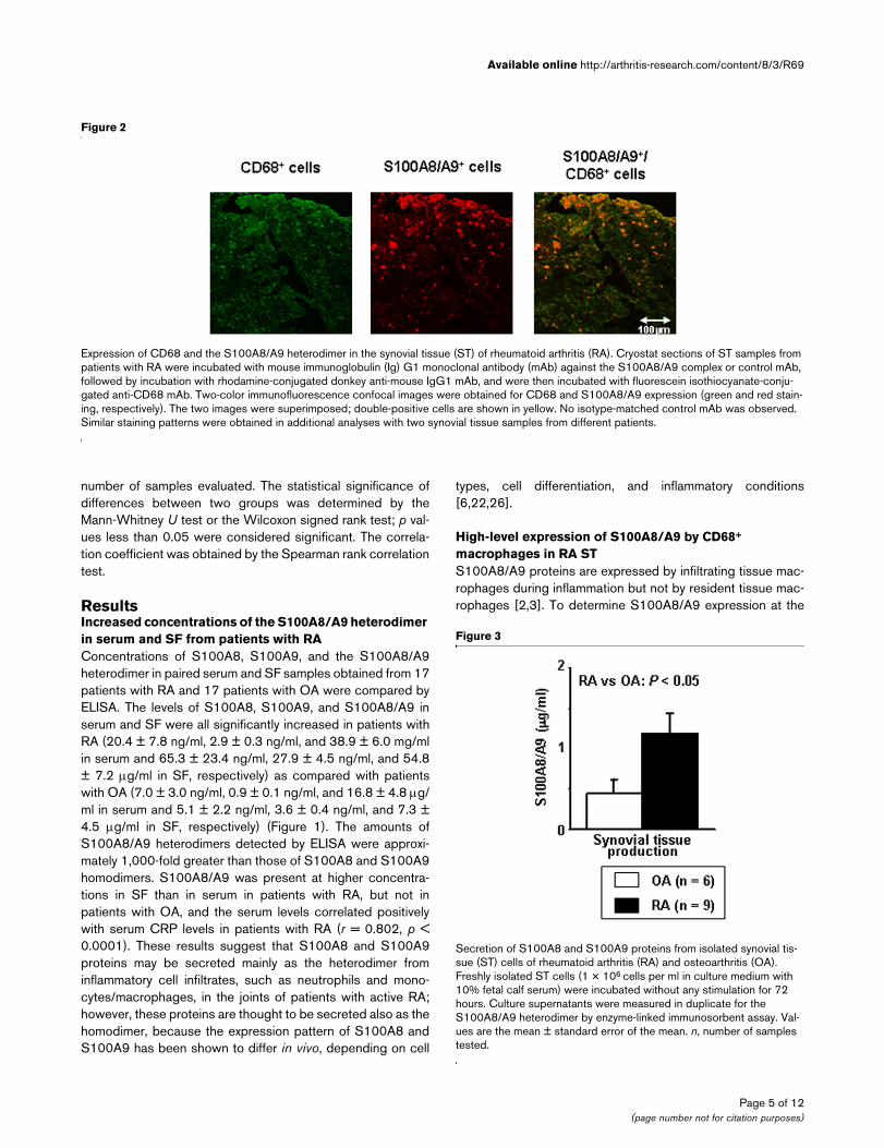
Figure 1
Concentrations of S100A8 and S100A9 proteins in the serum and syn-ovial fluid (SF) of rheumatoid arthritis (RA) and osteoarthritis (OA)Concentrations of S100A8 and S100A9 proteins in the serum and syn-ovial fluid (SF) of rheumatoid arthritis (RA) and osteoarthritis (OA). Paired serum and SF samples were prepared from patients with RA and patients with OA. Concentrations of the S100A8 and S100A9 homodimer and the S100A8/A9 heterodimer were measured in dupli-cate by enzyme-linked immunosorbent assay. Values are the mean ± standard error of the mean. n, number of samples tested.
Page 4 of 12(page number not for citation purposes)
Available online http://arthritis-research.com/content/8/3/R69
number of samples evaluated. The statistical significance ofdifferences between two groups was determined by theMann-Whitney U test or the Wilcoxon signed rank test; p val-ues less than 0.05 were considered significant. The correla-tion coefficient was obtained by the Spearman rank correlationtest.
ResultsIncreased concentrations of the S100A8/A9 heterodimer in serum and SF from patients with RAConcentrations of S100A8, S100A9, and the S100A8/A9heterodimer in paired serum and SF samples obtained from 17patients with RA and 17 patients with OA were compared byELISA. The levels of S100A8, S100A9, and S100A8/A9 inserum and SF were all significantly increased in patients withRA (20.4 ± 7.8 ng/ml, 2.9 ± 0.3 ng/ml, and 38.9 ± 6.0 mg/mlin serum and 65.3 ± 23.4 ng/ml, 27.9 ± 4.5 ng/ml, and 54.8± 7.2 µg/ml in SF, respectively) as compared with patientswith OA (7.0 ± 3.0 ng/ml, 0.9 ± 0.1 ng/ml, and 16.8 ± 4.8 µg/ml in serum and 5.1 ± 2.2 ng/ml, 3.6 ± 0.4 ng/ml, and 7.3 ±4.5 µg/ml in SF, respectively) (Figure 1). The amounts ofS100A8/A9 heterodimers detected by ELISA were approxi-mately 1,000-fold greater than those of S100A8 and S100A9homodimers. S100A8/A9 was present at higher concentra-tions in SF than in serum in patients with RA, but not inpatients with OA, and the serum levels correlated positivelywith serum CRP levels in patients with RA (r = 0.802, p <0.0001). These results suggest that S100A8 and S100A9proteins may be secreted mainly as the heterodimer frominflammatory cell infiltrates, such as neutrophils and mono-cytes/macrophages, in the joints of patients with active RA;however, these proteins are thought to be secreted also as thehomodimer, because the expression pattern of S100A8 andS100A9 has been shown to differ in vivo, depending on cell
types, cell differentiation, and inflammatory conditions[6,22,26].
High-level expression of S100A8/A9 by CD68+
macrophages in RA STS100A8/A9 proteins are expressed by infiltrating tissue mac-rophages during inflammation but not by resident tissue mac-rophages [2,3]. To determine S100A8/A9 expression at the
Figure 3
Secretion of S100A8 and S100A9 proteins from isolated synovial tis-sue (ST) cells of rheumatoid arthritis (RA) and osteoarthritis (OA)Secretion of S100A8 and S100A9 proteins from isolated synovial tis-sue (ST) cells of rheumatoid arthritis (RA) and osteoarthritis (OA). Freshly isolated ST cells (1 × 10⁶ cells per ml in culture medium with 10% fetal calf serum) were incubated without any stimulation for 72 hours. Culture supernatants were measured in duplicate for the S100A8/A9 heterodimer by enzyme-linked immunosorbent assay. Val-ues are the mean ± standard error of the mean. n, number of samples tested.
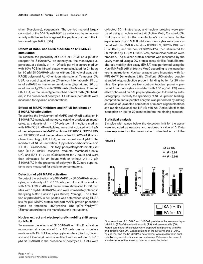
Figure 2
Expression of CD68 and the S100A8/A9 heterodimer in the synovial tissue (ST) of rheumatoid arthritis (RA)Expression of CD68 and the S100A8/A9 heterodimer in the synovial tissue (ST) of rheumatoid arthritis (RA). Cryostat sections of ST samples from patients with RA were incubated with mouse immunoglobulin (Ig) G1 monoclonal antibody (mAb) against the S100A8/A9 complex or control mAb, followed by incubation with rhodamine-conjugated donkey anti-mouse IgG1 mAb, and were then incubated with fluorescein isothiocyanate-conju-gated anti-CD68 mAb. Two-color immunofluorescence confocal images were obtained for CD68 and S100A8/A9 expression (green and red stain-ing, respectively). The two images were superimposed; double-positive cells are shown in yellow. No isotype-matched control mAb was observed. Similar staining patterns were obtained in additional analyses with two synovial tissue samples from different patients.
Page 5 of 12(page number not for citation purposes)
Arthritis Research & Therapy Vol 8 No 3 Sunahori et al.
site of macrophage infiltration, the proliferative ST samplesfrom three patients with RA were analyzed by two-colorimmunofluorescence labeling with anti-S100A8/A9 Ab andanti-CD68 Ab. Figure 2 shows representative staining pat-terns of the S100A8/A9 protein and the CD68 antigen in theST. These tissues were characterized by the extensive infiltra-tion of CD68-expressing macrophages. Colocalization analy-sis revealed that the heterodimeric S100A8/A9 was highlyexpressed by CD68+ macrophages, in particular the cellslocalized to the lining layer. However, S100A8/A9 stainingwas negligible in CD68-negative cells in the sublining layer,including lymphocytes and vascular endothelial cells. Immu-nostaining of the S100A8 and S100A9 homodimers showssimilar staining patterns, but the intensity and number ofS100A8 and S100A9 staining were less prominent (data notshown). These results indicate that S100A8 and S100A9 areexpressed predominantly in the form of a heterodimeric com-plex by highly activated macrophages in RA ST.
S100A8/A9 secretion in vitro by RA ST cellsTo confirm the local production of S100A8/A9 proteins at thesite of chronic inflammation in RA, isolated cells from ST sam-ples of nine patients with RA and six patients with OA wereincubated for 72 hours without any stimulation and culturesupernatants were measured for the heterodimer by ELISA. Asshown in Figure 3, ST cells from patients with RA spontane-
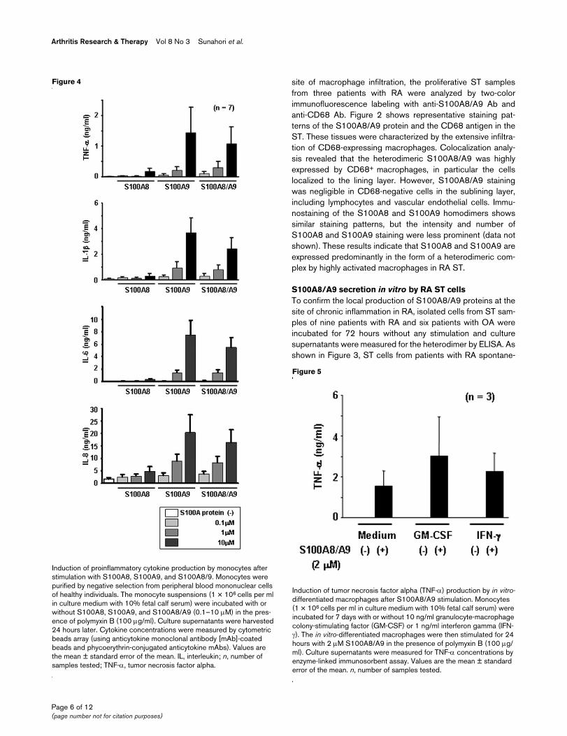
Figure 4
Induction of proinflammatory cytokine production by monocytes after stimulation with S100A8, S100A9, and S100A8/9Induction of proinflammatory cytokine production by monocytes after stimulation with S100A8, S100A9, and S100A8/9. Monocytes were purified by negative selection from peripheral blood mononuclear cells of healthy individuals. The monocyte suspensions (1 × 10⁶ cells per ml in culture medium with 10% fetal calf serum) were incubated with or without S100A8, S100A9, and S100A8/A9 (0.1–10 µM) in the pres-ence of polymyxin B (100 µg/ml). Culture supernatants were harvested 24 hours later. Cytokine concentrations were measured by cytometric beads array (using anticytokine monoclonal antibody [mAb]-coated beads and phycoerythrin-conjugated anticytokine mAbs). Values are the mean ± standard error of the mean. IL, interleukin; n, number of samples tested; TNF-α, tumor necrosis factor alpha.
Figure 5
Induction of tumor necrosis factor alpha (TNF-α) production by in vitro-differentiated macrophages after S100A8/A9 stimulationInduction of tumor necrosis factor alpha (TNF-α) production by in vitro-differentiated macrophages after S100A8/A9 stimulation. Monocytes (1 × 10⁶ cells per ml in culture medium with 10% fetal calf serum) were incubated for 7 days with or without 10 ng/ml granulocyte-macrophage colony-stimulating factor (GM-CSF) or 1 ng/ml interferon gamma (IFN-γ). The in vitro-differentiated macrophages were then stimulated for 24 hours with 2 µM S100A8/A9 in the presence of polymyxin B (100 µg/ml). Culture supernatants were measured for TNF-α concentrations by enzyme-linked immunosorbent assay. Values are the mean ± standard error of the mean. n, number of samples tested.
Page 6 of 12(page number not for citation purposes)
Available online http://arthritis-research.com/content/8/3/R69
ously secreted larger amounts of S100A8/A9 proteins (1.20± 0.25 µg/ml) than did the cells from patients with OA (0.45± 0.17 µg/ml). Lactate dehydrogenase activity in these culturesupernatants was undetectable or detectable only at negligi-ble levels, indicating that S100A8/A9 is released as a conse-quence of active secretion but not of cellular injury.
Although the cellular composition of ST cells was not deter-mined in this study, we had previously measured the frequen-cies of nonspecific esterase-staining macrophages and CD3-positive T cells in the ST cell population isolated in the samemanner and found that RA ST samples consisted of 31.7 ±5.3% (mean ± SD) macrophages and 44.7 ± 6.6% T cells (n= 7) and that OA ST samples consisted of 30.3 ± 4.9% mac-rophages and 28.8 ± 3.3% T cells (n = 4) (unpublished data).Additionally, we could not detect significant neutrophil con-tamination (≤1%) in two of the studied ST samples. Therefore,it would seem that the increased S100A8/A9 secretion fromRA ST cells may be due to the presence of in vivo-activatedmacrophages in the culture. This notion is likely consistentwith the fact that tissue macrophages are more activated in RAthan in OA [15,16].
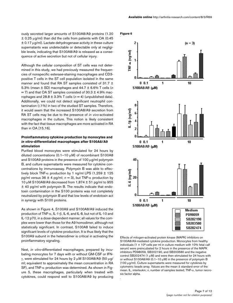
Proinflammatory cytokine production by monocytes and in vitro-differentiated macrophages after S100A8/A9 stimulationPurified blood monocytes were stimulated for 24 hours bydiluted concentrations (0.1–10 µM) of recombinant S100A8and S100A9 proteins in the presence of 100 µg/ml polymyxinB, and culture supernatants were measured for cytokine con-centrations by immunoassay. Polymyxin B was able to effec-tively block TNF-α production by 1 ng/ml LPS (1,259 ± 125pg/ml versus 36 ± 4 pg/ml; n = 3), but TNF-α production by10 µM S100A8/A9 decreased from 1,874 ± 51 pg/ml to 955± 40 pg/ml with polymyxin B. The results indicate that endo-toxin contamination in the S100 proteins was not completelyneutralized by polymyxin B and that low levels of endotoxin actin synergy with S100 proteins.
As shown in Figure 4, S100A9 and S100A8/A9 induced theproduction of TNF-α, IL-1-β, IL-6, and IL-8, but not of IL-10 andIL-12 p70, in a dose-dependent manner; all values for the com-plex were lower than those for the A9 homodimer, although notstatistically significant. In contrast, S100A8 failed to inducesignificant levels of cytokine production. It is thus likely that theS100A9 subunit in the heterodimer is critical in activating theproinflammatory signaling.
Next, in vitro-differentiated macrophages, prepared by incu-bating monocytes for 7 days with or without GM-CSF or IFN-γ, were stimulated for 24 hours by 2 µM S100A8/A9 (50 µg/ml; equivalent to approximately the mean concentration in RASF), and TNF-α production was determined. As shown in Fig-ure 5, these macrophages, particularly when treated withcytokines, could respond well to S100A8/A9 by producing
Figure 6
Effects of mitogen-activated protein kinase (MAPK) inhibitors on S100A8/A9-mediated cytokine productionEffects of mitogen-activated protein kinase (MAPK) inhibitors on S100A8/A9-mediated cytokine production. Monocytes from healthy individuals (1 × 10⁶ cells per ml in culture medium with 10% fetal calf serum) were preincubated for 2 hours in the presence of the MAPK inhibitors PD98059, SB202190, and SB203580 and the negative control SB202474 (1 µM) and were then stimulated for 24 hours with or without S100A8/A9 (0.1–10 µM) in the presence of polymyxin B (100 µg/ml). Culture supernatants were measured for cytokines by cytometric beads array. Values are the mean ± standard error of the mean. IL, interleukin; n, number of samples tested; TNF-α, tumor necro-sis factor alpha.
Page 7 of 12(page number not for citation purposes)
Arthritis Research & Therapy Vol 8 No 3 Sunahori et al.
TNF-α. These results suggest that the secreted S100A8/A9heterodimer may amplify the local expression of proinflamma-tory cytokines in joints of patients with RA.
Effects of RAGE and CD36 blockade on S100A8/A9 stimulationTo examine whether S100A8/A9 used RAGE and CD36 assignal transducing receptors on monocytes, monocytes werestimulated for 24 hours by 10 µM S100A8/A9 with or withoutanti-RAGE Ab, esRAGE (the splice variant lacking the trans-membrane-spanning domain), and anti-CD36 Ab, and culturesupernatants were measured for cytokine concentrations byimmunoassay. The S100A8/A9-stimulated cytokine responsewas not significantly reduced by blockade of the RAGE orCD36 receptor (percentage of inhibition, < 30%). Therefore,neither RAGE nor CD36 appears to be relevant to S100A8/A9 stimulation in monocytes.
Involvement of p38 MAPK activation in S100A8/A9 stimulationTo examine whether MAPK activation was involved in theS100A8/A9-mediated cytokine response, monocytes werepretreated with the MAPK inhibitors (PD98059, SB202190,and SB203580) and the control SB202474 and were thenstimulated for 24 hours by 1–10 µM S100A8/A9. Culturesupernatants were measured for cytokine concentrations byimmunoassay. As shown in Figure 6, TNF-α, IL-1-β, and IL-6production by S100A8/A9-stimulated monocytes was clearlyreduced by the specific p38 MAPK inhibitors SB202190 andSB203580, while it was not affected by the MEK (mitogen-activated/extracellular signal-regulated kinase) inhibitorPD98059, as well as by SB202474.
To support the significance of p38 MAPK in S100A8/A9 stim-ulation, monocytes were stimulated for 30 minutes by 10 µMS100A8/A9, and p38 MAPK protein and phosphorylated p38MAPK protein (pThr180/pTyr182) in cell lysates were measuredby ELISA. The phosphorylation ratio of p38 MAPK protein(phosphorylated p38 MAPK protein/total p38 MAPK protein)was markedly higher in S100A8/A9-stimulated monocytes(2.17 ± 0.74 U/pg; n = 3) than in unstimulated monocytes(0.52 ± 0.09 U/pg), proving the activation of p38 MAPK byS100A8/A9.
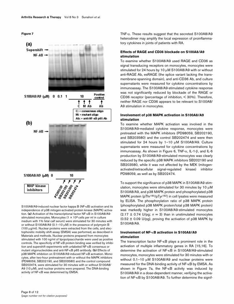
Involvement of NF-κB activation in S100A8/A9 stimulationThe transcription factor NF-κB plays a prominent role in theactivation of multiple inflammatory genes in RA [15,16]. Todetermine the activation of NF-κB in S100A8/A9-stimulatedmonocytes, monocytes were stimulated for 30 minutes with orwithout 0.1–10 µM S100A8/A9 and nuclear proteins weremeasured for the DNA-binding activity of NF-κB by EMSA. Asshown in Figure 7a, the NF-κB activity was induced byS100A8/A9 in a dose-dependent manner, verifying the activa-tion of NF-κB by S100A8/A9. To further determine the signif-
Figure 7
S100A8/A9-induced nuclear factor kappa B (NF-κB) activation and its independence of p38 mitogen-activated protein kinase (MAPK) activa-tionS100A8/A9-induced nuclear factor kappa B (NF-κB) activation and its independence of p38 mitogen-activated protein kinase (MAPK) activa-tion. (a) Activation of the transcriptional factor NF-κB in S100A8/A9-stimulated monocytes. Monocytes (1 × 10⁶ cells per ml in culture medium with 1% fetal calf serum) were stimulated for 30 minutes with or without S100A8/A9 (0.1–10 µM) in the presence of polymyxin B (100 µg/ml). Nuclear proteins were extracted from the cells, and elec-tophoretic mobility shift assay (EMSA) was performed, as described in Materials and methods. Nuclear proteins prepared from monocytes stimulated with 100 ng/ml of lipopolysaccharide were used as positive controls. The specificity of NF-κB protein binding was verified by inhibi-tion and supershift experiments with unlabeled NF-κB consensus or mutant oligonucleotides and anti-NF-κB p65 antibody. (b) Effects of p38 MAPK inhibition on S100A8/A9-induced NF-κB activation. Mono-cytes, after two-hour pretreatment with or without the MAPK inhibitors (PD98059, SB202190, and SB203580) and the control compound SB202474, were stimulated for 30 minutes with or without S100A8/A9 (10 µM), and nuclear proteins were prepared. The DNA-binding activity of NF-κB was determined by EMSA.
Page 8 of 12(page number not for citation purposes)
Available online http://arthritis-research.com/content/8/3/R69
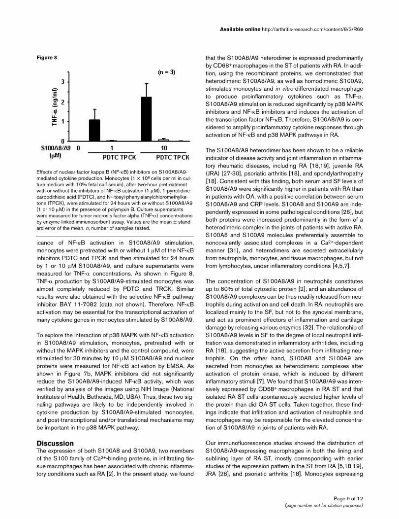
icance of NF-κB activation in S100A8/A9 stimulation,monocytes were pretreated with or without 1 µM of the NF-κBinhibitors PDTC and TPCK and then stimulated for 24 hoursby 1 or 10 µM S100A8/A9, and culture supernatants weremeasured for TNF-α concentrations. As shown in Figure 8,TNF-α production by S100A8/A9-stimulated monocytes wasalmost completely reduced by PDTC and TRCK. Similarresults were also obtained with the selective NF-κB pathwayinhibitor BAY 11-7082 (data not shown). Therefore, NF-κBactivation may be essential for the transcriptional activation ofmany cytokine genes in monocytes stimulated by S100A8/A9.
To explore the interaction of p38 MAPK with NF-κB activationin S100A8/A9 stimulation, monocytes, pretreated with orwithout the MAPK inhibitors and the control compound, werestimulated for 30 minutes by 10 µM S100A8/A9 and nuclearproteins were measured for NF-κB activation by EMSA. Asshown in Figure 7b, MAPK inhibitors did not significantlyreduce the S100A8/A9-induced NF-κB activity, which wasverified by analysis of the images using NIH Image (NationalInstitutes of Health, Bethesda, MD, USA). Thus, these two sig-naling pathways are likely to be independently involved incytokine production by S100A8/A9-stimulated monocytes,and post-transcriptional and/or translational mechanisms maybe important in the p38 MAPK pathway.
DiscussionThe expression of both S100A8 and S100A9, two membersof the S100 family of Ca2+-binding proteins, in infiltrating tis-sue macrophages has been associated with chronic inflamma-tory conditions such as RA [2]. In the present study, we found
that the S100A8/A9 heterodimer is expressed predominantlyby CD68+ macrophages in the ST of patients with RA. In addi-tion, using the recombinant proteins, we demonstrated thatheterodimeric S100A8/A9, as well as homodimeric S100A9,stimulates monocytes and in vitro-differentiated macrophageto produce proinflammatory cytokines such as TNF-α.S100A8/A9 stimulation is reduced significantly by p38 MAPKinhibitors and NF-κB inhibitors and induces the activation ofthe transcription factor NF-κB. Therefore, S100A8/A9 is con-sidered to amplify proinflammatoy cytokine responses throughactivation of NF-κB and p38 MAPK pathways in RA.
The S100A8/A9 heterodimer has been shown to be a reliableindicator of disease activity and joint inflammation in inflamma-tory rheumatic diseases, including RA [18,19], juvenile RA(JRA) [27-30], psoriatic arthritis [18], and spondylarthropathy[18]. Consistent with this finding, both serum and SF levels ofS100A8/A9 were significantly higher in patients with RA thanin patients with OA, with a positive correlation between serumS100A8/A9 and CRP levels. S100A8 and S100A9 are inde-pendently expressed in some pathological conditions [26], butboth proteins were increased predominantly in the form of aheterodimeric complex in the joints of patients with active RA.S100A8 and S100A9 molecules preferentially assemble tononcovalently associated complexes in a Ca2+-dependentmanner [31], and heterodimers are secreted extracellularlyfrom neutrophils, monocytes, and tissue macrophages, but notfrom lymphocytes, under inflammatory conditions [4,5,7].
The concentration of S100A8/A9 in neutrophils constitutesup to 60% of total cytosolic protein [2], and an abundance ofS100A8/A9 complexes can be thus readily released from neu-trophils during activation and cell death. In RA, neutrophils arelocalized mainly to the SF, but not to the synovial membrane,and act as prominent effectors of inflammation and cartilagedamage by releasing various enzymes [32]. The relationship ofS100A8/A9 levels in SF to the degree of local neutrophil infil-tration was demonstrated in inflammatory arthritides, includingRA [18], suggesting the active secretion from infiltrating neu-trophils. On the other hand, S100A8 and S100A9 aresecreted from monocytes as heterodimeric complexes afteractivation of protein kinase, which is induced by differentinflammatory stimuli [7]. We found that S100A8/A9 was inten-sively expressed by CD68+ macrophages in RA ST and thatisolated RA ST cells spontaneously secreted higher levels ofthe protein than did OA ST cells. Taken together, these find-ings indicate that infiltration and activation of neutrophils andmacrophages may be responsible for the elevated concentra-tion of S100A8/A9 in joints of patients with RA.
Our immunofluorescence studies showed the distribution ofS100A8/A9-expressing macrophages in both the lining andsublining layer of RA ST, mostly corresponding with earlierstudies of the expression pattern in the ST from RA [5,18,19],JRA [28], and psoriatic arthritis [18]. Monocytes expressing
Figure 8
Effects of nuclear factor kappa B (NF-κB) inhibitors on S100A8/A9-mediated cytokine productionEffects of nuclear factor kappa B (NF-κB) inhibitors on S100A8/A9-mediated cytokine production. Monocytes (1 × 10⁶ cells per ml in cul-ture medium with 10% fetal calf serum), after two-hour pretreatment with or without the inhibitors of NF-κB activation (1 µM), 1-pyrrolidine-carbodithioic acid (PDTC), and Nα-tosyl-phenylalanylchloromethylke-tone (TPCK), were stimulated for 24 hours with or without S100A8/A9 (1 or 10 µM) in the presence of polymyxin B. Culture supernatants were measured for tumor necrosis factor alpha (TNF-α) concentrations by enzyme-linked immunosorbent assay. Values are the mean ± stand-ard error of the mean. n, number of samples tested.
Page 9 of 12(page number not for citation purposes)
Arthritis Research & Therapy Vol 8 No 3 Sunahori et al.
cell surface S100A8/A9 represent a fast-migrating monocytesubpopulation across the endothelium barrier, and high levelsof the S100A8/A9 protein are secreted after their interactionwith inflammatory-activated endothelial cells [28,33].S100A8/A9 expression by sublining macrophages, particu-larly by perivascular macrophages thought to represent newlyinfiltrated macrophages, is attributable to selective infiltrationof S100A8/A9+ monocytes and their endothelium-dependentinduction of S100A8/A9 proteins, because the inflamed tis-sue of RA is characterized by increased vascularity andendothelial cell activation [34]. On the other hand, S100A8mRNA expression in macrophages has been shown to bestimulated by cytokines such as TNF-α, IFN-γ, and IL-10[35,36], which are relevant to RA pathogenesis [16]. Thus,cytokine stimulation may be responsible for S100A8/A9expression by synovial lining macrophages (that is, the highlyactivated phenotype of tissue macrophages that are capableof producing high levels of cytokines such as TNF-α) [15]. It isintriguing to note that there is a positive correlation betweenS100A8/A9 expression and TNF-α release in alveolar macro-phages from patients with active sarcoidosis [37].
The released S100A8/A9 heterodimer may play a role in thepropagation of inflammation by recruiting neutrophils andmonocytes to joints of patients with RA. Extracellular S100A8/A9 enhances the transendothelial migration of these inflamma-tory cells by enhancing CD11b expression and affinity forintracellular adhesion molecule-1 (ICAM-1) [33,38]. We foundthat S100A8/A9 stimulated monocytes and in vitro-differenti-ated macrophages to produce proinflammatory cytokinessuch as TNF-α. In addition, co-stimulation of S100A8/A9 withLPS and cytokines such as TNF-α, IL-1-β, and IFN-γ showedsynergistic effects on monocyte cytokine production (data notshown). However, S100A8/A9 was less effective in terms ofmonocyte stimulation compared with cytokines; the protein of10-7 to 10-5 M concentrations (2.4–240 µg/ml) was requiredfor significant cytokine induction, whereas cytokines areknown to produce their actions in the range of 10-12 to 10-9 Mconcentrations [39]. Despite its lower efficacy, S100A8/A9stimulation is believed to be effective in joints of patients withRA, where the protein is present at 54.8 ± 28.6 µg/ml,because in vitro-differentiated macrophages, as well as freshlyisolated monocytes (data not shown), when stimulated with 2µM (50 µg/ml) S100A8/A9, could produce significant levelsof TNF-α. Similarly, 15 µg/ml S100A8/A9 significantlyinduced IL-8 release from bronchial epithelial cells, and anti-S100A8/A9 Ab treatment reduced the IL-8-stimulating poten-tial of bronchial secretions [40]. More recently, the study usingmicroarray analysis demonstrated that S100A8/A9 proteins(100 µg/ml) directly activate endothelial cells to induce proin-flammatory chemokines and adhesion molecules and toincrease vascular permeability [41]. Taken together, thesefindings suggest the significance of S100A8/A9 as key effec-tors/amplifiers of inflammation with a wide range of activities,including cytokine induction.
Considering the potential inflammatory properties, thereseems to be a discrepancy between high concentrations ofserum S100A8/A9 and the lack of its peripheral effect inpatients with RA. We speculate that the yet-unidentified, inhib-itory molecules for S100A8/A9 activity might be present in theblood circulation, as is well recognized in the regulation ofTNF-α and IL-1 activities by soluble TNF receptors and IL-1receptor antagonist which bind to the protein or interferingwith its cell interaction, respectively [15,16]. However, it isalso conceivable that an abundance of the protein might par-tially contribute to monocyte activation before entry into thejoint, as evidenced by the increase of circulating CD16+
mature monocytes in patients with RA [42].
The S100A9 homodimer, but not the S100A8 homodimer,stimulated the monocyte/macrophage cytokine response.Similarly, the S100A9 homodimer, as well as the S100A8/A9complex, could enhance ICAM-1 binding to monocytes,whereas the S100A8 homodimer failed to do so [33]. TheS100A9 subunit is thus likely to be critical in eliciting theinflammatory signaling pathway. Interestingly, values ofcytokine production with S100A8/A9 tend to be lower thanthose with S100A9, which may indicate partial inhibition ofS100A9 stimulation by its heterodimeric partner S100A8.However, we could not clearly detect inhibitory effects ofS100A8 on S100A9-induced TNF-α production when titrat-ing S100A8 concentrations (0.1–10 µM) into the 1 µMS100A9 solution (data not shown), although the possibilitystill remains that much higher concentrations of S100A8 arerequired for its significant inhibition. The S100A9 protein stim-ulates the β2 integrin (CD11/CD18)-mediated neutrophiladhesion by inducing the high-affinity CD11b epitope, and thisS100A9 activity is specifically inhibited by S100A8 [38]. Moreimportantly, recent studies have demonstrated that numbersof S100A8/A9-expressing macrophages increased in the STof patients with RA after treatment with high dose of intrave-nous methylprednisolone and that high levels of S100A8induced by glucocorticoids have anti-inflammatory propertiesindependent of hetero-complex formation with S100A9 [43].These findings support the possible inhibition of the het-erodimer-induced cytokine response by the S100A8 subunit.
RAGE, originally defined by its binding to advanced glycationendproducts, has been identified as a multiligand receptor thatcan bind inflammatory molecules such as HMGB1 (high mobil-ity group box chromosomal protein 1), S100A12, andS100B1 [14,44]. Significant similarities in structure amongS100A8, S100A9, and S100A12 suggest that RAGE mayfunction as a signal-transducing receptor for these S100 pro-teins. In addition, the CD36 molecule, a member of the scav-enger receptor family, has been shown to bind the complex ofS100A8/A9 and AA and induce uptake of exogenous AA intothe endothelial cells [13]. However, S100A8/A9-mediatedcytokine production was not inhibited by anti-RAGE Ab,esRAGE (an endogenous inhibitor of RAGE activity), or anti-
Page 10 of 12(page number not for citation purposes)
Available online http://arthritis-research.com/content/8/3/R69
CD36, excluding the possibility of RAGE or CD36 as a puta-tive receptor for S100A8/A9. Thus, S100A8/A9 may inducecell activation after its nonspecific interaction with monocytes,because S100A8/A9 binds endothelial cells in conjunctionwith heparin sulfate proteoglycans and novel carboxylated gly-cans [11,12].
The significance of NF-κB and MAPK pathways in the inflam-matory gene activation in joints of patients with RA has beenwell established [15,16]. We found that S100A8/A9, likeTNF-α and IL-1, efficiently activate both the NF-κB and p38MAPK pathways, resulting in cytokine production in mono-cytes.
Because MAPK inhibitors did not reduce the DNA-bindingactivity of NF-κB, these two critical signaling pathways may beindependently involved in S100A8/A9-mediated cytokine pro-duction. NF-κB inhibitors could completely block the cytokineresponse. Therefore, we believe that the transcriptional activityof NF-κB may be most critical in S100A8/A9-inducedcytokine expression in monocytes/macrophages and that p38MAPK may regulate the synthesis of cytokines at the post-tran-scriptional and/or translational levels
ConclusionIn summary, the S100A8/A9 heterodimer, highly expressed bysynovial lining macrophage, may play a role in amplifying proin-flammatory cytokine responses via activation of NF-κB andp38 MAPK in RA.
Competing interestsThe authors declare that they have no competing interests.
Authors' contributionsKS was responsible for the experiments and data analysis andwrote the report. MY was responsible for the planning of theresearch and wrote up the manuscript. JY, KT, and MKassisted the experiments. HY prepared recombinant esRAGE,and WJC, YN, and SY prepared recombinant S100A8 and S9proteins. HM critically read the manuscript. All authors readand approved the final manuscript.
AcknowledgementsThe authors thank Dr H. Inoue and Dr K. Nishida (Okayama University, Okayama, Japan) for providing clinical samples. Dr W. J. Chazin was supported by NIH grant R01 GM62112. This work was supported in part by grants-in-aid (14570413/16590982) from the Ministry of Edu-cation, Science, Culture, and Technology of Japan.
References1. Donato R: Intracellular and extracellular roles of S100 proteins.
Microsc Res Tech 2003, 60:540-551.2. Nacken W, Roth J, Sorg C, Kerkhoff C: S100A9/S100A8: mye-
loid representatives of the S100 protein family as prominentplayers in innate immunity. Microsc Res Tech 2003,60:569-580.
3. Roth J, Vogl T, Sorg C, Sunderkotter C: Phagocyte-specific S100proteins: a novel group of proinflammatory molecules. TrendsImmunol 2003, 24:155-158.
4. Andersson KB, Sletten K, Berntzen HB, Fagerhol MK, Dale I,Brandtzaeg P, Jellum E: Leukocyte L1 protein and the cysticfibrosis antigen. Nature 1988, 332:688.
5. Koike T, Kondo K, Makita T, Kajiyama K, Yoshida T, Morikawa M:Intracellular localization of migration inhibitory factor-relatedprotein (MRP) and detection of cell surface MRP binding siteson human leukemia cell lines. J Biochem (Tokyo) 1998,123:1079-1087.
6. Odink K, Cerletti N, Bruggen J, Clerc RG, Tarcsay L, Zwadlo G,Gerhards G, Schlegel R, Sorg C: Two calcium-binding proteinsin infiltrate macrophages of rheumatoid arthritis. Nature 1987,330:80-82.
7. Rammes A, Roth J, Goebeler M, Klempt M, Hartmann M, Sorg C:Myeloid-related protein (MRP) 8 and MRP14, calcium-bindingproteins of the S100 family, are secreted by activated mono-cytes via a novel, tubulin-dependent pathway. J Biol Chem1997, 272:9496-9502.
8. Vandal K, Rouleau P, Boivin A, Ryckman C, Talbot M, Tessier PA:Blockade of S100A8 and S100A9 suppresses neutrophilmigration in response to lipopolysaccharide. J Immunol 2003,171:2602-2609.
9. Kerkhoff C, Klempt M, Kaever V, Sorg C: The two calcium-bind-ing proteins, S100A8 and S100A9, are involved in the metabo-lism of arachidonic acid in human neutrophils. J Biol Chem1999, 274:32672-32679.
10. Eue I, Sorg C: Arachidonic acid specifically regulates binding ofS100A8/9, a heterodimer complex of the S100 class of cal-cium binding proteins, to human microvascular endothelialcells. Atherosclerosis 2001, 154:505-508.
11. Robinson MJ, Tessier P, Poulsom R, Hogg N: The S100 familyheterodimer, MRP-8/14, binds with high affinity to heparin andheparan sulfate glycosaminoglycans on endothelial cells. JBiol Chem 2002, 277:3658-3665.
12. Srikrishna G, Panneerselvam K, Westphal V, Abraham V, Varki A,Freeze HH: Two proteins modulating transendothelial migra-tion of leukocytes recognize novel carboxylated glycans onendothelial cells. J Immunol 2001, 166:4678-4688.
13. Kerkhoff C, Sorg C, Tandon NN, Nacken W: Interaction ofS100A8/S100A9-arachidonic acid complexes with the scav-enger receptor CD36 may facilitate fatty acid uptake byendothelial cells. Biochemistry 2001, 40:241-248.
14. Hofmann MA, Drury S, Fu C, Qu W, Taguchi A, Lu Y, Avila C, Kam-bham N, Bierhaus A, Nawroth P, et al.: RAGE mediates a novelproinflammatory axis: a central cell surface receptor for S100/calgranulin polypeptides. Cell 1999, 97:889-901.
15. Burmester GR, Stuhlmuller B, Keyszer G, Kinne RW: Mononu-clear phagocytes and rheumatoid synovitis. Mastermind orworkhorse in arthritis? Arthritis Rheum 1997, 40:5-18.
16. Feldmann M, Brennan FM, Maini RN: Role of cytokines in rheu-matoid arthritis. Annu Rev Immunol 1996, 14:397-440.
17. Brun JG, Jonsson R, Haga HJ: Measurement of plasma calpro-tectin as an indicator of arthritis and disease activity inpatients with inflammatory rheumatic diseases. J Rheumatol1994, 21:733-738.
18. Kane D, Roth J, Frosch M, Vogl T, Bresnihan B, FitzGerald O:Increased perivascular synovial membrane expression ofmyeloid-related proteins in psoriatic arthritis. Arthritis Rheum2003, 48:1676-1685.
19. Youssef P, Roth J, Frosch M, Costello P, Fitzgerald O, Sorg C,Bresnihan B: Expression of myeloid related proteins (MRP) 8and 14 and the MRP8/14 heterodimer in rheumatoid arthritissynovial membrane. J Rheumatol 1999, 26:2523-2528.
20. Arnett FC, Edworthy SM, Bloch DA, McShane DJ, Fries JF, CooperNS, Healey LA, Kaplan SR, Liang MH, Luthra HS, et al.: The Amer-ican Rheumatism Association 1987 revised criteria for theclassification of rheumatoid arthritis. Arthritis Rheum 1988,31:315-324.
21. Altman R, Asch E, Bloch D, Bole G, Borenstein D, Brandt K,Christy W, Cooke TD, Greenwald R, Hochberg M, et al.: Develop-ment of criteria for the classification and reporting of osteoar-thritis. Classification of osteoarthritis of the knee. Diagnosticand Therapeutic Criteria Committee of the American Rheuma-tism Association. Arthritis Rheum 1986, 29:1039-1049.
Page 11 of 12(page number not for citation purposes)
http://www.ncbi.nlm.nih.gov/entrez/query.fcgi?cmd=Retrieve&db=PubMed&dopt=Abstract&list_uids=3357535
http://www.ncbi.nlm.nih.gov/entrez/query.fcgi?cmd=Retrieve&db=PubMed&dopt=Abstract&list_uids=3357535
http://www.ncbi.nlm.nih.gov/entrez/query.fcgi?cmd=Retrieve&db=PubMed&dopt=Abstract&list_uids=9603996
http://www.ncbi.nlm.nih.gov/entrez/query.fcgi?cmd=Retrieve&db=PubMed&dopt=Abstract&list_uids=9603996
http://www.ncbi.nlm.nih.gov/entrez/query.fcgi?cmd=Retrieve&db=PubMed&dopt=Abstract&list_uids=9603996
http://www.ncbi.nlm.nih.gov/entrez/query.fcgi?cmd=Retrieve&db=PubMed&dopt=Abstract&list_uids=3313057
http://www.ncbi.nlm.nih.gov/entrez/query.fcgi?cmd=Retrieve&db=PubMed&dopt=Abstract&list_uids=3313057
http://www.ncbi.nlm.nih.gov/entrez/query.fcgi?cmd=Retrieve&db=PubMed&dopt=Abstract&list_uids=9083090
http://www.ncbi.nlm.nih.gov/entrez/query.fcgi?cmd=Retrieve&db=PubMed&dopt=Abstract&list_uids=9083090
http://www.ncbi.nlm.nih.gov/entrez/query.fcgi?cmd=Retrieve&db=PubMed&dopt=Abstract&list_uids=9083090
http://www.ncbi.nlm.nih.gov/entrez/query.fcgi?cmd=Retrieve&db=PubMed&dopt=Abstract&list_uids=9008595
http://www.ncbi.nlm.nih.gov/entrez/query.fcgi?cmd=Retrieve&db=PubMed&dopt=Abstract&list_uids=9008595
http://www.ncbi.nlm.nih.gov/entrez/query.fcgi?cmd=Retrieve&db=PubMed&dopt=Abstract&list_uids=9008595
http://www.ncbi.nlm.nih.gov/entrez/query.fcgi?cmd=Retrieve&db=PubMed&dopt=Abstract&list_uids=8717520
http://www.ncbi.nlm.nih.gov/entrez/query.fcgi?cmd=Retrieve&db=PubMed&dopt=Abstract&list_uids=8717520
http://www.ncbi.nlm.nih.gov/entrez/query.fcgi?cmd=Retrieve&db=PubMed&dopt=Abstract&list_uids=8035402
http://www.ncbi.nlm.nih.gov/entrez/query.fcgi?cmd=Retrieve&db=PubMed&dopt=Abstract&list_uids=8035402
http://www.ncbi.nlm.nih.gov/entrez/query.fcgi?cmd=Retrieve&db=PubMed&dopt=Abstract&list_uids=8035402
http://www.ncbi.nlm.nih.gov/entrez/query.fcgi?cmd=Retrieve&db=PubMed&dopt=Abstract&list_uids=3358796
http://www.ncbi.nlm.nih.gov/entrez/query.fcgi?cmd=Retrieve&db=PubMed&dopt=Abstract&list_uids=3358796
http://www.ncbi.nlm.nih.gov/entrez/query.fcgi?cmd=Retrieve&db=PubMed&dopt=Abstract&list_uids=3358796
http://www.ncbi.nlm.nih.gov/entrez/query.fcgi?cmd=Retrieve&db=PubMed&dopt=Abstract&list_uids=3741515
Arthritis Research & Therapy Vol 8 No 3 Sunahori et al.
22. Zwadlo G, Schlegel R, Sorg C: A monoclonal antibody to a sub-set of human monocytes found only in the peripheral bloodand inflammatory tissues. J Immunol 1986, 137:512-518.
23. Hunter MJ, Chazin WJ: High level expression and dimer charac-terization of the S100 EF-hand proteins, migration inhibitoryfactor-related proteins 8 and 14. J Biol Chem 1998,273:12427-12435.
24. Yui S, Nakatani Y, Hunter MJ, Chazin WJ, Yamazaki M: Implicationof extracellular zinc exclusion by recombinant human calpro-tectin (MRP8 and MRP14) from target cells in its apoptosis-inducing activity. Mediators Inflamm 2002, 11:165-172.
25. Yonekura H, Yamamoto Y, Sakurai S, Petrova RG, Abedin MJ, LiH, Yasui K, Takeuchi M, Makita Z, Takasawa S, et al.: Novel splicevariants of the receptor for advanced glycation end-productsexpressed in human vascular endothelial cells and pericytes,and their putative roles in diabetes-induced vascular injury.Biochem J 2003, 370:1097-1109.
26. Zwadlo G, Bruggen J, Gerhards G, Schlegel R, Sorg C: Two cal-cium-binding proteins associated with specific stages of mye-loid cell differentiation are expressed by subsets ofmacrophages in inflammatory tissues. Clin Exp Immunol 1988,72:510-515.
27. Berntzen HB, Fagerhol MK, Ostensen M, Mowinckel P, HoyeraalHM: The L1 protein as a new indicator of inflammatory activityin patients with juvenile rheumatoid arthritis. J Rheumatol1991, 18:133-138.
28. Frosch M, Strey A, Vogl T, Wulffraat NM, Kuis W, Sunderkotter C,Harms E, Sorg C, Roth J: Myeloid-related proteins 8 and 14 arespecifically secreted during interaction of phagocytes andactivated endothelium and are useful markers for monitoringdisease activity in pauciarticular-onset juvenile rheumatoidarthritis. Arthritis Rheum 2000, 43:628-637.
29. Wulffraat NM, Haas PJ, Frosch M, De Kleer IM, Vogl T, BrinkmanDM, Quartier P, Roth J, Kuis W: Myeloid related protein 8 and 14secretion reflects phagocyte activation and correlates withdisease activity in juvenile idiopathic arthritis treated withautologous stem cell transplantation. Ann Rheum Dis 2003,62:236-241.
30. Frosch M, Vogl T, Seeliger S, Wulffraat N, Kuis W, Viemann D,Foell D, Sorg C, Sunderkotter C, Roth J: Expression of myeloid-related proteins 8 and 14 in systemic-onset juvenile rheuma-toid arthritis. Arthritis Rheum 2003, 48:2622-2626.
31. Teigelkamp S, Bhardwaj RS, Roth J, Meinardus-Hager G, Karas M,Sorg C: Calcium-dependent complex assembly of the myeloicdifferentiation proteins MRP-8 and MRP-14. J Biol Chem 1991,266:13462-13467.
32. Edwards SW, Hallett MB: Seeing the wood for the trees: theforgotten role of neutrophils in rheumatoid arthritis. ImmunolToday 1997, 18:320-324.
33. Eue I, Pietz B, Storck J, Klempt M, Sorg C: Transendothelialmigration of 27E10+ human monocytes. Int Immunol 2000,12:1593-1604.
34. Jackson JR, Seed MP, Kircher CH, Willoughby DA, Winkler JD:The codependence of angiogenesis and chronic inflammation.Faseb J 1997, 11:457-465.
35. Xu K, Geczy CL: IFN-gamma and TNF regulate macrophageexpression of the chemotactic S100 protein S100A8. J Immu-nol 2000, 164:4916-4923.
36. Xu K, Yen T, Geczy CL: IL-10 up-regulates macrophage expres-sion of the S100 protein S100A8. J Immunol 2001,166:6358-6366.
37. Zheng L, Teschler H, Guzman J, Hubner K, Striz I, Costabel U:Alveolar macrophage TNF-alpha release and BAL cell pheno-types in sarcoidosis. Am J Respir Crit Care Med 1995,152:1061-1066.
38. Newton RA, Hogg N: The human S100 protein MRP-14 is anovel activator of the beta 2 integrin Mac-1 on neutrophils. JImmunol 1998, 160:1427-1435.
39. Vilček J: The cytokines: an overview. In The Cytokine Handbook4th edition. Edited by: Thomson AW, Lotze MT. London: AcademicPress; 2003:3-18.
40. Ahmad A, Bayley DL, He S, Stockley RA: Myeloid related protein-8/14 stimulates interleukin-8 production in airway epithelialcells. Am J Respir Cell Mol Biol 2003, 29:523-530.
41. Viemann D, Strey A, Janning A, Jurk K, Klimmek K, Vogl T, HironoK, Ichida F, Foell D, Kehrel B, et al.: Myeloid-related proteins 8
and 14 induce a specific inflammatory response in humanmicrovascular endothelial cells. Blood 2005, 105:2955-2962.
42. Kawanaka N, Yamamura M, Aita T, Morita Y, Okamoto A,Kawashima M, Iwahashi M, Ueno A, Ohmoto Y, Makino H: CD14+,CD16+ blood monocytes and joint inflammation in rheumatoidarthritis. Arthritis Rheum 2002, 46:2578-2586.
43. Hsu K, Passey RJ, Endoh Y, Rahimi F, Youssef P, Yen T, Geczy CL:Regulation of S100A8 by glucocorticoids. J Immunol 2005,174:2318-2326.
44. Schmidt AM, Yan SD, Yan SF, Stern DM: The multiligand recep-tor RAGE as a progression factor amplifying immune andinflammatory responses. J Clin Invest 2001, 108:949-955.
Page 12 of 12(page number not for citation purposes)
http://www.ncbi.nlm.nih.gov/entrez/query.fcgi?cmd=Retrieve&db=PubMed&dopt=Abstract&list_uids=3722815
http://www.ncbi.nlm.nih.gov/entrez/query.fcgi?cmd=Retrieve&db=PubMed&dopt=Abstract&list_uids=3722815
http://www.ncbi.nlm.nih.gov/entrez/query.fcgi?cmd=Retrieve&db=PubMed&dopt=Abstract&list_uids=3722815
http://www.ncbi.nlm.nih.gov/entrez/query.fcgi?cmd=Retrieve&db=PubMed&dopt=Abstract&list_uids=9575199
http://www.ncbi.nlm.nih.gov/entrez/query.fcgi?cmd=Retrieve&db=PubMed&dopt=Abstract&list_uids=9575199
http://www.ncbi.nlm.nih.gov/entrez/query.fcgi?cmd=Retrieve&db=PubMed&dopt=Abstract&list_uids=9575199
http://www.ncbi.nlm.nih.gov/entrez/query.fcgi?cmd=Retrieve&db=PubMed&dopt=Abstract&list_uids=3048809
http://www.ncbi.nlm.nih.gov/entrez/query.fcgi?cmd=Retrieve&db=PubMed&dopt=Abstract&list_uids=3048809
http://www.ncbi.nlm.nih.gov/entrez/query.fcgi?cmd=Retrieve&db=PubMed&dopt=Abstract&list_uids=3048809
http://www.ncbi.nlm.nih.gov/entrez/query.fcgi?cmd=Retrieve&db=PubMed&dopt=Abstract&list_uids=2023183
http://www.ncbi.nlm.nih.gov/entrez/query.fcgi?cmd=Retrieve&db=PubMed&dopt=Abstract&list_uids=2023183
http://www.ncbi.nlm.nih.gov/entrez/query.fcgi?cmd=Retrieve&db=PubMed&dopt=Abstract&list_uids=2071612
http://www.ncbi.nlm.nih.gov/entrez/query.fcgi?cmd=Retrieve&db=PubMed&dopt=Abstract&list_uids=2071612
http://www.ncbi.nlm.nih.gov/entrez/query.fcgi?cmd=Retrieve&db=PubMed&dopt=Abstract&list_uids=9238834
http://www.ncbi.nlm.nih.gov/entrez/query.fcgi?cmd=Retrieve&db=PubMed&dopt=Abstract&list_uids=9238834
http://www.ncbi.nlm.nih.gov/entrez/query.fcgi?cmd=Retrieve&db=PubMed&dopt=Abstract&list_uids=9194526
http://www.ncbi.nlm.nih.gov/entrez/query.fcgi?cmd=Retrieve&db=PubMed&dopt=Abstract&list_uids=9194526
http://www.ncbi.nlm.nih.gov/entrez/query.fcgi?cmd=Retrieve&db=PubMed&dopt=Abstract&list_uids=7663784
http://www.ncbi.nlm.nih.gov/entrez/query.fcgi?cmd=Retrieve&db=PubMed&dopt=Abstract&list_uids=7663784
http://www.ncbi.nlm.nih.gov/entrez/query.fcgi?cmd=Retrieve&db=PubMed&dopt=Abstract&list_uids=7663784
http://www.ncbi.nlm.nih.gov/entrez/query.fcgi?cmd=Retrieve&db=PubMed&dopt=Abstract&list_uids=9570563