
Nuclear Receptor BioMed Central - core.ac.uk · dependent pathway), whereas ER β, and a short...
14
BioMed Central Page 1 of 14 (page number not for citation purposes) Nuclear Receptor Open Access Research A conserved lysine in the estrogen receptor DNA binding domain regulates ligand activation profiles at AP-1 sites, possibly by controlling interactions with a modulating repressor Rosalie M Uht* 1 , Paul Webb 2 , Phuong Nguyen 2 , Richard H Price Jr Jr 3 , Cathleen Valentine 4 , Helene Favre 4 and Peter J Kushner 4 Address: 1 Departments of Pathology, & Biochemistry and Molecular Genetics University of Virginia, School of Medicine, MR5 Rm. 3123, 415 Lane Rd., Charlottesville, VA 22908-0904, USA, 2 Center for Diabetes and Endocrinology, University of California San Francisco, CA 94143-0540, USA, 3 Amgen, Inc., 930 Scott St. #6, San Francisco, CA 94115, USA and 4 Department of Medicine, Room C430, 2200 Post St., University of California San Francisco, San Francisco CA 94115-1640, USA Email: Rosalie M Uht* - [email protected]; Paul Webb - [email protected]; Phuong Nguyen - [email protected]; Richard H Price Jr - [email protected]; Cathleen Valentine - [email protected]; Helene Favre - [email protected]; Peter J Kushner - [email protected] * Corresponding author Abstract Background: Estrogen receptors alpha and beta (ERα and ERβ) differentially activate genes with AP-1 elements. ERα activates AP-1 targets via activation functions with estrogens (the AF- dependent pathway), whereas ERβ, and a short version of ERα (ERα DBD-LBD) activate only with anti-estrogens (AF-independent pathway). The DNA binding domain (DBD) plays an important role in both pathways, even though neither pathway requires ERE recognition. Results: Mutations of a highly conserved DBD lysine (ERα.K206A/G), lead to super-activation of AP-1 through activation function dependent pathways, up to 200 fold. This super-activity can be elicited either through ER AFs 1 or 2, or that of a heterologous activation function (VP16). The homologous substitution in ERβ, K170A, or in ERα DBD-LBD leads to estrogen-dependent AP-1 activation and loss of the usually potent anti-estrogen effects. Each of numerous K206 substitutions in ERα, except K206R, eliminates anti-estrogen activation and this loss correlates perfectly with a loss of ability to titrate a repressive function from the RU486 bound progesterone receptor. Conclusion: We conclude that ER DBDs contain a complex regulatory function that influences ligand activation profiles at AP-1. This function, which requires the integrity of the conserved lysine, both allows for activation at AP-1 with anti-estrogens (with ERβ and ERα DBD-LBD), and prevents ERα from becoming superactive at AP-1 with estrogens. We discuss the possibility that a repressor interaction with the DBD both mediates the AF-independent pathway and dampens the AF dependent pathway. Mutations in the conserved lysine might, by this model, disrupt the binding or function of the repressor. Published: 07 May 2004 Nuclear Receptor 2004, 2:2 doi:10.1186/1478-1336-2-2 Received: 24 November 2003 Accepted: 07 May 2004 This article is available from: http://www.nuclear-receptor.com/content/2/1/2 © 2004 Uht et al; licensee BioMed Central Ltd. This is an Open Access article: verbatim copying and redistribution of this article are permitted in all media for any purpose, provided this notice is preserved along with the article's original URL.
Transcript of Nuclear Receptor BioMed Central - core.ac.uk · dependent pathway), whereas ER β, and a short...
BioMed CentralNuclear Receptor
ss
Open AcceResearchA conserved lysine in the estrogen receptor DNA binding domain regulates ligand activation profiles at AP-1 sites, possibly by controlling interactions with a modulating repressorRosalie M Uht*1, Paul Webb2, Phuong Nguyen2, Richard H Price Jr Jr3, Cathleen Valentine4, Helene Favre4 and Peter J Kushner4Address: 1Departments of Pathology, & Biochemistry and Molecular Genetics University of Virginia, School of Medicine, MR5 Rm. 3123, 415 Lane Rd., Charlottesville, VA 22908-0904, USA, 2Center for Diabetes and Endocrinology, University of California San Francisco, CA 94143-0540, USA, 3Amgen, Inc., 930 Scott St. #6, San Francisco, CA 94115, USA and 4Department of Medicine, Room C430, 2200 Post St., University of California San Francisco, San Francisco CA 94115-1640, USA
Email: Rosalie M Uht* - [email protected]; Paul Webb - [email protected]; Phuong Nguyen - [email protected]; Richard H Price Jr - [email protected]; Cathleen Valentine - [email protected]; Helene Favre - [email protected]; Peter J Kushner - [email protected]
* Corresponding author
AbstractBackground: Estrogen receptors alpha and beta (ERα and ERβ) differentially activate genes withAP-1 elements. ERα activates AP-1 targets via activation functions with estrogens (the AF-dependent pathway), whereas ERβ, and a short version of ERα (ERα DBD-LBD) activate only withanti-estrogens (AF-independent pathway). The DNA binding domain (DBD) plays an importantrole in both pathways, even though neither pathway requires ERE recognition.
Results: Mutations of a highly conserved DBD lysine (ERα.K206A/G), lead to super-activation ofAP-1 through activation function dependent pathways, up to 200 fold. This super-activity can beelicited either through ER AFs 1 or 2, or that of a heterologous activation function (VP16). Thehomologous substitution in ERβ, K170A, or in ERα DBD-LBD leads to estrogen-dependent AP-1activation and loss of the usually potent anti-estrogen effects. Each of numerous K206 substitutionsin ERα, except K206R, eliminates anti-estrogen activation and this loss correlates perfectly with aloss of ability to titrate a repressive function from the RU486 bound progesterone receptor.
Conclusion: We conclude that ER DBDs contain a complex regulatory function that influencesligand activation profiles at AP-1. This function, which requires the integrity of the conserved lysine,both allows for activation at AP-1 with anti-estrogens (with ERβ and ERα DBD-LBD), and preventsERα from becoming superactive at AP-1 with estrogens. We discuss the possibility that a repressorinteraction with the DBD both mediates the AF-independent pathway and dampens the AFdependent pathway. Mutations in the conserved lysine might, by this model, disrupt the binding orfunction of the repressor.
Published: 07 May 2004
Nuclear Receptor 2004, 2:2 doi:10.1186/1478-1336-2-2
Received: 24 November 2003Accepted: 07 May 2004
This article is available from: http://www.nuclear-receptor.com/content/2/1/2
© 2004 Uht et al; licensee BioMed Central Ltd. This is an Open Access article: verbatim copying and redistribution of this article are permitted in all media for any purpose, provided this notice is preserved along with the article's original URL.
Page 1 of 14(page number not for citation purposes)
Nuclear Receptor 2004, 2 http://www.nuclear-receptor.com/content/2/1/2
BackgroundEstrogen receptors alpha and beta (ERα and ERβ) classi-cally activate transcription by binding to cognate estrogenresponse elements (EREs). The receptors also activateexpression of target genes through alternate pathways, e.g.through AP-1 and CRE-like response elements [1,2]. AP-1/CRE-like elements bind Jun and related transcriptionfactors but not ERs [2,3]. ER action at these alternate ele-ments appears increasingly to be important. For example,ER/AP-1 pathways underlie estrogen activation of colla-genase, Cyclin D1 and IGF-1genes [1,2,4]. The relativecontributions of classic and alternate pathways to a givengene response in vivo have not yet been directly explored.However, there is suggestive evidence that AP-1 alternatepathways play an important role in estrogen-dependentproliferation [1,5-7].
There are two distinct ER/AP-1 pathways: one activated byestrogens, the other activated by anti-estrogens [1,3,8].The estrogen-activated ERα/AP-1 pathway is mediated byERα's two activation functions: the constitutive AF-1 inthe N'terminal domain (NTD), and the estrogen-activatedAF-2 in the C-terminal ligand binding domain (LBD) [8].The ligand dependent AF-2 is functional in virtually allcell types but is blocked by anti-estrogens. Cells ofendometrial origin also support strong AF-1 activity at AP-1 which is allowed by some anti-estrogens most notablytamoxifen (Tam) but not by raloxifene or ICI 182,780.These latter two "pure" anti-estrogens do not permit AF-1activity most likely because they allow for efficient bind-ing of the corepressor N-CoR [9]. It is inferred therefore,that when these pure anti-estrogens activate AP-1 medi-ated transcription (with ERβ or ERα DBD-LBD) they do sothrough an AF-independent mechanism possibly one thatinvolves N-CoR binding since a mutation that eliminatesN-CoR binding eliminates this anti-estrogen activation[9].
To summarize our previous work, we have identified twopathways by which the ER can activate transcription at AP-1 elements, AF dependent and AF independent. Threeclasses of ligands can be distinguished since the AP-1 tar-gets can be activated: (1) By estrogens through an AF-1and -2 dependent pathway; (2) By Tam through AF-1, andpartly through the AF-independent activity, and (3) By"pure" anti-estrogens (ICI and Raloxifene) that activateonly through an AF-independent pathway [9]. The latterpathway is most prominent in the presence of ERβ andcertain ERαNTD-deleted mutants [8].
The mechanisms that underlie the AF-independent activa-tion have not been elucidated. However, we have sug-gested that anti-estrogen-ER complexes increase AP-1activity by functionally inactivating an unidentified co-repressor normally associated with the AP-1 complex and/
or its chromatin environment [3,8]. The lack of AFinvolvement in this pathway leaves open the question asto which portions of the ER contribute to this mode ofanti-estrogen action through AP-1. Here we present datathat a domain generally not considered to contain tran-scriptional activation functions, the DNA binding domain(DBD), plays a critical role in regulating the balancebetween different pathways of ER action at AP-1 sites.
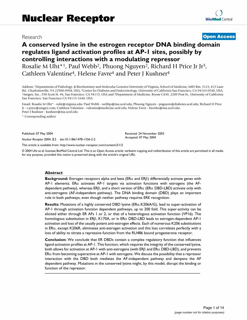
We noted that nuclear receptor DBDs contain a highlyconserved lysine (lys) residue at the base of the first zincbinding motif (Fig. 1A). In the context of the glucocorti-coid receptor (GR) this residue is required for GR's abilityto inhibit AP-1 mediated transcriptional activation. Itsmutation to gly or ala converts the GR from an inhibitorto a stimulator of the AP-1 activity in response to agonist[10,11]. We noted in a preliminary report that the equiv-alent ERα mutation (ERα.K206A) potentiates estrogenaction at AP-1 sites, and at hybrid AP-1/cAMP responseelements (CREs) in the context of much larger promoter,Cyclin D1 [2]. Here we report that mutations of this resi-due show complex effects on the ligand profile of ERaction at AP-1 sites and propose that DBD interacting pro-teins could regulate the type of ER pathway used at alter-nate response elements.
ResultsERα.K206A and K206G are superactive at AP-1 sites but not at EREsWe first mutated the conserved ERα lysine residue at thebase of the first zinc finger to ala or gly (ERα.K206A orK206G, Fig. 1A). Introduction of these mutations wasmotivated by reports that mutations in the homologousresidue convert rat and human GRs from inhibitors toactivators at AP-1 regulated genes [10,11]. Data in Fig. 1Bconfirm that both E2-liganded ERα.K206A andERα.G400V.K206A enhance the activity of an AP-1responsive reporter gene (Coll73-Luc) more efficientlythan their K206 counterparts in HeLa cells. Also apparentwas the inhibitory nature of the anti-estrogen ligands usedin presence of the K206A mutation, vide infra for furtherdiscussion.
ERα.K206A also showed increased activity in the absenceof exogenous ligands. Fully wild type ERα. G400 (ERαdesignated as ERα throughout the remainder of the man-uscript unless otherwise indicated) exhibits variable levelsof constitutive activity in cell culture because of very lowlevels of estrogens in serum [12]. This phenomenon prob-ably accounts for the increased ability of unliganded ERαbearing the K206A mutation to enhance AP-1 dependenttranscription because a mutation (G400V) that slightlyreduces ERα affinity for ligand [12] abolished the consti-tutive activity exhibited by the fully wild type receptor,without affecting overall E2 response (compare ERαG400
α
Page 2 of 14(page number not for citation purposes)
Nuclear Receptor 2004, 2 http://www.nuclear-receptor.com/content/2/1/2
ERαK206A and K206G mutations lead to super-stimulation at AP-1 sites through the estrogen pathwayFigure 1ERαK206A and K206G mutations lead to super-stimulation at AP-1 sites through the estrogen pathway. (A) Location of K206 at the base of the first zinc binding domain in the ERα DBD. (B) ERαK206A and ERα G400V.K206A ligand profiles in HeLa cells. The panel shows results of HeLa cell transfections in which activity of ERα, and ERα G400V with or without the K206A mutation, were assessed at the Coll73-Luc reporter (shown in schematic above), in the presence of a range of ligands. Note: ERα refers to the fully wild type human estrogen receptor (Fomerly pHEG0). All mutations in this manuscript are in this background unless performed in the human ERα that contains a G400V mutation in the ligand binding domain. This latter construct is designated ERαG400V (Formerly pHE0). "No ER" indicates cells transfected exactly as in all others in the experiment except that an empty expression vector (pSG5) is transfected instead of the same vector containing receptor cDNA, as indicated. Vehicle treatments (No H.) are represented by white bars, ICI by light grey bars, Ral by medium grey bars, Tam by dark grey bars and E2 by black bars. The figure represents a single representative experiment with standard deviations calculated from multiple wells. (C, D) ERα.K206A and ERα.K206G are both superactive at AP-1 sites, but not EREs. (C) The panel shows results from a transfection in which activities of ERα with and without the K206A/G mutations were assessed at an AP-1 site (Coll73-Luc reporter). (D) Results of a similar transfection in which activities of the same ERs were assessed at an ERE (ERE-Luc). The panel represents a single experiment with average results obtained from triplicate wells.
Page 3 of 14(page number not for citation purposes)
Nuclear Receptor 2004, 2 http://www.nuclear-receptor.com/content/2/1/2
to ERαG400V with and with out the K206A mutation, Fig.1B). ERα.K206G also showed marked ability to enhanceAP-1 activity in response to E2 (Fig. 1C). As mentionedabove, the degree of constitutive activity displayed by ERαis variable as seen by comparing Fig 1C to 1B. However,regardless of the amount of constitutive activity, treatmentwith E2 always increased the estrogen response in the pres-ence of the ERα.K206. Additionally, it exhibited enhancedactivity at the Cyclin D1 promoter (not shown, [2]). Curi-ously, of all mutations tested, be they DBD deletions, chi-meras, or substitution of K206 to other residues, onlyK206A/G sustained superactivity at AP-1 (Figs. 1C, 5A anddata not shown). In distinction, at an ERE, estrogen-bound ERα.K206A and G showed mildly reduced to nor-mal levels of activity (Fig. 1D). Thus, K206A and K206Gmutations enhance ERα and ERαG400V activity at AP-1and CRE-like elements but not at EREs.
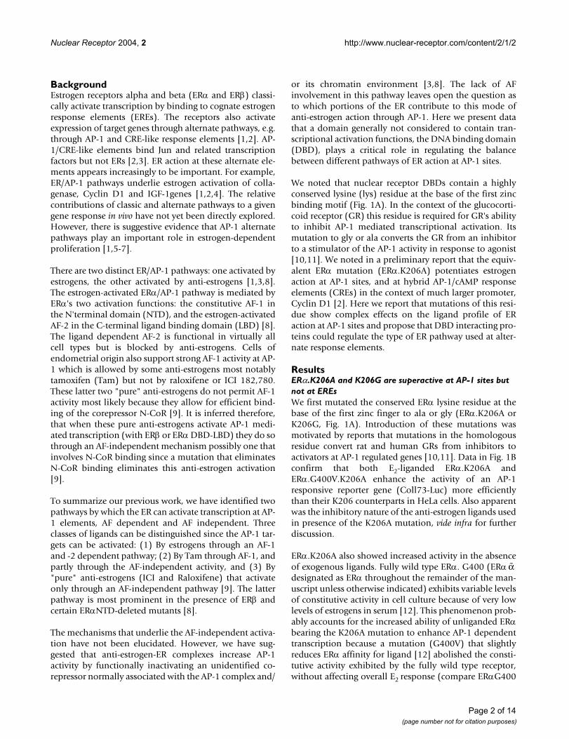
ERα enhances AP-1 activity in the presence of E2 in a vari-ety of cell lines ([3] and references therein). ERα alsoenhances AP-1 activity in the presence of the partial ago-nist/antagonist Tam in cell lines that support AF-1 activityat AP-1 sites [1,7]. Since the K206A mutation enhancesestrogen activity, we determined whether the ERα.K206Amutation would also amplify Tam action at AP-1. Consist-ent with our previous results [1], endogenous ERα acti-vated AP-1 in the presence of both E2 and Tam in Ishikawacells (Figs. 2A insert) whereas endogenous ERα activatedthe AP-1 responsive reporter only in the presence of E2 inMCF-7 breast cells, which do not support AF-1 activity atAP-1 (Fig. 2B, insert). Co-transfection of both cell typeswith the K206A mutation amplified these responses. InIshikawas the K206A mutation enhanced both Tam andE2 response, whereas in MCF-7s ERα.K206A primarilyenhanced the E2 response. The MCF-7 cell response is likethat seen in HeLas (Fig. 1B), which also have very little AF-1 activity. Enhanced tamoxifen response was also sup-ported by MDA-MB-435 (a breast cell line; Fig. 3A) andchicken embryo fibroblasts (CEFs; data not shown) bothof which support AF-1 activity. We conclude that theERα.K206A mutation amplifies E2 responses at AP-1 sitesand also amplifies the agonist properties of the mixedagonist/antagonist Tam in cells that support AF-1 activityat AP-1 sites.
ERα.K206A activity is dependent on linked activation functions, either endogenous or exogenousWe next determined whether the increased activity elicited
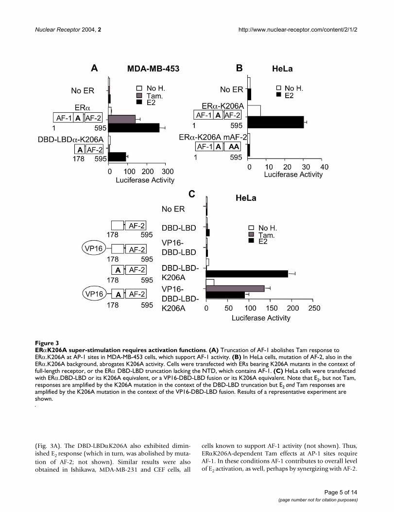
by the ERα K206A mutation was truly dependent on ERactivation functions. As indicated above, the ERα.K206Amutation amplifies Tam responses at AP-1 sites in cellsthat support AF-1 activity. To further test the dependenceof Tam's effect on AF-1, we evaluated the effect of an ERαAF-1 truncation (DBD-LBDα.K206A). This mutationcompletely abolished Tam response in MDA-MB-453 cells
ERαK206A super-stimulates in cells that contain endogenous ERsFigure 2ERαK206A super-stimulates in cells that contain endogenous ERs. Both Ishikawa (A) and MCF-7 cells (B) were transfected with Coll73-Luc and treated with ligands as indicated in Fig. 1B. The insets show the effect of treatment with endogenous receptor only. The main graphs show lig-and effect in the presence of the indicated transfected ERα (5 µg).
Page 4 of 14(page number not for citation purposes)
Nuclear Receptor 2004, 2 http://www.nuclear-receptor.com/content/2/1/2
(Fig. 3A). The DBD-LBDαK206A also exhibited dimin-ished E2 response (which in turn, was abolished by muta-tion of AF-2; not shown). Similar results were alsoobtained in Ishikawa, MDA-MB-231 and CEF cells, all
cells known to support AF-1 activity (not shown). Thus,ERαK206A-dependent Tam effects at AP-1 sites requireAF-1. In these conditions AF-1 contributes to overall levelof E2 activation, as well, perhaps by synergizing with AF-2.
ERαK206A super-stimulation requires activation functionsFigure 3ERαK206A super-stimulation requires activation functions. (A) Truncation of AF-1 abolishes Tam response to ERα.K206A at AP-1 sites in MDA-MB-453 cells, which support AF-1 activity. (B) In HeLa cells, mutation of AF-2, also in the ERα.K206A background, abrogates K206A activity. Cells were transfected with ERs bearing K206A mutants in the context of full-length receptor, or the ERα DBD-LBD truncation lacking the NTD, which contains AF-1. (C) HeLa cells were transfected with ERα.DBD-LBD or its K206A equivalent, or a VP16-DBD-LBD fusion or its K206A equivalent. Note that E2, but not Tam, responses are amplified by the K206A mutation in the context of the DBD-LBD truncation but E2 and Tam responses are amplified by the K206A mutation in the context of the VP16-DBD-LBD fusion. Results of a representative experiment are shown.
Page 5 of 14(page number not for citation purposes)
Nuclear Receptor 2004, 2 http://www.nuclear-receptor.com/content/2/1/2
As suggested by the data above, the K206A mutationexerts its effects via both AFs 1 and 2 in the presence of E2(Figs. 3A). To test for the contribution of AF-2, we used adouble hydrophobic mutation within the ERα LBD C-ter-minal helix 12 (M543A;L544A) that completely abolishesERα AF-2 activity at classical EREs [13]. We performed thisexperiment in HeLa cells, which only support minimalAF-1 activity. This mutation completely abolishedERα.K206A action at AP-1 sites in HeLa cells (Fig. 3B).Thus, ERα.K206A enhances AP-1 activity in HeLa cells ina manner that is completely dependent on AF-2. These
observations indicate that the enhancement of AP-1 activ-ity by the K206A ERα DBD is dependent on linked activa-tion functions AF-1 and AF-2.
We then investigated whether the K206A mutation wouldamplify the activity of a heterologous activation function.Substitution of a strong heterologous activation functionfor the ERα N-terminus (VP16) failed to enhance Tamresponse at AP-1 sites in HeLa cells. In contrast, fusion ofthe same activation function to the DBD-LBDαK206A N-terminus produced robust Tam activation (Fig 3C). Thus,the VP16 activation function when fused to the ER DBD isnearly inactive at AP-1unless the DBD carries the K206Amutation. This observation suggests that the K206A muta-tion may relieve a repressive function mediated throughthe DBD exerted on linked activation functions when ERαactivates at AP-1. This is further explored below.
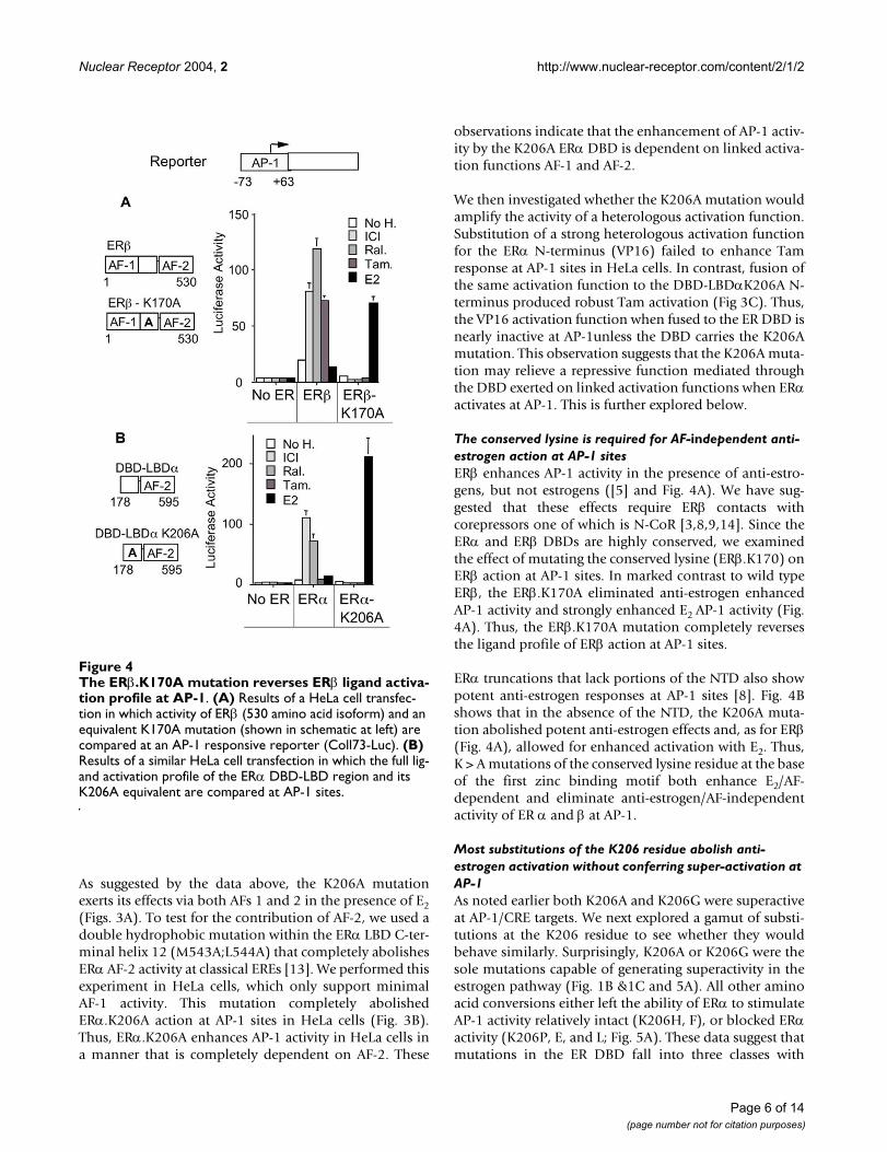
The conserved lysine is required for AF-independent anti-estrogen action at AP-1 sitesERβ enhances AP-1 activity in the presence of anti-estro-gens, but not estrogens ([5] and Fig. 4A). We have sug-gested that these effects require ERβ contacts withcorepressors one of which is N-CoR [3,8,9,14]. Since theERα and ERβ DBDs are highly conserved, we examinedthe effect of mutating the conserved lysine (ERβ.K170) onERβ action at AP-1 sites. In marked contrast to wild typeERβ, the ERβ.K170A eliminated anti-estrogen enhancedAP-1 activity and strongly enhanced E2 AP-1 activity (Fig.4A). Thus, the ERβ.K170A mutation completely reversesthe ligand profile of ERβ action at AP-1 sites.
ERα truncations that lack portions of the NTD also showpotent anti-estrogen responses at AP-1 sites [8]. Fig. 4Bshows that in the absence of the NTD, the K206A muta-tion abolished potent anti-estrogen effects and, as for ERβ(Fig. 4A), allowed for enhanced activation with E2. Thus,K > A mutations of the conserved lysine residue at the baseof the first zinc binding motif both enhance E2/AF-dependent and eliminate anti-estrogen/AF-independentactivity of ER α and β at AP-1.
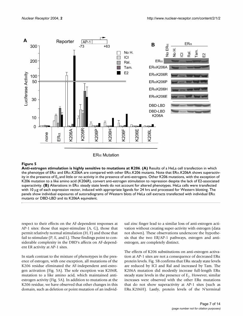
Most substitutions of the K206 residue abolish anti-estrogen activation without conferring super-activation at AP-1As noted earlier both K206A and K206G were superactiveat AP-1/CRE targets. We next explored a gamut of substi-tutions at the K206 residue to see whether they wouldbehave similarly. Surprisingly, K206A or K206G were thesole mutations capable of generating superactivity in theestrogen pathway (Fig. 1B &1C and 5A). All other aminoacid conversions either left the ability of ERα to stimulateAP-1 activity relatively intact (K206H, F), or blocked ERαactivity (K206P, E, and L; Fig. 5A). These data suggest thatmutations in the ER DBD fall into three classes with
The ERβ.K170A mutation reverses ERβ ligand activation pro-file at AP-1Figure 4The ERβ.K170A mutation reverses ERβ ligand activa-tion profile at AP-1. (A) Results of a HeLa cell transfec-tion in which activity of ERβ (530 amino acid isoform) and an equivalent K170A mutation (shown in schematic at left) are compared at an AP-1 responsive reporter (Coll73-Luc). (B) Results of a similar HeLa cell transfection in which the full lig-and activation profile of the ERα DBD-LBD region and its K206A equivalent are compared at AP-1 sites.
Page 6 of 14(page number not for citation purposes)
Nuclear Receptor 2004, 2 http://www.nuclear-receptor.com/content/2/1/2
respect to their effects on the AF-dependent responses atAP-1 sites: those that super-stimulate (A, G), those thatpermit relatively normal stimulation (H, F) and those thatfail to stimulate (P, E, and L). These findings point to con-siderable complexity in the DBD's affects on AF-depend-ent ER activity at AP-1 sites.
In stark contrast to the mixture of phenotypes in the pres-ence of estrogen, with one exception, all mutations of theK206 residue eliminated the AF-independent anti-estro-gen activation (Fig. 5A). The sole exception was K206R,mutation to a like amino acid, which maintained anti-estrogen activity (Fig. 5A). In addition to mutations at theK206 residue, we have observed that other changes in thisdomain, such as deletion or point mutation of an individ-
ual zinc finger lead to a similar loss of anti-estrogen acti-vation without creating super-activity with estrogen (datanot shown). These observations underscore the hypothe-sis that the two ER/AP-1 pathways, estrogen and anti-estrogen, are completely distinct.
The effects of K206 substitutions on anti-estrogen activa-tion at AP-1 sites are not a consequence of decreased ERαprotein levels. Fig. 5B confirms that ERα steady state levelsare reduced by ICI and Ral and increased by Tam. TheK206A mutation did modestly increase full-length ERαsteady state levels in the presence of E2. However, similarincreases were observed with the other ERα mutationsthat do not show superactivity at AP-1 sites (such asERα.K206H). Lastly, protein levels of the N'terminal
Anti-estrogen stimulation is highly sensitive to mutations at K206Figure 5Anti-estrogen stimulation is highly sensitive to mutations at K206. (A) Results of a HeLa cell transfection in which the phenotype of ERα and ERα.K206A are compared with other ERα.K206 mutants. Note that ERα.K206A shows superactiv-ity in the presence of E2 and little or no activity in the presence of anti-estrogens. Other K206 mutations, with the exception of K206 mutation to a like amino acid (K206R), convert anti-estrogen stimulation to repression despite the lack of E2-associated superactivity. (B) Alterations in ERα steady state levels do not account for altered phenotypes. HeLa cells were transfected with 10 µg of each expression vector, induced with appropriate ligands for 24 hrs and processed for Western blotting. The panels show individual exposures of autoradiograms of Western blots of HeLa cell extracts transfected with individual ERα mutants or DBD-LBD and its K206A equivalent.
Page 7 of 14(page number not for citation purposes)
Nuclear Receptor 2004, 2 http://www.nuclear-receptor.com/content/2/1/2
domain (AF-1) deletion mutants with and without theK206A mutation were virtually indistinguishable (Fig.5B). Thus, the dramatic loss of ICI and raloxifeneresponses exhibited by the DBD-LBDα.K206A mutant(Fig. 4B) cannot be explained by reduced protein levels(Fig. 5B). Overall, our results suggest that effects of theK206A mutation on steady state levels of ERα are unlikelyto explain ERα.K206A behavior at AP-1.
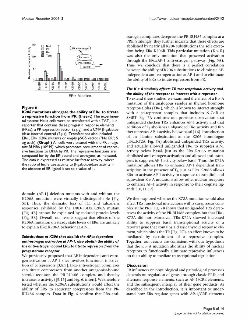
Substitutions at K206 that abolish the AF-independent anti-estrogen activation at AP-1, also abolish the ability of the anti-estrogen bound ERα to titrate repressors from the progesterone receptorWe previously proposed that AF-independent anti-estro-gen activation at AP-1 sites involves functional inactiva-tion of corepressors [3,8,9]. ERα-anti-estrogen complexescan titrate corepressors from another antagonist-boundsteroid receptor, the PR-RU486 complex, and therebyincrease its activity ([9,15] and Fig. 6, insert). We thereforetested whether the K206A substitutions would affect theability of ERα to sequester corepressors from the PR-RU486 complex. Data in Fig. 6 confirm that ERα-anti-
estrogen complexes derepress the PR-RU486 complex at aPRE. Strikingly, they further indicate that these effects areabolished by nearly all K206 substitutions the sole excep-tion being ERα.K206R. This particular mutation (K > R)was also the only mutation that preserved activationthrough the ERα/AP-1 anti-estrogen pathway (Fig. 5A).Thus, we conclude that there is a perfect correlationbetween the ability of K206 substitutions to eliminate AF-independent anti-estrogen action at AP-1 and to eliminatethe ability of ERα to titrate repressors from PR.
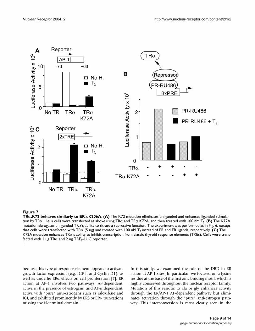
The K > A similarly affects TR transcriptional activity and the ability of the receptor to interact with a repressorTo extend these studies, we examined the effect of a K > Amutation of the analogous residue in thyroid hormonereceptor-alpha (TRα), which is known to interact stronglywith a co-repressor complex that includes N-CoR orSMRT. Fig. 7A confirms our previous observation thatunliganded chicken TRα enhances AP-1 activity and thataddition of T3 abolishes unliganded TRα activity and fur-ther represses AP-1 activity below basal [16]. Introductionof an alanine substitution at the K206 homologue(TRα.K72A; Fig. 7A) abolished unliganded TRα activity,and actually allowed unliganded TRα to suppress AP-1activity below basal, just as the ERα.K206A mutationabolished anti-estrogen activation and allowed anti-estro-gens to suppress AP-1 activity below basal. Thus, the K72Amutation allows TRα to enhance AP-1 dependent tran-scription in the presence of T3, just as ERα.K206A allowsERα to activate AP-1 activity in response to estradiol, andequivalent K > A mutations allow other nuclear receptorsto enhance AP-1 activity in response to their cognate lig-ands [10,11,17].
We then explored whether the K72A mutation would alsoaffect TRα functional interactions with a corepressor com-plex at the PRE. Fig. 7B shows that unliganded TRα derep-resess the activity of the PR-RU486 complex, but that TRα-K72A did not. Moreover, TRα.K72A showed increasedability to suppress basal transcriptional activity of areporter gene that contains a classic thyroid response ele-ment, which binds the TR (Fig. 7C), an effect known to bemediated by recruitment of a repressive complex.Together, our results are consistent with our hypothesisthat the K > A mutation abolishes the ability of nuclearreceptors to functionally eliminate repressive influenceson their ability to mediate transcriptional regulation.
DiscussionER influences on physiological and pathological processesdepends on regulation of genes through classic EREs andalternate response elements, such as AP-1/CRE elements,and the subsequent interplay of their gene products. Asdescribed in the Introduction, it is important to under-stand how ERs regulate genes with AP-1/CRE elements
K206 mutations abrogate the ability of ERα to titrate a repressive function from PRFigure 6K206 mutations abrogate the ability of ERα to titrate a repressive function from PR. (Insert) The experimen-tal system: HeLa cells were co-transfected with a TAT3-Luc reporter that contains three progestin response elements (PREs), a PR expression vector (5 µg), and a CMV β-galactos-idase internal control (2 µg). Transfections also included ERα, ERα K206 mutants or empty pSG5 vector ("No ER"; 5 µg each). (Graph) All cells were treated with the PR antago-nist RU486 (10-6 M), which promotes recruitment of repres-sive functions to DNA by PR. The repressive functions are competed for by the ER-bound anti-estrogens, as indicated. The data is expressed as relative luciferase activity, where the ratio of luciferase activity to β-galactosidase activity in the absence of ER ligand is set to a value of 1.
Page 8 of 14(page number not for citation purposes)
Nuclear Receptor 2004, 2 http://www.nuclear-receptor.com/content/2/1/2
because this type of response element appears to activategrowth factor expression (e.g. IGF I, and Cyclin D1), aswell as underlie ERα effects on cell proliferation [7]. ERaction at AP-1 involves two pathways: AF-dependent,active in the presence of estrogens; and AF-independent,active with "pure" anti-estrogens such as raloxifene andICI, and exhibited prominently by ERβ or ERα truncationsmissing the N-terminal domain.
In this study, we examined the role of the DBD in ERaction at AP-1 sites. In particular, we focused on a lysineresidue at the base of the first zinc binding motif, which ishighly conserved throughout the nuclear receptor family.Mutation of this residue to ala or gly enhances activitythrough the ER/AP-1 AF-dependent pathway but elimi-nates activation through the "pure" anti-estrogen path-way. This interconversion is most clearly seen in the
TRα.K72 behaves similarly to ERα.K206AFigure 7TRα.K72 behaves similarly to ERα.K206A. (A) The K72 mutation eliminates unliganded and enhances liganded stimula-tion by TRα. HeLa cells were transfected as above using TRα and TRα.K72A, and then treated with 100 nM T3. (B) The K72A mutation abrogates unliganded TRα's ability to titrate a repressive function. The experiment was performed as in Fig. 6, except that cells were transfected with TRα (5 ug) and treated with 100 nM T3 instead of ER and ER ligands, respectively. (C) The K72A mutation enhances TRα's ability to inhibit transcription from classic thyroid response elements (TREs). Cells were trans-fected with 1 ug TRα and 2 ug TRE2-LUC reporter.
Page 9 of 14(page number not for citation purposes)
Nuclear Receptor 2004, 2 http://www.nuclear-receptor.com/content/2/1/2
context of either the ERα deleted of its NTD (K206A), orin the context of ERβ (K170A; Fig. 4). Bjornstrom andSjoberg have also studied the role of the ERβ DBD in anti-estrogen activation at AP-1 and have similarly found thatmutations widely spaced through the DBD eliminate anti-estrogen activation and confer some activation with estro-gen. The K170 residue was not tested in that study and nosuperactivation was observed [18]. Their observations areconsistent with our model of two exclusive mechanisms atAP-1 and the role of the DBD in controlling choicebetween the mechanisms.
The homologous mutation in TRα (K72A; Fig 7) exerts ahighly similar phenotype to that of ERs α and β. It con-verts unliganded TRα stimulation at AP-1 to repression ofbasal transcription and converts liganded TRα from inhib-iting AP-1 activity to stimulating it. Thus, the pathwaysaffected by the ERα.K206A mutation are likely to be uti-lized by other members of the nuclear receptor super-family, as well. In addition, the ERα.K206A mutationamplifies ERα action at AP-1 sites with a ligand profilethat resembles that of endogenous ERα in MCF-7 andIshikawa cells (Fig. 2). Taken together these data suggeststhat the K206A and G mutations amplify pathways thatare likely to be physiologically relevant across nuclearreceptors.
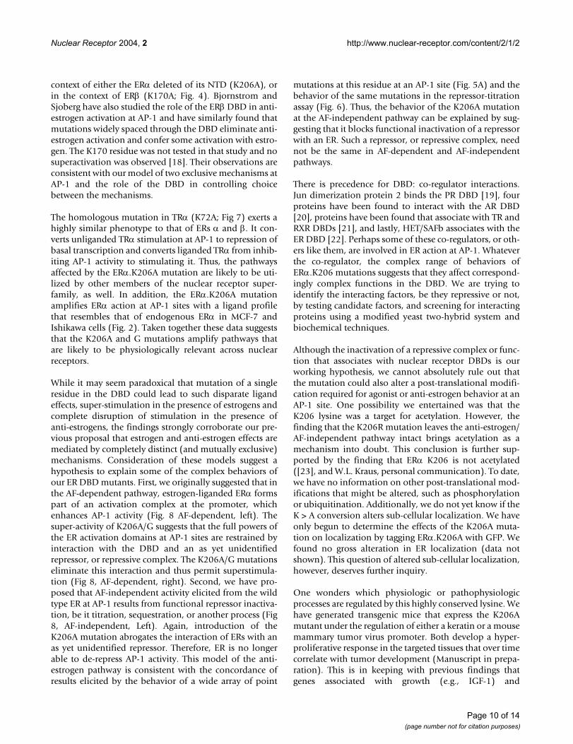
While it may seem paradoxical that mutation of a singleresidue in the DBD could lead to such disparate ligandeffects, super-stimulation in the presence of estrogens andcomplete disruption of stimulation in the presence ofanti-estrogens, the findings strongly corroborate our pre-vious proposal that estrogen and anti-estrogen effects aremediated by completely distinct (and mutually exclusive)mechanisms. Consideration of these models suggest ahypothesis to explain some of the complex behaviors ofour ER DBD mutants. First, we originally suggested that inthe AF-dependent pathway, estrogen-liganded ERα formspart of an activation complex at the promoter, whichenhances AP-1 activity (Fig. 8 AF-dependent, left). Thesuper-activity of K206A/G suggests that the full powers ofthe ER activation domains at AP-1 sites are restrained byinteraction with the DBD and an as yet unidentifiedrepressor, or repressive complex. The K206A/G mutationseliminate this interaction and thus permit superstimula-tion (Fig 8, AF-dependent, right). Second, we have pro-posed that AF-independent activity elicited from the wildtype ER at AP-1 results from functional repressor inactiva-tion, be it titration, sequestration, or another process (Fig8, AF-independent, Left). Again, introduction of theK206A mutation abrogates the interaction of ERs with anas yet unidentified repressor. Therefore, ER is no longerable to de-repress AP-1 activity. This model of the anti-estrogen pathway is consistent with the concordance ofresults elicited by the behavior of a wide array of point
mutations at this residue at an AP-1 site (Fig. 5A) and thebehavior of the same mutations in the repressor-titrationassay (Fig. 6). Thus, the behavior of the K206A mutationat the AF-independent pathway can be explained by sug-gesting that it blocks functional inactivation of a repressorwith an ER. Such a repressor, or repressive complex, neednot be the same in AF-dependent and AF-independentpathways.
There is precedence for DBD: co-regulator interactions.Jun dimerization protein 2 binds the PR DBD [19], fourproteins have been found to interact with the AR DBD[20], proteins have been found that associate with TR andRXR DBDs [21], and lastly, HET/SAFb associates with theER DBD [22]. Perhaps some of these co-regulators, or oth-ers like them, are involved in ER action at AP-1. Whateverthe co-regulator, the complex range of behaviors ofERα.K206 mutations suggests that they affect correspond-ingly complex functions in the DBD. We are trying toidentify the interacting factors, be they repressive or not,by testing candidate factors, and screening for interactingproteins using a modified yeast two-hybrid system andbiochemical techniques.
Although the inactivation of a repressive complex or func-tion that associates with nuclear receptor DBDs is ourworking hypothesis, we cannot absolutely rule out thatthe mutation could also alter a post-translational modifi-cation required for agonist or anti-estrogen behavior at anAP-1 site. One possibility we entertained was that theK206 lysine was a target for acetylation. However, thefinding that the K206R mutation leaves the anti-estrogen/AF-independent pathway intact brings acetylation as amechanism into doubt. This conclusion is further sup-ported by the finding that ERα K206 is not acetylated([23], and W.L. Kraus, personal communication). To date,we have no information on other post-translational mod-ifications that might be altered, such as phosphorylationor ubiquitination. Additionally, we do not yet know if theK > A conversion alters sub-cellular localization. We haveonly begun to determine the effects of the K206A muta-tion on localization by tagging ERα.K206A with GFP. Wefound no gross alteration in ER localization (data notshown). This question of altered sub-cellular localization,however, deserves further inquiry.
One wonders which physiologic or pathophysiologicprocesses are regulated by this highly conserved lysine. Wehave generated transgenic mice that express the K206Amutant under the regulation of either a keratin or a mousemammary tumor virus promoter. Both develop a hyper-proliferative response in the targeted tissues that over timecorrelate with tumor development (Manuscript in prepa-ration). This is in keeping with previous findings thatgenes associated with growth (e.g., IGF-1) and
Page 10 of 14(page number not for citation purposes)
Nuclear Receptor 2004, 2 http://www.nuclear-receptor.com/content/2/1/2
proliferative regulation (e.g., Cyclin D1) are regulated bythe ER through AP-1 or AP-1 like elements [2,4]. We areunaware of a naturally occurring germline or somaticmutations associated with squamous or breast carcino-mas that involve the homologous, highly conservedlysine. However, a naturally occurring mutation in theVitamin D Receptor (VDR) at this conserved site (K45;mutated to K45E) has been associated with a hereditaryform of vitamin D resistant rickets [24]. Therefore, eventhough we cannot point to this mutation in a givenhuman breast carcinoma, or growth disorder of squamousepithelium, data from our transgenic mice and the discov-ery of a disease associated with the homologous lysine ina different nuclear receptor (Vitamin D resistant rickets)underscore the potential importance that this residueplays in physiologic or pathophysiologic mechanisms. Infact, we hope that our work spurs a quest for mutations ofthis highly conserved lysine in tumors, or otherpathologies.
ConclusionsOur data indicate that the ER DBD acts as a regulatoryswitch that dictates the ligand activation profile at AP-1
sites and thereby distinguishes the AF-dependent (estro-gen) from the AF-independent (anti-estrogen) pathways.What are the factors that might bind the ER DBD andinfluence ER action at AP-1 sites? Data from three experi-ments described here corroborate our hypothesis that thefactors are part of a repressive complex: 1) The K > A muta-tion is able to unmask the activity of a heterologous acti-vation function (VP16; Fig. 3C), 2) Conversion of K206 toany residue except the like amino acid, arg, eliminates theAF-independent pathway (Fig. 5A), and 3) the profile ofactivity elicited by the same mutations parallels theirbehavior in the repressor titration assay (Fig. 6).
Regardless of the actual mechanism, the degree of super-stimulation elicited by ERα.K206A is unlikely to bebenign in normal growth and development. In fact, datafrom our ERα.K206A transgenic mice bear this out (Man-uscript in preparation). The extraordinary degree of ER-stimulated AP-1 activity possible likely needs to berestrained over a wide range of cellular conditions, thoughit may be selectively exposed in some. Given the potentialimportance of such mechanisms, it is likely that the pro-teins that mediate these effects will represent valuable tar-
Model of ER action at AP-1 sitesFigure 8Model of ER action at AP-1 sites. The underlying mechanisms of estrogen and anti-estrogen pathways are distinct. In the AF-dependent/Estrogen Pathway: the balance of co-activator and co-repressor functions is altered by the introduction of the K206A mutation. Loss of ER: repressor interactions lead to unopposed, potent, co-activator stimulation (right). In the AF-inde-pendent or anti-estrogen pathway, ERs titrate, or functionally inactivate, a repressive function associated with the promoter (left). The K206A mutation renders the ER unable to do so. The star represents a mutation at ERα K206: K > A/G in the AF-depend-ent pathway; or K > any mutation except arg, in the AF-independent pathway.
Page 11 of 14(page number not for citation purposes)
Nuclear Receptor 2004, 2 http://www.nuclear-receptor.com/content/2/1/2
gets for drugs designed specifically to dampen ERstimulated growth. We are presently searching for proteinsthat associate with the ER DBD in a manner influenced bymutations of this highly conserved lysine, in the hopesthat they will direct novel drug development.
MethodsCell lines and ER ligandsHeLa, Ishikawa, MCF-7, and MDA-MB-453 cells weregrown in Dulbecco's Modified Eagle's/F-12 Coon's modi-fied medium (Sigma), with 15 mM Hepes, L-glutamine(0.438 g/liter), NaHCO3 (1.38 g/liter), 10% iron supple-mented calf serum (Sigma) which we screen for negligibleE2 activity, and penicillin/streptomycin as previouslydescribed [5,8]. E2 was purchased from Sigma (St. Louis,Mo). ICI 182,780 was a gift from Dr. Alan Wakeling(Astra/Zeneca, Macclesfieled, UK). Ral was a gift from Dr.Stefan Nilsson (Karo Bio AB Huddinge, Sweden).
PlasmidsThe following expression vectors were previouslydescribed: ERα: G400, the fully wild type and G400Vwhich contains the indicated G > V conversion in the LBDof ERα. These vectors have previously been termed HEG0and HE0 respectively [12,25]; ERα.K206A [2], the ERβ530 amino acid isoform [8], and PR [26].
ERαG400V.K206A was generated by D. Barry Starr, usingsite directed mutagenesis [27]. ERα mAF-2.K206A wasprepared by ligating a BglII fragment from a mutated ver-sion of pSG5-ERαLBD [28] into BglII digested ERαK206A.ERα.K206 was mutated to ala in the context of ERαG400Vand ERα DBD-LBD mutations by site directed mutagene-sis of HE0 using the Stratagene Quickchange kit.ERβ.K170A was made similarly. VP16-ER-DBD-LBD andthe K206A equivalent (both in ERα) were generated byPCR amplification of ERα amino acids 164–595 usingERα and ERαK206A as templates. The primers weredesigned with EcoRI and SalI sites. The amplified cDNAsequences were digested with these enzymes and ligateddownstream (in frame) of a cDNA encoding the strongHSV VP16 activation domain in expression vector pVP16(Clontech). Primer sequences and PCR conditions areavailable on request.
Reporter genes Coll73-Luc [1], ERE-Luc [5], and GAL4-RE5-Luc (GK1) [29] have been previously described. ThePR (and GR) responsive reporter gene TAT3-Luc (3xPRE)was a gift from K. Yamamoto (UCSF) and is described in[30].
TransfectionsTransfections were performed as described [2,5,8]. Cellswere grown to an approximate density of 5 × 10⁴ cells/cm2
on 10cm diameter tissue culture plates. The cells were
trypsinized and resuspended in a single 0.4-cm gap elec-troporation cuvette in 0.5 ml electroporation buffer (PBSsupplemented with 10% glucose and 10 µg/ml BioBrenedetergent; Applied Biosystems, Foster City, CA). Transfec-tions to determine ER affects on AP-1 activity contained 2µg of Coll73-Luc and actin β-galactosidase reporters and 5µg ER vector or empty vector controls. Transfections todetermine ER effects on classical response elements con-tained 2 µg of the appropriate reporter, an actin-β-galac-tosidase control, and 1 µg ER expression vector.Transfections to determine ER affects on PR-RU486 com-plex activity contained 5 µg of TAT3-Luc reporter, 2 µg ofCMV-β-galactosidase and 5 µg each of PR and ERexpression vectors. Transfected cells were resuspended instandard growth medium and plated on 12-well dishes.Ligands were then added at the following final concentra-tions (ICI 10-7 M, Ral 10-7 M, Tam 5 × 10⁻⁶ M, E2 10-7 M,RU486 10-6 M).
Western Blots10 cm dishes of HeLa cells (transfected with 10 µg ERexpression vector and 2 µg CMV-β-galactosidase to permitdetection of equivalent transfection efficiency) weretreated with ligands for 24 hrs, chilled on ice, washed withice-cold PBS, then incubated in lysis buffer (0.2% TritonX-100, 150 mM NaCl, 50 mM Tris pH 7.8, 1 mM EDTA,and 1/1000 dilution Novagen protease inhibitor cocktail)for 10 mins. They were then scraped and centrifuged at4°C in an Eppendorf bench top centrifuge at maximumspeed for 15 mins. Protein concentration in cell extractwas determined by standard methods. The concentrationof each sample was normalized by addition of cold lysisbuffer to a final value of approximately 5 mg total proteinper ml. 20–30 µg of proteins were separated by SDS-PAGE. Proteins were transferred to a wet Immuno-BlotPVDF membrane (Bio-Rad, Hercules CA), overnight at 90mA, 30 V, using a standard transfer apparatus. The mem-brane was then incubated in 5% non-fat milk in PBS-T (1× PBS, 0.1% Tween-20) for 1 hour and washed twice inPBS-T for 10 minutes at room temperature. The primaryERα antibody used in this study was HC-20 (Santa CruzBiotechnology, Inc.) directed against the ERα C-terminus.Primary antibody was diluted 1:2000 in PBS-T and incu-bated with the membrane for 1 hour, followed by PBS-Twashes, 1 × 15 minutes and then 2 × 5 minutes. Themembrane was incubated for 45 minutes with horse-rad-ish peroxidase conjugated anti-rabbit IgG (Santa CruzBiotechnology, Inc.) diluted at 1:2000 in PBS-T, followedby PBS-T washes (1 × 15 minutes and 4 × 5 minutes). Afterthe last wash, the membrane was developed with astandard ECL kit (Amersham-Pharmacia Biotech), cov-ered with Saran wrap and exposed to X-ray film.
AbbreviationsAF-1 Activation Function 1
Page 12 of 14(page number not for citation purposes)
Nuclear Receptor 2004, 2 http://www.nuclear-receptor.com/content/2/1/2
AF-2 Activation Function 2
AP-1 Activator Protein-1
CEF Chicken Embryo Fibroblasts
CRE cAMP Response Element
DBD DNA binding Domain
ERα Estrogen Receptor-alpha, fully wild type: G400
ERαG400V Estrogen Receptor-alpha with LBD mutation:G400V
ERβ Estrogen Receptor-beta
ERE Estrogen Response Element
GR Glucocorticoid Receptor
ICI Imperial Chemical Industries #182,780
LBD Ligand Binding Domain
N-CoR Nuclear receptor Corepressor
NTD Amino (N)-terminal domain
PR Progesterone Receptor
PRE Progesterone Response Element
Ral Raloxifene (Evista)
SMRT Silencing mediator of retinoid and thyroidreceptors
Tam Tamoxifen (Nolvadex)
Competing interestsNone declared for any authors except PK who is a con-sultant, former member of the board of directors, and hassignificant financial interests in KaroBio AB, a Swedishcompany that develops pharmaceuticals that target the ERand other nuclear receptors.
Authors' contributionsRU Made the original observation that ERα.K206A super-stimulated the ER/AP-1 pathway, performed experimentswhile a post-doctoral fellow in PJK's lab, and finished theproject in her own lab at the University of Virginia, whichincluded writing the manuscript.
PW Provided considerable intellectual guidance throughout the project. He provided significant input in craftingthe manuscript.
RP Provided intellectual insight and designed and per-formed several experiements, in particular the VP16fusion experiment (Fig. 3C).
PN Performed many experiments under the guidance ofPW
CV & HF Performed many experiments under the guid-ance of PW and PJK.
PK Oversaw the entire project and provided the majorityof funding.
AcknowledgementsSome of these data were initially presented at the 1998 & 2000 Nuclear Receptor Keystone meetings, and the 2001 Cold Spring Harbor meeting. We also thank Carol M. Anderson and Kristen Hilty for their excellent technical support in the early part of the project. This work was supported by K08 DK 02335 and R03 DK56236 (R.M.U.) and DK51083 and CA80210 to (P.J.K.).
References1. Webb Paul, Lopez Gabriela N., Uht Rosalie M., Kushner Peter J.:
Tamoxifen activation of the estrogen receptor/AP-1 path-way: potential origin for the cell-specific estrogen-like effectsof antiestrogens. Mol Endocrinol 1995, 9:443-456.
2. Liu MM, Albanese C, Anderson CM, Hilty K, Webb P, Uht RM, PriceR. H., Jr., Pestell RG, Kushner PJ: Opposing action of estrogenreceptors alpha and beta on cyclin D1 gene expression. J BiolChem 2002, 277:24353-24360.
3. Kushner PJ, Agard DA, Greene GL, Scanlan TS, Shiau AK, Uht RM,Webb P: Estrogen receptor pathways to AP-1. Steroid Biochem-istry & Molecular Biology 2000, 74:311-317.
4. Umayahara Y, Kawamori R, Watada H, Imano E, Iwama N, MorishimaT, Yamasaki Y, Kajimoto Y, Kamada T: Estrogen regulation of theinsulin-like growth factor I gene transcription involves anAP-1 enhancer. J Biol Chem 1994, 269:16433-16442.
5. Paech K, Webb P, Kuiper GG, Nilsson S, Gustafsson J, Kushner PJ,Scanlan TS: Differential ligand activation of estrogen recep-tors ERalpha and ERbeta at AP1 sites [see comments]. Sci-ence 1997, 277:1508-1510.
6. Philips Alexandre, Teyssier Catherine, Galtier Florence, Rivier-CovasCorinne, Rey Jean-Marc, Rochefort Henri, Chalbos Dany: FRA-1Expression Level Modulates Regulation of Activator protein-1 Activity by Estradiol in Breast Cancer Cells. Mol Endocrinol1998, 12:973-985.
7. Shang Y, Brown M: Molecular determinants for the tissue spe-cificity of SERMs. [see comments.]. Science 2002,295:2465-2468.
8. Webb P, Nguyen P, Valentine C, Lopez GN, Kwok GR, McInerney E,Katzenellenbogen BS, Enmark E, Gustafsson J-Å, Nilsson S, KushnerPJ: The Estrogen Receptor Enhances AP-1 Activity by twoDistinct Mechanisms with Different Requirements forReceptor Transactivation Functions. Mol Endocrinol 1999,13:1672-1685.
9. Webb P, Nguyen P, Kushner PJ: Differential SERM effects oncorepressor binding dictate ERalpha activity in vivo. J BiolChem 2003, 278:6912-6920.
10. Yang-Yen Hsin-Fang, Chambard Jean-Claude, Sun Yu-Lin, Smeal Tod,Schmidt Thomas J., Drouin Jacques, Karin Michael: Transcriptionalinterference between c-Jun and the glucocorticoid receptor:mutual inhibition of DNA binding due to direct protein-pro-tein interaction. Cell 1990, 62:1205-1215.
Page 13 of 14(page number not for citation purposes)
http://www.ncbi.nlm.nih.gov/entrez/query.fcgi?cmd=Retrieve&db=PubMed&dopt=Abstract&list_uids=7659088
http://www.ncbi.nlm.nih.gov/entrez/query.fcgi?cmd=Retrieve&db=PubMed&dopt=Abstract&list_uids=8206951
http://www.ncbi.nlm.nih.gov/entrez/query.fcgi?cmd=Retrieve&db=PubMed&dopt=Abstract&list_uids=8206951
http://www.ncbi.nlm.nih.gov/entrez/query.fcgi?cmd=Retrieve&db=PubMed&dopt=Abstract&list_uids=8206951
http://www.ncbi.nlm.nih.gov/entrez/query.fcgi?cmd=Retrieve&db=PubMed&dopt=Abstract&list_uids=9278514
Nuclear Receptor 2004, 2 http://www.nuclear-receptor.com/content/2/1/2
Publish with BioMed Central and every scientist can read your work free of charge
"BioMed Central will be the most significant development for disseminating the results of biomedical research in our lifetime."
Sir Paul Nurse, Cancer Research UK
Your research papers will be:
available free of charge to the entire biomedical community
peer reviewed and published immediately upon acceptance
cited in PubMed and archived on PubMed Central
yours — you keep the copyright
Submit your manuscript here:http://www.biomedcentral.com/info/publishing_adv.asp
BioMedcentral
11. Starr D.Barry, Matsui William, R. Thomas Jay, Yamamoto Keith R.:Intracellular receptors use a common mechanism to inter-pret signaling information at response elements. Genes & Dev1996, 10:1271-1283.
12. Tora L, Mullick A, Metzger D, Ponglikitmongkol M, Park I, ChambonP: The cloned human oestrogen receptor contains a muta-tion which alters its hormone binding properties. EMBO J1989, 8:1981-1986.
13. Danielian PS, White R, Lees JA, Parker MG: Identification of a con-served region required for hormone dependent transcrip-tional activation by steroid hormone receptors. EMBO J 1992,11:1025-1033.
14. Webb P, Valentine C, Nguyen P, Price R. H., Jr., Marimuthu A, WestBL, Baxter JD, Kushner PJ: ERbeta Binds N-CoR in the Presenceof Estrogens via an LXXLL-like Motif in the N-CoR C-termi-nus. Nucl Recept 2003, 1:4.
15. Zhang Xun, Jeyakumar M, Petukhov Sergei, Bagchi Milan K.: ANuclear Receptor Corepressor Modulates TranscriptionalActivity of Antagonist-Occupied Steroid HormoneReceptor. Mol Endocrinol 1998, 12:513-524.
16. Lopez G, Schaufele F, Webb P, Holloway JM, Baxter JD, Kushner PJ:Positive and negative modulation of Jun action by thyroidhormone receptor at a unique AP1 site. Mol Cell Biol 1993,13:3042-3049.
17. Jaaskelainen Tiina, Pirskanen Asta, Ryhanen Sanna, Palvimo Jorma J.,Deluca Hector F., Maenpaa Pekka: Functional interferencebetween AP-1 and the vitamin D receptor on osteocalcingene expression in human osteosarcoma cells. Eur J Biochem1994, 224:11-20.
18. Bjornstrom L, Sjoberg M: Mutations in the estrogen receptorDNA-binding domain discriminate between the classicalmechanism of action and cross-talk with Stat5b and activat-ing protein 1 (AP-1). J Biol Chem 2002, 277:48479-48483.
19. Wardell SE, Boonyaratanakornkit V, Adelman JS, Aronheim A,Edwards DP: Jun dimerization protein 2 functions as a proges-terone receptor N-terminal domain coactivator. Mol Cell Biol2002, 22:5451-5466.
20. Janne OA, Moilanen AM, Poukka H, Rouleau N, Karvonen U, KotajaN, Hakli M, Palvimo JJ: Androgen-receptor-interacting nuclearproteins. Biochem Soc Trans 2000, 28:401-405.
21. Mathur Mukul, Tucker Philip W., Samuels Herbert H.: PSF is aNovel Corepressor that Mediates its Effect through Sin3Aand the DNA Binding Domain of Nuclear HormoneReceptors. Mol Cell Biol 2001, 21:2298-2311.
22. Oesterreich S, Zhang QP, Hopp T, Fuqua SAW, Michaelis M, ZhaoHH, Davie JR, Osborne CK, Lee AV: Tamoxifen-bound estrogenreceptor (ER) strongly interacts with the nuclear matrixprotein HET/SAF-B, a novel inhibitor of ER-mediatedtransactivation. Mol Endocrinol 2000, 14:369-381.
23. Wang C, Fu M, Angeletti RH, Siconolfi-Baez L, Reutens AT, AlbaneseC, Lisanti MP, Katzenellenbogen BS, Kato S, Hopp T, Fuqua SA, LopezGN, Kushner PJ, Pestell RG: Direct acetylation of the estrogenreceptor alpha hinge region by p300 regulates transactiva-tion and hormone sensitivity. J Biol Chem 2001,276:18375-18383.
24. Kahlen J-P, Carlberg C: Allosteric interaction of the 1a,25-dihy-droxyvitamin D3 receptor and the retinoid X receptor onDNA. NAR 1997, 25:4307-4313.
25. Green Stephen, Walter Philippe, Kumar Vijay, Krust Andree, BornertJean-Marc, Argos Patrick, Chambon Pierre: Human oestrogenreceptor cDNA: sequence, expression and homology to v-erb-A. Nature 1986, 320:134-139.
26. Uht Rosalie M., Anderson Carol M., Webb Paul, Kushner Peter J.:Transcriptional Activities of Estrogen and GlucocorticoidReceptors Are Functionally Integtrated at the AP-1Response Element. Endocrinology 1997, 138:2900-2908.
27. Kunkel TA: Rapid and efficient site-specific mutagenesis with-out phenotypic selection. Proc Natt Acad Sci, U S A 1985,82:488-492.
28. Lopez GN, Webb P, Shinsako JH, Baxter JD, Greene GL, Kushner PJ:Titration by estrogen receptor activation function-2 of tar-gets that are downstream from coactivators. Mol Endocrinol1999, 13:897-909.
29. Webb P, Nguyen P, Shinsako J, Anderson C, Feng W, Nguyen MP,Chen D, Huang SM, Subramanian S, McKinerney E, KatzenellenbogenBS, Stallcup MR, Kushner PJ: Estrogen receptor activation func-
tion 1 works by binding p160 coactivator proteins. MolEndocrinol 1998, 12:1605-1618.
30. Iniguez-Lluhi JA, Pearce D: A common motif within the negativeregulatory regions of multiple factors inhibits their tran-scriptional synergy. Mol Cell Biol 2000, 20:6040-6050.
Page 14 of 14(page number not for citation purposes)
http://www.ncbi.nlm.nih.gov/entrez/query.fcgi?cmd=Retrieve&db=PubMed&dopt=Abstract&list_uids=8675013
http://www.ncbi.nlm.nih.gov/entrez/query.fcgi?cmd=Retrieve&db=PubMed&dopt=Abstract&list_uids=8675013
http://www.ncbi.nlm.nih.gov/entrez/query.fcgi?cmd=Retrieve&db=PubMed&dopt=Abstract&list_uids=8675013
http://www.ncbi.nlm.nih.gov/entrez/query.fcgi?cmd=Retrieve&db=PubMed&dopt=Abstract&list_uids=2792078
http://www.ncbi.nlm.nih.gov/entrez/query.fcgi?cmd=Retrieve&db=PubMed&dopt=Abstract&list_uids=2792078
http://www.ncbi.nlm.nih.gov/entrez/query.fcgi?cmd=Retrieve&db=PubMed&dopt=Abstract&list_uids=1372244
http://www.ncbi.nlm.nih.gov/entrez/query.fcgi?cmd=Retrieve&db=PubMed&dopt=Abstract&list_uids=1372244
http://www.ncbi.nlm.nih.gov/entrez/query.fcgi?cmd=Retrieve&db=PubMed&dopt=Abstract&list_uids=1372244
http://www.ncbi.nlm.nih.gov/entrez/query.fcgi?cmd=Retrieve&db=PubMed&dopt=Abstract&list_uids=9544987
http://www.ncbi.nlm.nih.gov/entrez/query.fcgi?cmd=Retrieve&db=PubMed&dopt=Abstract&list_uids=8474460
http://www.ncbi.nlm.nih.gov/entrez/query.fcgi?cmd=Retrieve&db=PubMed&dopt=Abstract&list_uids=8474460
http://www.ncbi.nlm.nih.gov/entrez/query.fcgi?cmd=Retrieve&db=PubMed&dopt=Abstract&list_uids=8474460
http://www.ncbi.nlm.nih.gov/entrez/query.fcgi?cmd=Retrieve&db=PubMed&dopt=Abstract&list_uids=8076631
http://www.ncbi.nlm.nih.gov/entrez/query.fcgi?cmd=Retrieve&db=PubMed&dopt=Abstract&list_uids=8076631
http://www.ncbi.nlm.nih.gov/entrez/query.fcgi?cmd=Retrieve&db=PubMed&dopt=Abstract&list_uids=8076631
http://www.ncbi.nlm.nih.gov/entrez/query.fcgi?cmd=Retrieve&db=PubMed&dopt=Abstract&list_uids=3754034
http://www.ncbi.nlm.nih.gov/entrez/query.fcgi?cmd=Retrieve&db=PubMed&dopt=Abstract&list_uids=3754034
http://www.ncbi.nlm.nih.gov/entrez/query.fcgi?cmd=Retrieve&db=PubMed&dopt=Abstract&list_uids=3754034
http://www.ncbi.nlm.nih.gov/entrez/query.fcgi?cmd=Retrieve&db=PubMed&dopt=Abstract&list_uids=9202234


![er [ə:] her er [ ə ] sister er [ ə ] sister t [ t ] pet th [ δ] the a [æ] cat.](https://static.fdocument.org/doc/165x107/56649ef45503460f94c07882/er-her-er-sister-er-sister-t-t-pet-th-the-a-ae.jpg)