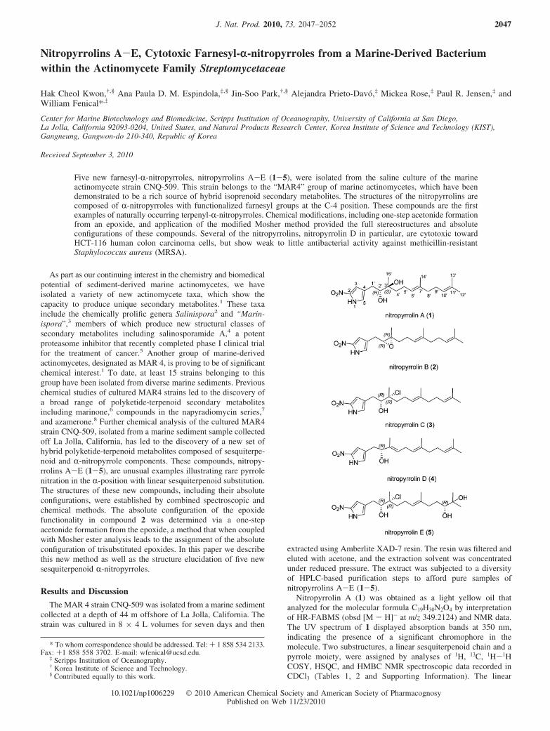
Nitropyrrolins A−E, Cytotoxic Farnesyl-α-nitropyrroles from a Marine-Derived Bacterium within the...
6
Nitropyrrolins A-E, Cytotoxic Farnesyl-r-nitropyrroles from a Marine-Derived Bacterium within the Actinomycete Family Streptomycetaceae Hak Cheol Kwon, †,§ Ana Paula D. M. Espindola, ‡,§ Jin-Soo Park, †,§ Alejandra Prieto-Davo ´, ‡ Mickea Rose, ‡ Paul R. Jensen, ‡ and William Fenical* ,‡ Center for Marine Biotechnology and Biomedicine, Scripps Institution of Oceanography, UniVersity of California at San Diego, La Jolla, California 92093-0204, United States, and Natural Products Research Center, Korea Institute of Science and Technology (KIST), Gangneung, Gangwon-do 210-340, Republic of Korea ReceiVed September 3, 2010 Five new farnesyl-R-nitropyrroles, nitropyrrolins A-E(1-5), were isolated from the saline culture of the marine actinomycete strain CNQ-509. This strain belongs to the “MAR4” group of marine actinomycetes, which have been demonstrated to be a rich source of hybrid isoprenoid secondary metabolites. The structures of the nitropyrrolins are composed of R-nitropyrroles with functionalized farnesyl groups at the C-4 position. These compounds are the first examples of naturally occurring terpenyl-R-nitropyrroles. Chemical modifications, including one-step acetonide formation from an epoxide, and application of the modified Mosher method provided the full stereostructures and absolute configurations of these compounds. Several of the nitropyrrolins, nitropyrrolin D in particular, are cytotoxic toward HCT-116 human colon carcinoma cells, but show weak to little antibacterial activity against methicillin-resistant Staphylococcus aureus (MRSA). As part as our continuing interest in the chemistry and biomedical potential of sediment-derived marine actinomycetes, we have isolated a variety of new actinomycete taxa, which show the capacity to produce unique secondary metabolites. 1 These taxa include the chemically prolific genera Salinispora 2 and “Marin- ispora”, 3 members of which produce new structural classes of secondary metabolites including salinosporamide A, 4 a potent proteasome inhibitor that recently completed phase I clinical trial for the treatment of cancer. 5 Another group of marine-derived actinomycetes, designated as MAR 4, is proving to be of significant chemical interest. 1 To date, at least 15 strains belonging to this group have been isolated from diverse marine sediments. Previous chemical studies of cultured MAR4 strains led to the discovery of a broad range of polyketide-terpenoid secondary metabolites including marinone, 6 compounds in the napyradiomycin series, 7 and azamerone. 8 Further chemical analysis of the cultured MAR4 strain CNQ-509, isolated from a marine sediment sample collected off La Jolla, California, has led to the discovery of a new set of hybrid polyketide-terpenoid metabolites composed of sesquiterpe- noid and R-nitropyrrole components. These compounds, nitropy- rrolins A-E(1-5), are unusual examples illustrating rare pyrrole nitration in the R-position with linear sesquiterpenoid substitution. The structures of these new compounds, including their absolute configurations, were established by combined spectroscopic and chemical methods. The absolute configuration of the epoxide functionality in compound 2 was determined via a one-step acetonide formation from the epoxide, a method that when coupled with Mosher ester analysis leads to the assignment of the absolute configuration of trisubstituted epoxides. In this paper we describe this new method as well as the structure elucidation of five new sesquiterpenoid R-nitropyrroles. Results and Discussion The MAR 4 strain CNQ-509 was isolated from a marine sediment collected at a depth of 44 m offshore of La Jolla, California. The strain was cultured in 8 × 4 L volumes for seven days and then extracted using Amberlite XAD-7 resin. The resin was filtered and eluted with acetone, and the extraction solvent was concentrated under reduced pressure. The extract was subjected to a diversity of HPLC-based purification steps to afford pure samples of nitropyrrolins A-E(1-5). Nitropyrrolin A (1) was obtained as a light yellow oil that analyzed for the molecular formula C 19 H 30 N 2 O 4 by interpretation of HR-FABMS (obsd [M - H] - at m/z 349.2124) and NMR data. The UV spectrum of 1 displayed absorption bands at 350 nm, indicating the presence of a significant chromophore in the molecule. Two substructures, a linear sesquiterpenoid chain and a pyrrole moiety, were assigned by analyses of 1 H, 13 C, 1 H- 1 H COSY, HSQC, and HMBC NMR spectroscopic data recorded in CDCl 3 (Tables 1, 2 and Supporting Information). The linear * To whom correspondence should be addressed. Tel: + 1 858 534 2133. Fax: +1 858 558 3702. E-mail: [email protected]. ‡ Scripps Institution of Oceanography. † Korea Institute of Science and Technology. § Contributed equally to this work. J. Nat. Prod. 2010, 73, 2047–2052 2047 10.1021/np1006229 2010 American Chemical Society and American Society of Pharmacognosy Published on Web 11/23/2010
Transcript of Nitropyrrolins A−E, Cytotoxic Farnesyl-α-nitropyrroles from a Marine-Derived Bacterium within the...
Nitropyrrolins A-E, Cytotoxic Farnesyl-r-nitropyrroles from a Marine-Derived Bacteriumwithin the Actinomycete Family Streptomycetaceae
Hak Cheol Kwon,†,§ Ana Paula D. M. Espindola,‡,§ Jin-Soo Park,†,§ Alejandra Prieto-Davo,‡ Mickea Rose,‡ Paul R. Jensen,‡ andWilliam Fenical*,‡
Center for Marine Biotechnology and Biomedicine, Scripps Institution of Oceanography, UniVersity of California at San Diego,La Jolla, California 92093-0204, United States, and Natural Products Research Center, Korea Institute of Science and Technology (KIST),Gangneung, Gangwon-do 210-340, Republic of Korea
ReceiVed September 3, 2010
Five new farnesyl-R-nitropyrroles, nitropyrrolins A-E (1-5), were isolated from the saline culture of the marineactinomycete strain CNQ-509. This strain belongs to the “MAR4” group of marine actinomycetes, which have beendemonstrated to be a rich source of hybrid isoprenoid secondary metabolites. The structures of the nitropyrrolins arecomposed of R-nitropyrroles with functionalized farnesyl groups at the C-4 position. These compounds are the firstexamples of naturally occurring terpenyl-R-nitropyrroles. Chemical modifications, including one-step acetonide formationfrom an epoxide, and application of the modified Mosher method provided the full stereostructures and absoluteconfigurations of these compounds. Several of the nitropyrrolins, nitropyrrolin D in particular, are cytotoxic towardHCT-116 human colon carcinoma cells, but show weak to little antibacterial activity against methicillin-resistantStaphylococcus aureus (MRSA).
As part as our continuing interest in the chemistry and biomedicalpotential of sediment-derived marine actinomycetes, we haveisolated a variety of new actinomycete taxa, which show thecapacity to produce unique secondary metabolites.1 These taxainclude the chemically prolific genera Salinispora2 and “Marin-ispora”,3 members of which produce new structural classes ofsecondary metabolites including salinosporamide A,4 a potentproteasome inhibitor that recently completed phase I clinical trialfor the treatment of cancer.5 Another group of marine-derivedactinomycetes, designated as MAR 4, is proving to be of significantchemical interest.1 To date, at least 15 strains belonging to thisgroup have been isolated from diverse marine sediments. Previouschemical studies of cultured MAR4 strains led to the discovery ofa broad range of polyketide-terpenoid secondary metabolitesincluding marinone,6 compounds in the napyradiomycin series,7
and azamerone.8 Further chemical analysis of the cultured MAR4strain CNQ-509, isolated from a marine sediment sample collectedoff La Jolla, California, has led to the discovery of a new set ofhybrid polyketide-terpenoid metabolites composed of sesquiterpe-noid and R-nitropyrrole components. These compounds, nitropy-rrolins A-E (1-5), are unusual examples illustrating rare pyrrolenitration in the R-position with linear sesquiterpenoid substitution.The structures of these new compounds, including their absoluteconfigurations, were established by combined spectroscopic andchemical methods. The absolute configuration of the epoxidefunctionality in compound 2 was determined via a one-stepacetonide formation from the epoxide, a method that when coupledwith Mosher ester analysis leads to the assignment of the absoluteconfiguration of trisubstituted epoxides. In this paper we describethis new method as well as the structure elucidation of five newsesquiterpenoid R-nitropyrroles.
Results and Discussion
The MAR 4 strain CNQ-509 was isolated from a marine sedimentcollected at a depth of 44 m offshore of La Jolla, California. Thestrain was cultured in 8 × 4 L volumes for seven days and then
extracted using Amberlite XAD-7 resin. The resin was filtered andeluted with acetone, and the extraction solvent was concentratedunder reduced pressure. The extract was subjected to a diversityof HPLC-based purification steps to afford pure samples ofnitropyrrolins A-E (1-5).
Nitropyrrolin A (1) was obtained as a light yellow oil thatanalyzed for the molecular formula C19H30N2O4 by interpretationof HR-FABMS (obsd [M - H]- at m/z 349.2124) and NMR data.The UV spectrum of 1 displayed absorption bands at 350 nm,indicating the presence of a significant chromophore in themolecule. Two substructures, a linear sesquiterpenoid chain and apyrrole moiety, were assigned by analyses of 1H, 13C, 1H-1HCOSY, HSQC, and HMBC NMR spectroscopic data recorded inCDCl3 (Tables 1, 2 and Supporting Information). The linear
* To whom correspondence should be addressed. Tel: + 1 858 534 2133.Fax: +1 858 558 3702. E-mail: [email protected].
‡ Scripps Institution of Oceanography.† Korea Institute of Science and Technology.§ Contributed equally to this work.
J. Nat. Prod. 2010, 73, 2047–2052 2047
10.1021/np1006229 2010 American Chemical Society and American Society of PharmacognosyPublished on Web 11/23/2010
sesquiterpenoid chain was defined by analysis of NMR data startingfrom the terminal olefinic gem dimethyl protons (H3-12′ and H3-13′) at δΗ 1.69 and 1.61, which showed HMBC correlations toone another, to C-10′ (δC 124.1), and to C-11′ (δC 131.6). Theproton signal (δH 5.10) derived from H-10′ showed a COSYcorrelation to an aliphatic methylene proton signal at δH 2.09 (H2-9′), which in turn showed a COSY correlation to another aliphaticmethylene proton signal at δH 2.01 (H2-8′). These NMR dataestablished a terminal isoprene unit, which was expanded to asequential isoprene unit by the observation of HMBC correlationsfrom the methyl singlet of H3-14′ (δH 1.65) to C-8′ (δC 39.7) andfrom an olefinic proton H-6′ (δH 5.18) to C-14′ (δC 16.1), and bya series of COSY correlations between the H-6′ signal and twoaliphatic methylene protons signals at δH 2.20, 2.14, 1.73, and 1.49(H2-5′ and H2-4′). A remaining isoprenoid fragment was constructedstarting from the last methyl proton at δH 1.27 (H3-15′), whichshowed HMBC correlations to a carbinol carbon (C-2′), anoxygenated quaternary carbon (C-3′), and a methylene carbon (C-4′) at δC 78.3, 74.6, and 36.2, respectively. In addition, the H-2′proton signal (δH 3.58) showed a COSY correlation to thediastereotopic methylene proton signals H2-1′ observed at δH 2.73and 2.53. Because the 13C NMR chemical shifts of C-2′ (δC 78.3)
and C-3′ (δC 74.6) in 1 (CDCl3) were very similar to those of ananti-R,�-dihydroxylated terpenoid structural unit (δC 78.4 and 74.8,respectively), previously reported in the literature,9 the hydroxygroups were positioned at C-2′ and C-3′. The geometry of theC-6′-C-7′ double bond was assigned an E configuration on thebasis of a NOESY correlation between H-6′ and H2-8′. This issupported by the 13C chemical shifts of C-14′ and C-8′ (δC 16.1and 39.7), while their chemical shifts would be approximately δC
23.5 and 40.0 in a Z double bond.9 In addition, the assignment oftwo terminal methyl groups, C-12′ and C-13′, was confirmed by aNOESY correlation between H-10′ and H3-12′, coupled with a lackof NOESY correlation between H-10′ and H3-13′.
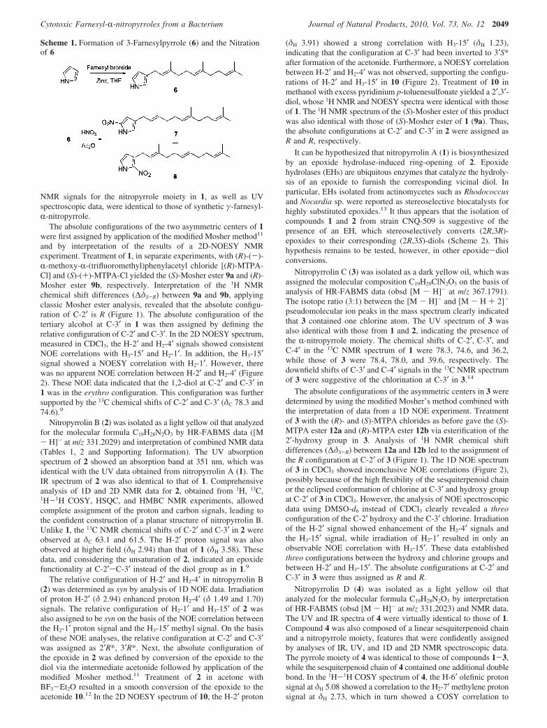
Other characteristic features of the 1H NMR spectrum of 1 werethe presence of two aromatic protons at δH 7.06 (1H, br s) and6.90 (1H, br s) and an exchangeable proton at δH 9.49 (1H, br s).The 1H-1H COSY spectrum showed cross correlations betweenthese three proton signals, indicating the presence of the NH groupin the aromatic ring. HSQC NMR data showed that the aromaticproton signals at δH 7.06 and 6.90 (H-3 and H-5) correlated with13C NMR bands at δC 111.2 and 122.2, respectively. In the HMBCspectrum of 1, proton H-5′ correlated to two unassigned aromaticquaternary carbons C-2 (δC 137.5) and C-4 (δC 124.3), and theH-3 signal showed an HMBC correlation with C-2. Furthermore,the chemical shift of C-2′, the UV absorption features of 1, andthe remaining unaccounted for elements (NO2) suggested that thearomatic ring of 1 was a disubstituted nitropyrrole. Strong IRabsorption at 1504 cm-1 also supported the presence of a nitro groupin 1. Confirming this assignment, the UV spectrum of 1 showed amaximum absorption at 350 nm characteristic for an R-nitropyrrole,while the UV absorption band for �-nitropyrroles has been recordedat 315 nm.10 HMBC correlations from the diastereotopic methyleneprotons at δH 2.73 and 2.53 (H2-1′) to the C-3, C-4, and C-5 signalsallowed the diol-substituted farnesyl chain of 1 to be connectedwith the R-nitropyrrole group through C-1′ and C-4. Finally, theγ-sesquiterpenyl-R-nitropyrrole structure of 1 was confirmed bythe comparison of NMR and UV data of 1 with those of syntheticγ-farnesyl-R-nitropyrrole and �-farnesyl-R-nitropyrrole. These twoisomers were synthesized for comparison to 1. A mixture of pyrrole(7.5 mmol), farnesyl bromide (2.8 mmol), and zinc powder (650mg) in THF (10 mL) was stirred at room temperature for 10 h toafford 3-farnesylpyrrole (6) (270 mg). Nitration of 6 (0.08 mmol)in acetic anhydride (1.0 mL) with a solution of HNO3 (0.08 mmol)in acetic anhydride (0.5 mL), followed by chromatographicpurification, yielded γ-farnesyl-R-nitropyrrole (7) (5.0 mg) and�-farnesyl-R-nitropyrrole (8) (8.7 mg) (Scheme 1). The 1H and 13C
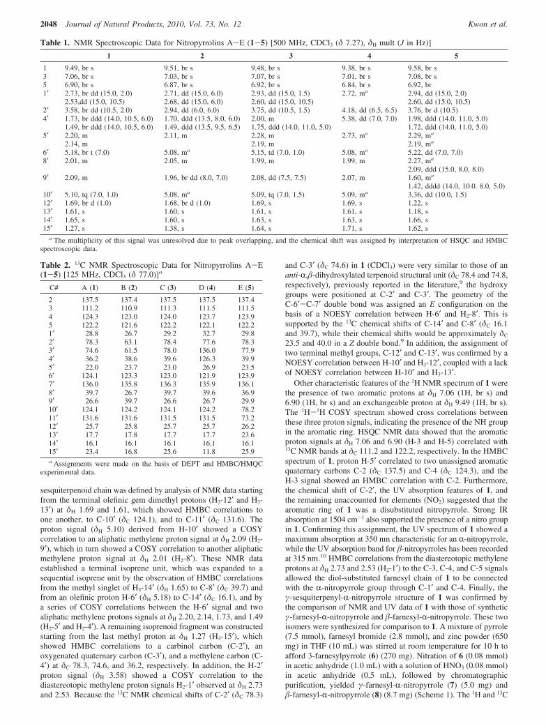
Table 1. NMR Spectroscopic Data for Nitropyrrolins A-E (1-5) [500 MHz, CDCl3 (δ 7.27), δH mult (J in Hz)]
1 2 3 4 5
1 9.49, br s 9.51, br s 9.48, br s 9.38, br s 9.58, br s3 7.06, br s 7.03, br s 7.07, br s 7.01, br s 7.08, br s5 6.90, br s 6.87, br s 6.92, br s 6.84, br s 6.92, br1′ 2.73, br dd (15.0, 2.0) 2.71, dd (15.0, 6.0) 2.93, dd (15.0, 1.5) 2.72, ma 2.94, dd (15.0, 2.0)
2.53,dd (15.0, 10.5) 2.68, dd (15.0, 6.0) 2.60, dd (15.0, 10.5) 2.60, dd (15.0, 10.5)2′ 3.58, br dd (10.5, 2.0) 2.94, dd (6.0, 6.0) 3.75, dd (10.5, 1.5) 4.18, dd (6.5, 6.5) 3.76, br d (10.5)4′ 1.73, br ddd (14.0, 10.5, 6.0) 1.70, ddd (13.5, 8.0, 6.0) 2.00, m 5.38, dd (7.0, 7.0) 1.98, ddd (14.0, 11.0, 5.0)
1.49, br ddd (14.0, 10.5, 6.0) 1.49, ddd (13.5, 9.5, 6.5) 1.75, ddd (14.0, 11.0, 5.0) 1.72, ddd (14.0, 11.0, 5.0)5′ 2.20, m 2.11, m 2.28, m 2.73, ma 2.29, ma
2.14, m 2.19, m 2.19, ma
6′ 5.18, br t (7.0) 5.08, ma 5.15, td (7.0, 1.0) 5.08, ma 5.22, dd (7.0, 7.0)8′ 2.01, m 2.05, m 1.99, m 1.99, m 2.27, ma
2.09, ddd (15.0, 8.0, 8.0)9′ 2.09, m 1.96, br dd (8.0, 7.0) 2.08, dd (7.5, 7.5) 2.07, m 1.60, ma
1.42, dddd (14.0, 10.0. 8.0, 5.0)10′ 5.10, tq (7.0, 1.0) 5.08, ma 5.09, tq (7.0, 1.5) 5.09, ma 3.36, dd (10.0, 1.5)12′ 1.69, br d (1.0) 1.68, br d (1.0) 1.69, s 1.69, s 1.22, s13′ 1.61, s 1.60, s 1.61, s 1.61, s 1.18, s14′ 1.65, s 1.60, s 1.63, s 1.63, s 1.66, s15′ 1.27, s 1.38, s 1.64, s 1.71, s 1.62, s
a The multiplicity of this signal was unresolved due to peak overlapping, and the chemical shift was assigned by interpretation of HSQC and HMBCspectroscopic data.
Table 2. 13C NMR Spectroscopic Data for Nitropyrrolins A-E(1-5) [125 MHz, CDCl3 (δ 77.0)]a
C# A (1) B (2) C (3) D (4) E (5)
2 137.5 137.4 137.5 137.5 137.43 111.2 110.9 111.3 111.5 111.54 124.3 123.0 124.0 123.7 123.95 122.2 121.6 122.2 122.1 122.21′ 28.8 26.7 29.2 32.7 29.82′ 78.3 63.1 78.4 77.6 78.33′ 74.6 61.5 78.0 136.0 77.94′ 36.2 38.6 39.6 126.3 39.95′ 22.0 23.7 23.0 26.9 23.56′ 124.1 123.3 123.0 121.9 123.97′ 136.0 135.8 136.3 135.9 136.18′ 39.7 26.7 39.7 39.6 36.99′ 26.6 39.7 26.6 26.7 29.910′ 124.1 124.2 124.1 124.2 78.211′ 131.6 131.6 131.5 131.5 73.212′ 25.7 25.8 25.7 25.7 26.213′ 17.7 17.8 17.7 17.7 23.614′ 16.1 16.1 16.1 16.1 16.115′ 23.4 16.8 25.6 11.8 25.9a Assignments were made on the basis of DEPT and HMBC/HMQC
experimental data.
2048 Journal of Natural Products, 2010, Vol. 73, No. 12 Kwon et al.
NMR signals for the nitropyrrole moiety in 1, as well as UVspectroscopic data, were identical to those of synthetic γ-farnesyl-R-nitropyrrole.
The absolute configurations of the two asymmetric centers of 1were first assigned by application of the modified Mosher method11
and by interpretation of the results of a 2D-NOESY NMRexperiment. Treatment of 1, in separate experiments, with (R)-(-)-R-methoxy-R-(trifluoromethyl)phenylacetyl chloride [(R)-MTPA-Cl] and (S)-(+)-MTPA-Cl yielded the (S)-Mosher ester 9a and (R)-Mosher ester 9b, respectively. Interpretation of the 1H NMRchemical shift differences (∆δS-R) between 9a and 9b, applyingclassic Mosher ester analysis, revealed that the absolute configu-ration of C-2′ is R (Figure 1). The absolute configuration of thetertiary alcohol at C-3′ in 1 was then assigned by defining therelative configuration of C-2′ and C-3′. In the 2D NOESY spectrum,measured in CDCl3, the H-2′ and H2-4′ signals showed consistentNOE correlations with H3-15′ and H2-1′. In addition, the H3-15′signal showed a NOESY correlation with H2-1′. However, therewas no apparent NOE correlation between H-2′ and H2-4′ (Figure2). These NOE data indicated that the 1,2-diol at C-2′ and C-3′ in1 was in the erythro configuration. This configuration was furthersupported by the 13C chemical shifts of C-2′ and C-3′ (δC 78.3 and74.6).9
Nitropyrrolin B (2) was isolated as a light yellow oil that analyzedfor the molecular formula C19H28N2O3 by HR-FABMS data ([M- H]- at m/z 331.2029) and interpretation of combined NMR data(Tables 1, 2 and Supporting Information). The UV absorptionspectrum of 2 showed an absorption band at 351 nm, which wasidentical with the UV data obtained from nitropyrrolin A (1). TheIR spectrum of 2 was also identical to that of 1. Comprehensiveanalysis of 1D and 2D NMR data for 2, obtained from 1H, 13C,1H-1H COSY, HSQC, and HMBC NMR experiments, allowedcomplete assignment of the proton and carbon signals, leading tothe confident construction of a planar structure of nitropyrrolin B.Unlike 1, the 13C NMR chemical shifts of C-2′ and C-3′ in 2 wereobserved at δC 63.1 and 61.5. The H-2′ proton signal was alsoobserved at higher field (δH 2.94) than that of 1 (δH 3.58). Thesedata, and considering the unsaturation of 2, indicated an epoxidefunctionality at C-2′-C-3′ instead of the diol group as in 1.9
The relative configuration of H-2′ and H2-4′ in nitropyrrolin B(2) was determined as syn by analysis of 1D NOE data. Irradiationof proton H-2′ (δ 2.94) enhanced proton H2-4′ (δ 1.49 and 1.70)signals. The relative configuration of H2-1′ and H3-15′ of 2 wasalso assigned to be syn on the basis of the NOE correlation betweenthe H2-1′ proton signal and the H3-15′ methyl signal. On the basisof these NOE analyses, the relative configuration at C-2′ and C-3′was assigned as 2′R*, 3′R*. Next, the absolute configuration ofthe epoxide in 2 was defined by conversion of the epoxide to thediol via the intermediate acetonide followed by application of themodified Mosher method.11 Treatment of 2 in acetone withBF3-Et2O resulted in a smooth conversion of the epoxide to theacetonide 10.12 In the 2D NOESY spectrum of 10, the H-2′ proton
(δH 3.91) showed a strong correlation with H3-15′ (δH 1.23),indicating that the configuration at C-3′ had been inverted to 3′S*after formation of the acetonide. Furthermore, a NOESY correlationbetween H-2′ and H2-4′ was not observed, supporting the configu-rations of H-2′ and H3-15′ in 10 (Figure 2). Treatment of 10 inmethanol with excess pyridinium p-toluenesulfonate yielded a 2′,3′-diol, whose 1H NMR and NOESY spectra were identical with thoseof 1. The 1H NMR spectrum of the (S)-Mosher ester of this productwas also identical with those of (S)-Mosher ester of 1 (9a). Thus,the absolute configurations at C-2′ and C-3′ in 2 were assigned asR and R, respectively.
It can be hypothesized that nitropyrrolin A (1) is biosynthesizedby an epoxide hydrolase-induced ring-opening of 2. Epoxidehydrolases (EHs) are ubiquitous enzymes that catalyze the hydroly-sis of an epoxide to furnish the corresponding vicinal diol. Inparticular, EHs isolated from actinomycetes such as Rhodococcusand Nocardia sp. were reported as stereoselective biocatalysts forhighly substituted epoxides.13 It thus appears that the isolation ofcompounds 1 and 2 from strain CNQ-509 is suggestive of thepresence of an EH, which stereoselectively converts (2R,3R)-epoxides to their corresponding (2R,3S)-diols (Scheme 2). Thishypothesis remains to be tested, however, in other epoxide-diolconversions.
Nitropyrrolin C (3) was isolated as a dark yellow oil, which wasassigned the molecular composition C19H29ClN2O3 on the basis ofanalysis of HR-FABMS data (obsd [M - H]- at m/z 367.1791).The isotope ratio (3:1) between the [M - H]- and [M - H + 2]-
pseudomolecular ion peaks in the mass spectrum clearly indicatedthat 3 contained one chlorine atom. The UV spectrum of 3 wasalso identical with those from 1 and 2, indicating the presence ofthe R-nitropyrrole moiety. The chemical shifts of C-2′, C-3′, andC-4′ in the 13C NMR spectrum of 1 were 78.3, 74.6, and 36.2,while those of 3 were 78.4, 78.0, and 39.6, respectively. Thedownfield shifts of C-3′ and C-4′ signals in the 13C NMR spectrumof 3 were suggestive of the chlorination at C-3′ in 3.14
The absolute configurations of the asymmetric centers in 3 weredetermined by using the modified Mosher’s method combined withthe interpretation of data from a 1D NOE experiment. Treatmentof 3 with the (R)- and (S)-MTPA chlorides as before gave the (S)-MTPA ester 12a and (R)-MTPA ester 12b via esterification of the2′-hydroxy group in 3. Analysis of 1H NMR chemical shiftdifferences (∆δS-R) between 12a and 12b led to the assignment ofthe R configuration at C-2′ of 3 (Figure 1). The 1D NOE spectrumof 3 in CDCl3 showed inconclusive NOE correlations (Figure 2),possibly because of the high flexibility of the sesquiterpenoid chainor the eclipsed conformation of chlorine at C-3′ and hydroxy groupat C-2′ of 3 in CDCl3. However, the analysis of NOE spectroscopicdata using DMSO-d6 instead of CDCl3 clearly revealed a threoconfiguration of the C-2′ hydroxy and the C-3′ chlorine. Irradiationof the H-2′ signal showed enhancement of the H2-4′ signals andthe H3-15′ signal, while irradiation of H2-1′ resulted in only anobservable NOE correlation with H3-15′. These data establishedthreo configurations between the hydroxy and chlorine groups andbetween H-2′ and H3-15′. The absolute configurations at C-2′ andC-3′ in 3 were thus assigned as R and R.
Nitropyrrolin D (4) was isolated as a light yellow oil thatanalyzed for the molecular formula C19H28N2O3 by interpretationof HR-FABMS (obsd [M - H]- at m/z 331.2023) and NMR data.The UV and IR spectra of 4 were virtually identical to those of 1.Compound 4 was also composed of a linear sesquiterpenoid chainand a nitropyrrole moiety, features that were confidently assignedby analyses of IR, UV, and 1D and 2D NMR spectroscopic data.The pyrrole moiety of 4 was identical to those of compounds 1-3,while the sesquiterpenoid chain of 4 contained one additional doublebond. In the 1H-1H COSY spectrum of 4, the H-6′ olefinic protonsignal at δH 5.08 showed a correlation to the H2-7′ methylene protonsignal at δH 2.73, which in turn showed a COSY correlation to
Scheme 1. Formation of 3-Farnesylpyrrole (6) and the Nitrationof 6
Cytotoxic Farnesyl-R-nitropyrroles from a Bacterium Journal of Natural Products, 2010, Vol. 73, No. 12 2049
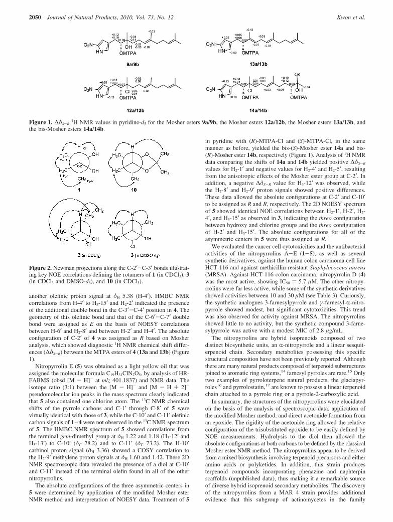
another olefinic proton signal at δH 5.38 (H-4′). HMBC NMRcorrelations from H-4′ to H3-15′ and H2-2′ indicated the presenceof the additional double bond in the C-3′-C-4′ position in 4. Thegeometry of this olefinic bond and that of the C-6′-C-7′ doublebond were assigned as E on the basis of NOESY correlationsbetween H-6′ and H2-8′ and between H-2′ and H-4′. The absoluteconfiguration of C-2′ of 4 was assigned as R based on Mosheranalysis, which showed diagnostic 1H NMR chemical shift differ-ences (∆δS-R) between the MTPA esters of 4 (13a and 13b) (Figure1).
Nitropyrrolin E (5) was obtained as a light yellow oil that wasassigned the molecular formula C19H31ClN2O5, by analysis of HR-FABMS (obsd [M - H]- at m/z 401.1837) and NMR data. Theisotope ratio (3:1) between the [M - H]- and [M - H + 2]-
pseudomolecular ion peaks in the mass spectrum clearly indicatedthat 5 also contained one chlorine atom. The 13C NMR chemicalshifts of the pyrrole carbons and C-1′ through C-8′ of 5 werevirtually identical with those of 3, while the C-10′ and C-11′ olefiniccarbon signals of 1-4 were not observed in the 13C NMR spectrumof 5. The HMBC NMR spectrum of 5 showed correlations fromthe terminal gem-dimethyl group at δH 1.22 and 1.18 (H3-12′ andH3-13′) to C-10′ (δC 78.2) and to C-11′ (δC 73.2). The H-10′carbinol proton signal (δH 3.36) showed a COSY correlation tothe H2-9′ methylene proton signals at δH 1.60 and 1.42. These 2DNMR spectroscopic data revealed the presence of a diol at C-10′and C-11′ instead of the terminal olefin found in all of the othernitropyrrolins.
The absolute configurations of the three asymmetric centers in5 were determined by application of the modified Mosher esterNMR method and interpretation of NOESY data. Treatment of 5
in pyridine with (R)-MTPA-Cl and (S)-MTPA-Cl, in the samemanner as before, yielded the bis-(S)-Mosher ester 14a and bis-(R)-Mosher ester 14b, respectively (Figure 1). Analysis of 1H NMRdata comparing the shifts of 14a and 14b yielded positive ∆δS-R
values for H2-1′ and negative values for H2-4′ and H2-5′, resultingfrom the anisotropic effects of the Mosher ester group at C-2′. Inaddition, a negative ∆δS-R value for H3-12′ was observed, whilethe H2-8′ and H2-9′ proton signals showed positive differences.These data allowed the absolute configurations at C-2′ and C-10′to be assigned as R and R, respectively. The 2D NOESY spectrumof 5 showed identical NOE correlations between H2-1′, H-2′, H2-4′, and H3-15′ as observed in 3, indicating the threo configurationbetween hydroxy and chlorine groups and the threo configurationof H-2′ and H3-15′. The absolute configurations for all of theasymmetric centers in 5 were thus assigned as R.
We evaluated the cancer cell cytotoxicities and the antibacterialactivities of the nitropyrrolins A-E (1-5), as well as severalsynthetic derivatives, against the human colon carcinoma cell lineHCT-116 and against methicillin-resistant Staphylococcus aureus(MRSA). Against HCT-116 colon carcinoma, nitropyrrolin D (4)was the most active, showing IC50 ) 5.7 µM. The other nitropy-rrolins were far less active, while some of the synthetic derivativesshowed activities between 10 and 30 µM (see Table 3). Curiously,the synthetic analogues 3-farnesylpyrrole and γ-farnesyl-R-nitro-pyrrole showed modest, but significant cytotoxicities. This trendwas also observed for activity against MRSA. The nitropyrrolinsshowed little to no activity, but the synthetic compound 3-farne-sylpyrrole was active with a modest MIC of 2.8 µg/mL.
The nitropyrrolins are hybrid isoprenoids composed of twodistinct biosynthetic units, an R-nitropyrrole and a linear sesquit-erpenoid chain. Secondary metabolites possessing this specificstructural composition have not been previously reported. Althoughthere are many natural products composed of terpenoid substructuresjoined to aromatic ring systems,14 farnesyl pyrroles are rare.15 Onlytwo examples of pyrroloterpene natural products, the glaciapyr-roles16 and pyrrolostatin,17 are known to possess a linear terpenoidchain attached to a pyrrole ring or a pyrrole-2-carboxylic acid.
In summary, the structures of the nitropyrrolins were elucidatedon the basis of the analysis of spectroscopic data, application ofthe modified Mosher method, and direct acetonide formation froman epoxide. The rigidity of the acetonide ring allowed the relativeconfiguration of the trisubstituted epoxide to be easily defined byNOE measurements. Hydrolysis to the diol then allowed theabsolute configurations at both carbons to be defined by the classicalMosher ester NMR method. The nitropyrrolins appear to be derivedfrom a mixed biosynthesis involving terpenoid precursors and eitheramino acids or polyketides. In addition, this strain producesterpenoid compounds incorporating phenazine and naphterpinscaffolds (unpublished data), thus making it a remarkable sourceof diverse hybrid isoprenoid secondary metabolites. The discoveryof the nitropyrrolins from a MAR 4 strain provides additionalevidence that this subgroup of actinomycetes in the family
Figure 1. ∆δS-R1H NMR values in pyridine-d5 for the Mosher esters 9a/9b, the Mosher esters 12a/12b, the Mosher esters 13a/13b, and
the bis-Mosher esters 14a/14b.
Figure 2. Newman projections along the C-2′-C-3′ bonds illustrat-ing key NOE correlations defining the rotamers of 1 (in CDCl3), 3(in CDCl3 and DMSO-d6), and 10 (in CDCl3).
2050 Journal of Natural Products, 2010, Vol. 73, No. 12 Kwon et al.

Streptomycetaceae is a rich source of secondary metabolitesproduced by mixed terpenoid biosynthetic pathways.
Experimental Section
General Experimental Procedures. Optical rotations were measuredon a PerkinElmer model 343 polarimeter. UV and IR spectra wererecorded using a PerkinElmer Lambda 35 UV/vis spectrophotometerand a Thermo Scientific Nicolet iS10 spectrometer, respectively. 1H,13C, and 2D NMR spectral data were obtained in CDCl3 or DMSO-d6
on a Varian Unity 500 MHz NMR spectrometer. Low-resolution ESIMSwere measured on a Waters Alliance 2695/Quattro micro API system.High-resolution mass spectral data were acquired on a JEOL/JMS-AX505WA instrument. Lichroprep RP-18 (Merck, 40-63 um) was usedfor column chromatography. Semipreparative HPLC separations wereperformed using a Gilson 321 HPLC system with a Phenomenex LunaC18(2) 10 µm column (10 × 250 mm) at a flow rate of 4 mL/min. AWaters 1525 HPLC-PDA system with a Phenomenex Luna C18(2) 5µm column (4.6 × 100 mm) was used for the analysis of extracts andfractions.
Isolation of Strain CNQ-509, Cultivation, and Extraction. StrainCNQ-509 was isolated on A1 agar medium (10 g of starch, 4 g ofpeptone, 2 g of yeast extract, and 18 g of agar in 1 L of seawater)from a marine sediment sample collected at a depth of 44.2 m onemile northwest of the Scripps Institution of Oceanography Pier (LaJolla, California). The 16S rRNA gene sequence for this strain (1451base pairs) has been deposited with GenBank (accession numberEF581384). It shares 99.2% sequence identity with the type strain forStreptomyces aculeolatus (AB184624), which interestingly is the onlynonmarine strain reported to date that falls within the MAR4 group.Strain CNQ-509 was cultured in eight replicate 7 L fermentors (LiFlusGR, BioTron Inc.) each containing 4 L of fermentation medium A1without agar for 7 days at 27 °C while stirring at 400 rpm. At the endof the fermentation period, 20 g/L Amberlite XAD-7 adsorbent resinwas added into each fermentor, and stirring was continued for oneadditional hour at 100 rpm. The resin was then collected by filtrationthrough cheesecloth, washed with deionized water, and eluted twicewith acetone. The acetone solution was concentrated to afford 3.5 g oforganic extract.
Isolation and Purification of Nitropyrrolins A-E (1-5). Theextract (3.5 g) was absorbed on Celite (5 g) and subjected to C-18 (50g) reversed-phase column chromatography eluting with a step gradientof MeOH-H2O solvent mixtures (increasing the MeOH by 20% per500 mL from 10% to 90% MeOH- H2O). The combined 70% and
90% MeOH fractions (360 mg) was then further fractionated by C-18reversed-phase HPLC with a MeCN-H2O gradient solvent system(Phenomenex Luna C-18(2) 21.2 × 250 mm, 10 mL/min, 50-100%MeCN-H2O for 40 min) to obtain five subfractions containing thenitropyrrolins (A, 16 mg; B, 30 mg; C, 27 mg; D, 11 mg; and E, 5mg). Each fraction was subsequently purified by C-18 reversed-phaseHPLC using a Phenomenex Luna C18(2) column (10 × 250 mm, 10µm) with 75% MeCN-H2O as an eluting solvent (flow rate: 4 mL/min) to yield five pure compounds, nitropyrrolin A (1, 6.0 mg),nitropyrrolin B (2, 16 mg), nitropyrrolin C (3, 12 mg), nitropyrrolin D(4, 4 mg), and nitropyrrolin E (5, 2 mg).
Nitropyrrolin A (1): light yellow oil, [R]20D +8 (c 0.05, CH2Cl2);
UV (MeOH) λmax (log ε) 350 nm (3.90) nm; IR (KBr) νmax 3360, 2967,2926, 1504, 1452, 1384, 1367, 1293, 1272 cm-1; see Table 1 for 1HNMR data and Table 2 for 13C NMR data; ESIMS m/z 373 [M + Na]+,349 [M - H]-; HR-FABMS m/z 349.2124 [M - H]- (calcd forC19H29N2O4,349.2127).
Nitropyrrolin B (2): light yellow oil, [R]20D +3 (c 0.05, CH2Cl2);
UV (MeOH) λmax (log ε) 351 nm (3.86); IR (KBr) νmax 3259, 3127,2963, 2926, 2873, 1510, 1455, 1383, 1368, 1302, 1272, 1251 cm-1;see Table 1 for 1H NMR data and Table 2 for 13C NMR data; ESIMSm/z 355 [M + Na]+, 331 [M - H]-; HR-FABMS m/z 331.2029 [M -H]- (calcd for C19H27N2O3, 331.2022).
Nitropyrrolin C (3): light yellow oil, [R]20D -2 (c 0.07, CH2Cl2);
UV (MeOH) λmax (log ε) 348 nm (3.82); IR (KBr) νmax 3303, 3128,2968, 2927, 2856, 2731, 1709, 1632, 1591, 1504, 1450, 1384, 1370,1295, 1273 cm-1; see Table 1 for 1H NMR data and Table 2 for 13CNMR data; ESIMS (rel int) m/z 391 [M + Na]+, 369 (40) [M - H +2]-, 367 (100) [M - H]-; HR-FABMS m/z 367.1791 [M - H]- (calcdfor C19H28ClN2O3, 367.1788).
Nitropyrrolin D (4): light yellow oil, [R]20D -9 (c 0.003, MeOH);
UV (MeOH) λmax (log ε) 351 nm (3.90); IR (KBr) νmax 3301, 2968,2926, 1503, 1452, 1383, 1366, 1276 cm-1; see Table 1 for 1H NMRdata and Table 2 for 13C NMR data; ESIMS m/z 355 [M + Na]+, 331[M - H]-; HR-FABMS m/z 331.2023 [M - H]- (calcd for C19H27N2O3,331.2021).
Nitropyrrolin E (5): light yellow oil, [R]20D +15 (c 0.002, MeOH);
UV (MeOH) λmax (log ε) 350 nm (3.85); IR (KBr) νmax 3403, 2973,2931, 1503, 1451, 1384, 1366, 1293, 1271 cm-1; see Table 1 for 1HNMR data and Table 2 for 13C NMR data; ESIMS (rel int) m/z 403 [M- H + 2]- (40), 401 [M - H]- (100); HR-FABMS m/z 401.1837 [M- H]- (calcd for C19H30ClN2O5, 401.1843).
Preparation of 3-Farnesylpyrrole by Zinc-Mediated Farnesyla-tion of Pyrrole. A mixture of pyrrole (7.5 mmol), farnesyl bromide(2.8 mmol), and zinc powder (650 mg) in THF (10 mL) was stirred atroom temperature for 10 h. After completion of the reaction as indicatedby TLC analysis, the reaction mixture was quenched with saturatedNH4Cl (20 mL), diluted with water (10 mL), and extracted with ethylacetate (2 × 40 mL). Evaporation of the solvent followed by purificationon normal-phase HPLC [Luna silica (2), 250 × 21.2 mm, flow rate 10mL/min] under isocratic conditions [hexane-EtOAc (20:1)] affordedpure 3-farnesylpyrrole (6) (270 mg): UV (MeOH) λmax (log ε) 227 nm(4.49); ESIMS m/z 270 [M - H]-; 1H NMR (500 MHz, CDCl3) δ8.02 (1H, NH), 6.75 (1H, br m, H-5), 6.58 (1H, br m, H-2), 6.12 (1H,br m, H-4), 5.41 (1H, br tq, J ) 7.5, 1.0 Hz, C-2′), 5.17 (1H, br tq, J) 7.0, 1.0 Hz, H-6′), 5.13 (1H, br tqq, J ) 7.0, 1.0, 1.0 Hz, H-10′),3.25 (2H, d, J ) 7.5 Hz, H2-2′), 2.15 (2H, m, H2-5′), 2.07-2.09 (4H,m, H2-4′ and H2-9′), 2.01 (2H, dd, J ) 8.5, 7.0 Hz, H2-8′), 1.72 (3H,br d, J ) 1.0 Hz, H3-15′), 1.71 (3H, br d, J ) 1.0 Hz, H3-12′), 1.63(6H, br s, H3-13′ and H3-14′).
Scheme 2. Formation of the Acetonide 10 and the Diol 11 from 2
Table 3. Cytotoxicities and Antibacterial Activities of theNitropyrrolins A-E (1-5) and Some Synthetic Derivatives
HCT-116 MRSA inhibition
compound IC50 (µM) MIC (µg/mL)
nitropyrrolin A (1) 31.1 NSAa
nitropyrrolin B (2) 31.0 NSAnitropyrrolin C (3) NSA NSAnitropyrrolin D (4) 5.7 NSAnitropyrrolin E (5) NSA NSA3-farnesylpyrrole 13.7 2.8γ-farnesyl-R-nitropyrrole 9.2 NSAR-nitropyrrole NSA NSA�-nitropyrrole NSA NSA2-farnesylpyrrole 24.4 NSAa NSA ) not significantly active (inhibition observed only at >20 µg/
mL).
Cytotoxic Farnesyl-R-nitropyrroles from a Bacterium Journal of Natural Products, 2010, Vol. 73, No. 12 2051
Preparation of γ-Farnesyl-r-nitropyrrole (7) and �-Farnesyl-r-nitropyrrole (8) by Nitration of 3-Farnesylpyrrole (6). A solutionof HNO3 (0.08 mmol) in Ac2O (0.5 mL) was added dropwise to asolution of 3-farnesylpyrrole (6) (0.08 mmol) in Ac2O (1 mL), and thereaction was stirred at -50 °C for 2 h and then at room temperaturefor 1 h. After the reaction was complete (as indicated by TLC analysis),the reaction mixture was diluted with H2O (5 mL) and extracted withEt2O (2 × 10 mL). The organic layer was washed with aqueousNaHCO3 solution (2%), concentrated under reduced pressure, thenpurified by normal-phase HPLC [Luna silica (2), 250 × 10 mm, flowrate 4 mL/min] under isocratic conditions using hexane-EtOAc (92:8) to afford γ-farnesyl-R-nitropyrrole (7) (5.0 mg) and �-farnesyl-R-nitropyrrole (8) (8.7 mg). For 7: light yellowish oil; UV (PDA detectorusing MeCN-H2O solvent mixture) λmax 353 nm; ESIMS m/z 315 [M- H]-; 1H NMR (500 MHz, CDCl3) δ 9.24 (1H, br s, NH), 6.96 (1H,br s, H-3), 6.74 (1H, br s, H-1), 5.30 (1H, br tq, J ) 7.5, 1.0 Hz, H-2′),5.11 (1H, m, H-6′), 5.10 (1H, m, H-10′), 3.19 (2H, d, J ) 7.5 Hz,H2-1′), 2.13 (2H, m, H2-5′), 2.06-2.08 (4H, m, H2-4′ and H2-9′), 1.99(2H, m, H2-8′), 1.69 (6H, s, H3-12′ and H3-15′), 1.61 (3H, s, H3-13′),1.60 (3H, s, H3-14′); 13C NMR (125 MHz, CDCl3) δ 137.3a (C-2),137.2 (C-3′), 135.3 (C-7′), 131.4 (C-11′), 127.1 (C-4), 124.3 (C-6′),123.9 (C-10′), 121.5 (C-2′), 121.0 (C-5), 110.8 (C-3), 39.7 (C-8′), 39.6(C-4′), 26.7 (C-9′), 26.4 (C-5′), 25.7 (C-12′), 25.3 (C-1′), 17.7 (C-13′),16.1 (C-15′), 16.0 (C-14′). aThis chemical shift was assigned by theanalysis of HSQC and HMBC spectra because it was not observed inthe 13C NMR spectrum. For 8: light yellowish oil; UV (PDA detectorusing MeCN-H2O solvent mixture) λmax 340 nm; ESIMS m/z 315 [M- H]-; 1H NMR (500 MHz, CDCl3) δ 9.35 (1H, br s, NH). 6.86 (1H,br dd, J ) 2.5, 2.0 Hz, H-5), 6.20 (1H, br dd, J ) 2.5, 2.0 Hz, H-4),5.35 (1H, br tq, J ) 7.0, 1.0 Hz, H-2′), 5.12 (1H, m, H-6′), 5.10 (1H,m, H-10′), 3.62 (2H, d, J ) 7.0 Hz, H2-1′), 2.13 (2H, m, H2-5′),2.06-2.07 (4H, m, H2-4′ and H2-9′), 1.99 (2H, m, H2-8′), 1.71 (3H, s,H3-15′), 1.68 (3H, s, H3-12′), 1.61 (6H, s, H3-13′ and H3-14′); 13C NMR(125 MHz, CDCl3) δ 137.7 (C-3′), 135.2 (C-7′), 134.1a (C-2), 131.3(C-11′), 129.3 (C-3), 124.3 (C-10′), 124.0 (C-6′), 121.9 (C-5), 120.2(C-2′), 112.4 (C-4), 39.7 (C-8′), 39.6 (C-4′), 26.7 (C-9′), 26.5 (C-5′),25.7 (C-1′ and C-12′), 17.7 (C-13′), 16.2 (C-15′), 16.0 (C-14′). aThischemical shift was assigned by the analysis of HSQC and HMBCspectra because it was not detected in the 1D 13C NMR spectrum.
Preparation of MTPA Esters. Compounds 1, 3, 4, 5, and 11 weredivided into two portions, and each was dissolved in 600 µL of pyridine-d5 in a 5 mm NMR tube. To each NMR tube were added a slight excessof dimethylaminopyridine. The samples were then treated with 5 µLof (R)-R-methoxy-a-(trifluoromethyl)phenylacetyl chloride (MTPA-Cl)and 5 µL of (S)-MTPA-Cl at room temperature. After 12 h, the reactionwas complete, and 1H NMR and 1H-1H gCOSY spectra for (S)-Mosheresters (9a, 12a, 13a, 14a, and the (S)-mosher ester of 11) and (R)-Mosher esters (9b, 12b, 13b, and 14b) were recorded (see SupportingInformation for NMR data). Because of the difficulty in interpretingsome crude ester mixtures, compounds 13a, 13b, 14a, and 14b werepurified by RP-HPLC with a Luna C-8 column (250 × 10 mm) using50% MeCN-H2O to 100% MeCN as a linear gradient for 40 min (flowrate 4 mL/min),
Preparation of the Acetonide 10. Nitropyrrolin B (2) (2 mg) wasdissolved in dry Me2CO (1 mL), and BF3 ·Et2O (20 µL) was added at0 °C. The reaction was allowed to stir for 3 h at 0 °C, the reaction wasthen quenched with saturated aqueous NaHCO3, and the aqueous phasewas extracted twice with EtOAc. The combined EtOAc solution wasconcentrated under reduced pressure, and the residue was purified byRP HPLC with a Luna C-8 column (250 × 10 mm) using a lineargradient solvent system of 50% MeCN-H2O to 100% MeCN for 20min to provide compound 10 (1.2 mg): 1H NMR (500 MHz, CDCl3) δ9.30 (1H, br s, NH), 7.07 (1H, br s, H-3), 6.90 (1H, br s, H-5), 5.16(1H, dd, J ) 7.0, 7.0 Hz, H-6′), 5.11 (1H, dd, J ) 7.0, 7.0 Hz, H-10′),3.91 (1H, dd, J ) 10.0, 3.5 Hz, H-2′), 2.76 (1H, J ) 15.0, 10.0 Hz,H2-1′a), 2.58 (1H, J ) 15.0, 3.5 Hz, H2-1′b), 2.23 (1H, m, H2-5′a),2.08 (1H, m, H2-5′b), 2.08 (2H, m, H2-9′), 2.00 (2H, m, H2-8′), 1.70(3H, br s, H3-12′), 1.65 (1H, m, H2-4′a), 1.64 (3H, br s, H3-14′), 1.62(3H, br s, H3-13′), 1.46 (3H, s, acetonide methyl), 1.37 (3H, s, acetonidemethyl), 1.27 (1H, m, H2-4′b), 1.23 (3H, s, H3-15′); ESIMS m/z 413[M + Na]+.
Preparation of the Diol 11 from 10. To a solution of 10 inanhydrous MeOH (2 mL) was added pyridinium-p-toluensulfonate (2mg), and the reaction was stirred for 24 h. The reaction was then
quenched with saturated aqueous NaHCO3, and the aqueous phase wasextracted twice with EtOAc. The EtOAc solution was concentratedunder reduced pressure, and the residue was purified by RP HPLC [LunaC-8 column (250 × 10 mm), flow rate 4 mL/min, 45% MeCN-H2Oto 95% MeCN as a linear gradient for 40 min] to afford diol 11: 1HNMR (500 MHz, CDCl3) δ 9.40 (1H, br s, NH), 7.06 (1H, br s, H-3),6.90 (1H, br s, H-5), 5.18 (1H, br t, J ) 7.0 Hz, H-6′), 5.10 (1H, tq,J ) 7.0, 1.0 Hz, H-10′), 3.58 (1H, ddd, J ) 11.0, 4.0, 2.0 Hz, H-2′),2.73 (1H, dd, J ) 15.0, 2.0 Hz, H2-1′a), 2.53 (1H, dd, J ) 15.0, 11.0Hz, H2-1′b), 2.20 (1H, m, H2-5′a), 2.14 (1H, m, H2-5′b), 2.14 (1H, d,J ) 4.0 Hz, 2′-OH), 2.19 (2H, m, H2-9′), 2.01 (2H, m, H2-8′), 1.97(1H, br s, 3′-OH), 1.73 (1H, m, H2-4′a), 1.69 (3H, br s, H3-12′), 1.66(3H, br s, H3-14′), 1.61 (3H, br s, H3-13′), 1.49 (1H, m, H2-4′b), 1.27(3H, br s, H3-15′); ESIMS m/z 373 [M + Na]+.
Acknowledgment. This research is a result of financial support fromthe NIH, National Cancer Institute, under grant R37 CA044848 (toW.F.) and the NOAA California Sea Grant College Program ProjectR/NMP-100 (NA100AR41700360 to P.R.J.) through NOAA’s NationalSea Grant College Program, U.S. Dept. of Commerce. The statements,findings, conclusions, and recommendations are those of the author(s)and do not necessarily reflect the views of California Sea Grant or theU.S. Dept. of Commerce. Fellowship support for A.P.E. from the CNPq(Conselho Nacional de Desenvolvimento Cientifico e Tecnologico,Brazil) is gratefully acknowledged.
Note Added in Proof: While this manuscript was in process, webecame aware of the publication of some similar structures, see:Heronapyrroles A-C: Farnesylated 2-Nitropyrroles from an AustralianMarine-Derived Streptomyces sp.; Raju, R.; Piggott, A. M.; Diaz,L. X. B.; Khalil, Z.; Capon, R. J. Org. Lett. 2010, 12, 5158-5161.
Supporting Information Available: Spectroscopic data sets (1Dand 2D NMR, ESIMS, UV/vis, HRMS, etc.) for 1-5, 1H NMR spectrafor 6 and (S)-Mosher ester of 11, 1H NMR and 1H-1H COSY spectrafor 9a, 9b, 12a, 12b, 13a, 13b, 14a, and 14b, 1H NMR and 2D NOESYspectra for 10 and 11, and 1H and 13C NMR spectra for 7 and 8. Thismaterial is available free of charge via the Internet at http://pubs.acs.org.
References and Notes
(1) Fenical, W.; Jensen, P. R. Nat. Chem. Biol. 2006, 2, 666–673.(2) (a) Mincer, T. J.; Jensen, P. R.; Kauffman, C. A.; Fenical, W. Appl.
EnViron. Microbiol. 2002, 68, 5005–5011. (b) Maldonado, L. A.;Fenical, W.; Jensen, P. R.; Kauffman, C. A.; Mincer, T. J.; Ward,A. C.; Bull, A. T.; Goodfellow, M. Int. J. Syst. EVol. Microbiol. 2005,55, 1759–1766.
(3) (a) Jensen, P. R.; Mincer, T. J.; Williams, P. G.; Fenical, W. AntonieVan Leeuwenhoek 2005, 87, 43–48. (b) Kwon, H. C.; Kauffman, C. A.;Jensen, P. R.; Fenical, W. J. Am. Chem. Soc. 2006, 128, 1622–1632.
(4) Feling, R. H.; Buchanan, G. O.; Mincer, T. J.; Kauffman, C. A.; Jensen,P. R.; Fenical, W. Angew. Chem., Int. Ed. 2003, 42, 355–357.
(5) Fenical, W.; Jensen, P. R.; Palladino., M. A.; Lam, K. S.; Lloyd, G. K.;Potts, B. C Bioorg. Med. Chem. 2009, 17, 2175–2180.
(6) Pathirana, C.; Jensen, P. R.; Fenical, W. Tetrahedron Lett. 1992, 33,7663–7666.
(7) Soria-Mercardo, I. E.; Prieto-Davo, A.; Jensen, P. R.; Fenical, W. J.Nat. Prod. 2005, 68, 904–910.
(8) Cho, J. Y.; Kwon, H. C.; Williams, P. G.; Jensen, P. R.; Fenical, W.Org. Lett. 2006, 8, 2471–2474.
(9) Deouet, D.; Cauret, L.; Brosse, J. C. Eur. Polym. J. 2003, 39, 671–686.
(10) Morgan, K. J.; Morrey, D. P. Tetrahedron 1966, 22, 57–62.(11) Ohtani, I.; Kusumi, T.; Kashman, Y.; Kakisawa, H. J. Am. Chem.
Soc. 1991, 113, 4092–4096.(12) Yadav, J. S.; Pratap, T. V.; Rajender, V. J. Org. Chem. 2007, 72,
5882–5885.(13) Steinreiber, A.; Farber, K. Curr. Opin. Biotechnol. 2001, 12, 552–
558.(14) Nii, K.; Tagami, K.; Kijima, M.; Munakata, T.; Ooi, T.; Kusumi, T.
Bull. Chem. Soc. Jpn. 2008, 81, 562–57.(15) (a) Dictionary of Natural Products on CD-ROM; Chapman & Hall,
Chemical Database, Version 13.1, 2004. (b) Harborne, J. B.; Williams,C. A. Nat. Prod. Rep. 1998, 15, 631–651.
(16) Macherla, V. R.; Liu, J.; Bellows, C.; Teisan, S.; Nicholson, B.; Lam,K. S.; Potts, B. C. M. J. Nat. Prod. 2005, 68, 780–783.
(17) Kato, S.; Shindo, K.; Kawai, H.; Odagawa, A.; Matsuoka, M.;Mochizuki, J. J. Antibiot. 1993, 46, 892–899.
NP1006229
2052 Journal of Natural Products, 2010, Vol. 73, No. 12 Kwon et al.