Neuronal and neurohormonal control of the heart in the ...Squilla oratoria (Watanabe et al., 1968,...
15
4663 Alexandrowicz (1934) first showed that three pairs of central cardioregulatory nerves (α, β and γ) control the heart of the stomatopod Squilla mantis. Of these, β and γ are most probably nerves originating from the 3rd and 4th segments of ‘the large thoracic ganglionic mass’, while α is a nerve originating from a more anterior segment of the ganglionic mass. He proposed that the α nerve was a cardio-inhibitor while the β and γ nerves were cardio-accelerators. The functions of α, β and γ nerves in control of the heart were verified by means of intracellular recording from neuronal cell bodies in the cardiac ganglion of Squilla oratoria (Watanabe et al., 1968, 1969). It is known that the heartbeat of stomatopods is triggered by the cardiac ganglion, located on the outer surface of the heart (reviewed by Maynard, 1960). Neither the anatomical studies of Alexandrowicz (1934) nor the functional studies of Watanabe et al. (1968, 1969), have been followed by more detailed studies of stomatopod neuroanatomy, and until the present study, a detailed description of the anatomy of the central ganglia and of the ganglionic nerve roots from which the cardioregulatory axons emerge has not been published, nor have their cell bodies been identified in the central ganglia. The neurotransmitter candidates for the intrinsic and extrinsic heart neurons have been identified in a few species within the sub-class Malacostraca from the results of pharmacological and immunocytochemical studies. Dealing with the decapods, Yazawa and Kuwasawa (1994) proposed that γ-amino-butyric acid (GABA) and dopamine (DA) are the extrinsic neurotransmitters of the cardio-inhibitory and cardio- acceleratory nerves, respectively, in the hermit crab Aniculus aniculus. They also proposed that acetylcholine (ACh) and DA The Journal of Experimental Biology 207, 4663-4677 Published by The Company of Biologists 2004 doi:10.1242/jeb.01272 The heart of Squilla oratoria contains a cardiac ganglion that consists of 15 intrinsic neurons, supplied by a pair of inhibitory nerves and two pairs of excitatory nerves, arising from the central nervous system. These comprise the extrinsic cardiac innervation. The paired cardio- inhibitor (CI) nerves run out in the 10th pair of nerve roots emerging from the subesophageal ganglion (SEG). The cell bodies of the CI neurons are found in the hemisphere of the 1st segment of the SEG contralateral to the nerve roots in which the CI axons emerge. The two pairs of 1st and 2nd cardio-accelerator (CA1 and CA2) nerves run out in the 16th and 19th pairs of nerve roots of the SEG. The cell bodies of the CA1 and CA2 neurons are found in the hemispheres of the 3rd and 4th segments of the SEG ipsilateral to the nerve roots in which the CA1 and CA2 axons are found. The heartbeat was activated by application of glutamate, serotonin, dopamine, octopamine or acetylcholine, which were applied to the heart by perfusion into an organ bath. Joro-spider toxin (JSTX) blocked myocardial excitatory junctional potentials evoked by the cardiac ganglion. Neuronal cell bodies and processes in the heart were examined using immunocytochemical techniques. All 15 neurons of the cardiac ganglion showed glutamate-like immunoreactivity. Glutamate may be a neurotransmitter of the cardiac ganglion neurons. JSTX also blocked cardiac acceleration by activation of CA1 and CA2 axons. CA1 and CA2 axons showed glutamate-like immunoreactivity. It is likely that glutamate is a neurotransmitter for the cardio- acceleratory neurons. The heartbeat was inhibited by application of γ-amino- butyric acid (GABA). Cardiac inhibition induced by activation of CI axons was blocked by picrotoxin. CI axons showed GABA-like immunoreactivity. These results may support the identification of GABA as an extrinsic inhibitory neurotransmitter. Key words: cardioregulatory neuron, cardiac ganglion neuron, neurotransmitter, stomatopod, Squilla oratoria, immunocytochemistry, GABA, glutamate. Summary Introduction Neuronal and neurohormonal control of the heart in the stomatopod crustacean, Squilla oratoria Hiroshi Ando 1 and Kiyoaki Kuwasawa 2, * 1 Department of Oral Physiology, Matsumoto Dental University School of Dentistry, Shiojiri 399-0781, Japan and 2 Neurobiology Laboratory, Faculty of Science, Okayama University of Science, Ridai-cho 1-1, Okayama 700-0005, Japan *Author for correspondence (e-mail: [email protected]) Accepted 6 September 2004
Transcript of Neuronal and neurohormonal control of the heart in the ...Squilla oratoria (Watanabe et al., 1968,...
4663
Alexandrowicz (1934) first showed that three pairs of centralcardioregulatory nerves (α, β and γ) control the heart of thestomatopod Squilla mantis. Of these, β and γ are most probablynerves originating from the 3rd and 4th segments of ‘the largethoracic ganglionic mass’, while α is a nerve originating froma more anterior segment of the ganglionic mass. He proposedthat the α nerve was a cardio-inhibitor while the β and γ nerveswere cardio-accelerators. The functions of α, β and γ nerves incontrol of the heart were verified by means of intracellularrecording from neuronal cell bodies in the cardiac ganglion ofSquilla oratoria (Watanabe et al., 1968, 1969). It is known thatthe heartbeat of stomatopods is triggered by the cardiacganglion, located on the outer surface of the heart (reviewedby Maynard, 1960). Neither the anatomical studies ofAlexandrowicz (1934) nor the functional studies of Watanabe
et al. (1968, 1969), have been followed by more detailedstudies of stomatopod neuroanatomy, and until the presentstudy, a detailed description of the anatomy of the centralganglia and of the ganglionic nerve roots from which thecardioregulatory axons emerge has not been published, norhave their cell bodies been identified in the central ganglia.
The neurotransmitter candidates for the intrinsic andextrinsic heart neurons have been identified in a few specieswithin the sub-class Malacostraca from the results ofpharmacological and immunocytochemical studies. Dealingwith the decapods, Yazawa and Kuwasawa (1994) proposedthat γ-amino-butyric acid (GABA) and dopamine (DA) are theextrinsic neurotransmitters of the cardio-inhibitory and cardio-acceleratory nerves, respectively, in the hermit crab Aniculusaniculus. They also proposed that acetylcholine (ACh) and DA
The Journal of Experimental Biology 207, 4663-4677Published by The Company of Biologists 2004doi:10.1242/jeb.01272
The heart of Squilla oratoria contains a cardiac ganglionthat consists of 15 intrinsic neurons, supplied by a pair ofinhibitory nerves and two pairs of excitatory nerves,arising from the central nervous system. These comprisethe extrinsic cardiac innervation. The paired cardio-inhibitor (CI) nerves run out in the 10th pair of nerveroots emerging from the subesophageal ganglion (SEG).The cell bodies of the CI neurons are found in thehemisphere of the 1st segment of the SEG contralateral tothe nerve roots in which the CI axons emerge. The twopairs of 1st and 2nd cardio-accelerator (CA1 and CA2)nerves run out in the 16th and 19th pairs of nerve roots ofthe SEG. The cell bodies of the CA1 and CA2 neurons arefound in the hemispheres of the 3rd and 4th segments ofthe SEG ipsilateral to the nerve roots in which the CA1and CA2 axons are found.
The heartbeat was activated by application ofglutamate, serotonin, dopamine, octopamine oracetylcholine, which were applied to the heart byperfusion into an organ bath. Joro-spider toxin (JSTX)blocked myocardial excitatory junctional potentials
evoked by the cardiac ganglion. Neuronal cell bodiesand processes in the heart were examined usingimmunocytochemical techniques. All 15 neurons of thecardiac ganglion showed glutamate-like immunoreactivity.Glutamate may be a neurotransmitter of the cardiacganglion neurons.
JSTX also blocked cardiac acceleration by activation ofCA1 and CA2 axons. CA1 and CA2 axons showedglutamate-like immunoreactivity. It is likely thatglutamate is a neurotransmitter for the cardio-acceleratory neurons.
The heartbeat was inhibited by application of γ-amino-butyric acid (GABA). Cardiac inhibition induced byactivation of CI axons was blocked by picrotoxin. CIaxons showed GABA-like immunoreactivity. These resultsmay support the identification of GABA as an extrinsicinhibitory neurotransmitter.
Key words: cardioregulatory neuron, cardiac ganglionneuron, neurotransmitter, stomatopod, Squilla oratoria,immunocytochemistry, GABA, glutamate.
Summary
Introduction
Neuronal and neurohormonal control of the heart in the stomatopod crustacean,Squilla oratoria
Hiroshi Ando1 and Kiyoaki Kuwasawa2,*1Department of Oral Physiology, Matsumoto Dental University School of Dentistry, Shiojiri 399-0781, Japan and
2Neurobiology Laboratory, Faculty of Science, Okayama University of Science, Ridai-cho 1-1, Okayama 700-0005,Japan
*Author for correspondence (e-mail: [email protected])
Accepted 6 September 2004

4664
are the intrinsic neurotransmitters of the small and largeneurons, respectively, in the cardiac ganglion of that species(Yazawa and Kuwasawa, 1992). On the other hand, glutamate(Glu) has been proposed for the neurotransmitter of both of thesmall and large neurons in the cardiac ganglion of the lobsterPanulirus argus (Delgado et al., 2000). In isopods, it has beenproposed that Glu is the intrinsic excitatory neurotransmitterreleased by the motoneurons of the cardiac ganglion ofBathynomus doederleini (Yazawa et al., 1998) and Ligiaexotica (Sakurai et al., 1998). Furthermore, GABA has beenproposed as the extrinsic cardio-inhibitory neurotransmitter,while ACh has been proposed as the extrinsic cardio-acceleratory neurotransmitter in B. doederleini (Tanaka et al.,1992).
In stomatopods, epinephrine (E), norepinephrine (NE), andACh have cardioexcitatory effects on the heart of S. mantis(Alexandrowicz and Carlisle, 1953; Florey and Rathmayer,1990). Additionally, pharmacological experiments indicatethat GABA may be a neurotransmitter of the extrinsic cardio-inhibitor nerves of S. oratoria (Watanabe et al., 1968).However, candidate neurotransmitters of the cardiac ganglionneurons and cardio-accelerator nerves have not yet beenproposed for the stomatopod (recently reviewed by Cooke,2002).
In this study, we describe the central ganglia and the nerveroots from which the cardioregulatory axons emerge, andidentify their cell bodies in the ganglia. Furthermore,we identify neurotransmitter candidates for all thecardioregulatory neurons and neurons of the cardiac ganglion,from the results of electrophysiological, pharmacologicaland immunocytochemical studies. Preliminary reports haveappeared elsewhere in abstract form (Ando and Kuwasawa,1993; Ando and Kuwasawa, 1994; Ando et al., 1995).
Materials and methodsThe stomatopods, Squilla oratoria De Haan used in this
study were about 10–15·cm in body length and about 20·g inmass. Specimens were collected in fishing trawl nets in TokyoBay and kept for a few days to several months in a laboratoryaquarium maintained at 13–15°C.
Neuroanatomy
Before dissection, the animals were anesthetized byinjection of an isotonic MgCl2 (0.36·mol·l–1) solution, througha syringe inserted into the thorax. After anesthetization, theywere pinned to the Silpot (Dow Corning, Kanagawa, Japan)-lined bottom of a chamber filled with cold seawater, kept in arefrigerator. The carapace in the cephalo-thorax was carefullypeeled off to expose the terminal region of the cardioregulatorynerves on the heart inside the pericardium. The peripheralcardioregulatory nerves were stained by means of a vitalstaining technique, in Methylene Blue filtered seawater (SW).The stained preparations were fixed in a 4% ammoniummolybdate–SW solution overnight and washed in runningtapwater for several hours (Alexandrowicz, 1932). Then, the
fixed preparations were stained in a Methylene Blue–tapwatersolution (Kihara and Kuwasawa, 1984), and dissection wascontinued to trace the cardioregulatory nerves up to the centralganglia.
According to their functions, as elucidated by physiologicalexperiments (see Electrophysiology section below), we refer tothe nerves named α, β and γ by Alexandrowicz (1934) as,respectively, the cardio-inhibitor (CI) nerve, and the 1st and2nd cardio-accelerator (CA1 and CA2) nerves. Those are termsthat have been commonly used in a variety of crustaceanspecies: in the decapods (Maynard, 1953; Yazawa andKuwasawa, 1984) and in the isopods (Kihara and Kuwasawa,1984; Sakurai and Yamagishi, 1998).
Electrophysiology
After the heart and cardioregulatory nerves were exposed,the nerves were cut in the pericardial cavity. The isolated heartwas pinned dorsal side up to a Silpot-lined experimental bath(3.5·ml). The distal cut-stumps of the nerves, extending tothe heart, were introduced one by one into a glass capillarysuction electrode containing a Ag–AgCl wire, used to applystimulus pulses to the nerve. Heartbeat of the whole heart wasmonitored using either a mechanogram, with a strain gaugetransducer (TB-611T Nihon Kohden, Tokyo, Japan) or anelectrocardiogram (ECG), which was recorded from the outersurface of the heart with a glass capillary suction electrode.
Myocardial intracellular potentials were recorded from theisolated heart, which was opened by longitudinal incision ofthe ventral wall of the heart. After incision, the heart wasspread out and then pinned dorsal side up to a Silpot-linedexperimental bath (1.5·ml). Intracellular potentials wererecorded with a conventional glass capillary microelectrodefilled with 3·mol·l–1 KCl (tip resistance, 20–35·MΩ).Excitatory junctional potentials (EJPs) in myocardial cellswere evoked by electrical stimuli applied to the cardiacganglion at the cut stump of the trunk of the ganglion.Preparations were routinely perfused with Squilla saline (inmmol·l–1: NaCl, 450; KCl, 15; CaCl2, 10; MgCl2, 20; Hepes,5; pH·7.8) by gravity-feeding through a cannula at the rate of3–4·ml·min–1. Saline was suctioned for removal. The salinewas modified from the saline of Watanabe et al. (1967). Theperfusate was maintained at 17–20°C by a cooling device (EC-201, Scinics, Tokyo, Japan).
Application of agents
The following agents were used: acetylcholine chloride(ACh), atropine sulfate monohydrate, dopamine hydrochloride(DA), γ-amino-N-butyric acid (GABA), sodium L-glutamatemonohydrate, histamine dihydrochloride, joro spider toxin 3(JSTX), L-noradrenaline bitartrate, picrotoxin (PTX),serotonin–creatinine sulfate, serotonin (5-HT), D-tubocurarinehydrochloride (Wako Pure Chemical Industries, Osaka,Japan), adrenaline bitartrate, chlorpromazine hydrochloride,octopamine hydrochloride (Sigma Chemicals, St Louis, MO,USA), phentolamine mesylate, Regitin (Ciba-Geigy, Basle,Switzerland).
H. Ando and K. Kuwasawa
4665Neurohormonal control of Squilla heart
The salines containing the agents were applied topreparations when the cock of a three-way valve wasturned in the perfusion line, except for JSTX, when salinecontaining the agent was added to the experimental bath bya pipette.
Resin sections for photomicroscopic observation
Pieces of nerve, isolated from the CI, CA1 and CA2, werepre-fixed in a 3% (w/v) glutaraldehyde solution and post-fixedin a 0.1% (w/v) osmium solution. After washing in the buffersolution and dehydrating in a graded ethanol series, they wereembedded in Quetol-812 (Nissin EM, Tokyo, Japan). Sectionswere cut at 1·µm and stained with the mixed 1% (w/v)Methylene Blue and 0.1% (w/v) Azur II solutions. They wereobserved under a microscope and photographed (BX-51,Olympus, Tokyo, Japan).
Back-filling of the cardioregulatory neurons with Co2+ andNi2+ ions
In order to locate the cell bodies of the CI, CA1 and CA2neurons in the central nervous system (CNS), a proximal cut-stump of the cardioregulatory nerve was introduced into a glasscapillary filled with a mixed solution of 1·mol·l–1 CoCl2 and1·mol·l–1 NiCl2. The preparation was incubated for 4–8 days at4°C, and then rinsed with SW. Some drops of a saturatedRubeanic Acid–ethanol solution were added to precipitate asulfate compound (Quicke and Brace, 1979). The stainedpreparation was fixed with 4% (w/v) formaldehyde, dehydratedwith a graded ethanol series, and cleared with methylsalicylate. The preparation was observed under a microscopeand photographed (BX-51, Olympus).
Immunocytochemistry
We examined immunoreactivity of neural processes in theheart and extrinsic nerves against four kinds of antibodies: anti-GABA, anti-glutamate, anti-serotonin and anti-histamine. Inimmunocytochemistry using anti-GABA and anti-glutamate,preparations were processed as described for the isopod by F.-Tsukamoto and Kuwasawa (2003). For controls, preparationswere processed in the same manner but without treatmentwith the primary antibodies. For each antibody, noimmunoreactivity was detected in the control preparations.The preparations were observed under a microscope andphotographed (BX-51, Olympus).
1. Treatment with anti-GABA antibodies
To locate GABA-like immunoreactive neural processes,specimens were fixed with a solution of 4% (w/v)paraformaldehyde–0.1% glutaraldehyde in 0.1·mol·l–1
phosphate buffer (pH·7.2) containing 15% (w/v) sucrose for2–3·h at 4°C. After fixation they were rinsed in a 0.1·mol·l–1
phosphate buffer solution containing 15% (w/v) sucrose over1 day at 4°C. The specimens were embedded in paraffin andsectioned serially at 10·µm. The sections were dried on theslides, deparaffinized, rehydrated and immersed in distilledwater (DW). In order to eliminate intrinsic peroxidase
activity in the sectioned tissues, the slides were treated with0.3% (w/v) H2O2 in DW for 30·min at room temperature.They were incubated with a 0.1·mol·l–1 phosphate buffercontaining 0.1% (w/v) Triton X-100 (pH·7.2) (0.1% PBT) for15·min, then with the primary antibody (rabbit anti-GABA;cat. no. 20094, Incstar, Stillwater, MN, USA), diluted 1:2000in a 0.1% PBT for 24·h and rinsed in a 0.1% PBT for 1·h.The specimens were treated with the secondary antibody(goat anti-rabbit IgG; Sigma), diluted 1:200 in a 0.1% PBTfor 1.5–2·h at room temperature and rinsed in a 0.1% PBTfor 1·h. They were treated with the third antibody [rabbitperoxidase–antiperoxidase (PAP) complex; Sigma], diluted1: 200 in a 0.1% PBT for 2·h and rinsed in a 0.1% PBT for1·h. 0.03% (w/v) 3,3′-diaminobenzidine-tetrahydrochloride(DAB) in 0.05·mol·l–1 Tris buffer (pH·7.6) containing0.006% (w/v) H2O2 was applied for about 10·min at roomtemperature in dark. The peroxidase reaction was stoppedby transferring the slides to DW. The sections werecounterstained with Methyl Green, dehydrated in a gradedethanol series, cleared in xylene and mounted in Bioleit(Oken Shoji, Tokyo, Japan).
For whole-mount preparations, fixed specimens wereimmersed in 0.2% (w/v) collagenase in SW, rinsed in 0.3%PBT, and immersed in 1% PBT overnight. They wereincubated with the primary antibody diluted 1:1000 in a 0.3%PBT for 48·h, with the secondary antibody for 2–4·h andfurther with PAP complex for 2–4·h. After each incubationthey were rinsed in 0.3% PBT for 1·h. The specimens werestained with DAB solutions, dehydrated with a graded ethanolseries, cleared in methyl salicylate and then in xylene andmounted on glass slides in Bioleit.
2. Treatment with anti-glutamate antibodies
Specimens were treated as described for GABAimmunocytochemistry before treatment with the antibodies.Specimens were treated with the primary antibody (mouse anti-glutamate; cat. no. 22523, Incstar or cat. no. G9282, Sigma)diluted 1:1000 (Incstar) or 1:20000 (Sigma) in a 0.1% PBT.The specimens were treated with the secondary antibody (goatanti-mouse IgG; Sigma), diluted 1:200 in a 0.1% PBT and withthe third antibody (mouse PAP complex; Sigma), diluted 1:200in a 0.1% PBT. Then the specimens were processed andexamined as described above.
3. Treatment with anti-serotonin antibodies
To locate serotonin-like immunoreactive neural cells andprocesses, specimens were fixed with a solution of 4% (w/v)paraformaldehyde in 0.1·mol·l–1 phosphate buffer containing15% (w/v) sucrose (pH·6.5) for 30·min at room temperature,followed by a 4% (w/v) paraformaldehyde in 0.1·mol·l–1
sodium tetraborate buffer containing 15% (w/v) sucrose(pH·9.0) for 2.5–3·h at 4°C. The specimens were treated withthe primary antibody (rabbit anti-serotonin; cat. no. 2008,Incstar), diluted 1:2000 in a 0.1% PBT for 24·h and rinsed ina 0.1% PBT. Then, the specimens were processed andexamined as described above.
4666
4. Treatment with anti-histamine antibodies
To locate histamine-like immunoreactive neural cells andprocesses, specimens were fixed with a solution of 4% (w/v)ethyl-dimethylcarbodiimide in 0.1·mol·l–1 phosphate buffer(pH·7.3) for 60·min at 4°C, followed by a 4% (w/v)paraformaldehyde in 0.1·mol·l–1 phosphate buffer containing15% sucrose (pH·7.2) overnight at 4°C. The specimens weretreated with the primary antibody (rabbit anti-histamine; cat.no. 22939, Incstar), diluted 1:1000 in a 0.1% PBT for 24·h andrinsed in a 0.1% PBT. Then, the specimens were processed andexamined as described above.
ResultsNeuroanatomy
In stomatopods, the cephalon and the 1st to 4th thoracicsegments are fused to form the cephalo-thorax, which is
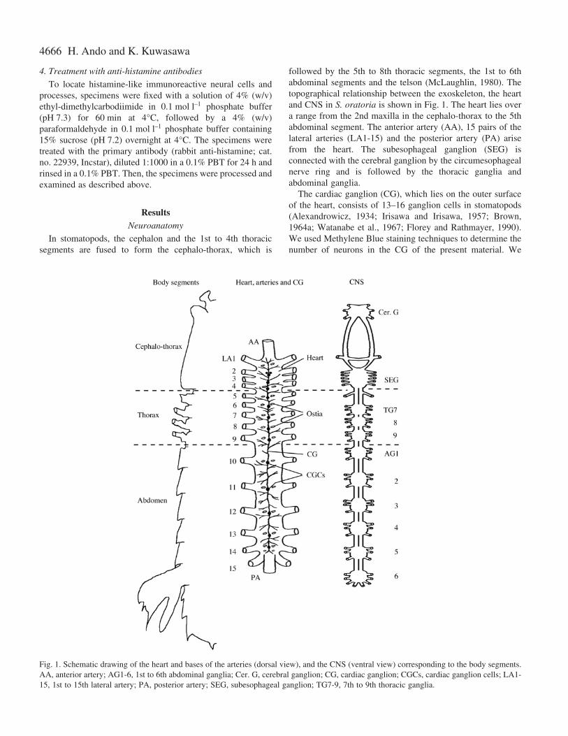
followed by the 5th to 8th thoracic segments, the 1st to 6thabdominal segments and the telson (McLaughlin, 1980). Thetopographical relationship between the exoskeleton, the heartand CNS in S. oratoria is shown in Fig.·1. The heart lies overa range from the 2nd maxilla in the cephalo-thorax to the 5thabdominal segment. The anterior artery (AA), 15 pairs of thelateral arteries (LA1-15) and the posterior artery (PA) arisefrom the heart. The subesophageal ganglion (SEG) isconnected with the cerebral ganglion by the circumesophagealnerve ring and is followed by the thoracic ganglia andabdominal ganglia.
The cardiac ganglion (CG), which lies on the outer surfaceof the heart, consists of 13–16 ganglion cells in stomatopods(Alexandrowicz, 1934; Irisawa and Irisawa, 1957; Brown,1964a; Watanabe et al., 1967; Florey and Rathmayer, 1990).We used Methylene Blue staining techniques to determine thenumber of neurons in the CG of the present material. We
H. Ando and K. Kuwasawa
Fig.·1. Schematic drawing of the heart and bases of the arteries (dorsal view), and the CNS (ventral view) corresponding to the body segments.AA, anterior artery; AG1-6, 1st to 6th abdominal ganglia; Cer. G, cerebral ganglion; CG, cardiac ganglion; CGCs, cardiac ganglion cells; LA1-15, 1st to 15th lateral artery; PA, posterior artery; SEG, subesophageal ganglion; TG7-9, 7th to 9th thoracic ganglia.
4667Neurohormonal control of Squilla heart
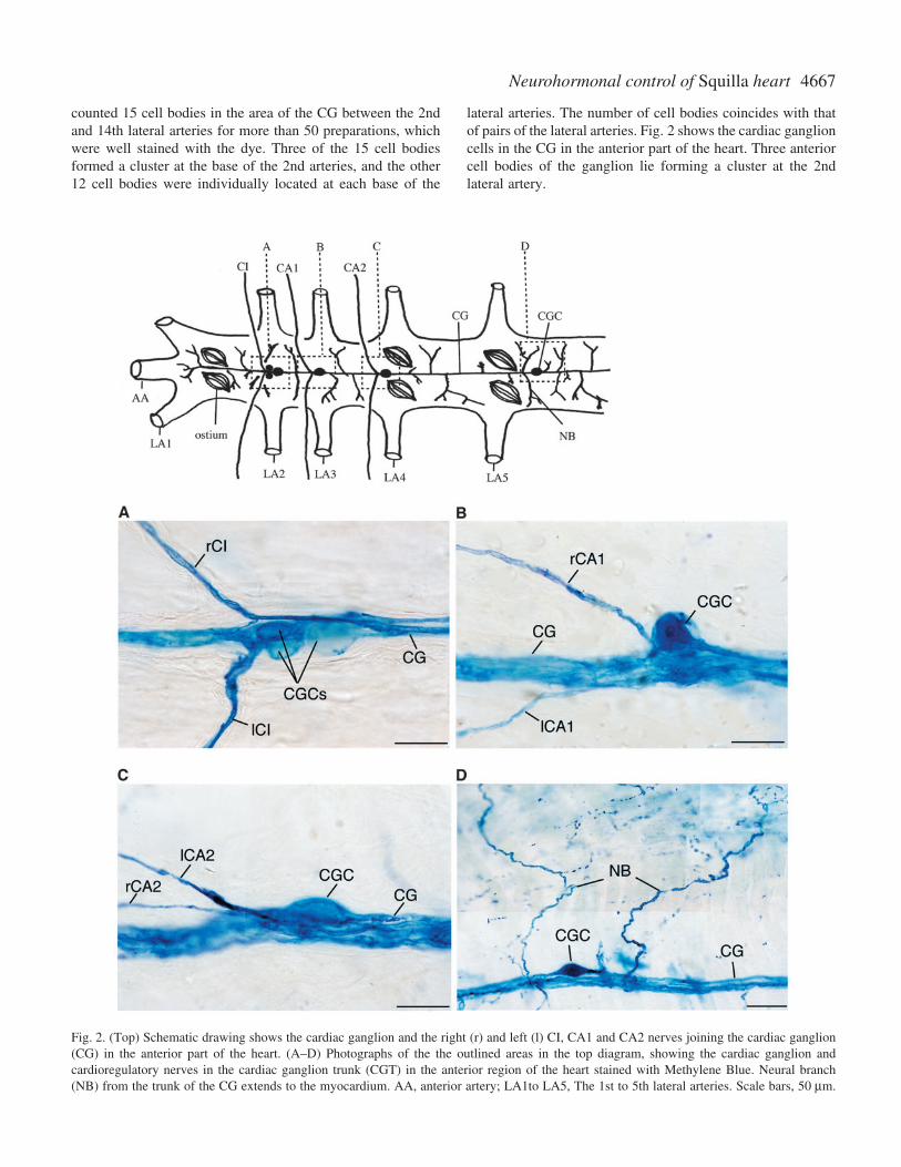
counted 15 cell bodies in the area of the CG between the 2ndand 14th lateral arteries for more than 50 preparations, whichwere well stained with the dye. Three of the 15 cell bodiesformed a cluster at the base of the 2nd arteries, and the other12 cell bodies were individually located at each base of the
lateral arteries. The number of cell bodies coincides with thatof pairs of the lateral arteries. Fig.·2 shows the cardiac ganglioncells in the CG in the anterior part of the heart. Three anteriorcell bodies of the ganglion lie forming a cluster at the 2ndlateral artery.
Fig.·2. (Top) Schematic drawing shows the cardiac ganglion and the right (r) and left (l) CI, CA1 and CA2 nerves joining the cardiac ganglion(CG) in the anterior part of the heart. (A–D) Photographs of the the outlined areas in the top diagram, showing the cardiac ganglion andcardioregulatory nerves in the cardiac ganglion trunk (CGT) in the anterior region of the heart stained with Methylene Blue. Neural branch(NB) from the trunk of the CG extends to the myocardium. AA, anterior artery; LA1to LA5, The 1st to 5th lateral arteries. Scale bars, 50·µm.
4668
Three pairs of cardioregulatory nerves join the CG at thelevel of the origins of the 2nd, 3rd and 4th lateral arteries inthe cephalo-thorax, for CI, CA1 and CA2 nerves, respectively(Fig.·2A–C).
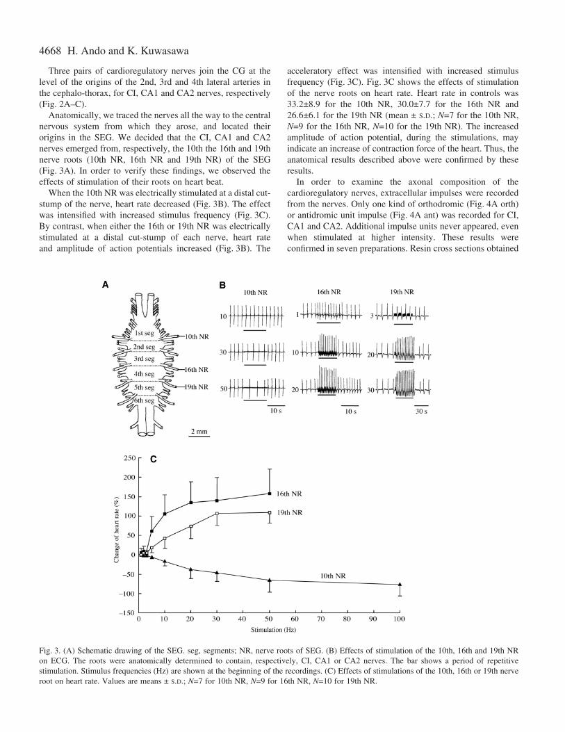
Anatomically, we traced the nerves all the way to the centralnervous system from which they arose, and located theirorigins in the SEG. We decided that the CI, CA1 and CA2nerves emerged from, respectively, the 10th the 16th and 19thnerve roots (10th NR, 16th NR and 19th NR) of the SEG(Fig.·3A). In order to verify these findings, we observed theeffects of stimulation of their roots on heart beat.
When the 10th NR was electrically stimulated at a distal cut-stump of the nerve, heart rate decreased (Fig.·3B). The effectwas intensified with increased stimulus frequency (Fig.·3C).By contrast, when either the 16th or 19th NR was electricallystimulated at a distal cut-stump of each nerve, heart rateand amplitude of action potentials increased (Fig.·3B). The
acceleratory effect was intensified with increased stimulusfrequency (Fig.·3C). Fig.·3C shows the effects of stimulationof the nerve roots on heart rate. Heart rate in controls was33.2±8.9 for the 10th NR, 30.0±7.7 for the 16th NR and26.6±6.1 for the 19th NR (mean ± S.D.; N=7 for the 10th NR,N=9 for the 16th NR, N=10 for the 19th NR). The increasedamplitude of action potential, during the stimulations, mayindicate an increase of contraction force of the heart. Thus, theanatomical results described above were confirmed by theseresults.
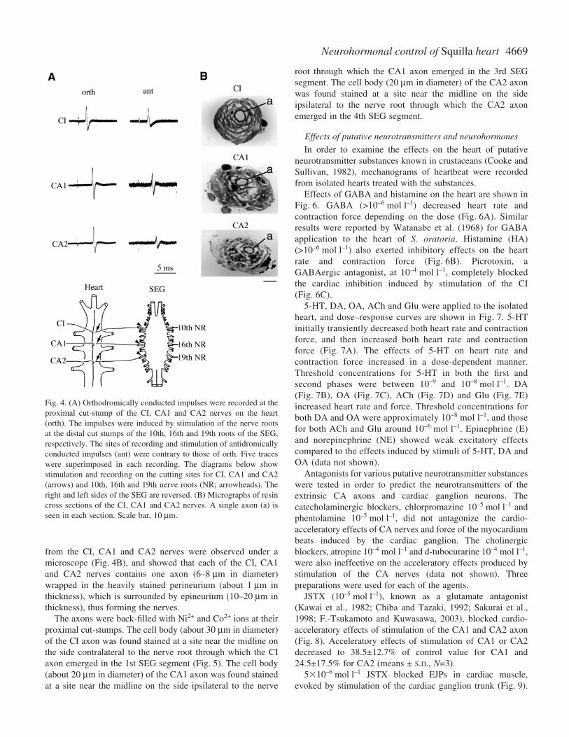
In order to examine the axonal composition of thecardioregulatory nerves, extracellular impulses were recordedfrom the nerves. Only one kind of orthodromic (Fig.·4A orth)or antidromic unit impulse (Fig.·4A ant) was recorded for CI,CA1 and CA2. Additional impulse units never appeared, evenwhen stimulated at higher intensity. These results wereconfirmed in seven preparations. Resin cross sections obtained
H. Ando and K. Kuwasawa
Fig.·3. (A) Schematic drawing of the SEG. seg, segments; NR, nerve roots of SEG. (B) Effects of stimulation of the 10th, 16th and 19th NRon ECG. The roots were anatomically determined to contain, respectively, CI, CA1 or CA2 nerves. The bar shows a period of repetitivestimulation. Stimulus frequencies (Hz) are shown at the beginning of the recordings. (C) Effects of stimulations of the 10th, 16th or 19th nerveroot on heart rate. Values are means ± S.D.; N=7 for 10th NR, N=9 for 16th NR, N=10 for 19th NR.
4669Neurohormonal control of Squilla heart
from the CI, CA1 and CA2 nerves were observed under amicroscope (Fig.·4B), and showed that each of the CI, CA1and CA2 nerves contains one axon (6–8·µm in diameter)wrapped in the heavily stained perineurium (about 1·µm inthickness), which is surrounded by epineurium (10–20·µm inthickness), thus forming the nerves.
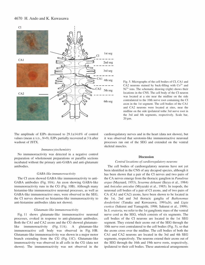
The axons were back-filled with Ni2+ and Co2+ ions at theirproximal cut-stumps. The cell body (about 30·µm in diameter)of the CI axon was found stained at a site near the midline onthe side contralateral to the nerve root through which the CIaxon emerged in the 1st SEG segment (Fig.·5). The cell body(about 20·µm in diameter) of the CA1 axon was found stainedat a site near the midline on the side ipsilateral to the nerve
root through which the CA1 axon emerged in the 3rd SEGsegment. The cell body (20·µm in diameter) of the CA2 axonwas found stained at a site near the midline on the sideipsilateral to the nerve root through which the CA2 axonemerged in the 4th SEG segment.
Effects of putative neurotransmitters and neurohormones
In order to examine the effects on the heart of putativeneurotransmitter substances known in crustaceans (Cooke andSullivan, 1982), mechanograms of heartbeat were recordedfrom isolated hearts treated with the substances.
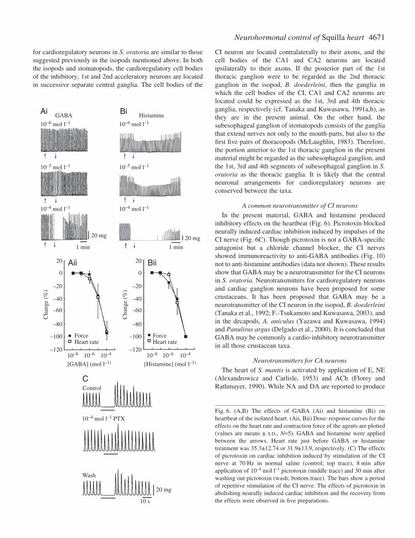
Effects of GABA and histamine on the heart are shown inFig.·6. GABA (>10–6·mol·l–1) decreased heart rate andcontraction force depending on the dose (Fig.·6A). Similarresults were reported by Watanabe et al. (1968) for GABAapplication to the heart of S. oratoria. Histamine (HA)(>10–6·mol·l–1) also exerted inhibitory effects on the heartrate and contraction force (Fig.·6B). Picrotoxin, aGABAergic antagonist, at 10–4·mol·l–1, completely blockedthe cardiac inhibition induced by stimulation of the CI(Fig.·6C).
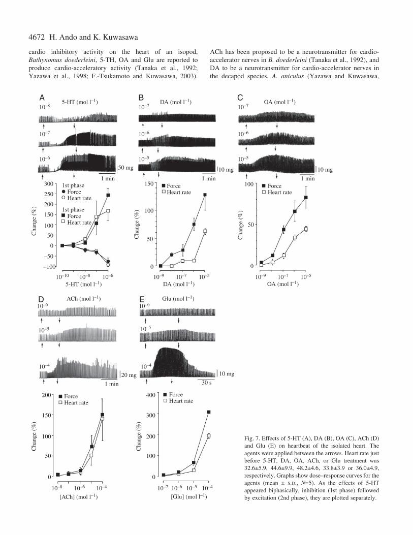
5-HT, DA, OA, ACh and Glu were applied to the isolatedheart, and dose–response curves are shown in Fig.·7. 5-HTinitially transiently decreased both heart rate and contractionforce, and then increased both heart rate and contractionforce (Fig.·7A). The effects of 5-HT on heart rate andcontraction force increased in a dose-dependent manner.Threshold concentrations for 5-HT in both the first andsecond phases were between 10–9 and 10–8·mol·l–1. DA(Fig.·7B), OA (Fig.·7C), ACh (Fig.·7D) and Glu (Fig.·7E)increased heart rate and force. Threshold concentrations forboth DA and OA were approximately 10–8·mol·l–1, and thosefor both ACh and Glu around 10–6·mol·l–1. Epinephrine (E)and norepinephrine (NE) showed weak excitatory effectscompared to the effects induced by stimuli of 5-HT, DA andOA (data not shown).
Antagonists for various putative neurotransmitter substanceswere tested in order to predict the neurotransmitters of theextrinsic CA axons and cardiac ganglion neurons. Thecatecholaminergic blockers, chlorpromazine 10–5·mol·l–1 andphentolamine 10–5·mol·l–1, did not antagonize the cardio-acceleratory effects of CA nerves and force of the myocardiumbeats induced by the cardiac ganglion. The cholinergicblockers, atropine 10–4·mol·l–1 and d-tubocurarine 10–4·mol·l–1,were also ineffective on the acceleratory effects produced bystimulation of the CA nerves (data not shown). Threepreparations were used for each of the agents.
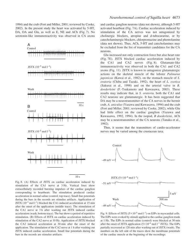
JSTX (10–5·mol·l–1), known as a glutamate antagonist(Kawai et al., 1982; Chiba and Tazaki, 1992; Sakurai et al.,1998; F.-Tsukamoto and Kuwasawa, 2003), blocked cardio-acceleratory effects of stimulation of the CA1 and CA2 axon(Fig.·8). Acceleratory effects of stimulation of CA1 or CA2decreased to 38.5±12.7% of control value for CA1 and24.5±17.5% for CA2 (means ± S.D., N=3).
510–6·mol·l–1 JSTX blocked EJPs in cardiac muscle,evoked by stimulation of the cardiac ganglion trunk (Fig.·9).
Fig.·4. (A) Orthodromically conducted impulses were recorded at theproximal cut-stump of the CI, CA1 and CA2 nerves on the heart(orth). The impulses were induced by stimulation of the nerve rootsat the distal cut stumps of the 10th, 16th and 19th roots of the SEG,respectively. The sites of recording and stimulation of antidromicallyconducted impulses (ant) were contrary to those of orth. Five traceswere superimposed in each recording. The diagrams below showstimulation and recording on the cutting sites for CI, CA1 and CA2(arrows) and 10th, 16th and 19th nerve roots (NR; arrowheads). Theright and left sides of the SEG are reversed. (B) Micrographs of resincross sections of the CI, CA1 and CA2 nerves. A single axon (a) isseen in each section. Scale bar, 10·µm.
4670
The amplitude of EJPs decreased to 29.1±14.6% of controlvalues (mean ± S.D., N=9). EJPs partially recovered at 3·h afterwashout of JSTX.
Immunocytochemistry
No immunoreactivity was detected in a negative controlpreparation of wholemount preparations or paraffin sectionsincubated without the primary anti-GABA and anti-glutamateantibodies.
GABA-like immunoreactivity
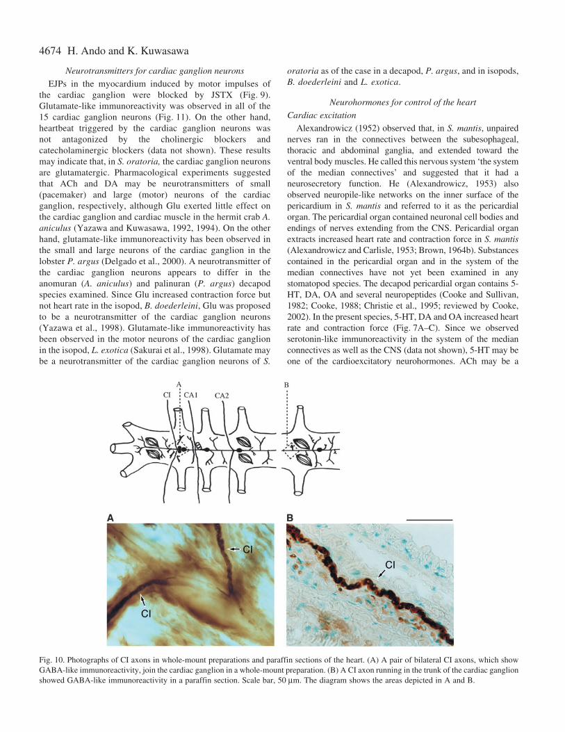
The CI axon showed GABA-like immunoreactivity to anti-GABA antibodies (Fig.·10A). An axon showing GABA-likeimmunoreactivity runs in the CG (Fig.·10B). Although manyhistamine-like immunoreactive neuronal processes, as well asGABA-like immunoreactive ones, were observed in the SEG,the CI nerves showed no histamine-like immunoreactivity toanti-histamine antibodies (data not shown).
Glutamate-like immunoreactivity
Fig.·11 shows glutamate-like immunoreactive neuronalprocesses, evoked in response to anti-glutamate antibodies.Both the CA1 and CA2 axons and the CG showed glutamate-like immunoreactivity (Fig.·11A). A glutamate-likeimmunoreactive cell body was observed in Fig.·10B.Glutamate-like immunoreactivity was shown in a major motorbranch extending from the CG (Fig.·11C). Glutamate-likeimunoreactivity was observed in all cells in the CG (data notshown). The immunoreactivity was not observed in the
cardioregulatory nerves and in the heart (data not shown), butit was observed that serotonin-like immunoreactive neuronalprocesses ran out of the SEG and extended on the ventralskeletal muscles.
DiscussionCentral locations of cardioregulatory neurons
The cell bodies of cardioregulatory neurons have not yetbeen identified in the CNS of any decapod species, although ithas been shown that a pair of the CI nerves and two pairs ofthe CA nerves emerge from the thoracic ganglion in Panulirusargus (Maynard, 1953), Sesarma dehaani (Ikeya et al., 1984)and Aniculus aniculus (Miyazaki et al., 1985). In isopods, theneuronal cell bodies of a pair of CI axons, and of two pairs ofCA (CA1 and CA2) axons, have been shown to be located inthe 1st, 2nd and 3rd thoracic ganglia of Bathynomusdoederleini (Tanaka and Kuwasawa, 1991a,b), and Ligiaexotica (Sakurai and Yamagishi, 1998; Sakurai et al., 1999).In S. oratoria, we refer to the 1st ganglionic mass of the ventralnerve cord as the SEG, which consists of six segments. Thecell bodies of the CI neurons are located in the 1st SEGsegment. They extend their axons out of the SEG through the10th nerve root contralateral to the cell bodies (Fig.·5), so thatthe axons cross over the midline. The cell bodies of both theCA1 and CA2 neurons are located in the 3rd and 4th SEGsegments, respectively. The neurons extend their axons out ofthe SEG through the 16th and 19th nerve roots, respectively,ipsilateral to their cell bodies. These anatomical arrangements
H. Ando and K. Kuwasawa
Fig.·5. Micrographs of the cell bodies of CI, CA1 andCA2 neurons stained by back-filling with Co2+ andNi2+ ions. The schematic drawing (right) shows theirlocations in the CNS. The cell body of the CI neuronwas located at a site near the midline on the sidecontralateral to the 10th nerve root containing the CIaxon in the 1st segment. The cell bodies of the CA1and CA2 neurons were located at sites, near themidline on the side ipsilateral tothe 3rd nerve root inthe 3rd and 4th segments, respectively. Scale bar,20·µm.
4671Neurohormonal control of Squilla heart
for cardioregulatory neurons in S. oratoria are similar to thosesuggested previously in the isopods mentioned above. In boththe isopods and stomatopods, the cardioregulatory cell bodiesof the inhibitory, 1st and 2nd acceleratory neurons are locatedin successive separate central ganglia. The cell bodies of the
CI neuron are located contralaterally to their axons, and thecell bodies of the CA1 and CA2 neurons are locatedipsilaterally to their axons. If the posterior part of the 1stthoracic ganglion were to be regarded as the 2nd thoracicganglion in the isopod, B. doederleini, then the ganglia inwhich the cell bodies of the CI, CA1 and CA2 neurons arelocated could be expressed as the 1st, 3rd and 4th thoracicganglia, respectively (cf. Tanaka and Kuwasawa, 1991a,b), asthey are in the present animal. On the other hand, thesubesophageal ganglion of stomatopods consists of the gangliathat extend nerves not only to the mouth-parts, but also to thefirst five pairs of thoracopods (McLaughlin, 1983). Therefore,the portion anterior to the 1st thoracic ganglion in the presentmaterial might be regarded as the subesophageal ganglion, andthe 1st, 3rd and 4th segments of subesophageal ganglion in S.oratoria as the thoracic ganglia. It is likely that the centralneuronal arrangements for cardioregulatory neurons areconserved between the taxa.
A common neurotransmitter of CI neurons
In the present material, GABA and histamine producedinhibitory effects on the heartbeat (Fig.·6). Picrotoxin blockedneurally induced cardiac inhibition induced by impulses of theCI nerve (Fig.·6C). Though picrotoxin is not a GABA-specificantagonist but a chloride channel blocker, the CI nervesshowed immunoreactivity to anti-GABA antibodies (Fig.·10)not to anti-histamine antibodies (data not shown). These resultsshow that GABA may be a neurotransmitter for the CI neuronsin S. oratoria. Neurotransmitters for cardioregulatory neuronsand cardiac ganglion neurons have been proposed for somecrustaceans. It has been proposed that GABA may be aneurotransmitter of the CI neuron in the isopod, B. doederleini(Tanaka et al., 1992; F.-Tsukamoto and Kuwasawa, 2003), andin the decapods, A. aniculus (Yazawa and Kuwasawa, 1994)and Panulirus argus (Delgado et al., 2000). It is concluded thatGABA may be commonly a cardio-inhibitory neurotransmitterin all those crustacean taxa.
Neurotransmitters for CA neurons
The heart of S. mantis is activated by application of E, NE(Alexandrowicz and Carlisle, 1953) and ACh (Florey andRathmayer, 1990). While NA and DA are reported to produce
GABA Histamine
10–6 mol l–1
10–8 10–6 10–610–4 10–8 10–4
10–6 mol l–1
10–5 mol l–1 10–5 mol l–1
10–4 mol l–1
10–4 mol l–1 PTX
10–4 mol l–1
20 mg20 mg
1 min
20 mg
10 s
1 min
Ai Bi
Aii
C
Bii20
0
–20
–60
–40
–80
–100
–120
Cha
nge
(%)
20
0
–20
–60
–40
–80
–100
–120
Cha
nge
(%)
ForceHeart rate
ForceHeart rate
[GABA] (mol l–1) [Histamine] (mol l–1)
Control
Wash
Fig.·6. (A,B) The effects of GABA (Ai) and histamine (Bi) onheartbeat of the isolated heart. (Aii, Bii) Dose–response curves for theeffects on the heart rate and contraction force of the agents are plotted(values are means ± S.D., N=5). GABA and histamine were appliedbetween the arrows. Heart rate just before GABA or histaminetreatment was 35.3±12.74 or 31.9±13.9, respectively. (C) The effectsof picrotoxin on cardiac inhibition induced by stimulation of the CInerve at 70·Hz in normal saline (control; top trace), 8·min afterapplication of 10–4·mol·l–1 picrotoxin (middle trace) and 30·min afterwashing out picrotoxin (wash; bottom trace). The bars show a periodof repetitive stimulation of the CI nerve. The effects of picrotoxin inabolishing neurally induced cardiac inhibition and the recovery fromthe effects were observed in five preparations.
4672
cardio inhibitory activity on the heart of an isopod,Bathynomus doederleini, 5-TH, OA and Glu are reported toproduce cardio-acceleratory activity (Tanaka et al., 1992;Yazawa et al., 1998; F.-Tsukamoto and Kuwasawa, 2003).
ACh has been proposed to be a neurotransmitter for cardio-accelerator nerves in B. doederleini (Tanaka et al., 1992), andDA to be a neurotransmitter for cardio-accelerator nerves inthe decapod species, A. aniculus (Yazawa and Kuwasawa,
H. Ando and K. Kuwasawa
10–8 10–7 10–7
10–7
10–10 10–8
10–8 10–6 10–4 10–7 10–6 10–5 10–4
10–6
10–6
10–9 10–7 10–5 10–9 10–7 10–5
10–5
10–6 10–6
10–6 10–5 10–5
10–4
10–6
10–5
10–4
10 mg
1 min
10 mg
1 min
10 mg
30 s20 mg
1 min
50 mg
1 min
50
0
–50
–100
250
300
200
0
50
100
150
400
0
100
200
300
150
200
50
50
0 0
150
100
100
100
Force1st phase
Heart rate
ForceHeart rate
ForceHeart rate
Force1st phase
Heart rate
ForceHeart rate
ForceHeart rate
5-HT (mol l–1)
ACh (mol l–1) Glu (mol l–1)
[ACh] (mol l–1) [Glu] (mol l–1)
5-HT (mol l–1) DA (mol l–1)
DA (mol l–1)
OA (mol l–1)
OA (mol l–1)
A B C
ED
Cha
nge
(%)
Cha
nge
(%)
Cha
nge
(%)
Cha
nge
(%)
Cha
nge
(%)
Fig.·7. Effects of 5-HT (A), DA (B), OA (C), ACh (D)and Glu (E) on heartbeat of the isolated heart. Theagents were applied between the arrows. Heart rate justbefore 5-HT, DA, OA, ACh, or Glu treatment was32.6±5.9, 44.6±9.9, 48.2±4.6, 33.8±3.9 or 36.0±4.9,respectively. Graphs show dose–response curves for theagents (mean ± S.D., N=5). As the effects of 5-HTappeared biphasically, inhibition (1st phase) followedby excitation (2nd phase), they are plotted separately.
4673Neurohormonal control of Squilla heart
1994) and the crab (Fort and Miller, 2001; reviewed by Cooke,2002). In the present study the heart was activated by 5-HT,DA, OA and Glu, as well as E, NE and ACh (Fig.·7). Noserotonin-like immunoreactivity was observed in CA axons
and cardiac ganglion neurons (data not shown), although 5-HTactivated heartbeat (Fig.·7A). Cardiac acceleration induced bystimulation of the CA nerves was not antagonized bycholinergic blockers, atropine and d-tubocurarine, or bycatecholaminergic blockers, chlorpromazine and phentolamine(data not shown). Thus, ACh, 5-HT and catecholamines maybe excluded from the list of transmitter candidates for the CAneurons.
Glu increased not only contraction force but also heart rate(Fig.·7E). JSTX blocked cardiac acceleration induced bythe CA1 and CA2 nerves (Fig.·8). Glutamate-likeimmunoreactivity was observed in both the CA1 and CA2axons (Fig.·11). JSTX is known to antagonize glutamatergicactions on the skeletal muscle of the lobster Palinurusjaponicus (Kawai et al., 1982), on the stomach muscle of S.oratoria (Chiba and Tazaki, 1992), the heart of L. exotica(Sakurai et al., 1998) and on the arterial valve in B.doederleini (F.-Tsukamoto and Kuwasawa, 2003). Theseresults may indicate that, in S. oratoria, both the CA1 andCA2 neurons are glutamatergic. It has been suggested thatDA may be a neurotransmitter of the CA nerves in the hermitcrab, A. aniculus (Yazawa and Kuwasawa, 1994) and the crab(Fort and Miller, 2001; reviewed by Cooke, 2002), while Gluhad little effect on the cardiac ganglion (Yazawa andKuwasawa, 1992, 1994). In the isopod, B doederleini, AChmay be a neurotransmitter of the CA neurons (Tanaka et al.,1992).
Thus, it seems that the transmitters of cardio-acceleratornerves may be varied among the crustacean taxa.
JSTX (10–5 mol l–1)
10 s
A
B
Control
Wash
JSTX (10–5 mol l–1)
10 s
Control
Wash
Fig.·8. (A) Effects of JSTX on cardiac acceleration induced bystimulation of the CA1 nerve at 3·Hz. Vertical lines showextracellularly recorded bursting impulses of the cardiac ganglioncorresponding to heartbeats. The stimulation produced cardiacacceleration in normal saline (control; top trace). Small fine potentialsduring the bars in the records are stimulus artifacts. Application ofJSTX (10–5·mol·l–1) blocked the CA1-induced acceleration at 15·minafter the onset of the application (middle trace). The stimulation ofthe CA1 nerve at 3·h after washing out JSTX induced cardiacacceleration (wash; bottom trace). The bar shows a period of repetitivestimulation. (B) Effects of JSTX on cardiac acceleration induced bystimulation of the CA2 nerve at 10·Hz. Application of JSTX blockedthe CA2 induced acceleration at 30·min after the onset of theapplication. The stimulation of the CA2 nerve at 1·h after washing outJSTX induced cardiac acceleration. Small fine potentials during thebars in the records are stimulus artifacts.
JSTX (510–6 mol l–1)
500 ms
5 mV
–49 mV
–51 mV
–49 mV
Control
Wash
Fig.·9. Effects of JSTX (510–6·mol·l–1) on EJPs in myocardial cells.The EJPs were evoked by stimuli applied to the cardiac ganglion trunkat 1·Hz. The EJPs in normal saline (control) were blocked at 30·minafter the onset of JSTX application (510–6·mol·l–1 JSTX). The EJPspartially recovered at 120·min after washing out of JSTX (wash). Thenumbers on the left side of the traces show the membrane potentialsof the cardiac muscle at the beginning of the recordings.
4674
Neurotransmitters for cardiac ganglion neurons
EJPs in the myocardium induced by motor impulses ofthe cardiac ganglion were blocked by JSTX (Fig.·9).Glutamate-like immunoreactivity was observed in all of the15 cardiac ganglion neurons (Fig.·11). On the other hand,heartbeat triggered by the cardiac ganglion neurons wasnot antagonized by the cholinergic blockers andcatecholaminergic blockers (data not shown). These resultsmay indicate that, in S. oratoria, the cardiac ganglion neuronsare glutamatergic. Pharmacological experiments suggestedthat ACh and DA may be neurotransmitters of small(pacemaker) and large (motor) neurons of the cardiacganglion, respectively, although Glu exerted little effect onthe cardiac ganglion and cardiac muscle in the hermit crab A.aniculus (Yazawa and Kuwasawa, 1992, 1994). On the otherhand, glutamate-like immunoreactivity has been observed inthe small and large neurons of the cardiac ganglion in thelobster P. argus (Delgado et al., 2000). A neurotransmitter ofthe cardiac ganglion neurons appears to differ in theanomuran (A. aniculus) and palinuran (P. argus) decapodspecies examined. Since Glu increased contraction force butnot heart rate in the isopod, B. doederleini, Glu was proposedto be a neurotransmitter of the cardiac ganglion neurons(Yazawa et al., 1998). Glutamate-like immunoreactivity hasbeen observed in the motor neurons of the cardiac ganglionin the isopod, L. exotica (Sakurai et al., 1998). Glutamate maybe a neurotransmitter of the cardiac ganglion neurons of S.
oratoria as of the case in a decapod, P. argus, and in isopods,B. doederleini and L. exotica.
Neurohormones for control of the heart
Cardiac excitation
Alexandrowicz (1952) observed that, in S. mantis, unpairednerves ran in the connectives between the subesophageal,thoracic and abdominal ganglia, and extended toward theventral body muscles. He called this nervous system ‘the systemof the median connectives’ and suggested that it had aneurosecretory function. He (Alexandrowicz, 1953) alsoobserved neuropile-like networks on the inner surface of thepericardium in S. mantis and referred to it as the pericardialorgan. The pericardial organ contained neuronal cell bodies andendings of nerves extending from the CNS. Pericardial organextracts increased heart rate and contraction force in S. mantis(Alexandrowicz and Carlisle, 1953; Brown, 1964b). Substancescontained in the pericardial organ and in the system of themedian connectives have not yet been examined in anystomatopod species. The decapod pericardial organ contains 5-HT, DA, OA and several neuropeptides (Cooke and Sullivan,1982; Cooke, 1988; Christie et al., 1995; reviewed by Cooke,2002). In the present species, 5-HT, DA and OA increased heartrate and contraction force (Fig.·7A–C). Since we observedserotonin-like immunoreactivity in the system of the medianconnectives as well as the CNS (data not shown), 5-HT may beone of the cardioexcitatory neurohormones. ACh may be a
H. Ando and K. Kuwasawa
Fig.·10. Photographs of CI axons in whole-mount preparations and paraffin sections of the heart. (A) A pair of bilateral CI axons, which showGABA-like immunoreactivity, join the cardiac ganglion in a whole-mount preparation. (B) A CI axon running in the trunk of the cardiac ganglionshowed GABA-like immunoreactivity in a paraffin section. Scale bar, 50·µm. The diagram shows the areas depicted in A and B.

4675Neurohormonal control of Squilla heart
neurotransmitter for small neurons of the pacemaker in thecardiac ganglion of the hermit crabs (Yazawa and Kuwasawa,1992) and for the CA axons of the isopod (Tanaka et al., 1992).It is suggested that in the stomatopod a transmitter of CAneurons is Glu but not ACh, although ACh increased bothheart rate and contraction force (Fig.·7D). It is of interest forfurther study to determine whether ACh is contained as aneurohormonal substance in the pericardial organ of S. oratoria.
Cardiac inhibition
Cardio-inhibitory substances other than GABA have notpreviously been reported in any stomatopod species. This studyshowed that HA induced inhibitory effects on the heart(Fig.·6B). However, no histamine-like immunoreactivity was
observed in the CI neurons. Since many histamine-likeimmunoreactive neurons were located throughout the CNS inthe present material (data not shown), it cannot be excludedthat HA is possibly a cardio-inhibitory neurohormone, asproposed for Homarus americanus (Hashemzadeh-Gargari andFreschi, 1992).
List of abbreviationsAA anterior arteryACh acetylcholineAG1-6 1st to 6th abdominal gangliaCA1 and CA2 1st and 2nd cardio-acceleratorCer. G cerebral ganglion
Fig.·11. Photographs of glutamate-like immunoreactivity in paraffin sections obtained from the heart. Glutamate-like immunoreactivity is seenin (Ai) the CA1 nerve and the cardiac ganglion trunk (CGT) and (Aii) the CA2 nerves on both sides and the CGT. (B) Glutamate-likeimmunoreactivity in the soma of the cardiac ganglion neuron. (C) Glutamate-like immunoreactivity in a major branch (MB) of the cardiacganglion extending to the myocardium. Scale bar, 50·µm. The diagram shows the areas depicted in A–C.
4676
Ceph.-Thorax cephalo-thoraxCG cardiac ganglionCGC cardiac ganglion cellCGT cardiac ganglion trunkCI cardio-inhibitorCNS central nervous systemDA dopamineDAB 3,3′-diaminobenzidine-tetrahydrochlorideDW distilled waterE epinephrineECG electrocardiogramEJP excitatory junctional potentialGABA γ-amino-butyric acidGlu glutamateHA histamine5-HT serotoninJSTX joro-spider toxinLA1-15 1st to 15th lateral arteriesMB major branchNB neural branchNE norepinephrineNR nerve rootPA posterior arteryPAP peroxidase–antiperoxidasePBT phosphate buffer containing Triton X-100PTX picrotoxinSEG subesophageal ganglionSW seawaterTG7-9 7th to 9th thoracic ganglia
This study was partly supported by the grant from JSPS No.12304050 (K.K.). We thank Mr K. Shishikura for generouslycollecting animals. We would like to thank Prof. R. B. Hill,University of Rhode Island, for revision of this manuscript.Contribution from the Shimoda Marine Research Center, No.707.
ReferencesAlexandrowicz, J. S. (1932). The innervation of the heart of Crustacea. I.
Decapoda. Q. J. Microsc. Sci. 75, 181-249.Alexandrowicz, J. S. (1934). The innervation of the heart of Crustacea. II.
Stomatopoda. Q. J. Microsc. Sci. 76, 511-548.Alexandrowicz, J. S. (1952). Notes on the nervous system in the
Stomatopoda. I. The system of median connectives. Publ. Stn. Zool. Napoli23, 201-214.
Alexandrowicz, J. S. (1953). Notes on the nervous system in theStomatopoda. II. The system of dorsal trunks. III. Small nerve cells in motornerves. Publ. Stn. Zool. Napoli 24, 29-45.
Alexandrowicz, J. S. and Carlisle, D. B. (1953). Some experiments on thefunction of the pericardial organs in Crustacea. J. Mar. Biol. Assn. UK 32,175-192.
Ando, H. and Kuwasawa, K. (1993). Central origins and functions ofextrinsic cardiac nerves in the stomatopod crustacean Squilla oratoria. Zool.Sci. 10, Suppl., 94.
Ando, H. and Kuwasawa, K. (1994). Identification of cardioinhibitory andcardioacceleratory neurons in the CNS of the stomatopod crustacean Squillaoratoria. Zool. Sci. 11, Suppl., 95.
Ando, H., Kuwasawa, K., Yazawa, T. and Kurokawa, M. (1995). Ananalysis of cardio-regulatory nerves in the stomatopod crustacean Squillaoratoria. Physiol. Zool. 68, 75.
Brown, H. F. (1964a). Electrophysiological investigations of the heart ofSquilla mantis. I. The ganglionic nerve trunk. J. Exp. Biol. 41, 689-700.
Brown, H. F. (1964b). Electrophysiological investigations of the heart ofSquilla mantis. III. The mode of action of pericardial organ extract on theheart. J. Exp. Biol. 41, 723-734.
Chiba, C. and Tazaki, K. (1992). Glutamatergic motoneurons in thestomatogastric ganglion of the mantis shrimp Squilla oratoria. J. Comp.Physiol. A 170, 773-786.
Christie, A. E., Skiebe, P. and Marder, E. (1995). Matrix ofneuromodulators in neurosecretory structures of the crab Cancer borealis.J. Exp. Biol. 198, 2431-2439.
Cooke, I. M. (1988). Studies on the crustacean cardiac ganglion. Comp.Biochem. Physiol. 91C, 205-218.
Cooke, I. M. (2002). Reliable, responsive pacemaking and pattern generationwith minimal cell numbers: the crustacean cardiac ganglion. Biol. Bull. 202,108-136.
Cooke, I. M. and Sullivan, R. E. (1982). Hormones and Neurosecretion. InThe Biology of Crustacea, vol. 3 (ed. H. L. Atwood and D. C. Sandeman),pp. 205-290. New York: Academic Press.
Delgado, J. Y., Oyola, E. and Miller, M. W. (2000). Localization of GABA-and glutamate-like immunoreactivity in the cardiac ganglion of the lobsterPanulirus argus. J. Neurocytol. 29, 605-619.
F.-Tsukamoto, Y. and Kuwasawa, K. (2003). Neurohormonal andglutamatergic neuronal control of the cardioarterial valves in the isopodcrustacean Bathynomus doederleini. J. Exp. Biol. 206, 431-443.
Florey, E. and Rathmayer, M. (1990). Facilitation and potentiation oftransmitter release at neuromuscular synapses in the heart of Squilla mantis;functional and theoretical implications. In Frontiers in CrustaceanNeurobiology (ed. K. Wiese, W.-D. Krenz, J. Tautz, H. Reichert and B.Mulloney), pp. 330-337. Basel: Birkhäuser Verlag.
Fort, T. J. and Miller, M. W. (2001). Functional organization of the cardiacsystem of the blue crab Callinectes Sapidus: GABAergic andcatecholaminergic regulatory fibers. Abstract, 2001 Society forNeuroscience.
Hashemzadeh-Gargari, H. and Freschi, J. E. (1992). Histamine activateschloride conductance in motor neurons of the lobster cardiac ganglion. J.Neurophysiol. 68, 9-15.
Ikeya, H., Uesaka, H., Yamagishi, H. and Ebara, A. (1984). Studies on thecardioregulatory nerves in a crab, Sesarma dehaani. Comp. Biochem.Physiol. 78A, 743-748.
Irisawa, H. and Irisawa, A. F. (1957). The electrocardiogram of astomatopod. Biol. Bull. 112, 358-362.
Kawai, N., Niwa, A. and Abe, T. (1982). Spider venom contains specificreceptor blocker of glutaminergic synapses. Brain Res. 247, 169-171.
Kihara, A. and Kuwasawa, K. (1984). A neuroanatomical andelectrophysiological analysis of nervous regulation in the heart of an isopodcrustacean, Bathynomus doederleini. J. Comp. Physiol. A 154, 883-894.
Maynard, D. M., Jr (1953). Activity in a crustacean ganglion. I. cardio-inhibition and acceleration in Panulirus argus. Biol. Bull. 104, 156-170.
Maynard, D. M. (1960). Circulation and heart function. In The Physiology ofCrustacea, vol. 1 (ed. T. H. Waterman), pp.161-226, New York: AcademicPress.
McLaughlin, P. A. (1980). Order Stomatopoda. In Comparative Morphologyof Recent Crustacea, pp. 63-69. San Francisco: Freeman.
McLaughlin, P. A. (1983). Internal anatomy. In The Biology of Crustacea,vol. 5 (ed. L. H. Mantel), pp. 1-52. New York: Academic Press.
Miyazaki, T., Kuwasawa, K., Yazawa, T. and Mashimo, K. (1985).Identification of the cardio-regulator nerves in a marine hermit crab and theshadow-induced cardiac inhibition in some decapods. Zool. Sci. 2, 35-47.
Quicke, D. L. J. and Brace, R. C. (1979). Differential staining of cobalt- andnickel-filled neurones using rubeanic acid. J. Microsc. 115, 161-163.
Sakurai, A. and Yamagishi, H. (1998). Identification of two cardio-acceleratory neurons in the isopod crustacean, Ligia exotica and their effectson cardiac ganglion cells. J. Comp. Physiol. A 182, 145-152.
Sakurai, A., Mori, A. and Yamagishi, H. (1998). Glutamatergicneuromuscular transmission in the heart of the isopod crustacean Ligiaexotica. J. Exp. Biol. 201, 2833-2842.
Sakurai, A., Mori, A. and Yamagishi, H. (1999). Cardioinhibitory neuronsin the isopod crustacean Ligia exotica. Zool. Sci. 16, 401-405.
Tanaka, K. and Kuwasawa, K. (1991a). Identification of cardio-acceleratoryneurons in the thoracic ganglion of the isopod crustacean Bathynomusdoederleini. Brain Res. 544, 311-314.
Tanaka, K. and Kuwasawa, K. (1991b). Identification of cardio-inhibitory
H. Ando and K. Kuwasawa
4677Neurohormonal control of Squilla heart
neurons in the thoracic ganglion of the isopod crustacean Bathynomusdoederleini. Brain Res. 558, 339-342.
Tanaka, K. Yazawa, T. and Kuwasawa, K. (1992). Cholinergic andGABAergic control of the heart in the isopod crustacean, Bathynomusdoederleini. In Phylogenetic Models in Functional Coupling of the CNS andthe Cardiovascular System, Comparative Physiology, vol. 11 (ed. R. B. Hill,K. Kuwasawa, R. R. McMahon and T. Kuramoto), pp. 132-140. Basel:Karger.
Watanabe, A., Obara, S. and Akiyama, T. (1967). Pacemaker potentials forthe periodic burst discharge in the heart ganglion of a stomatopod, Squillaoratoria. J. Gen. Physiol. 50, 839-862.
Watanabe, A., Obara, S. and Akiyama, T. (1968). Inhibitory synapses onpacemaker neurons in the heart ganglion of a stomatopod, Squilla oratoria.J. Gen. Physiol. 52, 908-924.
Watanabe, A., Obara, S. and Akiyama, T. (1969). Acceleratory synapses
on pacemaker neurons in the heart ganglion of a stomatopod, Squillaoratoria. J. Gen. Physiol. 54, 212-231.
Yazawa, T. and Kuwasawa, K. (1984). The cardio-regulator nerves of thehermit crabs: anatomical and electrophysiological identification of theirdistribution inside the heart. J. Comp. Physiol. A 154, 871-881.
Yazawa, T. and Kuwasawa, K. (1992). Intrinsic and extrinsic neural andneurohumoral control of the decapod heart. Experientia 48, 834-840.
Yazawa, T. and Kuwasawa, K. (1994). Dopaminergic acceleration andGABAergic inhibition in extrinsic neural control of the hermit crab heart.J. Comp. Physiol. A 174, 65-75.
Yazawa, T., Tanaka, K., Yasumatsu, M., Otokawa, M., Aihara, Y.,Ohsuga, K. and Kuwasawa, K. (1998). A pharmacological and HPLCanalysis of the excitatory transmitter of the cardiac ganglion in the heart ofthe isopod crustacean Bathynomus doederleini. Can. J. Physiol. Pharmacol.76, 599-604.