Odgovori Na Ispitna Pitanja Iz Predmeta Istorija Filozofije 1b (Platon)
of September 24, 2015.This information is current as
ActivationβSynthesis and NLRP3-Mediated IL-1
βIL-1−Multiple Cathepsins Promote Pro
RockMatthew Bogyo, Stephanie A. Robertson and Kenneth L. Gregory M. Orlowski, Jeff D. Colbert, Shruti Sharma,
http://www.jimmunol.org/content/195/4/1685doi: 10.4049/jimmunol.1500509July 2015;
2015; 195:1685-1697; Prepublished online 20J Immunol
MaterialSupplementary
9.DCSupplemental.htmlhttp://www.jimmunol.org/content/suppl/2015/07/18/jimmunol.150050
Referenceshttp://www.jimmunol.org/content/195/4/1685.full#ref-list-1
, 41 of which you can access for free at: cites 87 articlesThis article
Subscriptionshttp://jimmunol.org/subscriptions
is online at: The Journal of ImmunologyInformation about subscribing to
Permissionshttp://www.aai.org/ji/copyright.htmlSubmit copyright permission requests at:
Email Alertshttp://jimmunol.org/cgi/alerts/etocReceive free email-alerts when new articles cite this article. Sign up at:
Print ISSN: 0022-1767 Online ISSN: 1550-6606. Immunologists, Inc. All rights reserved.Copyright © 2015 by The American Association of9650 Rockville Pike, Bethesda, MD 20814-3994.The American Association of Immunologists, Inc.,
is published twice each month byThe Journal of Immunology
at stanford university on September 24, 2015
http://ww
w.jim
munol.org/
Dow
nloaded from
at stanford university on September 24, 2015
http://ww
w.jim
munol.org/
Dow
nloaded from
The Journal of Immunology
Multiple Cathepsins Promote Pro–IL-1b Synthesis andNLRP3-Mediated IL-1b Activation
Gregory M. Orlowski,* Jeff D. Colbert,* Shruti Sharma,† Matthew Bogyo,‡,x
Stephanie A. Robertson,{ and Kenneth L. Rock*
Sterile particles induce robust inflammatory responses that underlie the pathogenesis of diseases like silicosis, gout, and athero-
sclerosis. A key cytokine mediating this response is IL-1b. The generation of bioactive IL-1b by sterile particles is mediated by the
NOD-like receptor containing a pyrin domain 3 (NLRP3) inflammasome, although exactly how this occurs is incompletely
resolved. Prior studies have found that the cathepsin B inhibitor, Ca074Me, suppresses this response, supporting a model whereby
ingested particles disrupt lysosomes and release cathepsin B into the cytosol, somehow activating NLRP3. However, reports that
cathepsin B-deficient macrophages have no defect in particle-induced IL-1b generation have questioned cathepsin B’s involve-
ment. In this study, we examine the hypothesis that multiple redundant cathepsins (not just cathepsin B) mediate this process by
evaluating IL-1b generation in murine macrophages, singly or multiply deficient in cathepsins B, L, C, S and X. Using an activity-
based probe, we measure specific cathepsin activity in living cells, documenting compensatory changes in cathepsin-deficient cells,
and Ca074Me’s dose-dependent cathepsin inhibition profile is analyzed in parallel with its suppression of particle-induced IL-1b
secretion. Also, we evaluate endogenous cathepsin inhibitors cystatins C and B. Surprisingly, we find that multiple redundant
cathepsins, inhibited by Ca074Me and cystatins, promote pro–IL-1b synthesis, and to our knowledge, we provide the first evidence
that cathepsin X plays a nonredundant role in nonparticulate NLRP3 activation. Finally, we find cathepsin inhibitors selectively
block particle-induced NLRP3 activation, independently of suppressing pro–IL-1b synthesis. Altogether, we demonstrate that
both small molecule and endogenous cathepsin inhibitors suppress particle-induced IL-1b secretion, implicating roles for multiple
cathepsins in both pro–IL-1b synthesis and NLRP3 activation. The Journal of Immunology, 2015, 195: 1685–1697.
Sterile particles induce robust inflammatory responses thatunderlie the pathogenesis of many diseases. These path-ogenic particles are diverse and include silica (1–4), which
causes silicosis, monosodium urate (5), the etiologic agent ingout, and cholesterol crystals (CC) (6, 7), which are thought tocontribute to the pathogenesis of atherosclerosis. Importantly, thesterile inflammatory response and resultant diseases caused bythese particles all involve signaling through the IL-1R, IL-1R1 (8,9). Although IL-1R1 can be stimulated by either of two cytokines,IL-1a or IL-1b, it has been shown that IL-1b plays a pivotal role
in disease pathogenesis (10) because it not only directly stimulatesIL-1R1–dependent inflammatory signaling but is also needed forthe secretion of IL-1a from cells (11). Therefore, it is important tounderstand the exact mechanisms underlying the generation andsecretion of active IL-1b. However, this process is still incom-pletely understood and the focus of the present report.The generation of biologically active IL-1b is highly regulated
and usually proceeds in two distinct steps (12, 13). The first step(Signal 1 or “priming”) is initiated when cells such as macro-phages are stimulated by certain cytokines, pathogen-associatedmolecular patterns, or danger-associated molecular patterns. Sig-nal 1 leads to the nuclear translocation of NF-kB, which thenstimulates the synthesis of biologically inactive pro–IL-1b and,among other things, NOD-like receptor containing a pyrin domain3 (NLRP3), a protein important for IL-1b activation. The secondstep (Signal 2 or “activation”) induces the formation of a multi-molecular complex, known as the inflammasome. Inflammasomesare composed of a sensor protein, an adaptor protein, apoptosis-associated speck-like protein containing a CARD, and an execu-tioner protease, caspase-1. Each inflammasome sensor detectsdistinct stimuli, thereby initiating multimerization and activatingcaspase-1, which then cleaves pro–IL-1b and facilitates the secretionof bioactive mature IL-1b. Among the known inflammasomes, theNLRP3 inflammasome is unique. Although all inflammasomesrely on the availability of a newly synthesized pool of pro–IL-1b,basal levels of NLRP3 itself are limiting, making priming espe-cially critical for de novo NLRP3 transcription and subsequentactivation (14, 15). Moreover, the NLRP3 inflammasome is theexclusive mediator of IL-1b activation in response to sterile par-ticles (1–7).Although the NLRP3 inflammasome is located in the cytosol,
how this intracellular complex senses the presence of extracellularparticles has been of considerable interest. It has been shown that
*Department of Pathology, University of Massachusetts Medical School, Worcester,MA 01655; †Department of Medicine, University of Massachusetts Medical School,Worcester, MA 01655; ‡Department of Pathology, Stanford University School ofMedicine, Stanford, CA 94305; xDepartment of Microbiology and Immunology,Stanford University School of Medicine, Stanford, CA 94305; and {Sandler Centerfor Drug Discovery, University of California, San Francisco, San Francisco, CA94158
Received for publication March 3, 2015. Accepted for publication June 2, 2015.
This work was supported by National Institutes of Health Grants 5R01AI078287-05(to K.L.R.) and T32AI095213-01, services from the National Heart, Lung, and BloodInstitute, National Institutes of Health, Department of Health and Human Services,and the Science Moving Towards Research Translation and Therapy program viaContract HHSN268201100015C.
Address correspondence and reprint requests to Dr. Kenneth L. Rock, Departmentof Pathology, University of Massachusetts Medical School, 368 Plantation Street,Albert Sherman Center, AS9, Worcester, MA 01655. E-mail address: [email protected]
The online version of this article contains supplemental material.
Abbreviations used in this article: CC, cholesterol crystal; CHX, cycloheximide;dAdT, poly(deoxyadenylic-deoxythymidylic) acid; LDH, lactate dehydrogenase;LLOMe, Leu-Leu-OMe; LMD, lysosomal membrane disruption; LRR, leucine-richrepeat; NLRP3, NOD-like receptor containing a pyrin domain 3; PM, peritonealmacrophage; qPCR, quantitative PCR; ROS, reactive oxygen species; siRNA, smallinterfering RNA; WT, wild-type.
Copyright� 2015 by The American Association of Immunologists, Inc. 0022-1767/15/$25.00
www.jimmunol.org/cgi/doi/10.4049/jimmunol.1500509
at stanford university on September 24, 2015
http://ww
w.jim
munol.org/
Dow
nloaded from
internalization of particles by phagocytosis is a first essential step inactivating the NLRP3 inflammasome (2). Multiple mechanisms havebeen proposed as to how particles in phagosomes then lead to NLRP3inflammasome activation, including lysosomal membrane disruption(LMD) (2, 3, 6, 7, 13, 16–29), potassium efflux (1, 4, 7, 21, 29–37),and the generation of reactive oxygen species (ROS) (1, 27, 29, 30,32, 36, 38–40), among various other mechanisms (reviewed Ref. 12).All of these pathways may contribute to this process. In support of theLMD model, it has been shown that particles like silica, CC, and theadjuvant alum can cause LMD (2, 6, 7), leading to the leakage of thelysosomal cysteine protease cathepsin B into the cytosol, where thisprotease is thought to activate NLRP3 through an as yet undescribedmechanism. Consistent with this model, particle-induced activation ofthe NLRP3 inflammasome is blocked by inhibitors of lysosomalacidification (cathepsins are optimally active in acidic conditions) andinhibitors of cathepsin B. However, the requirement for cathepsin Bin this process is controversial.A role for cathepsin B in NLRP3 activation is supported by
a number of studies showing that Ca074Me, an inhibitor reported tobe specific for cathepsin B, suppresses IL-1b activation induced byparticulate and nonparticulate stimuli (2, 7, 17, 20, 21, 25–29, 41–46). However, despite a few subsequent studies showing that ca-thepsin B– or L–deficient macrophages show partial impairmentof this response (6, 25, 41), several follow-up studies have foundthat responses are intact in these same mutant cells (31, 42, 47).Thus, it has become unclear whether the efficacy of Ca074Me isreally a result of cathepsin B inhibition or whether this is an off-target effect. Indeed, there are there are several reports demon-strating that Ca074Me inhibits other cathepsins as well (48–52).Therefore, one hypothesis proposed to explain the discrepancybetween Ca074Me and genetic models is that multiple cathepsins,which are a highly conserved family of proteases, play redundantroles in NLRP3 activation (53). Redundancy of cathepsins B andL has been demonstrated in a mouse model, where deficiency ofboth results in neonatal mortality whereas deficiency of eitheralone does not (54). Similar redundancy has also been observed inmouse cancer models showing upregulation of cathepsin X whencathepsin B is knocked out (55). However, the role of redundantcathepsins has not been examined in the context of NLRP3 acti-vation and remains an open question.In this study, we use genetic inactivation of multiple cathepsins,
together with exogenous and endogenous inhibitors of theseproteases, and an activity-based probe to investigate the role ofcathepsins in NLRP3-dependent particle-induced IL-1b secretion.This analysis reveals that multiple cathepsins indeed contributeto IL-1b secretion. Surprisingly, our data also demonstrate thatcathepsins contribute not only to the inflammasome-mediatedcleavage of pro–IL-1b into mature IL-1b (Signal 2) but also tothe priming step of pro–IL-1b synthesis (Signal 1). In addition, wefound a unique role for cathepsin X in nigericin-induced NLRP3activation, a protease not previously implicated in the IL-1 re-sponse. Taken together, these data clarify the contribution ofcathepsins to particle-induced IL-1b responses and define a pre-viously unappreciated role for cathepsins and their inhibitors inregulating pro–IL-1b synthesis. In doing so, this study providesinsight into the mechanistic regulation of IL-1b production andpoints to cathepsins as unique therapeutic targets for controllingparticle-induced sterile inflammatory responses.
Materials and MethodsReagents and Abs
Abs for Western blots were against mouse IL-1b (R&D Systems), caspase-1p10 (sc-514; Santa Cruz Biotechnology), NLRP3 (Cryo2; Enzo LifeSciences), b-actin (C4; Santa Cruz Biotechnology), and GAPDH (6C5;
EMD Millopore). ELISA kits were purchased for mouse IL-1b (BD Bio-sciences), pro–IL-1b, and TNF-a (eBioscience). Ultrapure LPS was fromSalmonella minnesota (InvivoGen). Poly(deoxyadenylic-deoxythymidylic)acid and nigericin were purchased from Sigma-Aldrich (St. Louis, MO).Silica crystals (MIN- U-SIL 15) were obtained from U.S. Silica (Frederick,MD). Cholesterol crystals were synthesized by acetone supersaturation andcooling (6), Alum (Imject alum adjuvant; a mixture of aluminum hydroxideand magnesium hydroxide) was from Pierce Biotechnology, and Leu-Leu-OMe-HCl was from Chem-Impex International. ZVAD-FMK and Ca-074-Me were from Enzo Life Sciences, and K777 was initially gifted to us byS. A. Robertson and J. H. McKerrow at University of California (SanFrancisco, CA), and further stocks obtained through services from theNational Heart, Lung, and Blood Institute’s SMARTT Program. Lipo-fectamine 2000, RNAiMax, and all small interfering RNA (siRNA) smartpools were from Life Technologies and Endoporter was from Gene Tools.
Animal and cell lines
Wild-type C57BL/6 mice were purchased from The Jackson Laboratory.NLRP3-deficient mice (56) were provided by Millennium Pharmaceuticals.Cathepsin S–deficient mice (57) were provided by Dr. H. Chapman(University of California), cathepsin L–deficient (58), and cathepsinB–deficient (53) mice were provided by Dr. H. Ploegh (Harvard MedicalSchool, Boston, MA), cathepsin C–deficient mice (59) were providedby Dr. Christine Pham (Washington University School of Medicine,St. Louis), and all mice have been backcrossed to C57BL/6 background.Multiple cathepsin-deficient mice were bred from single cathepsin-deficientmice at the University of Massachusetts Medical School. All animal proto-cols were approved by the University of Massachusetts Institutional AnimalCare and Use Committee.
Generation of bone marrow chimeras
Adult wild-type (WT) C57BL/6 mice were lethally irradiated (1100 rad) andreconstituted for at least 8 wk with bone marrow collected from age-matchedWT or mutant donor mice 1–2 wk old. Some recipient mice in each groupexpressed the leukocyte marker Ly5.1 (CD45.1), whereas all donors ex-pressed Ly5.2 (CD45.2), allowing confirmation of .90% chimerism to bedetermined by flow cytometric analysis of peripheral blood samples.
Production and measurement of IL-1b, pro–IL-1b, and TNF-afrom in vitro cultures
Peritoneal exudate cells were elicited by i.p. injection of 3 ml 1% thio-glycollate and collected after 72 h by peritoneal lavage. Prior to experi-mentation, non-adherent cells were decanted, leaving primarily macrophagesbehind. Bone marrow–derived macrophages were generated as describedpreviously (60). Bone marrow neutrophils were isolated from whole bonemarrow, following RBC lysis, using the anti–Ly-6G Microbead Kit fromMiltenyi Biotec. Purity was assessed to be 95–98% by flow cytometry.Murine bone marrow–derived mast cells were derived from whole bonemarrow using murine rIL-3 (PeproTech), and purity was assessed to be 95%by toluidine blue (61). Cells were plated overnight in 96-well plates (ELISA)or 12-well plates (SDS-PAGE, cathepsin activity labeling with BMV109, andWestern blotting). Unless otherwise stated, the “standard protocol” followedin this study is as follows: priming in RPMI 1640 medium (or MC/9 mediumfor mast cells (61)) for 3 h with LPS (200 ng/ml), with or without the ad-dition of inhibitors after 2 h of priming, followed by 6 h of stimulation.Inhibitors were added in a final concentration of #0.1% DMSO, which hasno effect on readouts compared with medium alone. Supernatants werecollected, with or without addition of Promega’s 103 lysis solution formeasuring intracellular cytokines or lactate dehydrogenase (LDH) mea-surement by plate reader at OD490 using Promega’s Cytox96 Nonradioactivecytotoxicity assay, and cytokine levels were analyzed by ELISA.
siRNA knockdowns
Each siRNA was used at a 50 nM final concentration (or control siRNA at100 nM for double knockdowns) after complexation in a mixture of Endoporter(GeneTools) and RNAiMax (Life Technologies) at a ratio of 0.11 ml:0.15 ml inOptiMEM (Life Technologies), respectively, per 0.1 ml final volume (in 10%FCS). Complexes were combined with 10% FCS-containing RPMI 1640medium at a ratio 1:10 (complexes:10% FCS) and added to cells for 96 h.Medium was supplemented with 2 mM L-glutamine, 1 mM sodium pyruvate,0.2 mM 2-ME, 13 nonessential amino acids, and 100 mg/ml ciprofloxacin.
Immunoblotting and Live-cell intracellular cathepsin activitylabeling with BMV109
In 12-well plates, adherent macrophages were washed with RPMI 1640medium, and incubated with or without LPS as indicated for 3 h (inhibitors
1686 MULTIPLE CATHEPSINS IN IL-1b RESPONSES
at stanford university on September 24, 2015
http://ww
w.jim
munol.org/
Dow
nloaded from
added after 2 h), at which time BMV109 (62) was added at a final con-centration of 1 mM. After 1 h with BMV109, supernatants were collected,cells were washed with PBS, and lysates were made with Cell ExtractionBuffer from Life Technologies with complete protease inhibitor mixturefrom Roche. Supernatants were precipitated with chloroform/methanol,and lysate protein concentration was equalized using the Pierce BCAAssay. At least 15 mg was loaded for each sample and separated by 15%(or 8% for NLRP3 blots) SDS-PAGE, and gels were analyzed with a Ty-phoon Trio phosphor-imager from GE, and protein was transferred ontonitrocellulose membranes. Densitometry was performed using ImageJ.Images of gels or blots were cropped for the bands of interest, and anycontrast enhancement was applied evenly throughout using iPhoto.
Real-time measurement of lysosomal integrity
In black high-binding 96-well clear-bottom plates, 50 ml acridine orange(Life Technologies) in warm HBSS (with Ca2+ and Mg2+) was added tocells in 100 ml RPMI 1640 medium containing 10% FCS to reach a finalconcentration of 3.75 mg/ml and then incubated at 37˚C for 15 min prior towashing 13 with 200 ml HBSS. Cells were then treated and stimulated asindicated in phenol red-free CO2-independent Leibovitz’s medium. Fluo-rescence was measured in each well every 1–3 min using an incubatedVictorX5 plate reader. Background fluorescence was subtracted from wellstreated with dye-free HBSS.
ResultsGenetic and biochemical analysis of the impact of individualcathepsin deficiency on particle-induced IL-1b secretion
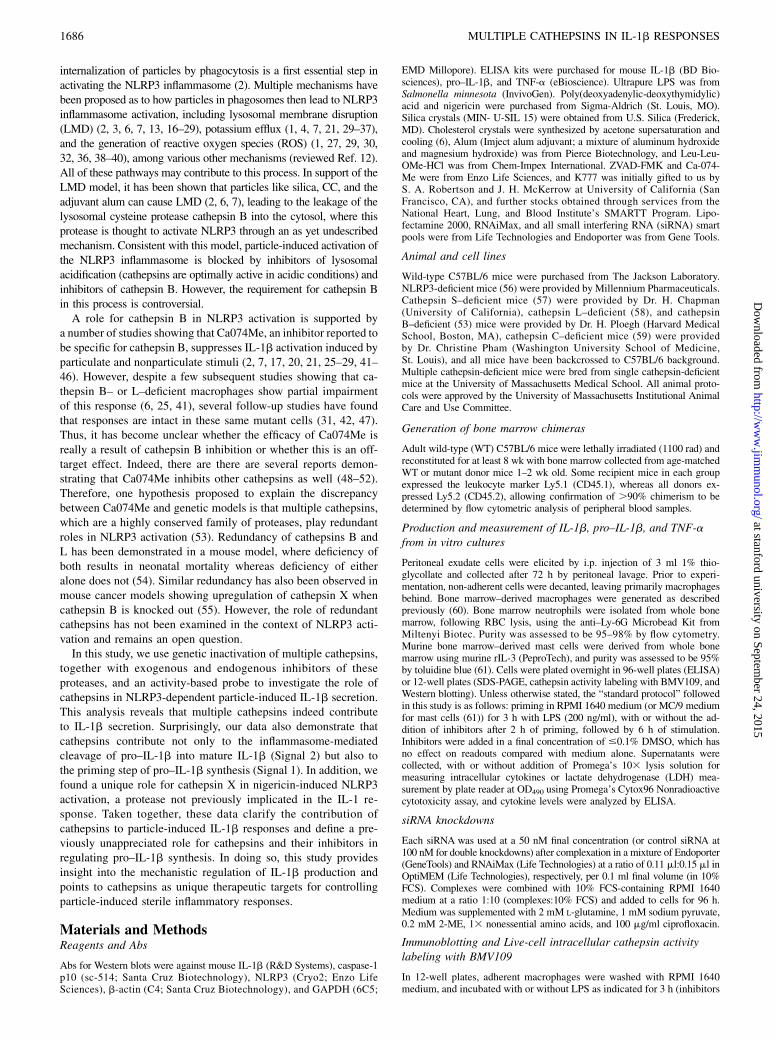
The role of cathepsins in NLRP3 activation remains controversial.Some studies describe a role for cathepsin B or L (6, 25, 41),whereas others show no role for either cathepsin in particle-induced NLRP3 activation and IL-1b secretion (31, 42, 47).One interpretation of these data suggests that other cathepsins,besides B or L, may be the key players in this response. Therefore,we examined the impact that genetic deficiency of five closelyrelated individual cathepsins has on particle-induced IL-1b se-cretion. Unless noted otherwise, IL-1b secretion was induced withvarious stimuli following 3 h of LPS priming. First, we examinedperitoneal macrophages (PMs) elicited from mice lacking cathe-psins B, L, S, or C. However, these cathepsin-deficient PMs dis-played no difference in IL-1b secretion in response to silicacompared with PMs derived from WT mice (Fig. 1A). To examinethe role of cathepsin X, we silenced cathepsin X in PMs by siRNAknockdown, and then, these cells were stimulated with silica, thesoluble NLRP3 activator nigericin, or the Absent in melanoma2 inflammasome activator poly(deoxyadenylic-deoxythymidylic)acid (dAdT) (Fig. 1B). We confirmed a 90–95% knockdown ofcathepsin X mRNA by quantitative PCR (qPCR) (Fig. 1C) andnoted a similar loss of cathepsin X activity using the fluorescentactivity–based probe BMV109 (Fig. 1D), which binds covalentlyto active cathepsins inside live cells (62). In lysates generatedfrom these cells, the proteins were separated by SDS-PAGE, andthen, the activity of specific cathepsins was assessed in the gelsusing a laser phosphor-imager to analyze the degree of fluores-cence for each cathepsin at the appropriate molecular mass.Again, we noted no significant difference in IL-1b secretion be-tween cathepsin X–sufficient and cathepsin X–deficient PMs inresponse to either silica or dAdT. Strikingly, cathepsin X defi-ciency significantly reduced IL-1b secretion in response tonigericin. In contrast, LPS-induced TNF-a secretion was unaf-fected by the loss of any of the cathepsins tested. Therefore, theindividual cathepsins B, L, S, C, and X are dispensable for silica-induced IL-1b secretion, but we found, unexpectedly, that in theresponse induced by nigericin, cathepsin X plays a nonredundantrole.Using cathepsin knockout animals to study IL-1b secretion
could potentially be confounded if some cathepsins are upregu-lated to compensate for the deficiency of others (54, 63, 64). Using
BMV109, we examined the activity of specific intracellularcathepsins in the LPS-primed WT and cathepsin-deficient PMsthat were tested above in Fig. 1A and 1B. Indeed, knockdown ofcathepsin X with siRNA resulted in an upregulation of cathepsin Land S activity (Fig. 1D). Moreover, PMs lacking cathepsin Lshowed increased cathepsin S activity, whereas those deficient incathepsin S upregulated cathepsin X activity (Fig. 1E). Takentogether, these data indicate that the cathepsins examined, in-cluding cathepsins B, L, S, and X, are not essential for particle-induced IL-1b secretion, and they cannot be readily studied usinggenetic methods because of compensation issues upon knock-down.
Analysis of small-molecule cathepsin inhibitors
The absence of a phenotype in cathepsin B–deficient macrophages,shown in this paper and reported by others, contradicts the resultsreported with cathepsin B inhibitors (31, 42, 47). Despite severalreports demonstrating that the cathepsin inhibitor Ca074Meinhibits multiple cathepsins in biochemical and cellular assays(48–52), Ca074Me is cited as a cathepsin B–specific inhibitor andused to implicate cathepsin B in NLRP3 activation in manystudies (2, 7, 17, 20, 21, 25–29, 41–46). The nonselective prodrugmethyl ester Ca074Me is processed in lysosomes into the highlycathepsin B–selective free acid Ca-074. However, this processingoccurs slowly and allows time for Ca074Me to inhibit multiple
FIGURE 1. Sterile particle-induced IL-1b secretion does not require
cathepsins B, L, C, S, or X, but nigericin is partially dependent on ca-
thepsin X. (A) LPS-primed PMs from WT mice or mice deficient for
cathepsins B (B2/2), L (L2/2), S (S2/2), or C (C2/2) were stimulated with
silica (40 mg/ml). (B) PMs were treated with nontargeting (NT) control
siRNA or siRNA targeting cathepsin X (siX) before priming with LPS and
stimulating with media control (2), silica (80 mg/ml), nigericin (1.5 mM),
or dAdT (0.5 mg/ml). IL-1b and TNF-a were measured in supernatants by
ELISA. (C) PMs from “B” were analyzed for cathepsin X (CtsX) ex-
pression by qPCR following siRNA (siX) treatment and LPS priming; data
are normalized to GAPDH expression and plotted relative to NT siRNA, or
(D) cathepsin X activity was probed with BMV109; lysates were processed
and pro–IL-1b and b-actin analyzed by Western blot; dashed box high-
lights upregulated cathepsin L/S activity; molecular mass markers are on
the right in kilodaltons. (E) LPS-primed PMs from WT or cathepsin-de-
ficient mice in (A) were probed for cathepsin activity with BMV109;
dashed boxes highlight upregulated cathepsin S or X activity in L2/2 and
S2/2 macrophages; molecular mass markers are on the right in kilodaltons.
Error bars represent range bars of technical duplicates (A), SD of technical
quadruplicates (B), and SD of technical triplicates (C). (B) All data are
representative of at least three independent experiments. Statistical anal-
ysis was performed by two-way ANOVA and Sidak’s multiple compar-
isons test, ****p , 0.0001.
The Journal of Immunology 1687
at stanford university on September 24, 2015
http://ww
w.jim
munol.org/
Dow
nloaded from
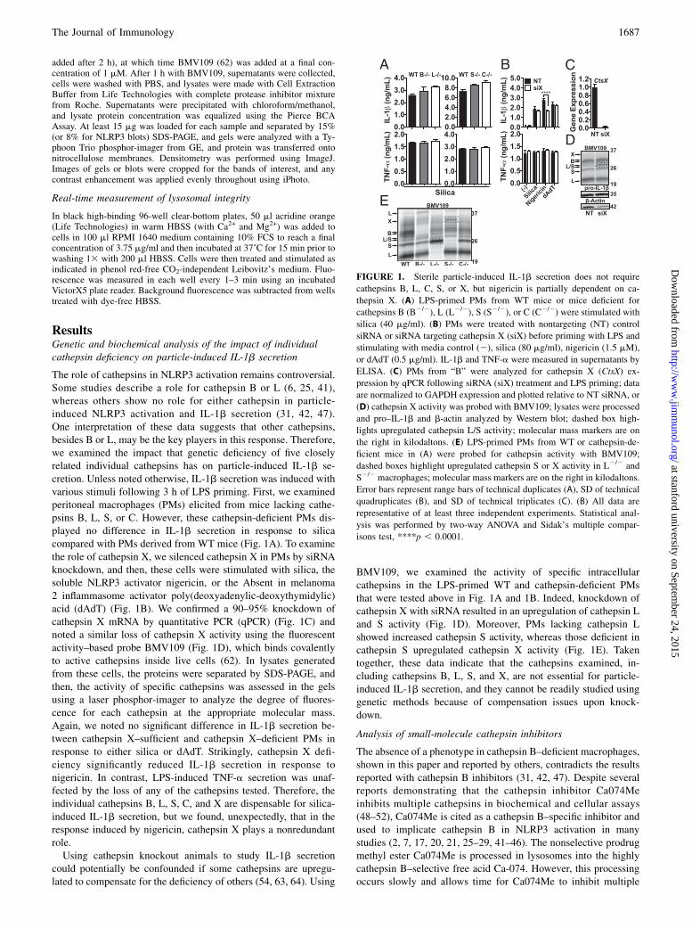
cathepsins (48–52). Therefore, in the context of NLRP3 acti-vation, Ca074Me’s targets in intact cells have not yet been verifiedand closely examined as a function of inhibitor concentration.In this study, we re-examine both Ca074Me and a newly de-scribed broad cathepsin inhibitor, K777 (N-methyl-piperazine-phenylalanyl-homophenylalanyl-vinylsulfone-phenyl), whose anti-inflammatory properties have not yet been tested. K777 inhibitscathepsins B, L, S, C, V, and K in cell-free assays (65). UsingCa074Me or K777 in combination with the active site probeallowed us to correlate their effects on IL-1b secretion with theextent of inhibition of specific cathepsins as a function of con-centration.To examine the inhibition profile of K777, we treated PMs with
K777 or solvent control (DMSO) for 1 h (after 2 h of LPS priming,unless stated otherwise), after which we probed for cathepsinactivity in the intact cells with BMV109. As previously reported,K777 inhibited cathepsins B, L, and S (65) over a titration rangefrom 0.1 to 30 mM (Fig. 2A, 2B). Interestingly, we also found thatK777 inhibited cathepsin X at high concentrations but unexpect-edly increased cathepsin X activity at lower concentrations. Theseparadoxical effects can be explained by K777’s greater potencytoward cathepsin S, which fits with our data, in Fig. 1E above,showing that cathepsin S deletion causes an increase in cathepsinX activity. Therefore, K777 inhibits cathepsin S at low concen-trations, which likely causes a compensatory increase in cathepsinX activity.In parallel to examining its effects on cathepsin activity, we also
tested the effect of K777 on IL-1b secretion (Fig. 2C). PMs wereprimed with LPS and treated with K777 as done above (2 h after
LPS priming and 1 h prior to stimulation), at which point theywere exposed to various stimuli for an additional 6 h of incuba-tion; this is the standard protocol used for the rest of this study,unless stated otherwise. At concentrations where multiple cathe-psins were inhibited, K777 suppressed silica-induced IL-1b se-cretion. In contrast to silica, K777 was much less effective atsuppressing IL-1b secretion induced by nigericin. Presumably,this is because K777 has opposing effects on cathepsin X, which isuniquely required for the nigericin response, shown in Fig. 1B.Moreover, K777 had a negligible affect on the IL-1b responseinduced by dAdT. We also confirmed that K777 is similarly se-lective and/or efficacious at suppressing IL-1b secretion inducedby other particles, including alum and CC, and in other primarymyeloid cell lines, including bone marrow–derived macrophages,mast cells, and neutrophils (Supplemental Fig. 1A, 1B). Impor-tantly, K777 did not affect LPS-induced TNF-a production withinthe tested concentration range, suggesting specific inhibition ofIL-1b secretion.We performed similar analyses for Ca074Me (Fig. 2D–F). Al-
though Ca074Me was selective for cathepsin B at concentrations, 1 mM, at higher concentrations (typically used in previousstudies) it inhibited cathepsins broadly (Fig. 2D, 2E). Moreover,.10 mM Ca074Me was required to completely inhibit cathepsinB. Unlike K777, Ca074Me suppressed nigericin and silica-induced IL-1b secretion with similar potency, presumably be-cause Ca074Me inhibits cathepsin X more potently than K777(Fig. 2F). Interestingly, the concentration required to achieve andmaximize these effects exceeds the range in which Ca074Me isselective for cathepsin B. In reviewing previous studies examining
FIGURE 2. Both Ca074Me and K777 inhibit multiple cathepsins at concentrations needed to block IL-1b secretion. (A) PMs were given media control
(No LPS) or LPS-primed (Plus LPS; +K777) and subsequently treated with media control (No LPS; Plus LPS) or the indicated concentrations of K777
(+K777), after which cathepsin activity was labeled with BMV109 in live cells before lysates were processed by SDS-PAGE and phosphor imaged;
molecular mass markers are on the right in kilodaltons. (B) Concentration-dependent inhibition of cathepsin activity by K777 analyzed by densitometry of
(A): cathepsin B (N) and X (s), large (L(l); N) and small (L(s);s) molecular mass isoforms of cathepsin L, cathepsin S (s), and overlapping molecular mass
isoforms of S and L (S/L; N). (C) LPS-primed PMs were treated with media control or the indicated concentrations of K777 and stimulated with silica
(40 mg/ml), nigericin (2 mM), or dAdT (0.5 mg/ml); data show percent inhibition of IL-1b secretion measured in supernatants compared with no inhibitor
treatment. (D–F) Same as (A–C) but with Ca074Me instead of K777. Error bars represent SE of means from three independent experiments (B), SD of
means from four independent experiments (0.1–15 mM) or S.D. of means from three independent experiments (30 mM) (C), range bars of the means from
two independent experiments (E), or SD of the means from three independent experiments (0.5–15 mM) (F), or range bars of the means from two in-
dependent experiments (30 mM).
1688 MULTIPLE CATHEPSINS IN IL-1b RESPONSES
at stanford university on September 24, 2015
http://ww
w.jim
munol.org/
Dow
nloaded from
Ca074Me’s effects on IL-1b responses, the concentrations usedwere also in the range that would inhibit multiple cathepsins (10–200 mM) (2, 7, 17, 20, 21, 25–29, 41–46). Therefore, our findingslikely explain the difference in results seen for the genetic lossof cathepsin B compared with small-molecule inhibitors of thisprotease. In summary, although both K777 and Ca074Me inhibitmultiple cathepsins at concentrations required to suppress IL-1bsecretion, K777 blocks particle-induced NLRP3 activation moreselectively than Ca074Me.To further investigate whether Ca074Me or K777 can inhibit IL-
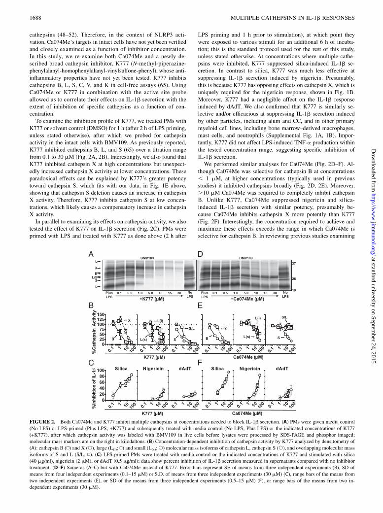
1b secretion in PMs from cathepsin-deficient mice, these cellswere LPS primed and treated with inhibitors prior to stimulation.Indeed, K777 inhibited IL-1b secretion to the same extent inWT cells as in cathepsins B, L, S, or C–deficient cells (Fig. 3A).Moreover, across a titration range for both K777 and Ca074Me,the extent to which they suppressed IL-1b secretion was the samein both WT and cathepsin B–deficient PMs (Fig. 3B). Again, LPS-induced TNF-a secretion was relatively unaffected by cathepsin Bdeficiency or inhibitor treatments. Taken together, these data in-dicate that the individual cathepsins examined, including cathep-sin B, are not essential for the activation of particle-induced IL-1bsecretion or as targets for cathepsin inhibitors that suppress thisresponse.
Analysis of compound cathepsin deficiencies
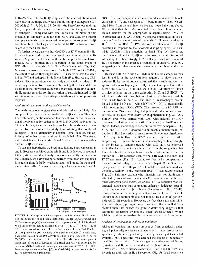
The analyses above suggest that multiple cathepsins likely playcompensatory roles in particle-induced IL-1b secretion. This is inline with some genetic evidence that has shown partial or condi-tional involvement for cathepsin B or L in NLRP3 activation (6,25, 41). In fact, these two cathepsins have been shown to com-pensate for one another in a study demonstrating that combinedcathepsin B and L deficiency is neonatal lethal in mice, but de-ficiency of either protease alone is nonlethal (54). Therefore,a dual deficiency of cathepsins B and L may have a greater effecton the IL-1b response (6).To test this hypothesis, we bred mice lacking both cathepsins B
and L. Because combined cathepsin B and L deficiency is neonatallethal (54), we could not analyze responses directly in these ani-mals. Instead, we harvested bone marrow from neonates and usedit to reconstitute lethally irradiated adult WT mice. In these chi-meric mice, cells of hematopoietic origin lack cathepsin B and L
(B&L2/2). For comparison, we made similar chimeras with WT,cathepsin B2/2, and cathepsin L2/2 bone marrow. Then, we eli-cited PMs from these chimeric mice and treated them as above.We verified that the PMs collected from these chimeric micelacked activity for the appropriate cathepsins using BMV109(Supplemental Fig. 2A). Again, we observed upregulation of ca-thepsin S activity upon loss of cathepsin L. However, cathepsinB2/2, L2/2, or B&L2/2 PMs showed no attenuation of IL-1bsecretion in response to the lysosome-disrupting agent Leu-Leu-OMe (LLOMe), silica, nigericin, or dAdT (Fig. 4A). Moreover,there was no defect in IL-1b secretion over a broad titration ofsilica (Fig. 4B). Interestingly, K777 still suppressed silica-inducedIL-1b secretion in the absence of cathepsins B and/or L (Fig. 4C),suggesting that other cathepsins potentially contribute to this re-sponse.Because both K777 and Ca074Me inhibit more cathepsins than
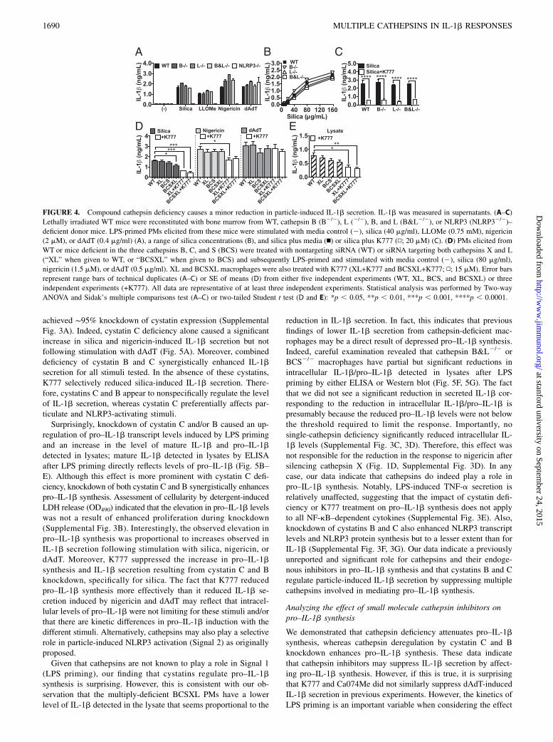
just B and L at the concentrations required to block particle-induced IL-1b secretion, we examined the particle-induced re-sponses of macrophages genetically deficient in up to five cathe-psins (Fig. 4D, 4E). To do this, we elicited PMs from WT miceor mice deficient in the three cathepsins B, C, and S (BCS2/2),which are viable with no obvious physical or behavioral pathol-ogy. In addition, in both WT and BCS2/2 macrophages, we si-lenced cathepsins X and L with siRNA (siXL; XL) or treated cellswith nontargeting siRNA (WT). This resulted in a 90–95% re-duction in mRNA of each targeted gene and reduction in enzymeactivity, as assayed with BMV109 (Supplemental Fig. 2B, 2C).Finally, PMs were primed with LPS, with medium or K777treatment, and stimulated with silica, nigericin, or dAdT as doneabove. Indeed, macrophages deficient in the five cathepsins B, C,S, X, and L (BCSXL) showed a significant, although small, re-duction in IL-1b secretion in response to silica but not nigericin ordAdT (Fig. 4D). However, K777 was still effective at furthersuppressing IL-1b secretion in these macrophages. Interestingly,in the lysates of samples treated with LPS only, we observeda similar decrease in intracellular IL-1b levels, suggesting thatlower levels of IL-1b synthesis may be contributing to the re-duction in IL-1b secretion seen for both BCSXL deficiency andK777 treatment (Fig. 4E). Again, we observed a compensatoryupregulation of cathepsin activity, with cathepsin B and S activityupregulated in the cathepsin XL knockdown and increased ca-thepsin X activity in the cathepsin BCS2/2 PMs (SupplementalFig. 2C). This may explain why nigericin was not significantlyaffected by knockdown of cathepsin X in combination with theseother cathepsin deficiencies. As above, TNF-a secretion was un-affected, suggesting that compound cathepsin deficiency specifi-cally impacts the IL-1b pathway (Supplemental Fig. 2D–H).Thus, compound deficiency of cathepsins B, C, S, X, and Ldemonstrates a reproducible, albeit minor, attenuation of particle-induced IL-1b secretion. However, the fact that cathepsin inhib-itors have shown, yet again, more profound effects on IL-1b se-cretion than that caused by genetic deficiency suggests thatadditional cathepsins or possibly other targets affected by theinhibitors might be involved in particle-induced IL-1b secretion.
Analysis of endogenous cathepsin inhibitors
Although technical limitations prevent us from genetically delet-ing all potentially relevant cathepsin activity, these proteases arespecifically inhibited by a family of endogenous regulators calledcystatins (66). Therefore, we examined the effects of geneticallydisabling the activity of the endogenous cathepsins inhibitors,cystatin C and B, on particle-induced IL-1b secretion.We used siRNA to silence cystatin C, B, or C and B in PMs to
investigate their role in IL-1b secretion (Fig. 5). In all cases, we
FIGURE 3. Cathepsin inhibitors suppress particle-induced IL-1b secre-
tion independently of individual cathepsins. IL-1b (upper graphs) and
TNF-a (lower graphs) were measured in supernatants. (A) LPS-primed
WT PMs or those lacking cathepsins B (B2/2), L (L2/2), S (S2/2), or
(C2/2) were treated with silica (n; 40 mg/ml) or silica plus K777 (N; 15 mM).
(B) LPS-primed WT (d, solid line) or cathepsin B–deficient (s, dashed line)
PMs were treated silica (50 mg/ml) or silica plus a range of K777 or
Ca074Me concentrations (1, 5, 10, 15, or 30 mM). Error bars represent
range bars of technical duplicates. Statistical analysis was performed by
two-way ANOVA and Sidak’s multiple comparisons test, ****p , 0.0001.
Data are representative of two [(B) for Ca074Me] or three [(A and B) for
K777] independent experiments.
The Journal of Immunology 1689
at stanford university on September 24, 2015
http://ww
w.jim
munol.org/
Dow
nloaded from
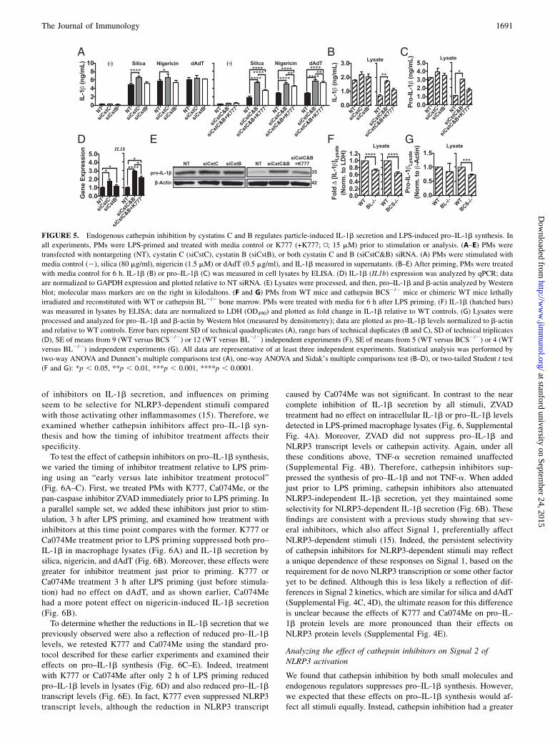
achieved ∼95% knockdown of cystatin expression (SupplementalFig. 3A). Indeed, cystatin C deficiency alone caused a significantincrease in silica and nigericin-induced IL-1b secretion but notfollowing stimulation with dAdT (Fig. 5A). Moreover, combineddeficiency of cystatin B and C synergistically enhanced IL-1bsecretion for all stimuli tested. In the absence of these cystatins,K777 selectively reduced silica-induced IL-1b secretion. There-fore, cystatins C and B appear to nonspecifically regulate the levelof IL-1b secretion, whereas cystatin C preferentially affects par-ticulate and NLRP3-activating stimuli.Surprisingly, knockdown of cystatin C and/or B caused an up-
regulation of pro–IL-1b transcript levels induced by LPS primingand an increase in the level of mature IL-1b and pro–IL-1bdetected in lysates; mature IL-1b detected in lysates by ELISAafter LPS priming directly reflects levels of pro–IL-1b (Fig. 5B–E). Although this effect is more prominent with cystatin C defi-ciency, knockdown of both cystatin C and B synergistically enhancespro–IL-1b synthesis. Assessment of cellularity by detergent-inducedLDH release (OD490) indicated that the elevation in pro–IL-1b levelswas not a result of enhanced proliferation during knockdown(Supplemental Fig. 3B). Interestingly, the observed elevation inpro–IL-1b synthesis was proportional to increases observed inIL-1b secretion following stimulation with silica, nigericin, ordAdT. Moreover, K777 suppressed the increase in pro–IL-1bsynthesis and IL-1b secretion resulting from cystatin C and Bknockdown, specifically for silica. The fact that K777 reducedpro–IL-1b synthesis more effectively than it reduced IL-1b se-cretion induced by nigericin and dAdT may reflect that intracel-lular levels of pro–IL-1b were not limiting for these stimuli and/orthat there are kinetic differences in pro–IL-1b induction with thedifferent stimuli. Alternatively, cathepsins may also play a selectiverole in particle-induced NLRP3 activation (Signal 2) as originallyproposed.Given that cathepsins are not known to play a role in Signal 1
(LPS priming), our finding that cystatins regulate pro–IL-1bsynthesis is surprising. However, this is consistent with our ob-servation that the multiply-deficient BCSXL PMs have a lowerlevel of IL-1b detected in the lysate that seems proportional to the
reduction in IL-1b secretion. In fact, this indicates that previousfindings of lower IL-1b secretion from cathepsin-deficient mac-rophages may be a direct result of depressed pro–IL-1b synthesis.Indeed, careful examination revealed that cathepsin B&L2/2 orBCS2/2 macrophages have partial but significant reductions inintracellular IL-1b/pro–IL-1b detected in lysates after LPSpriming by either ELISA or Western blot (Fig. 5F, 5G). The factthat we did not see a significant reduction in secreted IL-1b cor-responding to the reduction in intracellular IL-1b/pro–IL-1b ispresumably because the reduced pro–IL-1b levels were not belowthe threshold required to limit the response. Importantly, nosingle-cathepsin deficiency significantly reduced intracellular IL-1b levels (Supplemental Fig. 3C, 3D). Therefore, this effect wasnot responsible for the reduction in the response to nigericin aftersilencing cathepsin X (Fig. 1D, Supplemental Fig. 3D). In anycase, our data indicate that cathepsins do indeed play a role inpro–IL-1b synthesis. Notably, LPS-induced TNF-a secretion isrelatively unaffected, suggesting that the impact of cystatin defi-ciency or K777 treatment on pro–IL-1b synthesis does not applyto all NF-kB–dependent cytokines (Supplemental Fig. 3E). Also,knockdown of cystatins B and C also enhanced NLRP3 transcriptlevels and NLRP3 protein synthesis but to a lesser extent than forIL-1b (Supplemental Fig. 3F, 3G). Our data indicate a previouslyunreported and significant role for cathepsins and their endoge-nous inhibitors in pro–IL-1b synthesis and that cystatins B and Cregulate particle-induced IL-1b secretion by suppressing multiplecathepsins involved in mediating pro–IL-1b synthesis.
Analyzing the effect of small molecule cathepsin inhibitors onpro–IL-1b synthesis
We demonstrated that cathepsin deficiency attenuates pro–IL-1bsynthesis, whereas cathepsin deregulation by cystatin C and Bknockdown enhances pro–IL-1b synthesis. These data indicatethat cathepsin inhibitors may suppress IL-1b secretion by affect-ing pro–IL-1b synthesis. However, if this is true, it is surprisingthat K777 and Ca074Me did not similarly suppress dAdT-inducedIL-1b secretion in previous experiments. However, the kinetics ofLPS priming is an important variable when considering the effect
FIGURE 4. Compound cathepsin deficiency causes a minor reduction in particle-induced IL-1b secretion. IL-1b was measured in supernatants. (A–C)
Lethally irradiated WT mice were reconstituted with bone marrow from WT, cathepsin B (B2/2), L (2/2), B, and L (B&L2/2), or NLRP3 (NLRP32/2)–
deficient donor mice. LPS-primed PMs elicited from these mice were stimulated with media control (2), silica (40 mg/ml), LLOMe (0.75 mM), nigericin
(2 mM), or dAdT (0.4 mg/ml) (A), a range of silica concentrations (B), and silica plus media (n) or silica plus K777 (N; 20 mM) (C). (D) PMs elicited from
WT or mice deficient in the three cathepsins B, C, and S (BCS) were treated with nontargeting siRNA (WT) or siRNA targeting both cathepsins X and L
(“XL” when given to WT, or “BCSXL” when given to BCS) and subsequently LPS-primed and stimulated with media control (2), silica (80 mg/ml),
nigericin (1.5 mM), or dAdT (0.5 mg/ml). XL and BCSXL macrophages were also treated with K777 (XL+K777 and BCSXL+K777; N; 15 mM). Error bars
represent range bars of technical duplicates (A–C) or SE of means (D) from either five independent experiments (WT, XL, BCS, and BCSXL) or three
independent experiments (+K777). All data are representative of at least three independent experiments. Statistical analysis was performed by Two-way
ANOVA and Sidak’s multiple comparisons test (A–C) or two-tailed Student t test (D and E): *p , 0.05, **p , 0.01, ***p , 0.001, ****p , 0.0001.
1690 MULTIPLE CATHEPSINS IN IL-1b RESPONSES
at stanford university on September 24, 2015
http://ww
w.jim
munol.org/
Dow
nloaded from
of inhibitors on IL-1b secretion, and influences on primingseem to be selective for NLRP3-dependent stimuli comparedwith those activating other inflammasomes (15). Therefore, weexamined whether cathepsin inhibitors affect pro–IL-1b syn-thesis and how the timing of inhibitor treatment affects theirspecificity.To test the effect of cathepsin inhibitors on pro–IL-1b synthesis,
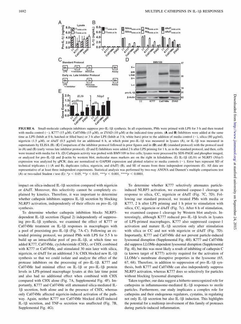
we varied the timing of inhibitor treatment relative to LPS prim-ing using an “early versus late inhibitor treatment protocol”(Fig. 6A–C). First, we treated PMs with K777, Ca074Me, or thepan-caspase inhibitor ZVAD immediately prior to LPS priming. Ina parallel sample set, we added these inhibitors just prior to stim-ulation, 3 h after LPS priming, and examined how treatment withinhibitors at this time point compares with the former. K777 orCa074Me treatment prior to LPS priming suppressed both pro–IL-1b in macrophage lysates (Fig. 6A) and IL-1b secretion bysilica, nigericin, and dAdT (Fig. 6B). Moreover, these effects weregreater for inhibitor treatment just prior to priming. K777 orCa074Me treatment 3 h after LPS priming (just before stimula-tion) had no effect on dAdT, and as shown earlier, Ca074Mehad a more potent effect on nigericin-induced IL-1b secretion(Fig. 6B).To determine whether the reductions in IL-1b secretion that we
previously observed were also a reflection of reduced pro–IL-1blevels, we retested K777 and Ca074Me using the standard pro-tocol described for these earlier experiments and examined theireffects on pro–IL-1b synthesis (Fig. 6C–E). Indeed, treatmentwith K777 or Ca074Me after only 2 h of LPS priming reducedpro–IL-1b levels in lysates (Fig. 6D) and also reduced pro–IL-1btranscript levels (Fig. 6E). In fact, K777 even suppressed NLRP3transcript levels, although the reduction in NLRP3 transcript
caused by Ca074Me was not significant. In contrast to the nearcomplete inhibition of IL-1b secretion by all stimuli, ZVADtreatment had no effect on intracellular IL-1b or pro–IL-1b levelsdetected in LPS-primed macrophage lysates (Fig. 6, SupplementalFig. 4A). Moreover, ZVAD did not suppress pro–IL-1b andNLRP3 transcript levels or cathepsin activity. Again, under allthese conditions above, TNF-a secretion remained unaffected(Supplemental Fig. 4B). Therefore, cathepsin inhibitors sup-pressed the synthesis of pro–IL-1b and not TNF-a. When addedjust prior to LPS priming, cathepsin inhibitors also attenuatedNLRP3-independent IL-1b secretion, yet they maintained someselectivity for NLRP3-dependent IL-1b secretion (Fig. 6B). Thesefindings are consistent with a previous study showing that sev-eral inhibitors, which also affect Signal 1, preferentially affectNLRP3-dependent stimuli (15). Indeed, the persistent selectivityof cathepsin inhibitors for NLRP3-dependent stimuli may reflecta unique dependence of these responses on Signal 1, based on therequirement for de novo NLRP3 transcription or some other factoryet to be defined. Although this is less likely a reflection of dif-ferences in Signal 2 kinetics, which are similar for silica and dAdT(Supplemental Fig. 4C, 4D), the ultimate reason for this differenceis unclear because the effects of K777 and Ca074Me on pro–IL-1b protein levels are more pronounced than their effects onNLRP3 protein levels (Supplemental Fig. 4E).
Analyzing the effect of cathepsin inhibitors on Signal 2 ofNLRP3 activation
We found that cathepsin inhibition by both small molecules andendogenous regulators suppresses pro–IL-1b synthesis. However,we expected that these effects on pro–IL-1b synthesis would af-fect all stimuli equally. Instead, cathepsin inhibition had a greater
FIGURE 5. Endogenous cathepsin inhibition by cystatins C and B regulates particle-induced IL-1b secretion and LPS-induced pro–IL-1b synthesis. In
all experiments, PMs were LPS-primed and treated with media control or K777 (+K777; N; 15 mM) prior to stimulation or analysis. (A–E) PMs were
transfected with nontargeting (NT), cystatin C (siCstC), cystatin B (siCstB), or both cystatin C and B (siCstC&B) siRNA. (A) PMs were stimulated with
media control (2), silica (80 mg/ml), nigericin (1.5 mM) or dAdT (0.5 mg/ml), and IL-1b measured in supernatants. (B–E) After priming, PMs were treated
with media control for 6 h. IL-1b (B) or pro–IL-1b (C) was measured in cell lysates by ELISA. (D) IL-1b (IL1b) expression was analyzed by qPCR; data
are normalized to GAPDH expression and plotted relative to NT siRNA. (E) Lysates were processed, and then, pro–IL-1b and b-actin analyzed by Western
blot; molecular mass markers are on the right in kilodaltons. (F and G) PMs from WT mice and cathepsin BCS2/2 mice or chimeric WT mice lethally
irradiated and reconstituted with WT or cathepsin BL2/2 bone marrow. PMs were treated with media for 6 h after LPS priming. (F) IL-1b (hatched bars)
was measured in lysates by ELISA; data are normalized to LDH (OD490) and plotted as fold change in IL-1b relative to WT controls. (G) Lysates were
processed and analyzed for pro–IL-1b and b-actin by Western blot (measured by densitometry); data are plotted as pro–IL-1b levels normalized to b-actin
and relative to WT controls. Error bars represent SD of technical quadruplicates (A), range bars of technical duplicates (B and C), SD of technical triplicates
(D), SE of means from 9 (WT versus BCS2/2) or 12 (WT versus BL2/2) independent experiments (F), SE of means from 5 (WT versus BCS2/2) or 4 (WT
versus BL2/2) independent experiments (G). All data are representative of at least three independent experiments. Statistical analysis was performed by
two-way ANOVA and Dunnett’s multiple comparisons test (A), one-way ANOVA and Sidak’s multiple comparisons test (B–D), or two-tailed Student t test
(F and G): *p , 0.05, **p , 0.01, ***p , 0.001, ****p , 0.0001.
The Journal of Immunology 1691
at stanford university on September 24, 2015
http://ww
w.jim
munol.org/
Dow
nloaded from
impact on silica-induced IL-1b secretion compared with nigericinor dAdT. Moreover, this selectivity cannot be completely ex-plained by kinetics. Therefore, it was important to determinewhether cathepsin inhibitors suppress IL-1b secretion by blockingNLRP3 activation, independently of their effects on pro–IL-1bsynthesis.To determine whether cathepsin inhibition blocks NLRP3-
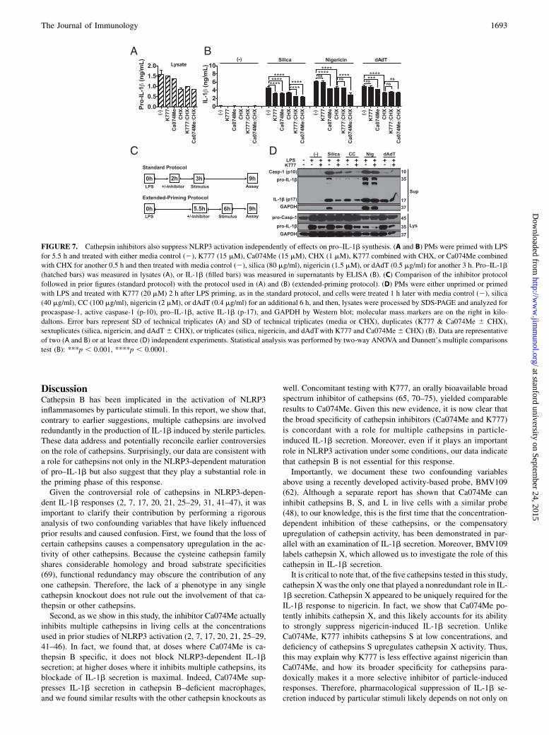
dependent IL-1b secretion (Signal 2) independently of suppress-ing pro–IL-1b synthesis, we examined the effect of K777 orCa074Me treatment on IL-1b responses in macrophages witha pool of preexisting pro–IL-1b (Fig. 7A–C). Following an ex-tended priming protocol, we primed PMs with LPS for 5.5 h tobuild up an intracellular pool of pro–IL-1b, at which time weadded K777, Ca074Me, cycloheximide (CHX), or CHX combinedwith K777 or Ca074Me, and stimulated 30 min later with silica,nigericin, or dAdT for an additional 3 h; CHX blocked new IL-1bsynthesis so that we could isolate and analyze the effect of theprotease inhibitors on the processing of pro–IL-1b. K777 andCa074Me had minimal effect on IL-1b or pro–IL-1b proteinlevels in LPS-primed macrophage lysates at this late time pointand also had no additional effect when combined with CHXcompared with CHX alone (Fig. 7A, Supplemental Fig. 4F). Im-portantly, K777 and Ca074Me still attenuated silica-mediated IL-1b secretion, both alone and in the presence of CHX, whereasonly Ca074Me affected nigericin-induced activation of the path-way. Again, neither K777 nor Ca074Me blocked dAdT-inducedIL-1b secretion, and TNF-a secretion was unaffected (Fig. 7B,Supplemental Fig. 4G).
To determine whether K777 selectively attenuates particle-induced NLRP3 activation, we examined caspase-1 cleavage inresponse to silica, CC, nigericin or dAdT (Fig. 7C, 7D). Fol-lowing our standard protocol, we treated PMs with media orK777, 2 h after LPS priming and 1 h prior to stimulation withsilica, CC, nigericin or dAdT (Fig. 7c). After 6 h of stimulation,we examined caspase-1 cleavage by Western blot analysis. In-terestingly, although K777 reduced pro–IL-1b levels in lysatesof LPS-primed macrophages, K777 also suppressed caspase-1activation and mature IL-1b secretion only after stimulationwith silica or CC and not with nigericin or dAdT (Fig. 7D).Importantly, K777 and Ca074Me did not prevent particle-inducedlysosomal disruption (Supplemental Fig. 4H). K777 and Ca074Medid suppress LLOMe-dependent lysosomal disruption (SupplementalFig. 4I), but this was most likely a result of inhibiting of cathepsin C(a known target of K777) activity required for the activation ofLLOMe’s membrane disruptive properties in the lysosome (65,67, 68). Therefore, in addition to suppression of pro–IL-1b syn-thesis, both K777 and Ca074Me can also independently suppressNLRP3 activation, whereas K777 does so selectively for particleswithout blocking lysosomal disruption.Taken together, our data suggest a hitherto unrecognized role for
cathepsins in inflammasome-mediated IL-1b responses to sterileparticles. Furthermore, our study implicates a complex role forcathepsins and their endogenous regulators, cystatins, in regulatingnot only IL-1b secretion but also IL-1b induction. This highlightsthe potential for a multistep involvement of this family of proteasesduring particle-induced inflammation.
FIGURE 6. Small-molecule cathepsin inhibitors suppress pro–IL-1b synthesis. In all experiments, PMs were primed with LPS for 3 h and then treated
with media control (2), K777 (15 mM), Ca074Me (15 mM), or ZVAD (10 mM) at the indicated time points. (A and B) Inhibitors were added at the same
time as LPS (Inhib at 0 h; hatched or filled bars) or 3 h after LPS (Inhib at 3 h; white bars) prior to the addition of media control (2), silica (80 mg/ml),
nigericin (1.5 mM), or dAdT (0.5 mg/ml) for an additional 6 h, at which point pro–IL-1b was measured in lysates (A), or IL-1b was measured in
supernatants by ELISA (B). (C) Comparison of the inhibitor protocol followed in prior figures and in (D) and (E) (standard protocol) with the protocol used
in (A) and (B) (early versus late inhibitor protocol). (D and E) Inhibitors were added 2 h after LPS priming for 1 h, as in the standard protocol, and then, cells
were treated with media for 4 h. (D) Cathepsin activity was probed with BMV109 in live cells; lysates were processed by SDS-PAGE and phosphor imaged,
or analyzed for pro–IL-1b and b-actin by western blot; molecular mass markers are on the right in kilodaltons. (E) IL-1b (IL1b) or NLRP3 (Nlrp3)
expression was analyzed by qPCR; data are normalized to GAPDH expression and plotted relative to media controls (2). Error bars represent SD of
technical triplicates (-) (A and B), duplicates (silica, nigericin, and dAdT) (B), and SE of means from three independent experiments (E). All data are
representative of at least three independent experiments. Statistical analysis was performed by two-way ANOVA and Dunnett’s multiple comparisons test
(A) or two-tailed Student t test (E): *p , 0.05, **p , 0.01, ***p , 0.001, ****p , 0.0001.
1692 MULTIPLE CATHEPSINS IN IL-1b RESPONSES
at stanford university on September 24, 2015
http://ww
w.jim
munol.org/
Dow
nloaded from
DiscussionCathepsin B has been implicated in the activation of NLRP3inflammasomes by particulate stimuli. In this report, we show that,contrary to earlier suggestions, multiple cathepsins are involvedredundantly in the production of IL-1b induced by sterile particles.These data address and potentially reconcile earlier controversieson the role of cathepsins. Surprisingly, our data are consistent witha role for cathepsins not only in the NLRP3-dependent maturationof pro–IL-1b but also suggest that they play a substantial role inthe priming phase of this response.Given the controversial role of cathepsins in NLRP3-depen-
dent IL-1b responses (2, 7, 17, 20, 21, 25–29, 31, 41–47), it wasimportant to clarify their contribution by performing a rigorousanalysis of two confounding variables that have likely influencedprior results and caused confusion. First, we found that the loss ofcertain cathepsins causes a compensatory upregulation in the ac-tivity of other cathepsins. Because the cysteine cathepsin familyshares considerable homology and broad substrate specificities(69), functional redundancy may obscure the contribution of anyone cathepsin. Therefore, the lack of a phenotype in any singlecathepsin knockout does not rule out the involvement of that ca-thepsin or other cathepsins.Second, as we show in this study, the inhibitor Ca074Me actually
inhibits multiple cathepsins in living cells at the concentrationsused in prior studies of NLRP3 activation (2, 7, 17, 20, 21, 25–29,41–46). In fact, we found that, at doses where Ca074Me is ca-thepsin B specific, it does not block NLRP3-dependent IL-1bsecretion; at higher doses where it inhibits multiple cathepsins, itsblockade of IL-1b secretion is maximal. Indeed, Ca074Me sup-presses IL-1b secretion in cathepsin B–deficient macrophages,and we found similar results with the other cathepsin knockouts as
well. Concomitant testing with K777, an orally bioavailable broadspectrum inhibitor of cathepsins (65, 70–75), yielded comparable
results to Ca074Me. Given this new evidence, it is now clear that
the broad specificity of cathepsin inhibitors (Ca074Me and K777)
is concordant with a role for multiple cathepsins in particle-
induced IL-1b secretion. Moreover, even if it plays an important
role in NLRP3 activation under some conditions, our data indicate
that cathepsin B is not essential for this response.Importantly, we document these two confounding variables
above using a recently developed activity-based probe, BMV109(62). Although a separate report has shown that Ca074Me caninhibit cathepsins B, S, and L in live cells with a similar probe(48), to our knowledge, this is the first time that the concentration-dependent inhibition of these cathepsins, or the compensatoryupregulation of cathepsin activity, has been demonstrated in par-allel with an examination of IL-1b secretion. Moreover, BMV109labels cathepsin X, which allowed us to investigate the role of thiscathepsin in IL-1b secretion.It is critical to note that, of the five cathepsins tested in this study,
cathepsin X was the only one that played a nonredundant role in IL-1b secretion. Cathepsin X appeared to be uniquely required for theIL-1b response to nigericin. In fact, we show that Ca074Me po-tently inhibits cathepsin X, and this likely accounts for its abilityto strongly suppress nigericin-induced IL-1b secretion. UnlikeCa074Me, K777 inhibits cathepsins S at low concentrations, anddeficiency of cathepsins S upregulates cathepsin X activity. Thus,this may explain why K777 is less effective against nigericin thanCa074Me, and how its broader specificity for cathepsins para-doxically makes it a more selective inhibitor of particle-inducedresponses. Therefore, pharmacological suppression of IL-1b se-cretion induced by particular stimuli likely depends on not only on
FIGURE 7. Cathepsin inhibitors also suppress NLRP3 activation independently of effects on pro–IL-1b synthesis. (A and B) PMs were primed with LPS
for 5.5 h and treated with either media control (2), K777 (15 mM), Ca074Me (15 mM), CHX (1 mM), K777 combined with CHX, or Ca074Me combined
with CHX for another 0.5 h and then treated with media control (2), silica (80 mg/ml), nigericin (1.5 mM), or dAdT (0.5 mg/ml) for another 3 h. Pro–IL-1b
(hatched bars) was measured in lysates (A), or IL-1b (filled bars) was measured in supernatants by ELISA (B). (C) Comparison of the inhibitor protocol
followed in prior figures (standard protocol) with the protocol used in (A) and (B) (extended-priming protocol). (D) PMs were either unprimed or primed
with LPS and treated with K777 (20 mM) 2 h after LPS priming, as in the standard protocol, and cells were treated 1 h later with media control (2), silica
(40 mg/ml), CC (100 mg/ml), nigericin (2 mM), or dAdT (0.4 mg/ml) for an additional 6 h, and then, lysates were processed by SDS-PAGE and analyzed for
procaspase-1, active caspase-1 (p-10), pro–IL-1b, active IL-1b (p-17), and GAPDH by Western blot; molecular mass markers are on the right in kilo-
daltons. Error bars represent SD of technical triplicates (A) and SD of technical triplicates (media or CHX), duplicates (K777 & Ca074Me 6 CHX),
sextuplicates (silica, nigericin, and dAdT 6 CHX), or triplicates (silica, nigericin, and dAdTwith K777 and Ca074Me6 CHX) (B). Data are representative
of two (A and B) or at least three (D) independent experiments. Statistical analysis was performed by two-way ANOVA and Dunnett’s multiple comparisons
test (B): ***p , 0.001, ****p , 0.0001.
The Journal of Immunology 1693
at stanford university on September 24, 2015
http://ww
w.jim
munol.org/
Dow
nloaded from
how many but which cathepsins are inhibited and at what con-centrations.Although Ca074Me and K777 could have noncathepsin off-
target effects responsible for their suppression of particle-inducedIL-1b secretion, we strongly favor the interpretation that they areachieving this effect by inhibiting multiple functionally redundantcathepsins. Although we observed a minor but insignificant re-duction of particle-induced IL-1b secretion in the cathepsinBL2/2 PMs, and a small but significant reduction in the pentuplecathepsin BCSXL2/2 PMs, we believe that the residual cathepsinactivity in these cells, as shown by BMV109 labeling, could besufficient to mediate NLRP3 activation. In fact, a recent studydemonstrated that inflammasome activation is an “all-or-none”response (76), which gives credence to earlier proposals thatonly a few molecules of active cathepsins may be sufficient toreach a minimum threshold for inflammasome activation (47).Whether this is true or not remains to be demonstrated. However,we did find more robust genetic evidence supporting an unex-pected role for cathepsins in regulating the priming phase of IL-1bsecretion.Because we could not genetically suppress cathepsin activity to
the same extent as inhibitors, which further reduced IL-1b se-cretion by these genetically deficient cells, we adopted an alter-native strategy. Instead of examining cathepsin deficiency, weevaluated the effect of cathepsin deregulation by silencing twobroadly active endogenous cathepsin inhibitors, cystatins C and B.Like the cathepsin family (69), the cystatin family is large (66), asmight be expected of regulators of a large family of proteases.Moreover, individual cystatins specifically regulate multiple cys-teine cathepsin proteases, including B, L, and S (66). Indeed,knockdown of cystatins C and B synergistically enhanced IL-1bsecretion but did so for all stimuli tested. Further analysesrevealed that the increase in IL-1b secretion we observed wasdirectly proportional to the upregulation of pro–IL-1b transcriptand synthesis. In fact, reexamination of the compound cathepsinknockouts (BL2/2 and BCS2/2) also showed that multiple re-dundant cathepsins play a partial but significant role in LPS-induced pro–IL-1b synthesis. As far as we know, these findingsare among the first to implicate and clarify the role of endogenouscathepsin inhibitors, cystatins, in regulating IL-1b responses.Although an association between cystatins and inflammation has
been widely reported, the mechanism underlying this associationhas not been established. Given this context, our evidence that bothcystatin B and especially cystatin C play a role in the IL-1b re-sponse is enlightening. In fact, lower serum levels of cystatin C,considered the “dominant” cystatin (77), are associated with nu-merous inflammatory conditions (66), including sterile inflam-matory arterial disease (78). Furthermore, cystatin B deficiency inmice exacerbated LPS-induced sepsis and elevated IL-1b levels inthe serum (79). This latter study demonstrated higher caspase-1and/or caspase-11 activity and mitochondrial ROS, suggesting thatloss of cystatin B increased inflammasome activation (79). How-ever, the authors noted that there were no signs of LMD or ele-vated cathepsin activity in the cytosol, and effects on pro-IL-1bwere not measured. Thus, our data demonstrating that cystatindeficiency increases pro–IL-1b and NLRP3 synthesis offer a dif-ferent perspective that may help to explain these results. In thiscontext, it is interesting that other studies have shown that cystatinB interacts with cathepsin L in the nucleus (80) and that cathepsinL can play a role in NF-kB activation (81). Moreover, cystatinB–deficient macrophages have lower IL-10 expression (82), andIL-10 transcriptionally downregulates IL-1b synthesis (83).While unexpected, our data with cystatins shed further light on
the mechanism by which small molecule cathepsin inhibitors may
impact IL-1b secretion by modulating pro-IL-1b synthesis. In-deed, we directly demonstrated that exogenous cathepsin inhib-itors also suppress LPS-induced pro-IL-1b synthesis, and thatthis effect contributes substantially to their suppression of IL-1bsecretion by inflammasome-activating particulates and non-particulates. Importantly, K777 and Ca074Me reduce pro-IL-1bsynthesis in response to LPS priming alone, prior to any IL-1bbeing secreted, and they do not affect TNF-a secretion. Thus, it isunlikely that inhibitors are reducing the autocrine-like priming ofpro-IL-1b synthesis simply by suppressing TNF-a or IL-1b se-cretion upon stimulation. Together, these findings reiterate theimportance of examining both Signal 1 and Signal 2 when inter-preting inflammasome studies. In fact, a recent paper emphasizedthis point by demonstrating that several ROS inhibitors thought tosuppress NLRP3 activation actually affect Signal 1 (15). We alsofind that the timing of inhibitor treatment relative to LPS primingcan confirm this phenomenon. If inhibitors are added earlier withrespect to LPS priming, effects on priming become more pro-nounced and less NLRP3-specific. In some contexts, this mayactually be a therapeutically advantageous characteristic.Our findings are consistent with a prior study demonstrating
that a cathepsin B inhibitor, Z-FA-fmk, suppresses LPS signaling(84). Finding discordant results with cathepsin B-deficient cells,the authors suggested this was a non-cathepsin off-target effect.Similarly, we cannot completely exclude the possibility that thevarious exogenous and endogenous cathepsin inhibitors are re-ducing IL-1b responses through off-target effects. However, givenour results, it is likely that redundant cathepsins compensated forthe loss of cathepsin B, and even more likely that Z-FA-fmk isnon-specific for cathepsin B. Moreover, since we observed con-cordant results with two chemically distinct cathepsin inhibitors,Ca074Me and K777, as well as the endogenous cathepsin inhib-itors, we favor the idea that the common effect of these inhibitorson pro-IL-1b synthesis is attributable to their common cathepsintargets.Importantly, Ca074Me and K777 were consistently more ef-
fective against NLRP3-mediated IL-1b secretion compared withthat mediated by Absent in melanoma 2 via dAdT, and the effectsof cystatin deficiencies were similarly biased. Therefore, it ap-peared that cathepsins may indeed have a role in mediatingstimulus-specific/priming-independent NLRP3 activation. Althoughthis is one interpretation, others would predict that NLRP3-mediated IL-1b secretion is particularly sensitive to the levels ofpro–IL-1b or that the levels of NLRP3 itself are significantlyimpacted by inhibitor treatment. Given the importance of LPSpriming kinetics, deducing priming-independent effects on IL-1bsecretion can be achieved via prolonged priming and/or con-comitant inhibition of protein synthesis. Indeed, by inhibitingfurther pro–IL-1b synthesis with CHX following a prolongedperiod of LPS priming, we showed that subsequent treatment withK777 and Ca074Me affects Signal 2, independently of Signal 1.This is consistent with our finding that cathepsin inhibition sup-pressed inflammasome activation, as assayed by examining cleav-age of caspase-1, indicating that cathepsins may also play a rolein NLRP3 activation, as originally proposed. Importantly, ourdata showed that cathepsins are not necessary for particle-inducedlysosome disruption, although this has been suggested previously(42, 85). Recently, cathepsins have also been implicated ininducing particle-stimulated K+ efflux (K+ efflux is thought tobe an absolute requirement for NLRP3 activation) (37) in LPS-primed macrophages. Whether cathepsins affect K+ efflux byinfluencing a K+ channel or the integrity plasma membrane (sec-ondary to inducing LMD-dependent cell death) (42, 48, 85) is notyet clear.
1694 MULTIPLE CATHEPSINS IN IL-1b RESPONSES
at stanford university on September 24, 2015
http://ww
w.jim
munol.org/
Dow
nloaded from
Whether cathepsins play a role in Signal 1 or Signal 2, it is likelythat the proteolytic activity of cathepsins is necessary, given theefficacy of inhibitors; if true, the substrate involved remains to beelucidated. Importantly, both TLR4 and NLRP3, which sequen-tially mediate the priming and activation of IL-1b secretion, re-spectively, have large leucine-rich repeats (LRRs). It is presumedthe LRRs act as autoinhibitory motifs that block activation untilinduction of structural changes or ligand binding. In fact, ca-thepsin inhibitors have been used to demonstrate that cleavage ofthe LRRs for TLRs 3, 7, and 9 is necessary for optimal activation(86, 87). Moreover, it has also been shown that NLRP1 activationcan be directly mediated by proteolytic cleavage of its LRR (88,89) and that expression of a transgenic NLRP3 protein lacking anLRR motif makes it constitutively active (90). Although this isstill all speculation, LRR-targeted cleavage of TLR4 and NLRP3by cathepsins remains an intriguing possibility that might explainour findings.Taken together, this study identifies a previously unappreciated
role for cathepsins and cystatins in the regulation of pro–IL-1bsynthesis (as well as IL-1b secretion) and provides compellingevidence that cathepsins play redundant and compensatory rolesin these processes. Furthermore, we have reconfirmed thatCa074Me inhibits multiple cathepsins and demonstrate conclu-sively that cathepsin B is not the sole target of this agent thatmediates its effect on IL-1b secretion. Moreover, we identifiedcathepsin X as a previously unappreciated player in nigericin-induced NLRP3 activation and raised important questions as tothe relative importance of cathepsins in mediating Signal 1 andSignal 2 during particle-induced NLRP3 activation and IL-1bsecretion. Finally, we have characterized a cathepsin inhibi-tor, K777, which selectively reduces particle-induced IL-1bresponses and possesses pharmacological properties warrant-ing its investigation as a potential anti-inflammatory thera-peutic (65, 70–75). Indeed, cathepsins are tractable targets forthe development of small-molecule inhibitors. Our data predictthat inhibitors that broadly inhibit cathepsins, like K777, mighthave potential as therapeutic inhibitors of particle-induced sterileinflammation.
AcknowledgmentsWe thank Havisha Karnam from the Brown and Khvorova laboratories
as well as Myriam Aouadi from the Czech laboratory at the University of
Massachusetts Medical School for help in the optimization of siRNA
knockdowns, as well as Kate Fitzgerald, Douglas Golenbock, and Eicke
Latz for advice and reagents.
DisclosuresThe authors have no financial conflicts of interest.
References1. Dostert, C., V. Petrilli, R. Van Bruggen, C. Steele, B. T. Mossman, and
J. Tschopp. 2008. Innate immune activation through Nalp3 inflammasomesensing of asbestos and silica. Science 320: 674–677.
2. Hornung, V., F. Bauernfeind, A. Halle, E. O. Samstad, H. Kono, K. L. Rock,K. A. Fitzgerald, and E. Latz. 2008. Silica crystals and aluminum salts activatethe NALP3 inflammasome through phagosomal destabilization. Nat. Immunol. 9:847–856.
3. Halle, A., V. Hornung, G. C. Petzold, C. R. Stewart, B. G. Monks, T. Reinheckel,K. A. Fitzgerald, E. Latz, K. J. Moore, and D. T. Golenbock. 2008. The NALP3inflammasome is involved in the innate immune response to amyloid-b. Nat.Immunol. 9: 857–865.
4. Cassel, S. L., S. C. Eisenbarth, S. S. Iyer, J. J. Sadler, O. R. Colegio,L. A. Tephly, A. B. Carter, P. B. Rothman, R. A. Flavell, and F. S. Sutterwala.2008. The Nalp3 inflammasome is essential for the development of silicosis.Proc. Natl. Acad. Sci. USA 105: 9035–9040.
5. Martinon, F., V. Petrilli, A. Mayor, A. Tardivel, and J. Tschopp. 2006. Gout-associated uric acid crystals activate the NALP3 inflammasome. Nature 440:237–241.
6. Duewell, P., H. Kono, K. J. Rayner, C. M. Sirois, G. Vladimer,F. G. Bauernfeind, G. S. Abela, L. Franchi, G. Nunez, M. Schnurr, et al. 2010.NLRP3 inflammasomes are required for atherogenesis and activated by cho-lesterol crystals. Nature 464: 1357–1361.
7. Rajamaki, K., J. Lappalainen, K. Oorni, E. Valimaki, S. Matikainen,P. T. Kovanen, and K. K. Eklund. 2010. Cholesterol crystals activate the NLRP3inflammasome in human macrophages: a novel link between cholesterol me-tabolism and inflammation. PLoS One 5: e11765.
8. Kono, H., G. M. Orlowski, Z. Patel, and K. L. Rock. 2012. The IL-1‑dependentsterile inflammatory response has a substantial caspase-1‑independent compo-nent that requires cathepsin C. J. Immunol. 189: 3734–3740.
9. Kono, H., D. Karmarkar, Y. Iwakura, and K. L. Rock. 2010. Identification of thecellular sensor that stimulates the inflammatory response to sterile cell death. J.Immunol. 184: 4470–4478.
10. Dinarello, C. A. 2011. Interleukin-1 in the pathogenesis and treatment of in-flammatory diseases. Blood 117: 3720–3732.
11. Fettelschoss, A., M. Kistowska, S. LeibundGut-Landmann, H. D. Beer,P. Johansen, G. Senti, E. Contassot, M. F. Bachmann, L. E. French, A. Oxenius,and T. M. K€undig. 2011. Inflammasome activation and IL-1b target IL-1a forsecretion as opposed to surface expression. Proc. Natl. Acad. Sci. USA 108:18055–18060.
12. Sutterwala, F. S., S. Haasken, and S. L. Cassel. 2014. Mechanism of NLRP3inflammasome activation. Ann. N. Y. Acad. Sci. 1319: 82–95.
13. Hornung, V., and E. Latz. 2010. Critical functions of priming and lysosomaldamage for NLRP3 activation. Eur. J. Immunol. 40: 620–623.
14. Bauernfeind, F. G., G. Horvath, A. Stutz, E. S. Alnemri, K. MacDonald,D. Speert, T. Fernandes-Alnemri, J. Wu, B. G. Monks, K. A. Fitzgerald, et al.2009. Cutting edge: NF-kB activating pattern recognition and cytokine receptorslicense NLRP3 inflammasome activation by regulating NLRP3 expression. J.Immunol. 183: 787–791.
15. Bauernfeind, F., E. Bartok, A. Rieger, L. Franchi, G. Nunez, and V. Hornung.2011. Cutting edge: reactive oxygen species inhibitors block priming, but notactivation, of the NLRP3 inflammasome. J. Immunol. 187: 613–617.
16. Lopez-Castejon, G., J. Theaker, P. Pelegrin, A. D. Clifton, M. Braddock, andA. Surprenant. 2010. P2X7 receptor-mediated release of cathepsins from mac-rophages is a cytokine-independent mechanism potentially involved in jointdiseases. J. Immunol. 185: 2611–2619.
17. Hentze, H., X. Y. Lin, M. S. Choi, and A. G. Porter. 2003. Critical role forcathepsin B in mediating caspase-1‑dependent interleukin-18 maturation andcaspase-1‑independent necrosis triggered by the microbial toxin nigericin. CellDeath Differ. 10: 956–968.
18. Fujisawa, A., N. Kambe, M. Saito, R. Nishikomori, H. Tanizaki, N. Kanazawa,S. Adachi, T. Heike, J. Sagara, T. Suda, et al. 2007. Disease-associated mutationsin CIAS1 induce cathepsin B‑dependent rapid cell death of human THP-1monocytic cells. Blood 109: 2903–2911.
19. Morishige, T., Y. Yoshioka, A. Tanabe, X. Yao, S. Tsunoda, Y. Tsutsumi,Y. Mukai, N. Okada, and S. Nakagawa. 2010. Titanium dioxide induces differentlevels of IL-1b production dependent on its particle characteristics throughcaspase-1 activation mediated by reactive oxygen species and cathepsin B.Biochem. Biophys. Res. Commun. 392: 160–165.
20. Duncan, J. A., X. Gao, M. T. Huang, B. P. O’Connor, C. E. Thomas,S. B. Willingham, D. T. Bergstralh, G. A. Jarvis, P. F. Sparling, and J. P. Ting.2009. Neisseria gonorrhoeae activates the proteinase cathepsin B to mediate thesignaling activities of the NLRP3 and ASC-containing inflammasome. J.Immunol. 182: 6460–6469.
21. Chu, J., L. M. Thomas, S. C. Watkins, L. Franchi, G. Nunez, and R. D. Salter.2009. Cholesterol-dependent cytolysins induce rapid release of mature IL-1bfrom murine macrophages in a NLRP3 inflammasome and cathepsin B‑de-pendent manner. J. Leukoc. Biol. 86: 1227–1238.
22. Barlan, A. U., P. Danthi, and C. M. Wiethoff. 2011. Lysosomal localization andmechanism of membrane penetration influence nonenveloped virus activation ofthe NLRP3 inflammasome. Virology 412: 306–314.
23. Bauer, C., P. Duewell, C. Mayer, H. A. Lehr, K. A. Fitzgerald, M. Dauer,J. Tschopp, S. Endres, E. Latz, and M. Schnurr. 2010. Colitis induced in micewith dextran sulfate sodium (DSS) is mediated by the NLRP3 inflammasome.Gut 59: 1192–1199.
24. Muruve, D. A., V. Petrilli, A. K. Zaiss, L. R. White, S. A. Clark, P. J. Ross,R. J. Parks, and J. Tschopp. 2008. The inflammasome recognizes cytosolicmicrobial and host DNA and triggers an innate immune response. Nature 452:103–107.
25. Terada, K., J. Yamada, Y. Hayashi, Z. Wu, Y. Uchiyama, C. Peters, andH. Nakanishi. 2010. Involvement of cathepsin B in the processing and se-cretion of interleukin-1b in chromogranin A‑stimulated microglia. Glia 58:114–124.
26. Meixenberger, K., F. Pache, J. Eitel, B. Schmeck, S. Hippenstiel, H. Slevogt,P. N’Guessan, M. Witzenrath, M. G. Netea, T. Chakraborty, et al. 2010. Listeriamonocytogenes‑infected human peripheral blood mononuclear cells produce IL-1b, depending on listeriolysin O and NLRP3. J. Immunol. 184: 922–930.
27. Barlan, A. U., T. M. Griffin, K. A. McGuire, and C. M. Wiethoff. 2011. Ade-novirus membrane penetration activates the NLRP3 inflammasome. J. Virol. 85:146–155.
28. Rintahaka, J., N. Lietzen, T. Ohman, T. A. Nyman, and S. Matikainen. 2011.Recognition of cytoplasmic RNA results in cathepsin-dependent inflammasomeactivation and apoptosis in human macrophages. J. Immunol. 186: 3085–3092.
29. Kankkunen, P., L. Teirila, J. Rintahaka, H. Alenius, H. Wolff, and S. Matikainen.2010. (1,3)-b-Glucans activate both dectin-1 and NLRP3 inflammasome in hu-man macrophages. J. Immunol. 184: 6335–6342.
The Journal of Immunology 1695
at stanford university on September 24, 2015
http://ww
w.jim
munol.org/
Dow
nloaded from
30. Saıd-Sadier, N., E. Padilla, G. Langsley, and D. M. Ojcius. 2010. Aspergillusfumigatus stimulates the NLRP3 inflammasome through a pathway requiringROS production and the Syk tyrosine kinase. PLoS One 5: e10008.
31. Dostert, C., G. Guarda, J. F. Romero, P. Menu, O. Gross, A. Tardivel, M. L. Suva,J. C. Stehle, M. Kopf, I. Stamenkovic, et al. 2009. Malarial hemozoin is a Nalp3inflammasome activating danger signal. PLoS One 4: e6510.
32. Gross, O., H. Poeck, M. Bscheider, C. Dostert, N. Hannesschlager, S. Endres,G. Hartmann, A. Tardivel, E. Schweighoffer, V. Tybulewicz, et al. 2009. Sykkinase signalling couples to the Nlrp3 inflammasome for anti-fungal host de-fence. Nature 459: 433–436.
33. Eisenbarth, S. C., O. R. Colegio, W. O’Connor, F. S. Sutterwala, andR. A. Flavell. 2008. Crucial role for the Nalp3 inflammasome in the immu-nostimulatory properties of aluminium adjuvants. Nature 453: 1122–1126.
34. Petrilli, V., S. Papin, C. Dostert, A. Mayor, F. Martinon, and J. Tschopp. 2007.Activation of the NALP3 inflammasome is triggered by low intracellular po-tassium concentration. Cell Death Differ. 14: 1583–1589.
35. Petrovski, G., G. Ayna, G. Majai, J. Hodrea, S. Benko, A. Madi, and L. Fesus.Phagocytosis of cells dying through autophagy induces inflammasome activationand IL-1b release in human macrophages. Autophagy 7: 321‑330.
36. Lindauer, M., J. Wong, and B. Magun. Ricin toxin activates the NALP3inflammasome. Toxins 2: 1500‑1514.
37. Munoz-Planillo, R., P. Kuffa, G. Martınez-Colon, B. L. Smith, T. M. Rajendiran,and G. Nunez. 2013. K⁺ efflux is the common trigger of NLRP3 inflammasomeactivation by bacterial toxins and particulate matter. Immunity 38: 1142–1153.
38. Tschopp, J., and K. Schroder. 2010. NLRP3 inflammasome activation: theconvergence of multiple signalling pathways on ROS production? Nat. Rev.Immunol. 10: 210–215.
39. Cruz, C. M., A. Rinna, H. J. Forman, A. L. Ventura, P. M. Persechini, andD. M. Ojcius. 2007. ATP activates a reactive oxygen species-dependent oxidativestress response and secretion of proinflammatory cytokines in macrophages. J.Biol. Chem. 282: 2871–2879.
40. Abdul-Sater, A. A., N. Saıd-Sadier, E. V. Padilla, and D. M. Ojcius. 2010.Chlamydial infection of monocytes stimulates IL-1b secretion through activationof the NLRP3 inflammasome. Microbes Infect. 12: 652–661.
41. Bruchard, M., G. Mignot, V. Derangere, F. Chalmin, A. Chevriaux, F. Vegran,W. Boireau, B. Simon, B. Ryffel, J. L. Connat, et al. 2013. Chemotherapy-triggered cathepsin B release in myeloid-derived suppressor cells activates theNlrp3 inflammasome and promotes tumor growth. Nat. Med. 19: 57–64.
42. Lima, H., Jr., L. S. Jacobson, M. F. Goldberg, K. Chandran, F. Diaz-Griffero,M. P. Lisanti, and J. Brojatsch. 2013. Role of lysosome rupture in controllingNlrp3 signaling and necrotic cell death. Cell Cycle 12: 1868–1878.
43. Heid, M. E., P. A. Keyel, C. Kamga, S. Shiva, S. C. Watkins, and R. D. Salter.2013. Mitochondrial reactive oxygen species induces NLRP3-dependent lyso-somal damage and inflammasome activation. J. Immunol. 191: 5230–5238.
44. Sun, L., Z. Wu, Y. Hayashi, C. Peters, M. Tsuda, K. Inoue, and H. Nakanishi.2012. Microglial cathepsin B contributes to the initiation of peripheralinflammation-induced chronic pain. J. Neurosci. 32: 11330–11342.
45. Masters, S. L., A. Dunne, S. L. Subramanian, R. L. Hull, G. M. Tannahill,F. A. Sharp, C. Becker, L. Franchi, E. Yoshihara, Z. Chen, et al. 2010. Activationof the NLRP3 inflammasome by islet amyloid polypeptide provides a mecha-nism for enhanced IL-1b in type 2 diabetes. Nat. Immunol. 11: 897–904.
46. Niemi, K., L. Teirila, J. Lappalainen, K. Rajamaki, M. H. Baumann, K. Oorni,H. Wolff, P. T. Kovanen, S. Matikainen, and K. K. Eklund. 2011. Serum amyloidA activates the NLRP3 inflammasome via P2X7 receptor and a cathepsinB‑sensitive pathway. J. Immunol. 186: 6119–6128.
47. Newman, Z. L., S. H. Leppla, and M. Moayeri. 2009. CA-074Me protectionagainst anthrax lethal toxin. Infect. Immun. 77: 4327–4336.
48. Jacobson, L. S., H. Lima, Jr., M. F. Goldberg, V. Gocheva, V. Tsiperson,F. S. Sutterwala, J. A. Joyce, B. V. Gapp, V. A. Blomen, K. Chandran, et al. 2013.Cathepsin-mediated necrosis controls the adaptive immune response by Th2(T helper type 2)-associated adjuvants. J. Biol. Chem. 288: 7481–7491.
49. Montaser, M., G. Lalmanach, and L. Mach. 2002. CA-074, but not its methylester CA-074Me, is a selective inhibitor of cathepsin B within living cells. Biol.Chem. 383: 1305–1308.
50. Mihalik, R., G. Imre, I. Petak, B. Szende, and L. Kopper. 2004. CathepsinB‑independent abrogation of cell death by CA-074-OMe upstream of lysosomalbreakdown. Cell Death Differ. 11: 1357–1360.
51. Klemencic, I., A. K. Carmona, M. H. Cezari, M. A. Juliano, L. Juliano,G. Guncar, D. Turk, I. Krizaj, V. Turk, and B. Turk. 2000. Biochemical char-acterization of human cathepsin X revealed that the enzyme is an exopeptidase,acting as carboxymonopeptidase or carboxydipeptidase. Eur. J. Biochem. 267:5404–5412.
52. Bogyo, M., S. Verhelst, V. Bellingard-Dubouchaud, S. Toba, and D. Greenbaum.2000. Selective targeting of lysosomal cysteine proteases with radiolabeledelectrophilic substrate analogs. Chem. Biol. 7: 27–38.
53. Deussing, J., W. Roth, P. Saftig, C. Peters, H. L. Ploegh, and J. A. Villadangos.1998. Cathepsins B and D are dispensable for major histocompatibility complexclass II‑mediated antigen presentation. Proc. Natl. Acad. Sci. USA 95: 4516–4521.
54. Felbor, U., B. Kessler, W. Mothes, H. H. Goebel, H. L. Ploegh, R. T. Bronson,and B. R. Olsen. 2002. Neuronal loss and brain atrophy in mice lacking cathe-psins B and L. Proc. Natl. Acad. Sci. USA 99: 7883–7888.
55. Vasiljeva, O., A. Papazoglou, A. Kr€uger, H. Brodoefel, M. Korovin, J. Deussing,N. Augustin, B. S. Nielsen, K. Almholt, M. Bogyo, et al. 2006. Tumor cell-derived and macrophage-derived cathepsin B promotes progression and lungmetastasis of mammary cancer. Cancer Res. 66: 5242–5250.
56. Kanneganti, T. D., N. Ozoren, M. Body-Malapel, A. Amer, J. H. Park,L. Franchi, J. Whitfield, W. Barchet, M. Colonna, P. Vandenabeele, et al. 2006.Bacterial RNA and small antiviral compounds activate caspase-1 through cry-opyrin/Nalp3. Nature 440: 233–236.
57. Shi, G. P., J. A. Villadangos, G. Dranoff, C. Small, L. Gu, K. J. Haley, R. Riese,H. L. Ploegh, and H. A. Chapman. 1999. Cathepsin S required for normal MHCclass II peptide loading and germinal center development. Immunity 10: 197–206.
58. Nakagawa, T., W. Roth, P. Wong, A. Nelson, A. Farr, J. Deussing,J. A. Villadangos, H. Ploegh, C. Peters, and A. Y. Rudensky. 1998. Cathepsin L:critical role in Ii degradation and CD4 T cell selection in the thymus. Science280: 450–453.
59. Pham, C. T., and T. J. Ley. 1999. Dipeptidyl peptidase I is required for theprocessing and activation of granzymes A and B in vivo. Proc. Natl. Acad. Sci.USA 96: 8627–8632.
60. Severa, M., E. M. Coccia, and K. A. Fitzgerald. 2006. Toll-like receptor‑de-pendent and ‑independent viperin gene expression and counter-regulation byPRDI-binding factor-1/BLIMP1. J. Biol. Chem. 281: 26188–26195.
61. Jensen, B. M., E. J. Swindle, S. Iwaki, and A. M. Gilfillan. 2006. Generation,isolation, and maintenance of rodent mast cells and mast cell lines. Curr. Protoc.Immunol. Chapter 3: Unit 3 23.
62. Verdoes, M., K. Oresic Bender, E. Segal, W. A. van der Linden, S. Syed,N. P. Withana, L. E. Sanman, and M. Bogyo. 2013. Improved quenched fluo-rescent probe for imaging of cysteine cathepsin activity. J. Am. Chem. Soc. 135:14726–14730.
63. Sevenich, L., U. Schurigt, K. Sachse, M. Gajda, F. Werner, S. M€uller,O. Vasiljeva, A. Schwinde, N. Klemm, J. Deussing, et al. 2010. Synergisticantitumor effects of combined cathepsin B and cathepsin Z deficiencies on breastcancer progression and metastasis in mice. Proc. Natl. Acad. Sci. USA 107:2497–2502.
64. Friedrichs, B., C. Tepel, T. Reinheckel, J. Deussing, K. von Figura, V. Herzog,C. Peters, P. Saftig, and K. Brix. 2003. Thyroid functions of mouse cathepsins B,K, and L. J. Clin. Invest. 111: 1733–1745.
65. Chen, Y. T., L. S. Brinen, I. D. Kerr, E. Hansell, P. S. Doyle, J. H. McKerrow, andW. R. Roush. 2010. In vitro and in vivo studies of the trypanocidal properties ofWRR-483 against Trypanosoma cruzi. PLoS Negl. Trop. Dis. 4: 4.
66. Ochieng, J., and G. Chaudhuri. 2010. Cystatin superfamily. J. Health Care PoorUnderserved 21(1 Suppl.): 51–70.
67. Thiele, D. L., and P. E. Lipsky. 1990. The action of leucyl-leucine methyl esteron cytotoxic lymphocytes requires uptake by a novel dipeptide-specific facili-tated transport system and dipeptidyl peptidase I‑mediated conversion to mem-branolytic products. J. Exp. Med. 172: 183–194.
68. Thiele, D. L., and P. E. Lipsky. 1990. Mechanism of L-leucyl-L-leucine methylester‑mediated killing of cytotoxic lymphocytes: dependence on a lysosomalthiol protease, dipeptidyl peptidase I, that is enriched in these cells. Proc. Natl.Acad. Sci. USA 87: 83–87.
69. Turk, V., V. Stoka, O. Vasiljeva, M. Renko, T. Sun, B. Turk, and D. Turk. 2012.Cysteine cathepsins: from structure, function and regulation to new frontiers.Biochim. Biophys. Acta 1824: 68–88.
70. Jacobsen, W., U. Christians, and L. Z. Benet. 2000. In vitro evaluation of thedisposition of A novel cysteine protease inhibitor. Drug Metab. Dispos. 28:1343–1351.
71. Engel, J. C., P. S. Doyle, I. Hsieh, and J. H. McKerrow. 1998. Cysteine proteaseinhibitors cure an experimental Trypanosoma cruzi infection. J. Exp. Med. 188:725–734.
72. Barr, S. C., K. L. Warner, B. G. Kornreic, J. Piscitelli, A. Wolfe, L. Benet, andJ. H. McKerrow. 2005. A cysteine protease inhibitor protects dogs from cardiacdamage during infection by Trypanosoma cruzi. Antimicrob. Agents Chemother.49: 5160–5161.
73. Doyle, P. S., Y. M. Zhou, J. C. Engel, and J. H. McKerrow. 2007. A cysteineprotease inhibitor cures Chagas’ disease in an immunodeficient-mouse model ofinfection. Antimicrob. Agents Chemother. 51: 3932–3939.
74. Abdulla, M. H., K. C. Lim, M. Sajid, J. H. McKerrow, and C. R. Caffrey. 2007.Schistosomiasis mansoni: novel chemotherapy using a cysteine protease inhib-itor. PLoS Med. 4: e14.
75. McKerrow, J. H., P. S. Doyle, J. C. Engel, L. M. Podust, S. A. Robertson,R. Ferreira, T. Saxton, M. Arkin, I. D. Kerr, L. S. Brinen, and C. S. Craik. 2009.Two approaches to discovering and developing new drugs for Chagas disease.Mem. Inst. Oswaldo Cruz 104(Suppl. 1): 263–269.
76. Liu, T., Y. Yamaguchi, Y. Shirasaki, K. Shikada, M. Yamagishi, K. Hoshino,T. Kaisho, K. Takemoto, T. Suzuki, E. Kuranaga, et al. 2014. Single-cell imagingof caspase-1 dynamics reveals an all-or-none inflammasome signaling response.Cell Reports 8: 974–982.
77. Chapman, H. A., Jr., J. J. Reilly, Jr., R. Yee, and A. Grubb. 1990. Identification ofcystatin C, a cysteine proteinase inhibitor, as a major secretory product of humanalveolar macrophages in vitro. Am. Rev. Respir. Dis. 141: 698–705.
78. Schulte, S., J. Sun, P. Libby, L. Macfarlane, C. Sun, M. Lopez-Ilasaca, G. P. Shi,and G. K. Sukhova. 2010. Cystatin C deficiency promotes inflammation in an-giotensin II‑induced abdominal aortic aneurisms in atherosclerotic mice. Am. J.Pathol. 177: 456–463.
79. Maher, K., B. Jeri�c Kokelj, M. Butinar, G. Mikhaylov, M. Man�cek-Keber,V. Stoka, O. Vasiljeva, B. Turk, S. A. Grigoryev, and N. Kopitar-Jerala. 2014. Arole for stefin B (cystatin B) in inflammation and endotoxemia. J. Biol. Chem.289: 31736–31750.
80. Ceru, S., S. Konjar, K. Maher, U. Repnik, I. Krizaj, M. Bencina, M. Renko,A. Nepveu, E. Zerovnik, B. Turk, and N. Kopitar-Jerala. 2010. Stefin B interactswith histones and cathepsin L in the nucleus. J. Biol. Chem. 285: 10078–10086.
1696 MULTIPLE CATHEPSINS IN IL-1b RESPONSES
at stanford university on September 24, 2015
http://ww
w.jim
munol.org/
Dow
nloaded from
81. Wang, Y. R., S. Qin, R. Han, J. C. Wu, Z. Q. Liang, Z. H. Qin, and Y. Wang.
2013. Cathepsin L plays a role in quinolinic acid-induced NF-Κb activation and
excitotoxicity in rat striatal neurons. PLoS One 8: e75702.82. Maher, K., J. Zavr�snik, B. Jeri�c-Kokelj, O. Vasiljeva, B. Turk, and N. Kopitar-
Jerala. 2014. Decreased IL-10 expression in stefin B‑deficient macrophages is
regulated by the MAP kinase and STAT-3 signaling pathways. FEBS Lett. 588:
720–726.83. Guarda, G., M. Braun, F. Staehli, A. Tardivel, C. Mattmann, I. Forster, M. Farlik,
T. Decker, R. A. Du Pasquier, P. Romero, and J. Tschopp. 2011. Type I interferon
inhibits interleukin-1 production and inflammasome activation. Immunity 34:
213–223.84. Schotte, P., R. Schauvliege, S. Janssens, and R. Beyaert. 2001. The cathepsin B
inhibitor z-FA.fmk inhibits cytokine production in macrophages stimulated by
lipopolysaccharide. J. Biol. Chem. 276: 21153–21157.85. Brojatsch, J., H. Lima, A. K. Kar, L. S. Jacobson, S. M. Muehlbauer,
K. Chandran, and F. Diaz-Griffero. 2014. A proteolytic cascade controls lyso-
some rupture and necrotic cell death mediated by lysosome-destabilizing adju-vants. PLoS One 9: e95032.
86. Park, B., M. M. Brinkmann, E. Spooner, C. C. Lee, Y. M. Kim, and H. L. Ploegh.2008. Proteolytic cleavage in an endolysosomal compartment is required foractivation of Toll-like receptor 9. Nat. Immunol. 9: 1407–1414.
87. Ewald, S. E., A. Engel, J. Lee, M. Wang, M. Bogyo, and G. M. Barton. 2011.Nucleic acid recognition by Toll-like receptors is coupled to stepwise processingby cathepsins and asparagine endopeptidase. J. Exp. Med. 208: 643–651.
88. Frew, B. C., V. R. Joag, and J. Mogridge. 2012. Proteolytic processing of Nlrp1bis required for inflammasome activity. PLoS Pathog. 8: e1002659.
89. Finger, J. N., J. D. Lich, L. C. Dare, M. N. Cook, K. K. Brown, C. Duraiswami,J. Bertin, and P. J. Gough. 2012. Autolytic proteolysis within the function to finddomain (FIIND) is required for NLRP1 inflammasome activity. J. Biol. Chem.287: 25030–25037.
90. Martinon, F., L. Agostini, E. Meylan, and J. Tschopp. 2004. Identification ofbacterial muramyl dipeptide as activator of the NALP3/cryopyrin inflamma-some. Curr. Biol. 14: 1929–1934.
The Journal of Immunology 1697
at stanford university on September 24, 2015
http://ww
w.jim
munol.org/
Dow
nloaded from


![J. Braz. Chem. Soc. Articlestatic.sites.sbq.org.br/jbcs.sbq.org.br/pdf/160177AR.pdf · 2020. 1. 8. · Henry reactions,27 we hypothesized that HT [Calc.] could promote the in situ](https://static.fdocument.org/doc/165x107/60e0c27bb7bbdf5b0f1361d4/j-braz-chem-soc-2020-1-8-henry-reactions27-we-hypothesized-that-ht-calc.jpg)