Malunion of long bones - Αθλητιατρικό ... · Operative treatment for malunion of most...
56
Malunion of long bones Andreas Panagopoulos Assistant professor in Orthopaedics University Hospital of Patras
Transcript of Malunion of long bones - Αθλητιατρικό ... · Operative treatment for malunion of most...
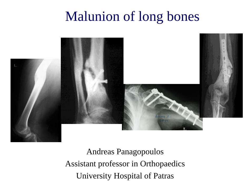
Malunion of long bones
Andreas Panagopoulos
Assistant professor in Orthopaedics
University Hospital of Patras
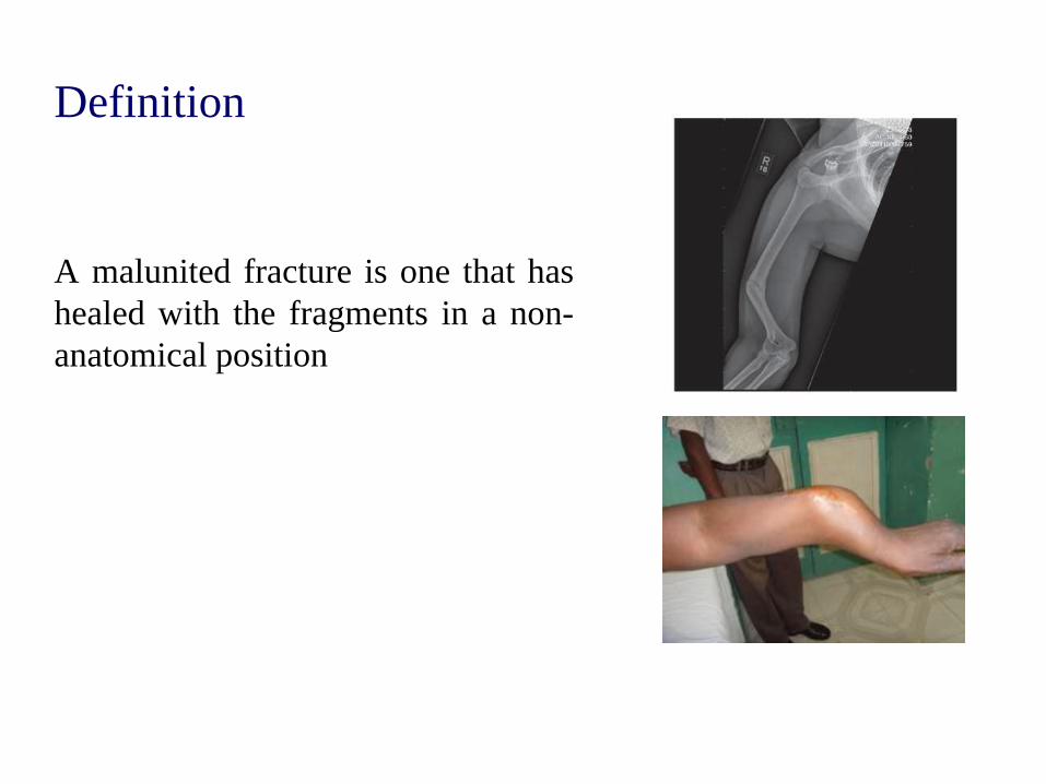
Definition
A malunited fracture is one that has
healed with the fragments in a non-
anatomical position
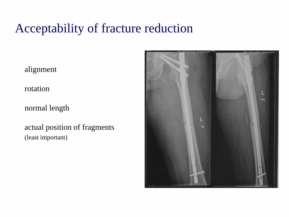
Acceptability of fracture reduction
alignment
rotation
normal length
actual position of fragments
(least important)
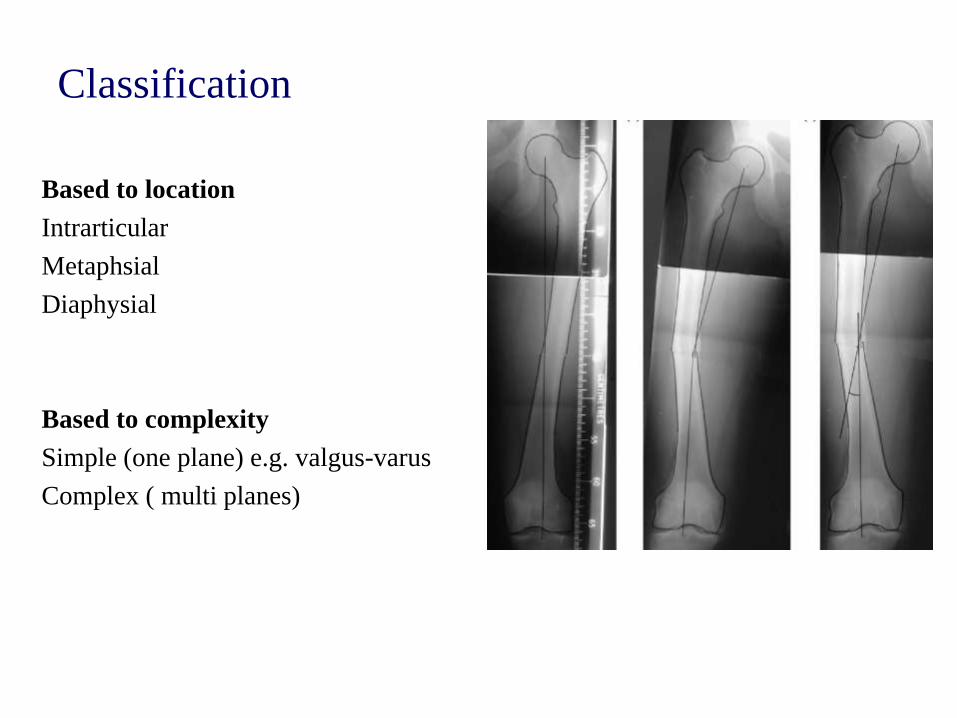
Classification
Based to location
Intrarticular
Metaphsial
Diaphysial
Based to complexity
Simple (one plane) e.g. valgus-varus
Complex ( multi planes)
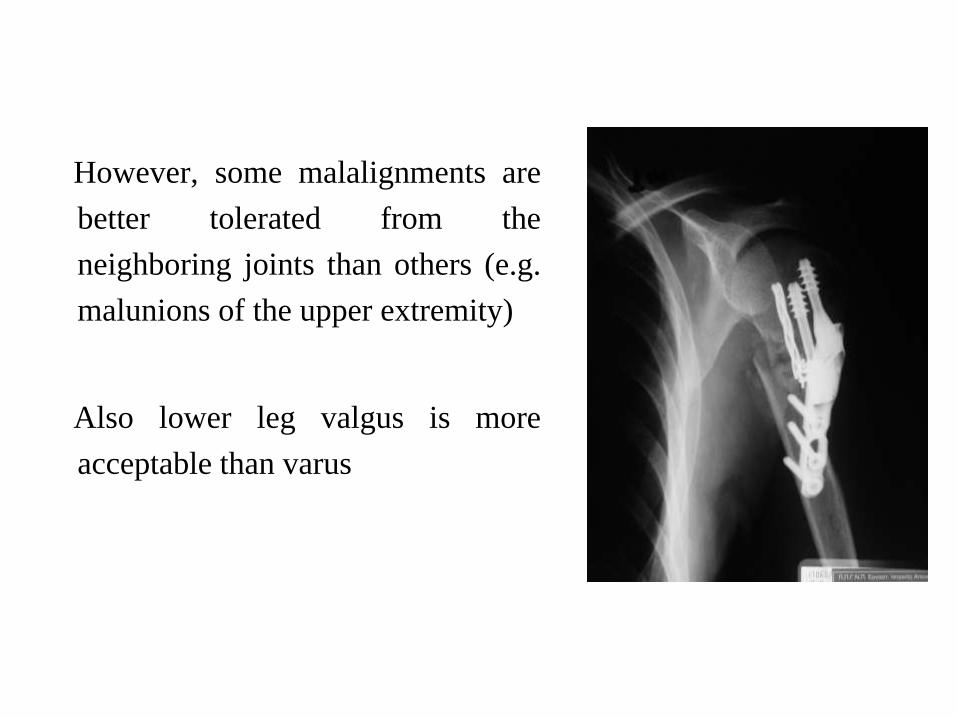
However, some malalignments are
better tolerated from the
neighboring joints than others (e.g.
malunions of the upper extremity)
Also lower leg valgus is more
acceptable than varus
This means there are both relative and absolute indications
to correct deformities and leg length discrepancies
Absolute Indications
- Presence of disabling pain
- Severe functional disability
Relative Indications
- Cosmetic reasons
- No response to nonoperative treatment
The object of surgery for malunion is to restore function
Operative treatment for malunion of most fractures should
not be considered until 6 to 12 months after the fracture
has occurred.
However, in intraarticular fractures, surgery may be
required sooner if satisfactory function is to be restored
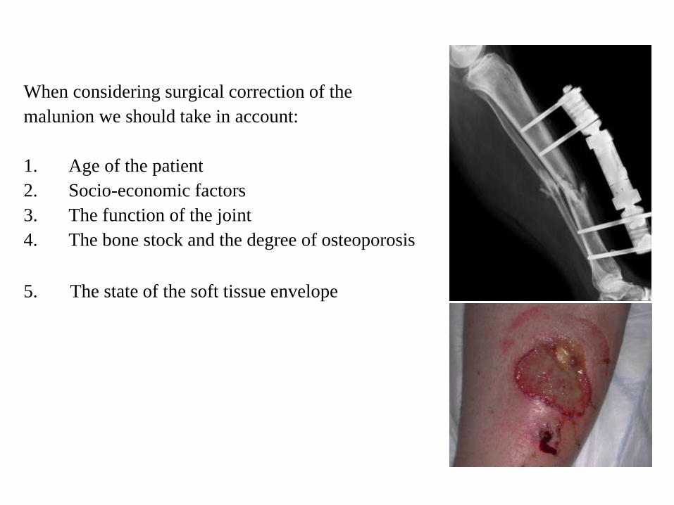
When considering surgical correction of the
malunion we should take in account:
1. Age of the patient
2. Socio-economic factors
3. The function of the joint
4. The bone stock and the degree of osteoporosis
5. The state of the soft tissue envelope
Corrective surgery at the site of malunion is not always
feasible.
In some instances, a compensatory procedure may be
necessary to restore function; in others, pain may be the
predominant symptom and may require fusion of a joint.
Preoperative Planning
X-rays (contralateral normal side) , CT imaging/3D
reconstruction, MRI scans (measurement of alignment )
Evaluation of the soft tissue and bone condition
Throughout physical examination and history
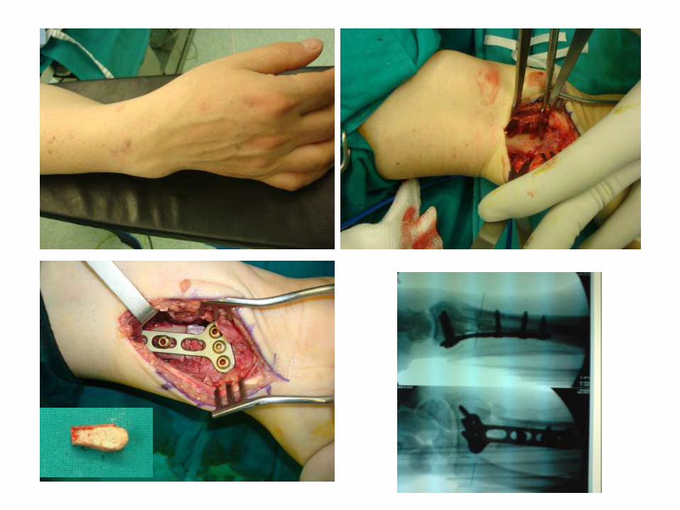
Implant Choices
Plates and screws
- Grants intrafragmetary compression & anatomical reduction
- Is mandatory to be covered adequately from soft tissues
- The implant of choice in case of periarthritic & metaphysic areas
External fixation devices
-Useful to avoid soft tissue irritation,
-Better to be used at the supramalleolar areas & tibial plafond
Intramedullary devises
-The implant of choice in case of the diaphysis of the long bone
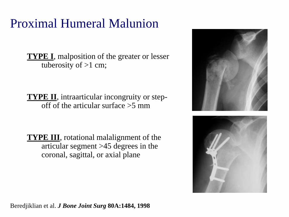
Proximal Humeral Malunion
TYPE I, malposition of the greater or lesser tuberosity of >1 cm;
TYPE II, intraarticular incongruity or step-off of the articular surface >5 mm
TYPE III, rotational malalignment of the articular segment >45 degrees in the coronal, sagittal, or axial plane
Beredjiklian et al. J Bone Joint Surg 80A:1484, 1998
3 m pop
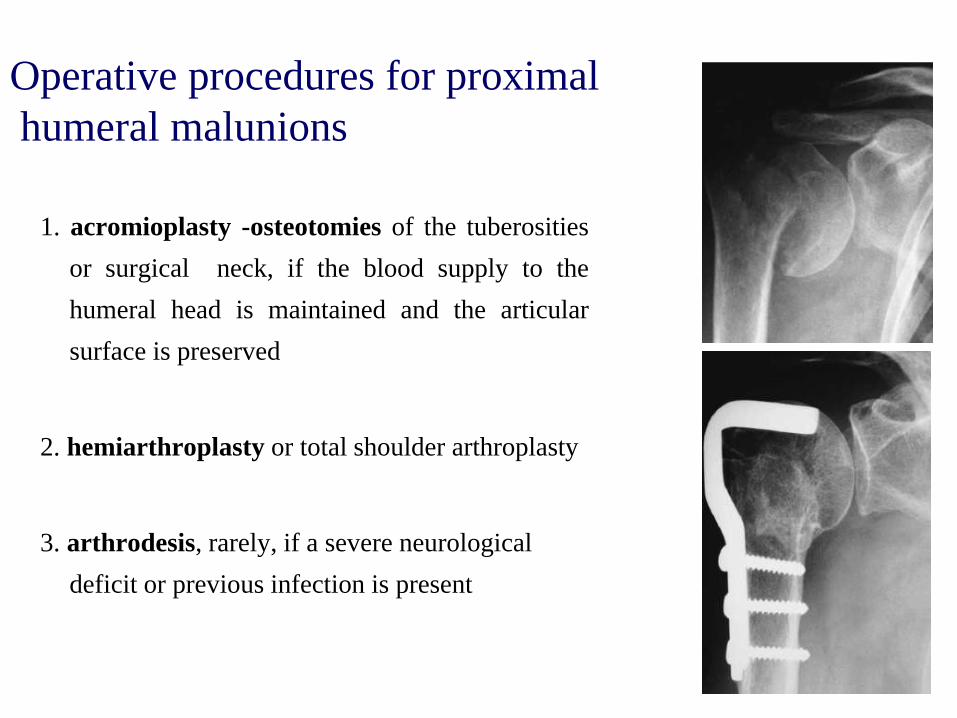
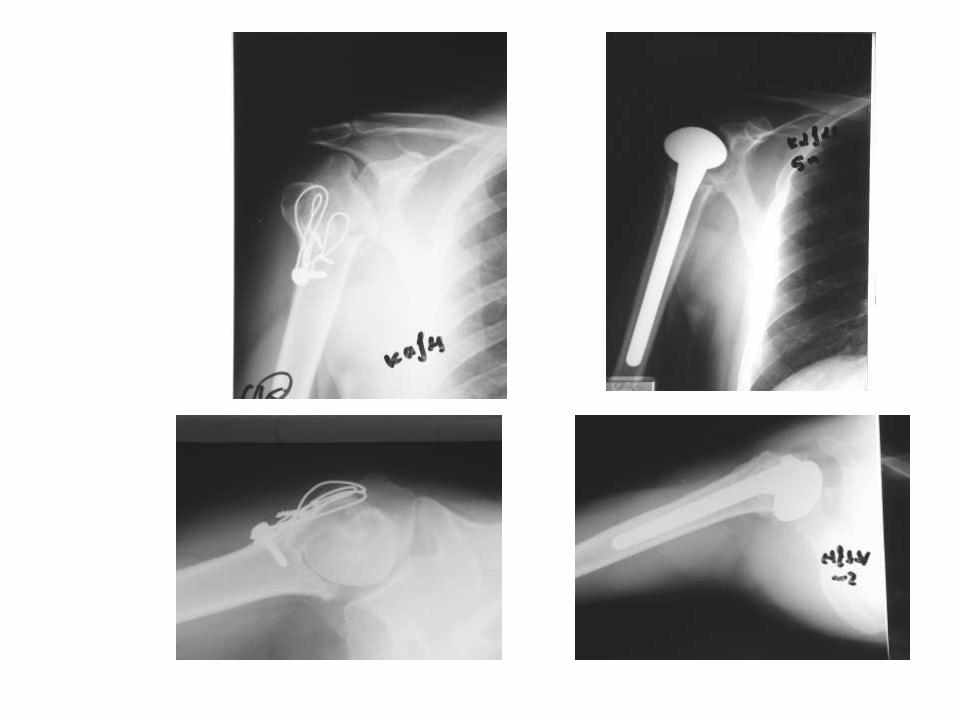
Operative procedures for proximal
humeral malunions
1. acromioplasty -osteotomies of the tuberosities
or surgical neck, if the blood supply to the
humeral head is maintained and the articular
surface is preserved
2. hemiarthroplasty or total shoulder arthroplasty
3. arthrodesis, rarely, if a severe neurological
deficit or previous infection is present
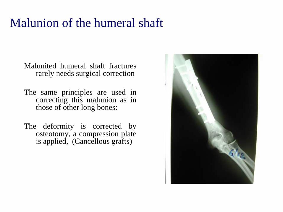
Malunion of the humeral shaft
Malunited humeral shaft fractures rarely needs surgical correction
The same principles are used in correcting this malunion as in those of other long bones:
The deformity is corrected by osteotomy, a compression plate is applied, (Cancellous grafts)
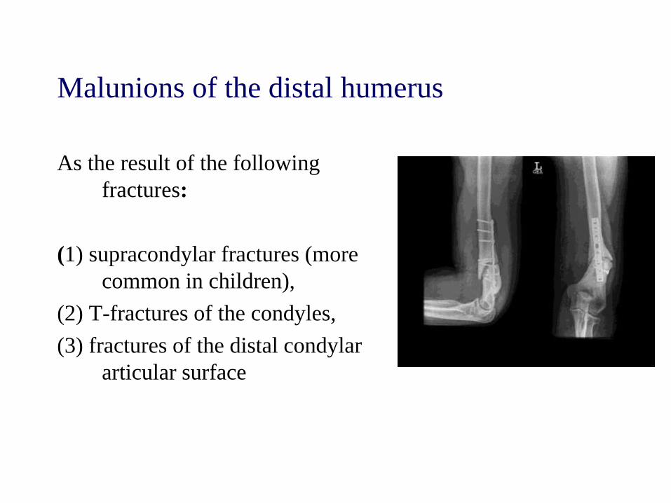
Malunions of the distal humerus
As the result of the following
fractures:
(1) supracondylar fractures (more
common in children),
(2) T-fractures of the condyles,
(3) fractures of the distal condylar
articular surface
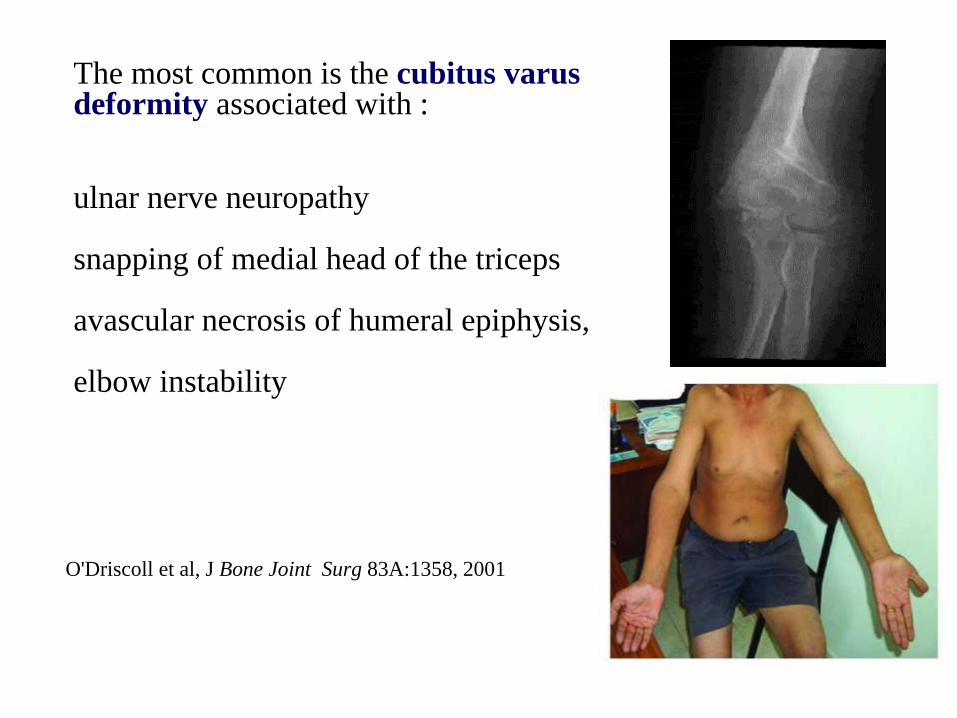
O'Driscoll et al, J Bone Joint Surg 83A:1358, 2001
The most common is the cubitus varus deformity associated with :
ulnar nerve neuropathy
snapping of medial head of the triceps
avascular necrosis of humeral epiphysis,
elbow instability
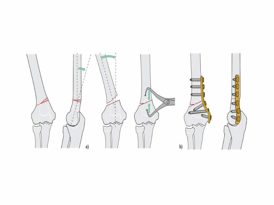
Treatment Options
Wedge osteotomy alone
Osteotomy combined with ligament reconstruction
( High demands & severe deformity > 15º)
Total elbow arthroplasty
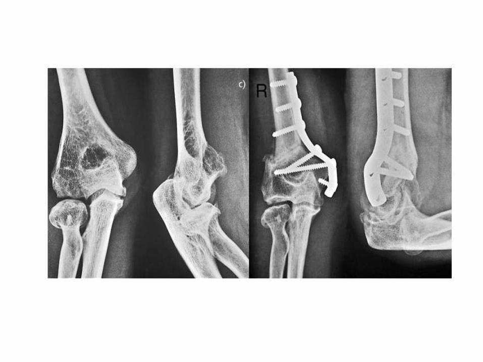
Proximal radius & ulna malunions
Malunions of the proximal third of the radius and ulna can be
classified:
(1) of the radial head
(2) of the radial neck
(3) of the olecranon
(4) with anterior dislocation of the proximal radius
(Monteggia fracture)
(5) with synostosis between the radius and ulna.
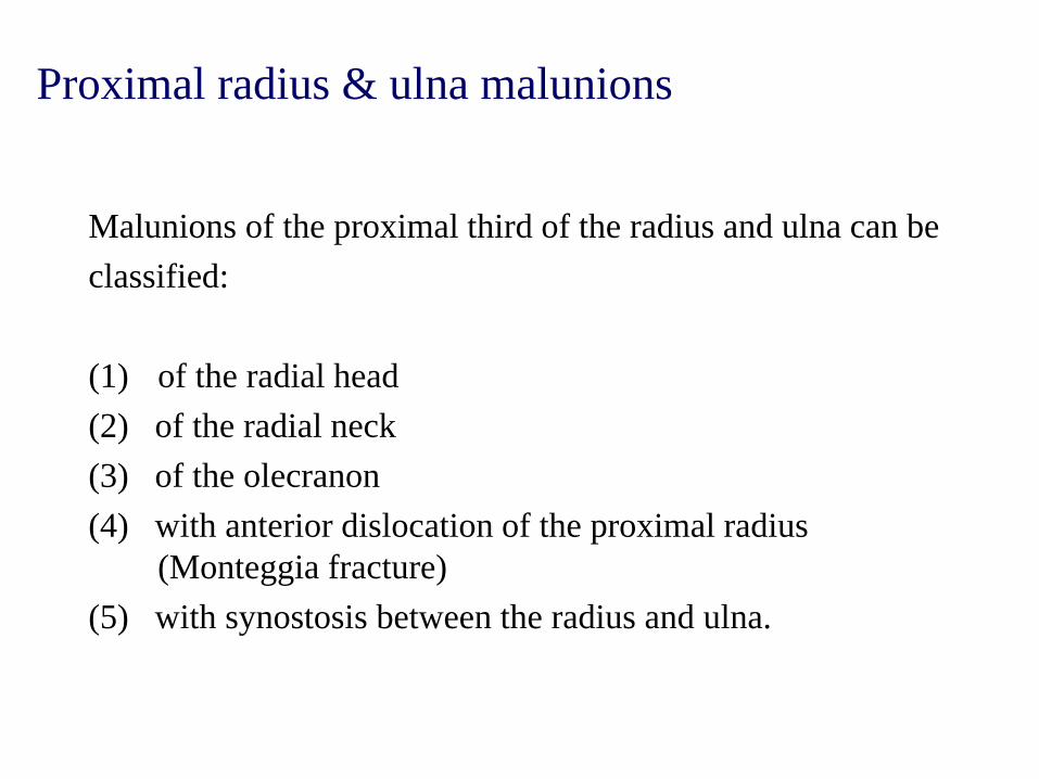
7-year old girl with a Bado I equivalent
lesion 5 months after trauma
reduction of the radial head and
fixation of the ulnar osteotomy with a
plate
Forearm Shaft Malunions
Malrotation, angulation and loss of
the radial bow all have been
associated with loss of motion and
compromised functional outcomes
Malunited forearm fractures may
lead to disturbances of the distal
radioulnar joint, and arthritis of the
proximal radioulnar joint
In a cadaveric study, Matthews et al. found an insignificant
reduction in forearm rotation with a 10º angulatory
deformity, whereas a 20º angulation caused a functional loss
of pronation and supination.
In a similar study, Tarr, Garfinkel, and Sarmiento showed
that angular or rotational deformities of <10º resulted in
minimal limitation of forearm rotation; however, with 15º of
total deformity, forearm motion was reduced > 27% except
in distal-third fractures
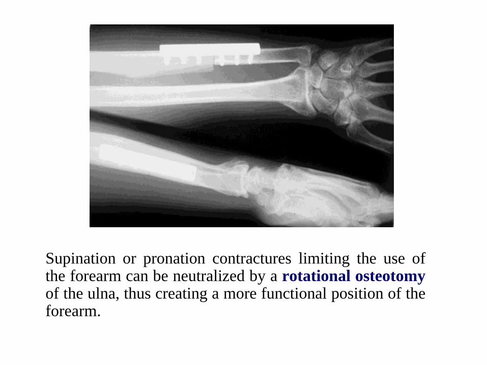
Supination or pronation contractures limiting the use of the forearm can be neutralized by a rotational osteotomy of the ulna, thus creating a more functional position of the forearm.
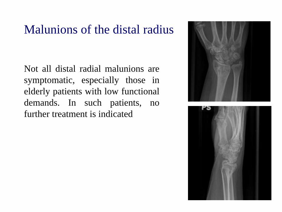
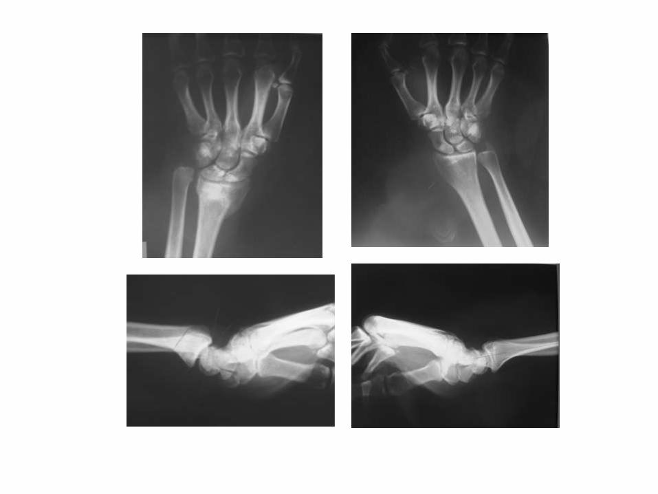
Malunions of the distal radius
Not all distal radial malunions are
symptomatic, especially those in
elderly patients with low functional
demands. In such patients, no
further treatment is indicated
pain and functional deficits severe enough to interfere
significantly with daily activities
a young, active patient (< 40 years old) with a deformity that
is likely to become symptomatic with time:
- articular step-off of > 2 mm
- carpal instability
- > 20 -30º of dorsal angulation
- incongruent distal radioulnar joint)
Indications for surgical intervention
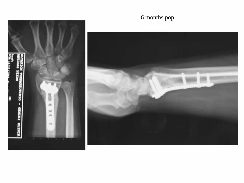
6 months pop
Proximal Femur Malunions
Surgical Indications:
varus and rotational deformities in combination with
shortening leading to limping and overuse of the
neighboring joints.
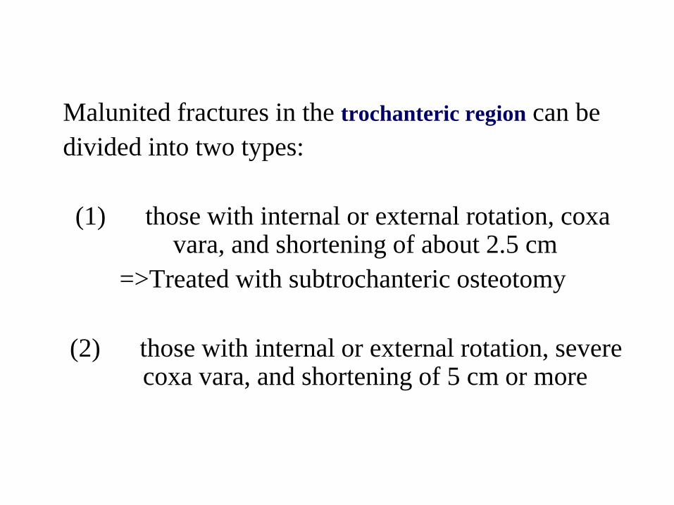
Malunited fractures in the trochanteric region can be
divided into two types:
(1) those with internal or external rotation, coxa vara, and shortening of about 2.5 cm
=>Treated with subtrochanteric osteotomy
(2) those with internal or external rotation, severe coxa vara, and shortening of 5 cm or more
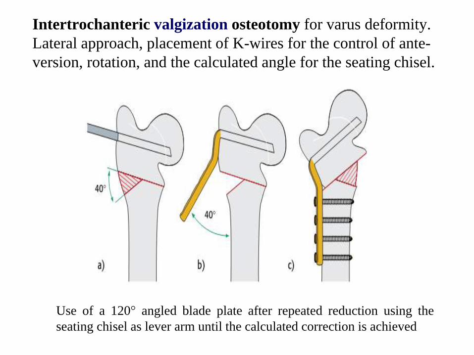
Intertrochanteric valgization osteotomy for varus deformity.
Lateral approach, placement of K-wires for the control of ante-
version, rotation, and the calculated angle for the seating chisel.
Use of a 120° angled blade plate after repeated reduction using the
seating chisel as lever arm until the calculated correction is achieved
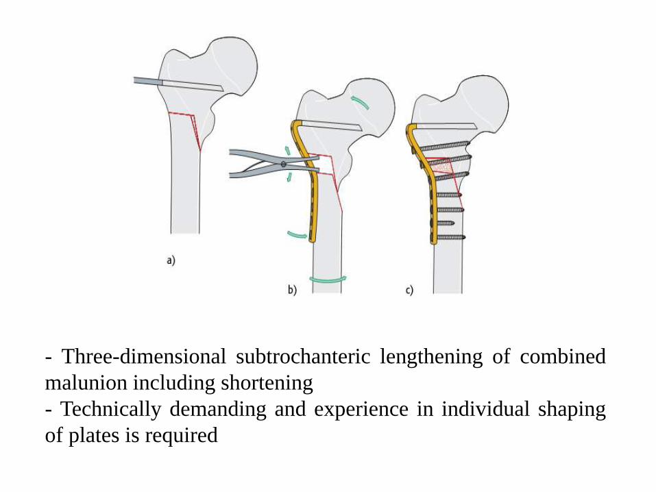
- Three-dimensional subtrochanteric lengthening of combined
malunion including shortening
- Technically demanding and experience in individual shaping
of plates is required
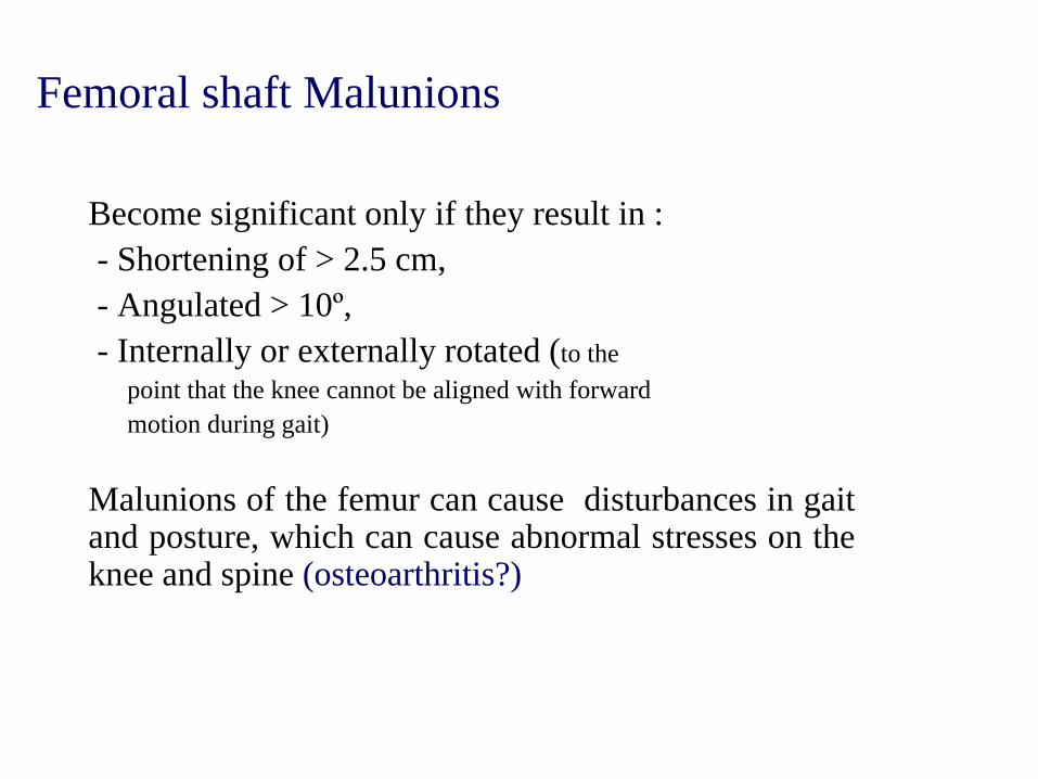
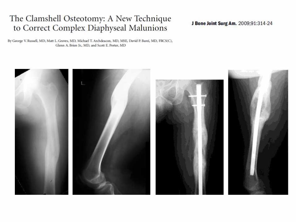
Femoral shaft Malunions
Become significant only if they result in :
- Shortening of > 2.5 cm,
- Angulated > 10º,
- Internally or externally rotated (to the
point that the knee cannot be aligned with forward
motion during gait)
Malunions of the femur can cause disturbances in gait and posture, which can cause abnormal stresses on the knee and spine (osteoarthritis?)
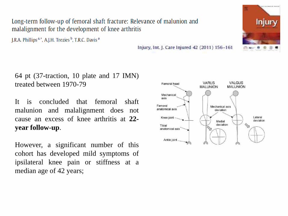
64 pt (37-traction, 10 plate and 17 IMN)
treated between 1970-79
It is concluded that femoral shaft
malunion and malalignment does not
cause an excess of knee arthritis at 22-
year follow-up.
However, a significant number of this
cohort has developed mild symptoms of
ipsilateral knee pain or stiffness at a
median age of 42 years;
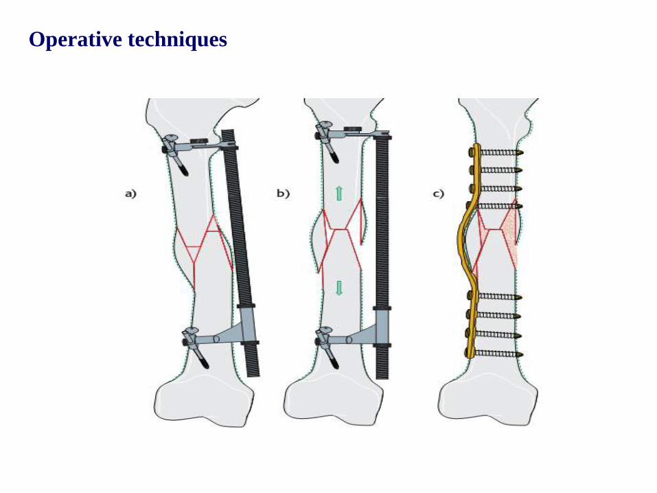
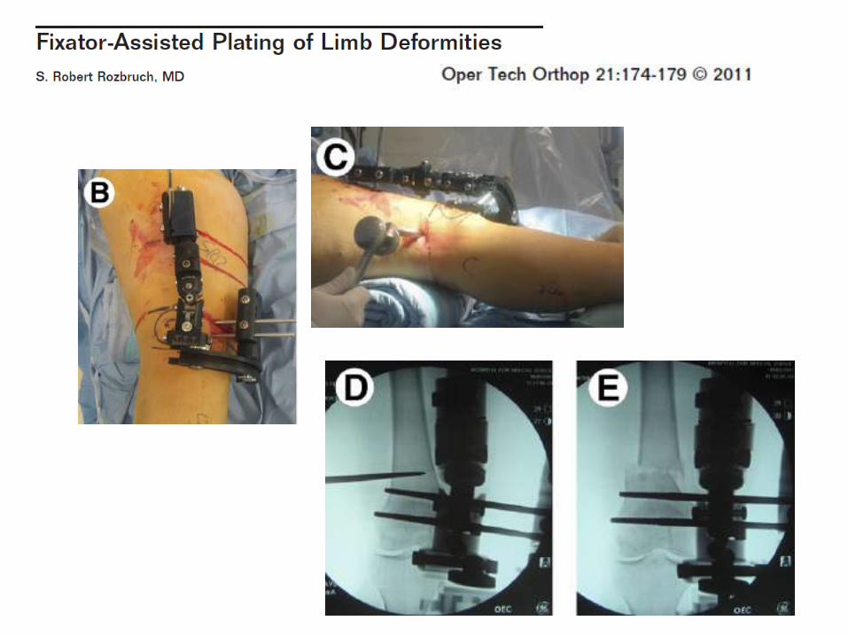
Operative techniques
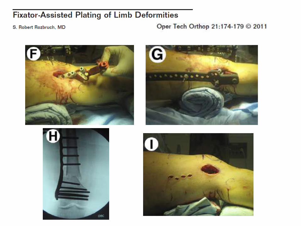
Distal Femur Malunion
Indications for surgery:
- Malunions in valgus & varus
- ante, or recurvation deformities
- rotation deformities
Treatment: Axial correction by open or close wedge
supracondylar osteotomy
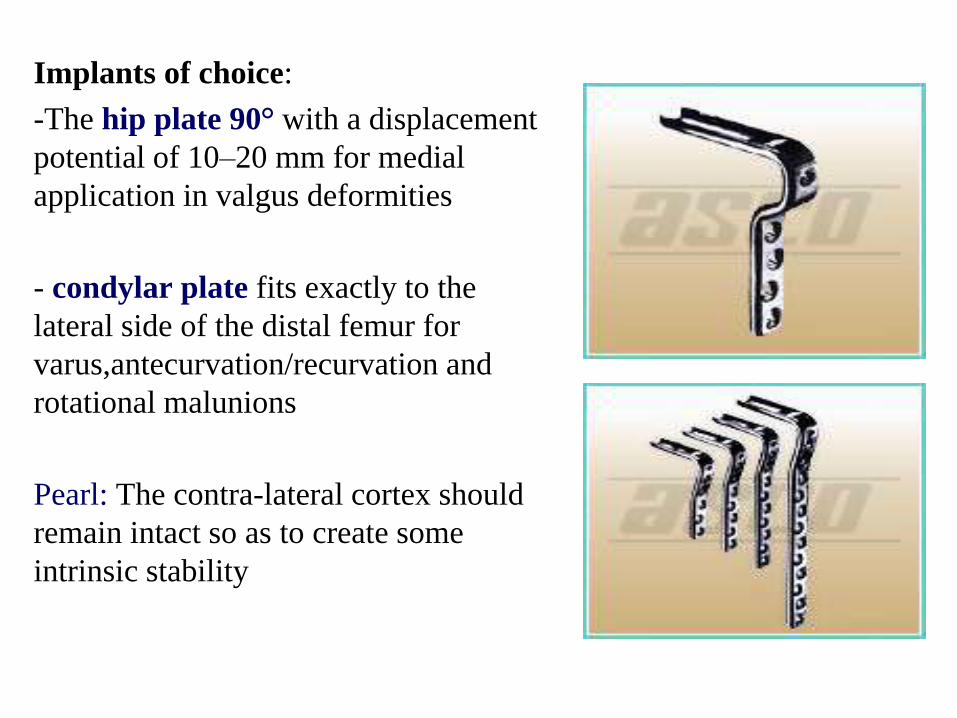
Implants of choice:
-The hip plate 90° with a displacement
potential of 10–20 mm for medial
application in valgus deformities
- condylar plate fits exactly to the
lateral side of the distal femur for
varus,antecurvation/recurvation and
rotational malunions
Pearl: The contra-lateral cortex should
remain intact so as to create some
intrinsic stability
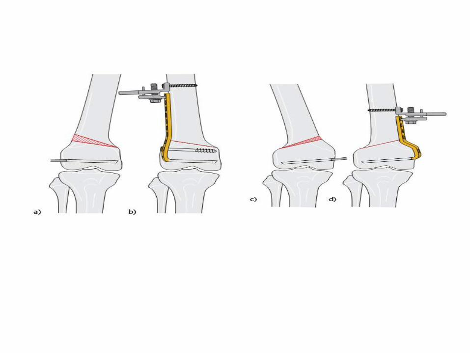
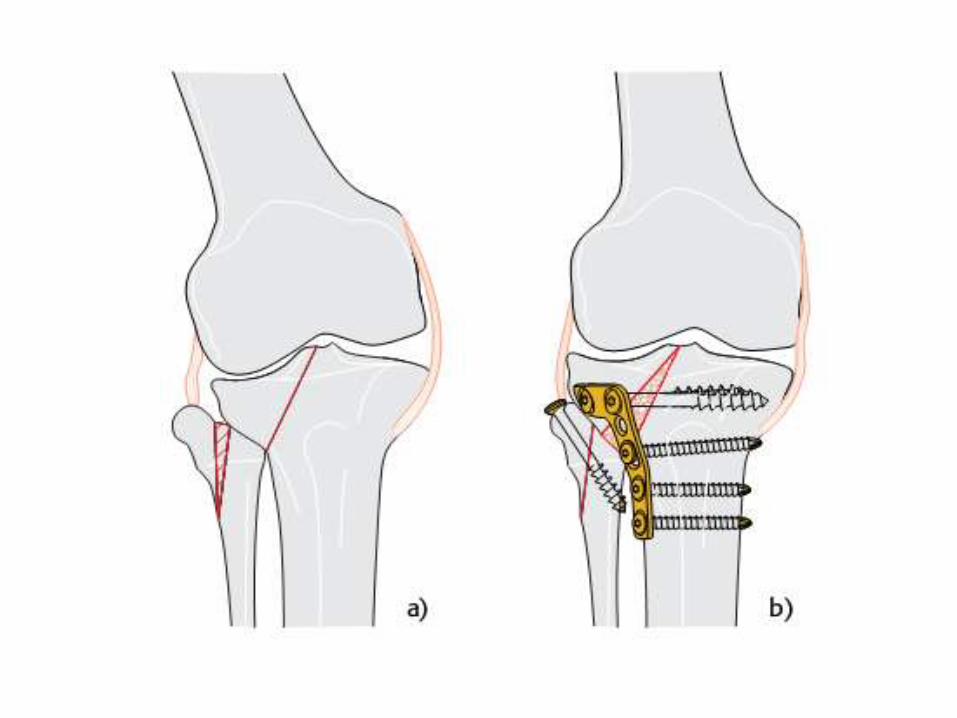
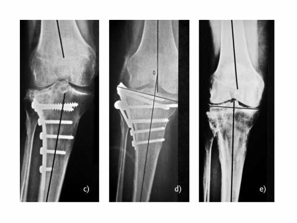
Proximal Tibia Malunions
Indications for surgery:
- Deformities of the proximal tibia in all three planes
- Intra-articular malunions after monocondylar fractures
- Residual joint impaction in combination with ligamentous
instability.
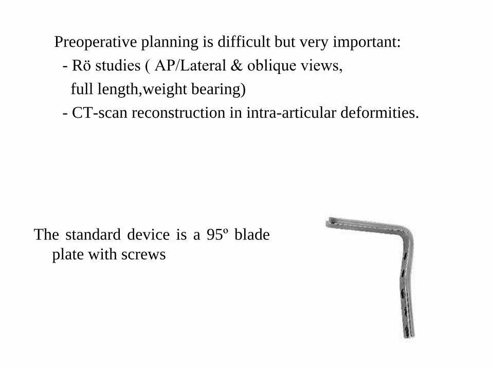
Preoperative planning is difficult but very important:
- Rö studies ( AP/Lateral & oblique views,
full length,weight bearing)
- CT-scan reconstruction in intra-articular deformities.
The standard device is a 95º blade
plate with screws
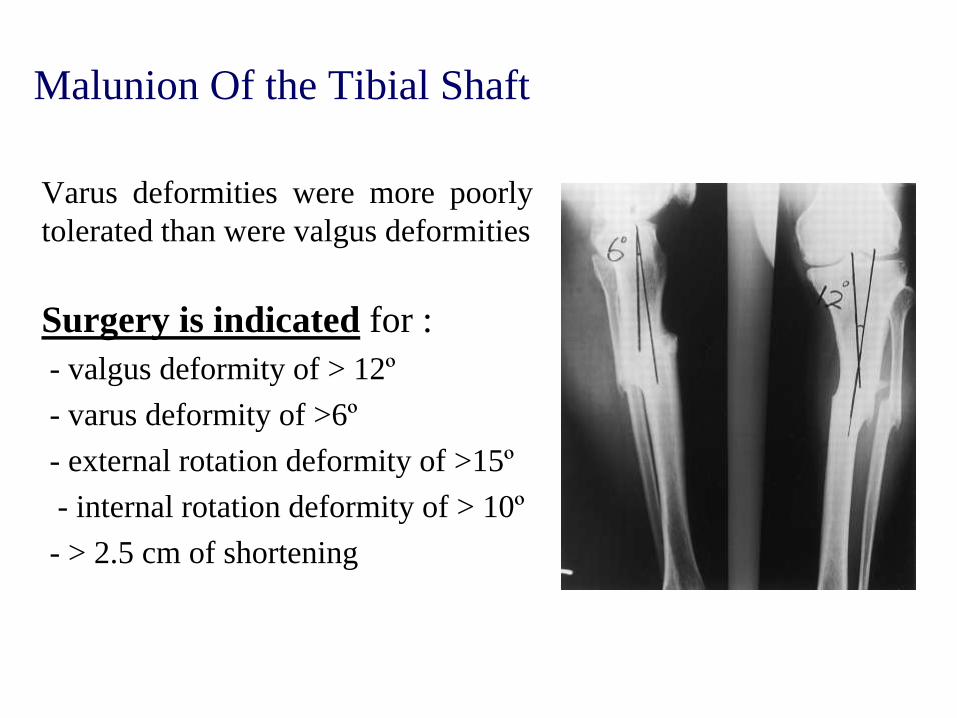
Malunion Of the Tibial Shaft
Varus deformities were more poorly
tolerated than were valgus deformities
Surgery is indicated for :
- valgus deformity of > 12º
- varus deformity of >6º
- external rotation deformity of >15º
- internal rotation deformity of > 10º
- > 2.5 cm of shortening

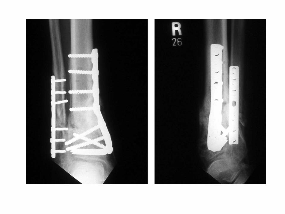
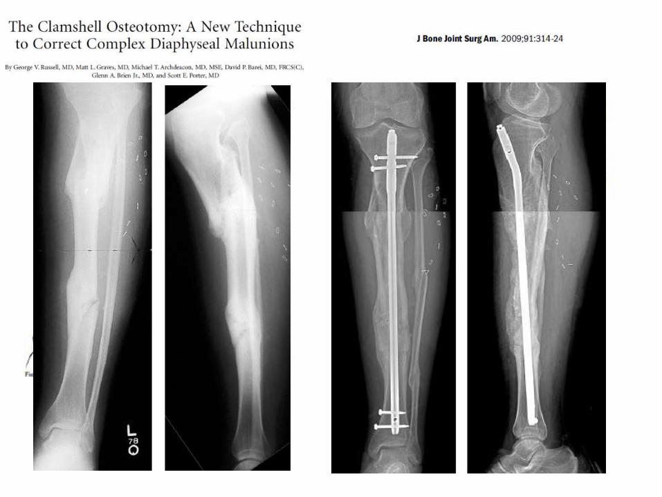
Several choises of treatment:
Simple opening wedge, closing wedge, or dome-shaped
osteotomies
Oblique osteotomies can be used to correct multiplanar
deformities
![Stromal fibroblast activation protein alpha promotes gastric … · 2018. 11. 12. · gional tumor progression majorly occurred in abdomen pelvic cavities [5, 6]. The underlying mechanisms](https://static.fdocument.org/doc/165x107/60dc1541981c0c65b612e293/stromal-fibroblast-activation-protein-alpha-promotes-gastric-2018-11-12-gional.jpg)