Lectures 1. Meiosis and Recombination in yeast. After this ... · Lectures 1. Meiosis and...
16
1 Lectures 1. Meiosis and Recombination in yeast. After this lecture you should: 1) Be able to predict the likelihood of outcome of genetic crosses where the markers in question are placed at different locations. I don’t care about predicting the absolute frequencies as much as having a conceptual understanding of the segregation biases. 2) Have a good understanding of how recombination is thought to work on a molecular level. 3) Understand how gene conversions and crossovers might occur. 4) Appreciate why crossovers are important for meiotic chromosome segregation.
Transcript of Lectures 1. Meiosis and Recombination in yeast. After this ... · Lectures 1. Meiosis and...

1
Lectures 1. Meiosis and Recombination in yeast.
After this lecture you should:
1) Be able to predict the likelihood of outcome of genetic crosses where the
markers in question are placed at different locations. I don’t care about
predicting the absolute frequencies as much as having a conceptual
understanding of the segregation biases.
2) Have a good understanding of how recombination is thought to work on a
molecular level.
3) Understand how gene conversions and crossovers might occur.
4) Appreciate why crossovers are important for meiotic chromosome
segregation.
2
References:
Experiments suggesting that COs (crossovers) and NCOs are not merely the resolution of the same product: Several papers reviewed in: Bishop DK, Zickler D. Cell. 2004 Apr 2;117(1):9-15. Youds and Boulton, J Cell Sci. 2011 Feb 15;124(Pt 4):501-13. The choice in meiosis - defining the factors that influence crossover or non-crossover formation.
Not very exciting reading, but potentially helpful. These papers outline some of the strategies given in class for protein tagging: Longtine MS, McKenzie A 3rd, Demarini DJ, Shah NG, Wach A, Brachat A, Philippsen P, Pringle JR.Additional modules for versatile and economical PCR-based gene deletion and modification in Saccharomyces cerevisiae. Yeast. 1998 Jul;14(10):953-61. Bahler J, Wu JQ, Longtine MS, Shah NG, McKenzie A 3rd, Steever AB, Wach A, Philippsen P, Pringle JR. Heterologous modules for efficient and versatile PCR-based gene targeting in Schizosaccharomyces pombe. Yeast. 1998 Jul;14(10):943-51.
3
I) Yeast-An introduction.
A) History- The oldest domesticated species, from at least 6000 B.C.
1. Enzymes were discovered in yeast by Pasteur, who discovered invertase. Enzyme
literally means “in yeast”, or en zymi.
B) Different yeasts are studied for different reasons.
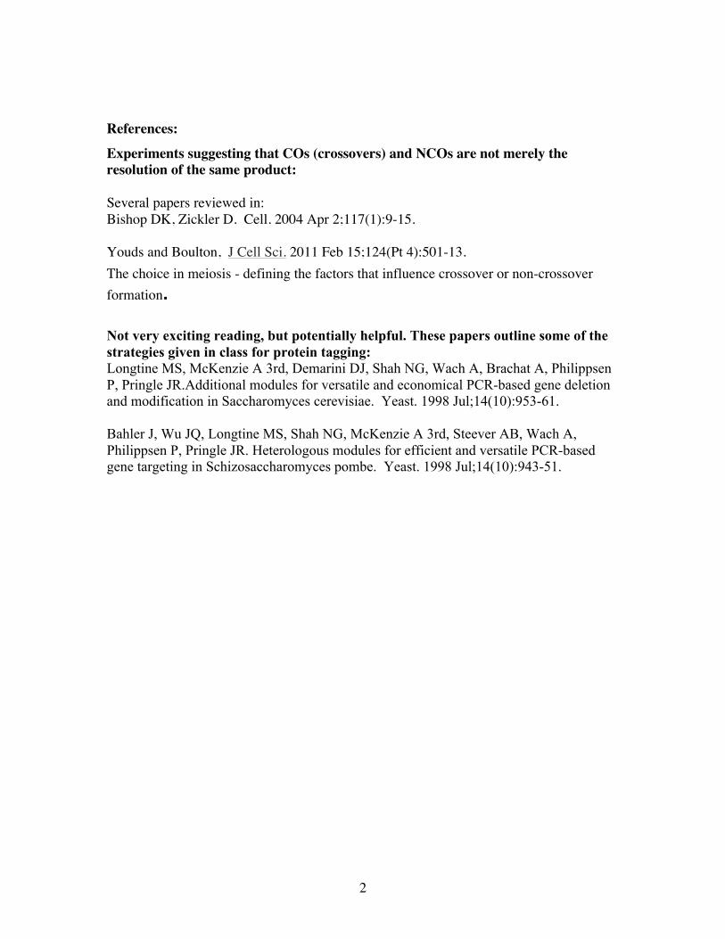
1. To study most processes in a simple model eukaryote:
Saccharomyces cerevisiae a.k.a. budding yeast, bakers’ yeast, brewers’ yeast
-NOTE
bud scars, where previous
buds had occurred
Schizosaccharomyces pombe a.k.a. fission yeast, “the other yeast”
! NOTE cells with septa forming.
-cerevisiae and pombe are very distantly related to each other.
2. To model pathogenesis . – pathogenic yeasts. e.g. Candida albicans
Candida has two growth forms. The yeast form looks just like
cerevisiae, whereas the pathogenic pseudal hyphal form is
filamentous
Fun candida fact: Candida has a modified genetic code
For the purposes of this course, I will always be referring to S. cerevisiae when I say
yeast, unless I state otherwise.
4
C). The Genome.
1. The yeast genome is about 1.2x10⁷ bps, 200x smaller than humans and 3x bigger
than E. coli
a) 16 chromosomes, all readily observable by pulse field gel electrophoresis
b) 70% of the yeast genome is coding! About 6000 genes, or 1 every 2KB
c) 274 tRNAs, ≈40 small RNA genes (harder to spot than ORFs),
d) 120 copies of the rDNA genes, in tandem arrays, on chromosome XII
-This forms a crescent-shaped nucleolus along the inner edge of the nucleus
e) about 50 copies of the Ty transposon.
f) additional DNA from an endogenous plasmid, called 2µ, is in most strains
g) some strains also have “killer” double stranded RNA. These are virus like,
but have no extracellular form and must be passed horizontally. They encode a toxin and
an immunity factor to that toxin
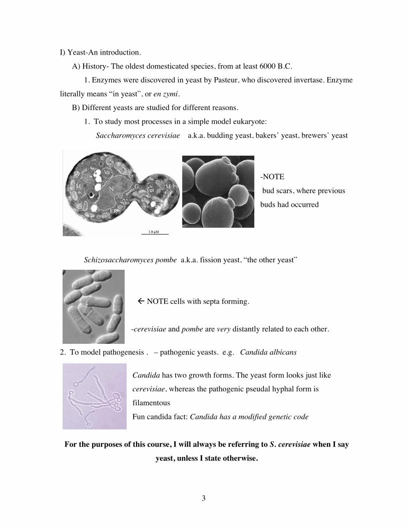
2. Yeast have small (few hundred base pair) CENs and origins with defined seq.
Note:
a) Tight spacing of genes. Typically less than one KB between genes.
b) Origins (e.g. ARS604) and centromere (black spot) are mapped and very small
c) Very few introns (none among these genes).
d) Most genes are annotated with gene designations (exceptions being YFR006W and
YFR007W), which typically means that they have been studied before.
5
3. Mitochondria. Circular 76 KB genome. Most mitochondrial proteins are made in the
cytoplasm and imported in.
a) The mitochondrial genome is not essential for life
-Cells lacking the mitochondrial genome still make mitochondria, but are
unable to grow on non-fermentable carbon sources (such as ethanol or glycerol), as they
can only generate ATP from glycolysis.
-Mutants unable to grow on non-fermentable carbon sources are called
petites (because of the size of the colonies, not the size of the cells)
-The growth difference between petites and wildtype is not normally large
because when glucose is plentiful, yeast grow anaerobically
However, you still need mitochondria, which do more than the krebs cycle.
-If they have a mutation in the mitochondrial genome, they are ρ− (rho
minus) if they lack the mitochondrial genome, they are ρ 0
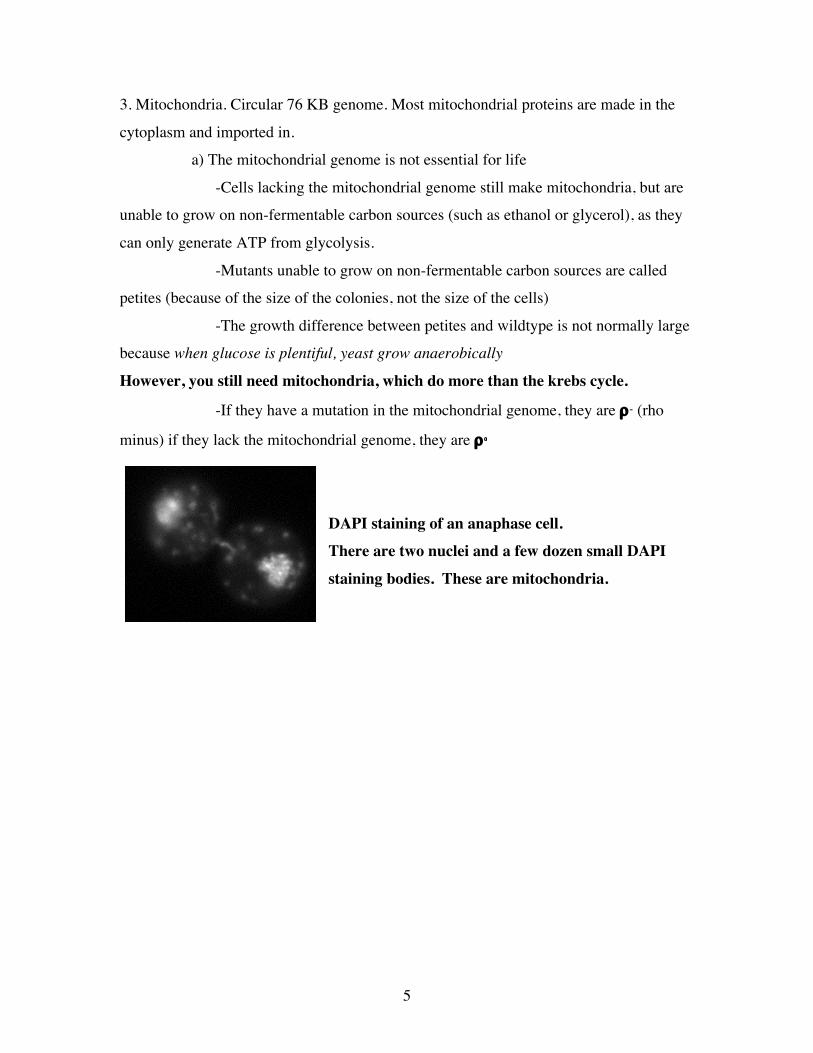
DAPI staining of an anaphase cell. There are two nuclei and a few dozen small DAPI
staining bodies. These are mitochondria.
6
II) Nomenclature: There are highly standardized ways of referring to genes and gene
products (proteins).
A) Genes names are italicized and have three letters.
-e.g. CDC stands for Cell Division Cycle, URA stands for URAcil biosynthesis.
C) The three letters are followed by a number, which typically represent different genes
that share a common phenotype.
-> The genes involved in uracil biosynthesis are URA1, URA2, URA3…
D) If the gene is wildtype it’s name will be given in all capitals, if the gene is in all small
case it is a recessive mutation.
-> a ura3 strain has a recessive mutation in the URA3 gene.
E) If the gene is a mutant allele, it is often represented by a hyphen with a number after
that. This isn’t always done, but it is nice to know the allele number because it allows you to
know exactly what you are dealing with. Sometimes people use letters to designate a special
phenotype, or will indicate the exact mutation
-> ura3-100 is a common allele of the URA3 gene.
-> ura3-S32A would mean that a serine at amino acid 32 had been mutated to an
alanine. This is typically done when a specific mutation has been engineered in, such as those
that get rid of a phosphorylation site.
F) Capital letters are used for any allele that is dominant. Note that wildtype is also in
CAPs, however dominant alleles will be capitals and have an allele designation.
- > URA3-123 could represent a dominant allele of URA3
G) If there is a deletion, there is a delta after (or occasionally before) the name
-> ura3Δ
I) If one gene has been placed at the locus of another gene, there are two colons
-> ura3∆::LEU2 (or just ura3::LEU2) implies that the URA3 gene has been disrupted
(and is therefore in small case) with a wildtype (and therefore large case) copy of LEU2
J) If the first letter is capitalized, and there are no italics, this is the protein
-> Ura3 is the protein encoded by URA3 (Old nomenclature is Ura3p)
K) The phenotype is designated by a + or -
-> Ura+ cells can make uracil, Ura- cells cannot. Note that this isn’t referring to any
specific URA gene, it is a phenotypic designation
7
III) Basic manipulations.
A) Markers. Yeast can make all of their organic molecules. Wildtype yeast can
grow if they are given an energy source (sugar), some nitrogen with which to make
things, and simple trace elements etc. In order to do genetics, people have isolated
mutants, like ura3, which are unable to make certain amino acids or bases.
1) commonly used “markers” genes are URA3, HIS3, LEU2, TRP1, and ADE2,
which require uracil, histidine, leucine, tryptophan, and adenine, respectively, to
be added to the media.
a) URA3 is an especially useful marker because you can select for the wildtype
or mutant
{DEF: Select means that you do something so that only the things you want live
Screen means that you look at things individually to see if they have a phenotype or
not.}
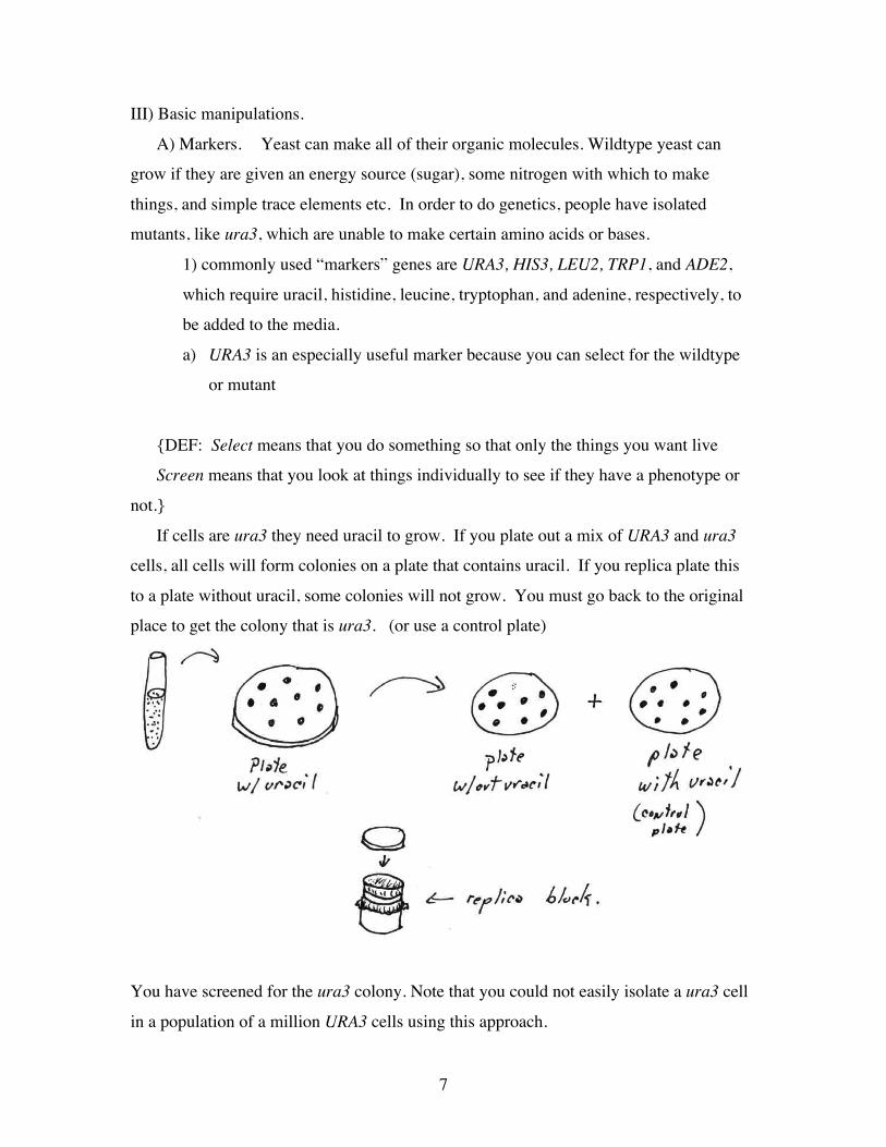
If cells are ura3 they need uracil to grow. If you plate out a mix of URA3 and ura3
cells, all cells will form colonies on a plate that contains uracil. If you replica plate this
to a plate without uracil, some colonies will not grow. You must go back to the original
place to get the colony that is ura3. (or use a control plate)
You have screened for the ura3 colony. Note that you could not easily isolate a ura3 cell
in a population of a million URA3 cells using this approach.
8
The chemical 5-FOA is not itself toxic, but is metabolized into a toxic chemical by the
uracil biosynthetic machinery. It will therefore kill a URA3 cell. It will not kill a ura3
cell, since an enzyme required to turn 5-FOA into a toxic chemical is missing. Using this
drug, you can plate a mixture of a million URA3 cells and one ura3 cell on a plate
containing 5-FOA and only the single ura3 cell will grow. This is a selection.
b) a similar drug, called FAA, exists for TRP1, but it doesn’t work as well.
c) Some of the mutants in the adenine biosynthesis pathway generate red colonies
(e.g.ade2).
d) There are also drug resistance genes that are now used in yeast, especially Kanomycin
(G418), Hygromycin B (Hyg) or Nourseothricin (NAT)
B) Mating. Haploid yeast have two mating types, called α and a. After that long
description of the “rules” for nomenclature, I will now tell you that there is a single
exception, and that is the genes that govern mating type. This is because these genes were
named at the dawn of time. So, an α or an a cell is referred to as a MATα and a MATa cell.
1) Each cell type produces a secreted peptide “pheromone” and a transmembrane
receptor that recognizes the opposite mating type’s pheromone.
2) After an a cell senses an α’s pheromone (called α factor), or visa versa, it will
arrest in G1 of the cell cycle.
3) Cells will polarize and fuse, first their plasma membranes, then their nuclear
envelope.
4) Now you have a diploid, if you grow this on very poor media is will undergo
meiosis and yield four spores. This is called a “tetrad”. The spores can be micro-dissected
from this tetrad and you can see all the products of meiosis. Note: You can’t really do this
in any metazoan. This is why the actual study of genetics (i.e. meiosis), is so very good in
fungi. Animals, e.g. mice or flies, produce 4 sperm from one meiosis, but they get all
mixed and can’t be looked at as a set.
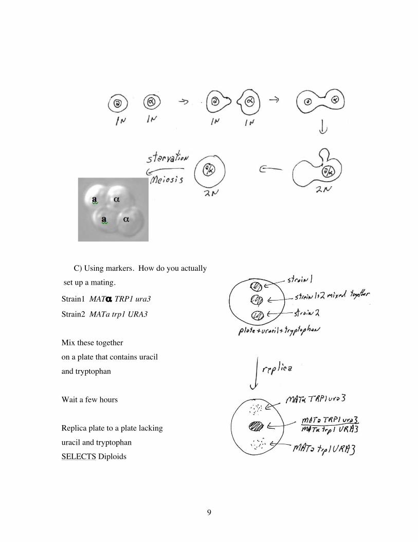
9
C) Using markers. How do you actually
set up a mating.
Strain1 MATα TRP1 ura3
Strain2 MATa trp1 URA3
Mix these together
on a plate that contains uracil
and tryptophan
Wait a few hours
Replica plate to a plate lacking
uracil and tryptophan
SELECTS Diploids
10
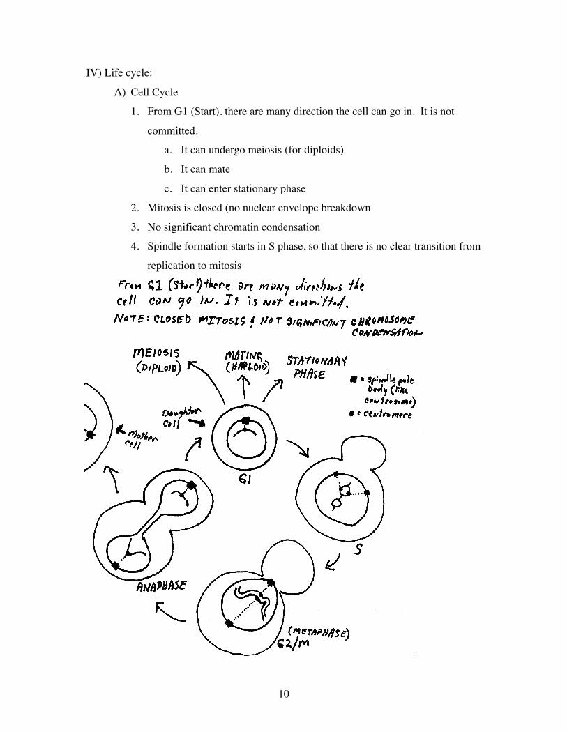
IV) Life cycle:
A) Cell Cycle
1. From G1 (Start), there are many direction the cell can go in. It is not
committed.
a. It can undergo meiosis (for diploids)
b. It can mate
c. It can enter stationary phase
2. Mitosis is closed (no nuclear envelope breakdown
3. No significant chromatin condensation
4. Spindle formation starts in S phase, so that there is no clear transition from
replication to mitosis
!
11
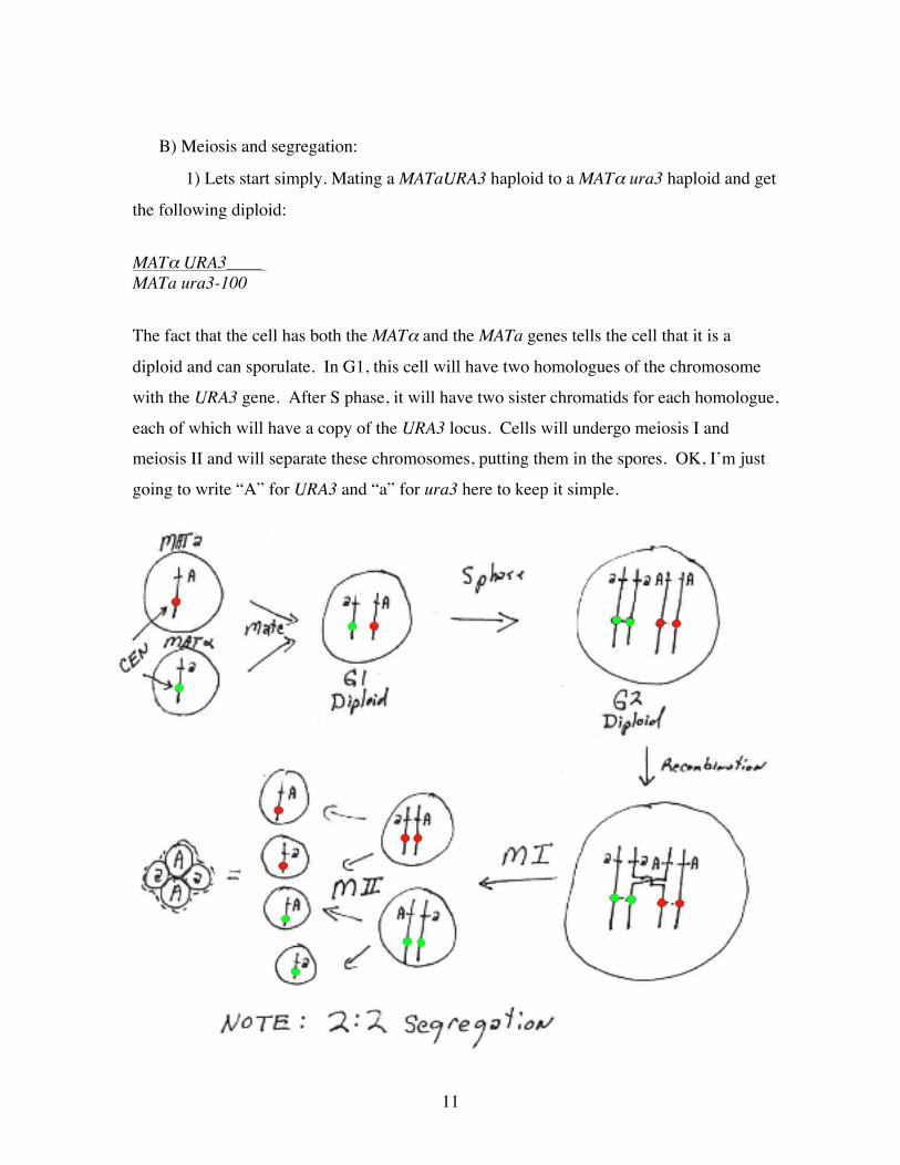
B) Meiosis and segregation:
1) Lets start simply. Mating a MATaURA3 haploid to a MATα ura3 haploid and get
the following diploid:
MATα URA3____ MATa ura3-100
The fact that the cell has both the MATα and the MATa genes tells the cell that it is a
diploid and can sporulate. In G1, this cell will have two homologues of the chromosome
with the URA3 gene. After S phase, it will have two sister chromatids for each homologue,
each of which will have a copy of the URA3 locus. Cells will undergo meiosis I and
meiosis II and will separate these chromosomes, putting them in the spores. OK, I’m just
going to write “A” for URA3 and “a” for ura3 here to keep it simple.
12
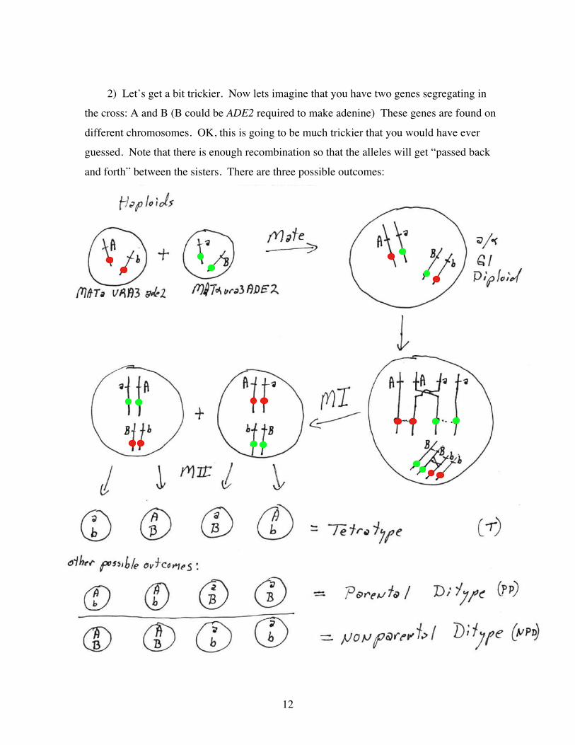
2) Let’s get a bit trickier. Now lets imagine that you have two genes segregating in
the cross: A and B (B could be ADE2 required to make adenine) These genes are found on
different chromosomes. OK, this is going to be much trickier that you would have ever
guessed. Note that there is enough recombination so that the alleles will get “passed back
and forth” between the sisters. There are three possible outcomes:
!
13
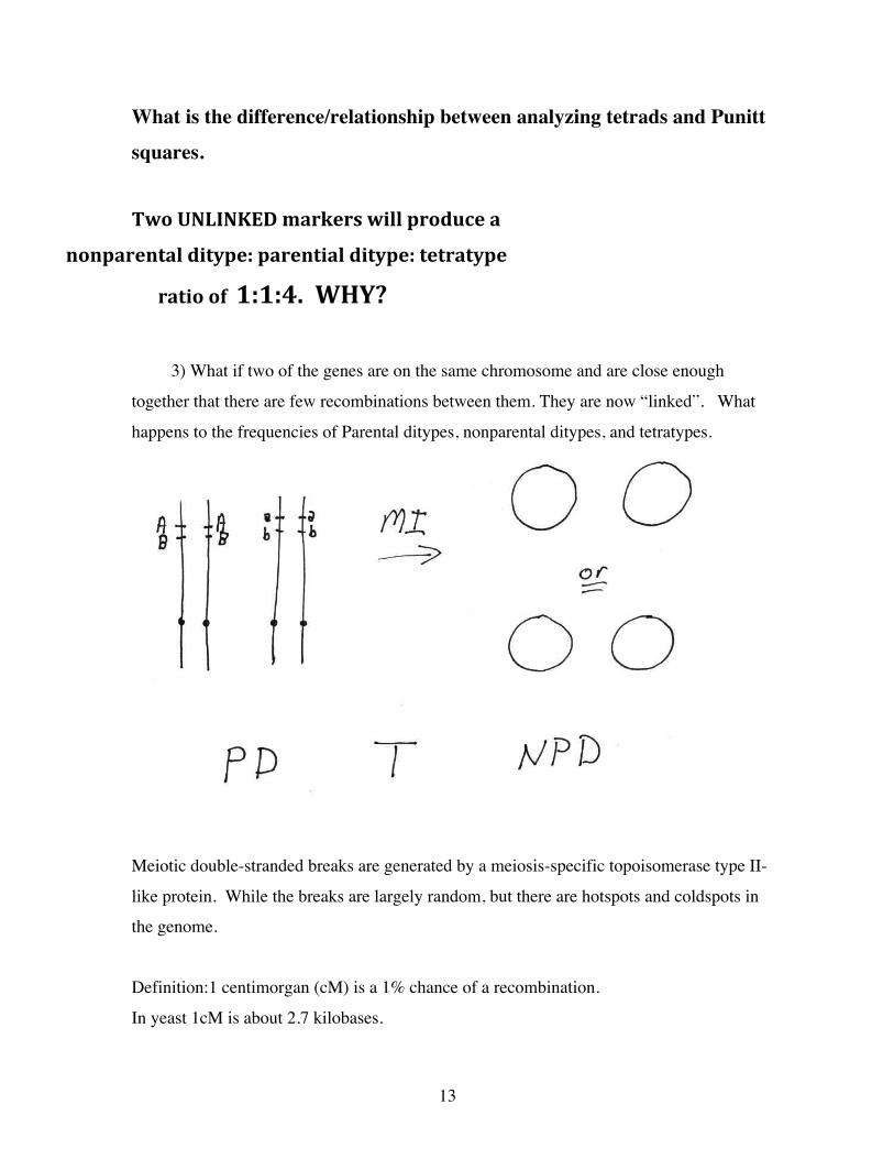
What is the difference/relationship between analyzing tetrads and Punitt
squares.
TwoUNLINKEDmarkerswillproducea
nonparentalditype:parentialditype:tetratype
ratioof1:1:4.WHY?
3) What if two of the genes are on the same chromosome and are close enough
together that there are few recombinations between them. They are now “linked”. What
happens to the frequencies of Parental ditypes, nonparental ditypes, and tetratypes.
Meiotic double-stranded breaks are generated by a meiosis-specific topoisomerase type II-
like protein. While the breaks are largely random, but there are hotspots and coldspots in
the genome.
Definition:1 centimorgan (cM) is a 1% chance of a recombination.
In yeast 1cM is about 2.7 kilobases.

14
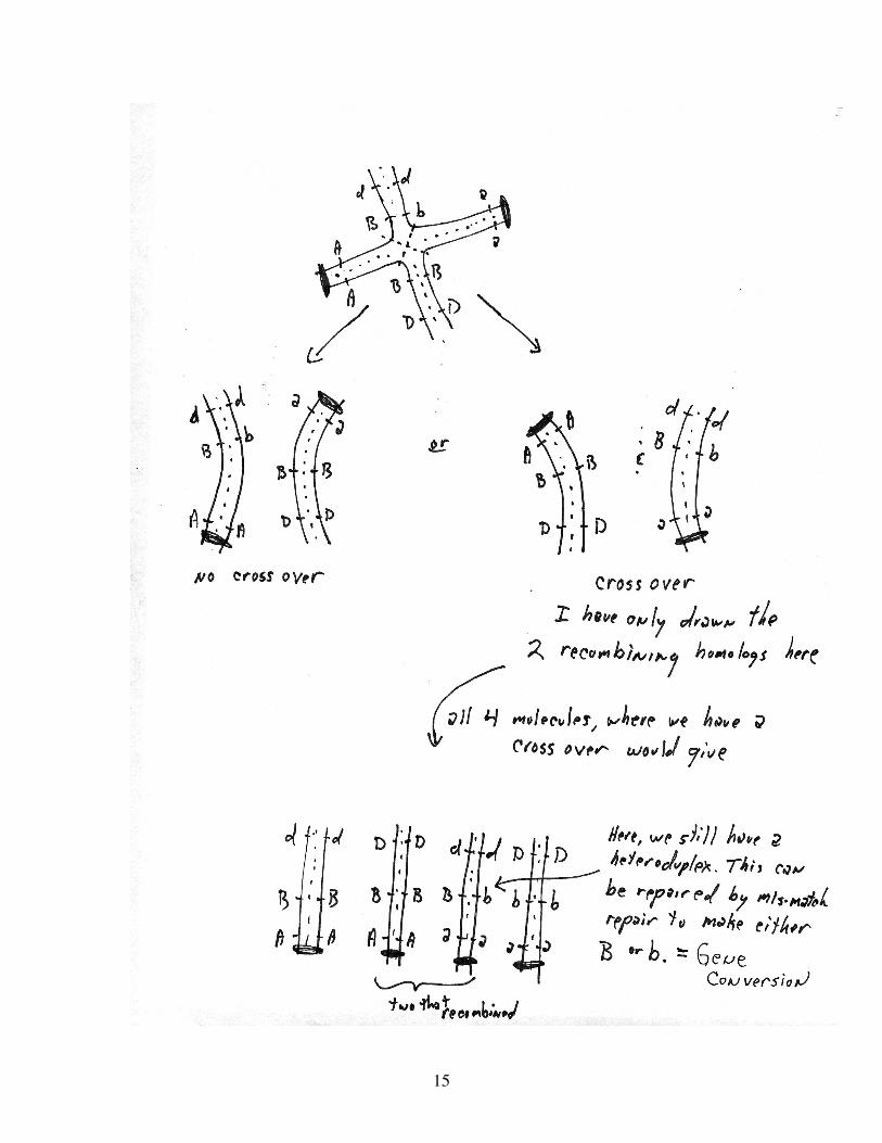
4) Recombination:
How does the recombination work. The molecular basis of gene conversion
Let’s just look at the recombination step, after S phase and before the first division.
15
16
5) The study of crossovers typically refers to gene conversions COs and NCOs
Thus, gene conversions could arise as a simple consequence of how the cross over is
resolved. That is, no mechanism other than the one shown needs to be involved. But, this
is unlikely the case. Several factors suggest that crossovers occur by a distinct mechanism.
a) Some mutations alter the ratio of crossovers to non-crossovers.
b) Crossovers occur much later in meiosis


![Engineering the oleaginous yeast Yarrowia lipolytica for ... · Liu et al. Biotechnol Biofuels Page 2 of 11 compounds.Forexample,artemisininisasesquiterpene endoperoxidewitheectiveantimalarialproperties[12],](https://static.fdocument.org/doc/165x107/5f7b487302e1e8790c2f553c/engineering-the-oleaginous-yeast-yarrowia-lipolytica-for-liu-et-al-biotechnol.jpg)