Lecture 3. α domain structures Coiled-coil, knobs and hole packing Four-helix bundle Donut ring...
15
Lecture 3. α domain structures •Coiled-coil, knobs and hole packing •Four-helix bundle •Donut ring large structure •Globin fold •Ridges and grooves model CS882, Fall 2006
-
Upload
linda-rose -
Category
Documents
-
view
245 -
download
1
Transcript of Lecture 3. α domain structures Coiled-coil, knobs and hole packing Four-helix bundle Donut ring...
Lecture 3. α domain structures
•Coiled-coil, knobs and hole packing•Four-helix bundle
•Donut ring large structure•Globin fold
•Ridges and grooves model
CS882, Fall 2006
Packing together, they are stabler
While isolated α helices often occur in proteins, they are only marginally stable.
They are stabilized by being packed together through hydrophobic side chains.
We will worry about membrane proteins later.
Project: classify all alpha helix occurrences in the PDB. In PDB, they tell you which amino acid belongs to an alpha helix. However, you will need to decide properly which structure an alpha helix belongs to.
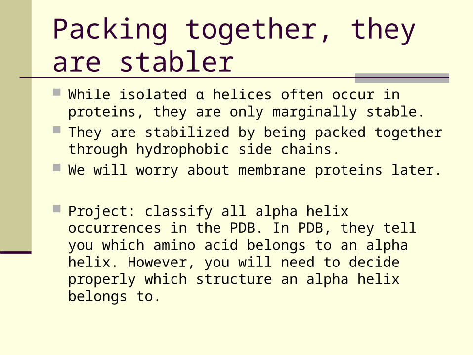
Coiled coil
In 1953, Francis Crick showed that the side chain interactions are maximized if the two alpha helices are wound around each other in a “coiled coil” arrangement.
Such structures are the basis of some fibrous proteins, and others. For fibers, sometimes many hundreds of amino acids to make long flexible dimmer.
Shorter coiled-coils are also used elsewhere.
Left handed supercoilfrom two right-handed helices
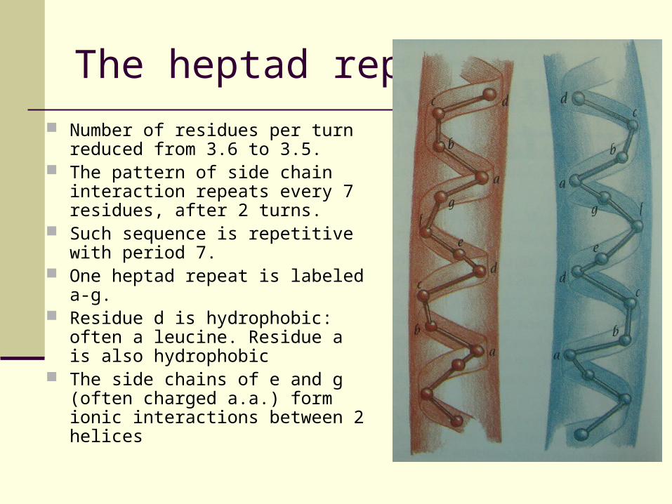
The heptad repeat
Number of residues per turn reduced from 3.6 to 3.5.
The pattern of side chain interaction repeats every 7 residues, after 2 turns.
Such sequence is repetitive with period 7.
One heptad repeat is labeled a-g. Residue d is hydrophobic: often a
leucine. Residue a is also hydrophobic
The side chains of e and g (often charged a.a.) form ionic interactions between 2 helices
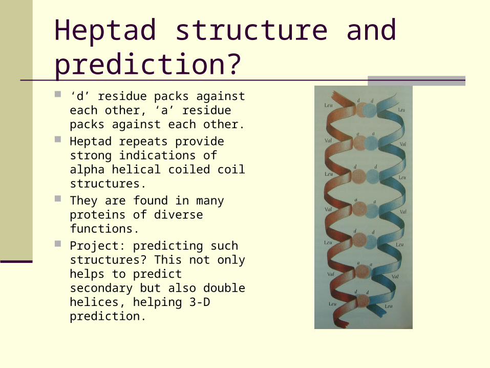
Heptad structure and prediction?
‘d’ residue packs against each other, ‘a’ residue packs against each other.
Heptad repeats provide strong indications of alpha helical coiled coil structures.
They are found in many proteins of diverse functions.
Project: predicting such structures? This not only helps to predict secondary but also double helices, helping 3-D prediction.
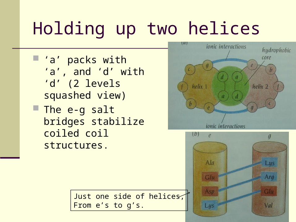
Holding up two helices
‘a’ packs with ‘a’, and ‘d’ with ‘d’ (2 levels squashed view)
The e-g salt bridges stabilize coiled coil structures.
Just one side of helices,From e’s to g’s.
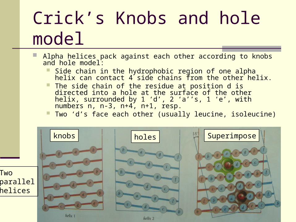
Crick’s Knobs and hole model Alpha helices pack against each other according to knobs and hole
model: Side chain in the hydrophobic region of one alpha helix can contact
4 side chains from the other helix. The side chain of the residue at position d is directed into a hole at
the surface of the other helix, surrounded by 1 ‘d’, 2 ‘a’’s, 1 ‘e’, with numbers n, n-3, n+4, n+1, resp.
Two ‘d’s face each other (usually leucine, isoleucine)
knobs holes Superimpose
Twoparallelhelices
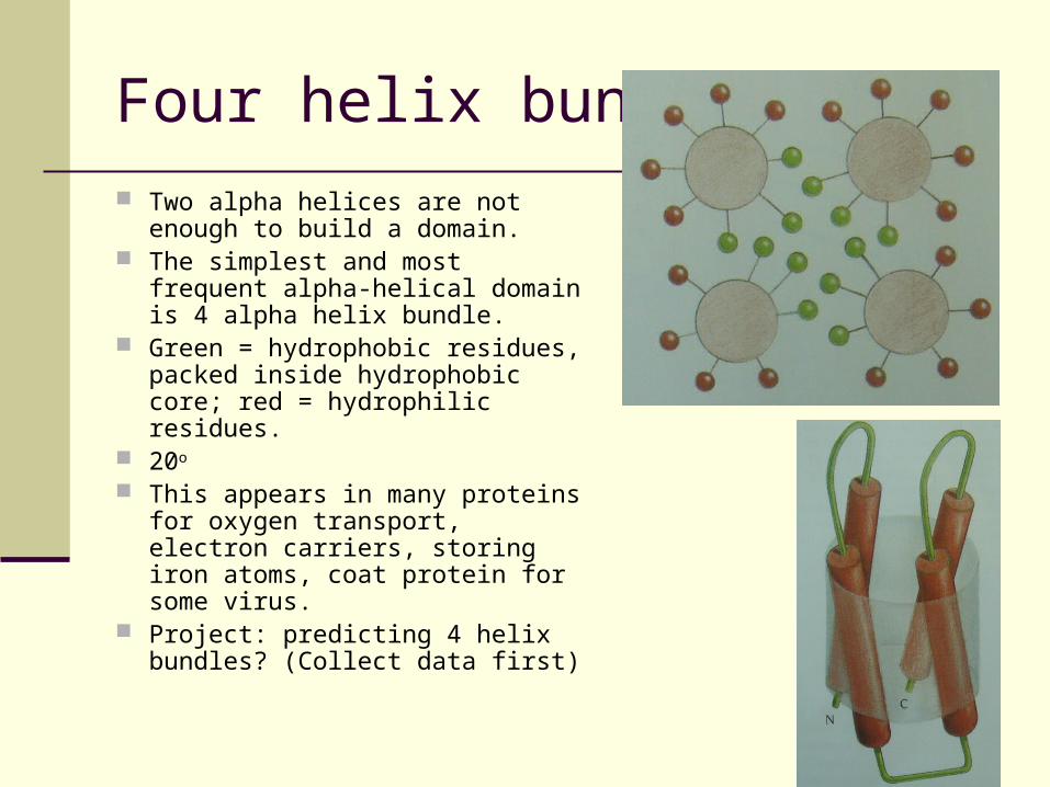
Four helix bundle
Two alpha helices are not enough to build a domain.
The simplest and most frequent alpha-helical domain is 4 alpha helix bundle.
Green = hydrophobic residues, packed inside hydrophobic core; red = hydrophilic residues.
20o
This appears in many proteins for oxygen transport, electron carriers, storing iron atoms, coat protein for some virus.
Project: predicting 4 helix bundles? (Collect data first)
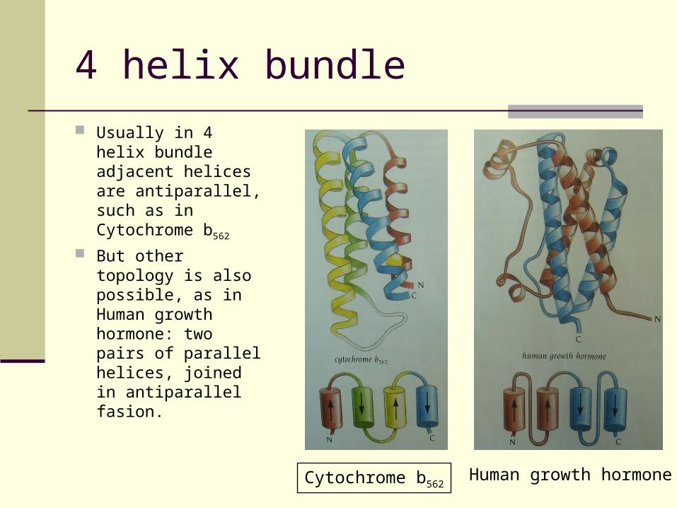
4 helix bundle
Usually in 4 helix bundle adjacent helices are antiparallel, such as in Cytochrome b562
But other topology is also possible, as in Human growth hormone: two pairs of parallel helices, joined in antiparallel fasion.
Cytochrome b562Human growth hormone
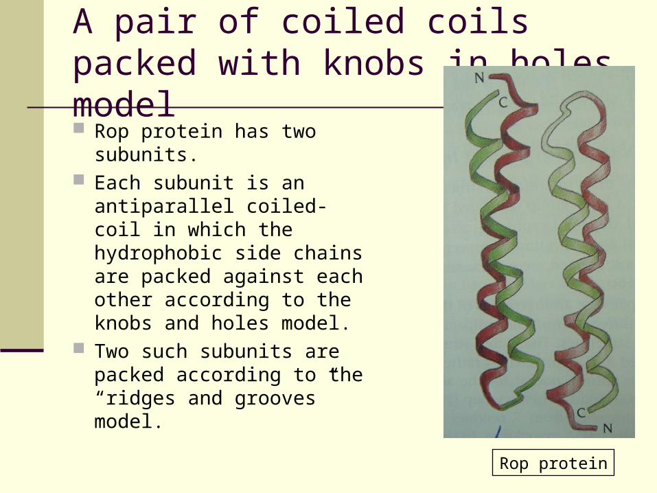
A pair of coiled coils packed with knobs in holes model Rop protein has two
subunits. Each subunit is an
antiparallel coiled-coil in which the hydrophobic side chains are packed against each other according to the knobs and holes model.
Two such subunits are packed according to the “ridges and grooves” model.
Rop protein
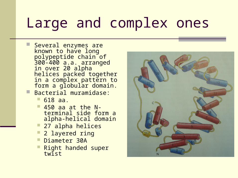
Large and complex ones Several enzymes are known
to have long polypeptide chain of 300-400 a.a. arranged in over 20 alpha helices packed together in a complex pattern to form a globular domain.
Bacterial muramidase: 618 aa. 450 aa at the N-terminal
side form a alpha-helical domain
27 alpha helices 2 layered ring Diameter 30A Right handed super twist
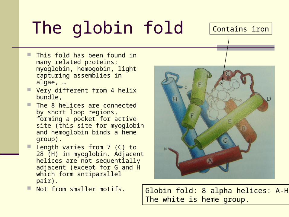
The globin fold
This fold has been found in many related proteins: myoglobin, hemogobin, light capturing assemblies in algae, …
Very different from 4 helix bundle, The 8 helices are connected by
short loop regions, forming a pocket for active site (this site for myoglobin and hemoglobin binds a heme group).
Length varies from 7 (C) to 28 (H) in myoglobin. Adjacent helices are not sequentially adjacent (except for G and H which form antiparallel pair).
Not from smaller motifs.
Globin fold: 8 alpha helices: A-H.The white is heme group.
Contains iron
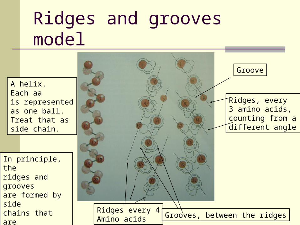
Ridges and grooves model
Ridges every 4Amino acids Grooves, between the ridges
Ridges, every 3 amino acids,counting from a different angle
Groove
A helix.Each aais representedas one ball.Treat that as side chain.
In principle, the ridges and grooves are formed by sidechains that are 4 or 3 aa apart, ridges fit into grooves, when fold.
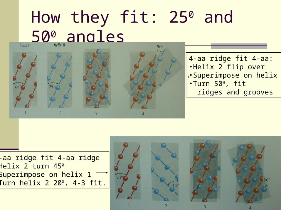
How they fit: 250 and 500 angles
4-aa ridge fit 4-aa:•Helix 2 flip over•Superimpose on helix 1•Turn 500, fit ridges and grooves
3-aa ridge fit 4-aa ridge•Helix 2 turn 450
•Superimpose on helix 1•Turn helix 2 200, 4-3 fit.
Evolution: what affects protein fold?
People studied 9 globin structures, trying to figure our what positions are important for protein to be folded that way.
They choose some “important” positions (functionally, or structurally) trying to see what made the difference.
They found essentially nothing (including size of amino acids, matters, except there is a striking preferential conservation of the hydrophobic character of the amino acids at the buried positions.
On the other hand, at the exposed positions, it actually does not matter whether we have mutations from hydrophilic to hydrophobic or vice versa, with one exception: the amino acid 6 in the beta chain in myoglobin (sickle cell disease)
Project: do this study in a larger class of proteins, and more carefully. (You can find protein class information at PDB, PFam, SCOP websites.) Other structures, not just helix?