J. Bacteriol. 2015 McGuffie JB.00784 15
63
1 1 2 3 A σ factor and anti-σ factor that control swarming motility and biofilm formation 4 in Pseudomonas aeruginosa 5 6 Bryan A. McGuffie a , Isabelle Vallet-Gely a *, Simon L. Dove a # 7 8 a Division of Infectious Diseases, Boston Children's Hospital, Harvard Medical School, 9 Boston, Massachusetts, USA 10 11 Running Head: A σ factor controls surface behavior in P. aeruginosa 12 13 #Address correspondence to Simon L. Dove, [email protected] 14 *Present address: Isabelle Vellet-Gely, CNRS, Centre de Génétique Moléculaire, 15 UPR3404, Gif-sur-Yvette, France. 16 17 JB Accepted Manuscript Posted Online 30 November 2015 J. Bacteriol. doi:10.1128/JB.00784-15 Copyright © 2015, American Society for Microbiology. All Rights Reserved.
-
Upload
anonymous-6oplc9u -
Category
Documents
-
view
218 -
download
1
description
Biofilm Research
Transcript of J. Bacteriol. 2015 McGuffie JB.00784 15

1
1
2
3
A σ factor and anti-σ factor that control swarming motility and biofilm formation 4
in Pseudomonas aeruginosa 5
6
Bryan A. McGuffiea, Isabelle Vallet-Gelya*, Simon L. Dovea# 7
8
aDivision of Infectious Diseases, Boston Children's Hospital, Harvard Medical School, 9
Boston, Massachusetts, USA 10
11
Running Head: A σ factor controls surface behavior in P. aeruginosa 12
13
#Address correspondence to Simon L. Dove, [email protected] 14
*Present address: Isabelle Vellet-Gely, CNRS, Centre de Génétique Moléculaire, 15
UPR3404, Gif-sur-Yvette, France. 16
17
JB Accepted Manuscript Posted Online 30 November 2015J. Bacteriol. doi:10.1128/JB.00784-15Copyright © 2015, American Society for Microbiology. All Rights Reserved.
2
Abstract 18
Pseudomonas aeruginosa is capable of causing a variety of acute and chronic 19
infections. Here, we provide evidence that sbrR (PA2895), a gene previously identified 20
as required during chronic P. aeruginosa respiratory infection, encodes an anti-σ factor 21
that inhibits the activity of its cognate extra-cytoplasmic function σ factor SbrI 22
(PA2896). Bacterial two-hybrid analysis identified an N-terminal region of SbrR that 23
interacts directly with SbrI and was sufficient for inhibition of SbrI-dependent gene 24
expression. We show that SbrI associates with RNA polymerase in vivo and identify the 25
SbrIR regulon. In cells lacking SbrR, the SbrI-dependent expression of muiA was found 26
to inhibit swarming motility and promote biofilm formation. Our findings uncover 27
SbrR and SbrI as a novel set of regulators of swarming motility and biofilm formation 28
in P. aeruginosa that mediate their effects through muiA, a gene not previously known 29
to influence surface-associated behaviors in this organism. 30
31
32
3
IMPORTANCE 33
This study characterizes a σ factor/anti-σ factor system that reciprocally regulates the 34
surface-associated behaviors of swarming motility and biofilm formation in the 35
opportunistic pathogen Pseudomonas aeruginosa. We present evidence that SbrR is an 36
anti-σ factor specific for its cognate σ factor SbrI and identify the SbrIR regulon in P. 37
aeruginosa. We find that cells lacking SbrR are severely defective for swarming motility 38
and exhibit enhanced biofilm formation. Moreover, we identify muiA (PA1494) as the 39
SbrI-dependent gene responsible for mediating these effects. SbrIR have been 40
implicated in virulence and in responding to antimicrobial and cell envelope stress. 41
SbrIR may therefore represent a stress-response system that influences the surface 42
behaviors of P. aeruginosa during infection.43
4
INTRODUCTION 44
The Gram-negative bacterium Pseudomonas aeruginosa is an opportunistic 45
human pathogen notorious for being the principal cause of morbidity and mortality in 46
cystic fibrosis (CF) patients (1). In patients with CF, chronic pulmonary colonization by 47
P. aeruginosa leads to chronic inflammation, progressive loss of lung function, and 48
eventually respiratory failure and death (1). P. aeruginosa is also the fifth leading cause 49
of nosocomial infections overall in the US and is the second most common cause of 50
ventilator-associated pneumonia (VAP) and catheter-associated urinary tract 51
infections (CAUTI) (2, 3). In patients with VAP or CAUTI, P. aeruginosa grows as a biofilm 52
on endotracheal tubes and catheters, respectively (4-6). In addition, P. aeruginosa is 53
thought to persist as a biofilm in the CF lung (7). P. aeruginosa biofilms are associated 54
with chronic infection and exhibit increased antibiotic resistance and resistance to 55
clearance by the immune system (8). Thus, the ability to form biofilms contributes 56
significantly to the clinical burden of P. aeruginosa infection. 57
In P. aeruginosa, growth as a biofilm is inversely regulated with a cooperative 58
form of multicellular surface motility called swarming (9-12). Swarming motility is 59
flagella-dependent and requires the secretion of surfactants regulated by quorum 60
sensing (13-16). In addition, swarming motility correlates with increased expression of 61
5
virulence factors and is associated with acute infection (9, 17). Several systems are 62
known to mediate the transition from motile, swarming cells, to cells growing as 63
sessile biofilms, including c-di-GMP signaling and the GacS/GacA two-component 64
system (9, 11, 12). 65
PA2895, which we refer to here as sbrR, was identified in a signature-tagged 66
mutagenesis (STM) screen as being required for persistence in a rat-lung model of 67
chronic P. aeruginosa respiratory infection (18). Although SbrR has no significant 68
homology to previously characterized proteins, sbrR is located in a putative bicistronic 69
operon downstream of the PA2896 gene encoding a putative extracytoplasmic 70
function (ECF) σ factor that we refer to here as SbrI (19). As a group, ECF σ factors are 71
frequently cotranscribed with their own negative regulator, a transmembrane anti-σ 72
factor (20, 21). Upon stimulation by the appropriate extracytoplasmic signal, the ECF σ 73
factor is released from the anti-σ factor, allowing it to associate with RNA polymerase 74
(RNAP) and activate expression of its regulon. 75
Here we present evidence that SbrI and SbrR are an ECF σ and anti-σ factor pair. 76
We identify the SbrIR regulon and show that SbrI and SbrR influence biofilm formation 77
and swarming motility by controlling the expression of muiA (PA1494). In particular, 78
we show that cells lacking sbrR are unable to engage in swarming motility and exhibit 79
6
increased biofilm formation due to the SbrI-dependent increase in muiA expression 80
observed in these cells. We have named PA2896 and PA2895 SbrI and SbrR, 81
respectively, as a result of the swarming and biofilm related phenotypes we observe in 82
ΔsbrR mutant cells. SbrI and SbrR constitute a pair of regulators controlling swarming 83
motility and biofilm formation in P. aeruginosa that mediate their effects through 84
MuiA.85
7
MATERIALS AND METHODS 86
Bacterial strains 87
E. coli DH5αFʹIQ (Invitrogen) was used as the recipient strain for all plasmid 88
constructions. E. coli SM10 λpir served as the conjugative donor for transferring 89
plasmids into P. aeruginosa during strain construction. P. aeruginosa strains used 90
included PAO1 (provided by A. Rietsch) and PA14 (provided by L. Rahme). Bacterial 91
cultures were routinely grown at 37°C in lysogeny broth (LB), or on plates containing 92
LB solidified with 1.5% agar unless otherwise noted. When appropriate, gentamicin (30 93
μg/ml) and carbenicillin (200 μg/ml) were used for selection in P. aeruginosa cultures. 94
A list of strains and plasmids used in this study are available in Table S1. 95
96
Plasmids and strains for tandem affinity purification (TAP)-tag experiments 97
Plasmid pP30Δ-PA2896-TAP was made by cloning an ~300 bp fragment of DNA 98
corresponding to a 3ʹ portion of the sbrI gene into pP30Δ-YTAP cut with HindIII and 99
NotI; the portion of the PA2896 gene was cloned such that it was in-frame with the 100
DNA specifying the TAP-tag. pP30Δ-SbrI-TAP was used to generate PAO1 SbrI-TAP as 101
previously described (22). Strain PAO1 RpoS-TAP was constructed in a similar way 102
using vector pP30Δ-RpoS-TAP, which contains a portion of the P. aeruginosa rpoS gene 103
8
fused in-frame to DNA specifying the TAP-tag. The PAO1 AceF-TAP strain, which 104
expresses AceF-TAP and serves as a control, has been described previously (22). 105
Plasmid pP30ΔFRT-SbrI-VSV-G was made by subcloning the HindIII/NotI sbrI fragment 106
from pP30Δ-SbrI-TAP into pP30ΔFRT-MvaT-VSV-G to replace mvaT, such that the 3ʹ 107
end of sbrI is in-frame with the VSV-G tag (23). PAO1 βʹ-TAP SbrI-V was constructed in 108
a similar manner by integrating pP30ΔFRT-SbrI-VSV-G into the previously described 109
strain PAO1 βʹ-TAP (24). 110
111
Reporter strain and plasmids for bacterial two-hybrid assays 112
Bacterial two-hybrid assays were performed with the E. coli reporter strain KS1 113
(25); KS1 harbors on its chromosome the placOR2-62 test promoter driving expression 114
of a linked lacZ reporter gene. Plasmids pACλcI32 and pBRαLN have been described 115
previously (26), and were used to create fusions to the C-terminus of λcI and the C-116
terminus of the α-linker, respectively. Plasmid pACλcI-SbrR-NTR encodes λcI (residues 117
1-236) fused to residues 2-64 of SbrR from P. aeruginosa via a small linker composed of 118
three alanine residues. Plasmid pACλcI-SbrR-NTR was made by cloning the 119
appropriate NotI-BamHI-digested PCR product into pACλcI32 that had been digested 120
with NotI and BstYI, thus placing expression of the λcI-SbrR-NTR fusion protein under 121
9
the control of the IPTG-inducible lacUV5 promoter. Plasmid pBRα-SbrI encodes 122
residues 1-248 of the α subunit of E. coli RNA polymerase fused to residues 2-194 of 123
SbrI from P. aeruginosa via a small linker composed of three alanine residues. Plasmid 124
pBRα-SbrI was made by cloning the appropriate NotI-BamHI-digested PCR product 125
into pBRαLN digested with NotI and BamHI, thus placing the α-fusion under the 126
control of tandem lpp and IPTG-inducible lacUV5 promoters. Plasmid pBRα encodes 127
wild type α under the control of tandem lpp and IPTG-inducible lacUV5 promoters and 128
has been described previously (25). 129
130
Promoter-lacZ fusion reporter strains 131
The PAO1 promoter-lacZ fusion reporter strains PAO1 attB::PsbrI-lacZ , PAO1 132
attB::PmuiA-lacZ, and PAO1 attB::PPA4495-lacZ contain the putative promoter regions of 133
sbrI, muiA, and PA4495, respectively, fused to the lacZ gene and integrated in single 134
copy into the ΦCTX locus in the PAO1 chromosome. The putative sbrI promoter region 135
consisted of the 327 bp intergenic region upstream of the sbrI start codon (see 136
www.pseudomonas.com) (27). This region was amplified by PCR and cloned into mini-137
CTX-lacZ as a BamHI/PstI fragment to generate mini-CTX-PsbrI-lacZ. The upstream 138
intergenic regions of muiA (122 bp) and PA4495 (275 bp) were also PCR amplified and 139
10
cloned into mini-CTX-lacZ on HindIII/BamHI fragments to generate mini-CTX-PmuiA-lacZ 140
and mini-CTX-PPA4495-lacZ, respectively. The resulting plasmids were integrated in single 141
copy into the ΦCTX site to create reporter strains PAO1 attB::PsbrI-lacZ , PAO1 attB::PmuiA-142
lacZ, and PAO1 attB::PPA4495-lacZ as previously described (28). 143
144
Construction of deletion mutant strains 145
The deletion construct for the sbrR gene was generated by amplifying regions 146
~ 700 bp in length that flank sbrR in the PAO1 genome by PCR and then splicing the 147
flanking regions together by overlap extension PCR. Due to a 4 bp overlap between 148
the 3ʹ end of sbrI and the 5ʹ end of sbrR, the deletion was designed such that sbrI 149
would not be disrupted by the sbrR deletion construct. The deletion was in-frame and 150
contained a 9-bp NotI-linker sequence 5ʹ-GCGGCCGCC-3ʹ separating the two flanking 151
regions. The resulting PCR product was cloned on a HindIII/XbaI fragment into plasmid 152
pEXG2 (29), yielding plasmid pEXG2-ΔsbrR. The PAO1 ΔsbrR and PA14 ΔsbrR deletion 153
strains were generated with this plasmid by allelic replacement as previously 154
described (30). Plasmid pEXG2-ΔsbrR was also used to generate the ΔsbrR mutant 155
reporter strains in a similar manner. The ΔsbrIR deletion construct was generated by 156
amplifying the ~ 700 bp 5ʹ flanking region of sbrI in the PAO1 genome by PCR. This 157
11
PCR product was digested with XbaI and NotI and cloned into pEXG2-ΔsbrR digested 158
with XbaI and NotI, such that the 5ʹ-flanking sbrI XbaI/NotI fragment replaced the 5ʹ 159
flanking region used for deleting sbrR, yielding plasmid pEXG2-ΔsbrIR. This plasmid 160
was used to create the ΔsbrIR deletion strains as previously described (30). The muiA 161
deletion construct was made in a similar fashion to the sbrR deletion construct. 162
Flanking regions ~ 700 bp in length on either side of muiA in the PAO1 genome were 163
amplified by PCR and spliced together by overlap extension PCR. The deletion was in-164
frame and included the NotI-linker as above. This PCR product was cloned into pEXG2 165
to generate pEXG2-ΔmuiA. The resulting plasmid was used to delete muiA as described 166
above. Deletions were confirmed by PCR. 167
168
Tandem affinity purification 169
Cells were grown at 37°C with aeration in 200 ml of LB in 1L flasks to an OD600 of 170
~ 1, then harvested by centrifugation at 4°C. TAP was then performed as described 171
(29). Purified proteins were concentrated using Amicon Ultra-4 centrifugal filtration 172
units with a 10 kDa molecular weight cut-off (Millipore), separated on 4-12% Bis-Tris 173
NuPAGE gel (Invitrogen) and stained with Coomassie blue. 174
12
175
Western blots 176
Purified proteins and cell lysates were separated on 4-12% Bis-Tris NuPAGE 177
(Invitrogen) and Western blotting was performed as described previously (22). The 178
VSV-G-tag was detected using polyclonal rabbit anti-VSV-G (Sigma-Aldrich) and 179
peroxidase-conjugated goat anti-rabbit IgG antibodies (Sigma-Aldrich). 180
181
Bacterial two-hybrid assays 182
Cells were grown with aeration at 37°C in LB supplemented with kanamycin (50 183
μg/ml), carbenicillin (100 μg/ml), chloramphenicol (25 μg/ml), and IPTG at the 184
concentration indicated. β-galactosidase assays were performed as described (26). 185
Assays were performed three times in duplicate on separate occasions. A 186
representative data set is shown. Values are averages based on one experiment; 187
duplicate measurements differed by <10%. 188
189
Construction of sbrR, sbrI, and muiA expression plasmids 190
To make expression plasmid pPSV38-SbrR, a DNA fragment containing the P. 191
aeruginosa PAO1 sbrR coding sequence flanked by HindIII and BamHI sites was 192
13
amplified by PCR and cloned into pPSV38 (31). pPSV38-SbrR directs IPTG-inducible 193
synthesis of SbrI and confers resistance to gentamicin. The same process was used to 194
generate expression plasmid pPSV38-SbrI. To construct pPSV38-SbrR-NTR, a DNA 195
fragment encoding the first 64 residues of SbrR flanked by HindIII and BamHI sites was 196
amplified by PCR and cloned into pPSV38. To make pHERD20T-MuiA, a DNA fragment 197
containing the P. aeruginosa PA14 muiA coding sequence flanked by XbaI and PstI was 198
amplified by PCR and cloned into pHERD20T (32). pHERD20T-MuiA directs the 199
arabinose-inducible synthesis of MuiA and confers resistance to ampicillin. 200
201
Microarray experiments 202
Cells of PAO1 (pPSV38), PAO1 ΔsbrR (pPSV38), PAO1 ΔsbrR (pPSV38-SbrR), and 203
PAO1 ΔsbrIR (pPSV38) were grown with aeration at 37°C in 200 ml LB supplemented 204
with gentamicin (30 μg/ml). Biological duplicate cultures of each strain were 205
inoculated at a starting OD600 of 0.01 and grown to an OD600 of •0.5 (corresponding to 206
the mid-logarithmic phase of growth). RNA isolation, cDNA synthesis, cDNA 207
fragmentation, and labeling were performed as described previously (33). Labeled 208
cDNA was hybridized to Affymetrix GeneChip P. aeruginosa genome arrays 209
(Affymetrix) and GeneSpring GX was used to analyze data for statistically significant 210
14
changes in gene expression. The genes with changes in expression ≥2-fold (P value of 211
≤0.01) are listed in Table 1. The data discussed in this publication have been deposited 212
in NCBI's Gene Expression Omnibus (34) and are accessible through GEO Series 213
accession number GSE74917 (https://www.ncbi.nlm.nih.gov/geo/query/acc.cgi?acc= 214
GSE74917). 215
216
Reporter strain β-galactosidase assays 217
Cells were grown at 37°C with aeration in LB supplemented with gentamicin 218
(30 μg/ml). Cells were permeabilized with sodium dodecyl sulfate and CHCl3 and 219
assayed for β-galactosidase activity as described previously (26). Assays were 220
performed at least twice in biological triplicate. Representative data sets are shown. 221
222
Motility assays 223
Swimming motility and swarming motility assays were performed as previously 224
described (35, 36). Agar plates for assessing swimming motility and swarming motility 225
consisted of M8 medium supplemented with glucose, MgSO4, CAA, and 0.3% agar for 226
swim plates or 0.5% agar for swarm plates. 0.1% arabinose was added where indicted. 227
Swim plates were stab inoculated from colonies grown overnight on LB agar plates. 228
15
Swarm plates were inoculated with 3μl of liquid culture grown overnight in LB. Swim 229
plates and swarm plates were incubated ~20 hours at 37°C. Quantification of swim 230
and swarm zones was performed using ImageJ software. Experiments for testing 231
swimming motility and swarming motility were performed in biological triplicate or 232
quadruplicate on three separate days. The data shown is an aggregate of 3 separate 233
experiments normalized to wild type (WT). Subsurface twitching motility was assayed 234
as described previously (37). Bacteria were stab inoculated through a layer of LB agar 235
(1% agar) to the bottom of the petri dish. After incubation for ~24 hours at 37°C, the 236
twitching motility was examined by removing the agar and staining the attached cells 237
with Coomassie blue (Sigma-Aldrich). Quantification of twitching motility was 238
performed by measuring the maximum diameter in millimeters of the circular zones 239
formed by attached cells. Twitching motility experiments were performed in 240
quadruplicate on two separate occasions. Data shown represent the results of those 241
experiments shown in aggregate and normalized to WT. Statistical analysis of motility 242
data was performed using Prism version 6.0 (GraphPad). 243
244
Biofilm formation assay 245
16
Biofilm formation in 96-well microtiter plates was assayed as previously 246
described with modifications (38). Overnight cultures grown in LB were used to 247
inoculate fresh media to a starting OD600 of 0.1. Media was supplemented with 248
carbenicillin (200 μg/ml) and 1% arabinose as indicated. 100 μl of each bacterial 249
suspension was dispensed in quadruplicate into the wells of a Costar 96-well 250
polyvinylchloride microtiter plate and incubated for 8 hours at 37°C. Following 251
incubation, plates were washed twice with water and adherent biofilms were stained 252
with 150 μl of 0.1% Crystal violet for 15 minutes. Following staining, plates were 253
washed twice with water and allowed to dry overnight. Stained biofilms were 254
solubilized with 200 μl of 33% acetic acid and absorbance at 595 nm was read with a 255
Tecan Infinite 200 plate reader. Experiments were performed on at least two separate 256
occasions. Representative results are shown. 257
258
17
RESULTS 259
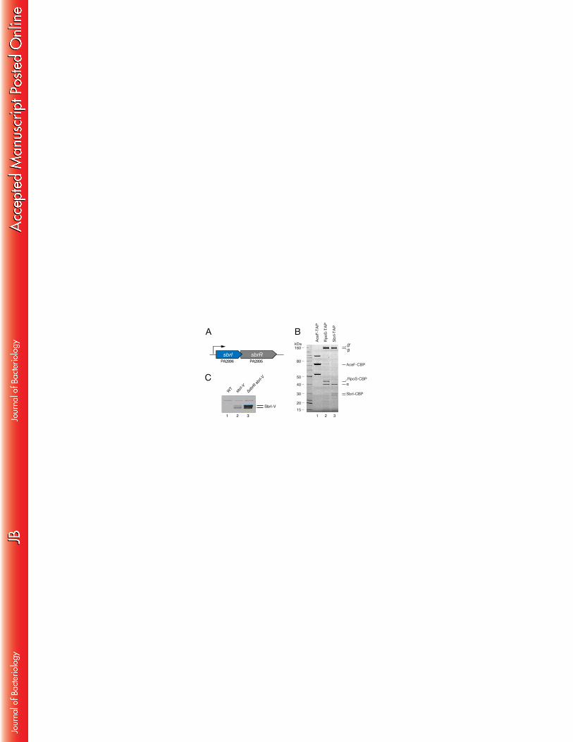
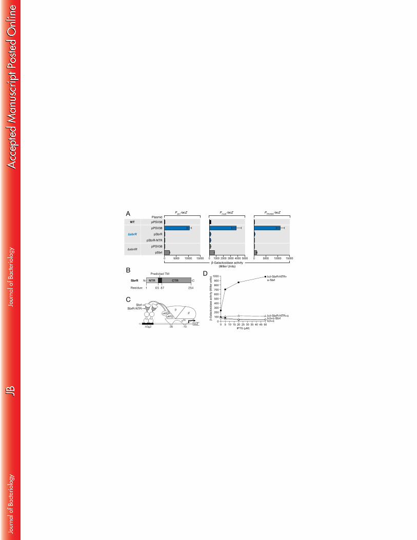
SbrI associates with RNA polymerase 260
sbrR was previously identified through a signature tagged transposon 261
mutagenesis screen as essential for the persistence of P. aeruginosa in a chronic 262
respiratory infection model (18). sbrR is predicted to be a component of a bicistronic 263
operon together with sbrI (Fig. 1A). A four base pair overlap in the coding sequences of 264
sbrR and sbrI suggests strong translational coupling of these genes. While SbrR has no 265
homology to any previously characterized proteins, SbrI is annotated as a probable 266
ECF σ factor on the Pseudomonas Genome Database and shares significant sequence 267
homology with other ECF σ factors (27). To determine if SbrI might function as a σ 268
factor, we purified SbrI from cells of P. aeruginosa and asked whether subunits of RNAP 269
co-purified with it. 270
To facilitate the purification of SbrI from P. aeruginosa we constructed a strain 271
of PAO1 that synthesized SbrI with a tandem affinity purification (TAP) tag fused to its 272
C-terminus (SbrI-TAP) from its native chromosomal location. As a positive control for 273
our ability to detect an association between a σ factor and RNAP, we also constructed 274
a second strain that synthesized the stationary phase-specific σ factor RpoS with a C-275
terminal TAP-tag (RpoS-TAP). As a negative control we used a previously constructed 276
18
strain that synthesizes AceF (a subunit of pyruvate dehydrogenase that is not 277
expected to interact with RNAP), with a TAP-tag fused to its C-terminus (AceF-TAP) 278
(22). We then purified SbrI, RpoS, and AceF by TAP and analyzed those proteins that 279
co-purified by SDS-PAGE followed by staining with Coomassie blue. Proteins with the 280
expected molecular weights for the β, βʹ, and α subunits of RNAP co-purify with both 281
RpoS-TAP and SbrI-TAP, but not the negative control AceF-TAP (Fig. 1B). This suggests 282
that SbrI associates with RNAP in vivo, consistent with its predicted function as a σ 283
factor. SbrI appears as a doublet in Fig. 1B, suggesting it can exist as a high molecular 284
weight and low molecular weight species. It is unclear if this doublet represents SbrI 285
processing, the use of an alternative translational start site, or if there is any functional 286
difference between these two forms. However, both forms co-purify with βʹ-TAP (Fig. 287
S1), suggesting that both forms are capable of associating with RNAP. 288
289
SbrR negatively influences the abundance of SbrI 290
The genomic arrangement of sbrI and sbrR is consistent with that of an ECF σ 291
factor and its cognate anti-σ factor. In the absence of their cognate anti-σ factor 292
autoregulated σ factors can become constitutively active, resulting in increased 293
abundance of the σ factor. However, in some cases ECF σ factors exhibit reduced 294
19
activity and abundance in the absence of their cognate anti-σ factors, which are 295
thought to stabilize and protect the σ factor from degradation (39, 40). Alternatively, 296
anti-σ factors can also promote the proteolysis of their cognate σ factor (41). A 297
comparison of the abundance of SbrI with a C-terminal VSV-G epitope tag (SbrI-V) in 298
wild-type (WT) and ΔsbrR mutant cells revealed that SbrI-V (synthesized from its native 299
locus) is more abundant in the absence of SbrR (Fig. 1C). This suggests that SbrR 300
negatively influences the abundance of SbrI and that SbrR might inhibit the 301
expression of sbrI. 302
303
Transcription from the sbrI promoter is negatively regulated by SbrR 304
After observing increased SbrI protein abundance in cells of the ΔsbrR mutant 305
strain, we were interested in determining whether SbrR represses transcription from 306
the sbrI promoter. To test this, we integrated a construct with the putative sbrI 307
promoter region upstream of a lacZ reporter at the ɸCTX phage attachment site on the 308
PAO1 chromosome to generate the reporter strain PAO1 attB::PsbrI-lacZ. We then 309
created an in-frame ΔsbrR deletion in this strain to test the effects of SbrR on 310
expression from the sbrI promoter. In cells of the ΔsbrR mutant reporter strain, β-311
galactosidase activity increased 45-fold relative to that observed in cells of the WT 312
20
reporter strain (Fig. 2A, left graph), suggesting that SbrR inhibits transcription from the 313
sbrI promoter. This increase was restored to WT levels by complementation with sbrR 314
from a plasmid (Fig. 2A, left graph). Cells of the ΔsbrIR double mutant strain exhibited 315
basal levels of β-galactosidase activity similar to that observed in cells of the WT 316
reporter strain (Fig. 2A, left graph), suggesting that expression from the sbrI promoter 317
is sbrI-dependent. Ectopic expression of sbrI in cells of the ΔsbrIR mutant strain 318
resulted in an increase in β-galactosidase activity (Fig. 2A), confirming the positive 319
regulatory effect of SbrI on its own promoter. Expression of the PsbrI-lacZ reporter was 320
higher in cells of the ΔsbrR mutant than in cells of the ΔsbrIR double mutant in which 321
sbrI was under the control of a heterologous promoter (Fig. 2A). This difference might 322
be explained by a SbrI-dependent positive feedback loop that serves to amplify sbrI 323
expression only when sbrI is under the control of its native promoter. Taken together, 324
these results suggest that SbrR inhibits SbrI-dependent transcription and that sbrI is 325
positively autoregulated, consistent with a model in which SbrI is an ECF σ factor that 326
controls its own expression and SbrR is its cognate anti-σ factor. 327
328
The N-terminal region of SbrR inhibits the activity of SbrI 329
It has been shown that the N-terminal cytoplasmic region of ECF anti-σ factors 330
21
can be sufficient for anti-σ factor activity (40, 42-44). SbrR is predicted to contain a 331
single transmembrane α-helix from residue 65 to 87, with a cytoplasmic N-terminal 332
domain and a periplasmic C-terminal domain, consistent with the membrane 333
topology of other ECF anti-σ factors (Fig. 2C). To determine if the N-terminal region of 334
SbrR was capable of inhibiting SbrI-dependent gene expression, we truncated SbrR at 335
the start of the predicted transmembrane domain to produce SbrR-NTR (residues 1-64) 336
(Fig. 2B). Levels of β-galactosidase activity in the ΔsbrR reporter strain expressing SbrR-337
NTR are indistinguishable from those expressing full length SbrR (Fig. 2A), which 338
suggests SbrR-NTR contains the region of SbrR necessary for inhibiting SbrI activity. It 339
further suggests that SbrR-NTR may contain a domain that is capable of interacting 340
with SbrI. 341
342
The N-terminal region of SbrR interacts with SbrI 343
ECF anti-σ factors inhibit their cognate σ factors by binding to them directly 344
and preventing their association with RNAP (42, 44-46). We have shown that both SbrR 345
and SbrR-NTR inhibit SbrI-dependent gene expression (Fig. 2B), and we next sought to 346
determine whether SbrR directly interacts with SbrI using a bacterial two-hybrid 347
system. 348
22
In this two-hybrid system, the detection of a protein-protein interaction relies 349
on the observation that an interaction between a DNA-bound protein and a subunit of 350
RNAP can result in transcription activation of a test promoter (25, 26, 47). In the version 351
of the assay used here, contact between a protein fused to the α subunit of E. coli 352
RNAP and another protein (or protein domain) fused to the λcI DNA-binding protein 353
activates the transcription of a lacZ reporter gene situated downstream of an 354
appropriate test promoter containing a λcI binding site (Fig. 2C). 355
To test whether SbrR could interact directly with SbrI we created two 356
compatible plasmids, one expressing SbrR-NTR (residues 2-64) fused to the C-terminus 357
of λcI and the other expressing an α fusion protein where the C-terminal domain (CTD) 358
of α has been replaced with full-length SbrI (residues 2-194). We then determined 359
whether the resulting λcI-SbrR-NTR fusion protein could activate transcription from 360
the test promoter in cells that also synthesized the α-SbrI fusion protein. Plasmids 361
directing the synthesis of the λcI-SbrR-NTR and the α-SbrI fusion proteins were used to 362
transform E. coli strain KS1, which harbors the Plac-OR2-62 test promoter (depicted in 363
Fig. 2C) linked to lacZ and integrated in single copy in the E. coli chromosome (26). We 364
found the λcI-SbrR-NTR fusion protein strongly activated the transcription of the lacZ 365
reporter in cells that also synthesize the α-SbrI fusion protein, but not in cells that only 366
23
contained WT α (Fig. 2D). Additional controls revealed that WT λcI fails to activate 367
expression of the lacZ reporter in the presence of the α-SbrI fusion protein or in the 368
presence of WT α (Fig. 2D). These results suggest that SbrR and SbrI directly interact, 369
consistent with the hypothesis that SbrR is the cognate anti-σ factor of SbrI. 370
371
Defining the SbrIR regulon 372
Next, we wanted to identify the genes controlled by SbrI and SbrR. Based on 373
our above results that show SbrR inhibits the expression of sbrI and that SbrI is 374
positively autoregulated, we reasoned the SbrI regulon would be constitutively 375
expressed in ΔsbrR mutant cells. Using DNA microarrays, we compared gene 376
expression in ΔsbrR mutant cells, ΔsbrIR mutant cells, and WT cells containing an 377
empty vector, together with ΔsbrR mutant cells containing a vector that expresses 378
sbrR. 379
Compared to WT cells, the expression of 21 genes changed >2-fold in cells of 380
the ΔsbrR mutant (Table 1). The expression of three genes in particular was strongly 381
influenced by the deletion of sbrR. Specifically, the expression of muiA (PA1494) was 382
103-fold higher in cells of the ΔsbrR mutant than in WT cells, while expression of 383
PA4495 and sbrI was 22- and 18-fold higher, respectively, in cells of the ΔsbrR mutant 384
24
when compared to WT (Table 1). The effects of the ΔsbrR deletion on muiA, PA4495 385
and sbrI expression could be complemented by providing sbrR in trans from a plasmid 386
(Table 1). Furthermore, upregulation of muiA and PA4495 did not occur in cells of the 387
ΔsbrIR double mutant, suggesting that the upregulation of these genes observed in 388
cells of the ΔsbrR single mutant is dependent upon SbrI (Table 1). Consistent with the 389
results of our microarray analyses, the expression of putative muiA promoter- and 390
PA4495 promoter-lacZ fusions was upregulated in cells of a ΔsbrR mutant compared to 391
WT (Fig. 2A). Moreover, this upregulation could be complemented by ectopic 392
synthesis of SbrR or SbrR-NTR from a plasmid, and was dependent upon SbrI (Fig. 2A). 393
Taken together, our findings suggest that transcription from the PmuiA, PPA4495, and PsbrI 394
promoters is positively regulated by the ECF σ factor SbrI and inhibited by SbrI’s 395
cognate anti-σ factor SbrR. 396
Microarray analyses revealed that several genes exhibited differential 397
expression in ΔsbrR mutant cells relative to WT cells that did not respond to 398
complementation with pPSV38-SbrR, suggesting SbrR may not control the expression 399
of these genes (Table 1). Indeed, previous work in our lab may explain the differential 400
regulation of three genes (PA1202, PA1203, and PA2432) that fit this profile. We have 401
previously shown that expression of PA1202, PA1203, and PA2432 (bexR) is bistable 402
25
and subject to the control of a bistable switch mediated by the transcription regulator 403
BexR (48). Therefore, the differential expression of these genes may result from 404
bistable BexR activity, rather than changes in expression attributable to SbrI activation. 405
PA3876 (narK2) also exhibited higher expression levels in ΔsbrR mutant cells relative to 406
WT that were unaffected by complementation with pPSV38-SbrR (Table 1). The 407
expression of narK2 was also elevated in ΔsbrIR double mutant cells relative to WT 408
(Table 1), suggesting that the observed changes in narK2 expression are independent 409
of SbrI. 410
Several genes were found to be downregulated in ΔsbrR mutant cells relative 411
to WT. In particular, the mmsAB operon was found to be down-regulated roughly 9-412
fold in ΔsbrR mutant cells relative to WT (Table 1). MmsA and MmsB are enzymes 413
involved in valine metabolism (49). The mmsAB operon is positively regulated by the 414
divergently transcribed AraC-like transcription regulator MmsR (49), however no 415
changes in mmsR expression were observed by DNA microarray. These findings 416
suggest that although SbrR functions principally as a negative regulator of the muiA, 417
PA4495 and sbrI genes, SbrR might also exert positive effects on the expression of 418
some genes. 419
420
26
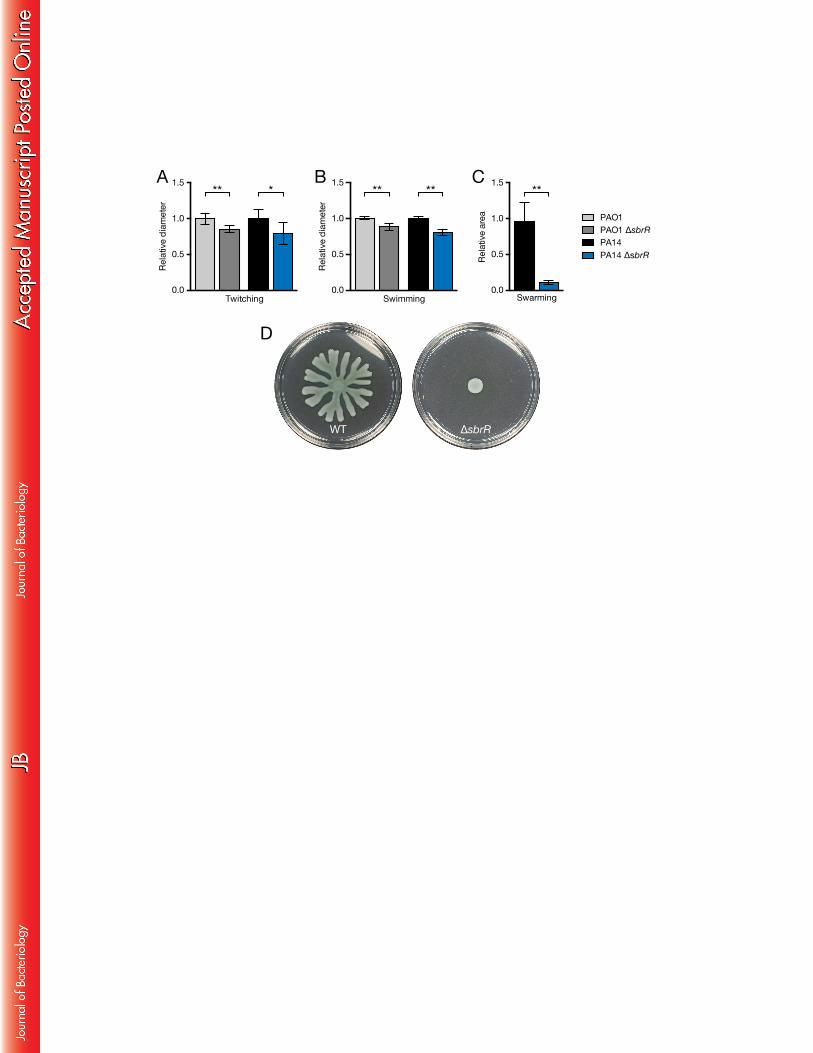
ΔsbrR mutants exhibit a severe swarming motility defect 421
P. aeruginosa is capable of several types of motility, including twitching, 422
swimming, and swarming. Twitching motility is a form of surface motility and is 423
mediated by type-IV pili (50). Swimming motility occurs in low viscosity liquid 424
environments and is a unicellular behavior dependent upon flagella and the 425
chemotaxis system (13). Swarming is a form of cooperative multicellular motility that 426
occurs on hydrated surfaces and in viscous liquid environments (13). In P. aeruginosa, 427
swarming motility is dependent upon flagella and secreted surfactants, the 428
production of which is controlled by quorum sensing (14, 15). On agar plates, WT P. 429
aeruginosa colonies grown overnight expand their borders via surface motility (51). 430
When we compared our WT and ΔsbrR mutant strains, we noticed colonies of ΔsbrR 431
mutant cells were slightly smaller (data not shown), suggesting these cells might have 432
a motility defect. To determine if SbrR influences motility, we compared twitching, 433
swimming, and swarming motility in WT and ΔsbrR mutants of P. aeruginosa strains 434
PAO1 and PA14. 435
Compared to WT, PAO1 ΔsbrR and PA14 ΔsbrR mutants exhibit reductions in 436
twitching motility of 14% and 20%, respectively (Fig. 3A). These findings suggest that 437
although ΔsbrR mutants have reduced twitching motility, cells of these mutants 438
27
continue to produce functional type-IV pili and engage in twitching motility. 439
Next we tested the swimming motility of our strains. PAO1 ΔflgK and PA14 440
ΔflgK mutants that do not produce flagella were unable to swim from the point of 441
inoculation (data not shown). Compared to WT, PAO1 and PA14 ΔsbrR mutants 442
exhibited reductions in swimming motility of 12% and 19%, respectively (Fig. 3B). 443
Thus, swimming motility is only slightly reduced in ΔsbrR mutant cells, suggesting that 444
these mutant cells continue to engage in chemotaxis and continue to produce 445
functional flagella. Fluorescent staining of WT PAO1 and PAO1 ΔsbrR revealed both WT 446
and mutant strains produce normal flagella (data not shown). 447
To test whether cells of our ΔsbrR mutant strains exhibited a swarming defect, 448
we inoculated cells from overnight cultures onto plates containing a minimal media 449
solidified with 0.5% agar. WT PA14 is a robust swarmer, forming colonies with 450
dendrites that extended radially from the point of inoculation in an irregular starburst 451
(Fig. 3D). In contrast to our WT PA14 strain, cells of our WT PAO1 strain did not swarm 452
appreciably under these conditions, precluding an analysis of swarming motility in this 453
strain background (data not shown). When we examined our PA14 ΔsbrR mutant strain 454
we discovered it does not spread from the point of inoculation and is completely 455
28
defective for swarming motility (Fig. 3C and D). This suggests that SbrR promotes 456
swarming motility or that SbrR represses an inhibitor(s) of swarming motility. 457
458
The effect of SbrR on swarming motility is dependent upon SbrI and MuiA 459
Given SbrR’s role as an anti-σ factor, we reasoned that constitutive activation of 460
SbrI might be responsible for the motility defect in ΔsbrR mutant cells. To determine if 461
the swarming defect in PA14 ΔsbrR mutants was dependent upon sbrI, we constructed 462
the double mutant PA14 ΔsbrIR. Swarming motility of cells of the PA14 ΔsbrIR strain 463
was restored to WT levels (Fig. 4A), demonstrating that SbrI is necessary for swarming 464
inhibition in PA14 ΔsbrR mutant cells. 465
Next, we reasoned that SbrI-dependent inhibition of swarming motility in PA14 466
ΔsbrR mutants was likely due to the constitutive expression of a gene(s) in the SbrI 467
regulon. The three most highly upregulated genes in the cells of the ΔsbrR mutant in 468
our microarrays were muiA, PA4495, and sbrI itself (Table 1). MuiA and PA4495 have 469
not previously been implicated in swarming motility. However, ectopic expression of 470
muiA (mucoidy inhibitor A) has been shown to suppress alginate overproduction in 471
mucoid strains that retain WT MucA (52). Neither MuiA nor PA4495 have any 472
significant homology to any previously characterized proteins, but both proteins are 473
29
predicted to contain N-terminal secretion signals, and have been found 474
experimentally in the periplasm (53). To test whether MuiA or PA4495 contributed to 475
the inhibition of swarming motility exhibited by the ΔsbrR mutant strain, we 476
generated PA14 ΔsbrR ΔmuiA double mutants and PA14 ΔsbrR ΔPA4495 double 477
mutants. Unlike the cells of a ΔsbrR single mutant, cells of a ΔsbrR ΔmuiA mutant did 478
not exhibit a swarming motility defect and instead resembled WT PA14 with respect to 479
their ability to swarm (Fig. 4A). This finding indicates that MuiA is necessary for 480
swarming inhibition in ΔsbrR mutant cells. The PA14 ΔsbrR ΔmuiA double mutant 481
strain could be complemented by ectopic expression of muiA, which restored 482
swarming inhibition (Fig. 4B). In contrast, the PA14 ΔsbrR ΔPA4495 double mutant 483
strain exhibited a swarming motility defect similar to that of the PA14 ΔsbrR single 484
mutant (Fig. S2), suggesting PA4495 is not necessary for the inhibition of swarming 485
motility in ΔsbrR mutant cells. Together, these results suggest that the inhibition of 486
swarming motility in ΔsbrR mutant cells is the result of increased SbrI-dependent 487
expression of MuiA. 488
489
Expression of muiA is sufficient for the inhibition of swarming motility 490
In cells of the ΔsbrR mutant, the expression of muiA, PA4495, and sbrI is 491
30
increased (Table 1), and swarming motility is inhibited (Fig. 4A and B). Moreover, MuiA 492
is necessary for the inhibition of swarming motility in the ΔsbrR mutant strain (Fig. 4A). 493
Therefore we wondered whether ectopic expression of muiA would suffice to inhibit 494
swarming motility, or if MuiA-mediated inhibition of swarming motility in the ΔsbrR 495
mutant strain required activation of the entire SbrI regulon. To test this, we 496
transformed WT PA14 cells with the same muiA expressing plasmid used to 497
complement the PA14 ΔsbrR ΔmuiA double mutant (pHERD20T-MuiA) and an empty 498
vector control (pHERD20T). Swarming motility was inhibited to levels comparable to 499
that of the ΔsbrR mutant strain in WT PA14 cells transformed with pMuiA (pHERD20T-500
MuiA), but not in WT PA14 cells transformed with the empty vector control 501
(pHERD20T) (Fig. 4B). Thus, ectopic expression of muiA is sufficient for the inhibition of 502
swarming motility. 503
504
ΔsbrR mutant cells exhibit enhanced biofilm formation 505
The surface-associated behaviors of swarming motility and biofilm formation 506
are inversely co-regulated in P. aeruginosa PA14 (9-12). That is, strains that exhibit 507
increased swarming motility produce less biofilm, while strains that produce increased 508
levels of biofilm exhibit reduced or inhibited swarming motility. Given this 509
31
relationship, we next asked whether PA14 ΔsbrR mutant cells were altered with 510
respect to their ability to form biofilms. Following 8 hours of static growth, cells of the 511
non-swarming PA14 ΔsbrR mutant strain formed ~2-fold more biofilm than cells of the 512
WT strain (Fig. 4C). This finding suggests that SbrR reduces biofilm formation or 513
inhibits factors that facilitate biofilm formation. 514
515
The effect of SbrR on biofilm formation is dependent on SbrI and MuiA 516
Next, we tested the PA14 ΔsbrIR double mutant strain to determine if the 517
increase in biofilm formation in the ΔsbrR mutant strain was SbrI-dependent. PA14 518
ΔsbrIR formed biofilms at levels similar to WT (Fig. 4C), suggesting that in addition to 519
inhibiting swarming motility, increased expression of the SbrI regulon in the ΔSbrR 520
mutant strain also promotes biofilm formation. 521
Our previous finding that the swarming motility defect of the ΔsbrR mutant 522
strain was MuiA-dependent led us to ask whether MuiA was also required for 523
enhanced biofilm formation in the ΔsbrR strain. Cells of a PA14 ΔsbrR ΔmuiA mutant 524
strain formed biofilms at levels similar to WT (Fig. 4C), indicating MuiA is necessary for 525
the enhanced biofilm formation observed in the ΔsbrR mutant strain. In addition, 526
biofilm formation in PA14 ΔsbrR ΔmuiA mutants could be restored to ΔsbrR mutant 527
32
levels by expressing muiA from a plasmid (Fig. 4D). These findings suggest that 528
enhanced biofilm formation in the ΔsbrR mutant strain may be the result of increased 529
SbrI-dependent expression of muiA. Lastly, while MuiA is required for increased biofilm 530
formation in the ΔsbrR mutant strain, the PA14 ΔmuiA mutant strain formed biofilms at 531
WT levels (Fig. 4C). This indicates MuiA is not required for biofilm formation in WT cells 532
of P. aeruginosa PA14 under the conditions of our experiments. 533
534
Expression of muiA is sufficient to promote biofilm formation 535
Ectopic expression of muiA in WT PA14 results in a swarming motility defect 536
equivalent to that observed in cells of the ΔsbrR mutant strain (Fig. 4B), suggesting it is 537
sufficient for the inhibition of swarming motility. In light of this observation and the 538
inverse relationship between swarming motility and biofilm formation, we next asked 539
whether ectopic expression of muiA was also sufficient to enhance biofilm formation. 540
In WT PA14 cells transformed with a muiA expression construct, but not those 541
transformed with an empty vector, there is an increase in biofilm formation that is 542
comparable to that seen in ΔsbrR mutants (Fig. 4D), demonstrating that ectopic 543
expression of muiA is sufficient to promote biofilm formation. Taken together, our 544
findings suggest that in ΔsbrR mutant cells, constitutive activation of SbrI results in 545

33
high levels of muiA expression, which enhances biofilm formation via an unknown 546
mechanism.547
34
DISCUSSION 548
We have presented evidence that SbrI and SbrR constitute a σ factor and anti-σ 549
factor pair with the N-terminal portion of SbrR interacting directly with SbrI. Cells 550
lacking SbrR are defective for swarming motility and exhibit enhanced biofilm 551
formation as a result of the SbrI-dependent increase in muiA expression that occurs in 552
these cells. SbrR and SbrI represent a novel set of regulators of swarming motility and 553
biofilm formation in P. aeruginosa that mediate their effects through MuiA, a protein 554
not previously known to influence either of these processes. 555
556
The SbrIR regulon 557
Our transcription profiling experiments identified a relatively small number of 558
genes that appear to be negatively controlled by SbrR and are expressed in an SbrI-559
dependent manner. In particular, the expression of the putative sbrIR operon, muiA, 560
and PA4495 exhibited the largest changes in expression in cells of the ΔsbrR anti-σ 561
factor mutant relative to WT (Table 1). Using promoter-lacZ fusion reporter strains we 562
demonstrated that SbrR negatively regulates transcription from the promoters of 563
these genes. We also showed that the increase in transcription that occurs from these 564
promoters in the absence of SbrR is dependent upon SbrI. These findings support the 565
35
idea that SbrR is an anti-σ factor specific for SbrI and are consistent with those of a 566
previous study that reported PA2896 (SbrI)-dependent expression of muiA (PA1494) 567
and PA4495 in response to the overexpression of the periplasmic protease ctpA (19). 568
By aligning the transcription start sites of sbrI, muiA, and PA4495 derived from 569
RNA-seq studies, Seo and Darwin identified putative -10 and -35 promoter elements, 570
with the sequence TAACCCG-N16-CGTCTCA-N6-A (+1) (19). Using the Find Individual 571
Motif Occurrences (FIMO) program to search for this putative SbrI-dependent 572
promoter sequence in the PAO1 and PA14 genomes revealed statistically significant 573
matches to this consensus only in the promoters of sbrI, muiA, and PA4495 (False 574
Discovery Rate q-value ≤0.01) (data not shown). The sbrI, muiA, and PA4495 promoters 575
may therefore be the only ones that are recognized directly by RNAP containing SbrI. 576
We note that the SbrIR regulon defined here is not unusually small for ECF σ factors, 577
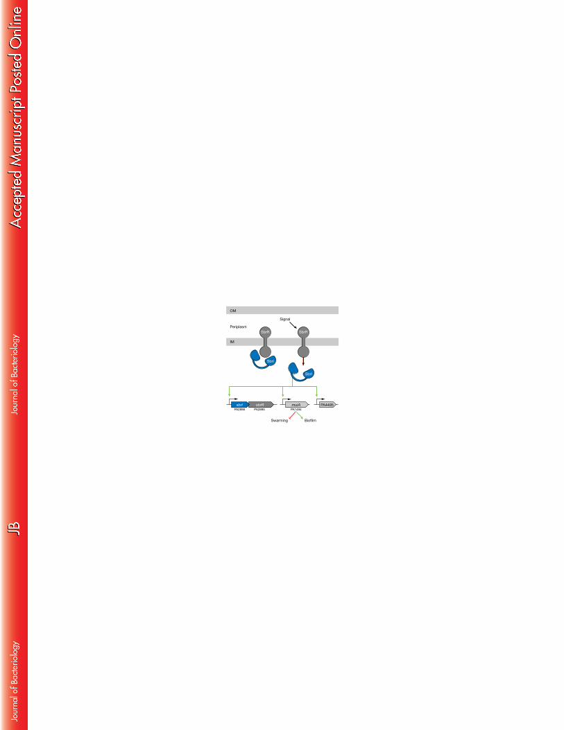
which frequently control the expression of relatively small sets of genes (20). 578
Taken together, our results suggest a model in which the anti-σ factor SbrR 579
binds to SbrI and sequesters it at the membrane, preventing it from associating with 580
RNAP (Fig. 5). In response to an unknown extracytoplasmic signal, SbrI is released from 581
SbrR and associates with RNAP, resulting in expression of the SbrI regulon (Fig. 5). The 582
SbrI regulon likely consists of the putative sbrIR operon, muiA, and PA4495. SbrI-583
36
dependent expression of SbrI results in a positive feedback loop, amplifying the 584
expression of the SbrI regulon. muiA and PA4495 are expressed at high levels and 585
exported to the periplasm, leading to muiA-dependent inhibition of swarming motility 586
and enhanced biofilm formation. Previous work has shown that the expression of sbrI, 587
muiA, and PA4495 becomes elevated following osmotic shock (54), following 588
treatment with the cell wall inhibitory antibiotic D-cycloserine (55), and following 589
ectopic expression of the periplasmic protease CtpA, which leads to disruption of the 590
cell envelope (19). We suggest that cell envelope stress might be sensed by SbrR 591
leading to SbrI activation, expression of muiA, and a transition from motile swarming 592
cells to growth as a biofilm (Fig. 5). It is also possible that CtpA is capable of directly 593
degrading SbrR, resulting in the activation of SbrI (19). 594
595
SbrR and SbrI control swarming motility and biofilm formation in P. aeruginosa 596
PA14 through MuiA 597
The muiA gene was the most highly upregulated member of the SbrI regulon in 598
ΔsbrR mutant cells relative to WT (Table 1), and expression from the PmuiA promoter was 599
shown to be SbrI-dependent (Fig. 2A) (19). We have shown that increased SbrI-600
dependent expression of muiA in ΔsbrR mutant cells inhibits swarming motility and 601
37
enhances biofilm formation, and that ectopic expression of muiA in WT cells has the 602
same effect. 603
Swarming motility in PA14 is dependent upon flagella and secreted surfactants 604
(14, 15). As demonstrated by our swimming assays, PA14 ΔsbrR mutants are capable of 605
chemotaxis and produce functional flagella (Fig. 3B). On the 0.5% agar plates used to 606
observe swarming, surfactant production can be observed as an area of wetness 607
surrounding surfactant-producing colonies (15, 16). We observed this region 608
surrounding non-swarming PA14 ΔsbrR mutant colonies, suggesting this strain 609
continues to produce surfactants (data not shown). These findings suggest the MuiA-610
dependent swarming motility defect in PA14 ΔsbrR cells is unlikely to be caused by a 611
defect in either flagella or surfactant production. 612
MuiA has previously been shown to inhibit the mucoid phenotype of certain 613
clinical isolates in which alginate is overproduced due to the presence of mutations 614
that activate AlgW (52). However, we think it unlikely that MuiA inhibits swarming 615
motility and promotes biofilm formation in PA14 through an inhibitory effect on 616
alginate production. Indeed, MuiA does not appear to be a general inhibitor of 617
alginate production; e.g. MuiA did not detectably influence the production of alginate 618
38
in cells that produce truncated versions of MucA (52). Furthermore, alginate does not 619
appear to be produced by WT PA14 during growth as a biofilm (56). 620
In addition to MuiA, several other systems have been shown to exert reciprocal 621
control over swarming motility and biofilm formation in P. aeruginosa, including c-di-622
GMP and the GacS/GacA two-component system (9, 11, 12). We note that the 623
abundance of the muiA and PA4495 transcripts is elevated in cells in which the 624
GacS/GacA system is artificially activated through deletion of retS (9). The GacS/GacA 625
system functions by activating the expression of genes encoding the small RNAs RsmY 626
and RsmZ (57). These small RNAs in turn sequester the RNA-binding protein RsmA that 627
binds many mRNAs directly to influence their translation or abundance (or both) (58). 628
Neither sbrI nor sbrR transcript abundance was altered in cells of a retS mutant relative 629
to WT (9), suggesting that the muiA and PA4495 transcripts may be direct targets of 630
RsmA. Future work will be aimed at identifying the mechanism by which MuiA 631
represses swarming motility and enhances biofilm formation in P. aeruginosa PA14. 632
633
Connections to virulence 634
A signature-tagged mutagenesis screen identified sbrR (PA2895) as a gene 635
required during chronic respiratory infection in a rat lung model (18). Interestingly, a 636
39
significant portion of the mutants identified in this screen exhibited impaired 637
swarming motility, indicating that swarming motility is an important virulence 638
determinant in this model of chronic respiratory infection (18). Although a sbrR 639
(PA2895) mutant in P. aeruginosa strain PA103 was previously shown to be defective 640
for protease secretion, we do not observe any protease secretion defect in cells of our 641
PAO1 ΔsbrR or PA14 ΔsbrR mutant cells (data not shown). The results presented here 642
suggest the virulence defect of sbrR mutants in the rat lung model of chronic 643
respiratory infection could be explained in whole or in part by their swarming motility 644
defect, or possibly through the misregulation of either swarming motility, biofilm 645
formation, or both. Recent Tn-Seq analyses indicate that SbrI is required for 646
colonization of the murine GI tract in a neutropenic model of acute infection (59). 647
SbrIR may therefore play important regulatory roles during both chronic and acute 648
infections.649
40
FUNDING INFORMATION 650
This work was funded by grant AI105013 from the NIH (to S.L.D). The funders had no 651
role in study design, data collection and interpretation, or the decision to submit the 652
work for publication. 653
654
ACKNOWLEDGEMENTS 655
We thank Heather McManus for constructing plasmid pP30ΔFRT-SbrI-VSV-G, Kirsty 656
McFarland for help with microarray data analysis, and Roger Levesque and Ann 657
Hochschild for discussions.658
41
FIGURE LEGENDS 659
660
FIG. 1. SbrI interacts with RNAP and is more abundant in ΔsbrR mutants. (A) The 661
putative sbrIR operon. (B) The β, βʹ, and α subunits of RNAP copurify with RpoS-TAP 662
(lane 2) and SbrI-TAP (lane 3), but not with AceF-TAP (lane 1). Purified proteins were 663
separated by SDS-PAGE and stained with Coomassie blue. AceF-CBP, RpoS-CBP, and 664
SbrI-CBP indicate the purified proteins with the calmodulin binding protein (CBP) 665
moiety that remains after cleaving the protein A moiety of the TAP tag during 666
purification. (C) SbrR has a negative effect on SbrI-V protein abundance. Anti-VSV-G 667
Western blot of WT PAO1 (lane 1), PAO1 SbrI-V (lane 2), and PAO1 ΔsbrR SbrI-V (lane 3). 668
669
FIG. 2. SbrR is an anti-σ factor that directly interacts with SbrI and inhibits its activity. 670
(A) β-galactosidase activity of PAO1 PsbrI-lacZ, PAO1 PmuiA-lacZ, and PAO1 PPA4495-lacZ 671
reporter strains. ΔsbrR and ΔsbrIR mutants were generated in each reporter strain and 672
transformed with the indicated plasmids. pPSV38 is an empty vector control. Error bars 673
represent standard deviations between three biological replicates. (B) Schematic 674
representation of SbrR and the location of its predicted transmembrane (TM) domain. 675
(C) Schematic representation of the bacterial two-hybrid system. Interaction between 676
42
the N-terminal region of SbrR (SbrR-NTR) fused to λcI and SbrI fused to the α subunit of 677
RNA polymerase activates transcription from the test promoter driving expression of 678
lacZ. The diagram depicts test promoter PlacλOR2-62, which bears the λ operator OR2 679
centered 62 bp upstream from the transcription start site of the lac core promoter. 680
This test promoter is linked to lacZ and located on the chromosome. (D) Effect of λcI-681
SbrR-NTR on transcription from PlacλOR2-62 in the presence of the α-SbrI chimera. Cells 682
harboring compatible plasmids directing the synthesis of the indicated proteins were 683
grown in the presence of different concentrations of IPTG and assayed for β-684
galactosidase activity. 685
686
FIG. 3. Swimming, twitching, and swarming motility of WT and ΔsbrR mutant strains. 687
(A) The relative diameter of twitching motility zones for the indicated PAO1 and PA14 688
strains normalized to WT for each strain. *p=0.02, **p<0.001 (B) The relative diameter 689
of swimming motility zones of the indicated PAO1 and PA14 strains normalized to WT 690
for each strain. **p<0.001 (C) The relative swarming motility of the indicated PA14 691
strains. Plates were photographed and the area of motile cells was quantified with 692
ImageJ. **p<0.001 (D) Representative image of PA14 swarming motility in WT and 693
ΔsbrR strains. Significant differences between strains were determined by t-test. Error 694
43
bars represent 95% confidence intervals. 695
696
FIG. 4. MuiA inhibits swarming motility and enhances biofilm formation. (A) Swarming 697
motility inhibition in ΔsbrR mutants is dependent upon sbrI and muiA. Representative 698
images of swarming plates are shown above the quantification of the area of each 699
strain’s swarm relative to WT PA14. (B) Swarming motility is inhibited by ectopic 700
expression of muiA. (C) Enhanced biofilm formation in PA14 ΔsbrR mutants is 701
dependent upon sbrI and muiA. (D) Ectopic expression of muiA results in enhanced 702
biofilm formation. Error bars represent 95% confidence intervals. 703
704
705
FIG. 5. A model of the SbrIR regulon. SbrR is an anti-σ factor that binds to SbrI and 706
inhibits its activity. In response to an unknown signal, SbrI is released from SbrR, 707
resulting in increased expression of the SbrI-regulon. Increased expression of muiA 708
results in the inhibition of swarming motility and enhanced biofilm formation. 709
710
44
REFERENCES 711
1. Govan JR, Deretic V. 1996. Microbial pathogenesis in cystic fibrosis: mucoid 712
Pseudomonas aeruginosa and Burkholderia cepacia. Microbiol Rev 60:539–574. 713
2. Hidron AI, Edwards JR, Patel J, Horan TC, Sievert DM, Pollock DA, Fridkin SK, 714
National Healthcare Safety Network Team, Participating National 715
Healthcare Safety Network Facilities. 2008. NHSN Annual 716
Update:Antimicrobial‐Resistant Pathogens Associated With Healthcare‐717
Associated Infections: Annual Summary of Data Reported to the National 718
Healthcare Safety Network at the Centers for Disease Control and Prevention, 719
2006–2007 • . Infect Control Hosp Epidemiol 29:996–1011. 720
3. Sievert DM, Ricks P, Edwards JR, Schneider A, Patel J, Srinivasan A, Kallen A, 721
Limbago B, Fridkin S, for the National Healthcare Safety Network (NHSN) 722
Team and Participating NHSN Facilities. 2013. Antimicrobial-Resistant 723
Pathogens Associated with Healthcare-Associated Infections: Summary of Data 724
Reported to the National Healthcare Safety Network at the Centers for Disease 725
Control and Prevention, 2009–2010. Infect Control Hosp Epidemiol 34:1–14. 726
4. Williams BJ, Dehnbostel J, Blackwell TS. 2010. Pseudomonas aeruginosa: host 727
45
defence in lung diseases. Respirology 15:1037–1056. 728
5. Hardalo C, Edberg SC. 1997. Pseudomonas aeruginosa: assessment of risk from 729
drinking water. Crit Rev Microbiol 23:47–75. 730
6. Donlan RM, Costerton JW. 2002. Biofilms: survival mechanisms of clinically 731
relevant microorganisms. Clin Microbiol Rev 15:167–193. 732
7. Singh PK, Schaefer AL, Parsek MR, Moninger TO, Welsh MJ, Greenberg EP. 733
2000. Quorum-sensing signals indicate that cystic fibrosis lungs are infected 734
with bacterial biofilms. Nature 407:762–764. 735
8. Costerton JW, Stewart PS, Greenberg EP. 1999. Bacterial biofilms: a common 736
cause of persistent infections. Science 284:1318–1322. 737
9. Goodman AL, Kulasekara B, Rietsch A, Boyd D, Smith RS, Lory S. 2004. A 738
signaling network reciprocally regulates genes associated with acute infection 739
and chronic persistence in Pseudomonas aeruginosa. Dev Cell 7:745–754. 740
10. Caiazza NC, Merritt JH, Brothers KM, O'Toole GA. 2007. Inverse regulation of 741
biofilm formation and swarming motility by Pseudomonas aeruginosa PA14. J 742
Bacteriol 189:3603–3612. 743
46
11. Kuchma SL, Brothers KM, Merritt JH, Liberati NT, Ausubel FM, O'Toole GA. 744
2007. BifA, a cyclic-Di-GMP phosphodiesterase, inversely regulates biofilm 745
formation and swarming motility by Pseudomonas aeruginosa PA14. J Bacteriol 746
189:8165–8178. 747
12. Merritt JH, Brothers KM, Kuchma SL, O'Toole GA. 2007. SadC reciprocally 748
influences biofilm formation and swarming motility via modulation of 749
exopolysaccharide production and flagellar function. J Bacteriol 189:8154–750
8164. 751
13. Kearns DB. 2010. A field guide to bacterial swarming motility. Nat Rev Microbiol 752
8:634–644. 753
14. Köhler T, Curty LK, Barja F, Van Delden C, Pechère JC. 2000. Swarming of 754
Pseudomonas aeruginosa is dependent on cell-to-cell signaling and requires 755
flagella and pili. J Bacteriol 182:5990–5996. 756
15. Rashid MH, Kornberg A. 2000. Inorganic polyphosphate is needed for 757
swimming, swarming, and twitching motilities of Pseudomonas aeruginosa. 758
Proc Natl Acad Sci USA 97:4885–4890. 759
47
16. Caiazza NC, Shanks RMQ, O'Toole GA. 2005. Rhamnolipids modulate 760
swarming motility patterns of Pseudomonas aeruginosa. J Bacteriol 187:7351–761
7361. 762
17. Overhage J, Bains M, Brazas MD, Hancock REW. 2008. Swarming of 763
Pseudomonas aeruginosa is a complex adaptation leading to increased 764
production of virulence factors and antibiotic resistance. J Bacteriol 190:2671–765
2679. 766
18. Potvin E, Lehoux DE, Kukavica-Ibrulj I, Richard KL, Sanschagrin F, Lau GW, 767
Levesque RC. 2003. In vivo functional genomics of Pseudomonas aeruginosa 768
for high-throughput screening of new virulence factors and antibacterial 769
targets. Environ Microbiol 5:1294–1308. 770
19. Seo J, Darwin AJ. 2013. The Pseudomonas aeruginosa periplasmic protease 771
CtpA can affect systems that impact its ability to mount both acute and chronic 772
infections. Infect Immun 81:4561-4570. 773
20. Helmann JD. 2002. The extracytoplasmic function (ECF) sigma factors. Adv 774
Microb Physiol 46:47–110. 775
48
21. Llamas MA, Imperi F, Visca P, Lamont IL. 2014. Cell-surface signaling in 776
Pseudomonas: stress responses, iron transport, and pathogenicity. FEMS 777
Microbiol Rev 38:569–597. 778
22. Vallet-Gely I, Donovan KE, Fang R, Joung JK, Dove SL. 2005. Repression of 779
phase-variable cup gene expression by H-NS-like proteins in Pseudomonas 780
aeruginosa. Proc Natl Acad Sci USA 102:11082–11087. 781
23. Castang S, McManus HR, Turner KH, Dove SL. 2008. H-NS family members 782
function coordinately in an opportunistic pathogen. Proc Natl Acad Sci USA 783
105:18947–18952. 784
24. Goldman SR, Sharp JS, Vvedenskaya IO, Livny J, Dove SL, Nickels BE. 2011. 785
NanoRNAs prime transcription initiation in vivo. Mol Cell 42:817–825. 786
25. Dove SL, Joung JK, Hochschild A. 1997. Activation of prokaryotic transcription 787
through arbitrary protein-protein contacts. Nature 386:627–630. 788
26. Dove SL, Hochschild A. 2004. A bacterial two-hybrid system based on 789
transcription activation. Methods Mol Biol 261:231–246. 790
27. Winsor GL, Lam DKW, Fleming L, Lo R, Whiteside MD, Yu NY, Hancock REW, 791
49
Brinkman FSL. 2011. Pseudomonas Genome Database: improved comparative 792
analysis and population genomics capability for Pseudomonas genomes. 793
Nucleic Acids Res 39:D596–600. 794
28. Hoang TT, Kutchma AJ, Becher A, Schweizer HP. 2000. Integration-proficient 795
plasmids for Pseudomonas aeruginosa: site-specific integration and use for 796
engineering of reporter and expression strains. Plasmid 43:59–72. 797
29. Rietsch A, Vallet-Gely I, Dove SL, Mekalanos JJ. 2005. ExsE, a secreted 798
regulator of type III secretion genes in Pseudomonas aeruginosa. Proc Natl Acad 799
Sci USA 102:8006–8011. 800
30. Hoang TT, Karkhoff-Schweizer RR, Kutchma AJ, Schweizer HP. 1998. A 801
broad-host-range Flp-FRT recombination system for site-specific excision of 802
chromosomally-located DNA sequences: application for isolation of unmarked 803
Pseudomonas aeruginosa mutants. Gene 212:77–86. 804
31. Vvedenskaya IO, Sharp JS, Goldman SR, Kanabar PN, Livny J, Dove SL, 805
Nickels BE. 2012. Growth phase-dependent control of transcription start site 806
selection and gene expression by nanoRNAs. Genes Dev 26:1498–1507. 807
50
32. Qiu D, Damron FH, Mima T, Schweizer HP, Yu HD. 2008. PBAD-based shuttle 808
vectors for functional analysis of toxic and highly regulated genes in 809
Pseudomonas and Burkholderia spp. and other bacteria. Appl Environ Microbiol 810
74:7422–7426. 811
33. Vallet-Gely I, Sharp JS, Dove SL. 2007. Local and Global Regulators Linking 812
Anaerobiosis to cupA Fimbrial Gene Expression in Pseudomonas aeruginosa. J 813
Bacteriol 189:8667–8676. 814
34. Edgar R, Domrachev M, Lash AE. 2002. Gene Expression Omnibus: NCBI gene 815
expression and hybridization array data repository. Nucleic Acids Res 30:207–816
210. 817
35. Ha D-G, Kuchma SL, O'Toole GA. 2014. Plate-based assay for swarming motility 818
in Pseudomonas aeruginosa. Methods Mol Biol 1149:67–72. 819
36. Ha D-G, Kuchma SL, O'Toole GA. 2014. Plate-based assay for swimming 820
motility in Pseudomonas aeruginosa. Methods Mol Biol 1149:59–65. 821
37. Castang S, Dove SL. 2012. Basis for the essentiality of H-NS family members in 822
Pseudomonas aeruginosa. J Bacteriol 194:5101–5109. 823
51
38. O'Toole GA, Kolter R. 1998. Flagellar and twitching motility are necessary for 824
Pseudomonas aeruginosa biofilm development. Mol Microbiol 30:295–304. 825
39. Mahren S, Braun V. 2003. The FecI extracytoplasmic-function sigma factor of 826
Escherichia coli interacts with the beta' subunit of RNA polymerase. J Bacteriol 827
185:1796–1802. 828
40. Mettrick KA, Lamont IL. 2009. Different roles for anti-sigma factors in 829
siderophore signalling pathways of Pseudomonas aeruginosa. Mol Microbiol 830
74:1257–1271. 831
41. Spencer MR, Beare PA, Lamont IL. 2008. Role of cell surface signaling in 832
proteolysis of an alternative sigma factor in Pseudomonas aeruginosa. J 833
Bacteriol 190:4865–4869. 834
42. Las Peñas De A, Connolly L, Gross CA. 1997. The sigmaE-mediated response to 835
extracytoplasmic stress in Escherichia coli is transduced by RseA and RseB, two 836
negative regulators of sigmaE. Mol Microbiol 24:373–385. 837
43. Missiakas D, Mayer MP, Lemaire M, Georgopoulos C, Raina S. 1997. 838
Modulation of the Escherichia coli sigmaE (RpoE) heat-shock transcription-factor 839
52
activity by the RseA, RseB and RseC proteins. Mol Microbiol 24:355–371. 840
44. Campbell EA, Tupy JL, Gruber TM, Wang S, Sharp MM, Gross CA, Darst SA. 841
2003. Crystal structure of Escherichia coli sigmaE with the cytoplasmic domain 842
of its anti-sigma RseA. Mol Cell 11:1067–1078. 843
45. Helmann JD. 1999. Anti-sigma factors. Curr Opin Microbiol 2:135–141. 844
46. Hughes KT, Gillen KL, Semon MJ, Karlinsey JE. 1993. Sensing structural 845
intermediates in bacterial flagellar assembly by export of a negative regulator. 846
Science 262:1277–1280. 847
47. Dove SL, Hochschild A. 1998. Conversion of the omega subunit of Escherichia 848
coli RNA polymerase into a transcriptional activator or an activation target. 849
Genes Dev 12:745–754. 850
48. Turner KH, Vallet-Gely I, Dove SL. 2009. Epigenetic control of virulence gene 851
expression in Pseudomonas aeruginosa by a LysR-type transcription regulator. 852
PLoS Genet 5:e1000779. 853
49. Steele MI, Lorenz D, Hatter K, Park A, Sokatch JR. 1992. Characterization of 854
the mmsAB operon of Pseudomonas aeruginosa PAO encoding 855
53
methylmalonate-semialdehyde dehydrogenase and 3-hydroxyisobutyrate 856
dehydrogenase. J Biol Chem 267:13585–13592. 857
50. Mattick JS. 2002. Type IV pili and twitching motility. Annu Rev Microbiol 858
56:289–314. 859
51. Semmler AB, Whitchurch CB, Mattick JS. 1999. A re-examination of twitching 860
motility in Pseudomonas aeruginosa. Microbiology 145:2863–2873. 861
52. Withers TR, Yin Y, Yu HD. 2013. Identification and characterization of a novel 862
inhibitor of alginate overproduction in Pseudomonas aeruginosa. Pathog Dis 863
70:185-188. 864
53. Imperi F, Ciccosanti F, Perdomo AB, Tiburzi F, Mancone C, Alonzi T, Ascenzi 865
P, Piacentini M, Visca P, Fimia GM. 2009. Analysis of the periplasmic proteome 866
of Pseudomonas aeruginosa, a metabolically versatile opportunistic pathogen. 867
Proteomics 9:1901–1915. 868
54. Aspedon A, Palmer K, Whiteley M. 2006. Microarray analysis of the osmotic 869
stress response in Pseudomonas aeruginosa. J Bacteriol 188:2721–2725. 870
55. Wood LF, Leech AJ, Ohman DE. 2006. Cell wall-inhibitory antibiotics activate 871
54
the alginate biosynthesis operon in Pseudomonas aeruginosa: roles of sigma22 872
(AlgT) and the AlgW and Prc proteases. Mol Microbiol 62:412–426. 873
56. Wozniak DJ, Wyckoff TJO, Starkey M, Keyser R, Azadi P, O'Toole GA, Parsek 874
MR. 2003. Alginate is not a significant component of the extracellular 875
polysaccharide matrix of PA14 and PAO1 Pseudomonas aeruginosa biofilms. 876
Proc Natl Acad Sci USA 100:7907–7912. 877
57. Brencic A, McFarland KA, McManus HR, Castang S, Mogno I, Dove SL, Lory S. 878
2009. The GacS/GacA signal transduction system of Pseudomonas aeruginosa 879
acts exclusively through its control over the transcription of the RsmY and RsmZ 880
regulatory small RNAs. Mol Microbiol 73:434–445. 881
58. Brencic A, Lory S. 2009. Determination of the regulon and identification of 882
novel mRNA targets of Pseudomonas aeruginosa RsmA. Mol Microbiol 72:612–883
632. 884
59. Skurnik D, Roux D, Aschard H, Cattoir V, Yoder-Himes D, Lory S, Pier GB. 885
2013. A comprehensive analysis of in vitro and in vivo genetic fitness of 886
Pseudomonas aeruginosa using high-throughput sequencing of transposon 887

55
libraries. PLoS Pathog 9:e1003582. 888
889
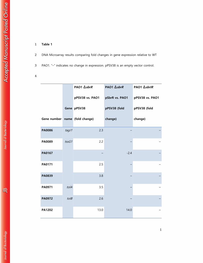
1
Table 1 1 DNA Microarray results comparing fold changes in gene expression relative to WT 2 PAO1. “–“ indicates no change in expression. pPSV38 is an empty vector control. 3 4
Gene number
Gene
name
PAO1 ∆sbrR
pPSV38 vs. PAO1
pPSV38
(fold change)
PAO1 ∆sbrR
pSbrR vs. PAO1
pPSV38 (fold
change)
PAO1 ∆sbrIR
pPSV38 vs. PAO1
pPSV38 (fold
change)
PA0086 tagJ1 2.3 – –
PA0089 tssG1 2.2 – –
PA0167 – -2.4 –
PA0171 2.5 – –
PA0839 3.8 – –
PA0971 tolA 3.5 – –
PA0972 tolB 2.6 – –
PA1202 13.0 14.0 –
2
PA1203 4.2 3.1 –
PA1493 cysP 2.8 – –
PA1494 muiA 103.1 -2.2 -2.2
PA2432 bexR 14.2 9.5 –
PA2895 sbrR – 6.0 -2.0
PA2896 sbrI 18.0 – -2.4
PA3179 – 2.1 2.2
PA3569 mmsB -9.7 – –
PA3570
mms
A
-9.3 – –
PA3876 narK2 2.8 6.9 4.9
PA3923 -5.4 – –
PA4495 21.9 – –
PA5172 arcB -2.6 – –
PA5315 rpmG 2.7 – –

3
PA5435 -2.4 – –
PA5445 2.5 – –
5 6 7


![Serdica Math. J. · Serdica Math. J. 33 (2007), 125{162 ON SOME EXTREMAL PROBLEMS OF LANDAU Szil ard R ev esz Communicated by V. Drensky ... Primzahlen" [15] Edmund Landau provided](https://static.fdocument.org/doc/165x107/5c64ca3b09d3f2a36e8bcb2a/serdica-math-j-serdica-math-j-33-2007-125162-on-some-extremal-problems.jpg)

![Ba^QdPc E RPW lPMcW^] - Farnell element145 P^\_McWOWZWch 5 § 5 @^ §@^ BVhbWPMZ EWjR HI g : g 5 I \\ ?MW] J J 7a^]c E_RMYRa J J 4R]cRa E_RMYRa J J DRMa E_RMYRa J J EdOf^^SRa g g 5WbP](https://static.fdocument.org/doc/165x107/5f62e0104f48cc34e33e05f9/baqdpc-e-rpw-lpmcw-farnell-5-pmcwowzwch-5-5-bvhbwpmz-ewjr-hi.jpg)