Involvement of Akt/NF-?B pathway in antitumor effects of
11
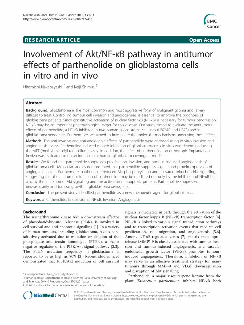
RESEARCH ARTICLE Open Access Involvement of Akt/NF-κB pathway in antitumor effects of parthenolide on glioblastoma cells in vitro and in vivo Hiromichi Nakabayashi 1* and Keiji Shimizu 2 Abstract Background: Glioblastoma is the most common and most aggressive form of malignant glioma and is very difficult to treat. Controlling tumour cell invasion and angiogenesis is essential to improve the prognosis of glioblastoma patients. Since constitutive activation of nuclear factor-κB (NF-κB) is necessary for tumour progression, NF-κB may be an important pharmacological target for this disease. Our study aimed to evaluate the antitumour effects of parthenolide, a NF-κB inhibitor, in two human glioblastoma cell lines (U87MG and U373) and in glioblastoma xenografts. Furthermore, we aimed to investigate the molecular mechanisms underlying these effects. Methods: The anti-invasive and anti-angiogenic effects of parthenolide were analysed using in vitro invasion and angiogenesis assays. Parthenolide-induced growth inhibition of glioblastoma cells in vitro was determined using the MTT (methyl thiazolyl tetrazolium) assay. In addition, the effect of parthenolide on orthotropic implantation in vivo was evaluated using an intracerebral human glioblastoma xenograft model. Results: We found that parthenolide suppresses proliferation, invasion, and tumour- induced angiogenesis of glioblastoma cells. Molecular studies demonstrated that parthenolide suppresses gene and protein expression of angiogenic factors. Furthermore, parthenolide reduced Akt phosphorylation and activated mitochondrial signalling, suggesting that the antitumour function of parthenolide may be mediated not only by the inhibition of NF-κB but also by the inhibition of Akt signalling and the activation of apoptotic proteins. Parthenolide suppressed neovascularity and tumour growth in glioblastoma xenografts. Conclusion: The present study identified parthenolide as a new therapeutic agent for glioblastomas. Keywords: Parthenolide, Glioblastoma, NF-κB, Invasion, Angiogenesis Background The serine/threonine kinase Akt, a downstream effector of phosphatidylinositol 3-kinase (PI3K), is involved in cell survival and anti-apoptotic signalling [1]. In a variety of human tumours, including glioblastoma, Akt is con- stitutively activated due to mutation or deletion of the phosphatase and tensin homologue (PTEN), a major negative regulator of the PI3K/Akt signal pathway [2,3]. The PTEN mutation frequency in glioblastoma is reported to be as high as 80% [3]. Recent studies have demonstrated that PI3K/Akt induction of cell survival signals is mediated, in part, through the activation of the nuclear factor kappa B (NF-κB) transcription factor [4]. NF-κB is linked to various signal transduction pathways and to transcription activation events that mediate cell proliferation, cell migration, and angiogenesis [5,6]. Among NF-κB-regulated genes [7], matrix metallopro- teinase (MMP)-9 is closely associated with tumour inva- sion and tumour-induced angiogenesis, and vascular endothelial growth factor (VEGF) promotes tumour- induced angiogenesis. Therefore, inhibition of NF-κB may serve as an effective treatment strategy for many tumours through MMP-9 and VEGF downregulation and disruption of Akt signalling. Parthenolide, a major sesquiterpene lactone from the plant Tanacetum parthenium, inhibits NF-κB both * Correspondence: [email protected] 1 Human Biology, Department of Health Sciences, Oita University of Nursing and Sciences, 2944-9 Megusuno, Oita 870-1201, Japan Full list of author information is available at the end of the article © 2012 Nakabayashi and Shimizu; licensee BioMed Central Ltd. This is an Open Access article distributed under the terms of the Creative Commons Attribution License (http://creativecommons.org/licenses/by/2.0), which permits unrestricted use, distribution, and reproduction in any medium, provided the original work is properly cited. Nakabayashi and Shimizu BMC Cancer 2012, 12:453 http://www.biomedcentral.com/1471-2407/12/453
Transcript of Involvement of Akt/NF-?B pathway in antitumor effects of
Nakabayashi and Shimizu BMC Cancer 2012, 12:453http://www.biomedcentral.com/1471-2407/12/453
RESEARCH ARTICLE Open Access
Involvement of Akt/NF-κB pathway in antitumoreffects of parthenolide on glioblastoma cellsin vitro and in vivoHiromichi Nakabayashi1* and Keiji Shimizu2
Abstract
Background: Glioblastoma is the most common and most aggressive form of malignant glioma and is verydifficult to treat. Controlling tumour cell invasion and angiogenesis is essential to improve the prognosis ofglioblastoma patients. Since constitutive activation of nuclear factor-κB (NF-κB) is necessary for tumour progression,NF-κB may be an important pharmacological target for this disease. Our study aimed to evaluate the antitumoureffects of parthenolide, a NF-κB inhibitor, in two human glioblastoma cell lines (U87MG and U373) and inglioblastoma xenografts. Furthermore, we aimed to investigate the molecular mechanisms underlying these effects.
Methods: The anti-invasive and anti-angiogenic effects of parthenolide were analysed using in vitro invasion andangiogenesis assays. Parthenolide-induced growth inhibition of glioblastoma cells in vitro was determined usingthe MTT (methyl thiazolyl tetrazolium) assay. In addition, the effect of parthenolide on orthotropic implantationin vivo was evaluated using an intracerebral human glioblastoma xenograft model.
Results: We found that parthenolide suppresses proliferation, invasion, and tumour- induced angiogenesis ofglioblastoma cells. Molecular studies demonstrated that parthenolide suppresses gene and protein expression ofangiogenic factors. Furthermore, parthenolide reduced Akt phosphorylation and activated mitochondrial signalling,suggesting that the antitumour function of parthenolide may be mediated not only by the inhibition of NF-κB butalso by the inhibition of Akt signalling and the activation of apoptotic proteins. Parthenolide suppressedneovascularity and tumour growth in glioblastoma xenografts.
Conclusion: The present study identified parthenolide as a new therapeutic agent for glioblastomas.
Keywords: Parthenolide, Glioblastoma, NF-κB, Invasion, Angiogenesis
BackgroundThe serine/threonine kinase Akt, a downstream effectorof phosphatidylinositol 3-kinase (PI3K), is involved incell survival and anti-apoptotic signalling [1]. In a varietyof human tumours, including glioblastoma, Akt is con-stitutively activated due to mutation or deletion of thephosphatase and tensin homologue (PTEN), a majornegative regulator of the PI3K/Akt signal pathway [2,3].The PTEN mutation frequency in glioblastoma isreported to be as high as 80% [3]. Recent studies havedemonstrated that PI3K/Akt induction of cell survival
* Correspondence: [email protected] Biology, Department of Health Sciences, Oita University of Nursingand Sciences, 2944-9 Megusuno, Oita 870-1201, JapanFull list of author information is available at the end of the article
© 2012 Nakabayashi and Shimizu; licensee Biothe Creative Commons Attribution License (htdistribution, and reproduction in any medium
signals is mediated, in part, through the activation of thenuclear factor kappa B (NF-κB) transcription factor [4].NF-κB is linked to various signal transduction pathwaysand to transcription activation events that mediate cellproliferation, cell migration, and angiogenesis [5,6].Among NF-κB-regulated genes [7], matrix metallopro-teinase (MMP)-9 is closely associated with tumour inva-sion and tumour-induced angiogenesis, and vascularendothelial growth factor (VEGF) promotes tumour-induced angiogenesis. Therefore, inhibition of NF-κBmay serve as an effective treatment strategy for manytumours through MMP-9 and VEGF downregulationand disruption of Akt signalling.Parthenolide, a major sesquiterpene lactone from the
plant Tanacetum parthenium, inhibits NF-κB both
Med Central Ltd. This is an Open Access article distributed under the terms oftp://creativecommons.org/licenses/by/2.0), which permits unrestricted use,, provided the original work is properly cited.
Nakabayashi and Shimizu BMC Cancer 2012, 12:453 Page 2 of 11http://www.biomedcentral.com/1471-2407/12/453
indirectly, by inhibiting the IκB kinase (IκK), and dir-ectly, by modifying p65 at a key cysteine residue in itsactivation loop [8,9]. Due to its anti-inflammatory andlow toxicity properties, parthenolide has been used totreat migraine and rheumatoid arthritis [10]. In addition,many studies have investigated the effect of parthenolidetreatment on human malignancies [5-7,9], although onlytwo have examined the effect of parthenolide on glio-blastoma cell proliferation in vitro [11,12]. However,their results were contradictory. Anderson et al. reportedthat parthenolide inhibition of glioblastoma cell prolif-eration is independent of the NF-κB pathway [11]. Incontrast, Zanotto-Filho et al. reported that parthenolide-induced glioblastoma cell death is mediated by NF-κBinhibition [12]. We, therefore, aimed to clarify the mech-anism involved in parthenolide inhibition of glioblast-oma cell proliferation in vitro, and to examine thein vivo effect of parthenolide on tumour growth by usinga xenograft model of glioblastoma. We also aimed toevaluate the effect of parthenolide on invasion capacityand tumour-induced angiogenesis in the PTEN-mutanthuman glioblastoma cell lines U87MG [13] and U373[14]. To the best of our knowledge, this study is the firstto investigate the antitumour effects of parthenolide onhuman glioblastoma in vivo, and to define the anti-invasive and anti-angiogenic effects of parthenolidetreatment on glioblastoma cells in vitro.
MethodsCell cultures and drugPTEN-mutant human glioblastoma cell lines, U87MGand U373, were obtained from the American Type Cul-ture Collection (Rockville, MD). Glioblastoma cells weremaintained in Dulbecco’s modified Eagle’s medium(DMEM; Gibco, Gaithersburg, MD) supplemented with10% foetal bovine serum (FBS; Gibco), penicillin (100unit/mL), and streptomycin (100 mg/mL). Human brainmicrovascular endothelial cells (HBMECs) were pur-chased from Science Cell Research Laboratories (Carlsbad,CA) and maintained in RPMI 1640 (Gibco) supplementedwith 10% FBS, 10% NuSerum (BD Bioscience, MountainView, CA), modified Eagle’s medium nonessential aminoacids (1%) and vitamins (1%) (Gibco), sodium pyruvate(1 mM), and EC growth supplement (30 μg/mL). Cultureflasks were coated with 0.2% type-I collagen to supportthe growth of HBMEC monolayers, and experimentswere performed using passages 3–10. All cells were cul-tured at 37°C in a humidified atmosphere containing5% CO2.Parthenolide was purchased from Sigma-Aldrich (St.
Louis, MO), dissolved in dimethyl sulphoxide (DMSO)to a concentration of 10 mmol/L and stored in the darkat −80°C. A sample of DMSO was stored under the sameconditions and used as a control treatment.
ELISA based NF-κB transcription factor activity assayGlioblastoma cells were treated with different concentra-tions (0.1–50 μM) of parthenolide for 48 h, and then nu-clear extracts were analysed using the TransAM NF-κBp65 transcription factor assay kit (Active Motif, Carlsbad,CA), according to the manufacturer’s recommendations.NF-κB complexes were captured by binding to a consen-sus 50-GGGACTTTCC-30 oligonucleotide immobilised ona 96-well plate. Bound NF-κB was quantified by incubat-ing with anti-p65 primary antibody followed by horserad-ish peroxidase (HRP)-conjugated goat anti-rabbit IgG andspectrophotometric detection at a wavelength of 450 nm.Data were expressed as the percentage of NF-κB/DNAbinding relative to control cells. The assay was conductedthree times for each cell line.
Methyl thiazolyl tetrazolium (MTT) assayGlioblastoma cells (1 × 10⁴ cells) were plated into 96-well plates in 100 μL of DMEM containing 10% FBS.HBMECs (1 × 10⁴ cells) were plated into 96-well platesin 100 μL of EGM-2 medium (Kurabo, Osaka, Japan)containing 2% FBS. After 24 h, parthenolide was addedto each well at the indicated concentrations (0.1–50μM). After 24 h or 48 h, 50 μL of MTT (2 mg/mL) wasadded to each well and incubated at 37°C for a further3 h. The 570 nm absorbance of the dissolved precipitatewas measured using a microplate reader. The cell prolif-eration index was calculated as the ratio of the absorb-ance of parthenolide-treated cells to that of control cells.The assay was conducted three times for each cell line.
In vitro invasion assayIn vitro invasion assay was performed using Transwellinvasion chambers (BioCoat; BD Biosciences). Glioblast-oma cells were cultured in 24-well plates. An insert wasused to divide each well of the plate into lower andupper chambers. The bottom of the insert comprised an8.0-μm pore size PET membrane coated with Matrigel(BD Biosciences). The lower chamber was filled with700 μL of DMEM supplemented with 0.1% bovine serumalbumin (BSA) culture medium and human fibronectin(12.5 μg/mL). Subconfluent glioblastoma cells wereharvested and resuspended in 500 μL of DMEMsupplemented with 0.1% BSA containing parthenolide(1–50 μM), and 5.0 × 10⁴ cells/well were added to theupper chamber. After incubation for 23 h, the cells onthe upper surface of the filters were removed with cot-ton swabs. Cells on the lower surface of the filters werefixed using 70% ethanol, stained with Giemsa stain,and five randomised fields were counted at 200× magni-fication. The assay was conducted three times for eachcell line.
Nakabayashi and Shimizu BMC Cancer 2012, 12:453 Page 3 of 11http://www.biomedcentral.com/1471-2407/12/453
Tube formation assay using a HBMEC-glioblastoma cellco-culture methodA modified tube formation assay using culture insertswas used to assess in vitro angiogenesis. Culture plates(24-well) were coated with 300 μL/well Matrigel (BDBiosciences) and polymerised at 37°C for 30 min.HBMECs in 5% EBM-2 medium containing FBS wereplated (8 × 10⁴ cells/well) into the coated wells, and aninsert plate containing a 1-μm pore PET membrane (Fal-con HTS Multiwell Insert Systems, BD Biosciences) wasplaced onto the plate. U87MG cells or U377 cells werealso cultured in the insert plate to allow soluble angio-genic factors secreted from the glioblastoma cells toreach the endothelial cells in the lower chambers.Parthenolide (2.5-50 μM) was added to the culturemedium. After 7 days, four randomly selected fields ofcells from each treatment were digitally photographed,and the length of the capillary-like structures within thegel matrix was measured using ImageJ software (http://rsb.info.nih.gov/ij/). The assay was conducted threetimes for each cell line.
Western blot analysisAkt phosphorylation was analysed in glioblastoma cellsby western blotting. Glioblastoma cells were starved inserum-free condition for 24 h, stimulated with 5 ng/mLPMA for 45 min, and then treated with parthenolide(2.5–10 μM) for 2 h. Control cells were not stimulatedwith PMA. Cells were harvested in buffer containing50 mM Tris–HCl (pH 7.4), 150 mM NaCl, 1 mM EDTA,1% (v/v) Triton X-100, and protease and phosphataseinhibitors (Sigma– Aldrich). Protein concentration wasmeasured using the Bradford assay with a BSA standard.Cell lysate samples (50 μg) were separated by 10% SDS-PAGE and transferred onto nitrocellulose membranes.Membranes were incubated overnight at 4°C with anti-phospho-Akt (Ser473) rabbit antibody (Cell SignallingTechnology, Danvers, MA) or total Akt rabbit antibody(Cell Signalling Technology), incubated with HRP-conjugated anti-rabbit IgG sheep antibody (GE Health-care, Piscataway, NJ) for 1 h at room temperature, andthen, the proteins were visualised using a ECL+ Chemi-luminescence kit (GE Healthcare).To analyse the effect of parthenolide on Bcl-2 family
protein expression and caspase activation, glioblastomacells were in serum-free condition starved for 24 h, andthen treated with parthenolide (5 μM) for 6 h, 12 h, or24 h. Bcl-2 family protein expression was assessed bywestern blotting using anti-Bcl-2 rabbit antibody (CellSignalling Technology), anti-Bak rabbit antibody (CellSignalling Technology), and anti-Bax rabbit antibody(Cell Signalling Technology) primary antibodies. Caspaseactivation was assessed using rabbit anti-caspase 3 (CellSignalling Technology), rabbit anti-cleaved caspase 3 (Cell
Signalling Technology), rabbit anti-caspase 9 antibody(Cell Signalling Technology), and rabbit anti-cleaved cas-pase 9 (Cell Signalling Technology) antibodies.β-Actin expression, detected using mouse anti-β-actin
monoclonal antibody (Sigma-Aldrich), was used as aloading control. Western blot experiments were per-formed at least twice, and protein expression was deter-mined by quantitative densitometry.
Evaluation of VEGF and MMP-9 gene expressionThe effect of parthenolide on VEGF and MMP-9 geneexpression was analysed by semi-quantitative RT-PCR.Total cellular RNA was extracted from parthenolide-treated (2.5, 5, or 10 μM; 12 h) glioblastoma cellsusing an RNeasy Kit (Qiagen, Valencia, CA) accordingto the manufacturer’s instructions. RT-PCRs were per-formed using a Takara RNA PCR kit (AMV) ver. 3.0(Takara, Shiga, Japan) according to the manufac-turer’s protocol. PCR amplification was performed withthe following primers: VEGF forward, 50-CCATGAACTTTCTGCTGTCTT-30; VEGF reverse, 50-TCG-ATCGTTCTGTATCAGTCT- 3; MMP-9 forward, 50-GAATTCAGAACCAATCTCGACAGGCA-30; MMP-9reverse, 50-GAATCC AGA ACCAATCTCACCGACAG-GCA-30. As an internal control, a human GAPDH frag-ment was amplified using the following primers:forward, 50-TGTTGCCATCAATGACCC-30; and reverse,50-GCAGAGATGATGACCCTT-30. RT-PCR was per-formed in at least two independent experiments.
VEGF and MMP-9 secretion from glioblastoma cellsTo determine the effect of parthenolide on the secretionof VEGF and MMP-9 from glioblastoma cells, VEGF andMMP-9 protein levels were measured in the culturemedium of parthenolide-treated (5 or 10 μM; 12 h) glio-blastoma cells by using a VEGF and MMP-9 ELISA kit (GEHealthcare). Data from parthenolide-treated cells wereexpressed as percentage of untreated controls, and theELISA assay was conducted three times for each cell line.
Effect of parthenolide on orthotopic cell transplantationAthymic female mice (BALB/c nu/nu), aged 6–8 weeks,were obtained from Charles River Japan (Atsugi, Japan).Mice were anaesthetised with an intraperitoneal injec-tion of sodium pentobarbital (60 mg/kg), and theninjected intracerebrally with U87MG cells (1 × 10⁵)through a small hole drilled 2 mm anterior and 2 mmlateral to the bregma. Starting immediately after tumourcell transplantation, parthenolide (10 mg�kg-1�day-1) wasadministered intraperitoneally to mice (n = 10) everyday. Control mice (n = 10) were treated with DMSO ve-hicle. All mice were sacrificed on day 22, and theirbrains were excised and snap-frozen in liquid nitrogen.Growth of intracerebral tumours was confirmed by
Nakabayashi and Shimizu BMC Cancer 2012, 12:453 Page 4 of 11http://www.biomedcentral.com/1471-2407/12/453
histological evaluation. Serial coronal sections (30 μm)were cut using a cryo-microtome from the rostral to thecaudal edge of tumour-containing brain tissue. Tumoursize was estimated in sequential tumour sections by usingan Image-Pro system (Media Cybernetics Inc., SilverSpring, MD). All procedures involving animals wereapproved by the animal care committee of Kochi Univer-sity and were done in accordance with institutional andJapanese government guidelines for animal experiments.
Histological assessment of xenograft tumoursFor immunohistological examination of xenograft tumours,mice were treated as described above, and their brainswere harvested, fixed in buffered formalin, and embeddedin paraffin. Paraffin sections (4 μm) were deparaffinisedand dehydrated. MVD was assessed by counting the num-ber of microvessels in one microscope field (at 200× mag-nification) following haematoxylin-eosin (HE) staining.Immunohistochemical staining for VEGF and MMP-9was done by incubating paraffin sections with anti-VEGF(Abcam Inc., Cambridge, MA) and anti-MMP-9 (AbcamInc.) antibodies, followed by incubation with HRP(DAKO, Glostrup, Denmark) by using the EnVision+ Sys-tem, and detection with diaminobenzidine. The percent-age of VEGF and MMP-9 immunoreactive cells wasdetermined in 10 high-power microscopy fields.
Statistical analysisThe statistical significance of ELISA data was analysedusing an unpaired Student’s t-test. The results of inva-sion and angiogenesis assays were statistically analysedby one-way analysis of variance (ANOVA). The statis-tical significance of differences in tumour volume be-tween control and treated mice in the orthotopic
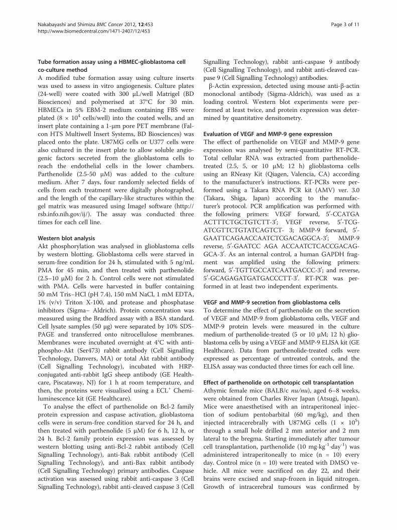
Figure 1 NF-κB transcription factor activity. NF-κB transcriptional activitparthenolide by using an ELISA-based assay. The assay showed that the tradecreased as the concentration of parthenolide increased. Data is presentep < 0.0001; ** indicates p < 0.01.
implantation model was analysed using unpaired andpaired Student’s t-tests. For all statistical analyses, a pvalue of <0.05 was considered to be statistically signifi-cant. All values are presented as mean ± standard devi-ation (SD).
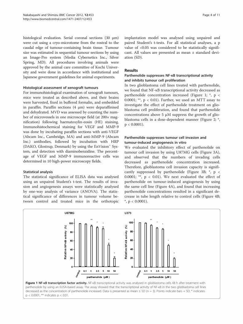
ResultsParthenolide suppresses NF-κB transcriptional activityand inhibits tumour cell proliferationIn two glioblastoma cell lines treated with parthenolide,we found that NF-κB transcriptional activity decreased asparthenolide concentration increased (Figure 1; *, p <0.0001; **, p < 0.01). Further, we used an MTT assay toinvestigate the effect of parthenolide treatment on glio-blastoma cell proliferation, and found that parthenolideconcentrations above 5 μM suppress the growth of glio-blastoma cells in a dose-dependent manner (Figure 2; *,p < 0.0001).
Parthenolide suppresses tumour cell invasion andtumour-induced angiogenesis in vitroWe evaluated the inhibitory effect of parthenolide ontumour cell invasion by using U87MG cells (Figure 3A),and observed that the numbers of invading cellsdecreased as parthenolide concentration increased.Therefore, glioblastoma cell invasion capacity is signifi-cantly suppressed by parthenolide (Figure 3B; *, p <0.0001; **, p < 0.01). We next evaluated the effect ofparthenolide on tumour-induced angiogenesis by usingthe same cell line (Figure 4A), and found that increasingparthenolide concentrations resulted in a significant de-crease in tube length relative to control cells (Figure 4B;*, p < 0.0001).
y was analysed in glioblastoma cells 48 h after treatment withnscriptional activity of NF-κB in the two glioblastoma cell linesd as mean ± SD (n = 3). Points indicate bars + SD; * indicates
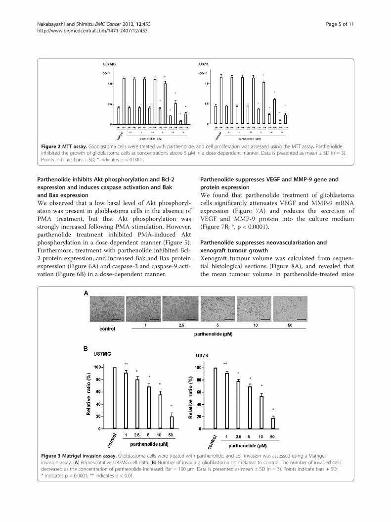
Figure 2 MTT assay. Glioblastoma cells were treated with parthenolide, and cell proliferation was assessed using the MTT assay. Parthenolideinhibited the growth of glioblastoma cells at concentrations above 5 μM in a dose-dependent manner. Data is presented as mean ± SD (n = 3).Points indicate bars + SD; * indicates p < 0.0001.
Nakabayashi and Shimizu BMC Cancer 2012, 12:453 Page 5 of 11http://www.biomedcentral.com/1471-2407/12/453
Parthenolide inhibits Akt phosphorylation and Bcl-2expression and induces caspase activation and Bakand Bax expressionWe observed that a low basal level of Akt phosphoryl-ation was present in glioblastoma cells in the absence ofPMA treatment, but that Akt phosphorylation wasstrongly increased following PMA stimulation. However,parthenolide treatment inhibited PMA-induced Aktphosphorylation in a dose-dependent manner (Figure 5).Furthermore, treatment with parthenolide inhibited Bcl-2 protein expression, and increased Bak and Bax proteinexpression (Figure 6A) and caspase-3 and caspase-9 acti-vation (Figure 6B) in a dose-dependent manner.
Figure 3 Matrigel invasion assay. Glioblastoma cells were treated with pinvasion assay. (A) Representative U87MG cell data. (B) Number of invadingdecreased as the concentration of parthenolide increased. Bar = 100 μm. D* indicates p < 0.0001; ** indicates p < 0.01.
Parthenolide suppresses VEGF and MMP-9 gene andprotein expressionWe found that parthenolide treatment of glioblastomacells significantly attenuates VEGF and MMP-9 mRNAexpression (Figure 7A) and reduces the secretion ofVEGF and MMP-9 protein into the culture medium(Figure 7B; *, p < 0.0001).
Parthenolide suppresses neovascularisation andxenograft tumour growthXenograft tumour volume was calculated from sequen-tial histological sections (Figure 8A), and revealed thatthe mean tumour volume in parthenolide-treated mice
arthenolide, and cell invasion was assessed using a Matrigelglioblastoma cells relative to control. The number of invaded cellsata is presented as mean ± SD (n = 3). Points indicate bars + SD;
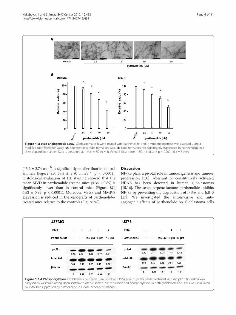
Figure 4 In vitro angiogenesis assay. Glioblastoma cells were treated with parthenolide, and in vitro angiogenesis was assessed using amodified tube formation assay. (A) Representative tube formation data. (B) Tube formation was significantly suppressed by parthenolide in adose-dependent manner. Data is presented as mean ± SD (n = 3). Points indicate bars + SD; * indicates p < 0.0001. Bar = 5 mm.
Nakabayashi and Shimizu BMC Cancer 2012, 12:453 Page 6 of 11http://www.biomedcentral.com/1471-2407/12/453
(45.2 ± 2.74 mm3) is significantly smaller than in controlanimals (Figure 8B; 59.5 ± 3.80 mm3; *, p < 0.0001).Histological evaluation of HE staining showed that themean MVD in parthenolide-treated mice (4.34 ± 0.89) issignificantly lower than in control mice (Figure 8C;6.52 ± 0.95; p < 0.0001). Moreover, VEGF and MMP-9expression is reduced in the xenografts of parthenolide-treated mice relative to the controls (Figure 8C).
Figure 5 Akt Phosphorylation. Glioblastoma cells were stimulated with Panalysed by western blotting. Representative blots are shown. Akt expressioby PMA, but suppressed by parthenolide in a dose-dependent manner.
DiscussionNF-κB plays a pivotal role in tumourigenesis and tumourprogression [5,6]. Aberrant or constitutively activatedNF-κB has been detected in human glioblastomas[15,16]. The sesquiterpene lactone parthenolide inhibitsNF-κB by preventing the degradation of IκB-α and IκB-β[17]. We investigated the anti-invasive and anti-angiogenic effects of parthenolide on glioblastoma cells
MA prior to parthenolide treatment, and Akt phosphorylation wasn and phosphorylation in both glioblastoma cell lines was stimulated
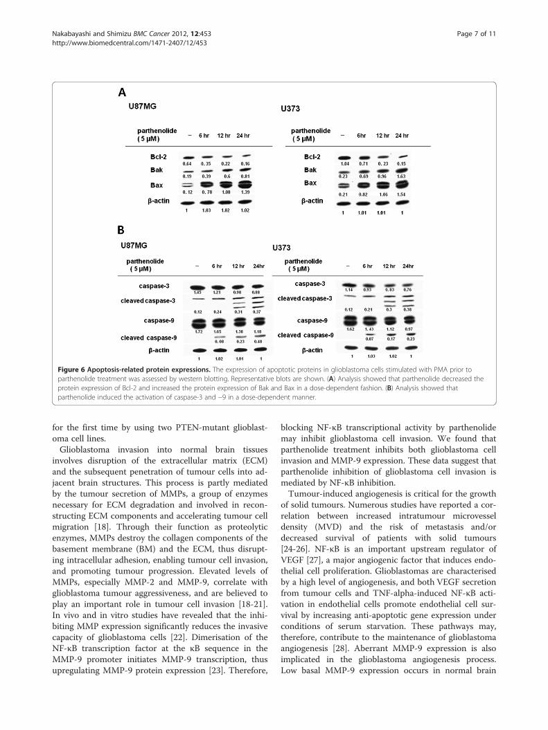
Figure 6 Apoptosis-related protein expressions. The expression of apoptotic proteins in glioblastoma cells stimulated with PMA prior toparthenolide treatment was assessed by western blotting. Representative blots are shown. (A) Analysis showed that parthenolide decreased theprotein expression of Bcl-2 and increased the protein expression of Bak and Bax in a dose-dependent fashion. (B) Analysis showed thatparthenolide induced the activation of caspase-3 and −9 in a dose-dependent manner.
Nakabayashi and Shimizu BMC Cancer 2012, 12:453 Page 7 of 11http://www.biomedcentral.com/1471-2407/12/453
for the first time by using two PTEN-mutant glioblast-oma cell lines.Glioblastoma invasion into normal brain tissues
involves disruption of the extracellular matrix (ECM)and the subsequent penetration of tumour cells into ad-jacent brain structures. This process is partly mediatedby the tumour secretion of MMPs, a group of enzymesnecessary for ECM degradation and involved in recon-structing ECM components and accelerating tumour cellmigration [18]. Through their function as proteolyticenzymes, MMPs destroy the collagen components of thebasement membrane (BM) and the ECM, thus disrupt-ing intracellular adhesion, enabling tumour cell invasion,and promoting tumour progression. Elevated levels ofMMPs, especially MMP-2 and MMP-9, correlate withglioblastoma tumour aggressiveness, and are believed toplay an important role in tumour cell invasion [18-21].In vivo and in vitro studies have revealed that the inhi-biting MMP expression significantly reduces the invasivecapacity of glioblastoma cells [22]. Dimerisation of theNF-κB transcription factor at the κB sequence in theMMP-9 promoter initiates MMP-9 transcription, thusupregulating MMP-9 protein expression [23]. Therefore,
blocking NF-κB transcriptional activity by parthenolidemay inhibit glioblastoma cell invasion. We found thatparthenolide treatment inhibits both glioblastoma cellinvasion and MMP-9 expression. These data suggest thatparthenolide inhibition of glioblastoma cell invasion ismediated by NF-κB inhibition.Tumour-induced angiogenesis is critical for the growth
of solid tumours. Numerous studies have reported a cor-relation between increased intratumour microvesseldensity (MVD) and the risk of metastasis and/ordecreased survival of patients with solid tumours[24-26]. NF-κB is an important upstream regulator ofVEGF [27], a major angiogenic factor that induces endo-thelial cell proliferation. Glioblastomas are characterisedby a high level of angiogenesis, and both VEGF secretionfrom tumour cells and TNF-alpha-induced NF-κB acti-vation in endothelial cells promote endothelial cell sur-vival by increasing anti-apoptotic gene expression underconditions of serum starvation. These pathways may,therefore, contribute to the maintenance of glioblastomaangiogenesis [28]. Aberrant MMP-9 expression is alsoimplicated in the glioblastoma angiogenesis process.Low basal MMP-9 expression occurs in normal brain
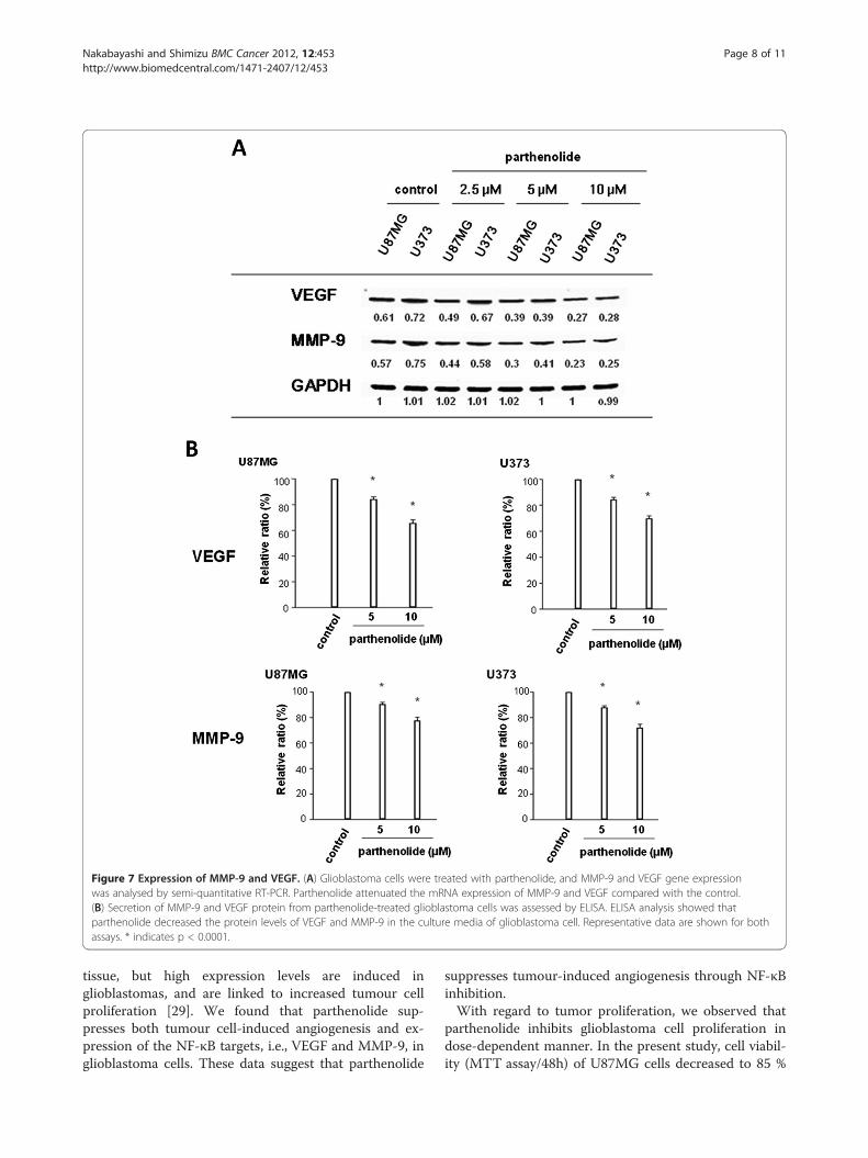
Figure 7 Expression of MMP-9 and VEGF. (A) Glioblastoma cells were treated with parthenolide, and MMP-9 and VEGF gene expressionwas analysed by semi-quantitative RT-PCR. Parthenolide attenuated the mRNA expression of MMP-9 and VEGF compared with the control.(B) Secretion of MMP-9 and VEGF protein from parthenolide-treated glioblastoma cells was assessed by ELISA. ELISA analysis showed thatparthenolide decreased the protein levels of VEGF and MMP-9 in the culture media of glioblastoma cell. Representative data are shown for bothassays. * indicates p < 0.0001.
Nakabayashi and Shimizu BMC Cancer 2012, 12:453 Page 8 of 11http://www.biomedcentral.com/1471-2407/12/453
tissue, but high expression levels are induced inglioblastomas, and are linked to increased tumour cellproliferation [29]. We found that parthenolide sup-presses both tumour cell-induced angiogenesis and ex-pression of the NF-κB targets, i.e., VEGF and MMP-9, inglioblastoma cells. These data suggest that parthenolide
suppresses tumour-induced angiogenesis through NF-κBinhibition.With regard to tumor proliferation, we observed that
parthenolide inhibits glioblastoma cell proliferation indose-dependent manner. In the present study, cell viabil-ity (MTT assay/48h) of U87MG cells decreased to 85 %
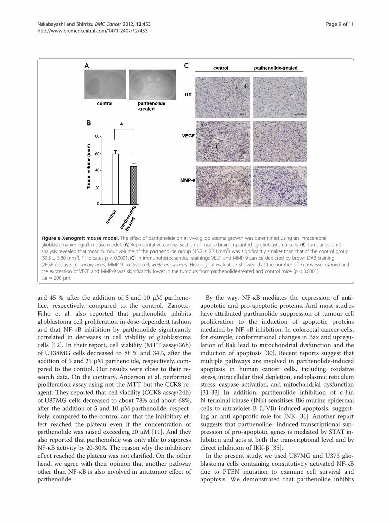
Figure 8 Xenograft mouse model. The effect of parthenolide on in vivo glioblastoma growth was determined using an intracerebralglioblastoma xenograft mouse model. (A) Representative coronal section of mouse brain implanted by glioblastoma cells. (B) Tumour volumeanalysis revealed that mean tumour volume of the parthenolide group (45.2 ± 2.74 mm3) was significantly smaller than that of the control group(59.5 ± 3.80 mm3). * indicates p < 0.0001. (C) In immunohistochemical stainings VEGF and MMP-9 can be depicted by brown DAB staining(VEGF-positive cell; arrow head, MMP-9-positive cell; white arrow head. Histological evaluation showed that the number of microvessel (arrow) andthe expression of VEGF and MMP-9 was significantly lower in the tumours from parthenolide-treated and control mice (p < 0.0001).Bar = 200 μm.
Nakabayashi and Shimizu BMC Cancer 2012, 12:453 Page 9 of 11http://www.biomedcentral.com/1471-2407/12/453
and 45 %, after the addition of 5 and 10 μM partheno-lide, respectively, compared to the control. Zanotto-Filho et al. also reported that parthenolide inhibitsglioblastoma cell proliferation in dose-dependent fashionand that NF-κB inhibition by parthenolide significantlycorrelated in decreases in cell viability of glioblastomacells [12]. In their report, cell viability (MTT assay/36h)of U138MG cells decreased to 88 % and 34%, after theaddition of 5 and 25 μM parthenolide, respectively, com-pared to the control. Our results were close to their re-search data. On the contrary, Anderson et al. performedproliferation assay using not the MTT but the CCK8 re-agent. They reported that cell viability (CCK8 assay/24h)of U87MG cells decreased to about 78% and about 68%,after the addition of 5 and 10 μM parthenolide, respect-ively, compared to the control and that the inhibitory ef-fect reached the plateau even if the concentration ofparthenolide was raised exceeding 20 μM [11]. And theyalso reported that parthenolide was only able to suppressNF-κB activity by 20-30%. The reason why the inhibitoryeffect reached the plateau was not clarified. On the otherhand, we agree with their opinion that another pathwayother than NF-κB is also involved in antitumor effect ofparthenolide.
By the way, NF-κB mediates the expression of anti-apoptotic and pro-apoptotic proteins. And most studieshave attributed parthenolide suppression of tumour cellproliferation to the induction of apoptotic proteinsmediated by NF-κB inhibition. In colorectal cancer cells,for example, conformational changes in Bax and upregu-lation of Bak lead to mitochondrial dysfunction and theinduction of apoptosis [30]. Recent reports suggest thatmultiple pathways are involved in parthenolide-inducedapoptosis in human cancer cells, including oxidativestress, intracellular thiol depletion, endoplasmic reticulumstress, caspase activation, and mitochondrial dysfunction[31-33]. In addition, parthenolide inhibition of c-JunN-terminal kinase (JNK) sensitises JB6 murine epidermalcells to ultraviolet B (UVB)-induced apoptosis, suggest-ing an anti-apoptotic role for JNK [34]. Another reportsuggests that parthenolide- induced transcriptional sup-pression of pro-apoptotic genes is mediated by STAT in-hibition and acts at both the transcriptional level and bydirect inhibition of IKK-β [35].In the present study, we used U87MG and U373 glio-
blastoma cells containing constitutively activated NF-κBdue to PTEN mutation to examine cell survival andapoptosis. We demonstrated that parthenolide inhibits
Nakabayashi and Shimizu BMC Cancer 2012, 12:453 Page 10 of 11http://www.biomedcentral.com/1471-2407/12/453
Akt phosphorylation, upregulates Bak and Bax, and acti-vates caspase-3 and caspase-9. These results suggest thatparthenolide induces apoptosis in glioblastoma cells byactivating the mitochondrial apoptosis cascade and redu-cing survival signals through the inhibition of the Aktpathway. Parthenolide was previously reported to induceapoptosis in colorectal cancer and cholangiocarcinomacells via the mitochondrial pathway [30,36]. In contrast,NF-κB inhibition by parthenolide markedly enhances thesensitivity of resistant breast cancer tumour cells to tam-oxifen through suppression of the Akt pathway [37].Taken together, the pro-apoptotic effect of parthenolideincludes both stimulating the intrinsic apoptotic pathwayand modulating expression of Bcl-2 family proteins.In our in vivo study, parthenolide inhibited the growth
of transplanted glioblastoma cells in mouse xenografts.This suggests that inhibitory effect of parthenolide onPTEN-mutant glioblastoma cells is caused by a combin-ation of three mechanisms: suppression of tumour cellinvasion, suppression of angiogenesis, and induction oftumour cell apoptosis.We investigated tumour-induced angiogenesis by
using a new in vitro angiogenesis assay that measuresEC tube formation in collagen gels. Tumour-inducedangiogenesis is usually examined by adding tumour cellmedium directly to ECs without direct contact betweenthe endothelial and tumour cells. In contrast, the newmethod allows the exchange of secreted factors betweenthe endothelial and tumour cells. HUVECs have beenused for many angiogenesis studies, especially those in-cluding in vitro experiments. However, microvascularendothelial cells from the organ being studied are pre-sumed to be the most appropriate tool for tumourangiogenesis assays. Therefore, for the first time, weinvestigated the effect of parthenolide on angiogenesisby using HBMECs.The effect of parthenolide treatment on neuro-
inflammatory disorders has been examined in the recentyears [38-40]. Runmel et al. investigated the potential ofparthenolide to reduce brain inflammation and reportedthat parthenolide can cross the blood–brain barrier [40].Thus, the combined antitumour and anti-inflammatoryproperties of parthenolide make it a promising candidatefor further studies of neurological diseases.
ConclusionWe report that parthenolide inhibits tumour progressionin PTEN-mutant glioblastoma cells in vitro and in vivo,and suggest that parthenolide has a therapeutic potentialas an antitumour agent for glioblastoma.
Competing interestsThe authors declare that they have no competing interests.
Authors’ contributionsHN was responsible for the study design, interpretation of the data andrevision of the manuscript. KS supervised the studies and helped to revisethe manuscript. Both authors read and approved the final manuscript.
AcknowledgementsThe authors declare that they have no acknowledgements.
Author details1Human Biology, Department of Health Sciences, Oita University of Nursingand Sciences, 2944-9 Megusuno, Oita 870-1201, Japan. 2Department ofNeurosurgery, Kochi Medical School Kochi University, 783-8505, Nankoku,Japan.
Received: 13 December 2011 Accepted: 27 September 2012Published: 5 October 2012
References1. Franke TF, Kaplan DR, Cantley LC: PI3K: downsteam AKTion blocks
apoptosis. Cell 1997, 88:435–437.2. Steck PA, Pershouse MA, Jasser SA, Yung WK, Lin H, Ligon AH, Langford LA,
Baumgard ML, Hattier T, Davis T, Frye C, Hu R, Swedlund B, Teng DH,Tavtigian SV: Identification of a candidate tumor suppressor gene,MMAC1, at chromosome 10q23.3 that is mutated in multiple advancedcancers. Nat Genet 1997, 15:356–362.
3. Wang SI, Puc J, Li J, Bruce JN, Cairns P, Sidransky D, Parsons R: Somaticmutations of PTEN in glioblastoma multiforme. Cancer Res 1997, 57:4183–4186.
4. Madrid LV, Mayo MW, Reuther JY, Baldwin AS Jr: Akt stimulates thetransactivation potential of the RelA/p65 subunit of NF-κB throughutilization of the IκB kinase and activation of the mitogen activatedprotein kinase p38. J Biol Chem 2001, 276:18934–18940.
5. Gilmore TD: Introduction to NF-kappaB: players, pathways, perspectives.Oncogene 2006, 25:6680–6684.
6. Haffner MC, Berlato C, Doppler W: Exploiting our knowledge of NF-κBsignaling for treatment of mammary cancer. J Mammary Gland BiolNeoplasia 2006, 11:63–73.
7. Wu JT, Kral JG: The NF- NF- NF-κB/IκB signaling system: A moleculartarget inbreast cancer therapy. J Surg Res 2005, 123:158–169.
8. Kwok BH, Koh B, Ndubuisi MI, Elofsson M, Crews CM: The anti-inflammatorynatural product parthenolide from the medicinal herb Feverfew directlybinds to and inhibits IB kinase. Chem Biol 2001, 8:759–766.
9. Hehner SP, Hofmann TG, Droge W, Schmitz ML: The antiinflammatorysesquiterpene lactone parthenolide inhibits NF-B by targeting the IBkinase complex. J Immunol 1999, 163:5617–5623.
10. Heptinstall S: Feverfew-an ancient remedy for modern times? J R Soc Med1988, 81:373–374.
11. Anderson KN, Bejcek BE: Parthenolide induces apoptosis in glioblastomaswithout affecting NF-kappaB. J Pharmacol Sci 2008, 106:318–320.
12. Zanotto-Filho A, Braganhol E, Schröder R, de Souza LH, Dalmolin RJ,Pasquali MA, Gelain DP, Battastini AM, Moreira JC: NFκB inhibitors inducecell death in glioblastomas. Biochem Pharmacol 2011, 81:412–424.
13. Cheney IW, Johnson DE, Vaillancourt MT, Avanzini J, Morimoto A, DemersGW, Wills KN, Shabram PW, Bolen JB, Tavtigian SV, Bookstein R: Suppressionof tumorigenicity of glioblastoma cells by adenovirus-mediated MMAC1/PTEN gene transfer. Cancer Res 1998, 58:2331–2334.
14. Morimoto AM, Berson AE, Fujii GH, Teng DH, Tavtigian SV, Bookstein R, SteckPA, Bolen JB: Phenotypic analysis of human glioma cells expressing theMMAC1 tumor suppressor phosphatase. Oncogene 1999, 18:1261–1266.
15. Kanzawa T, Ito H, Kondo Y, Kondo S: Current and Future Gene Therapy forMalignant Gliomas. J Biomed Biotechnol 2003, 2003:25–34.
16. Ravi R, Bedi A: NF-kappaB in cancer–a friend turned foe. Drug Resist Updat2004, 7:53–67.
17. Hehner SP, Heinrich M, Bork PM, Vog M, Ratter F, Lehmann V, Schulze-Osthoff K, Droge W, Schmitz ML: Sesquiterpene lactones specificallyinhibit activation of NF-B by preventing the degradation of IκB-α andIκB-β. J Biol Chem 1998, 273:1288–1297.
18. Rao JS: Molecular mechanisms of glioma invasiveness: the role ofproteases. Nat Rev Cancer 2003, 3:489–501.
19. Wild-Bode C, Weller M, Wick W: Molecular determinants of glioma cellmigration and invasion. J Neurosurg 2001, 94:978–984.
Nakabayashi and Shimizu BMC Cancer 2012, 12:453 Page 11 of 11http://www.biomedcentral.com/1471-2407/12/453
20. Kunishio K, Okada M, Matsumoto Y, Nagao S:Matrix metalloproteinase-2 and −9expression in astrocytic tumors. Brain Tumor Pathol 2003, 20:39–45.
21. Björklund M, Kojvunen E: Gelatinase-mediated migration and invasion ofcancer cells. Biochim Biophys Acta 2005, 1755:37–69.
22. Kondraganti S, Mohanam S, Chintala SK, Kin Y, Jasti SL, Nirmala C, Lakka SS, AdachiY, Kyritis AP, All-Osman F, Sawaya R, Fuller GN, Rao JS: Selective suppression ofmatrix metalloproteinase-9 in human glioblastoma cells by antisense genetransfer impairs glioblastoma cell invasion. Cancer Res 2000, 60:6851–6855.
23. Sato HM, Seiki M: Regulatory mechanism of 92-kDa type IV collagenasegene expression which is associtated with invasiveness of tumor cells.Oncogene 1993, 8:395–405.
24. Van Hoef ME, Knox WF, Dhesi SS, Howell A, Schor AM: Assessment oftumour vascularity as a prognostic factor in lymph node negativeinvasive breast cancer. Eur J Cancer 1993, 29A:1141–1145.
25. Gasparini G, Weidner N, Maluta S, Pozza F, Boracchi P, Mezzetti M, TestolinA, Bevilacqua P: Intratumoral microvessel density and p53 protein:correlation with metastasis in head-and-neck squamous-cell carcinoma.Int J Cancer 1993, 55:739–744.
26. Leon SP, Folkerth RD, Black PM: Microvessel density is a prognostic indicatorfor patients with astroglial brain tumors. Cancer 1996, 77:362–372.
27. Shibata A, Nagaya T, Imai T, Funahashi H, Nakao A, Seo H: Inhibition of NF-kappaB activity decreases the VEGF mRNA expression in MDA-MB-231breast cancer cells. Breast Cancer Res Treat 2002, 73:237–243.
28. Ueda Y, Nakagawa T, Kubota T, Ido K, Sato K: Glioma cells under hypoxicconditions block the brain microvascular endothelial cell death inducedby serum starvation. J Neurochem 2005, 95:99–110.
29. Zhao JX, Yang LP, Wang YF, Qin LP, Liu DQ, Bai CX, Nan X, Shi SS, Pei XJ:Gelatinolytic activity of matrix metalloproteinase-2 and matrixmetalloproteinase-9 in rat brain after implantation of 9L rat glioma cells.Eur J Neurol 2007, 14:510–516.
30. Zhang S, Ong CN, Shen HM: Involvement of proapoptotic Bcl-2 familymembers in parthenolide-induced mitochondrial dysfunction andapoptosis. Cancer Lett 2004, 211:175–188.
31. Cory AH, Cory JG: Lactacystin, a proteasome inhibitor, potentiates theapoptotic effect of parthenolide, an inhibitor of NFB activation, on drug-resistant mouse leukemia L1210 cells. Anticancer Res 2002, 22:3805–3809.
32. Wen J, You KR, Lee SY, Song CH, Kim DG: Oxidative stress-mediatedapoptosis. The anticancer effect of the sesquiterpene lactoneparthenolide. J Biol Chem 2002, 277:38954–38964.
33. Zhang S, Ong CN, Shen HM: Critical roles of intracellular thiols andcalcium in parthenolide-induced apoptosis in human colorectal cancercells. Cancer Lett 2004, 208:143–153.
34. Won YK, Ong CN, Shi X, Shen HM: Chemopreventive activity ofparthenolide against UVB-induced skin cancer and its mechanisms.Carcinogenesis 2004, 25:1449–1458.
35. Mathema VB, Koh YS, Thakuri BC, Sillanpää M: Parthenolide, a SesquiterpeneLactone. Inflammation: Expresses Multiple Anti-cancer and Anti-inflammatory Activities; 2011.
36. Kim JH, Liu L, Lee SO, Kim YT, You KR, Kim DG: Susceptibility ofcholangiocarcinoma cells to parthenolide-induced apoptosis. Cancer Res2005, 65:6312–6320.
37. de Graffenried LA, Chandrasekar B, Friedrichs WE, Donzis E, Silva J, HidalgoM, Freeman JW, Weiss GR: NF-kappa B inhibition markedly enhancessensitivity of resistant breast cancer tumor cells to tamoxifen. Ann Oncol2004, 15:885–890.
38. Tassorelli C, Greco R, Morazzoni P, Riva A, Sandrini G, Nappi G: Parthenolideis the component of tanacetum parthenium that inhibits nitroglycerin-induced Fos activation: studies in an animal model of migraine.Cephalalgia 2005, 25:612–621.
39. Lei ZN, Zeng SL, Wang L, Zhu JB, Li T: Parthenolide inhibitsneuroinflammation and promotes neurogenesis in the ischemic striatumfollowing transient middle cerebral artery occlusion in the adult ratbrain. Xi Bao Yu Fen Zi Mian Yi Xue Za Zhi 2009, 25:994–997. Chinese.
40. Rummel C, Gerstberger R, Roth J, Hübschle T: Parthenolide attenuates LPS-induced fever, circulating cytokines and markers of brain inflammationin rats. Cytokine 2011, 56:739–748.
doi:10.1186/1471-2407-12-453Cite this article as: Nakabayashi and Shimizu: Involvement of Akt/NF-κBpathway in antitumor effects of parthenolide on glioblastoma cellsin vitro and in vivo. BMC Cancer 2012 12:453.
Submit your next manuscript to BioMed Centraland take full advantage of:
• Convenient online submission
• Thorough peer review
• No space constraints or color figure charges
• Immediate publication on acceptance
• Inclusion in PubMed, CAS, Scopus and Google Scholar
• Research which is freely available for redistribution
Submit your manuscript at www.biomedcentral.com/submit
![Ferulic acid regulates the AKT/GSK-3 β/CRMP-2 signaling ... · linositol 3-kinase (PI3K) and extracellular signal-regulated kinase (ERK) pathways [10]. The PI3K/Akt pathway is an](https://static.fdocument.org/doc/165x107/5e5c6b03e0248c23f76fce82/ferulic-acid-regulates-the-aktgsk-3-crmp-2-signaling-linositol-3-kinase.jpg)

![Layout 1 (Page 1) - Antibodies, Proteins, Kits and … P WB Rb Hu 28225 ADAM17 P WB Rb Hu, Mm, Rt 2051 AKT (phospho S473) [14-6] M ICC, WB Rb Hu, Mm 27773 AKT (phospho T308) P ELISA,](https://static.fdocument.org/doc/165x107/5b0df7317f8b9af65e8e7090/layout-1-page-1-antibodies-proteins-kits-and-p-wb-rb-hu-28225-adam17-p-wb.jpg)