Inhibition of NF-κB/IL-33/ST2 Axis Ameliorates Acute ...
11
Research Article Inhibition of NF-κB/IL-33/ST2 Axis Ameliorates Acute Bronchiolitis Induced by Respiratory Syncytial Virus Liwen Zhang, 1 Yu Wan, 1 Liang Ma, 2 Kaihong Xu , 1 and Baojin Cheng 1 1 Department of Pediatrics, The Second People’s Hospital of Changzhou, Affiliated Hospital of Nanjing Medical University, China 2 Department of Digestive Disease, The First People’s Hospital of Changzhou, the Third Affiliated Hospital of Soochow University, China Correspondence should be addressed to Kaihong Xu; [email protected] and Baojin Cheng; [email protected] Received 16 December 2020; Revised 4 June 2021; Accepted 16 July 2021; Published 4 August 2021 Academic Editor: Zhipeng Xu Copyright © 2021 Liwen Zhang et al. This is an open access article distributed under the Creative Commons Attribution License, which permits unrestricted use, distribution, and reproduction in any medium, provided the original work is properly cited. Background/Aim. Bronchiolitis is a common acute lower respiratory tract infectious disease in infants. Respiratory syncytial virus (RSV) infection is one of the main causes. Bronchiolitis can lead to a significant increase in the incidence of asthma in young children, but the mechanism of bronchiolitis transforming into asthma is still unclear. The study was aimed at investigating the role of NF-κB/IL-33/ST2 axis on RSV-induced acute bronchiolitis. Methods. A total of 40 infants diagnosed with acute bronchiolitis infected by RSV, and 20 normal infants were included in this study. BALB/c mice (6-8 weeks old, 20 ± 1:1 g) were used as study models. Enzyme-linked immunosorbent assay (ELISA), quantitative real time PCR, western blot analysis, immunohistochemical staining, and flow cytometry analysis were performed to examine relevant indicators. Results. IL-33 level was significantly elevated, and Th1/Th2 ratio is imbalance after in infants with acute bronchiolitis. In vivo study, we found that NF-κB/IL-33/ST2 axis is mediated the Th2 cytokine levels and BAL cell number induced by RSV. Acute bronchiolitis induced by RSV in a mouse model is attenuated after inhibition of NF-κB/IL-33/ST2 pathway. Moreover, we also confirmed that macrophages are important sources of IL-33 and are regulated by NF-κB pathway in RSV-induced mice. Conclusion. We confirmed that inhibition of NF-κB/IL-33/ST2 axis could attenuate acute bronchiolitis by RSV infected. Our findings not only demonstrate the potential role of IL-33 antibody in attenuating RSV-induced lung damage but also provide a new insight into better prevention of RSV-induced asthma by mediating NF-κB/IL-33/ST2 axis. 1. Introduction Asthma is a respiratory disease involving a variety of inflammatory mediators and cytokines, which has been a serious threat to human health [1]. Numerous studies have shown that viral infection can directly invade 75-300 μm bronchioles, causing necrosis and exfoliation of epithelial cells of bronchioles, infiltration of peripheral lymphocytes, glandular hyperplasia, increased mucus secretion, and nar- rowing or even blockage of the lumen [2, 3]. Repeated or persistent viral infection eventually leads to chronic airway inflammation and airway hyperreflux [4]. Respiratory syncytial virus (RSV), as the main pathogen of bronchiolitis and pneumonia in infants and young chil- dren, is one of the main causes of asthma in young patients [5]. Previous reports showed that RSV infection accounts for 80-85% and 75-80% of asthma cases in children and adults, respectively [6, 7]. Another important reason RSV can induce asthma is that it can activate T helper 2 (Th2) cell-related cytokines such as IL-4, IL-5, and IL-10 and break the body’s immune balance [8, 9]. Recent studies have found that the treatment of cytokines secreted by Th2 cells can effectively alleviate the symptoms of acute asthma attack, while the reduction of cytokines secreted by Th1 cells does not significantly alleviate the symptoms of asthma [8, 10]. These findings enrich and improve the classic Th1/Th2 imbalance theory in asthma. IL-33 is a recently discovered proinflammatory cytokine with a variety of biological functions. It belongs to the IL-1 family with a molecular weight of about 1800 and is located on human 9p24.1 chromosome [11]. Current studies have shown that IL-33 not only induces Th0 cells to differentiate Hindawi Journal of Immunology Research Volume 2021, Article ID 6625551, 11 pages https://doi.org/10.1155/2021/6625551
Transcript of Inhibition of NF-κB/IL-33/ST2 Axis Ameliorates Acute ...
Research ArticleInhibition of NF-κB/IL-33/ST2 Axis Ameliorates AcuteBronchiolitis Induced by Respiratory Syncytial Virus
Liwen Zhang,1 Yu Wan,1 Liang Ma,2 Kaihong Xu ,1 and Baojin Cheng 1
1Department of Pediatrics, The Second People’s Hospital of Changzhou, Affiliated Hospital of Nanjing Medical University, China2Department of Digestive Disease, The First People’s Hospital of Changzhou,the Third Affiliated Hospital of Soochow University, China
Correspondence should be addressed to Kaihong Xu; [email protected] and Baojin Cheng; [email protected]
Received 16 December 2020; Revised 4 June 2021; Accepted 16 July 2021; Published 4 August 2021
Academic Editor: Zhipeng Xu
Copyright © 2021 Liwen Zhang et al. This is an open access article distributed under the Creative Commons Attribution License,which permits unrestricted use, distribution, and reproduction in any medium, provided the original work is properly cited.
Background/Aim. Bronchiolitis is a common acute lower respiratory tract infectious disease in infants. Respiratory syncytial virus(RSV) infection is one of the main causes. Bronchiolitis can lead to a significant increase in the incidence of asthma in youngchildren, but the mechanism of bronchiolitis transforming into asthma is still unclear. The study was aimed at investigating therole of NF-κB/IL-33/ST2 axis on RSV-induced acute bronchiolitis. Methods. A total of 40 infants diagnosed with acutebronchiolitis infected by RSV, and 20 normal infants were included in this study. BALB/c mice (6-8 weeks old, 20 ± 1:1 g) wereused as study models. Enzyme-linked immunosorbent assay (ELISA), quantitative real time PCR, western blot analysis,immunohistochemical staining, and flow cytometry analysis were performed to examine relevant indicators. Results. IL-33 levelwas significantly elevated, and Th1/Th2 ratio is imbalance after in infants with acute bronchiolitis. In vivo study, we found thatNF-κB/IL-33/ST2 axis is mediated the Th2 cytokine levels and BAL cell number induced by RSV. Acute bronchiolitis inducedby RSV in a mouse model is attenuated after inhibition of NF-κB/IL-33/ST2 pathway. Moreover, we also confirmed thatmacrophages are important sources of IL-33 and are regulated by NF-κB pathway in RSV-induced mice. Conclusion. Weconfirmed that inhibition of NF-κB/IL-33/ST2 axis could attenuate acute bronchiolitis by RSV infected. Our findings not onlydemonstrate the potential role of IL-33 antibody in attenuating RSV-induced lung damage but also provide a new insight intobetter prevention of RSV-induced asthma by mediating NF-κB/IL-33/ST2 axis.
1. Introduction
Asthma is a respiratory disease involving a variety ofinflammatory mediators and cytokines, which has been aserious threat to human health [1]. Numerous studies haveshown that viral infection can directly invade 75-300μmbronchioles, causing necrosis and exfoliation of epithelialcells of bronchioles, infiltration of peripheral lymphocytes,glandular hyperplasia, increased mucus secretion, and nar-rowing or even blockage of the lumen [2, 3]. Repeated orpersistent viral infection eventually leads to chronic airwayinflammation and airway hyperreflux [4].
Respiratory syncytial virus (RSV), as the main pathogenof bronchiolitis and pneumonia in infants and young chil-dren, is one of the main causes of asthma in young patients[5]. Previous reports showed that RSV infection accounts
for 80-85% and 75-80% of asthma cases in children andadults, respectively [6, 7]. Another important reason RSVcan induce asthma is that it can activate T helper 2 (Th2)cell-related cytokines such as IL-4, IL-5, and IL-10 and breakthe body’s immune balance [8, 9]. Recent studies have foundthat the treatment of cytokines secreted by Th2 cells caneffectively alleviate the symptoms of acute asthma attack,while the reduction of cytokines secreted by Th1 cells doesnot significantly alleviate the symptoms of asthma [8, 10].These findings enrich and improve the classic Th1/Th2imbalance theory in asthma.
IL-33 is a recently discovered proinflammatory cytokinewith a variety of biological functions. It belongs to the IL-1family with a molecular weight of about 1800 and is locatedon human 9p24.1 chromosome [11]. Current studies haveshown that IL-33 not only induces Th0 cells to differentiate
HindawiJournal of Immunology ResearchVolume 2021, Article ID 6625551, 11 pageshttps://doi.org/10.1155/2021/6625551
into Th2 cells but also promotes the secretion of Th2-relatedcytokines IL-4, IL-5, IL-6, and IL-10 in vitro and in vivo [12,13]. In the study of children with asthma, it was found thatthe proportion of Th2 cells and the level of IL-33 in serumincreased significantly, which were positively correlated withthe level of autoantibody IgE in vivo [14].
Suppression of tumorigenesis 2 (ST2) receptor can spe-cifically bind to IL-33 in two forms; one is transmodel ST2(ST2L), mainly expressed on Th2 surface, and the other issoluble ST2, which can compete with ST2L and bind to IL-33, leading to the decrease of IL-33 function [13]. WhenST2 is combined with IL-33, it activates the NF-κB signalingpathway and promotes the release of Th2 cytokines such asIL-4, IL-5, and IL-10 [15]. However, the role of IL-33/ST2and NF-κB in RSV-induced bronchitis is still unclear.
In our present study, we explored the expression of IL-33in serum of acute bronchiolitis infants and in lung tissues ofRSV-induced mice and investigated its role on Th1/Th2 cellratio in acute bronchiolitis infants and RSV infected mice.Moreover, we demonstrated that NF-κB/IL-33/ST2 axis isinvolved in the RSV-induced acute bronchiolitis. Our resultssuggested that IL-33, ST2, and NF-κB can serve as therapeu-tic targets in the treatment of RSV infected asthma.
2. Materials and Methods
2.1. Reagents and Antibodies. Respiratory syncytial virus(RSV) was purchased from Hipower Pharmaceutical(Hipower Pharmaceutical, Guangzhou, China). Fetal bovineserum (FBS) and Dulbecco’s modified Eagle’s medium(DMEM) were obtained from GIBCOBRL (Gibco, CA,USA). Anti-IL-33 antibody and anti-IgG were obtained fromSanta Cruz Biotechnology (SantaCruz, CA, USA). SolubleST2 (sST2) was purchased from KeyGEN (KeyGEN, Nan-jing, China). Pyrrolidine dithiocarbamic acid ammonium salt(PDTC) was obtained from Beyotime (Beyotime, Shanghai,China). Antibodies of p50, p65, ST2, and GAPDH were pur-chased from Cell Signaling Technology (CST, CA, USA).Lipofectamine 2000 and Opti-MEM were purchased fromInvitrogen (Carlsbad, CA, USA). Trizol was obtained fromInvitrogen (Carlsbad, CA, USA). Western blot detectionchemiluminescence reagents were purchased from ThermoScientific (Thermo Scientific, CA, USA).
2.2. Study Subjects.A total of 40 infants from the Second Peo-ple’s Hospital of Changzhou, Affiliated Hospital of NanjingMedical University between 2016 and 2018 diagnosed withacute bronchiolitis infected by RSV and 20 normal infantswere included in this study. The diagnosis of acute bronchi-
olitis was based on the latest diagnostic guidelines [16]. Thevenous blood samples of all the selected cases were takenbefore treatment and used for later experiments. All caseswere excluded from congenital heart disease, cardiopulmo-nary dysplasia, immunodeficiency, and other serious dis-eases. The present study was approved by the ethicscommittee of the Second People’s Hospital of Changzhou,Affiliate Hospital of Nanjing Medical University (No.SPH1904880). The written informed consent was obtainedfrom all parents of subjects.
2.3. Animal Experiments. BALB/c mice (6-8 weeks old, 20± 1:1 g) were randomly divided into six groups: normal con-trol (NC), respiratory syncytial virus (RSV), anti-IgG+RSV,anti-IL-13+RSV, sST2+RSV, and PDTC+RSV. Protocolsfor the RSV-induced mouse models were as previouslydescribed [17]. The mice in the RSV infection group wereanesthetized by intraperitoneal injection of 3% pentobarbitalsodium 0.1ml/kg, followed by nasal drip of RSV with 100ml106 PFU on 6 consecutive days, while the mice in the controlgroup were anesthetized by nasal drip of the same dose ofsaline. The anti-IgG+ RSV, anti-IL-33+ RSV, sST2+ RSV,and PDTC+RSV groups were pretreated with anti-RabbitIgG antibody and anti-Mouse IL-33 antibody (30μg/mouse)by intranasal injection 100μl 2 h, Ribavirin, sST2, and PTDCby intraperitoneal injection 2h before challenge with RSV on3 consecutive days.
All the mice were killed and sampled on 3, 5, and 7 daysafter RSV infected; the present study was approved by theethics committee of the Second People’s Hospital of Chang-zhou, Affiliate Hospital of Nanjing Medical University (No.CZ0004-1732).
Serum samples were taken from the spleen of mice afterexecution, and erythrocytes were removed. The cells weresuspended in 10ml of HBSS wash buffer and counted by ablood cell counter. The cell suspension was centrifuged at4°C for 10min at 200 g. Then, the supernatant was discarded,and the precipitate was mixed at a ratio of 0.9ml of MACSbuffer per 108 cells. 0.lml of magnetic beads (CD90) wasadded per 10 cells and incubate for 15min at 4°C in orderto extract total T cells. After centrifugation and cell suspen-sion at 4°C, 200 g for 10min, sufficient MACS buffer wasadded to the precipitate to reach a concentration of 108
cells/ml and mix well. Pass the cells through 30μm nylonmesh or 40μmpreseparation membrane. Rinse the filter with0.1~ 0.4ml of MACS buffer. Place the cells into the uppersample channel of autoMACS. Select the possel program sothat the labeled cells will elute from the positive lane.
Table 1: Sequences of primers for PCR.
Genes Forward primer Reverse primer
IL-33 5′-GTCGCCCTGGTACCAGTCCAG-3′ 5′-AGGCCTGGCCCGAGTTGTCAG-3′IL-4 5′-GGCCCGCTATTTGTTTGGTCA-3′ 5′-GCTCCTCCCTTGCTTACCAG-3′IL-10 5′-TAGACGCGCTGGGCGACAG-3′ 5′-GTCGCCCCCTAACGCCGTAA-3′GAPDH 5′-TTCGAAGCACCGGTCCC-3′ 5′-TCTCAAGAGCAGCTCCAGT-3′
2 Journal of Immunology Research
2.4. Enzyme-Linked Immunosorbent Assay (ELISA). Thelevels of IL-33, IL-4, IL-10, IL-2, INF-γ, and ST2 in infantserum were examined by specific ELISA kits (Cloud CloneCorp, Wuhan, China) according to manufacturer’sinstructions.
2.5. Extraction and Detection of Bronchoalveolar LavageFluid (BALF). PBS (1ml) was injected into the trachea afterthe separation of surrounding connective tissue and repeated
for 3 times, and the rinsed PBS was recovered. Collectedlavage fluid for inflammatory cell counting, and stored lavagefluid at -20°C for cell flow cytometry assay detection.
2.6. Quantitative Real Time PCR. Total RNA was extractedfrom lung tissues by using Trizol (Invitrogen). SuperscriptII (Invitrogen) was used to carry out reverse transcriptionqualified with 400 ng according to the manufacturer’sinstructions. For quantitative real time PCR, SYBR Green
0
30
60
90
120
IL-3
3 (p
g/m
l)
Normal group Case group
⁎⁎⁎
(a)
0
200
400
600
IL-4
(pg/
ml)
Normal group Case group
⁎⁎⁎
(b)
0
50
100
150
200
IL-1
0 (p
g/m
l)
Normal group Case group
⁎⁎⁎
(c)
0
20
40
60
IL-2
(pg/
ml)
Normal group Case group
⁎⁎⁎
(d)
0
20
40
60
80
INF-𝛾
(pg/
ml)
Normal group Case group
⁎⁎⁎
(e)
0.0
0.3
0.6
0.9
1.2Th
1/Th
2 ra
tio
Normal group Case group
⁎⁎
(f)
0
200
400
600
800
Seru
m sS
T2 (p
g/m
l)
Normal group Case group
⁎⁎
(g)
0 50 100 1500
200
400
600
800
1000
IL-33
Seru
m sS
T2 (p
g/m
l)
r = –0.669 p < 0.001
(h)
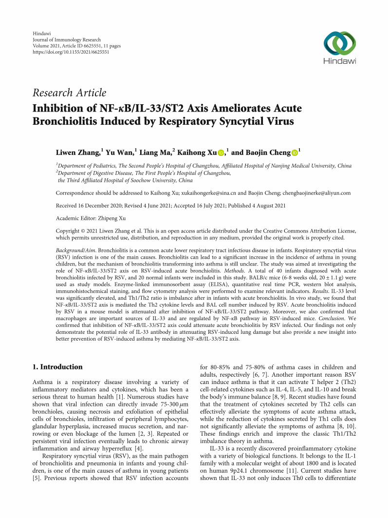
Figure 1: IL-33 expression is elevated, and Th1/Th2 ratio is imbalance in infants with acute bronchiolitis. (a) ELISA assay was used to detectthe production of IL-33 in serum. (b, c) The levels of Th2-related cytokines IL-4 and IL-10 were examined by ELISA assay. (d, e) The levels ofTh1-related cytokines IL-2 and INF-γwere examined by ELISA assay. (f) Cell flow cytometry was used to detect the ratio of Th1/Th2 cells. (g)ELISA assay was used to detect the production of ST2 in serum. (h) Correlation analysis is used to calculate the correlation between IL-33 andST2 in serum. All data were expressed as the mean ± SD (n = 3). Each value of ∗p < 0:05, ∗∗p < 0:01, and ∗∗∗p < 0:001 was deem to havesignificant differences.
3Journal of Immunology Research
p-p50
p-p65
ST2
GAPDH
p-I𝜅Ba
NC
RSV
RSV
+ant
i-IL-
33 A
b
(a)
0
1
2
3
4
5
NC
RSV
RSV
+ant
i-IL-
33 A
b
⁎⁎⁎ ⁎
Rela
tive I
L-4
mRN
A ex
pres
sion
(b)
0
1
2
3
4
5
NC
RSV
RSV
+ant
i-IL-
33 A
b
⁎⁎⁎ ⁎
Rela
tive I
L-10
mRN
A ex
pres
sion
(c)
0
20
40
60
NC
RSV
RSV
+ant
i-IL-
33 A
b
⁎⁎⁎ ⁎
Num
ber o
f BA
Lce
lls (×
104 )
(d)
ST2
GAPDH
NC
RSV
RSV
+PD
TC
(e)
0
2
4
6
8
NC
RSV
RSV
+PD
TC⁎⁎⁎ ⁎
Rela
tive I
L-33
mRN
A ex
pres
sion
(f)
0
1
2
3
4
5
NC
RSV
RSV
+PD
TC
⁎⁎⁎ ⁎
Rela
tive I
L-4
mRN
A ex
pres
sion
(g)
0
1
2
3
4
5
NC
RSV
RSV
+PD
TC
⁎⁎⁎ ⁎
Rela
tive I
L-10
mRN
A ex
pres
sion
(h)
Figure 2: Continued.
4 Journal of Immunology Research
Master Mix (Roche) and ABI-7900 system were used, andGAPDH functioned as a loading control. Primers of relatedgenes are listed in Table 1.
2.7. Western Blot Analysis. Protein samples were prepared,followed with the SDS polyacrylamide gel electrophoresis,and protein was subsequently transferred onto PVDF mem-branes. For further detection of related genes, the followingantibodies were used: IκB, p65, p50, ST2, and GAPDH.Membranes were blocking in 5% BSA for 2 h at room tem-perature prior to incubation with primary antibody at 4°Covernight. Membranes were washed 10min for three timesin TBST and then cultured with secondary antibody(1 : 5000) (CST, CA, USA) at -20°C for 2 h. Then, membraneswere washed 20min for three times with TBST. The blotswere detected with an ECL plus reagent (Thermo Scientific,Waltham, USA).
2.8. Immunohistochemical Staining. The lung tissues in dif-ferent treatment groups were collected separately and werestained with a rabbit monoclonal anti-mouse IL-33, p65,and ST2 antibody overnight at room temperature, washed,then incubated with the secondary Ab (CST, CA, USA) for2 h, and washed again. The specific detail steps were per-
formed according to DAB substrate kit (Thermo Scientific,Ma, USA).
2.9. Flow Cytometry Analysis. Peripheral blood samples andsingle cell suspension from lung tissue of mice were collected.FITC, anti-CD30, anti-CD49, and CD11 antibodies were addedin samples in turn according to the concentration of the instruc-tions. The 100 P1 antibody system fully suspended the cells andincubated on ice for 30min under the condition of dark. Add2% FBS and PBS 1ml/tube, gently whirl, 1500 rpm, 4°C centri-fuge for 5 minutes, discard supernatant; repeat this step once.Stored at 2% FBS. PBS overhanging cells were detected and ana-lyzed using the FlowJo software (version 7.6.5).
2.10. Statistical Analysis. Continuous variables were shown as“means ± SD.” For multiple comparisons, One-way ANOVAwas performed using the SPSS 22.0 software (SPSS, Inc.,USA), and p value < 0.05 was considered significantstatistically.
3. Results
3.1. IL-33 Level Is Significantly Elevated and Th1/Th2 Ratio IsImbalance after RSV Infection in Infants. It was reported that
0
20
40
60
NC
RSV
RSV
+PD
TC
⁎⁎⁎ ⁎
Num
ber o
f BA
Lce
lls (×
104 )
(i)
0
1
2
3
4
5
NC
RSV
RSV
+sST
2
⁎⁎⁎ ⁎
Rela
tive I
L-4
mRN
A ex
pres
sion
(j)
0
1
2
3
4
5
NC
RSV
RSV
+sST
2
⁎⁎⁎ ⁎
Rela
tive I
L-10
mRN
A ex
pres
sion
(k)
0
20
40
60
NC
RSV
RSV
+sST
2
⁎⁎⁎ ⁎⁎
Num
ber o
f BA
Lce
lls (×
104 )
(l)
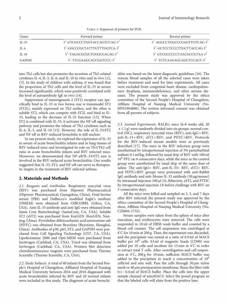
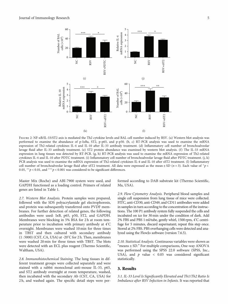
Figure 2: NF-κB/IL-33/ST2 axis is mediated the Th2 cytokine levels and BAL cell number induced by RSV. (a) Western blot analysis wasperformed to examine the abundance of p-IκBa, ST2, p-p65, and p-p50. (b, c) RT-PCR analysis was used to examine the mRNAexpression of Th2-related cytokines IL-4 and IL-10 after IL-33 antibody treatment. (d) Inflammatory cell number of bronchoalveolarlavage fluid after IL-33 antibody treatment. (e) ST2 protein abundance was examined by western blot analysis. (f) The IL-33 mRNAexpression in lung tissues was detected by RT-PCR. (g, h) RT-PCR analysis was used to examine the mRNA expression of Th2-relatedcytokines IL-4 and IL-10 after PDTC treatment. (i) Inflammatory cell number of bronchoalveolar lavage fluid after PDTC treatment. (j, k)PCR analysis was used to examine the mRNA expression of Th2-related cytokines IL-4 and IL-10 after sST2 treatment. (l) Inflammatorycell number of bronchoalveolar lavage fluid after sST2 treatment. All data were expressed as the mean ± SD (n = 3). Each value of ∗p <0:05, ∗∗p < 0:01, and ∗∗∗p < 0:001 was considered to be significant differences.
5Journal of Immunology Research
the production of Th2 cytokines IL-4 and IL-10 was pro-moted by IL-33 [18], and Th2 immune response wasdescribed to influence the pathogenesis of respiratorysyncytial virus (RSV) acute bronchiolitis [19]. Firstly, theexpression of IL-33 in serum of acute bronchiolitis infantsinfected by RSV was examined by ELISA assay; we foundthat IL-33 concentrations in AVB infants were significantlyincreased compared to normal subjects (Figure 1(a)). Acutebronchiolitis elicited increased levels of IL-4 and IL-10(Figures 1(b) and 1(c)) but decreased level of IFN-γ and IL-2(Figures 1(d) and 1(e)). We also examined that the ratio ofTh1 and Th2 cell was lowered in peripheral blood of acutebronchiolitis than normal infants by flow cytometry(Figure 1(f)). In addition, infants with acute bronchiolitishad a 2.87-fold higher level of serum (sST2) than those normalcases (Figure 1(g)). The ST2 protein expression was positivelycorrelated (r = 0:669, p < 0:001) with IL-33 production inacute bronchiolitis cases (Figure 1(h)).
3.2. NF-κB/IL-33/ST2 Axis Is Mediated the Th2 CytokineLevels and BAL Cell Number Induced by RSV. Previous stud-ies have shown that NF-κB is involved in the process of RSV-induced acute bronchiolitis [20], and NF-κB inhibitordimethyl fumarate inhibited IL-33 production [21], but itsspecific mechanism is unclear. Moreover, sST2 is anothermode of existence of ST2. It is reported that sST2 can compe-tently bind with IL-33, interfere with the coupling of IL-33and ST2, and then inhibit the biological effects of IL-33. Toconfirm whether the level of Th2 cytokines IL-4 and IL-10was mediated by IL-33, we used anti-IL-33 antibody by intra-nasal injection 30μg/mouse 2 h before challenge with RSV on3 consecutive days (14, 15, and 16). Western blot analysisshowed that abundances of ST2, p-IκBa, p-p65, and p-p50were all induced by RSV, and ST2 abundance was markedlyinhibited after treated with anti-IL-33 antibody prior toexpose to RSV. However, the abundance of IκBa, p65, andp50 was not significantly decreased in the anti-IL-33+ RSV
ST2IL-33 p65
NC
RSV
0.0
0.5
1.0
1.5
ST2P65
IL-33
Imm
unol
nten
sity
ofIL
-33/
ST2/
P65
NC RSV NC RSV NC RSV
⁎⁎⁎⁎⁎⁎
⁎⁎
(a)
NC RSV RSV+IgG
RSV+anti-IL-33 RSV+sST2 RSV+PDTC 0N
C
RSV
+sST
2
RSV
+IgG
RSV
+ant
i-IL-
33
RSV
+sST
2
RSV
+PD
TC
1
2
3
4
5
Qua
ntifi
catio
n of
HE
stain
ing
⁎⁎⁎
⁎⁎
⁎⁎
⁎
⁎
(b)
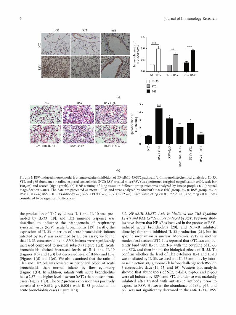
Figure 3: RSV-inducedmouse model is attenuated after inhibition of NF-κB/IL-33/ST2 pathway. (a) Immunohistochemical analysis of IL-33,ST2, and p65 abundance in saline-exposed control mice (NC); RSV-treated mice (RSV) was performed (original magnification ×400, scale bar100μm) and scored (right graph). (b) H&E staining of lung tissue in different group mice was analyzed by Image-proplus 6.0 (originalmagnification ×400). The data are presented as mean ± SEM and were analyzed by Student’s t-test (NC group, n = 8; RSV group, n = 7;RSV + IgG = 6; RSV + IL − 33 antibody = 6; RSV + PDTC = 7; RSV + sST2 = 8). Each value of ∗p < 0:05, ∗∗p < 0:01, and ∗∗∗p < 0:001 wasconsidered to be significant differences.
6 Journal of Immunology Research
0.0
0.5
1.020
30
40
50
PBS
MФ Eos Lym
RSVAnti-IL-33 ab
ns
Num
ber o
f cel
ls (×
104 )
⁎⁎⁎
⁎⁎⁎ ⁎⁎
⁎⁎⁎
(a)
0
10
20
30
40
50
Num
ber o
f mac
roph
age c
ells
(×10
4 )
RSV
0 3 5 7 (d)
0 d 3 d
5 d 7 d
⁎⁎⁎
⁎
101 102 103 104 105 106 107101
102
103
104
105
101 102 103 104 105 106 107101
102
103
104
105
101 102 103 104 105 106 107101
102
103
104
105
101 102 103 104 105 106 107101
102
103
104
105
Macrophage Macrophage
MacrophageMacrophage
(b)
Figure 4: Continued.
7Journal of Immunology Research
groups (Figure 2(a)). The mRNA expression of Th2 cyto-kines IL-4 and IL-10 elevated by RSV in lung tissues was sig-nificantly decreased in the anti-IL-33+ RSV groups(Figures 2(b) and 2(c)). Moreover, IL-33 antibody stronglysuppressed the BAL cells activated by RSV (Figure 2(d)).In order to further verify whether there is a relationshipbetween NF-κB and IL-33, we used NF-κB specific inhibi-tors (PDTC) to inject the mice prior 2 h to activate byRSV. As shown in Figures 2(e) and 2(f), the protein abun-dances of the ST2 and mRNA expression of IL-33 inmouse lung tissues induced by RSV were elevated andwere significantly inhibited in the PDTC+RSV groups.
Consistent with the role of IL-33 antibody, the mRNAexpression of Th2 cytokines IL-4 and IL-10 elevated byRSV in lung tissues was also significantly inhibited in thePDTC+ RSV groups (Figures 2(g) and 2(h)). Besides,PDTC treatment strongly suppressed the BAL cells acti-vated by RSV (Figure 2(i)). Furthermore, the levels ofIL-4 and IL-10 (Figures 2(j) and 2(k)), and BAL cells(Figure 2(l)) elevated in the RSV groups were significantlyinhibited in the RSV+ sST2 groups. However, the abun-dance of IL-33 and NF-κB (p-p50, p-p65, and p-IκBa)increased in the RSV groups was not suppressed in thesST2+RSV groups.
0 d
101 102 103 104 105 106 107
Macrophage
Macrophage
MacrophageMacrophage
Macrophage
MacrophageMacrophage
MacrophageMacrophage
Macrophage
MacrophageMacrophage12.0
101
0102
103 103
104 104
105 105
3 d 5 d 7 dPB
SRS
VRS
C+PD
TC
0
2
4
6
Relat
ive I
L-33
mRN
A ex
pres
sion
PBSRSVRSV+PDTC
0 3 5 7 (d)
⁎⁎⁎ ⁎⁎
⁎⁎⁎ ⁎⁎
⁎⁎ ⁎101 102 103 104 1051051041030
0
103
104
105
1051041030106 107101
102
103
104
105
101 102 103 104 105 106 107101
0102
103 103
104 104
105105
101 102 103 104 1051051041030
0
103
104
105
1051041030106 107101
102
103
104
105
101 102 103 104 105 106 107101
0102
103 103
104 104
105 105
101 102 103 104 1051051041030
0
103
104
105
1051041030106 107101
102
103
104
105
(c)
00 3 5 7
10
20
30
40
50
Num
ber o
f mac
roph
age c
ells
(×10
4 )
⁎⁎⁎ ⁎⁎
⁎⁎⁎ ⁎⁎
⁎⁎ ⁎
(d)
PBSRSVRSV+PDTC
(d)
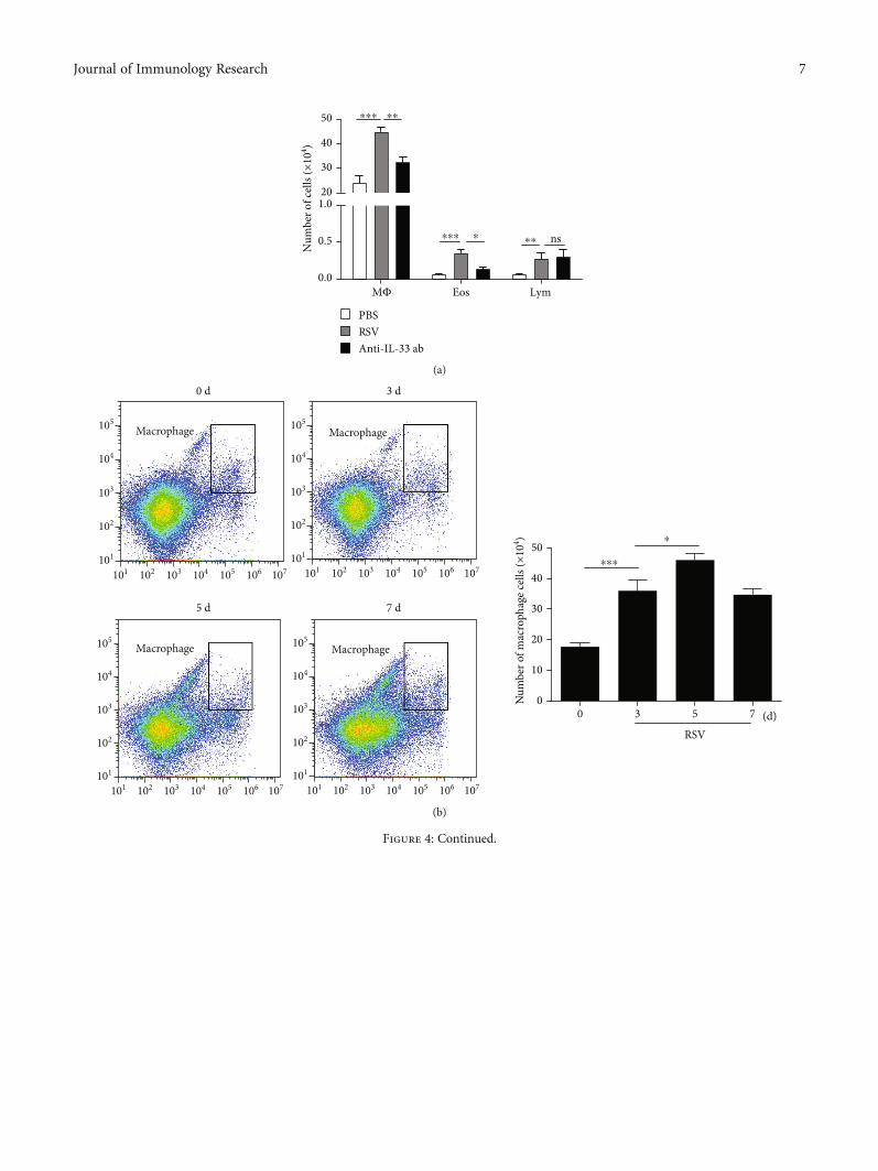
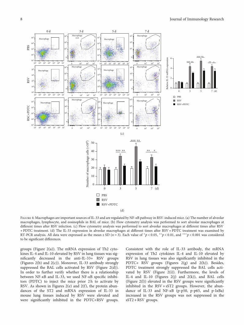
Figure 4: Macrophages are important sources of IL-33 and are regulated by NF-κB pathway in RSV-induced mice. (a) The number of alveolarmacrophages, lymphocyte, and eosinophils in BAL of mice. (b) Flow cytometry analysis was performed to sort alveolar macrophages atdifferent times after RSV infection. (c) Flow cytometry analysis was performed to sort alveolar macrophages at different times after RSV+PDTC treatment. (d) The IL-33 expression in alveolar macrophages at different times after RSV+PDTC treatment was examined byRT-PCR analysis. All data were expressed as the mean ± SD (n = 3). Each value of ∗p < 0:05, ∗∗p < 0:01, and ∗∗∗p < 0:001 was consideredto be significant differences.
8 Journal of Immunology Research
3.3. Acute Bronchiolitis Induced by RSV in a Mouse Model IsAttenuated after Inhibition of NF-κB/IL-33/ST2 Pathway. Tofurther confirm the role of NF-κB/IL-33/ST2 pathway inRSV-induced AVB in a mouse model, immunohistochemicalstaining was performed in this study. Representative IL-33,ST2, and p65 immunostaining was examined in saline-exposed control mice (NC) and RSV-treated mice (RSV).Immunostaining results showed that the abundances of IL-33, ST2, and p65 were all increased in acute bronchiolitisinduced by RSV than NC groups (Figure 3(a)). In addition,HE staining assay indicated that RSV significantly thickenedthe trachea wall, widened intercellular space, and enhancedinflammatory cell infiltration which was attenuated by anti-IL-13 antibody, PDTC, and sST2 treatment, representatively(Figure 3(b)).
3.4. Macrophages Are Important Sources of IL-33 and AreRegulated by NF-κB Pathway in RSV-Induced Mice. To fur-ther clarify the main cellular source of increased IL-33 secre-tion after RSV infection, inflammatory cells in BAL wereclassified and counted. We found that the number of alveolarmacrophages and eosinophils in BAL mice increased signifi-cantly after RSV infection (Figure 4(a)); flow cytometry anal-ysis showed that the number of macrophages was the largestand reached the peak on the fifth day after RSV infection(Figure 4(b)). We further verified whether NF-κB pathwaywas involved in RSV-induced macrophage number and IL-
33 production in macrophages; we found that both the num-ber of macrophages and expression of IL-33 increased in theRSV groups were reduced in the PDTC+RSV groups(Figures 4(c) and 4(d)).
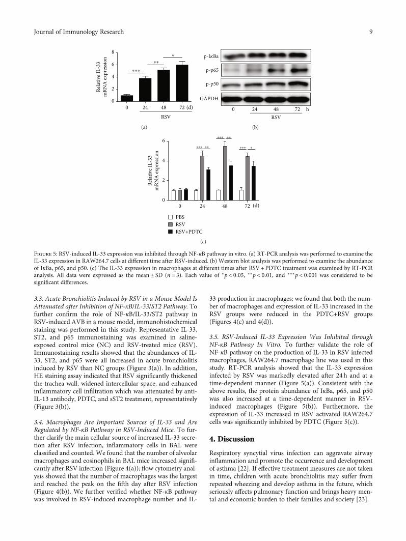
3.5. RSV-Induced IL-33 Expression Was Inhibited throughNF-κB Pathway In Vitro. To further validate the role ofNF-κB pathway on the production of IL-33 in RSV infectedmacrophages, RAW264.7 macrophage line was used in thisstudy. RT-PCR analysis showed that the IL-33 expressioninfected by RSV was markedly elevated after 24 h and at atime-dependent manner (Figure 5(a)). Consistent with theabove results, the protein abundance of IκBa, p65, and p50was also increased at a time-dependent manner in RSV-induced macrophages (Figure 5(b)). Furthermore, theexpression of IL-33 increased in RSV activated RAW264.7cells was significantly inhibited by PDTC (Figure 5(c)).
4. Discussion
Respiratory syncytial virus infection can aggravate airwayinflammation and promote the occurrence and developmentof asthma [22]. If effective treatment measures are not takenin time, children with acute bronchiolitis may suffer fromrepeated wheezing and develop asthma in the future, whichseriously affects pulmonary function and brings heavy men-tal and economic burden to their families and society [23].
0
2
4
6
8
RSV
(d)
Rela
tive I
L-33
mRN
A ex
pres
sion
0 24 48 72
⁎⁎⁎
⁎⁎
⁎
(a)
RSVh0 24 48 72
p-p50
GAPDH
p-I𝜅Ba
p-p65
(b)
(d)0 24 48 720
2
4
6
PBSRSVRSV+PDTC
Rela
tive I
L-33
mRN
A ex
pres
sion
⁎⁎⁎ ⁎⁎
⁎⁎⁎ ⁎⁎
⁎⁎⁎ ⁎
(c)
Figure 5: RSV-induced IL-33 expression was inhibited through NF-κB pathway in vitro. (a) RT-PCR analysis was performed to examine theIL-33 expression in RAW264.7 cells at different time after RSV-induced. (b) Western blot analysis was performed to examine the abundanceof IκBa, p65, and p50. (c) The IL-33 expression in macrophages at different times after RSV+PDTC treatment was examined by RT-PCRanalysis. All data were expressed as the mean ± SD (n = 3). Each value of ∗p < 0:05, ∗∗p < 0:01, and ∗∗∗p < 0:001 was considered to besignificant differences.
9Journal of Immunology Research
IL-33 can promote helminth infection and alleviate ath-erosclerosis by promoting Th2 immune response [11]. Inthe process of airway inflammation induced by RSV, IL-33and its specific receptor of ST2 increased significantly [24].However, the role of IL-33/ST2 pathway on RSV-inducedacute bronchiolitis and its molecular mechanism isunknown. In our study, we demonstrated that the levels ofIL-33 and Th2-related cytokines IL-4 and IL-10 in RSVinfected acute bronchiolitis in serum of infants were signifi-cantly increased. Besides, the Th1-associated cytokines IL-2and INF-γ and Th1/Th2 cell ratio were markedly reducedin infant serum of acute bronchiolitis. Furthermore, RSV-induced Th2-related cytokines IL-4 and IL-10 in lung tissueswere decreased by IL-33 antibody treatment. In addition, wealso found that the increase of inflammatory cells in lung tis-sue infected by RSV was inhibited by IL-33 antibody. Thesedata indicated that IL-33 plays an important role in acutebronchiolitis infants infected by RSV.
ST2 is one of the receptors of IL-1 family and widelyexpressed in many kinds of cells, especially mast cells andhelper T cells [25]. Without proinflammatory stimulation,IL-33 only exists in the nucleus of inflammation and immunecells. In many disease processes, activated and released IL-33can play an important role by combining with ST2 [26, 27].In addition, soluble ST2 (sST2) as a bait receptor of IL-33can directly bind to IL-33 and inhibit the biological functionof IL-33 [28, 29]. In this study, we confirmed that the ST2gene expression was positively correlated with IL-33 produc-tion in acute bronchiolitis cases. Both IL-33 antibody andsST2 could reversed RSV-induced Th2-related cytokine andpulmonary inflammatory damage. In addition, ST2 abun-dance can be inhibited in vivo and in vitro by IL-33 antibodytreatment. Our results suggested that IL-33/ST2 pathway ismediated the RSV-induced acute bronchiolitis and maybethe potential targets for the prevention of asthma after acutebronchiolitis in infants.
Many kinds of cells can secrete IL-33 after stimulatinginflammation. In addition to nonimmune cells such as epi-thelial cells and fibroblasts, macrophages, dendritic cells,and mast cells can also secrete IL-33 after stimulation [30].In recent years, increasing evidences indicate that innateimmune cells may be an important source of IL-33 in theprocess of respiratory viral infection [30]. By flow cytometryanalysis, we found that inflammatory cells increased signifi-cantly after RSV infection in mice, mainly macrophages,and the IL-33 expression in macrophages was elevated mark-edly. In addition, we also found that IL-33 antibody couldreduce the number of macrophages induced by RSV. Theseresults indicated that RSV increases the number of macro-phages and leads to the increase of IL-33 secretion; IL-33could also further promote the increase of macrophages.
Although evidence showed that IL-33 plays a biologicalrole through NF-κB pathway [20], IL-33 antibody treatmentdid not inhibit the expression of NF-pathway induce by RSVin our study, so we speculate that the NF-κB pathway is notmediated by the inhibitory effect of IL-33 antibody on RSV-induced lung damage. Interestingly, we found that NF-κBinhibitors can reduce RSV-induced lung damage in mice,and we also found that NF-κB inhibitors can reduce the
abundance of IL-33 in vivo and in vitro. This finding indi-cated that the NF-κB pathway is mainly involved in the pro-cess of RSV-induced IL-33 secretion in mice.
In conclusion, we confirmed that NF-κB/IL-33/ST2 axisis mediated the acute bronchiolitis by RSV infected. Ourfindings not only demonstrate the potential role of IL-33antibody in attenuating RSV-induced lung damage but alsoprovide a new insight into better prevention of RSV-induced asthma by mediating NF-κB/IL-33/ST2 axis.
Data Availability
All data collection and analysis were conducted under dou-ble-blind and supported by The Second People’s Hospitalof Changzhou, Affiliated Hospital of Nanjing MedicalUniversity.
Conflicts of Interest
There are no conflicts of interest.
Authors’ Contributions
Liwen Zhang designed and performed the in vivo experi-ments, analyzed the data, and wrote the manuscript. YuWan and Liang Ma are responsible for collecting clinicalblood samples and detecting inflammatory markers; theyare also responsible for in vitro studies. Kaihong Xu partici-pated in the animal experiments. Baojin Cheng designedand supervised the study and performed the manuscript edit-ing. Liwen Zhang and Yu Wan contributed equally to thiswork.
Acknowledgments
This study was supported by grants from the Research Pro-ject of Jiangsu Provincial Commission of Health and FamilyPlanning (No. H201547), the Major Scientific and Techno-logical Project of Changzhou City Commission of Healthand Family Planning (No. ZD201612), the Scientific andTechnological Project of Nanjing Medical University (No.2017NJMU042), the National Natural Science Foundationof China (No. 81700500), and the Applied Basic ResearchPrograms of Science, Technology Department of Changzhoucity (CJ20160031).
Supplementary Materials
Uncropped western blotting image. (SupplementaryMaterials)
References
[1] E. M. Abrams, A. B. Becker, and S. J. Szefler, “Where doesworsening asthma end and an asthma exacerbation begin?,”Annals of Allergy, Asthma & Immunology, vol. 123, no. 4,pp. 329-330, 2019.
[2] J. P. Lynch, R. B. Werder, Z. Loh et al., “Plasmacytoid dendriticcells protect from viral bronchiolitis and asthma throughsemaphorin 4a-mediated T reg expansion,” The Journal ofExperimental Medicine, vol. 215, no. 2, pp. 537–557, 2018.
10 Journal of Immunology Research
[3] L. Petrarca, R. Nenna, A. Frassanito et al., “Acute bronchiolitis:influence of viral co-infection in infants hospitalized over 12consecutive epidemic seasons,” Journal of Medical Virology,vol. 90, no. 4, pp. 631–638, 2018.
[4] S. M. Teo, H. H. F. Tang, D. Mok et al., “Airway microbiotadynamics uncover a critical window for interplay of patho-genic bacteria and allergy in childhood respiratory disease,”Cell Host & Microbe, vol. 24, no. 3, pp. 341–352.e5, 2018, e5.
[5] R. P. Tórtora, M. A. A. M. Guimarães, L. M. de Souza et al.,“Adenovirus species C detection in children under four yearsof age with acute bronchiolitis or recurrent wheezing,” Journalof Clinical Virology, vol. 73, pp. 77–80, 2015.
[6] A. M. Nathan, F. Rani, R. J. Y. Lee et al., “Clinical risk factorsfor life-threatening lower respiratory tract infections in chil-dren: a retrospective study in an urban city in Malaysia,” PLoSOne, vol. 9, no. 10, article e111162, 2014.
[7] J. Teeratakulpisarn, C. Pientong, T. Ekalaksananan,H. Ruangsiripiyakul, and R. Uppala, “Rhinovirus infection inchildren hospitalized with acute bronchiolitis and its impacton subsequent wheezing or asthma: a comparison of etiolo-gies,” Asian Pacific Journal of Allergy and Immunology,vol. 32, no. 3, pp. 226–234, 2014.
[8] J. Y. Chung, T. H. Han, J. S. Kim, S. W. Kim, C. G. Park, andE. S. Hwang, “Th1 and Th2 cytokine levels in nasopharyngealaspirates from children with human bocavirus bronchiolitis,”Journal of Clinical Virology, vol. 43, no. 2, pp. 223–225, 2008.
[9] X. Hu, X. Li, C. Hu et al., “Respiratory syncytial virus exacer-bates OVA-mediated asthma in mice through C5a-C5aR regu-lating CD4+T cells immune responses,” Scientific Reports,vol. 7, no. 1, article 15207, 2017.
[10] T. Jartti, M. Paul-Anttila, P. Lehtinen et al., “Systemic T-helperand T-regulatory cell type cytokine responses in rhinovirus vs.respiratory syncytial virus induced early wheezing: an observa-tional study,” Respiratory Research, vol. 10, no. 1, p. 85, 2009.
[11] Y. Liang, N. Yang, G. Pan, B. Jin, S. Wang, andW. Ji, “ElevatedIL-33 promotes expression ofMMP2 andMMP9 via activatingSTAT3 in alveolar macrophages during LPS-induced acutelung injury,” Cellular & Molecular Biology Letters, vol. 23,no. 1, p. 52, 2018.
[12] C. M. Finlay, A. M. Stefanska, K. P. Walsh et al., “Helminthproducts protect against autoimmunity via innate type 2 cyto-kines IL-5 and IL-33, which promote eosinophilia,” Journal ofImmunology, Virus Research and Experimental Chemotherapy,vol. 196, no. 2, pp. 703–714, 2016.
[13] H. Y. Lee, C. K. Rhee, J. Y. Kang et al., “Blockade of IL-33/ST2ameliorates airway inflammation in a murine model of allergicasthma,” Experimental Lung Research, vol. 40, no. 2, pp. 66–76,2014.
[14] X. Han, R. Chai, F. Qi et al., “Natural helper cells mediaterespiratory syncytial virus-induced airway inflammation byproducing type 2 cytokines in an IL-33-dependent manner,”Immunotherapy, vol. 9, no. 9, pp. 715–722, 2017.
[15] H. Yin, P. Li, F. Hu, Y. Wang, X. Chai, and Y. Zhang, “IL-33attenuates cardiac remodeling following myocardial infarctionvia inhibition of the p38 MAPK and NF-κB pathways,”Molec-ular Medicine Reports, vol. 9, no. 5, pp. 1834–1838, 2014.
[16] J. J. Zorc and C. B. Hall, “Bronchiolitis: recent evidence ondiagnosis and management,” Pediatrics, vol. 125, no. 2,pp. 342–349, 2010.
[17] A. Tahamtan, Y. Samieipoor, F. S. Nayeri et al., “Effects of can-nabinoid receptor type 2 in respiratory syncytial virus infec-
tion in human subjects and mice,” Virulence, vol. 9, no. 1,pp. 217–230, 2018.
[18] M. de Martinis, L. Ginaldi, M. M. Sirufo et al., “IL-33/vitaminD crosstalk in psoriasis-associated osteoporosis,” Frontiers inImmunology, vol. 11, article 604055, 2021.
[19] J. E. Vargas, A. P. D. de Souza, B. N. Porto et al., “Immuno-modulator plasmid projected by systems biology as a candi-date for the development of adjunctive therapy forrespiratory syncytial virus infection,” Medical Hypotheses,vol. 88, pp. 86–90, 2016.
[20] K. Fink, A. Duval, A. Martel, A. Soucy-Faulkner, andN. Grandvaux, “Dual role of NOX2 in respiratory syncytialvirus- and Sendai virus-induced activation of NF-κB in airwayepithelial cells,” Journal of Immunology, vol. 180, no. 10,pp. 6911–6922, 2008.
[21] S. Demyanets, C. Kaun, R. Pentz et al., “Components of theinterleukin-33/ST2 system are differentially expressed and reg-ulated in human cardiac cells and in cells of the cardiac vascu-lature,” Journal of Molecular and Cellular Cardiology, vol. 60,pp. 16–26, 2013.
[22] X. Carbonell-Estrany, “RSV prevention in infancy and asthmain later life,” The Lancet Respiratory Medicine, vol. 6, no. 7,article e31, 2018.
[23] D. You, D. T. Siefker, B. Shrestha, J. Saravia, and S. A. Cormier,“Building a better neonatal mouse model to understand infantrespiratory syncytial virus disease,” Respiratory Research,vol. 16, no. 1, p. 91, 2015.
[24] S. Zeng, J. Wu, J. Liu, F. Qi, and B. Liu, “IL-33 receptor (ST2)signalling is important for regulation of Th2-mediated airwayinflammation in a murine model of acute respiratory syncytialvirus infection,” Scandinavian Journal of Immunology, vol. 81,no. 6, pp. 494–501, 2015.
[25] A. Bayes-Genis, J. L. Januzzi, H. K. Gaggin et al., “ST2 patho-genetic profile in ambulatory heart failure patients,” Journalof Cardiac Failure, vol. 21, no. 4, pp. 355–361, 2015.
[26] X. Liu, L. Zhu, X. Lu et al., “IL-33/ST2 pathway contributes tometastasis of human colorectal cancer,” Biochemical and Bio-physical Research Communications, vol. 453, no. 3, pp. 486–492, 2014.
[27] S. M. Jung, J. Lee, S. Y. Baek et al., “The interleukin 33/ST2 axisin patients with primary Sjögren syndrome: expression inserum and salivary glands, and the clinical association,” TheJournal of Rheumatology, vol. 42, no. 2, pp. 264–271, 2015.
[28] S. E. Piper, R. A. Sherwood, G. F. Amin-Youssef, A. M. Shah,and T. A. McDonagh, “Serial soluble ST2 for the monitoringof pharmacologically optimised chronic stable heart failure,”International Journal of Cardiology, vol. 178, pp. 284–291,2015.
[29] K. Broch, A. K. Andreassen, T. Ueland et al., “Soluble ST2reflects hemodynamic stress in non-ischemic heart failure,”International Journal of Cardiology, vol. 179, pp. 378–384,2015.
[30] B. M. Matta, J. M. Lott, L. R. Mathews et al., “IL-33 is anunconventional alarmin that stimulates IL-2 secretion by den-dritic cells to selectively expand IL-33R/ST2+ regulatory Tcells,” Journal of Immunology, vol. 193, no. 8, pp. 4010–4020,2014.
11Journal of Immunology Research
![ACUTE SYSTEMIC INFLAMMATION · 5 parameter of inflammation. Increased levels of the acute phase protein fibrinogen are a main determinant of an elevated ESR [10]. A large number of](https://static.fdocument.org/doc/165x107/6147c476a830d0442101a684/acute-systemic-inflammation-5-parameter-of-inflammation-increased-levels-of-the.jpg)
![Oridonin protects LPS-induced acute lung injury by ......and acute lung injury (ALI) [1, 2]. Lipopolysaccharide (LPS), from the outer membrane of gram-negative bacteria, has been widely](https://static.fdocument.org/doc/165x107/608e9a4b0654131b49646243/oridonin-protects-lps-induced-acute-lung-injury-by-and-acute-lung-injury.jpg)