(1,3)-β-d-Glucan-based empirical antifungal interruption ...

Journal de Mycologie Médicale (2014) 24, 1—12
Available online at
www.sciencedirect.com
ORIGINAL ARTICLE/ARTICLE ORIGINAL
Inhibition of conidiophore development in
Aspergillus fumigatus by an Escherichia coliDH5a strain, a promising antifungalcandidate against aspergillosis Inhibition du developpement des conidiophores chez Aspergillusfumigatus par une souche d’Escherichia coli DH5a, un candidatantifongique prometteur dans l’aspergilloseM. Balhara, S. Ruhil, M. Kumar, S. Dhankhar, A.K. Chhillar *
Centre for Biotechnology, Maharshi Dayanand University, Rohtak, Haryana, India
Received 1 January 2013; received in revised form 23 June 2013; accepted 18 July 2013Available online 24 August 2013
KEYWORDSAspergillus fumigatus;E. coli;Time kill;Conidiophore;Scanningelectron microscopy
* Corresponding author.E-mail addresses: anil.chhillar@gm
1156-5233/$ — see front matter # 20http://dx.doi.org/10.1016/j.mycmed.
Summary The opportunistic human pathogen Aspergillus fumigatus produces a massivenumber of asexual spores (conidia) as the primary means of dispersal, survival, genomeprotection and infection of hosts. In this report, we investigated secretory and cytosolic proteinsof non-pathogenic bacterial species (mostly belonging to human microbiome) for antifungalpotential against A. fumigatus, A. flavus and A. niger. Our preliminary results revealed thatcytosolic proteins of E. coli DH5a were most active and the less toxic against various pathogenicisolates of A. fumigatus (the major pathogenic species), depicting a minimum inhibitoryconcentration (MIC) of 62.50 mg/mL, 62.50 mg/mL and 12.50 mg/disc using microbroth dilutionassay (MDA), percentage spore germination inhibition assay (PSGI) and disc diffusion assay (DDA),respectively. E. coli protein was non-toxic against human erythrocytes at doses up to 1000 mg/mLas compared to standard drug, amphotericin B which lysed 100% of erythrocytes at a concen-tration of 37.50 mg/mL. Time kill analysis proved it to be fungicidal in a concentration and time-dependent manner. Scanning electron microscopic studies (SEM) were carried out to prevailwhat kind of damage it causes to A. fumigatus. SEM results reported that conidiophore(structures forming conidia) development was halted as a major consequence, reducing thenumber of conidiophores to insignificant values as well as alteration in their morphologicalattributes. This feature may contribute to the development of new prevention strategies againstAspergillus infections. Hyphal atrophy was also observed, evidenced by shrinking and flattening
ail.com, [email protected] (A.K. Chhillar).
13 Elsevier Masson SAS. All rights reserved.2013.07.055

2 M. Balhara et al.
of hyphal walls and reduced, abrupt hyphal branching. Such actions may effectively reduce theinvasive ability of Aspergillus as well as it can sterilize the fungal burden by obstructing theconidiation pathway of A. fumigatus. Hence, E. coli DH5a, being a commensal species, can leadto the development of antifungal molecule with novel targets in fungal metabolism, which willhelp in combating the antifungal resistance and toxicity associated with current therapy.# 2013 Elsevier Masson SAS. All rights reserved.
MOTS CLÉSAspergillus fumigatus ;E. coli ;Courbe de survie ;Conidiophore ;Microscopieélectronique
Resume Le champignon opportuniste, Aspergillus fumigatus, pathogène pour l’homme pro-duit un nombre considérable de spores asexuées (conidies) principal moyen de dispersion, desurvie, de protection du génome et d’infection de l’hôte. Dans ce rapport, nous avons étudié lesprotéines secrétées et cytosoliques d’espèces bactériennes non-pathogènes (appartenant pourla plupart au microbiome humain) pour leur potentiel antifongique contre A. fumigatus, A. flavuset A. niger. Nos résultats préliminaires ont montré que les protéines cytosoliques de E. coli DH5aétaient les plus actives et les moins toxiques contres divers isolats pathogènes d’A. fumigatus (laprincipale espèce pathogène) avec une concentration minimale inhibitrice (CMI), respective-ment de 62,50 mg/mL, 62,50 mg/mL and 12,50 mg/disc dans le test de microdilution en milieuliquide, dans le test de pourcentage d’inhibition de germination des spores et dans le test dediffusion par la méthode des disques. La protéine d’E. coli n’était pas toxique vis-à-vis desérythrocytes humains à des concentrations jusqu’à 1000 mg/mL, comparativement à l’ampho-téricin B qui lysait 100 % des érythrocytes à une concentration de 37,50 mg/mL. La courbe desurvie en fonction du temps a montré une fongicidie concentration et temps dépendante. Lesétudes en microscopie électronique ont été faites pour préciser le type de lésions occasionnéessur A. fumigatus. Elles ont montré que le développement des conidiophores (structure produi-sant les conidies) était inhibé, réduisant le nombre des conidiophores jusqu’à une quantitéinsignifiante et altérant leurs caractéristiques morphologiques. Ces faits peuvent contribuer audéveloppement de nouvelles stratégies de prévention contre les infections aspergillaires. Uneatrophie des filaments a également été observée, traduite par un rétrécissement et unaplatissement de la paroi des hyphes et une diminution de la ramification. De telles actionspeuvent réduire de façon efficace la capacité invasive d’Aspergillus et stériliser la massefongique en bloquant la voie de synthèse des conidies d’A. fumigatus. Ainsi, E. coli DH5a,une espèce commensale, peut mener au développement d’une molécule antifongique avec denouvelles cibles au niveau du métabolisme fongique qui aidera à lutter contre la résistanceantifongique et contre la toxicité due aux traitements actuels.# 2013 Elsevier Masson SAS. Tous droits réservés.
Abbreviations
MDA M
icrobroth dilution assay DDA D isc diffusion assay PSGI P ercentage spore germination inhibition Amp B A mphotericin B MIC M inimum inhibitory concentration rpm R evolutions per minute EDTA E thylene diamine tetra acetic acid DTT D ithiotritol B. amylo B . amyloliquefaciensIntroduction
The frequency of invasive, opportunistic mycoses has increa-sed significantly over the past two decades [25]. Thisincrease in infection is associated with excessive morbidityand mortality and is directly related to the increasing num-bers of patients who are at risk for the development ofserious fungal infections, including patients undergoingblood marrow transplantation (BMT), solid-organ transplan-tation, and major surgery (especially gastrointestinal sur-gery); patients with AIDS, neoplastic disease, and advanced
age; patients receiving immunosuppressive therapy; andpremature infants [19,28]. Among invasive fungal infections,aspergillosis remains the most significant problem in thepatients with weakened immune system. It is the mostprevalent fungal infection, particularly in those with hae-matological malignancy and patients undergoing haemato-poietic stem cell transplantation (HSCT) [17]. Currently, theincidence of invasive aspergillosis in the US ranges from 0.5%after autologous hematopoietic stem cell transplantation to3.90% after transplantation from an unrelated donor. Inthese patients, after diagnosis of aspergillosis within3 months, mortality was 53.8% in autologous transplant reci-pients and 84.6% in those with unrelated donor transplants[18]. But in the most recent data from the US, 12-monthmortality from invasive aspergillosis (IA) was 25% [9]. Thisshows that there is improvement but the risk of fungalinfection after transplantion is multifactorial.
Although 19 species of Aspergillus that have been docu-mented as agents of human disease, the majority of infec-tions are caused by A. fumigatus, A. flavus and A. niger [2].The most common causing invasive disease is A. fumigatus.Remarkably, A. fumigatus causes 90% of all systemic Asper-gillus infections. But unfortunately, these fungi are one ofthe most neglected pathogens as demonstrated by the factthat amphotericin B, a polyene antibiotic discovered as long

Inhibition of conidiophore development in Aspergillus fumigatus 3
ago as 1956 is still used as a gold standard for antifungaltherapy. But it has been found to be less effective in invasiveaspergillosis and highly nephrotoxic. New liposomal formu-lations of amphotericin B were found to have reduced toxi-city, but cure rate did not improve much. In January 2001,the echinocandins became the first semisynthetic antifungalnatural product to be approved since polyene was approved40 years earlier. A new antifungal agent called caspofunginwas launched in 2002 and included in the existing therapeuticoptions for candidiasis and invasive aspergillosis. It showedgood activity against Aspergillus and Candida, however, itsclinical efficacy guided market is yet to be established [24].Although the new generation azoles (voriconazole, posaco-nazole) have been much improved as compared to previouscompounds of this class, the concerns with respect to limitedefficacy could not be resolved. The fungistatic character hasbeen the main drawback of these compounds. Recent increa-ses in fungal infections, the few available antifungal drugs,and increasing fungal resistance to the available antifungaldrugs have resulted in a broadening of the search for newantifungal agents.
Encouragingly, naturally occurring antifungal proteinsand peptides have the potential to be very interesting cli-nical leads. Various strains of Bacillus subtilis produce theiturin peptide family. One family member, Bacillomycin Finhibited the growth of fungi including A. niger, Candidaalbicans, and Fusarium oxysporum [23]. A Bacillus licheni-formis isolate, M-4, produces fungicin M-4. It is a hydrophilic,narrow-spectrum antifungal peptide that is resistant to pro-teolytic enzymes, lipase and inhibited the growth of Micro-sporum canis, Mucor species, and Sporothrix schenckii [11].Nikkomycins, which are produced by Streptomyces tendae,enter target cells via dipeptide permeases and inhibit chitinbiosynthesis in C. albicans both in vitro and in vivo [5].Various lactic acid bacteria isolates, including Lactobacilluscornyiformis, L. plantarum showed strong inhibitory activityagainst A. fumigatus, A. nidulans, etc [14]. An antifungalcompound d-dodecalactone from Lactobacillus plantarumAF1 isolated from kimchi showed strong inhibitory effectagainst A. flavus, A. fumigatus, A. petrakii, A. ochraceus,A. nidulans, and Penicillium roqueforti [26].
Similarly other bacterial species, like Streptomyceshygroscopicus strain BS-112 has been reported to possessantifungal activity against A. flavus [29]. Antifungal meta-bolites produced by Pseudomonas spp. have been implicatedin control of a variety of fungal diseases [12]. Bacillusamyloliquefaciens has been found to have antifungal activityagainst many phytopathogenic fungi [3]. While the mainemphasis has been on B. amyloliquefaciens metabolitesfor biocontrol of plant pathogens [1] but they also havepotential against human pathogens. One of its metabolites,Macrolactin A displays antimicrobial activity toward humanpathogens, such as Klebsiella pneumoniae and Staphylococ-cus aureus [3]. B. amyloliquefaciens LBM 5006 showedsignificant activity against phytopatogenic fungi, showingpotential for use as a biocontrol agent or production ofantifungal preparations [4]. In the microbial world, theEscherichia coli has been a very important member of greatmedical, biological, biotechnological and industrial signifi-cance. As part of the normal flora of humans, it plays acrucial role in food digestion by producing vitamin K andcreates barrier effect against enteropathogens [20].
The EM0 and JM105 K-12 strains of E. coli isolated fromhuman faecal sample and soil respectively were found tohave ability of preventing in vivo and in vitro infection bySalmonella typhimurium C5 and allergies [8]. But, E. coli hasnot been explored much for antifungal potential so that itcould be used as a resource of antimycotic molecules. Avariety of microbial products have been found to have anti-fungal potential but data establishing their clinical efficacy isscarce [16]. However, the commensal bacteria like E. coli,which are part of human biotic flora, can be used in the formof probiotics. Therefore, focal theme of the present studyinvolved detailed analysis of secretory and cytosolic proteinsof bacteria’s mostly belonging to human microbiome andsome natural antagonist bacterial species for antifungalpotential against A. fumigatus, A. flavus and A. niger.
Material and methods
Bacterial strains
Lactococcus lactis (MTCC440), E. coli DH5a (MTCC1652),E. coli (MTCC 1674), Streptococcus thermophilus(MTCC1938), Staphylococcus epidermidis (MTCC3615), Sta-phylococcus epidermidis (MTCC6810), Streptococcus subsp.equi (MTCC3522), Bacillus amyloliquefaciens (MTCC2248),Bacillus pumilus (MTCC2299), Bacillus subtilis (MTCC1133),Bacillus licheniformis (MTCC1483), Streptomyces hygrosco-picus (MTCC4003) and Pseudomonas fluorescens(MTCC1748), were purchased from Institute of MicrobialTechnology, Chandigarh, India. The bacterial strains wereharvested in Luria Broth (LB) for 72 h at 37 8C inan incubator shaker. The concentration or density of theculture (bacterial cells/mL) was evaluated by turbidometrymethod.
Fungal pathogens
Pathogenic isolates of A. fumigatus [ITCC4517 (IARI, IndianAgricultural Research Institute, Delhi), ITCC1634 (IARI, Delhi),190/96 (VPCI, Vallabhbhai Patel Chest Institute, Delhi)],A. flavus [ITCC5192 (IARI, Delhi), ITCC5076 (IARI, Delhi),223/96 (VPCI, Delhi)] and A. niger [ITCC5405 (IARI, Delhi),56/96 (VPCI, Delhi)] were employed in the current study.These pathogenic species of Aspergillus were cultured inlaboratory on Sabouraud dextrose agar plates. The plates wereinoculated with stock cultures of A. fumigatus, A. flavus,A. niger and incubated at 37 8C for 96 h. The freshly revivedfungal plates were used for preparing spore suspension.
Preparation of bacterial secretory and cytosolicprotein fractions
The bacterial strains were cultured for 72 h and the cultureswere centrifuged at 3256 g for 30 min. The culture super-natant or secretory protein fraction was separated frompellet. The cell free supernatant was filtered through0.22 mm filter and was dialysed against distilled water at4 8C for 24 h with several changes of water and lyophilized.The pellet was washed with PBS thrice and suspended insonication buffer (50 mM L�1 Tris—HCl, 50 mM L�1 EDTA,5 mM L�1 DTT and 1 mM L�1 PMSF). The cell suspension was
4 M. Balhara et al.
sonicated at 20 s bursts at 200 W and 10 s cool period using asonicator (Bio-Age, Sonicator Mohali, India). The sonicatedcell suspension was centrifuged at 16350 g for 30 min usingSigma refrigerated centrifuge (Osterode, Germany). Thesupernatant was collected and used as lysate. The lysateor cytosolic protein fraction was dialysed against distilledwater at 4 8C for 24 h with several changes of water andlyophilized. Protein concentration of cell free supernatantand lysate was determined by Bradford method [6].
Antifungal activity
The antifungal activity of bacterial proteins was analysed bymicrobroth dilution, disc diffusion and spore germinationinhibition assays (SGIA) [7]. Each experiment was repeated atleast three times.
Microbroth dilution assay
The test was performed in 96-well culture plates (Tarson,Kolkata India). Autoclaved Sabouraud dextrose broth (90 mL)was added to the wells of culture plates. An amount of 100 mLof secretory or cytosolic protein fractions, having concen-tration of 2000 mg/mL, was added to the wells which werethen serially diluted by two-fold dilution method, giving aconcentration range of 1000—3.9 mg/mL and these wellswere inoculated with 10 mL of spore suspension containing1 � 106 spores/mL. The plates were incubated at 37 8C andexamined macroscopically after 24 h for the growth ofAspergillus mycelia. Appropriate control wells treated withamphotericin B and without any treatment were included inthe study. A protein fraction was considered to be active ifthe wells appeared clear without any visible growth ofAspergillus and that protein concentration was expressedas minimum inhibitory concentration (MIC).
Agar-disc diffusion assay
The disc diffusion test was performed in radiation sterilizedPetri plates of 10.0 cm diameter (Tarsons, Kolkata, India).Different concentrations ranging from 50.00 to 3.20 mg ofproteins/disc of the bacterial proteins were impregnated onthe sterilized discs (5.0 mm in diameter) of Whatman filterpaper No. 1. These discs were placed on the surface of theagar plates, which were already inoculated with 1 � 106
spores/mL. The plates were incubated at 37 8C and examinedafter 24 h for zone of inhibition, if any, around the discs. Thediameter of zone of inhibition was recorded. The concen-tration, which developed the zone of inhibition of at least6.0 mm diameter, was considered as MIC. Amphotericin Bwas used in assay as a standard control drug. An additionalcontrol disc without any sample but impregnated with equi-valent amount of solvent was also used in the assay.
Percentage spore germination inhibition assay
The Aspergillus species were grown on Sabouraud dextroseagar plates and their homogenous spore suspension wasprepared in the Sabouraud dextrose broth. Various concen-trations (1000—3.9 mg/mL) of the bacterial proteins in 90 mLof culture medium were prepared in 96-well flat bottom
microculture plates (Tarsons, Kolkata, India) by double dilu-tion method. The wells were prepared in duplicates for eachconcentration. The wells were inoculated with 10 mL of sporesuspension containing 100 � 5 spores. Appropriate controlwells treated with amphotericin B or without any treatmentwere included in the study. The plates were incubated at37 8C for 16 h and then examined for spore germination undercompound microscope (Labomed, USA). The number of ger-minated and non-germinated spores was counted and thepercent spore germination inhibition (PSGI) was calculatedusing following formula:
PSGI = (100 — No. of spores germinated in drug treatedwell/No. of spores germinated in control well) � 100
The protein concentration, which inhibits the germinationof spores in the range of 90—100%, was represented as MIC.
The antifungal susceptibility tests were also performed ondifferent test medium. Effects of different media weredetermined by using RPMI 1640 (Himedia), yeast nitrogenbroth (pH 7.0; Himedia), antibiotic assay medium no. 3(Himedia). The MIC90 was considered to be the lowestconcentration of the compound that inhibited the visiblegrowth of fungi.
Haemolytic assay
The basic method of Latoud [10] with slight modifications wasemployed to determine the haemolytic effect of bacterialproteins with antifungal activity. Human erythrocytes, col-lected from apparently healthy individuals, were washedthree times with PBS by centrifugation at 1500 rpm for10 min. A 2% erythrocyte suspension was incubated at 37 8Cfor 1 h with different concentrations of bacterial proteinsranging from 1000.0 mg/mL to 15.6 mg protein/mL. Afterincubation, cells were pelleted at 5000 rpm for 10 min. Thesupernatant was collected and the absorbance at 450 nm wasdetermined using a spectrophotometer (UV Vis Spect LambdaBio 20; Perkin Elmer). In negative control sets, only buffer wasused for background lysis, whereas in positive controls, lysisbuffer was used for completely lysing the erythrocytes. Foreach sample, the percentage of maximum haemolytic activitywas determined. A protein concentration that causes a lysis ofless than 5% was considered as non-toxic.
Time kill analysis
Sabourd dextrose agar plates were inoculated with stockcultures of A. fumigatus and incubated at 37 8C for 96 h.The freshly revived fungal plates wereused for preparing sporesuspension. The homogenous spore suspension was made anddiluted with 0.9% NaCl to get 1 � 108 spores/mL. Aliquots with5 mL of RPMI 1640 as culture medium were inoculated withspore suspension of 1 � 108 spores/mL. Aliquots were treatedwith protein concentration equal to 1/2 MIC, MIC and 2� MIC.For each concentration, corresponding controls without anydrug treatment were prepared simultaneously. The aliquotswere incubated at 37 8C with shaking for up to 48 h. At definedtime intervals (6, 12, 24, 48 h), aliquots were removed and thenumber of viable colonies per millilitre were determined onSabouraud dextrose agar plates after serial dilution in 0.9%NaCl. The results were expressed as log10 cfu/mL against timeof incubation at each drug concentration tested. The protein

Table 1 Antifungal evaluation of secretory proteins against pathogenic isolates of Aspergillus spp. using microbroth dilutionassay.Evaluation de l’activite antifongique des proteines secretees contre des isolats d’Aspergillus sp. avec le test de microdilution enmilieu liquide.
MTCC No. Bacterial species Mean of MIC of BSP (mg/mL)
A. fumigatus A. flavus A. niger
440 Lactococcus lactis 250.0 250.0 500.01652 E. coli DH5a 62.5 62.5 250.01748 Pseudomonas fluorescens 15.6 15.6 31.21938 Streptococcus thermophilus 125.0 250.0 —3615 Staphylococcus epidemidis — — —6810 Staphylococcus epidemidis — — —3522 Streptococcus equisubsp. equi 250.0 250.0 —2248 Bacillus amyloliquefaciens 125.0 250.0 250.02299 Bacillus pumilus 250.0 250.0 500.01133 Bacillus subtilis 125.0 250.0 500.01483 Bacillus licheniformis 62.5 125.0 125.04003 Streptomyces hygroscopicus 62.5 62.5 125.01674 E. coli 125.0 125.0 250.0
BSP: Bacterial supernatant protein; —: no activity up to tested concentration i.e. 1000 mg/mL; MIC of Amp. B: 1.95 mg/mL.
Inhibition of conidiophore development in Aspergillus fumigatus 5
concentration, which causes an average reduction of 3 log10 incolony forming units per mL as compared to control at 0 h wasconsidered as fungicidal [15].
Scanning electron microscopy
A. fumigatus was cultured in 100 mL of L-asparagine brothusing a spore suspension of 1 � 106 spores/mL for 24 h in aBOD incubator shaker at 37 8C, 100 rpm and treatment withMIC50 of E. coli DH5a cytosolic protein to examine its effecton fungus morphology and spore germination along withcultures without any drug treatment (control). Fungal mass
Table 2 Antifungal evaluation of cytosolic proteins against patassay.Evaluation de l’activite antifongique de proteines cytosoliques coen milieu liquide.
MTCC No. Bacterial species
440 Lactococcus lacti1652 E. coli DH5a1748 Pseudomonas fluorescens1938 Streptococcus thermophilus3615 Staphylococcus epidemidis6810 Staphylococcus epidemidis3522 Streptococcus equisubsp. equi2248 Bacillus amyloliquefaciens2299 Bacillus pumilus1133 Bacillus subtilis1483 Bacillus licheniformis4003 Streptomyces hygroscopicus1674 E. coli
BLP: Bacterial lysate proteins; —: no activity up to tested concentrat
recovered after culture was used for SEM studies. It was fixedwith 2.5% glutaraldehyde in 0.1 mol/L phosphate buffer (pH7.2) at 48 8C for 2 h. All specimens were washed four timeswith phosphate buffer and post-fixed with phosphate-buffe-red 2% osmium tetroxide at 48 8C for 4 h. After washingovernight with the same buffer, the specimens were dehy-drated in graded ethanol and finally freeze-dried in t-butylalcohol. The specimens were coated with gold film (approxi-mately 10 nm). Samples were examined using a Carl ZeissEV040 Gemini field emission scanning microscope with anacceleration voltage of 20 kV using the Everhart Thornley SEdetector and the in-lens SE detector in a 50:50 ratio.
hogenic isolates of Aspergillus spp. using microbroth dilution
ntre des isolats d’Aspergillus sp. avec le test de microdilution
Mean of MIC of BLP (mg/mL)
A. fumigatus A. flavus A. niger
250.0 500.0 500.062.5 62.5 125.0125 125 250.0250.0 250.0 500.0— — —— — —500.0 500.0 —31.2 31.2 62.5500.0 — —250.0 500 —250.0 250 500500.0 — —500.0 500.0 —
ion i.e 1000 mg/mL, MIC of Amp.B: 1.95 mg/mL.

6 M. Balhara et al.
Results
In vitro evaluation of antifungal potential ofsecretory and cytosolic proteins
The secretory and cytosolic protein fractions of chosenbacteria’s were evaluated for antifungal activity againstpathogenic isolates of A. fumigatus, A. flavus and A. nigerusing microbroth dilution assay. The mean value of MICs ofsecretory and cytosolic fractions of various bacterial speciesis represented in Tables 1 and 2. The bacterial proteinsexhibited almost similar (showing no variation) antifungalactivity against different isolates of Aspergillus spp. Theresults revealed that secretory fractions of Pseudomonasfluorescens, E. coli DH5a, Streptomyces hygroscopicus andBacillus licheniformis had good antifungal activity againstA. fumigatus and A. flavus having MIC in the range of
igure 1 Inhibitory effect on the growth of A. fumigatusnalysed by a microbroth dilution assay: lane I: control (firsthree wells only medium and other wells normal growth of. fumigatus), lane II: treated with different concentrationsf E. coli DH5a lysate (MIC: 62.50 mg/mL), lane III: treated withifferent concentrations of B. amyloliquefaciens lysate (MIC:1.25 mg/mL).ffet inhibiteur sur la croissanced’A. fumigatuspar microdilutionn milieu liquide : rangee 1 : temoins (three premiers puits milieueul, autres puits : croissance normale d’A. fumigatus), rangee II :ifferentes concentrations de lysat d’ E. coli DH5a (CMI :2.50 mg/mL), rangee III : traitement avec differentes concen-
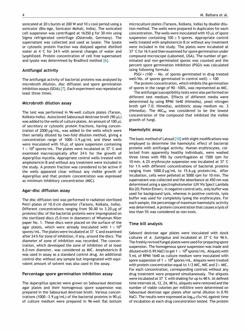
Figure 2 Cytotoxicity of E. coli DH5a lysate protein and B.amyloliquefaciens lysate against human erythrocytes in compa-rison with standard amphotericin B (Amp B).Cytotoxicite d’une proteine de lysat de E. coli DH5a et de B.amyloliquefaciens contre des erythrocytes humains en compa-
[(Figure_1)TD$FIG]
FatAod3Eesd6
trations de lysat de B. amyloliquefaciens (CMI : 31.25 mg/mL).15.62 mg/mL to 62.50 mg/mL. However, the degree of inhibi-tion against A. niger was low. Among the cytosolic proteinfractions, Bacillus amyloliquefaciens and E. coli DH5a werefound to be most active showing a MIC of 31.25 mg/mL and62.50 mg/mL, respectively against A. fumigatus (Fig. 1) andA. flavus while the MIC was two-fold higher against A. niger.
In vitro cytotoxicity results
Cytotoxicity of bacterial proteins with potent antifungalactivity was checked using a haemolytic assay against humanerythrocytes. The cytosolic and secretory fractions ofB. amyloliquefaciens, E. coli DH5a, Pseudomonas fluores-cens, Streptomyces hygroscopicus, Bacillus licheniformiswere evaluated for cytotoxicity. E. coli DH5a proteins werefound to be non-toxic up to a concentration of 1000 mg/mL(i.e. causes less than 5%lysis) which was remarkable ascompared to standard drug amphotericin B which causes100% lysis at a concentration of 37.50 mg/mL while Bacillusamyloliquefaciens protein was non-toxic up to a concentra-tion of 250 mg/mL (Fig. 2). The remaining bacterial proteinsshowed high cytotoxicity, which was their major limitationfor future consideration despite of good antifungal activity(P. fluorescens being non-toxic up to 31.25 mg/mL,S. hygroscopicus and B. licheniformis being non-toxic upto 62.50 mg/mL). As the cytosolic protein of E. coli DH5awas found to have ground level cytotoxicity as well as goodantifungal activity, its antimycotic potential was furtherinvestigated using various antifungal susceptibility assays.
Antifungal evaluation of E. coli DH5a lysate usingvarious antifungal susceptibility assays
The minimum inhibitory concentration of E. coli DH5a cyto-solic protein (lysate) using MDA was found to be 62.50 mg/mLagainst A. fumigatus and A. flavus while A. niger was lesssusceptible showing a MIC of 125 mg/mL. The broad spectrumantifungal activity of E. coli lysate protein was also examined[(Figure_2)TD$FIG]
raison avec l’amphotericine B (Amp B).

[(Figure_3)TD$FIG]
Figure 3 Inhibitory effect of E. coli DH5a lysate against A. fumigatus, A.flavus and A. niger using PSGI assay.Effet inhibiteur d’un lysat de E. coli DH5a contre A. fumigatus, A. flavus et A. niger dans le test d’inhibition de germination desspores.
Figure 4 Inhibitory effect of E. coli DH5a lysate againstA. fumigatus using DDA. A: 50 mg lysate, B: 25 mg Lysate, C:12.5 mg lysate, D: 6.25 mg lysate.Effet inhibiteur d’un lysat de E. coli DH5a contre A. fumigatuspar le test des disques : lysat. A : 50 mg, B : 25 mg, C : 12.5 mg, D :6.25 mg.
Inhibition of conidiophore development in Aspergillus fumigatus 7
through percent spore germination inhibition assay (PSGI)and disc diffusion assay (DDA). The percent spore germina-tion inhibition assay was performed for E. coli DH5a lysateusing A. fumigatus, A. flavus and A. niger spores. The doseresponse curve obtained by plotting percent spore germina-tion inhibition against concentration of E. coli DH5a lysateproteins against A. fumigatus, A. avus and A. niger is shownin Fig. 3. The percent spore germination inhibition increasedwith increase in concentration of lysate protein used in thetest. It was observed that a protein concentration of 62.5 mg/mL of DH5a lysate inhibited the growth of A. fumigatus andA. flavus. The susceptibility of A. niger to treatment with E.coli DH5a lysate was less than that of A. fumigatus andA. flavus. Various test concentrations ranging from 50.0 mgto 3.12 mg/disc of E. coli DH5a lysate were used to determinethe antifungal activity by disc diffusion assay. The lowestconcentration of E. coli DH5a lysate, which inhibited thegrowth of A. fumigatus, was found to be 12.5 mg/disc in discdiffusion assay (Fig. 4). The concentrations of E. coli DH5alysate lower than 12.5 mg/disc were found to be ineffectiveas there was no effect on the growth of A. fumigatus and themycelia of the pathogen could grow even over to the discsimpregnated with 6.25 mg/disc of E. coli DH5a lysate. Themean diameter of zone of inhibition developed by variousconcentrations of E. coli DH5a lysate against differentpathogenic species of Aspergillus is shown in Table 3. Thesusceptibility of A. niger to the lysate was comparatively lessas 25.0 mg/disc of the lysate was required to develop thezone of inhibition of activity significant in disc diffusionassay. The MICs of E. coli DH5a lysates obtained by usingdifferent assays are given in Table 4.
Antibiotic assay medium 3 was used to identify resistance,if any, in Aspergillus spp. against the E. coli cytosolic proteinand amphotericin B as described by Rex et al. [21]. The MIC90
against Aspergillus spp. was found to be 31.25 mg/mL inantibiotic medium 3 (Table 5). Variations in susceptibilityin the group of fungal strains tested with E. coli cytosolicprotein were observed; however, resistance against theprotein was not seen. The test method can affect the levelof antifungal activity determined. The in vitro activity ofE. coli DH5a cytosolic protein was compared against eight
strains of Aspergillus in four test media, namely RPMI 1640,YNB, antibiotic assay medium 3 and SD broth. The MIC90 washighest in RPMI 1640 and YNB medium while it was lowest inantibioic assay medium 3 and SD broth. Amphotericin B wasalso found to be most active in antibiotic medium 3 and SDbroth.
Time kill study
In vitro time kill kinetics was determined for E. coli DH5acytosolic protein against A. fumigatus, which is the majorpathogenic species responsible for 90% of systemic fungalinfections, caused by the genus Aspergillus. The result of timekill studies is presented in Fig. 5A—C. Data are presented in[(Figure_4)TD$FIG]

Table 3 Antifungal evaluation of E. coli DH5a cytosolic protein by disc diffusion method.Evaluation de l’activite antifongique d’une proteine cytosolique d’ E. coli DH5a par la methode des disques.
E. coli Mean diameter of zone of inhibition (mm)
Concentration/mg/disc A. fumigatus A. flavus A. niger
Mean � SD CV Mean � SD CV Mean � SD CV
50.0 10.50 � 0.10 0.01 10.70 � 0.05 0.003 8.90 � 0.10 0.0125.0 9.20 � 0.20 0.04 9.10 � 0.10 0.010 6.50 � 0.05 0.00312.5 6.10 � 0.15 0.02 6.06 � 0.05 0.003 0.0 0.06.2 0.0 0.0 0.0 0.0 0.0 0.03.1 0.0 0.0 0.0 0.0 0.0 0.0
Amphotericin B2.5 8.30 � 0.05 0.003 8.40 � 0.15 0.020 8.03 � 0.15 0.023
8 M. Balhara et al.
terms of the log10 cfu/mL against time of incubation at ½ MIC,MIC and 2�MIC for E. coli DH5a cytosilic protein and standarddrug amphotericin B. Growth inhibition and efficacy of theE. coli cytosolic protein was observed to be dose- and time-dependent, producing distinct time kill profiles againstA. fumigatus. In terms of reduction in value of log10 cfu/mL, E. coli DH5a cytosolic protein showed an average log10
reduction of 3.1 as compared to control at MIC after 24 h ofincubation while at 2� MIC, the average log10 reduction was3.3 after 12 h of incubation. Exposure of A. fumigatus sporesto various concentrations of active protein significantlydecreased their ability to produce fungal colonies in a time-and concentration-dependent manner. E. coli cytosolicprot � 95% loss at MIC after 12 h of incubation. At 2� MIC,E. coli protein causes � 90% killing after 12 h of incubationwhile amphotericin B inhibited 99% fungal growth after 6 h ofincubation. On the contrary, there was an insignificant growthinhibition of fungus when subjected to ½ MIC of E. coli proteinas it causes � 60% loss even after 48 h of incubation while thestandard drug Ampho. B at MIC50 causes � 90% loss after 24 hof incubation. The variation between the activities of thecrude protein and the standard antifungal drug may be due tothe mixtures of bioactive compounds present in the crudeprotein as compared to the pure compound contained in thestandard antibiotics that rarely have the same degree of
Table 4 MICs of lysate proteins of Escherichia coli (DH5a)by various antifungal susceptibility tests against Aspergillusspecies.CMI de proteines de lysat d’ Escherichia coli (DH5a) dansdifferents tests de sensibilite contre Aspergillus sp.
Pathogens MIC (mg/mL) Lysate protein of E. coliDH5a
MDA PSGI DDA (mg/disc)
A. fumigatus 62.50 62.50 12.50A. flavus 62.50 62.50 12.50A. niger 125.00 125.00 25.00
MIC of Amp. B using DDA was found to be 0.75 mg/disc. MIC ofAmp. B using PSGI was found to be 1.90 mg/mL.
activity as the unrefined fractions at comparable concentra-tions or dose of the active component.
Scanning electron microscopic findings
The effect of treatment of E. coli cytosolic protein (MIC50) onfungal growth and reproduction was examined throughscanning electron microscopy. The control samples (withoutany treatment) showed typical morphology of A. fumigatushyphae. Hyphae were generally elongated and grew denselyin various directions. The thickness was almost uniform andsurfaces were smooth. Various conidia were seen on thehyphal surface that have been shed from germinating coni-diophores (Fig. 6A). On the contrary, in treated samples,hyphal branching was reduced and altered (diffused andirregular) as compared to control; hyphae were short inlength and branched at trunk region, suggesting inhibitionof apical branching (Fig. 6B). Hyphae also displayed signifi-cantly wrinkled, flattened and fibrillar appearance with non-uniform thickness, revealing leakage of intracellular mate-rial (Fig. 7B). Conidiophore formation was severely reducedas compared to control in treated samples (Fig. 7A and B).The number of conidiophores was nominal in treated samplesas well as the morphology was also altered, showing inhibi-tion in the formation of conidial chains as compared tocontrol (Fig. 8A and C). Some conidiophores showed onlythe conidiogenous cells at their surface depicting delayed
Table 5 Effect of different test media on in vitro activityof E. coli cytosolic protein by microbroth dilution assay.Effets de differents milieux sur l’activite in vitro de pro-teines cytosoliques par microdilution en milieu liquide.
MIC of E. coli DH5a lysate in various testmedia (mg/mL)
Strain RPMI1640
YNB Antibioticmedium 3
SDBroth
A. fumigatus 125.0 125.0 31.25 62.5A. flavus 125.0 125.0 31.25 62.5A. niger 125.0 250.0 31.25 125.0

[(Figure_5)TD$FIG]
Figure 5 A. Time kill curve at ½ MIC. B. Time kill curve at MIC. C. Time kill curve at 2�MIC. In vitro time kill kinetics of E. coli DH5acytosolic protein and Amphotericin B.A. Courbe de fongicidie en fonction du temps avec ½ CMI. B. Courbe de fongicidie en fonction du temps a la CMI. C. Courbe defongicidie en fonction du temps avec 2�CMI. Courbe de fongicidie en fonction du temps d’une proteine cytosolique de E. coli DH5a etd’amphotericine B contre A. fumigatus.
Inhibition of conidiophore development in Aspergillus fumigatus 9
development (Fig. 8B). Resting conidia were observed whichindicated germination inhibition (Fig. 8C).
Discussion
Currently available antifungal compounds except echinocan-dins act on targets also found in mammalian cells, which mayresult in toxicity or an adverse drug interaction. It is there-fore imperative to find antifungal compounds that are not[(Figure_6)TD$FIG]
Figure 6 A. Hyphae are linearly elongated, grew densly, numerouhyphal morphology. B. Hyphae are short, branched frequently at irAction de la proteine de E. coli DH5a sur la morphologie des hyph
toxic to mammalian cells. In invasive aspergillosis infections,most of the antifungal drugs (caspofungin, amphotericin B)are administered intravenously; hence, haemolytic activitybecomes a major concern for drug dosage [24]. The pastdecade has witnessed a dramatic growth in knowledge ofnatural peptides. Among the first antifungal peptides isola-ted were the iturin and Bacillomycin families produced byBacillus subtilis. But they are found to be hemolytic [13].Syringomycins are small lipodepsipeptides and are among themost potent bacterial antifungal peptides yet discovered.
s conidia are visible (arrows). Effect of E. coli DH5a protein onregular sites, no conidia are visible.es.

[(Figure_7)TD$FIG]
Figure 7 A. Numerous conidiophores without abundant conidial chains are visible. B. Conidiophore formation is severely inhibiteddepicting 2—3 conidiophores only (arrows). Effect of E. coli DH5a protein on conidiophores developmentAction de la proteine de of E. coli DH5a sur le developpement des conidiophores.
10 M. Balhara et al.
But they were more toxic than Amp. B against human ery-throcytes [22]. Therefore, the toxicity of most of the repor-ted antifungal peptides has impeded their role as potentialcandidates. However, in the present study cytosolic proteinsof E. coli DH5a and Bacillus amyloliquefaciens showedsignificant antifungal activity against various Aspergilli spe-cies (showing a MIC of 62.5 mg/mL and 31.25 mg/mL) as wellas very less cytotoxicity. Although the MIC of active fractionsis in the range of 30—60 mg/mL, which is less as compared tostandard drugs but they are 10—25-fold less toxic thanstandard, which can compensate the higher, doses. Furtherpurification leading to the single molecule (compound) willincrease their efficacy as an antifungal. It has been reportedfor the first time that Bacillus amyloliquefaciens showedantifungal activity against A. fumigatus. Such properties ofB. amyloliquefaciens may be associated with its biologicallyactive protein molecules as indicated by present investiga-tion. But the cytotoxicity of E. coli DH5a was very near to theground as compared to standard drug amphotericin B. Such alow level of toxicity makes this strain of E. coli a potentialsource for development of novel antifungal molecules.[(Figure_8)TD$FIG]
Figure 8 A. Control: conidiophore with philadae and conidialconidiogenous cells (arrows). C. Treated: conidiophore showing in(arrows). Effect of E. coli DH5a protein on morphology of conidiopAction de la proteine de E. coli DH5a sur la morphologie des conid
Currently, most available data related to this topic areexclusively based on in vitro studies employing the minimalinhibitory concentration (MIC) method. This establishedapproach, however, describes growth inhibition after 18—24 h only, and does not represent fungicidal activity nor canit provide information on the time versus killing course ofantifungals. Therefore time kill studies were carried out atdifferent concentrations of E. coli DH5a cytosolic protein todetermine its killing behaviour, which was found to befungicidal in a concentration and exposure time-dependentmanner that further proves its efficacy as an antifungal.
The infectious life cycle of A. fumigatus usually beginswith the production of conidia that are small enough to bedeposited in the alveoli after being inhaled. The conidia cangerminate into invasive hyphae in immunocompromisedhosts, which penetrate the vasculature and migrate to distalsites. Asexual sporulation (conidiation) in A. fumigatus invol-ves the elaboration of a multicellular reproductive structure,called a conidiophore that requires activity of various keyregulators for commencing conidiation and further develop-ment. Although in immunocompromissed patients, invasive
chain at tip (arrow). B. Treated: conidiophore showing onlyhibition of conidial chains (arrows), resting conidia are shownhores.iophores.

Inhibition of conidiophore development in Aspergillus fumigatus 11
forms of aspergillosis are due to development of hyphae inthe tissue. Conidiophores and spores are exceptionally seenbut if conidiophore development is distressed then theremight be alteration in hyphal morphology like septum forma-tion, which may affect the invasive capability of Aspergillusfumigatus [27]. E. coli DH5a cytosolic protein is found tohave inhibitory effect on conidiophores formation thus,might be hindering the functioning of some key regulatorrequired for initiation of conidiation. Inhibition of apicalbranching and conidial germination supplements its capacityfor development as a novel antifungal agent. Further studiesrelated to its mechanism of action may reveal novel targets.
Conclusion and future prospects
The incidence of infection with invasive aspergillosis willcontinue to increase with the increasing immunosuppressedpatient population. Hence the exploration of microbialresources for novel antifungal protein with least toxicity isan urgent medical need. Our results also revealed that E. coliDH5a cytosolic protein could act as a potential candidate fordevelopment of effective and non- toxic antifungal therapy.The potent fungicidal activity, ground level cytotoxicity andits effect on conidiophore development provides scientificjustification for further investigation. Therefore purificationand characterization of active molecule is necessary fordevelopment of novel antifungal agent, which will help incombating antifungal resistance and toxicity limitations ofcurrent antifungal armamentarium. The work leading to thepurification and characterization of active molecule is inprogress.
Disclosure of interest
The authors have not supplied their declaration of conflict ofinterest.
Acknowledgements
The authors wish to thank University Grant commission (UGC)India, Haryana State Council for Science and Technology(HSCST) India and Council of Scientific and IndustrialResearch (CSIR), India for the necessary support.
References
[1] Arrebola E, Sivakumar D, Bacigalupo R, Korsten L. Combinedapplication of antagonist Bacillus amyloliquefaciens and essen-tial oils for the control of peach postharvest diseases. CropProtect 2009;29:369—77.
[2] Baddley JW, Stroud TP, Salzman D, Pappas PG. Invasive moldinfection in allogeneic bone marrow transplant recipients. ClinInfect Dis 2001;32:1319—24.
[3] Balhara M, Ruhil S, Dhankhar S, Chhillar AK. Bioactivecompounds holds up Bacillus amyloliquefaciens as a potentbio-control agent. Nat Prod J 2011;1:20—8.
[4] Benitez LB, Velho RV, Lisboa MP, Medina LF, Brandelli A.Isolation and characterization of antifungal peptides producedby Bacillus amyloliquefaciens LBM5006. J Microbiol 2010;48:791—7.
[5] Bormann C, Baier D, Horr I, Raps C, Berger J, Jung G, Schwaz H.Characterization of a novel antifungal chitin binding protein
from Streptomyces tendae Tu 901 that interferes with growthpolarity. J Bacteriol 1999;181:2710—3.
[6] Bradford MM. A rapid and sensitive method for the quantitationof microgram quantities of protein utilizing the principle ofprotein-dye binding. Anal Biochem 1976;72:248—54.
[7] Chhillar Anil K, Arya P, Mukherjee C, Gupta P, Yadav Y, Yadav V,et al. Microwave-assisted synthesis of antimicrobial dihydropy-ridines andtetrahydropyrimidin-2-ones: novel compoundsagainst aspergillosis. Bioorg Med Chem 2006;14:973—81.
[8] Hudault S, Guiqnot J, Servin AL. Escherichia coli strains colo-nising the gastrointestinal tract protect germfree mice againstSalmonella typhimurium infection. Gut 2001;49:47—55.
[9] Kontoyiannis D, Marr K, Park B, et al. Prospective surveillancefor invasive fungal infection in hematopoietic stem cell trans-plant recipients, 2001—2006: overview of the transplant-asso-ciated infection surveillance network (TRANSENT) database.Clin Infect Dis 2010;50:1091—100.
[10] Latoud C, Peypoux F, Michel G, Genet R, Morgat J. Interactionsof antibiotics of the iturin group with human erythrocytes.Biochim Biophys Acta 1986;35:526—35.
[11] Lebbadi M, Galvez A, Maqueda M, Martinez-Bueno M, ValdiviaE. Fungicin M-4: a narrow spectrum peptide antibiotic fromBacillus licheniformis M-4. J Appl Bacteriol 1994;77:49—53.
[12] Ligon JM, Hill DS, Hammer PE, Torkewitz NR, Hofmann D,Kempf HJ, et al. Natural products with antifungal activity fromPseudomonas biocontrol bacteria. Pest Manag Sci 2000;56:688—95.
[13] Liu Y, Chen Z, Ng TB, Zhang J, Zhou M, Song F, et al. Bacisubin,an antifungal protein with ribonuclease and hemagglutinatingactivities from Bacillus subtilis strain B-916. Peptides2007;28:553—9 [doi:10.1016/j.peptides.2006.10.009].
[14] Magnusson J, Strom K, Roos S, Sjogren J, Schnurer J. Broad andcomplex antifungal activity among environmental isolates oflactic acid bacteria. FEMS Microbiol Lett 2003;219:129—35.
[15] Manavathu EK, Cutright JL, Loebenberg D, et al. A comparativestudy of the in vitro susceptibilities of clinical and laboratory-selected resistant isolates of Aspergillus spp. to amphotericinB, itraconazole, voriconazole and posaconazole (SCH 56592). JAntimicrob Chemother 2000;46:229—34.
[16] Matricardi PM, Bjorksten B, Bonini S, Bousquet J, Djukenovic R,Dreborg S, et al. Microbial products in allergy prevention andtherapy. Allergy 2003;58:461—71.
[17] Marr KA, Carter RA, Crippa F, Wald A, Corey L. Epidemiologyand outcome of mould infections in hematopoietic stem celltransplant recipients. Clin Infect Dis 2002;34:909—17.
[18] Morgan J, Wannemuehler KA, Marr KA, Hardley S, KontoyianisDP, Walsh TJ, et al. Incidence of invasive aspergillosis followinghematopoietic stem cell and solid organ transplantation:interim results of a prospective multicenter surveillance pro-gram. Med Mycol 2005;43:49—58.
[19] Pfaller MA, Diekema DJ. Rare and emerging opportunisticfungal pathogens: concern for resistance beyond Candida albi-cans and Aspergillus fumigatus. J Clin Microbiol 2004;42:4419—31.
[20] Portrait V, Gendron-Gaillard S, Cottenceau G, Pons AM. Inhibi-tion of pathogenic Salmonella enteritidis growth mediated byEscherichia coli microcin J25 producing strains. Can J Microbiol1999;45:988—94.
[21] Rex JH, Pfaller MA, Rinaldi MG, Polak A, Galgiani JN. Antifungalsusceptibility testing. Clin Microbiol Rev 1993;6:367—81.
[22] Sorensen K, Kim KH, Takemoto JY. In vitro antifungaland fungicidal activities and erythrocytes toxicities ofcyclic lipodepsinonapeptides produced by Pseudomonas syrin-gae pv. syringae. Antimicrob Agents Chemother 1996;40:2710—3.
[23] Stein T. Bacillus subtilis antibiotics: structures, syntheses andspecific functions. Mol Microbiol 2005;56:845—57.

12 M. Balhara et al.
[24] Wagner C, Graninger W, Presterl E. The echinocandins: compa-rison of their pharmacokinetics, pharmacodynamics and clini-cal applications. Pharmacology 2006;78:161—77.
[25] Wisplinghoff H, Bischoff T, Tallent SM, Seifert H, Wenzel RP,Edmond MB. Nosocomial bloodstream infections in US hospitals:analysis of 24,179 cases from a prospective nationwide sur-veillance study. Clin Infect Dis 2004;39:309—17.
[26] Yang EJ, Kim YS, Chang HC. Purification and characterization ofantifungal—dodecalactone from Lactobacillus plantarum AF1isolated from kimchi. J Food Prot 2011;74(4):651—7.
[27] Yu JH. Regulation of Development in Aspergillus nidulans andAspergillus fumigatus. Mycobiology 2010;38:229—37.
[28] Zaoutis TE, Argon J, Chu J, Berlin JA, Walsh TJ, Feudtner C. Theepidemiology and attributable outcomes of candidemia inadults and children hospitalized in the United States: propen-sity analysis. Clin Infect Dis 2005;41:1232—9.
[29] Zhang N, Sun C, Song Z, Guo H, Zhang B, Qiu N, et al. Isolation,purification and characterization of antifungal substances fromStreptomyces hygroscopicus BS-112. Wei Sheng Wu Xue Bao2011;51:224—32.

![High cell density cultivation of [i]Escherichia coli[i] DH5α in ...cell growth and biomass formation. This study demonstrated the utility of a new semi-defined formulated medium in](https://static.fdocument.org/doc/165x107/611bbc3f68acba3f9c2ecb94/high-cell-density-cultivation-of-iescherichia-colii-dh5-in-cell-growth.jpg)