α-Amylase from Mon Thong durian (Durio zibethinus Murr. cv ... · E. coli DH5α competent cells....
10
Copyright © 2015 The Japanese Society for Plant Cell and Molecular Biology Plant Biotechnology 32, 1–10 (2015) DOI: 10.5511/plantbiotechnology.14.1122a Original Paper α-Amylase from Mon Thong durian (Durio zibethinus Murr. cv. Mon Thong)-nucleotide sequence analysis, cloning and expression Saijai Posoongnoen 1 , Raksmont Ubonbal 1 , Sompong Thammasirirak 1 , Jureerut Daduang 2 , Hiromichi Minami 3 , Kenji Yamamoto 3 , Sakda Daduang 1, * 1 Protein and Proteomics Research Center for Commercial and Industrial Purposes (ProCCI), Department of Biochemistry, Faculty of Science, Khon Kaen University, Khon Kaen, 40002, Thailand; 2 Department of Clinical Chemistry, Faculty of Associated Medical Sciences, Khon Kaen University, Khon Kaen, 40002, Thailand; 3 Research Institute for Bioresources and Biotechnology, Ishikawa Prefectural University, Nonoichi, Ishikawa 921-8836, Japan * E-mail: [email protected] Tel & Fax: +66-43-342-911 Received November 4, 2014; accepted November 22, 2014 (Edited by M. Otani) Abstract Amylase is hypothesized to involve in the initiation of intracellular starch granule digestion in the plastids of ripening durians. A putative α-amylase from Mon ong durian (Durio zibethinus Murr. cv. Mon ong; DzAmy3) was successfully isolated and its gene contain a 2,679 base pair open reading frame, which encodes 892 amino acids with a calculated molecular mass of 93.7 kDa and an isoelectric point of 5.77. e DzAmy3 contains starch binding domain with a putative plastid transit peptide and α-amylase like domain. Phylogenetic tree analysis proved it into the family three of plant α-amylases. e predicted structural model proposed a catalytic triad (Asp611, Glu636 and Asp719) for general acid/base hydrolysis. Recombinant DzAmy3 (rDzAmy3) was successfully expressed in Escherichia coli. rDzAmy3 hydrolyzed starch and ethylidene-pNP-G7 which confirms the authenticity of DzAmy3 gene and functional α-amylase. e rDzAmy3 had an optimum activity at pH 8.0 and 30°C. It was stable in the pH range of 7–8 at 37°C, temperature range of 5–20°C and in the presence of 1% (v/v) Tween 20 and Triton X-100. Substrate specificity analysis revealed that rDzAmy3 was active toward β-limit dextrin, starch, amylopectin, amylose and glycogen. Key words: α-Amylase, Mon ong durian, structural characterization, expression, amylase activity. Mon Thong durian ( Durio zibethinus Murr. cv. Mon Thong), one of Southeast Asia tropical fruits, is a climacteric fruit with high bioactive properties, nutritional value, especially unique aroma and taste, well- known as “King of Fruit” (Ho and Bhat 2015). Starch content in its pulp decreased from 40.08–40.42 to 9.44– 11.76% in ripening process (Bai-Ngew et al. 2014). e large stored starch is converted into sugars, becoming sweetness in ripen stage (Anabesa et al. 2006). e aril of ripened durian has very sweet taste containing 19– 23% of high total sugars content (Ketsa and Daengkanit 1998; Subhadrabandhu and Ketsa 2001). The starch degradation in a short period time of ripening process is predicted from high amylase activity. α-Amylase is enzyme that hydrolyzes starch into sugar to increase sweetness in ripening fruits (Seymour et al. 1993). The α-amylase (EC 3.2.1.1), 1,4-2-D-glucan-4- glucanohydrolase, which belongs to glycosyl hydrolase family 13 based on its amino acid sequence similarity (http://www.cazy.org) (Cantarel et al. 2009), catalyzes the hydrolysis of inner α-D-(1,4)-glucosidic bonds in starch, glycogen, related oligo- and polysaccharides resulting in release of glucose, maltodextrins and maltooligosaccharides. α-Amylases are found in animal organs, microorganisms, germinated cereal seeds and higher plants (Brena et al. 1996). e amylase family currently accounts for approximately 30% of the world’s enzyme production (van der Maarel et al. 2002). is family has wide application in industries (Aiyer 2005; Souza and Magalhaes 2010). Therefore, new sources of amylases with high activity and stability have been explored in various natural resources. The plant α-amylase structure which has been determined using X-ray crystal structure analysis in barley (Hordeum Abbreviations: aa, amino acid; Amy1, barley α-amylase isozyme 1; Amy2, barley α-amylase isozyme 2; cDNA, complementary deoxyribonucleic acid; DzAmy3, α-amylase from Mon ong durian; rDzAmy3, recombinant α-amylase from Mon ong durian; E. coli, escherichia coli; GSP, gene- specific primers; IPTG, isopropyl β-D-1-thiogalactopyranoside; RACE, rapid amplification of cDNA ends; RNA, ribonucleic acid; RT-PCR, reverse transcription polymerase chain reaction; SDS-PAGE, sodium dodecyl sulfate polyacrylamide gel electrophoresis. is article can be found at http://www.jspcmb.jp/ Published online January 27, 2015
Transcript of α-Amylase from Mon Thong durian (Durio zibethinus Murr. cv ... · E. coli DH5α competent cells....
Copyright © 2015 The Japanese Society for Plant Cell and Molecular Biology
Plant Biotechnology 32, 1–10 (2015)DOI: 10.5511/plantbiotechnology.14.1122a
Original Paper
α-Amylase from Mon Thong durian (Durio zibethinus Murr. cv. Mon Thong)-nucleotide sequence analysis, cloning and expression
Saijai Posoongnoen1, Raksmont Ubonbal1, Sompong Thammasirirak1, Jureerut Daduang2, Hiromichi Minami3, Kenji Yamamoto3, Sakda Daduang1,*1 Protein and Proteomics Research Center for Commercial and Industrial Purposes (ProCCI), Department of Biochemistry, Faculty of Science, Khon Kaen University, Khon Kaen, 40002, Thailand; 2 Department of Clinical Chemistry, Faculty of Associated Medical Sciences, Khon Kaen University, Khon Kaen, 40002, Thailand; 3 Research Institute for Bioresources and Biotechnology, Ishikawa Prefectural University, Nonoichi, Ishikawa 921-8836, Japan* E-mail: [email protected] Tel & Fax: +66-43-342-911
Received November 4, 2014; accepted November 22, 2014 (Edited by M. Otani)
Abstract Amylase is hypothesized to involve in the initiation of intracellular starch granule digestion in the plastids of ripening durians. A putative α-amylase from Mon Thong durian (Durio zibethinus Murr. cv. Mon Thong; DzAmy3) was successfully isolated and its gene contain a 2,679 base pair open reading frame, which encodes 892 amino acids with a calculated molecular mass of 93.7 kDa and an isoelectric point of 5.77. The DzAmy3 contains starch binding domain with a putative plastid transit peptide and α-amylase like domain. Phylogenetic tree analysis proved it into the family three of plant α-amylases. The predicted structural model proposed a catalytic triad (Asp611, Glu636 and Asp719) for general acid/base hydrolysis. Recombinant DzAmy3 (rDzAmy3) was successfully expressed in Escherichia coli. rDzAmy3 hydrolyzed starch and ethylidene-pNP-G7 which confirms the authenticity of DzAmy3 gene and functional α-amylase. The rDzAmy3 had an optimum activity at pH 8.0 and 30°C. It was stable in the pH range of 7–8 at 37°C, temperature range of 5–20°C and in the presence of 1% (v/v) Tween 20 and Triton X-100. Substrate specificity analysis revealed that rDzAmy3 was active toward β-limit dextrin, starch, amylopectin, amylose and glycogen.
Key words: α-Amylase, Mon Thong durian, structural characterization, expression, amylase activity.
Mon Thong durian (Durio zibethinus Murr. cv. Mon Thong), one of Southeast Asia tropical fruits, is a climacteric fruit with high bioactive properties, nutritional value, especially unique aroma and taste, well-known as “King of Fruit” (Ho and Bhat 2015). Starch content in its pulp decreased from 40.08–40.42 to 9.44–11.76% in ripening process (Bai-Ngew et al. 2014). The large stored starch is converted into sugars, becoming sweetness in ripen stage (Anabesa et al. 2006). The aril of ripened durian has very sweet taste containing 19–23% of high total sugars content (Ketsa and Daengkanit 1998; Subhadrabandhu and Ketsa 2001). The starch degradation in a short period time of ripening process is predicted from high amylase activity.
α-Amylase is enzyme that hydrolyzes starch into sugar to increase sweetness in ripening fruits (Seymour et al. 1993). The α-amylase (EC 3.2.1.1), 1,4-2-D-glucan-4-
glucanohydrolase, which belongs to glycosyl hydrolase family 13 based on its amino acid sequence similarity (http://www.cazy.org) (Cantarel et al. 2009), catalyzes the hydrolysis of inner α-D-(1,4)-glucosidic bonds in starch, glycogen, related oligo- and polysaccharides resulting in release of glucose, maltodextrins and maltooligosaccharides. α-Amylases are found in animal organs, microorganisms, germinated cereal seeds and higher plants (Brena et al. 1996). The amylase family currently accounts for approximately 30% of the world’s enzyme production (van der Maarel et al. 2002). This family has wide application in industries (Aiyer 2005; Souza and Magalhaes 2010). Therefore, new sources of amylases with high activity and stability have been explored in various natural resources. The plant α-amylase structure which has been determined using X-ray crystal structure analysis in barley (Hordeum
Abbreviations: aa, amino acid; Amy1, barley α-amylase isozyme 1; Amy2, barley α-amylase isozyme 2; cDNA, complementary deoxyribonucleic acid; DzAmy3, α-amylase from Mon Thong durian; rDzAmy3, recombinant α-amylase from Mon Thong durian; E. coli, escherichia coli; GSP, gene-specific primers; IPTG, isopropyl β-D-1-thiogalactopyranoside; RACE, rapid amplification of cDNA ends; RNA, ribonucleic acid; RT-PCR, reverse transcription polymerase chain reaction; SDS-PAGE, sodium dodecyl sulfate polyacrylamide gel electrophoresis.This article can be found at http://www.jspcmb.jp/Published online January 27, 2015
2 α-Amylase from Mon Thong durian (Durio zibethinus Murr. cv. Mon Thong)-nucleotide sequence analysis, cloning and expression
Copyright © 2015 The Japanese Society for Plant Cell and Molecular Biology
vulgare L.) contains three domains (domain A, B and C) of single polypeptide chain folding. Domain A is a central N-terminal domain with super-secondary structure of parallel (β/α)8 barrel having catalytic site. The irregular loop that bulges from the (β/α)8 barrel between β3 and α3 of domain A is determined as domain B. The C-terminal C domain is forming of a five-stranded anti parallel β sheet structure (Kadziola et al. 1994; Robert et al. 2003).
For plant, the α-amylase was classified into three subfamilies in accordance with structure and cellular localization (Stanley et al. 2002, 2005). Family one has a signal peptide for secretory pathway including cereal grain α-amylase. Family two is unclear function enzyme which is characterized by lacking of predicted targeting peptide and localizing in cytoplasm. Family three contain 400–500 amino acids of a large N-terminal extension having a tandem carbohydrate-binding modules and predicted chloroplast transit peptide. This family has been reported in apple (Malus domestica) and Arabidopsis thaliana (Stanley et al. 2002). For α-amylase in Arabidopsis thaliana (AtAMY3), it has been reported that contains N-terminal starch-binding domain for starch granule surface interaction and C-terminal catalytic domain (Glaring et al. 2011; Yu et al. 2005). Biochemical characterization revealed adapted unique properties of AtAMY3 for activity in the chloroplast (Seung et al. 2013). In this study, complete sequence of a plastid α-amylase from Mon Thong durian (DzAmy3) was determined. It was analyzed using a bioinformatics approach. A recombinant DzAmy3 (rDzAmy3) was expressed using a prokaryotic system and characterized
about biochemical properties.
Materials and methods
Total RNA extractionThe tissue of ripening Mon Thong durian (Chantaburi Province, Thailand) pulp was ground under liquid nitrogen. The total RNA was then isolated using TRIzol reagent (Invitrogen, USA) following the manufacturer’s instructions.
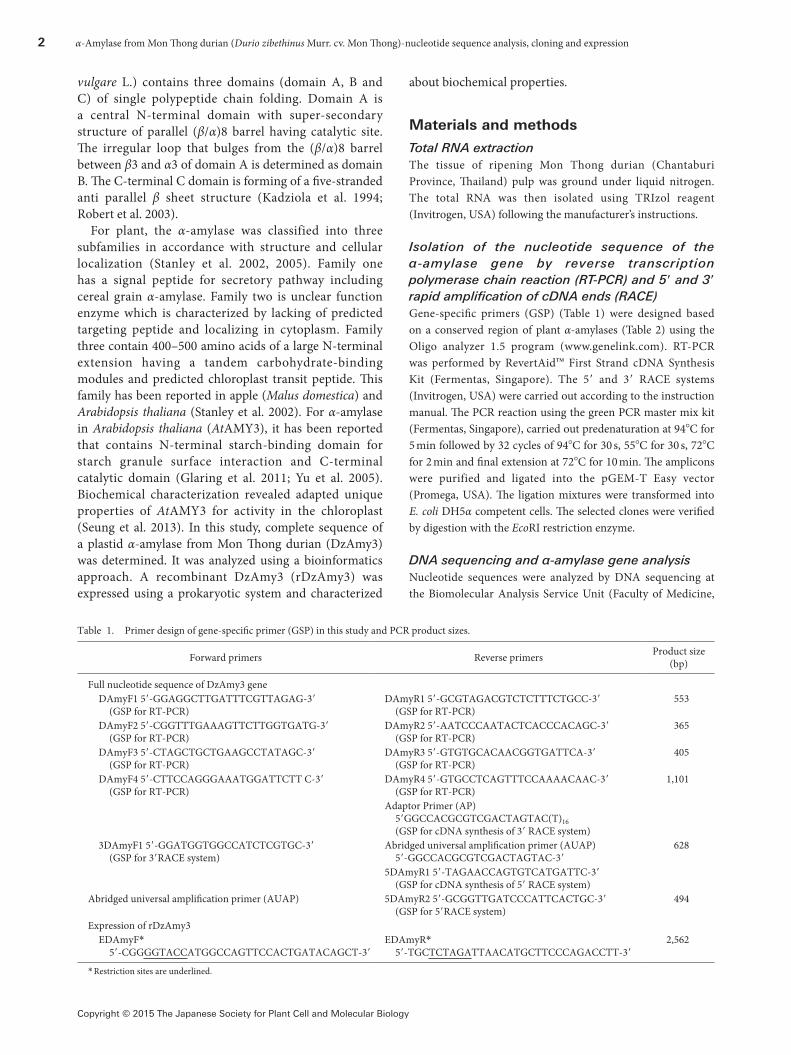
Isolation of the nucleotide sequence of the α-amylase gene by reverse transcription polymerase chain reaction (RT-PCR) and 5′ and 3′ rapid amplification of cDNA ends (RACE)Gene-specific primers (GSP) (Table 1) were designed based on a conserved region of plant α-amylases (Table 2) using the Oligo analyzer 1.5 program (www.genelink.com). RT-PCR was performed by RevertAid™ First Strand cDNA Synthesis Kit (Fermentas, Singapore). The 5′ and 3′ RACE systems (Invitrogen, USA) were carried out according to the instruction manual. The PCR reaction using the green PCR master mix kit (Fermentas, Singapore), carried out predenaturation at 94°C for 5 min followed by 32 cycles of 94°C for 30 s, 55°C for 30 s, 72°C for 2 min and final extension at 72°C for 10 min. The amplicons were purified and ligated into the pGEM-T Easy vector (Promega, USA). The ligation mixtures were transformed into E. coli DH5α competent cells. The selected clones were verified by digestion with the EcoRI restriction enzyme.
DNA sequencing and α-amylase gene analysisNucleotide sequences were analyzed by DNA sequencing at the Biomolecular Analysis Service Unit (Faculty of Medicine,
Table 1. Primer design of gene-specific primer (GSP) in this study and PCR product sizes.
Forward primers Reverse primers Product size (bp)
Full nucleotide sequence of DzAmy3 geneDAmyF1 5′-GGAGGCTTGATTTCGTTAGAG-3′
(GSP for RT-PCR)DAmyR1 5′-GCGTAGACGTCTCTTTCTGCC-3′
(GSP for RT-PCR)553
DAmyF2 5′-CGGTTTGAAAGTTCTTGGTGATG-3′ (GSP for RT-PCR)
DAmyR2 5′-AATCCCAATACTCACCCACAGC-3′ (GSP for RT-PCR)
365
DAmyF3 5′-CTAGCTGCTGAAGCCTATAGC-3′ (GSP for RT-PCR)
DAmyR3 5′-GTGTGCACAACGGTGATTCA-3′ (GSP for RT-PCR)
405
DAmyF4 5′-CTTCCAGGGAAATGGATTCTT C-3′ (GSP for RT-PCR)
DAmyR4 5′-GTGCCTCAGTTTCCAAAACAAC-3′ (GSP for RT-PCR)
1,101
Adaptor Primer (AP) 5′GGCCACGCGTCGACTAGTAC(T)16 (GSP for cDNA synthesis of 3′ RACE system)
3DAmyF1 5′-GGATGGTGGCCATCTCGTGC-3′ (GSP for 3′RACE system)
Abridged universal amplification primer (AUAP) 5′-GGCCACGCGTCGACTAGTAC-3′
628
5DAmyR1 5′-TAGAACCAGTGTCATGATTC-3′ (GSP for cDNA synthesis of 5′ RACE system)
Abridged universal amplification primer (AUAP) 5DAmyR2 5′-GCGGTTGATCCCATTCACTGC-3′ (GSP for 5′RACE system)
494
Expression of rDzAmy3EDAmyF*
5′-CGGGGTACCATGGCCAGTTCCACTGATACAGCT-3′EDAmyR*
5′-TGCTCTAGATTAACATGCTTCCCAGACCTT-3′2,562
* Restriction sites are underlined.
S. Posoongnoen et al. 3
Copyright © 2015 The Japanese Society for Plant Cell and Molecular Biology
Khon Kaen University, Khon Kaen, Thailand). The sequences were confirmed by First Base Laboratories (Malaysia). The nucleotide sequences were translated into protein sequences by the translate tool program (http://web.expasy.org/translate/). The gene and protein sequences were analyzed using the Basic Local Alignment Search Tool (BLAST, http://blast.ncbi.nlm.nih.gov/), InterProScan analysis (Hunter et al. 2009) and conserved domain database (CDD) analysis (Marchler-Bauer et al. 2007, 2009). The isoelectric point (pI) and molecular weight (Mw) were computed using Compute pI/Mw tool of the ExPASy Bioinformatics Resource Portal Server (http://web.expasy.org/compute_pi/). The amino acid sequence alignment for analyzing of the conserved regions and functional comparisons were performed using the ClustalW2 program (http://www.ebi.ac.uk/Tools/msa/clustalw2/) (Larkin et al. 2007). The targeting peptide was predicted by the Center for Biological Sequence Analysis (http://www.cbs.dtu.dk/services/). The TargetP program (Emanuelsson et al. 2000) was used to primarily predict the targeting protein. The signal peptide and plastid-targeting peptide were then predicted using the SignalP3 (Nielsen et al. 1997) and ChloroP programs (Emanuelsson et al. 1999), respectively. The nucleotide sequence of α-amylase from Mon Thong durian (DzAmy3) was deposited in the GenBank database (Accession no. KP164993)
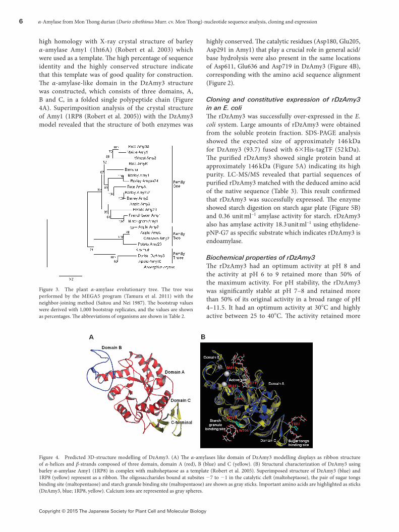
Phylogenic tree analysis and three-dimensional (3D) structure by homology modellingThe phylogenetic tree was constructed using the neighbor-joining method (Saitou and Nei 1987) in the Molecular Evolutionary Genetics Analysis version 5 program (MEGA5) (Tamura et al. 2011). The bootstrap values for the individual branches were analyzed by 1,000 replications of bootstrapping. The protein sequences of plants α-amylases (Table 2) were explored from the NCBI database (http://www.ncbi.nlm.nih.gov/). To root the tree, Aspergillus oryzae (Taka-amylase A) was used as an uncontroversial outgroup, which is most likely far enough to be a clear outgroup. The 3D-structure models of DzAmy3 were constructed by the automated SWISS-MODEL sever at ExPasy (http://swissmodel.expasy.org/) (Arnold et al. 2006; Kiefer et al. 2009; Schwede et al. 2003) based on protein homology. The X-ray crystal structure of barley α-amylase at 1.5 Å resolution (Robert et al. 2003) was used as a template. The 3D-structure models were then analyzed and superimposed using the PyMOL program (http://www.pymol.org/).
Cloning and expression of rDzAmy3 in E. coliThe mature α-amylase insert was prepared from the cDNA using PCR with the EDAmyF and EDAmyR primers (Table 1), which contain KpnI and XbaI restriction sites, respectively. PCR amplification was performed with proofreading KOD-plus Taq polymerase (Takara, Japan) under the following conditions: predenaturation at 94°C for 2 min, followed by 30 cycles of 94°C for 10 s, 55°C for 30 s and 68°C for 1.15 min. For construction, the gene insert (KpnI-XbaI fragment) was purified with the Wizard SV gel and PCR clean-up kit (Promega, USA) according
to the manufacturer’s protocol and ligated into a KpnI/XbaI-digested pCold-TF vector (Takara, Japan) using a DNA ligation kit (Takara, Japan). The constructed pCold-DzAmy3 plasmid and pCold-TF vector were transformed into the Rosetta-gami (DE3) (Novagen, Germany) expression host. Transformants were grown at 37°C in 50 ml LB medium supplemented with 50 µg ml−1 ampicillin until the optical density at 600 nm reached 0.4–0.5, and then the culture was refrigerated at 15°C for 30 min. The expression of DzAmy3 was induced with 1 mM IPTG and grown at 15°C for 36 h. After the culture period, the cell pellet was harvested by centrifugation at 9,000×g, 4°C for 10 min.
One-step purification of rDzAmy3The cell pellet was resuspened in lysis buffer (50 mM phosphate buffer, 500 mM NaCl, 20 mM imidazole, pH 7.0) and lysed with 0.1 mg ml−1 lysozyme for 30 min on ice. The solution was sonicated and centrifuged at 14,000×g at 4°C for 20 min. The supernatant was used as crude extract for further studies. The crude extract was purified using Talon metal affinity resin (Clontech, USA) by a hybrid batch/gravity-flow procedure. The nonspecific proteins were removed by washing with 30 bed volumes of washing buffer (50 mM phosphate buffer, 500 mM NaCl, pH 7.0) with 50 mM imidazole twice. The rDzAmy3 was eluted using stepwise of 100–200 mM imidazole in washing buffer. The protein concentration was analyzed by Bradford method. The purified rDzAmy3 was partially sequenced by liquid chromatography tandem mass spectrometry (LC-MS/MS) analysis at the Research Instrument Center (Khon Kaen University, Khon Kaen, Thailand).
Amylase activity assayStarch agar plate analysis was performed using starch agar plate (Lee et al. 2001) containing 0.5% (w/v) soluble starch, 50 mM phosphate buffer pH 7.0 and 1.5% (w/v) agar. The plates were incubated at 37°C, overnight and stained with staining solution (10 mM I2, 14 mM KI and 1% acetic acid). For dinitrosalicylate (DNS) method (Miller 1959), the hydrolysis reaction mixture consisting of 0.25 ml of 1% (w/v) soluble starch solution in 0.1 M acetate buffer pH 6.5 and 0.25 ml of diluted enzyme was incubated at 37°C for 10 min, terminated by 0.5 ml of DNS reagent and heated for 5 min, and observed the absorbance at 540 nm using glucose as standard of reducing sugar. One unit of enzyme was defined as the amount of enzyme that produced 1 µmol of reducing sugar per min. α-Type was analyzed using ethylidene-pNP-G7 as substrate according to the manufacturer’s protocol (Abnova, Taiwan). One unit was defined as the amount of amylase that cleaves ethylidene-pNP-G7 to generate 1.0 nmol of nitrophenol per min at pH 7.2, 37°C.
Enzyme characterizationThe optimum pH of amylase was determined the activity in the pH range of 2.5–11.5 at 37°C. For pH stability, sample was incubated in pH 2.5–11.5 for 17 h at 37°C before determining
4 α-Amylase from Mon Thong durian (Durio zibethinus Murr. cv. Mon Thong)-nucleotide sequence analysis, cloning and expression
Copyright © 2015 The Japanese Society for Plant Cell and Molecular Biology
by DNS method. The pH conditions were adjusted using the following buffers: 0.1 M glycine-HCl buffer (pH 2.0–4.0), 0.1 M sodium acetate buffer (pH 4.0–6.0), 0.1 M potassium phosphate buffer (pH 6.0–8.0) and 0.1 M Tris-HCl buffer (pH 8.0–9.0) and 0.1 M glycine-NaOH buffer (pH 10.0–11.5). The optimum temperature was analyzed the amylase activity at 5–95°C. For heat stability, the sample was pre-incubated at various temperatures for 1 h. The remaining activity was measured by DNS assay. The substrate specificity was analyzed in 0.1 M phosphate buffer (pH 7.0) at 37°C for 10 min using 1% (w/v) starch, glycogen, amylose, amylopectin, β-limit dextrin, pullulan, dextran and β-cyclodextrin as substrates. For enzymatic stability in the presence of surfactant, the sample was pre-incubated with detergents (1%v/v tween 20 and 1%v/v triton X-100) at 37°C for 1 h. The remaining activity was analyzed by DNS assay.
Results
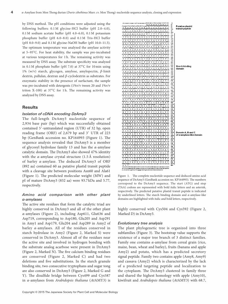
Isolation of cDNA encoding DzAmy3The full-length DzAmy3 nucleotide sequence of 2,934 base pair (bp) which was successfully obtained contained 5′-untranslated region (UTR) of 32 bp, open reading frame (ORF) of 2,679 bp and 3′ UTR of 223 bp (GenBank accession no. KP164993 (Figure 1). The sequence analysis revealed that DzAmy3 is a member of glycosyl hydrolase family 13 and has the α-amylase catalytic domain. The DzAmy3 also showed 47% identity with the α-amylase crystal structure (1.5 Å resolution) of barley α-amylase. The deduced DzAmy3 of ORF (892 aa) contained 60 aa putative plastid transit peptide with a cleavage site between positions Asn60 and Ala61 (Figure 1). The predicted molecular weight (MW) and pI of mature DzAmy3 (832 aa) were 93.7 kDa and 5.77, respectively.
Amino acid comparison with other plant α-amylasesThe active site residues that form the catalytic triad are highly conserved in DzAmy3 and all of the other plant α-amylases (Figure 2), including Asp611, Glu636 and Asp719, corresponding to Asp180, Glu205 and Asp291 in Amy1 and Asp179, Glu204 and Asp289 in Amy2 of barley α-amylases. All of the residues conserved in starch hydrolase in Amy2 (Figure 2, Marked S) were conserved in DzAmy3. Almost all of the residues near the active site and involved in hydrogen bonding with the substrate analog acarbose were present in DzAmy3 (Figure 2, Marked N). The five calcium binding residues are conserved (Figure 2, Marked C) and had two deletions and five substitutions. In the starch granule binding site, two consecutive tryptophans and sugar tong are also conserved in DzAmy3 (Figure 2, Marked G and Y). The disulfide bridge between Cys499 and Cys587 in α-amylases from Arabidopsis thaliana (AtAMY3) is
highly conserved with Cys504 and Cys592 (Figure 2, Marked D) in DzAmy3.
Evolutionary tree analysisThe plant phylogenetic tree is organized into three subfamilies (Figure 3). The bootstrap value supports the existence of a major tree branch of 3 distinct families. Family one contains α-amylase from cereal grain (rice, maize, bean, wheat and barley), fruits (banana and apple Amy2) and potato, which has a predicted secretory signal peptide. Family two contains apple (Amy8, Amy9) and cassava (Amy2) which is characterized by the lack of a predicted targeting peptide and localization to the cytoplasm. The DzAmy3 clustered in family three and shared the highest homology with apple (Amy10), kiwifruit and Arabidopsis thaliana (AtAMY3) with 68.7,
Figure 1. The complete nucleotide sequence and deduced amino acid sequence of DzAmy3 (GenBank accession no. KP164993). The numbers correspond to the DzAmy3 sequence. The start (ATG) and stop (TAA) codons are represented with bold italic letters and an asterisk, respectively. The predicted putative plastid transit peptide is indicated by underlined letters. The starch binding domain and α-amylase-like domains are highlighted with italic and bold letters, respectively.

S. Posoongnoen et al. 5
Copyright © 2015 The Japanese Society for Plant Cell and Molecular Biology
68.5 and 67.98% identity, respectively. Family three α-amylases consist of approximatry 440 amino acids of a large N-terminal domain and a plastid transit peptide, which is consistent with our previous results for the
subcellular localization of DzAmy3 (Figure 1).
Predicted 3D-structure modelling of DzAmy3The DzAmy3 displays 46% of sequence identity of
Figure 2. The amino acid sequence alignment of DzAmy3 with other plant α-amylases. The 440 amino acids of first N-terminal of DzAmy3 were removed. The start (Gln25) of mature protein at N-terminal of barley amylase Amy2 (Kadziola et al. 1994) is labeled with marked M. Residues conserved in starch hydrolase are labeled with S. Residues labeled with C are in close proximity with three calcium ions in barley α-amylase (Kadziola et al. 1994). The residues that are near the active site in barley α-amylase and have hydrogen bond interaction with the acarbose inhibitor (Kadziola et al. 1998) are labeled with N. Residues labeled with G are doublets of starch granule binding in barley α-amylase (Sogaard et al. 1993). The sugar binding site (Robert et al. 2003) is indicated by Y. The catalytic residues (Asp, Glu and Asp) are highlighted with bold letters. The gray shading represents an amino acid deletion in the domain B. The residues conserved in disulfide bond in AtAMY3 (Seung et al. 2013) are marked with D.
6 α-Amylase from Mon Thong durian (Durio zibethinus Murr. cv. Mon Thong)-nucleotide sequence analysis, cloning and expression
Copyright © 2015 The Japanese Society for Plant Cell and Molecular Biology
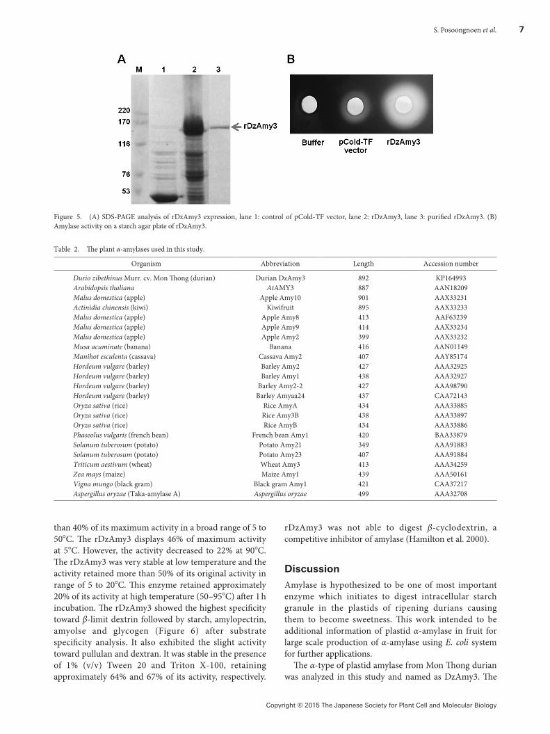
high homology with X-ray crystal structure of barley α-amylase Amy1 (1ht6A) (Robert et al. 2003) which were used as a template. The high percentage of sequence identity and the highly conserved structure indicate that this template was of good quality for construction. The α-amylase-like domain in the DzAmy3 structure was constructed, which consists of three domains, A, B and C, in a folded single polypeptide chain (Figure 4A). Superimposition analysis of the crystal structure of Amy1 (1RP8 (Robert et al. 2005)) with the DzAmy3 model revealed that the structure of both enzymes was
highly conserved. The catalytic residues (Asp180, Glu205, Asp291 in Amy1) that play a crucial role in general acid/base hydrolysis were also present in the same locations of Asp611, Glu636 and Asp719 in DzAmy3 (Figure 4B), corresponding with the amino acid sequence alignment (Figure 2).
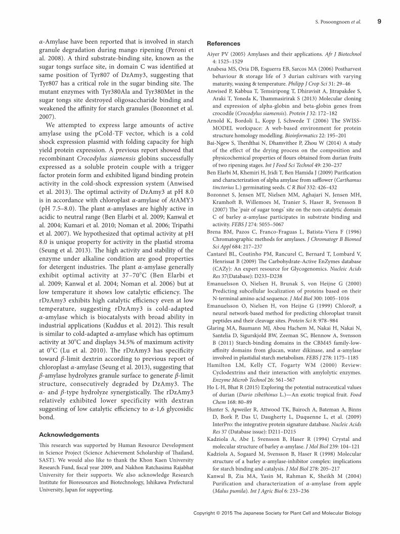
Cloning and constitutive expression of rDzAmy3 in an E. coliThe rDzAmy3 was successfully over-expressed in the E. coli system. Large amounts of rDzAmy3 were obtained from the soluble protein fraction. SDS-PAGE analysis showed the expected size of approximately 146 kDa for DzAmy3 (93.7) fused with 6×His-tagTF (52 kDa). The purified rDzAmy3 showed single protein band at approximately 146 kDa (Figure 5A) indicating its high purity. LC-MS/MS revealed that partial sequences of purified rDzAmy3 matched with the deduced amino acid of the native sequence (Table 3). This result confirmed that rDzAmy3 was successfully expressed. The enzyme showed starch digestion on starch agar plate (Figure 5B) and 0.36 unit ml−1 amylase activity for starch. rDzAmy3 also has amylase activity 18.3 unit ml−1 using ethylidene-pNP-G7 as specific substrate which indicates rDzAmy3 is endoamylase.
Biochemical properties of rDzAmy3The rDzAmy3 had an optimum activity at pH 8 and the activity at pH 6 to 9 retained more than 50% of the maximum activity. For pH stability, the rDzAmy3 was significantly stable at pH 7–8 and retained more than 50% of its original activity in a broad range of pH 4–11.5. It had an optimum activity at 30°C and highly active between 25 to 40°C. The activity retained more
Figure 3. The plant α-amylase evolutionary tree. The tree was performed by the MEGA5 program (Tamura et al. 2011) with the neighbor-joining method (Saitou and Nei 1987). The bootstrap values were derived with 1,000 bootstrap replicates, and the values are shown as percentages. The abbreviations of organisms are shown in Table 2.
Figure 4. Predicted 3D-structure modelling of DzAmy3. (A) The α-amylases like domain of DzAmy3 modelling displays as ribbon structure of α-helices and β-strands composed of three domain, domain A (red), B (blue) and C (yellow). (B) Structural characterization of DzAmy3 using barley α-amylase Amy1 (1RP8) in complex with maltoheptaose as a template (Robert et al. 2005). Superimposed structure of DzAmy3 (blue) and 1RP8 (yellow) represent as a ribbon. The oligosaccharides bound at subsites −7 to −1 in the catalytic cleft (maltoheptaose), the pair of sugar tongs binding site (maltopentaose) and starch granule binding site (maltopentaose) are shown as gray sticks. Important amino acids are highlighted as sticks (DzAmy3, blue; 1RP8, yellow). Calcium ions are represented as gray spheres.
S. Posoongnoen et al. 7
Copyright © 2015 The Japanese Society for Plant Cell and Molecular Biology
than 40% of its maximum activity in a broad range of 5 to 50°C. The rDzAmy3 displays 46% of maximum activity at 5°C. However, the activity decreased to 22% at 90°C. The rDzAmy3 was very stable at low temperature and the activity retained more than 50% of its original activity in range of 5 to 20°C. This enzyme retained approximately 20% of its activity at high temperature (50–95°C) after 1 h incubation. The rDzAmy3 showed the highest specificity toward β-limit dextrin followed by starch, amylopectrin, amyolse and glycogen (Figure 6) after substrate specificity analysis. It also exhibited the slight activity toward pullulan and dextran. It was stable in the presence of 1% (v/v) Tween 20 and Triton X-100, retaining approximately 64% and 67% of its activity, respectively.
rDzAmy3 was not able to digest β-cyclodextrin, a competitive inhibitor of amylase (Hamilton et al. 2000).
Discussion
Amylase is hypothesized to be one of most important enzyme which initiates to digest intracellular starch granule in the plastids of ripening durians causing them to become sweetness. This work intended to be additional information of plastid α-amylase in fruit for large scale production of α-amylase using E. coli system for further applications.
The α-type of plastid amylase from Mon Thong durian was analyzed in this study and named as DzAmy3. The
Figure 5. (A) SDS-PAGE analysis of rDzAmy3 expression, lane 1: control of pCold-TF vector, lane 2: rDzAmy3, lane 3: purified rDzAmy3. (B) Amylase activity on a starch agar plate of rDzAmy3.
Table 2. The plant α-amylases used in this study.
Organism Abbreviation Length Accession number
Durio zibethinus Murr. cv. Mon Thong (durian) Durian DzAmy3 892 KP164993Arabidopsis thaliana AtAMY3 887 AAN18209Malus domestica (apple) Apple Amy10 901 AAX33231Actinidia chinensis (kiwi) Kiwifruit 895 AAX33233Malus domestica (apple) Apple Amy8 413 AAF63239Malus domestica (apple) Apple Amy9 414 AAX33234Malus domestica (apple) Apple Amy2 399 AAX33232Musa acuminate (banana) Banana 416 AAN01149Manihot esculenta (cassava) Cassava Amy2 407 AAY85174Hordeum vulgare (barley) Barley Amy2 427 AAA32925Hordeum vulgare (barley) Barley Amy1 438 AAA32927Hordeum vulgare (barley) Barley Amy2-2 427 AAA98790Hordeum vulgare (barley) Barley Amyaa24 437 CAA72143Oryza sativa (rice) Rice AmyA 434 AAA33885Oryza sativa (rice) Rice Amy3B 438 AAA33897Oryza sativa (rice) Rice AmyB 434 AAA33886Phaseolus vulgaris (french bean) French bean Amy1 420 BAA33879Solanum tuberosum (potato) Potato Amy21 349 AAA91883Solanum tuberosum (potato) Potato Amy23 407 AAA91884Triticum aestivum (wheat) Wheat Amy3 413 AAA34259Zea mays (maize) Maize Amy1 439 AAA50161Vigna mungo (black gram) Black gram Amy1 421 CAA37217Aspergillus oryzae (Taka-amylase A) Aspergillus oryzae 499 AAA32708

8 α-Amylase from Mon Thong durian (Durio zibethinus Murr. cv. Mon Thong)-nucleotide sequence analysis, cloning and expression
Copyright © 2015 The Japanese Society for Plant Cell and Molecular Biology
putative sequence suggests containing 60 aa putative transit peptide according to those from apple (Mdamy10) and Arabidopsis (AtAMY3) (Stanley et al. 2002). The DzAmy3 belongs to subfamily three of plant α-amylases that was proposed to initiate starch granule breakdown in the plastid, producing soluble substrates (Stanley et al. 2005). The three subfamilies of plant α-amylases agreed well with previous reports (Stanley et al. 2002, 2005). In case of Arabidopsis (Stanley et al. 2002), the three α-amylase genes were identified in different cellular compartments. The structures of the three families were also different and possibly have different substrate specificities, but all three families contain the same catalytic triad for α-amylase activity. Suggesting, three families were classified on the basis of their intron structure, predicted subcellular targeting and domain B size, which is shorter in families two and three. Amino acid sequence comparison revealed that the B domains of α-amylase from barley (Amy1, Amy2) and banana have 8 and 6 more amino acid residues than the corresponding domains of family two (cassava and potato) and family three (DzAmy3, kiwifruit and apple Amy10), respectively. These results support the phylogenetic tree analysis indicates that the size of the B domain can be used to identify three groups of plant α-amylases. The DzAmy3 is predicted to have a starch binding domain of 440 aa in the N-terminus when compared with previously derived X-ray crystal structures of subfamily one plant α-amylases such as Amy1 (Robert et al. 2003)
and Amy2 (Kadziola et al. 1994) which only contains three domains (A, B and C). This starch binding domain at the N-terminus may be possibly important for binding with the starch granule surface (Glaring et al. 2011; Mikkelsen et al. 2006; Yu et al. 2005) in plastidial starch process. The starch binding domain has not yet been clearly characterized based on the X-ray crystal structure. Therefore, only α-amylase-like domain of DzAmy3 is supposed by the model that the large domain A is a major central and catalytic domain including eight inner parallel β-strands surrounded by eight α-helices folded into super-secondary structure of a parallel (β/α)8 barrel. The smallest domain B was organized as an irregular loop protruding between β3 and α3 of domain A. Domain C was established with five stranded antiparallel β sheet structure containing parallel β1, β3 and β4 and antiparallel β2 and β5 (Kadziola et al. 1994; Robert et al. 2003). The predicted disulfide bridge of DzAmy3 suggested that is central to redox-regulated for unique property of plastid amylase according to chloroplast α-amylase (Seung et al. 2013). The superimposition analysis hypothesized that Asp611, Glu636 and Asp719 are catalytic residues that play a crucial role in general acid/base hydrolysis, corresponding with the amino acid sequence alignment. The conserved active site residues are critical for catalysis because the replacement of these residues (Asp180Asn, Glu205Gln and Asp291Asn, in Amy1) using site-directed mutagenesis resulted in catalytic inactivity (Sogaard et al. 1993). The Met53 in Amy1, which is situated at high affinity subsite −2 of domain A, is also conserved at Met491 of DzAmy3, suggesting crucial for substrate specificity and activity (Mori et al. 2002). The starch granule binding site (pair of consecutive tryptophans) that is critical for adsorption onto granular starch has been previously reported in the X-ray crystal structure of Amy1 (Trp278 and Trp279) and Amy2 (Trp276 and Trp277), and this site is situated on the surface of the enzyme at α-helix 6 in the (β/α)8 barrel structure of catalytic A domain (Kadziola et al. 1998; Robert et al. 2003, 2005; Sogaard et al. 1993). The presence of a pair of consecutive tryptophans in DzAmy3 suggested that this position has the function of binding starch granules and is critical to adsorb onto granular starch for plastid α-amylase activity.
Table 3. LC-MS/MS analysis of purified rDzAmy3.
Sample Protein annotation [organism] Accession number Mr theor./exp. Peptide sequence Mascot score
Purified rDzAmy3 Alpha-amylase, choloplastic [Citrus sinensis]
gi|568859401 102.7/145.7 R.LNWDDR.A 509R.WYMELK.ER.GFWGGYVK.DK.DYAIETPLK.KR.DVYAAIIDEK.VK.LAAEAYSIFR.TK.DEETGAWYQHR.GK.GKPPGVVGWWPSR.A
Figure 6. Substrate specificity analysis of rDzAmy3, the highest activity of rDzAmy3 toward β-limit dextrin was considered as 100% activity. Values are means of three independent experiments.
S. Posoongnoen et al. 9
Copyright © 2015 The Japanese Society for Plant Cell and Molecular Biology
α-Amylase have been reported that is involved in starch granule degradation during mango ripening (Peroni et al. 2008). A third substrate-binding site, known as the sugar tongs surface site, in domain C was identified at same position of Tyr807 of DzAmy3, suggesting that Tyr807 has a critical role in the sugar binding site. The mutant enzymes with Tyr380Ala and Tyr380Met in the sugar tongs site destroyed oligosaccharide binding and weakened the affinity for starch granules (Bozonnet et al. 2007).
We attempted to express large amounts of active amylase using the pCold-TF vector, which is a cold shock expression plasmid with folding capacity for high yield protein expression. A previous report showed that recombinant Crocodylus siamensis globins successfully expressed as a soluble protein couple with a trigger factor protein form and exhibited ligand binding protein activity in the cold-shock expression system (Anwised et al. 2013). The optimal activity of DzAmy3 at pH 8.0 is in accordance with chloroplast α-amylase of AtAMY3 (pH 7.5–8.0). The plant α-amylases are highly active in acidic to neutral range (Ben Elarbi et al. 2009; Kanwal et al. 2004; Kumari et al. 2010; Noman et al. 2006; Tripathi et al. 2007). We hypothesized that optimal activity at pH 8.0 is unique property for activity in the plastid stroma (Seung et al. 2013). The high activity and stability of the enzyme under alkaline condition are good properties for detergent industries. The plant α-amylase generally exhibit optimal activity at 37–70°C (Ben Elarbi et al. 2009; Kanwal et al. 2004; Noman et al. 2006) but at low temperature it shows low catalytic efficiency. The rDzAmy3 exhibits high catalytic efficiency even at low temperature, suggesting rDzAmy3 is cold-adapted α-amylase which is biocatalysts with broad ability in industrial applications (Kuddus et al. 2012). This result is similar to cold-adapted α-amylase which has optimum activity at 30°C and displays 34.5% of maximum activity at 0°C (Lu et al. 2010). The rDzAmy3 has specificity toward β-limit dextrin according to previous report of chloroplast α-amylase (Seung et al. 2013), suggesting that β-amylase hydrolyzes granule surface to generate β-limit structure, consecutively degraded by DzAmy3. The α- and β-type hydrolyze synergistically. The rDzAmy3 relatively exhibited lower specificity with dextran suggesting of low catalytic efficiency to α-1,6 glycosidic bond.
Acknowledgements
This research was supported by Human Resource Development in Science Project (Science Achievement Scholarship of Thailand, SAST). We would also like to thank the Khon Kaen University Research Fund, fiscal year 2009, and Nakhon Ratchasima Rajabhat University for their supports. We also acknowledge Research Institute for Bioresources and Biotechnology, Ishikawa Prefectural University, Japan for supporting.
References
Aiyer PV (2005) Amylases and their applications. Afr J Biotechnol 4: 1525–1529
Anabesa MS, Oria DB, Esguerra EB, Sarcos MA (2006) Postharvest behaviour & storage life of 3 durian cultivars with varying maturity, waxing & temperature. Philipp J Crop Sci 31: 29–46
Anwised P, Kabbua T, Temsiripong T, Dhiravisit A, Jitrapakdee S, Araki T, Yoneda K, Thammasirirak S (2013) Molecular cloning and expression of alpha-globin and beta-globin genes from crocodile (Crocodylus siamensis). Protein J 32: 172–182
Arnold K, Bordoli L, Kopp J, Schwede T (2006) The SWISS-MODEL workspace: A web-based environment for protein structure homology modelling. Bioinformatics 22: 195–201
Bai-Ngew S, Therdthai N, Dhamvithee P, Zhou W (2014) A study of the effect of the drying process on the composition and physicochemical properties of flours obtained from durian fruits of two ripening stages. Int J Food Sci Technol 49: 230–237
Ben Elarbi M, Khemiri H, Jridi T, Ben Hamida J (2009) Purification and characterization of alpha amylase from safflower (Carthamus tinctorius L.) germinating seeds. C R Biol 332: 426–432
Bozonnet S, Jensen MT, Nielsen MM, Aghajari N, Jensen MH, Kramhoft B, Willemoes M, Tranier S, Haser R, Svensson B (2007) The ‘pair of sugar tongs’ site on the non-catalytic domain C of barley α-amylase participates in substrate binding and activity. FEBS J 274: 5055–5067
Brena BM, Pazos C, Franco-Fraguas L, Batista-Viera F (1996) Chromatographic methods for amylases. J Chromatogr B Biomed Sci Appl 684: 217–237
Cantarel BL, Coutinho PM, Rancurel C, Bernard T, Lombard V, Henrissat B (2009) The Carbohydrate-Active EnZymes database (CAZy): An expert resource for Glycogenomics. Nucleic Acids Res 37(Database): D233–D238
Emanuelsson O, Nielsen H, Brunak S, von Heijne G (2000) Predicting subcellular localization of proteins based on their N-terminal amino acid sequence. J Mol Biol 300: 1005–1016
Emanuelsson O, Nielsen H, von Heijne G (1999) ChloroP, a neural network-based method for predicting chloroplast transit peptides and their cleavage sites. Protein Sci 8: 978–984
Glaring MA, Baumann MJ, Abou Hachem M, Nakai H, Nakai N, Santelia D, Sigurskjold BW, Zeeman SC, Blennow A, Svensson B (2011) Starch-binding domains in the CBM45 family-low-affinity domains from glucan, water dikinase, and α-amylase involved in plastidial starch metabolism. FEBS J 278: 1175–1185
Hamilton LM, Kelly CT, Fogarty WM (2000) Review: Cyclodextrins and their interaction with amylolytic enzymes. Enzyme Microb Technol 26: 561–567
Ho L-H, Bhat R (2015) Exploring the potential nutraceutical values of durian (Durio zibethinus L.)—An exotic tropical fruit. Food Chem 168: 80–89
Hunter S, Apweiler R, Attwood TK, Bairoch A, Bateman A, Binns D, Bork P, Das U, Daugherty L, Duquenne L, et al. (2009) InterPro: the integrative protein signature database. Nucleic Acids Res 37 (Database issue): D211–D215
Kadziola A, Abe J, Svensson B, Haser R (1994) Crystal and molecular structure of barley α-amylase. J Mol Biol 239: 104–121
Kadziola A, Sogaard M, Svensson B, Haser R (1998) Molecular structure of a barley α-amylase-inhibitor complex: implications for starch binding and catalysis. J Mol Biol 278: 205–217
Kanwal B, Zia MA, Yasin M, Rahman K, Sheikh M (2004) Purification and characterization of α-amylase from apple (Malus pumila). Int J Agric Biol 6: 233–236
10 α-Amylase from Mon Thong durian (Durio zibethinus Murr. cv. Mon Thong)-nucleotide sequence analysis, cloning and expression
Copyright © 2015 The Japanese Society for Plant Cell and Molecular Biology
Ketsa S, Daengkanit T (1998) Physiological changes during postharvest ripening of durian fruit (Durio zibethinus Murry). J Hortic Sci Biotechnol 73: 575–577
Kiefer F, Arnold K, Kunzli M, Bordoli L, Schwede T (2009) The SWISS-MODEL Repository and associated resources. Nucleic Acids Res 37 (Database issue): D387–D392
Kuddus M, Roohi, Arif JM, Ramteke PW (2012) Structural adaptation and biocatalytic prospective of microbial cold-active α-amylase. Afr J Microbiol Res 6: 206–213
Kumari A, Singh VK, Fitter J, Polen T, Kayastha AM (2010) α-amylase from germinating soybean (Glycine max) seeds-purification, characterization and sequential similarity of conserved and catalytic amino acid residues. Phytochemistry 71: 1657–1666
Larkin MA, Blackshields G, Brown NP, Chenna R, McGettigan PA, McWilliam H, Valentin F, Wallace IM, Wilm A, Lopez R, et al. (2007) Clustal W and Clustal X version 2.0. Bioinformatics 23: 2947–2948
Lee CC, Wong DW, Robertson GH (2001) An E. coli expression system for the extracellular secretion of barley α-amylase. J Protein Chem 20: 233–237
Lu M, Wang S, Fang Y, Li H, Liu S, Liu H (2010) Cloning, expression, purification, and characterization of cold-adapted α-amylase from Pseudoalteromonas arctica GS230. Protein J 29: 591–597
Marchler-Bauer A, Anderson JB, Chitsaz F, Derbyshire MK, DeWeese-Scott C, Fong JH, Geer LY, Geer RC, Gonzales NR, Gwadz M, et al. (2009) CDD: specific functional annotation with the Conserved Domain Database. Nucleic Acids Res 37 (Database issue): D205–D210
Marchler-Bauer A, Anderson JB, Derbyshire MK, DeWeese-Scott C, Gonzales NR, Gwadz M, Hao L, He S, Hurwitz DI, Jackson JD, et al. (2007) CDD: a conserved domain database for interactive domain family analysis. Nucleic Acids Res 35 (Database issue): D237–D240
Mikkelsen R, Suszkiewicz K, Blennow A (2006) A novel type carbohydrate-binding module identified in α-glucan, water dikinases is specific for regulated plastidial starch metabolism. Biochemistry 45: 4674–4682
Miller GL (1959) Use of dinitrosalicylic acid reagent for determination of reducing sugar. Anal Chem 31: 426–428
Mori H, Bak-Jensen KS, Svensson B (2002) Barley α-amylase Met53 situated at the high-affinity subsite −2 belongs to a substrate binding motif in the β→α loop 2 of the catalytic (β/α)8-barrel and is critical for activity and substrate specificity. Eur J Biochem 269: 5377–5390
Nielsen H, Engelbrecht J, Brunak S, von Heijne G (1997) Identification of prokaryotic and eukaryotic signal peptides and prediction of their cleavage sites. Protein Eng 10: 1–6
Noman ASM, Hoque MA, Sen PK, Karim MR (2006) Purification and some properties of α-amylase from post-harvest Pachyrhizus erosus L. tuber. Food Chem 99: 444–449
Peroni FH, Koike C, Louro RP, Purgatto E, do Nascimento JR, Lajolo FM, Cordenunsi BR (2008) Mango starch degradation. II.
The binding of α-amylase and β-amylase to the starch granule. J Agric Food Chem 56: 7416–7421
Robert X, Haser R, Gottschalk TE, Ratajczak F, Driguez H, Svensson B, Aghajari N (2003) The structure of barley α-amylase isozyme 1 reveals a novel role of domain C in substrate recognition and binding: A pair of sugar tongs. Structure 11: 973–984
Robert X, Haser R, Mori H, Svensson B, Aghajari N (2005) Oligosaccharide binding to barley α-amylase 1. J Biol Chem 280: 32968–32978
Saitou N, Nei M (1987) The neighbor-joining method: A new method for reconstructing phylogenetic trees. Mol Biol Evol 4: 406–425
Schwede T, Kopp J, Guex N, Peitsch MC (2003) SWISS-MODEL: An automated protein homology-modeling server. Nucleic Acids Res 31: 3381–3385
Seung D, Thalmann M, Sparla F, Abou Hachem M, Lee SK, Issakidis-Bourguet E, Svensson B, Zeeman SC, Santelia D (2013) Arabidopsis thaliana AMY3 is a unique redox-regulated chloroplastic α-Amylase. J Biol Chem 288: 33620–33633
Seymour GB, Taylor JE, Tucker GA (1993) Biochemistry of Fruit Ripening. Chapman & Hall, London
Sogaard M, Kadziola A, Haser R, Svensson B (1993) Site-directed mutagenesis of histidine 93, aspartic acid 180, glutamic acid 205, histidine 290, and aspartic acid 291 at the active site and tryptophan 279 at the raw starch binding site in barley α-amylase 1. J Biol Chem 268: 22480–22484
Souza PM, Magalhaes PO (2010) Application of microbial α-amylase in industry—A review. Braz J Microbiol 41: 850–861
Stanley D, Farnden KJF, MacRae EA (2005) Plant α-amylases: Functions and roles in carbohydrate metabolism. Bio Brat 60: 65–71
Stanley D, Fitzgerald AM, Farnden KJF, MacRae EA (2002) Characterisation of putative α-amylases from apple (Malus domestica) and Arabidopsis thaliana. Bio Brat 57: 137–148
Subhadrabandhu S, Ketsa S (2001) Durian King of Tropical Fruit. Daphne Brasell Associates Ltd and CABI Publishing, Wellington
Tamura K, Peterson D, Peterson N, Stecher G, Nei M, Kumar S (2011) MEGA5: Molecular evolutionary genetics analysis using maximum likelihood, evolutionary distance, and maximum parsimony methods. Mol Biol Evol 28: 2731–2739
Tripathi P, Lo Leggio L, Mansfeld J, Ulbrich-Hofmann R, Kayastha AM (2007) α-Amylase from mung beans (Vigna radiata)—Correlation of biochemical properties and tertiary structure by homology modelling. Phytochemistry 68: 1623–1631
van der Maarel MJ, van der Veen B, Uitdehaag JC, Leemhuis H, Dijkhuizen L (2002) Properties and applications of starch-converting enzymes of the α-amylase family. J Biotechnol 94: 137–155
Yu T-S, Zeeman SC, Thorneycroft D, Fulton DC, Dunstan H, Lue W-L, Hegemann B, Tung S-Y, Umemoto T, Chapple A, et al. (2005) α-Amylase is not required for breakdown of transitory starch in Arabidopsis leaves. J Bio Chem 280: 9773–9779

![High cell density cultivation of [i]Escherichia coli[i] DH5α in ...cell growth and biomass formation. This study demonstrated the utility of a new semi-defined formulated medium in](https://static.fdocument.org/doc/165x107/611bbc3f68acba3f9c2ecb94/high-cell-density-cultivation-of-iescherichia-colii-dh5-in-cell-growth.jpg)