Increasing progesterone secretion and 3β-hydroxysteroid dehydrogenase activity of human cumulus...
7
Pergamon J. Stermd Biochem. Molec.Bzol. Vol.51, No. 5/6, pp. 299-305,1994 Copyright© 1994Elsevier Science Ltd 0960-0760(94)00133-2 Printed in GreatBritain.All rights reserved 0960-0760/94$7.00+ 0.00 Increasing Progesterone Secretion and 3fl-Hydroxysteroid Dehydrogenase Activity of Human Cumulus Cells and Granulosa-lutein Cells Concurrent with Successful Fertilization of the Corresponding Oocyte Shalom Bar-Ami* The Bruce Rappaport Faculty of Medicine, Technion-Israel Institute of Technology, P.O.B. 9649, 31096 Haifa, Israel and Department of Obstetrics and Gynecology, Rambam Medical Center, Haifa, Israel In many studies it has been documented that the induction of multiple follicular growth in humans results in an asynchrony between the degree of cumulus mucification, oocyte meiotic maturation, fertilizability, and follicular cell progesterone (P4) secretion. The present study was carried out on oocytes enclosed in fully mucified cumulus. Thus, oocyte fertilizability was correlated to human cumulus cell (hCC) and human granulosa-lutein (G-L) cell competence for P4 secretion in culture. In the G-L cells, P4 secretion and percentage of cells manifesting 3ig-hydroxysteroid dehydrogenase (31g-HSD) activity increased concurrently with the period of culture. In the hCC, however, P4 secretion decreased concurrently with elongation of the culture period, whereas the percentage of 3/~-HSD-positive cells increased. In hCC corresponding to the fertilized oocytes, P4 accumulation in culture medium was 1.9-fold (P < 0.001) and 1.6-fold (P < 0.02) higher on days 0-3 and 3-5 of culture, respectively, as compared to P4 accumulation in hCC of unfertilized oocytes. Also, in hCC corresponding to the fertilized oocytes, the degree of 3//-HSD activity was found to be significantly higher shortly after aspiration and after either 3 or 5 days, compared to hCC of unfertilized oocytes. In the G-L cells pooled from all follicles yielding mature cumulus-oocyte complexes, P4 accumu- lation and percentage of 31g-HSD-positive cells increased concurrently with the increase in percent- age of fertilized eggs of each individual woman. These results indicate that in stimulated cycles, follicles yielding mature cumulus-oocyte complex, oocyte fertilizability, and G-L cell or hCC competence for P4 secretion are correlated and synchronous. J. Steroid Biochem. Molec. Biol., Vol. 51, No. 5/6, pp. 299-305 1994 INTRODUCTION Administration of FSH/LH to women as part of treatment for in vitro fertilization/embryo transfer (IVF/ET) results in multiple follicular growth of a nonhomogeneous population. Thus, these follicles do not manifest homogeneous responses to the adminis- tration of HCG in terms of oocyte meiotic maturation, *Correspondence to S. Bar-Ami, The Bruce Rappaport Faculty of Medicine, Technion-Israel Institute of Technology, P.O.B. 9649, 31096 Haifa, Israel. Received 19 Nov. 1993; accepted 27 Jul. 1994. cumulus mucification, and the expected change in the steroidogenic pattern from estrogen-producing to progesterone (P4)-producing cells. Furthermore, an asynchrony between the degree of oocyte meiotic matu- ration and cumulus mucification has been reported in women subjected to massive FSH/LH treatment [1, 2]. However, in follicles yielding mature cumulus-oocyte complexes (COC), oocyte fertilizability and subsequent cleavage are correlated to cumulus culture morphology and steroidogenic pattern of the cumulus cells [3]. Furthermore, the steroidogenic pattern of human gran- ulosa-lutein (G-L) cells and the steroid concentration in follicular fluid (FF) are altered in relation to the 299
Transcript of Increasing progesterone secretion and 3β-hydroxysteroid dehydrogenase activity of human cumulus...
Pergamon J. Stermd Biochem. Molec. Bzol. Vol. 51, No. 5/6, pp. 299-305, 1994
Copyright © 1994 Elsevier Science Ltd 0960-0760(94)00133-2 Printed in Great Britain. All rights reserved
0960-0760/94 $7.00 + 0.00
Increasing Progesterone Secretion and 3fl-Hydroxysteroid Dehydrogenase Activity
of Human Cumulus Cells and Granulosa-lutein Cells Concurrent with
Successful Fertil ization of the Corresponding Oocyte
Shalom B a r - A m i * The Bruce Rappaport Faculty of Medicine, Technion-Israel Institute of Technology, P.O.B. 9649, 31096 Haifa,
Israel and Department of Obstetrics and Gynecology, Rambam Medical Center, Haifa, Israel
In m a n y s tud ies it has b e e n d o c u m e n t e d t h a t t he i n d u c t i o n o f m u l t i p l e f o l l i c u l a r g r o w t h in h u m a n s r e su l t s in a n a s y n c h r o n y b e t w e e n the d e g r e e o f c u m u l u s m u c i f i c a t i o n , o o cy t e m e i o t i c m a t u r a t i o n , f e r t i l i z a b i l i t y , a n d f o l l i c u l a r cell p r o g e s t e r o n e (P4) s ec re t i on . T h e p r e s e n t s t u d y was c a r r i e d ou t on o o c y t e s e n c l o s e d in fu l ly m u c i f i e d c u m u l u s . Thus , o o c y t e f e r t i l i z a b i l i t y was c o r r e l a t e d to h u m a n c u m u l u s cell ( hCC) a n d h u m a n g r a n u l o s a - l u t e i n (G-L) cell c o m p e t e n c e fo r P4 s e c r e t i o n in c u l t u r e . In the G - L cells, P4 s e c r e t i o n a n d p e r c e n t a g e o f cells m a n i f e s t i n g 3 i g - h y d r o x y s t e r o i d d e h y d r o g e n a s e (31g-HSD) a c t i v i t y i n c r e a s e d c o n c u r r e n t l y wi th the p e r i o d o f c u l t u r e . In the hCC, h o w e v e r , P4 s e c r e t i o n d e c r e a s e d c o n c u r r e n t l y wi th e l o n g a t i o n o f the c u l t u r e p e r i o d , w h e r e a s the p e r c e n t a g e o f 3 /~ -HSD-pos i t ive cells i n c r e a s e d . In h C C c o r r e s p o n d i n g to the f e r t i l i z e d oocy tes , P4 a c c u m u l a t i o n in c u l t u r e m e d i u m was 1.9-fold (P < 0.001) a n d 1.6-fold (P < 0.02) h i g h e r on days 0-3 a n d 3-5 o f c u l t u r e , r e s p e c t i v e l y , as c o m p a r e d to P4 a c c u m u l a t i o n in h C C o f u n f e r t i l i z e d oocy tes . Also, in h C C c o r r e s p o n d i n g to t he f e r t i l i z e d oocy tes , t he d e g r e e o f 3 / / -HSD ac t i v i t y was f o u n d to be s ign i f i can t ly h i g h e r s h o r t l y a f t e r a s p i r a t i o n a n d a f t e r e i t h e r 3 o r 5 days , c o m p a r e d to h C C o f u n f e r t i l i z e d oocy tes . In the G-L cells p o o l e d f r o m all fol l ic les y i e ld ing m a t u r e c u m u l u s - o o c y t e c o m p l e x e s , P4 a c c u m u - l a t i on a n d p e r c e n t a g e o f 31g-HSD-posi t ive cells i n c r e a s e d c o n c u r r e n t l y wi th the i n c r e a s e in p e r c e n t - age o f f e r t i l i z e d eggs o f e a c h i n d i v i d u a l w o m a n . Th ese r e su l t s i n d i c a t e t h a t in s t i m u l a t e d cycles , fo l l ic les y i e ld ing m a t u r e c u m u l u s - o o c y t e c o m p l e x , o o cy t e f e r t i l i z ab i l i t y , a n d G -L cell o r h C C c o m p e t e n c e fo r P4 s e c r e t i o n a r e c o r r e l a t e d a n d s y n c h r o n o u s .
J. Steroid Biochem. Molec. Biol., Vol. 51, No. 5/6, pp. 299-305 1994
INTRODUCTION
Administration of F S H / L H to women as part of treatment for in vitro fertilization/embryo transfer ( IVF/ET) results in multiple follicular growth of a nonhomogeneous population. Thus , these follicles do not manifest homogeneous responses to the adminis- tration of H C G in terms of oocyte meiotic maturation,
*Correspondence to S. Bar-Ami, The Bruce Rappaport Faculty of Medicine, Technion-Israel Institute of Technology, P.O.B. 9649, 31096 Haifa, Israel.
Received 19 Nov. 1993; accepted 27 Jul. 1994.
cumulus mucification, and the expected change in the steroidogenic pattern from estrogen-producing to progesterone (P4)-producing cells. Furthermore, an asynchrony between the degree of oocyte meiotic matu- ration and cumulus mucification has been reported in women subjected to massive F S H / L H treatment [1, 2]. However, in follicles yielding mature cumulus-oocyte complexes (COC), oocyte fertilizability and subsequent cleavage are correlated to cumulus culture morphology and steroidogenic pattern of the cumulus cells [3]. Furthermore, the steroidogenic pattern of human gran- ulosa-lutein (G-L) cells and the steroid concentration in follicular fluid (FF) are altered in relation to the
299
300 Shalom Bar-Ami .
degree of mucification/maturation of the corresponding cumulus mass [4-7] and in relation to the cumulus culture morphology [8]. The type and rate of steroid biosynthesis is dependent on many factors, including intracellular cholesterol availability, degree of proper enzyme activity, etc. [9-11]. The main factor that initially determines P4 synthesis in human cumulus cells (hCC) and human G - L cells as part of corpus luteum formation, in response to the endogenous L H surge or exogenous H C G administration, is the t remendous increase in the activity of various enzymes such as side-chain-cleavage cytochrome P-450 (P-450scc) and 3//-hydroxysteroid dehydrogenase (3f l -HSD) [12-14]. The 3 f l -HSD activity can easily be determined by a histochemical approach [15]. Thus , in the present study the correlation between oocyte fertilizability and degree of 3 f l -HSD activity in the corresponding hCC and G - L cells as well as the rate of Pa secretion were evaluated.
E X P E R I M E N T A L
Subjects
Twenty-seven women admit ted to the I V F / E T program at R a m b a m Medical Center who had normal menstrual cycles and whose husbands had been proven fertile were the subjects of this study. The i r age ranged between 30 and 35 years. Mult iple follicular growth was stimulated by daily t reatment with either 50rag/day clomiphene citrate (Ikaclomin; Ikapharm, Kfar Saba, Israel) combined with 150 IU/day human menopausal gonadotropin (Pergonal; Teva, Petah Tiqva, Israel) or 150 IU/day human F S H (Metrodin; Teva) and 150IU/day Pergonal. These t reatment protocols resulted in the development of 6.7 + 0.5 follicles/woman. All women received 10,000 U H C G (Chorigon; Teva) 36 h before aspiration of oocytes.
G - L cell cultures
The G - L cells were isolated only f rom follicles containing COC characterized as mature at the time of aspiration, according to criteria detailed earlier by Bar-Ami et al. [3]. Immediate ly after follicular content was aspirated, the COC were removed and cultured under proper conditions for fertilization, and the remaining follicular aspirates were pooled and then centrifuged at 300 g for 10 min to recover the G - L cells in the pellet. T h e G - L cells were suspended in culture medium, which consisted of H a m ' s F-10 medium (Gibco, Grand Island, NY) buffered with 25 m M sodium bicarbonate (Gibco) and 10 m M Hepes (Gibco) and enriched with l m M glutamine (Gibco), 5% inactivated fetal calf serum (Gibco), and streptomycin (50 IU/ml)/penici l l in (50 pg/ml) solution (Gibco). Natural ly the aspirated G - L cells contained red blood cells (RBC), which were removed by allowing them to sediment in 60-mm Petri dishes for 20 min, and the floating clumps of G - L cells were collected with a
Pasteur pipette. This procedure was repeated two or three times until the preparat ion of G - L cells was relatively clear of RBC. Ten thousand viable G - L cells/200/~l were plated in 96-well plates (Falcon, Oxnard, CA); cell viability was assessed using 0.04°o t rypan blue (w/v) for the dye-exclusion test [16].
T h e G - L cells were cultured for 7 days in a hu- midified 5°0 CO2-95°/o air incubator at 37°C. On days 3 and 5, medium was replaced with fresh medium, then collected and frozen at - 2 0 ~ C for later steroid radio- immunoassay (RIA). Sometimes cells were cultured in the presence of 40ng /ml H C G (CR 121, kindly provided by the Center for Population Research of the National Insti tute of Child Health and H u m a n Development , National Insti tutes of Health, l~ethesda, M D ) or 2 0 p M forskolin (Sigma Chemical Co., St Louis, MO).
Oocyte and sperm coculture
H u m a n COC were collected 36h after H C G administration, usually between 0800 and 1200 h; at this t ime oocyte fertilizability is greatest, and ovulation rarely occurs [17, 18]. The complexes were placed immediately in 1 ml of insemination medium, which consisted of H a m ' s F-10 medium buffered with 25 m M sodium bicarbonate and supplemented with 2 m M lactic acid (Calbiochem-Behring, La Jolla, CA), s treptomycin (Sigma), penicillin (Sigma), and 10% human cord serum. Preparat ion of human cord serum was as described previously [3]. Each single COC was cultured in one organ-culture dish (Falcon) at 37°C in 5°,o CO2-95°% air. Fresh semen obtained by masturba- tion was divided into 2-ml aliquots. Semen (2 ml) was mixed with H a m ' s F-10 medium (8 ml) and centrifuged at 200 g for 10 min. The supernatant was decanted, and the pellet was resuspended in 5 m l medium and recentrifuged. After removal of the supernatant, 1 ml of insemination medium was added without disturbing the pellet. One hour later, the media containing the motile sperm were collected and counted in a Makler chamber. One hundred thousand motile sperm were added to each culture dish within 4 - 6 h after recovery of the COC. Between 18 and 20 h after addition of sperm, the remaining corona cells attached to the oocytes were mechanically removed to view fertiliza- tion, which was marked by the appearance of two pronuclei in the ooplasm. T h e oocytes or zygotes were transferred to a new dish filled with fresh medium. The dishes containing the hCC were cultured for 2 additional days (3 days' total) to evaluate the steroid secretion of each individual cumulus mass. Culture was continued for another 4 days, with a med ium change on day 5. All the conditioned media, i.e. media collected after days 0-3, 3-5, and 5-7, were stored at - 20°C for further P4 RIA.
Sometimes, in cases where the husband 's sperm had failed to fertilize any of his wife's oocytes in at least two previous trials by regular IVF, I V F / E T was a t tempted
Oocyte Fertilizability and P~ Secretion 301
by micromanipulat ion of the oocytes. Thus , following 4 - 6 h incubation, each three complexes were placed in a single organ culture dish filled with 1 ml of a medium composed of a synthetic tubal fluid ( H T F ; Irvine Scientific, Santa Ana, CA) supplemented with 1 mg/ml bovine testis hyaluronidase (Sigma). hCC removal was assisted mechanically by repeated pipett ing with a Pasteur pipette, within about 1-2 min at room tempera- ture.
T h e naked oocytes were washed twice in 1.5ml hyaluronidase-free H T F and immediately placed in 1.5 ml fresh H T F medium supplemented with 10% human cord serum, three oocytes per dish. These oocytes were later subjected to micromanipulat ion as described by Cohen et al. [19]. Usually the hCC are discarded, since they have no clinical application. Thus , permission was obtained to utilize these cells for the present study.
T h e HTF-hya lu ron idase solution containing the re- moved hCC was collected f rom all dishes into a conical test tube and centrifuged at 300 g for 10 min at room temperature. T h e HTF-hya lu ron idase solution was discarded and the cell pellet washed again by centrifu- gation in fresh H a m ' s F-10 culture medium. After discarding the H a m ' s F-10 medium, cells were suspended in 2 ml culture medium, and 2 × 1 0 4 viable cells were plated in 0.5 ml culture medium in the organ culture dishes (Falcon No. 3037, Oxnard, CA). This cell system was utilized for determining the effect of 40 ng/ml H C G or 20/LM forskolin on the change in percentage of 3 /%HSD-posi t ive cells.
R I A for steroids
Steroid R I A was employed on the unextracted crude samples. T h e crude culture media were diluted in 50 m M Tris buffer, p H 8.0. The amount of P4 was measured as described by Bar-Ami et al. [3] using tritiated hormone (Amersham Radiochemical Centre, Amersham, Bucks., England) labeled at six places and rabbit anti-P 4 (Bio-Makor, Rehovot, Israel). This anti- serum has very low cross-reactivity (measured at 50°0 displacement), which did not exceed 2.5% for a similar steroid such as 5~-pregnane-3,20-dione. Assay sensitivity and intra- and interassay coefficients of variation were 50 pg/ml and 2 and 8°0. In all instances, data represent the net steroid level obtained after subtraction of the steroid contents of serum- supplemented medium.
Cell harvest and counting
At the end of the incubation period, the G - L cells or hCC were detached as described previously [20]. Briefly, following incubation of the cells for 20 min in a buffer consisting of 0 . 0 5 M phosphate-buffered saline, p H 7.4, with 1% (w/v) sodium E D T A and 5% (v/v) dimethyl sulfoxide (both f rom Sigma), most of the cells were detached from the plastic substrate. Detachment of cells was completed by repeating
pipetting. The G - L cells f rom two culture wells were collected into a single Eppendor f test tube. Also, each individual cumulus was collected into a single Eppendor f test tube. T h e detachment solution was removed by centrifugation at 300 g for 15 min at 22°C and decanting of the supernatant. Nuclear staining was observed by adding 50/~1 of crystal violet solution (5 rag/100 ml in 0.15 M trisodium citrate) and incubat- ing at 37°C for 20-30 rain. Nuclei were counted in a hemocytometer; this method resulted in reproducible data.
Determination of 3 ~ - H S D activity
3/~-HSD activity was determined according to modifications by Barkey et al. (unpublished data) of the method of Levy et al. [15]. The reaction solution was prepared as follows: T e n milligrams of nicotinamide- adenine dinucleotide (NAD, Sigma) were dissolved in 3 m l of an already prepared solution consisting of Tr is -HC1 (110 mM), sodium cyanide (0.224 raM), and MgC12.6H2 O (11 mM). The NAD-conta in ing solution was mixed with 1.67ml of a solution consisting of 40 mg nitroblue tetrazolium, 0.5 ml N ,N-d ime thy l fo r - mamide, and 9.5 ml double-distilled water, as well as with 1.33 ml of double-distilled water. This solution was buffered to p H 8 by N a O H . The final reaction solution contained 0.935 ml of the above mixture and either 0.065 ml of N ,N-d ime thy l fo rmamide (control) or 0.065 ml N ,N-d ime thy l fo rmamide containing 0.4 mg dehydroepiandrosterone (DHA). Cells of each individual cumulus or G - L cells of a single woman were harvested as indicated above. The cells were divided into two Eppendor f test tubes. The cells were suspended in 100/~1 of the reaction solution in the absence (control) or presence of D H A (experimental). Following incubation for 1 h at 37°C, the cells were visualized under a light microscope in a hemocytome- ter. Cells containing 3/~-HSD activity were stained blue violet. The degree of 3/~-HSD activity was expressed as percentage of stained cells per total cell count. The addition of the control group was to assure that the measured value of 3/~-HSD-posit ive cells in the DHA-conta in ing solution was specific.
About 2-3 h after aspiration, in 11 COC two small pieces were cut from each individual cumulus mass. Each piece was placed immediately in a single Eppendor f test tube and incubated for 20 min with a testicular hyaluronidase solution [50/~1 of 20 I U / m l enzyme (Sigma)]. Then the hCC in the hyaluronidase solution were diluted by adding 1450/~1 of culture medium. After mixing and centrifugation at 300 g for 15 rain, the supernatant was decanted, and the reaction solution for 3/~-HSD determination was added with and without D H A , as detailed above.
T o validate the approach for assessing 3/~-HSD activity in our cell system, the optimal p H and length of incubation time were determined. In addition, 38- H S D activity was tested in various nonsteroidogenic
302 Shalom Bar-Ami
cells. In any o f these cases, the pe rcen tage o f 3 [ 1 - H S D - pos i t ive cells was subs tan t i a l ly low, such as 0 .5% in ep i the l ia l cells co l lec ted f rom rat m a m m a r y g land.
Statistical analysis
E x p e r i m e n t a l da ta are p r e s e n t e d as the mean + S E M . T h e da ta were sub jec ted to one -way analysis o f var iance ( A N O V A ) , fo l lowed b y u n p a i r e d S t u d e n t ' s t - t e s t for i nd iv idua l c o m p a r i s o n s be tween means .
R E S U L T S
Altered rate of P4 secretion and 3fl-HSD activity during 7 days' culture
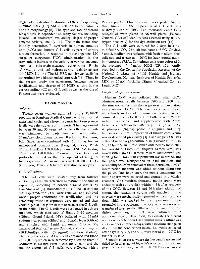
Dai ly P4 secre t ion by G - L cells inc reased 1.6-fold (P < 0.001) and 3 .1- fo ld (P < 0.001) d u r i n g days 3-5 and 5 - 7 o f cu l tu re , respec t ive ly , c o m p a r e d to s te ro id secre t ion d u r i n g days 0 - 3 (Fig . 1). I n h C C , however , P4 secre t ion d u r i n g days 0 - 3 was 2 .5- fo ld (P < 0.001) and 2 .9- fo ld ( P < 0.001) h ighe r than in h C C d u r i n g days 3-5 and 5 -7 , r espec t ive ly (Fig . 1).
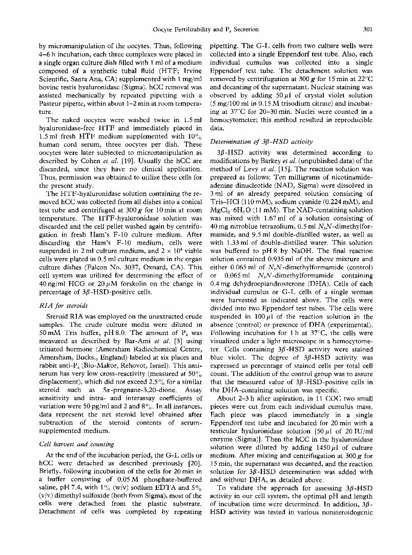
T h e pe rcen tage o f G - L cells man i fe s t ing 3 [1 -HSD act iv i ty inc reased c o n c o m i t a n t l y wi th p rog res s of the cu l tu re pe r iod , f rom 39.5 + 3 .1% shor t ly af ter a sp i ra -
~0
b0
eL
160 •
120
80
40
40
30
2O
lO I
ol 0-3
+
i
3-5 5 - 7
Days in culture
Fig. 1. In vitro accumulation of P4 in hCC (upper panel) and in G-L cells (lower panel), both of which were cultured for 7 days as detailed under Experimental . Data were subjected to one-way ANOVA. The altered P4 secretion with extension of the culture period gave the following statistical values: hCC, F = 42.3, P < 0.001; G-L cells: F = 9.6, P < 0.001, *P < 0.001 vs the corresponding values on days 0-3 of culture, by Student's
t-test.
60
40"
20
~ o
"-" 80
I
~ 6o
N D
40
20 Wl/ 0 0 -3 3 -5
Days m culture 5 -7
Fig. 2. Degree of 3/I-HSD activity in hCC (upper panel) and in G-L cells (lower panel) during 7 days of culture, where day of aspiration is day 0. Data were subjected to one-way ANOVA for the hCC or G-L cells. Thus, in the presence of DHA the following statistical values were observed: in the hCC, F = 28.3, P < 0.001; in the G-L cells, F = 4.1, P < 0.001. Other details are as described in the legend to Fig. 1. *P < 0.01, **P < 0.001, vs the corresponding values on day 0,
by Student's t-test. ND, not done.
t ion to 70 + 4.9 af ter 5 - 7 days ' cu l tu re (P < 0.001) (Fig . 2). h C C showed a s imi la r pa t te rn ; tha t is, 3[1- H S D act iv i ty inc reased wi th p rog res s of the cu l tu re pe r iod , f rom 24.3 + 3.4 abou t 2-3 h af ter a sp i ra t ion to 58.5 + 2.1 (P < 0.001) af ter 3 -5 days ' cu l tu re (Fig . 2).
W h e n h C C were r e m o v e d by hya lu ron ida se shor t ly af ter a sp i ra t ion and cu l tu r ed for 5 days wi th H C G or forskol in , the pe rcen tage of 3 [ 1 - H S D - p o s i t i v e cells inc reased f rom 46.5 + 3.0 to 72.4 + 7.1 (P < 0.01) and 79.8 + 5.8 (P < 0.001), respec t ive ly . S imi la r ly , in G - L cells c u l t u r e d for 5 days wi th H C G or forskol in , the pe rcen tage of 3 [ 1 - H S D - p o s i t i v e cells inc reased f rom 6 6 . 7 + 3 . 4 to 7 8 . 5 + 4 . 0 ( P < 0 . 0 5 ) and 8 1 . 2 + 5 . 0 (P < 0.05), respect ive ly .
Correlation of oocyte fertilizability to P4 and 3[t-HSD
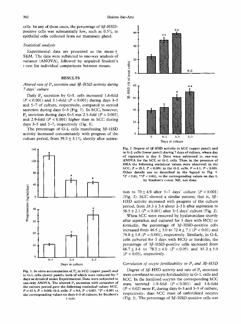
D e g r e e of 3 [1 -HSD act iv i ty and rate of P4 secre t ion were co r re l a t ed to oocyte fe r t i l i zab i l i ty in G - L cells and hCC. I n the fer t i l ized oocytes the c o r r e s p o n d i n g h C C mass secre ted 1.9-fold (P < 0.001) and 1.6-fold (P < 0.02) more P4 d u r i n g days 0 - 3 and 3-5 of cu l tu re , respec t ive ly , t han h C C mass o f un fe r t i l i zed oocytes (Fig . 3). T h e pe rcen tage o f 3 f l - H S D - p o s i t i v e cells was

Oocyte Fertilizability and P4 Secretion 303
o
v
I
100'
80.
60,
40.
20.
0
I--'1 Unfertilized I ~ Fertilized
~" 200
E
150
E ~ ]oo
O
0-3
Days in culture
3-5
Fig. 3. E v a l u a t i o n of degree of 3 ~ - H S D act iv i ty a n d P 4
sec re t i on in ind iv idua l h C C in r e l a t i on to fe r t i l i za t ion of corresponding oocyte. D a t a were sub jec t ed to o n e - w a y ANOVA. Thus , the a l t e r ed p e r c e n t a g e of 3 f l -HSD-pos i t ive h C C gave the fo l lowing s ta t i s t i ca l values: F = 5.4, P < 0.01. The change in P4 sec re t i on gave the fo l lowing s ta t i s t ica l values: F = 14.06, P < 0.001. *P < 0.05, **P < 0.02, ***P < 0.001, vs the c o r r e s p o n d i n g va lues in h C C of u n f e r t i l -
ized oocytes, by S t u d e n t ' s t - t e s t .
1.9-fold (P < 0.05) and 1.2-fold (P < 0.01) higher in hCC mass of oocytes undergoing fertilization, measured after 3 and 5 days, respectively, than the 3 f l -HSD value in hCC mass of unfertilized oocytes (Fig. 3). Fur ther- more, when the hCC were examined about 2-3 h after COC aspiration, it appeared that the percentage of
3f l -HSD-posi t ive cells was 31.3 + 3.4 (n = 6) in hCC corresponding to the fertilized oocytes and 15.9 _ 3.8 (n = 5) in the hCC of the unfertilized oocytes.
Since the G - L cells were pooled f rom all follicles yielding mature COC of each individual woman, it was impossible to correlate in a given follicle the compe- tence of the G - L cells for P4 secretion with the fertiliz- ability of the corresponding oocyte. Therefore , the degree of 3 f l -HSD activity and the rate of P4 secretion of the pooled G - L cells in a given woman were correlated with the average fertilization in the corresponding oocyte population of that woman. Thus , in women yielding an oocyte population manifesting a fertilization rate above 75°,0, 3 f l -HSD activity and P4
secretion were 1.4-fold ( P < 0 . 0 1 ) and 2.0-fold (P < 0.02) greater, respectively, during days 3-5 of culture, and 1.3-fold (P < 0.05) and 1.7-fold (P < 0.01) greater, respectively, during days 5-7 of culture, than in women yielding an oocyte population achieving a fertilization rate of less than 50% in either culture period (Table 1). However, no significant difference was noted in P4 secretion after days 0-3 of culture (Table 1) or in percentage of 3f l -HSD-posi t ive cells when examined shortly after aspiration (data not shown) in relation to successful fertilization of the corresponding oocytes.
DISCUSSION
The data collected in the present study were limited to stimulated follicles yielding mature mucified COC. This study demonstrated that the induction of COC mucifi- cation by H C G did not always advance synchronously with the other ovulatory processes such as an increase in hCC and G - L cell P4 secretion. However, a correlation between oocyte fertilization and hCC and G - L cell P4
secretion was observed. Administrat ion of H C G at I V F / E T causes
t remendous changes in the various follicular compartments . Among these changes, the oocyte
Table 1. Correlation of oocyte fertilizability to G-L cell P4 secretion and 3fl-HSD activity *
Fertilization rate (% of fertilized oocytes)
0-50 51-75 76-100 Days of
culture P4 a 3fl-HSD b P4 3fl-HSD P4 3fl-HSD
0-3 9.2 _ 1.1 ID 12.1 _+ 0.9 ¢ ID 12.0 _+ 4.2 ID 3-5 11.4+1.1 48.3_+6.1 23 .8+4 .6 d 60 .0+5.5 22 .2+1 .9 e 68.1 _ 1.9 d 5-7 38 .7+3 .7 58.6_+5.7 44 .9+6.1 65.1 -t-6.3 64 .5+5 .3 e 75 .7+6 .0 c
*Values represent mean _+ SEM. Data were analyzed by Student's t-test. Comparisons were made between the group with 0-50°0 fertilization rate and the groups with more than 50°,o fertilization rate. ID, insufficient data to be presented.
*Amount of steroid accumulation during indicated days of culture at ng-ml/day normalized per 103 cells.
bpercentage of 3fl-HSD-positive cells in the presence of DHA. In the absence of DHA, the percentage of 3fl-HSD-positive cells did not exceed 20°0 of the total cells. This value did not vary significantly among the different groups that were classified by fertilization rate.
cp < 0.05; dp < 0.02; ep < 0.01.
304 Shalom Bar-Ami
resumes meiosis f rom the diplotene state to second metaphase [21, 22], and P4, instead of estradiol-17/? (E2), becomes the major steroid product of the G - L cells [23]. The changes in the steroidogenic pattern apparently occur due to a change in the activity of various enzymes.
T h e conversion of pregnenolone into P4 is carried out by the microsomal-enzymat ic complex exerting the activity of both 3/3-HSD and 3-oxosteroid isomerase [24]. In human, follicles excised at the early follicular stage contain a detectable amount of m R N A encoding P-450scc, which probably is localized in the thecal tissue [14]. T h e quantity of the m R N A for P-450scc and of the enzyme itself increases significantly shortly before ovulation, and P-450scc is present in large quantity in the corpus luteum [12, 14]. On the other hand, m R N A encoding other enzymes essential to steroid biosynthesis, such as cytochrome P-450 aroma- tase and 3 ~ - H S D , was detected only in the corpora lutea, but not in follicles excised at different stages of growth from the human ovary [14]. Thus , the detection of a significant increase in percentage of 3 f i -HSD- positive cells in either hCC or G - L cells collected at the periovulatory state in follicles yielding fertilizable oocytes may suggest that these follicles undergo adequate luteinization compared to follicles yielding oocytes that fail to undergo fertilization at I V F / E T .
In G - L cells, both P4 secretion (Fig. 1) and the percentage of 3/?-HSD-posi t ive cells (Fig. 2) increased with elongation of the culture period. These observations are in agreement with previous studies. Thus , P4 secretion in G - L cells did not decrease in the course of 5 days' culture [20] or longer [25]. The concomitant increase in both processes may suggest that the increase in P4 secretion of G - L cells was a consequence of the increase in activity of 3f l -HSD. In hCC, however, the increase in percentage of 3 ~ - H S D - positive cells with elongation of the culture period (Fig. 2) was not associated with a parallel increase in P4
secretion (Fig. 1). Fur thermore , this pat tern of change in hCC-P4 secretion and 3/?-HSD activity was not altered, whether or not the corresponding oocyte had been fertilized at IVF. T h e reason for the decrease in hCC-P4 secretion and for the difference between hCC and G - L cells is not clear. However , a new series of studies has indicated a significant decrease in hCC-P4 secretion which could be alleviated by supplying the cells with 20:~-hydroxycholesterol or low-density l ipoprotein (C. Khoury and S. Bar-Ami, in preparation). Thus , these data may suggest that in the hCC there is insufficient endogenous cholesterol reservoirs or that cholesterol t ransport to the mitochon- dria is less efficient in long-term culture.
COC performance in terms of fertilizability and cleavability has also been studied in correlation with the steroid contents in FF and steroid secretion by G - L cells. There have been contradictory conclusions con- cerning the correlation between FF steroid contents
and the fertilizability and cleavability of the corresponding oocyte. I t is generally agreed that oocyte fertilizability is associated with a greater concentration of E 2 and/or a higher Ee/P 4 ratio [26-28]. However, some studies have indicated that the level of F F - E 2 concentration in follicles yielding oocytes that undergo cleavage and give rise to viable embryos is lower than the F F - E 2 in follicles yielding oocytes that fail to do so [29]. In other studies no correlation was found between F F - E 2 level and functionality of the corresponding oocyte [30]. Contradictory data have also been reported about a correlation between FF-P 4 level and functional- ity of the corresponding oocyte [5, 6, 26, 27, 30].
When oocyte functionality and serum E 2 and P4 levels have been compared, a clear correlative trend has not been reported by various researchers. Thus , con- tradictory data have been demonstrated regarding a correlation between I V F / E T outcomes and the se- cretory patterns of these steroids [28, 31, 32].
Taken together, it is very hard to derive a strong connection between oocyte functionality and P4 and E 2
levels in the FF of the corresponding follicles or in the systemic serum. The difficulty may be associated with the dynamic changes in E2 secretion. Serum E2, which progressively increases with follicular growth and maturat ion as well as shortly after H C G adminis- tration, significantly decreases at the t ime of oocyte pickup. Thus , when a low level of E 2 is measured, it is hard to associate it with the lower potential of the follicles to secrete steroids or with the decrease in E 2
level that occurs about 30h following L H / H C G administration. On the contrary, in G - L cells the levels of P-450scc and 3/~-HSD/3-oxosteroid isomerase progressively increase, with a dramatic rise at the final stages of follicular growth and maturat ion and follow- ing L H / H C G stimulation. Therefore, differences in the degree of activity of these enzymes and cell potential for in vitro P4 secretion in hCC and G - L cells should be a better candidate for evaluating oocyte functionality.
In small follicles, or follicles which did not reach their full size and maturi ty, L H is less effective in inducing granulosa cell luteinization [33], dispersion and mucification of the cumulus mass [33, 34], and oocyte meiotic maturat ion [33, 35]. In the follicles yielding mature mucified COC there is a correlation between the cumulus culture morphology and oocyte fertilizability and steroidogenic activity of the corresponding hCC and G - L cells [3, 8]. Figure 3 and Table 1 provide supplementary data indicating that the increase in percentage of 3f i -HSD-posi t ive cells in hCC and G - L cells as well as the rate of P4 secretion in the hCC and G - L cells were significantly higher when the corresponding oocytes were fertilized. The increase in 3/3-HSD activity in hCC and G - L cells in the presence of H C G or forskolin further supports our hypothesis that higher 3/~-HSD activity manifests ad- vanced luteinization of these cells. Therefore , as a
Oocyte Ferti l izabil i ty and P4 Secretion 305
r e s u l t o f i n d u c t i o n o f t h e o v u l a t o r y p r o c e s s b y H C G
in v ivo , t h e h i g h e r i n c r e a s e in p e r c e n t a g e o f 3 / ~ - H S D -
p o s i t i v e ce l l s s h o u l d i n d i c a t e t h a t t h e fo l l ic le is
su f f i c i en t l y r i p e to u n d e r g o p r o p e r l u t e i n i z a t i o n o f t h e
fo l l i cu l a r cell . A d d i t i o n a l l y , h i g h e r f e r t i l i z a b i l i t y o f t h e
c o r r e s p o n d i n g o o c y t e is g e n e r a l l y e x p e c t e d in m a t u r e
fo l l i c les in r e s p o n s e to H C G s t i m u l a t i o n .
Acknowledgements--This research was supported by grant 184-210 from the Technion V.P.R. Fund--Hedson Fund for Medical Research. I would like to thank Dr Hela Gitay-Goren and Miss Avital Regev for technical assistance and Miss Ruth Singer for typing and editing the manuscript.
R E F E R E N C E S
1. Bomsel-Helmreich O., Huyen L. V. N., Durand-Gasselin I., Salat-Baroux J. and Antoine J.-M.: Timing of nuclear matu- ration and cumulus dissociation in human oocytes stimulated with clomiphene citrate, human menopausal gonadotropin, and human chorionic gonadotropin. Fert. Steril. 48 (1987) 586-595.
2. Laufer N., Tarlatzis B. C., DeCherney A. H., Masters J. T., Haseltme F. P., MacLusky N. and Naftolin F.: Asynchrony between human cumulus-corona cell complex and oocyte matu- ration after human menopausal gonadotropin treatment for m vitro fertilization. Fert. Steril. 42 (1984) 366-372.
3. Bar-Ami S., Gitay-Goren H. and Brandes J. M.: Different morphological and steroidogenic patterns in oocyte-cumulus- corona cell complexes aspirated at in vitro ferulization. Biol. Reprod. 41 (1989) 761-770.
4. Polan M. L., Laufer N., Ohkawa R., Boters-Ru~z W., Haseltine F. P., DeCherney A. H. and Behrman H. R.: The association between granulosa cell aromatase activity and oocyte-corona- cumulus complex maturity from individual human follicles. J. Clin. Endocr. Metab. 59 (1984) 170-174.
5. Lobo R. A., diZerega G. S. and Marrs R. P.: Follicular fluid steroid levels in dysmature and mature follicles from spontaneous and hyperstimulated cycles m normal and anovulatory women. ,7. Chn. Endocr. Metab. 60 (1985) 81-87.
6. Botero-Ruiz W., Laufer N., DeCherney A. H., Polan M. L., Haseltine F. P. and Behrman H. R.: The relationship between folhcular fluid steroid concentration and successful fertilization of human oocytes in vztro. Fert. Steril. 41 (1984) 820-826.
7. VanLuchene E., Hinting A., Dhont M., De Sutter P., Van Maele G. and Vandekerckhove D.: Follicular fluid steroid levels in relauon to oocyte maturity and in vitro fertilizauon. J. Steroid Biochem. Molec. Biol. 38 (1991) 83-87.
8. Gitay-Goren H., Brandes J. M. and Bar-Am1 S.: Altered steroidogenic pattern of human granulosa-lutem cells in relation to cumulus cell culture morphology. J. Steroid Biochem. 36 (1990) 457-464.
9. Parmaud J., Perret B., Ribbes H., Chap H., Pontonnier G. and Douste-Blazy L.: High-density lipoprotein and low-density lipo- protein utilization by human granulosa cells for progesterone synthesis in serum-free culture: respective contributions of free and esterified cholesterol. J. Clin. Endocr. Metab. 64 (1987) 409-417.
10. Soto E., Silavin S. L., Tureck R. W. and Strauss J. F.: Stimulation of progesterone synthesis in luteinized human gran- ulosa cells by human choriomc gonadotropin and 8-bromo- adenosine 3',5'-monophosphate: the effect of low-density lipoprotein. J. Clin. Endocr. Metab. 58 (1984) 831-837.
11. Voutilainen R., Tapanainen J., Chung B. C., Matteson K. J. and Miller W. L.: Hormonal regulation of P450SCC (20,22-desmo- lase) and P450c17 (17~-hydroxylase/17,20-1yase) in cultured human granulosa cells. J. Chn. Endocr. Metab. 63 (1986) 202-207.
12. Sano Y., Suzuki K., Arai K., Okinaga S. and Tamaoki B.-I.: Changes in enzyme activities related to steroidogenesis in human ovaries during the menstrual cycle. J. Clin. Endocr. Metab. 52 (1981) 994-1001.
13. Goldschmit D., Kraicer P. and Orly J.: Periovulatory expression of cholesterol side-chain-cleavage cytochrome P-450 m cumulus cells. Endocrinology 124 (1989) 369-378.
14. Doody K. J., Lorence M. C., Mason J. I. and Simpson E. R.:
Expression of messenger ribonucleic acid species encoding steroidogemc enzymes m human follicles and corpora lutea throughout the menstrual cycle. J. Clin. Endocr. Metab. 70 (1990) 1041-1045.
15. Levy H., Dean H. W. and Rubin B. L.: Visualization of steroid-3fl-ol-dehydrogenase activity in tissues of intact and hypophysectomized rats. Endocrinology 65 (1959) 932-943.
16. Tennant J. R.: Evaluation of the trypan blue technique for determination of cell viability transplantation. Transplantation 2 (1964) 685-694.
17. Testart Z., Frydman R., Felnstein M. C., Thebault A., Roger M. and Scholler R.: Interpretation of plasma luteinizing hormone assay for the collection of mature oocytes from women: definition of a luteinizing hormone surge initiating rise. Fert. Sterzl. 36 (1981) 50-54.
18. Lopata A. and Leung P. C.: The fertilizability of human oocytes at different stages of meiotic maturation. Ann. N .Y . Acad. Sci. 541 (1988) 325-336.
19. Cohen J., Matler H. E., Talansky B. E. and Grifo J.: M~croma- nipulation of Human Gametes and Embryos. Raven Press, New York (1992) pp. 140-162.
20. Bar-Ami S and Gitay-Goren H.: Altered steroidogenic activity in human granulosa-lutem cells at different cell densities in culture. Molec. Cell. Endocr. 90 (1993) 157-164.
21. Lindner H. R., Bar-Ami S. and Tsafnri A.: Control of the resumption of meiosis m mammals. In The Ovary (Edited by G. Serra). Raven Press, New York (1983) pp. 83-94.
22. T6rnell J., Bilhg H. and Hillensjo T.: Regulation of oocyte maturation by changes in ovarian levels of cyclic nucleotides. Hum. Reprod. 6 (1991) 411-422.
23. Jones G. S.: Corpus luteum: composition and function. Fert. Steril. 54 (1990) 21-26.
24. Koide S. S. and Torres M. T.: Distribution of 3-/3-hydroxy- steroid dehydrogenase and AS-3-oxosteroid isomerase m hom- ogenate fractions of human term placenta. Biochim. Biophys. Acta 105 (1965) 115-120.
25. Sj6gren A., Hlllensj6 T. and Hamberger L.: The regulatory role of FSH in steroid formation by luteinized human granulosa cells in culture. Hum. Reprod. 2 (1988) 469-473.
26. Carson R. S., Trounson A. O. and Findlay J. K.: Successful fertilization of human oocytes in vitro : concentration of estradiol- 17/~ progesterone and androstenedione m the antral fluid of donor follicles. J. Clin. Endocr. Metab. 55 (1982) 798-800.
27. Fishel S. B., Edwards R. G. and Waiters D. E.: Follicular steroids as a prognosticator of successful fertilization of human oocytes in vitro. J. Endocr. 99 (1983) 335-344.
28. Nylund L., Beskow C., Carlstr6m B., Fredricsson B., Gustafson O., Lunell N. O., Poasette A., Rosenborg L., Slotte H. and Akerlof E.: The early luteal phase m successful and unsuccessful implantation after IVF-ET. Hum. Reprod. 5 (1990) 40-42.
29. Andersen C. Y.: Concentrations of free oestradiol and progester- one in human preovulatory follicular fluid. Hum. Reprod. 6 (1991) 359-364.
30. Franchimont P., H~izee-Hagelstein M.-T., Hazout A., Frydman R., Schatz B. and Demerl6 F.: Correlation between follicular fluid content and the results of in vitro fertilization and embryo transfer. I. Sex steroids. Fert. Steril. 52 (1989) 1006-1011.
31. Yovich J. L., McColm S. C., Yovich J. M. and Matson P. L.: Early luteal serum progesterone concentrations are higher in pregnancy cycles. Fert. Steril. 44 (1985) 185-189.
32. Dlugi A. M., Laufer N., DeCherney A. H., MacLusky N. J., Haseltine F. P., Polan M. L., Mezer H. C., Tarlatzis B. and Naftolin F.: The periovulatory and luteal phase of conception cycles following in vitro fertilization and embryo transfer. Fert. Steril. 41 (1984) 530-537.
33. Bomsel-Helmreich O , Huyen L. V. N., Durand-Gasselin I., Salat-Baroux J. and Antoine J.-M.: Mature and immature oocytes in large and medium follicles after clomlphene citrate and human menopausal gonadotropin stimulation without human choriomc gonadotropin. Fert. Steril. 48 (1987) 596-604b.
34. Simonetti S., Veeck L. L. and Jones H. W,: Correlation of follicular fluid volume with oocyte morphology from folhcles stimulated by human menopausal gonadotropin. Fert. Steril. 44 (1985) 177-180.
35. Tsuji K., Sowa M. and Nakano R.: Relationship between human oocyte maturation and different follicular sizes. Bwl. Reprod. 32 (1985) 413-417.