Increased TNF-α Initiates Cytoplasmic Vacuolization in ...
10
Research Article Increased TNF-α Initiates Cytoplasmic Vacuolization in Whole Blood Coculture with Dengue Virus Rahmat Dani Satria, 1,2,3,4 Tzu-Wen Huang , 4,5 Ming-Kai Jhan, 4,5 Ting-Jing Shen , 4,5 Po-Chun Tseng, 4,6 Yun-Ting Wang, 4 Zhen-Yu Yang, 4,5 Chung-Hsi Hsing , 7,8 and Chiou-Feng Lin 4,5,6 1 International Ph.D. Program in Medicine, College of Medicine, Taipei Medical University, Taipei 110, Taiwan 2 Department of Clinical Pathology and Laboratory Medicine, Faculty of Medicine, Public Health and Nursing, Universitas Gadjah Mada, Yogyakarta 55281, Indonesia 3 Clinical Laboratory Installation, Dr. Sardjito Central General Hospital, Yogyakarta 55281, Indonesia 4 Department of Microbiology and Immunology, School of Medicine, College of Medicine, Taipei Medical University, Taipei 110, Taiwan 5 Graduate Institute of Medical Sciences, College of Medicine, Taipei Medical University, Taipei 110, Taiwan 6 Core Laboratory of Immune Monitoring, Office of Research & Development, Taipei Medical University, Taipei 110, Taiwan 7 Department of Medical Research, Chi Mei Medical Center, Tainan 710, Taiwan 8 Department of Anesthesiology, Chi Mei Medical Center, Tainan 710, Taiwan Correspondence should be addressed to Chung-Hsi Hsing; [email protected] and Chiou-Feng Lin; cfl[email protected] Received 12 November 2020; Revised 9 April 2021; Accepted 26 April 2021; Published 7 May 2021 Academic Editor: Luiz Felipe Domingues Passero Copyright © 2021 Rahmat Dani Satria et al. This is an open access article distributed under the Creative Commons Attribution License, which permits unrestricted use, distribution, and reproduction in any medium, provided the original work is properly cited. During the acute febrile phase of dengue virus (DENV) infection, viremia can cause severe systemic immune responses accompanied by hematologic disorders. This study investigated the potential induction and mechanism of the cytopathic effects of DENV on peripheral blood cells ex vivo. At one day postinfection, there was viral nonstructural protein NS1 but no further virus replication measured in the whole blood culture. Notably, DENV exposure caused significant vacuolization in monocytic phagocytes. With a minor change in the complete blood cell count, except for a minor increase in neutrophils and a significant decrease in monocytes, the immune profiling assay identified several changes, particularly a significant reduction in CD14- positive monocytes as well as CD11c-positive dendritic cells. Abnormal production of TNF-α was highly associated with the induction of vacuolization. Manipulating TNF-α expression resulted in cytopathogenic effects. These results demonstrate the potential hematological damage caused by ex vivo DENV-induced TNF-α. 1. Introduction Dengue virus (DENV) infection is one of the most critical global health problems, especially in subtropical regions. Unfortunately, DENV causes disease in 50–100 million indi- viduals per year [1]. Dengue-infected patients have different manifestations ranging from mild acute febrile illness, den- gue fever, and dengue hemorrhagic fever to severe dengue shock syndrome, leading to plasma leakage hypovolemic shock, causing death [2]. In addition to hematologic disor- ders, patients with severe dengue infection may display vari- ous diseases, including multiple organ dysfunction and neurological complications [3]. According to clinicopatho- logical studies, hematologic changes, such as leukopenia and thrombocytopenia, are possibly involved in the coagu- lopathy and vasculopathy of dengue-infected patients Hindawi Journal of Immunology Research Volume 2021, Article ID 6654617, 10 pages https://doi.org/10.1155/2021/6654617
Transcript of Increased TNF-α Initiates Cytoplasmic Vacuolization in ...
Research ArticleIncreased TNF-α Initiates Cytoplasmic Vacuolization in WholeBlood Coculture with Dengue Virus
Rahmat Dani Satria,1,2,3,4 Tzu-Wen Huang ,4,5 Ming-Kai Jhan,4,5 Ting-Jing Shen ,4,5
Po-Chun Tseng,4,6 Yun-Ting Wang,4 Zhen-Yu Yang,4,5 Chung-Hsi Hsing ,7,8
and Chiou-Feng Lin 4,5,6
1International Ph.D. Program in Medicine, College of Medicine, Taipei Medical University, Taipei 110, Taiwan2Department of Clinical Pathology and Laboratory Medicine, Faculty of Medicine, Public Health and Nursing,Universitas Gadjah Mada, Yogyakarta 55281, Indonesia3Clinical Laboratory Installation, Dr. Sardjito Central General Hospital, Yogyakarta 55281, Indonesia4Department of Microbiology and Immunology, School of Medicine, College of Medicine, Taipei Medical University,Taipei 110, Taiwan5Graduate Institute of Medical Sciences, College of Medicine, Taipei Medical University, Taipei 110, Taiwan6Core Laboratory of Immune Monitoring, Office of Research & Development, Taipei Medical University, Taipei 110, Taiwan7Department of Medical Research, Chi Mei Medical Center, Tainan 710, Taiwan8Department of Anesthesiology, Chi Mei Medical Center, Tainan 710, Taiwan
Correspondence should be addressed to Chung-Hsi Hsing; [email protected] and Chiou-Feng Lin; [email protected]
Received 12 November 2020; Revised 9 April 2021; Accepted 26 April 2021; Published 7 May 2021
Academic Editor: Luiz Felipe Domingues Passero
Copyright © 2021 Rahmat Dani Satria et al. This is an open access article distributed under the Creative Commons AttributionLicense, which permits unrestricted use, distribution, and reproduction in any medium, provided the original work isproperly cited.
During the acute febrile phase of dengue virus (DENV) infection, viremia can cause severe systemic immune responsesaccompanied by hematologic disorders. This study investigated the potential induction and mechanism of the cytopathic effectsof DENV on peripheral blood cells ex vivo. At one day postinfection, there was viral nonstructural protein NS1 but no furthervirus replication measured in the whole blood culture. Notably, DENV exposure caused significant vacuolization in monocyticphagocytes. With a minor change in the complete blood cell count, except for a minor increase in neutrophils and a significantdecrease in monocytes, the immune profiling assay identified several changes, particularly a significant reduction in CD14-positive monocytes as well as CD11c-positive dendritic cells. Abnormal production of TNF-α was highly associated with theinduction of vacuolization. Manipulating TNF-α expression resulted in cytopathogenic effects. These results demonstrate thepotential hematological damage caused by ex vivo DENV-induced TNF-α.
1. Introduction
Dengue virus (DENV) infection is one of the most criticalglobal health problems, especially in subtropical regions.Unfortunately, DENV causes disease in 50–100 million indi-viduals per year [1]. Dengue-infected patients have differentmanifestations ranging from mild acute febrile illness, den-gue fever, and dengue hemorrhagic fever to severe dengue
shock syndrome, leading to plasma leakage hypovolemicshock, causing death [2]. In addition to hematologic disor-ders, patients with severe dengue infection may display vari-ous diseases, including multiple organ dysfunction andneurological complications [3]. According to clinicopatho-logical studies, hematologic changes, such as leukopeniaand thrombocytopenia, are possibly involved in the coagu-lopathy and vasculopathy of dengue-infected patients
HindawiJournal of Immunology ResearchVolume 2021, Article ID 6654617, 10 pageshttps://doi.org/10.1155/2021/6654617
following the acute febrile phase of infection [4]. In dengue-related hematological pathogenesis initiation, a direct viralattack and indirect host effects are generally involved [5–8].
The complex interaction between the host and viral fac-tors makes it challenging to explain the pathogenesis ofDENV infection. However, it is believed that the main factorscausing disease severity are typically due to numerous hostfactors. Several hypotheses have been formulated to explainsevere dengue causes, including genetic involvement, under-lying disorders, viral load, viral virulence, and immuneresponses [9–12]. Antibody-dependent enhancement isassumed to be pathogenic, mainly in the secondary infectionof DENV, when patients are infected with a different serotypefrom the previous one [13]. Also, the imbalance of cytokinesin some patients, namely, the cytokine storm, was related todengue disease severity [14, 15]. Clinical studies have shownthat tumor necrosis factor-α (TNF-α) is associated withincreased severity and progression of DENV infection dueto exacerbated proinflammatory cytokine production leadingto instability in vascular endothelial cell function [16–18].The impact of serum TNF-α on immune cells remainsundefined.
Innate immune cells, such as neutrophils and monocytes,are believed to have an essential role in dengue pathogenesis[19, 20]. There is increased neutrophil degranulation inpatients with DENV infection, as indicated by an increasein the levels of interleukin-8 (IL-8), elastase, and lactoferrin[21]. Following degranulation, in the acute febrile phase ofDENV infection, there is a significant reduction in neutrophilcounts, namely, neutropenia, in most patients with denguedisease [22]. Moreover, DENV infection triggers neutrophilactivation and degranulation during the febrile phase, associ-ated with increased plasma levels of proinflammatory medi-ators, such as IL-8 and TNF-α. Upon hematologicalimmunity, activated neutrophils and monocytes can destroymicrobes by releasing various toxic components, such asreactive oxygen species and granular enzymes [20, 21, 23].The establishment of vacuolization is caused by the fusionprocess of endosomes, autophagosomes, and secretory vesi-cles [24]. In this study, we investigated the induction of cellu-lar vacuolization and its possible regulation by TNF-α in anex vivo whole blood (WB) model of DENV infection.
2. Materials and Methods
2.1. Antibodies and Reagents. The reagents and antibodies(Abs) used were as follows: recombinant human TNF-α(hTNF-α, PeproTech, Rocky Hill, NJ); crystal violet (Sigma-Aldrich Co., St. Louis, MO, USA); neutralizing antibodiesagainst TNF-α (Abcam, Cambridge, MA); PerCP-conjugated anti-CD4 (Catalog# MA119775); PE-Cyanine 7-conjugated anti-CD8 (Catalog# 25-0086-42); PE-conjugatedanti-CD11c (Catalog# 12-0116-42); APC-conjugated anti-CD14 (Catalog# 17-0149-42); eFluor 506-conjugated anti-CD19 (Catalog# 69-0199-42); APC-eFluor 780-conjugatedanti-CD25 (Catalog# 47-0257-42); Super Bright 600-conjugated anti-CD56 (Catalog# 63-0566-42); Qdot 705-conjugated anti-HLA-DR (Catalog# Q22159) (Invitrogen,Thermo Fisher Scientific, Waltham, MA); Alexa Fluor 488-
conjugated anti-CD16 (Catalog# 302019); and Alexa Fluor700-conjugated anti-CD62L (Catalog# 304820) (BioLegend,San Diego, CA).
2.2. Cell Culture and Virus Culture. Baby hamster kidney-(BHK-) 21 cells (ATCC® CCL-10™) and Aedes albopictusclone C6/36 cells (ATCC® CRL-1660™) were cultured inDulbecco’s Modified Eagle’s Medium (DMEM, InvitrogenLife Technologies) containing 10% heat-inactivated fetalbovine serum (FBS) (Sigma-Aldrich). The DENV serotype(DENV2 PL046) was maintained in C6/36 cells. C6/36 cellmonolayers were seeded in a 75 cm2 tissue culture flask withDENV coculture at a multiplicity of infection (MOI) of 0.01and incubated at 28°C in 5% CO2 for 5 days. The virus super-natant was concentrated and filtered with Amicon Ultra cen-trifugal filters (Millipore, Billerica, MA, USA) and thenstored at −80°C before use.
2.3. Human Blood Collection. The human study was per-formed according to guidelines established by the TaipeiMedical University- (TMU-) Joint Institutional ReviewBoard (TMU-JIRB). Informed consent from all participants,as approved by TMU-JIRB, was obtained. All five partici-pants were volunteers with confirmed functional health sta-tus and good physical condition who were free frommedication and had no current infectious disease. All sam-ples were collected at the same time by sodium heparin BDvacutainer collection tubes (5ml; Becton Drive Vacutainer,Franklin Lakes, USA). All blood collection tubes were gentlyinverted to mix additives with the blood after collection.
2.4. DENV Ex Vivo Infection.One hundred microliters ofWBwas seeded in a 24-well plate supplemented with 100μl ofRoswell Park Memorial Institute (RPMI) 1640 medium con-taining DENV (MOI = 1). For infection, the total number ofWB leukocytes was calculated using a hematology analyzer asdescribed below. The WB was incubated with the calculatedplaque-forming units of DENV at 37°C for 24 h. The culturesupernatants were collected for measuring viral replicationand protein expression.
2.5. DENV and Antigen Detection. For the plaque assay,BHK-21 cells were plated in a 12-well plate (2 × 10⁵ cells/-well) and cultured in DMEM at 28°C in 5% CO2. Afteradsorption with serially diluted culture supernatants for 1 h,the solution was replaced with fresh DMEM containing 2%FBS and 0.5% methylcellulose (Sigma-Aldrich). Five dayspostinfection, the medium was removed, and the cells werefixed and stained with a crystal violet solution containing1% crystal violet, 0.64% NaCl, and 2% formalin. To calculatethe viral titer, the formation of plaques was counted at eachdilution. For the DENV nonstructural protein NS1 detection,NS1 Antigen Rapid Test Cassette obtained from AsiaGen(Tainan, Taiwan) was used according to the manufacturer’sinstructions.
2.6. Wright-Giemsa Staining. Following DENV ex vivo infec-tion for 24 and 48 h, a drop of WB approximately 3mm indiameter on each sample was placed at one end of the slideand then spread across the width of the slide. All smears of
2 Journal of Immunology Research
blood were air-dried and stained with Wright-Giemsa stain(Tonyar Biotech, Taipei, Taiwan). Cells were photographedand counted under an optical microscope (Olympus CX23;Olympus, Tokyo, Japan).
2.7. Complete Blood Counts (CBCs). A complete blood count(CBC) test was conducted on heparinized peripheral WB fol-lowing DENV infection for 24h by using a DxH 500 hema-tology analyzer (Beckman Coulter, Clare, Ireland).
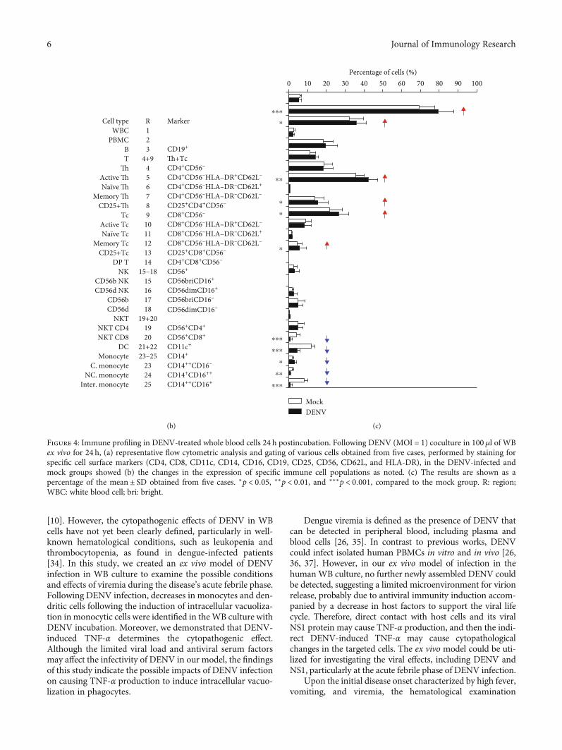
2.8. Immune Profiling. Following DENV (MOI = 1) infectionin 200μl of WB ex vivo for 24 h, representative flow cytomet-ric analysis was conducted using an Attune NxT Flow Cyt-ometer (Thermo Fisher Scientific) and performed by no-wash no-lyse staining for specific cell surface markers(CD4, CD8, CD11c, CD14, CD16, CD19, CD25, CD56,CD62L, and HLA-DR) according to the manufacturer’sinstructions (https://www.thermofisher.com/order/catalog/product/100022776#/100022776).
2.9. Enzyme-Linked Immunosorbent Assay (ELISA). Accord-ing to the manufacturer’s instructions, the concentration ofhuman TNF-α in the plasma samples was determined usingDuoSet ELISA Development System kits (R&D Systems,Minneapolis, MN). In brief, we coated microwells with cap-ture Abs against TNF-α and then blocked the wells with 1%bovine serum albumin in phosphate-buffered saline (PBS).We added the tested serum, added the hTNF-α detectionAbs, and then developed the signal with HRP-conjugateddetection Abs against human IgG. The relative optical den-sity was determined using a microplate reader set to 450nm.
2.10. Statistical Analysis. Values are expressed as the mean± standard deviation ðSDÞ. Groups were compared by usingStudent’s two-tailed unpaired t-test. These analyses were per-formed using GraphPad Prism 4 software (GraphPad Soft-ware, La Jolla, CA). Statistical significance was set atp < 0:05.
3. Results
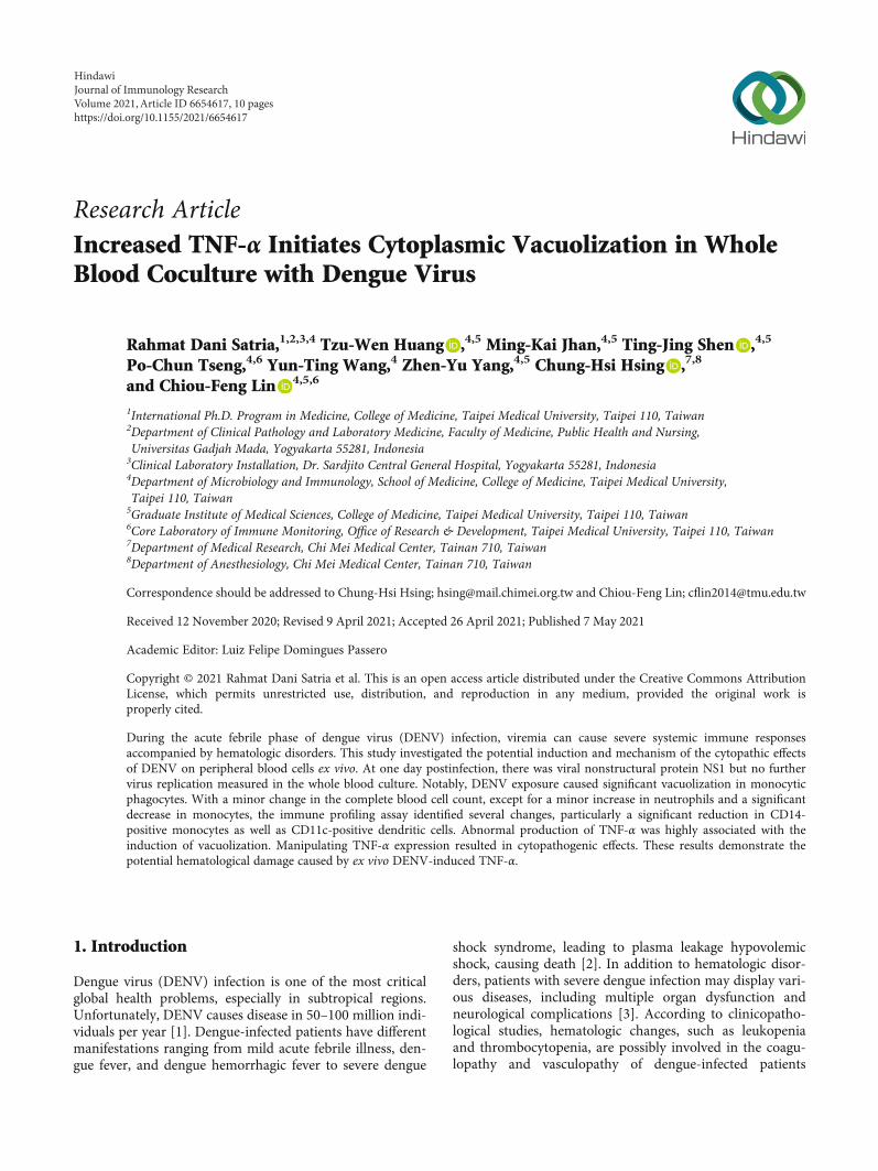
3.1. DENV Causes Infection but Not Replication in the BloodEx Vivo. To mimic a circulation situation during the acutefebrile phase of DENV infection with viremia as demon-strated previously [25, 26], we created a novel ex vivo modelof WB infection without peripheral blood mononuclear cell(PBMC) isolation. DENV was inoculated in the WB cultureex vivo. At 24 h postinfection of the coculture system, as sum-marized in the experimental flowchart (Figure 1(a)), in addi-tion to analyzing the viral infection and antigen expression,several approaches were conducted to evaluate the inductionof cytopathology, immune cell number and populationchanges, and cytokine response. For detecting DENV repli-cation and release, the supernatant of the WB culture washarvested, and a plaque assay was then performed in a stan-dard BHK-21 cell system [27]. Simultaneously, a commercialNS1-based rapid test cassette was performed to measure viralprotein secretion [28]. Compared with the positive control(DENV-containing supernatant), DENV (Figure 1(b)) wasnot detectable, but viral NS1 (Figure 1(c)) was significantly
detected in all five tests (Cases 1-5). The results showed thatDENV could cause infection but failed to release the virion inWB culture ex vivo.
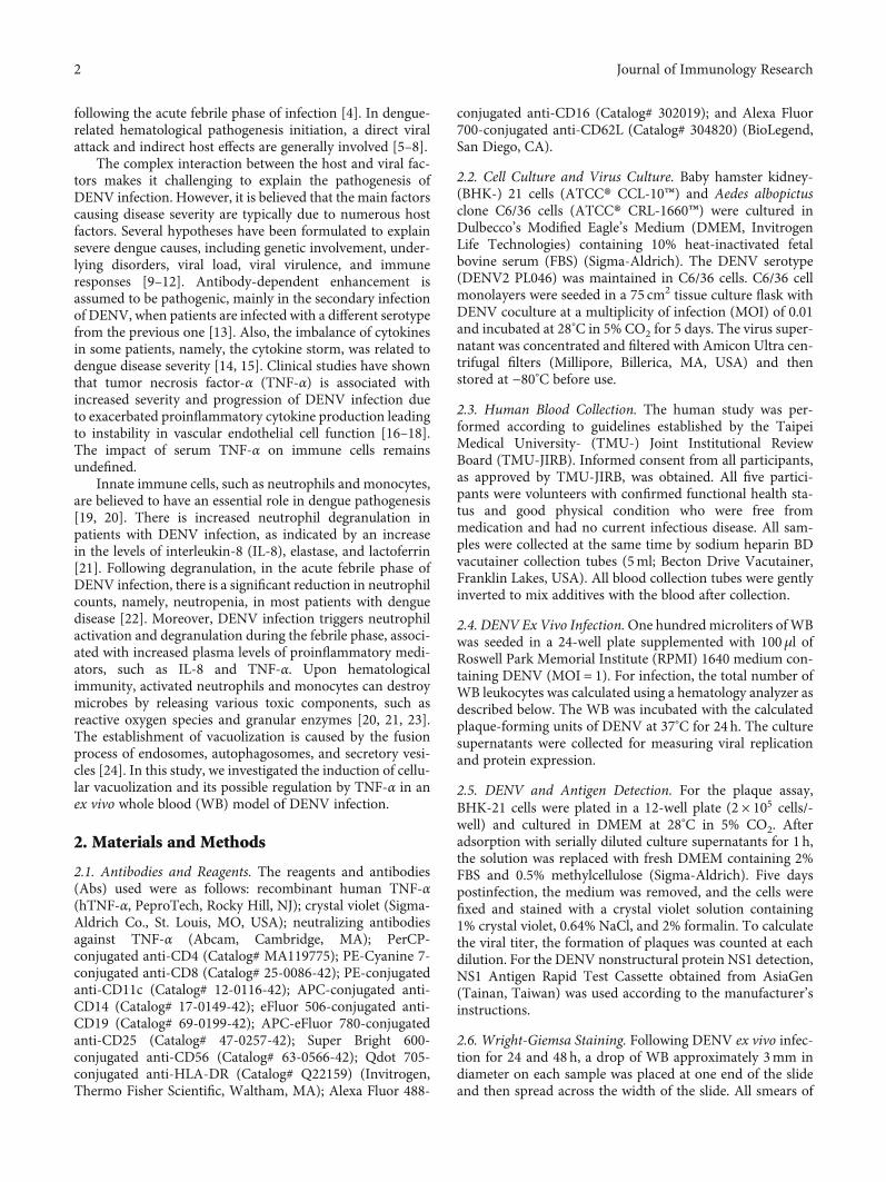
3.2. DENV Coculture Causes Mononuclear Phagocytic CellVacuolization in the Blood Ex Vivo. After DENV coculturecaused infection in the WB culture, we evaluated the mor-phological changes that occurred in leukocytes after viralincubation. In the DENV-WB coculture ex vivo, the sus-pected targets of DENV infection are the myeloid lineage’simmune cells, such as neutrophils and monocytes [20, 29,30]. Following DENV (MOI = 1) coculture in 100μl of WBex vivo for 24 and 48 h, Wright-Giemsa staining showed his-topathological changes significantly in mononuclear cells inall five tests (Figure 2(a)). There was an increase in the per-centage of vacuolated cells after inoculation with DENV at24 h (DENV, 82:53 ± 4:84%; mock, 24:06 ± 12:07%; p =0:001) (Figure 2(b)). While DENV was able to cause infec-tion in WB culture ex vivo, the results showed intracellularvacuolization in monocytes.
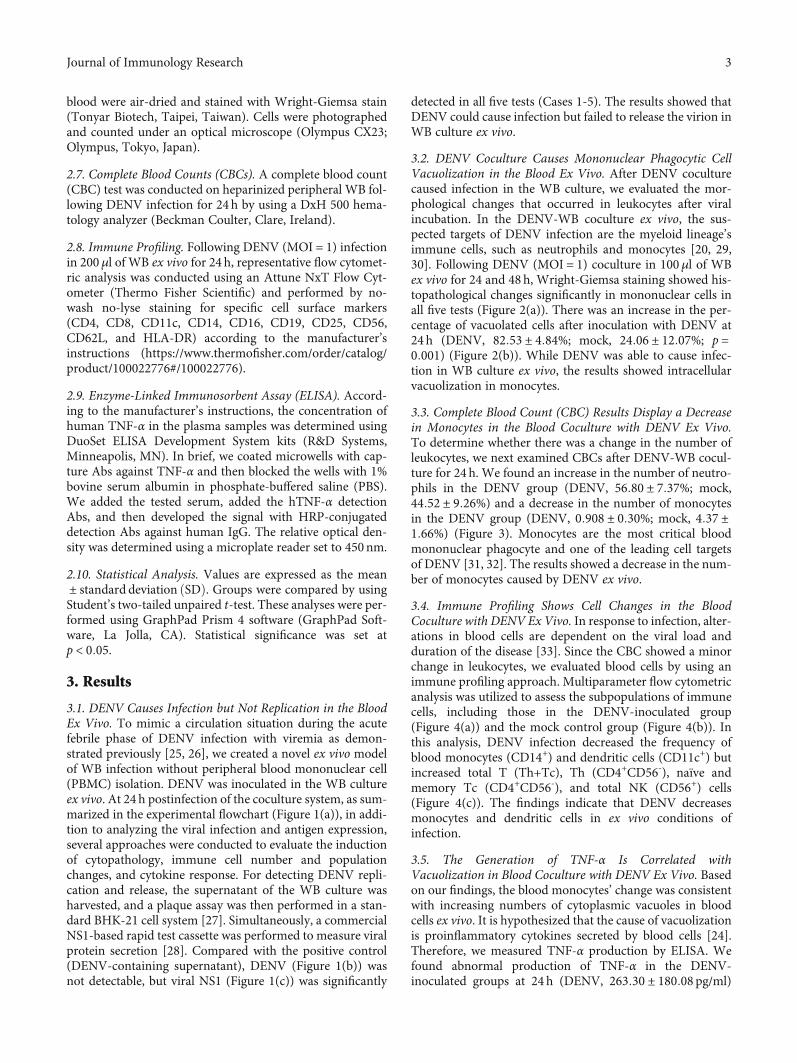
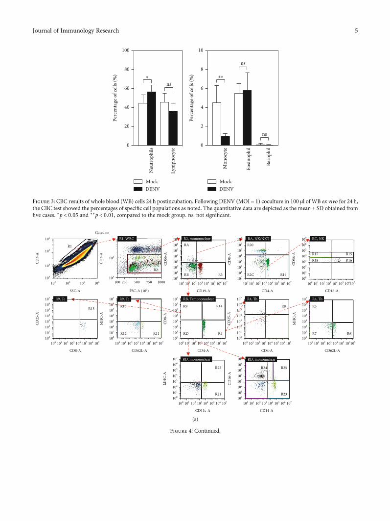
3.3. Complete Blood Count (CBC) Results Display a Decreasein Monocytes in the Blood Coculture with DENV Ex Vivo.To determine whether there was a change in the number ofleukocytes, we next examined CBCs after DENV-WB cocul-ture for 24 h. We found an increase in the number of neutro-phils in the DENV group (DENV, 56:80 ± 7:37%; mock,44:52 ± 9:26%) and a decrease in the number of monocytesin the DENV group (DENV, 0:908 ± 0:30%; mock, 4:37 ±1:66%) (Figure 3). Monocytes are the most critical bloodmononuclear phagocyte and one of the leading cell targetsof DENV [31, 32]. The results showed a decrease in the num-ber of monocytes caused by DENV ex vivo.
3.4. Immune Profiling Shows Cell Changes in the BloodCoculture with DENV Ex Vivo. In response to infection, alter-ations in blood cells are dependent on the viral load andduration of the disease [33]. Since the CBC showed a minorchange in leukocytes, we evaluated blood cells by using animmune profiling approach. Multiparameter flow cytometricanalysis was utilized to assess the subpopulations of immunecells, including those in the DENV-inoculated group(Figure 4(a)) and the mock control group (Figure 4(b)). Inthis analysis, DENV infection decreased the frequency ofblood monocytes (CD14+) and dendritic cells (CD11c+) butincreased total T (Th+Tc), Th (CD4+CD56-), naïve andmemory Tc (CD4+CD56-), and total NK (CD56+) cells(Figure 4(c)). The findings indicate that DENV decreasesmonocytes and dendritic cells in ex vivo conditions ofinfection.
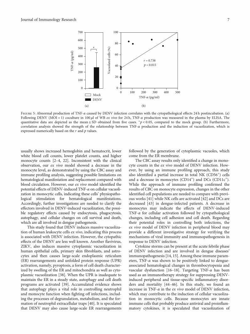
3.5. The Generation of TNF-α Is Correlated withVacuolization in Blood Coculture with DENV Ex Vivo. Basedon our findings, the blood monocytes’ change was consistentwith increasing numbers of cytoplasmic vacuoles in bloodcells ex vivo. It is hypothesized that the cause of vacuolizationis proinflammatory cytokines secreted by blood cells [24].Therefore, we measured TNF-α production by ELISA. Wefound abnormal production of TNF-α in the DENV-inoculated groups at 24 h (DENV, 263:30 ± 180:08pg/ml)
3Journal of Immunology Research
postincubation (Figure 5(a)). Notably, we found a strong cor-relation between TNF-α production and the number of vac-uolated cells (r = 0:738; p = 0:015) (Figure 5(b)). Theseresults indicate that the occurrence of vacuolization is highlycorrelated with DENV-induced TNF-α.
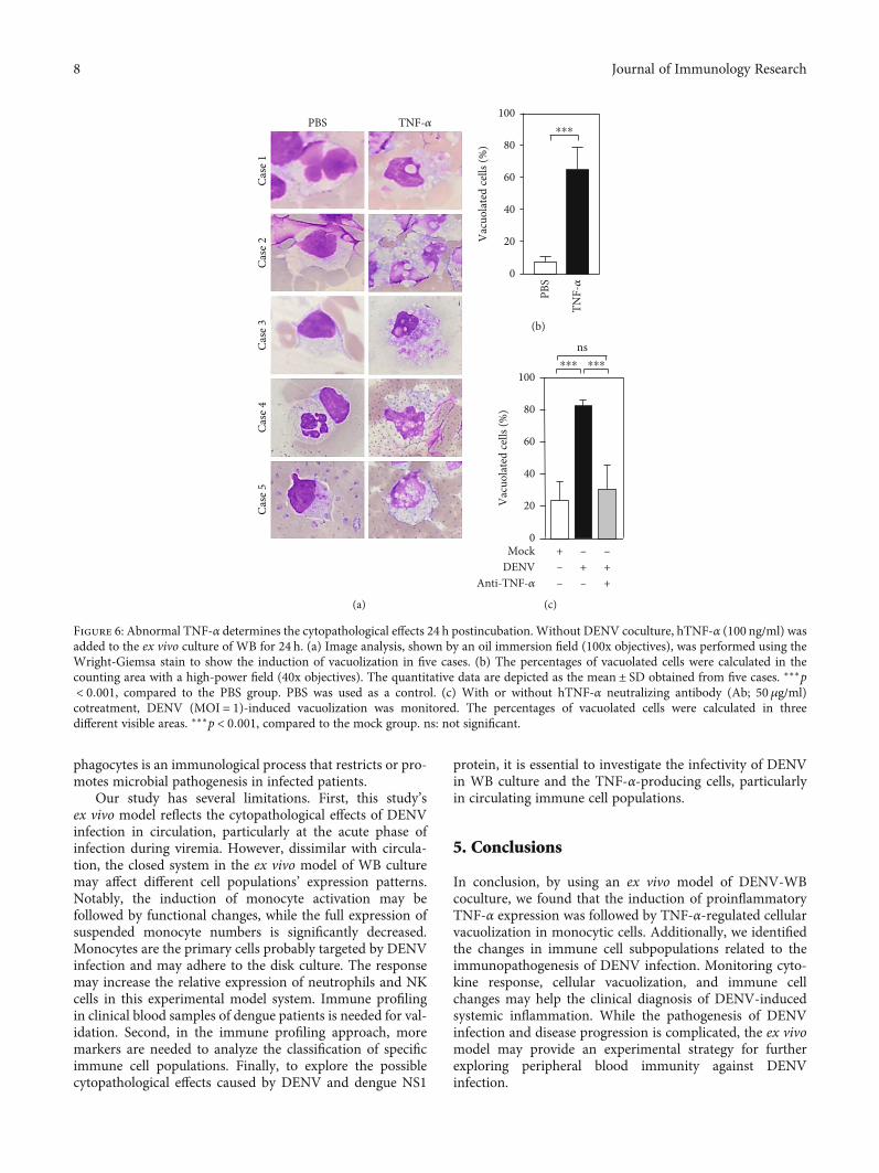
3.6. TNF-αDetermines Vacuolization in Blood Coculture withDENV Ex Vivo. To explore the mechanisms of induction ofvacuolization by TNF-α, we added human TNF-α (hTNF-α,100ng/ml) to the WB culture ex vivo. After 24h of treatment,Wright-Giemsa staining showed histopathological changes inmononuclear and polymorphonuclear cells in both tests(Figure 6(a)). After quantification, there was an increase inthe percentage of vacuolated cells after cotreatment with
hTNF-α for 24h (TNF-α, 64:83 ± 13:77%; mock, 7:90 ± 2:83%; p = 0:01) (Figure 6(b)). To ensure that TNF-α causedvacuolization, we administered anti-TNF-α to DENV-WBcoculture, and the results showed a large decrease in the num-ber of vacuolated cells after cotreatment with anti-TNF-α(DENV, 82:53 ± 4:84%; DENV+ anti-TNF-α, 30:70 ± 15:26%; and mock, 24:06 ± 12:08%) (Figure 6(c)). These resultsdemonstrated that the vacuolization is not caused by viral rep-lication but is caused by DENV-induced TNF-α.
4. Discussion
In the acute febrile phase of dengue-infected patients, theviral load in circulation is associated with disease severity
0 h 24 h
DENV
(Time of post-infection)
Whole blood culture
․Plaque assay․NS1 strip test․Histology․CBC test․Immune profile․ELISA
24 h24 h24 h24 h24 h24 h
Time Approach
(a)
Moc
kD
ENV
Case 1 Case 2 Case 3 Positive 1Case 4 Case 5
Plaq
ue as
say
Positive 2
(b)
Mock DENV
Case 1 Case 2 Case 3 Case 4 Case 5
Control line (C)-Test line (T)-
(c)
Figure 1: DENV infection of whole blood (WB) cells 24 h postincubation. (a) Experimental flowchart of this study. Following DENV(MOI = 1) infection in 100μl of WB ex vivo for 24 h, five plasma samples were harvested to detect DENV infection. (b) The images (10xobjectives) of plaque assay showed no viral replication. (c) An NS1 rapid test showed the expression of the NS1 dengue antigen in theplasma samples. Two positive controls were also performed for the plaque assay.
Vac
uola
ted
cells
Case 1 Case 2 Case 3 Case 4 Case 5
Moc
kD
ENV
(a)
Vac
uola
ted
cells
(%)
Mock DENV0
20
40
60
80
⁎⁎⁎100
(b)
Figure 2: Wright-Giemsa staining images of whole blood (WB) cells 24 h postincubation. Following DENV (MOI = 1) coculture in 100μl ofWB ex vivo for 24 h, Wright-Giemsa staining, shown by an oil immersion field (100x objectives), presented histopathological changes inmononuclear cells (a). The percentages of vacuolated cells are shown (b). The quantitative data are depicted as the mean ± SD obtainedfrom five cases. ∗∗∗p < 0:001, compared to the mock group.
4 Journal of Immunology Research
0N
eutr
ophi
ls
Lym
phoc
yte
Mon
ocyt
e
Eosin
ophi
l
Baso
phil
20
40
60
80
100
⁎
ns
Perc
enta
ge o
f cel
ls (%
)
0
2
4
6
8
10
Perc
enta
ge o
f cel
ls (%
) ⁎⁎
ns
ns
MockDENV
MockDENV
Figure 3: CBC results of whole blood (WB) cells 24 h postincubation. Following DENV (MOI = 1) coculture in 100μl of WB ex vivo for 24 h,the CBC test showed the percentages of specific cell populations as noted. The quantitative data are depicted as themean ± SD obtained fromfive cases. ∗p < 0:05 and ∗∗p < 0:01, compared to the mock group. ns: not significant.
Gated on10
10
10
1010 10 10 10 100 250 500 750 1000
1010 10 10 10 10 10 10 10
10101010101010
CD3-
A
10
10
CD3-
A
CD25
-A
CD8-A
1010 10 10 10 10 10 10 10
10101010101010
MH
C-A
CD11c-A
1010 10 10 10 10 10 10 10
10101010101010
CD16
-A
CD14-A
R23
R6R7
R5
R4
R14
RD
R9
R11R12
R10R13
R1
R2R3 R19
R17R18
R15R16
RB
RA R20
R2C
R8
R25
R21
R22 R24
1010 10 10 10 10 10 10 10
10101010101010
MH
C-A
CD62L-A
1010 10 10 10 10 10 10 10
10101010101010
CD8-
A
CD4-A
1010 10 10 10 10 10 10 10
10101010101010
CD25
-A
CD4-A
1010 10 10 10 10 10 10 10
10101010101010
MH
C-A
CD62L-A
1010 10 10 10 10 10 10 10
10101010101010
CD56
-A
CD19-AFSC-A (103)SSC-A
1010 10 10 10 10 10 10 10
10101010101010
CD8-
A
CD4-A
1010 10 10 10 10 10 10 10
10101010101010
CD56
-ACD16-A
R1, WBC R2, mononuclear RA, NK/NKT RC, NK
R9, Tc R9, Tc R4, Th R4, ThRB, T/mononuclear
RD, mononuclear RD, mononuclear
6
5
4
3
3 4 5 6
0
0 1 2 3 4 5 6 7
1
2
3
4
5
6
7
3
4
0
0 1 2 3 4 5 6 7
1
2
3
4
5
6
7
0
0 1 2 3 4 5 6 7
1
2
3
4
5
6
7
0
0 1 2 3 4 5 6 7
1
2
3
4
5
6
7
0
0 1 2 3 4 5 6 7
1
2
3
4
5
6
7
0
0 1 2 3 4 5 6 7
1
2
3
4
5
6
7
0
0 1 2 3 4 5 6 7
1
2
3
4
5
6
7
0
0 1 2 3 4 5 6 7
1
2
3
4
5
6
7
0
0 1 2 3 4 5 6 7
1
2
3
4
5
6
7
0
0 1 2 3 4 5 6 7
1
2
3
4
5
6
7
(a)
Figure 4: Continued.
5Journal of Immunology Research
[10]. However, the cytopathogenic effects of DENV in WBcells have not yet been clearly defined, particularly in well-known hematological conditions, such as leukopenia andthrombocytopenia, as found in dengue-infected patients[34]. In this study, we created an ex vivo model of DENVinfection in WB culture to examine the possible conditionsand effects of viremia during the disease’s acute febrile phase.Following DENV infection, decreases in monocytes and den-dritic cells following the induction of intracellular vacuoliza-tion in monocytic cells were identified in theWB culture withDENV incubation. Moreover, we demonstrated that DENV-induced TNF-α determines the cytopathogenic effect.Although the limited viral load and antiviral serum factorsmay affect the infectivity of DENV in our model, the findingsof this study indicate the possible impacts of DENV infectionon causing TNF-α production to induce intracellular vacuo-lization in phagocytes.
Dengue viremia is defined as the presence of DENV thatcan be detected in peripheral blood, including plasma andblood cells [26, 35]. In contrast to previous works, DENVcould infect isolated human PBMCs in vitro and in vivo [26,36, 37]. However, in our ex vivo model of infection in thehumanWB culture, no further newly assembled DENV couldbe detected, suggesting a limited microenvironment for virionrelease, probably due to antiviral immunity induction accom-panied by a decrease in host factors to support the viral lifecycle. Therefore, direct contact with host cells and its viralNS1 protein may cause TNF-α production, and then the indi-rect DENV-induced TNF-α may cause cytopathologicalchanges in the targeted cells. The ex vivo model could be uti-lized for investigating the viral effects, including DENV andNS1, particularly at the acute febrile phase of DENV infection.
Upon the initial disease onset characterized by high fever,vomiting, and viremia, the hematological examination
Cell type R MarkerWBC 1
PBMC 2B 3 CD19+
T 4+9 Th+TcTh 4 CD4+CD56–
Active Th 5 CD4+CD56–HLA–DR+CD62L–
Naïve Th 6 CD4+CD56–HLA–DR–CD62L+
Memory Th 7 CD4+CD56–HLA–DR–CD62L–
CD25+Th 8 CD25+CD4+CD56–
Tc 9 CD8+CD56–
Active Tc 10 CD8+CD56–HLA–DR+CD62L–
Naïve Tc 11 CD8+CD56–HLA–DR–CD62L+
Memory Tc 12 CD8+CD56–HLA–DR–CD62L–
CD25+Tc 13 CD25+CD8+CD56–
DP T 14 CD4+CD8+CD56–
NK 15–18 CD56+
CD56b NK 15 CD56briCD16+
CD56d NK 16 CD56dimCD16+
CD56b 17 CD56briCD16–
CD56d 18 CD56dimCD16–
NKT 19+20NKT CD4 19 CD56+CD4+
NKT CD8 20 CD56+CD8+
DC 21+22 CD11c+
Monocyte 23–25 CD14+
C. monocyte 23 CD14++CD16–
NC. monocyte 24 CD14+CD16++
Inter. monocyte 25 CD14++CD16+
(b)
Percentage of cells (%)0 2010 30 40 50 60 8070 90 100
⁎⁎⁎
⁎
⁎⁎
⁎
⁎
⁎
⁎⁎⁎
⁎⁎⁎
⁎
⁎⁎
⁎⁎⁎
MockDENV
(c)
Figure 4: Immune profiling in DENV-treated whole blood cells 24 h postincubation. Following DENV (MOI = 1) coculture in 100 μl of WBex vivo for 24 h, (a) representative flow cytometric analysis and gating of various cells obtained from five cases, performed by staining forspecific cell surface markers (CD4, CD8, CD11c, CD14, CD16, CD19, CD25, CD56, CD62L, and HLA-DR), in the DENV-infected andmock groups showed (b) the changes in the expression of specific immune cell populations as noted. (c) The results are shown as apercentage of the mean ± SD obtained from five cases. ∗p < 0:05, ∗∗p < 0:01, and ∗∗∗p < 0:001, compared to the mock group. R: region;WBC: white blood cell; bri: bright.
6 Journal of Immunology Research
usually shows increased hemoglobin and hematocrit, lowerwhite blood cell counts, lower platelet counts, and highermonocyte counts [2–4, 22]. Inconsistent with the clinicalobservation, our ex vivo model showed a decrease in themonocyte level, as demonstrated by using the CBC assay andimmune profiling analysis, suggesting possible limitations onhematological manifestation and replacement compared withblood circulation. However, our ex vivo model identified thepotential effects of DENV-induced TNF-α on cellular vacuoli-zation in monocytic cells, indicating these cells’ physiopatho-logical stimulation for hematological manifestations.Accordingly, further investigations are needed to clarify theeffectors involved in DENV-induced vacuolization, the possi-ble regulatory effects caused by endocytosis, phagocytosis,autophagy, and cellular changes on cell survival and death,which are all involved in dengue pathogenesis.
This study found that DENV induces massive vacuoliza-tion of human leukocyte cells ex vivo, indicating this processis associated with DENV infection. However, the cytopathiceffects of the DENV are less well known. Another flavivirus,ZIKV, also induces massive cytoplasmic vacuolization inhuman epithelial cells, primary skin fibroblasts, and astro-cytes and then causes large-scale endoplasmic reticulum(ER) rearrangements and unfolded protein response (UPR)activation, namely, pyroptosis, a form of cell death character-ized by swelling of the ER and mitochondria as well as cyto-plasmic vacuolization [38]. When the UPR is inadequate tomaintain the ER in a steady state, autophagy and cell deathprograms are activated [39]. Accumulated evidence showsthat autophagy plays a vital role in controlling neutrophiland monocyte function when fighting off infections, includ-ing the processes of degranulation, metabolism, and the for-mation of neutrophil extracellular traps [40]. It is speculatedthat DENV may also cause large-scale ER rearrangements
followed by the generation of cytoplasmic vacuoles, whichcome from the ER membrane.
The CBC assay results only identified a change in mono-cyte counts in the ex vivo model of DENV infection. How-ever, by using an immune profiling approach, this studyalso identified a partial increase in total NK (CD56+) cellsand a decrease in monocytes (CD14+) and DCs (CD11c+).While the approach of immune profiling confirmed theresults of CBC on monocyte expression, changes in the otheridentified cell populations are needed to compare with previ-ous works [41] while NK cells are activated [42] and DCs aredecreased [43] in dengue-infected patients. A decrease inmonocytes may indicate the effects of DENV-inducedTNF-α for cellular activation followed by cytopathologicalchanges, including cell adhesion and cell death. Regardingtheir potential roles in controlling both infections, theex vivo model of DENV infection in peripheral blood mayprovide a different investigative strategy for verifying themechanisms of viral immunity and immunopathogenesis inresponse to DENV infection.
Cytokine storms can be present at the acute febrile phaseof DENV infection and are involved in dengue diseases’immunopathogenesis [14, 15]. Among these immune param-eters, TNF-α was shown to be positively linked to dengue-associated hematological changes in thrombocytopenia andvascular dysfunction [16–18]. Targeting TNF-α has beenused as an immunotherapy strategy for suppressing DENV-induced peripheral and tissue-specific inflammatory disor-ders and mortality [44–46]. In this study, we found anincrease in TNF-α in the ex vivo model of DENV infection,which may contribute to the induction of cellular vacuoliza-tion in monocytic cells. Because monocytes are innateimmune cells that probably produce antiviral and proinflam-matory cytokines, it is speculated that vacuolization of
100
0
200
300
400
500
600
700
TNF-𝛼
(pg/
ml)
⁎
MockDENV
(a)
0
20
40
60
80
100
0 100 200 300 400 500TNF-𝛼 (pg/ml)
Vac
uola
ted
cells
(%)
r = 0.738 p = 0.015
(b)
Figure 5: Abnormal production of TNF-α caused by DENV infection correlates with the cytopathological effects 24 h postincubation. (a)Following DENV (MOI = 1) coculture in 100 μl of WB ex vivo for 24 h, TNF-α production was measured in the plasma by ELISA. Thequantitative data are depicted as the mean ± SD obtained from five cases. ∗p < 0:05, compared to the mock group. (b) Furthermore,correlation analysis showed the strength of the relationship between TNF-α production and the induction of vacuolization, which isexpressed numerically based on the r and p values.
7Journal of Immunology Research
phagocytes is an immunological process that restricts or pro-motes microbial pathogenesis in infected patients.
Our study has several limitations. First, this study’sex vivo model reflects the cytopathological effects of DENVinfection in circulation, particularly at the acute phase ofinfection during viremia. However, dissimilar with circula-tion, the closed system in the ex vivo model of WB culturemay affect different cell populations’ expression patterns.Notably, the induction of monocyte activation may befollowed by functional changes, while the full expression ofsuspended monocyte numbers is significantly decreased.Monocytes are the primary cells probably targeted by DENVinfection and may adhere to the disk culture. The responsemay increase the relative expression of neutrophils and NKcells in this experimental model system. Immune profilingin clinical blood samples of dengue patients is needed for val-idation. Second, in the immune profiling approach, moremarkers are needed to analyze the classification of specificimmune cell populations. Finally, to explore the possiblecytopathological effects caused by DENV and dengue NS1
protein, it is essential to investigate the infectivity of DENVin WB culture and the TNF-α-producing cells, particularlyin circulating immune cell populations.
5. Conclusions
In conclusion, by using an ex vivo model of DENV-WBcoculture, we found that the induction of proinflammatoryTNF-α expression was followed by TNF-α-regulated cellularvacuolization in monocytic cells. Additionally, we identifiedthe changes in immune cell subpopulations related to theimmunopathogenesis of DENV infection. Monitoring cyto-kine response, cellular vacuolization, and immune cellchanges may help the clinical diagnosis of DENV-inducedsystemic inflammation. While the pathogenesis of DENVinfection and disease progression is complicated, the ex vivomodel may provide an experimental strategy for furtherexploring peripheral blood immunity against DENVinfection.
Case
1Ca
se 2
Case
3Ca
se 4
Case
5
PBS TNF-𝛼
(a)
100
0
PBS
TNF-𝛼
⁎⁎⁎
Vac
uola
ted
cells
(%)
20
40
60
80
(b)
⁎⁎⁎
ns⁎⁎⁎
20
40
60
0
80
100
Vac
uola
ted
cells
(%)
Mock + – –DENV – + +
Anti-TNF-𝛼 – – +
(c)
Figure 6: Abnormal TNF-α determines the cytopathological effects 24 h postincubation. Without DENV coculture, hTNF-α (100 ng/ml) wasadded to the ex vivo culture of WB for 24 h. (a) Image analysis, shown by an oil immersion field (100x objectives), was performed using theWright-Giemsa stain to show the induction of vacuolization in five cases. (b) The percentages of vacuolated cells were calculated in thecounting area with a high-power field (40x objectives). The quantitative data are depicted as the mean ± SD obtained from five cases. ∗∗∗p< 0:001, compared to the PBS group. PBS was used as a control. (c) With or without hTNF-α neutralizing antibody (Ab; 50μg/ml)cotreatment, DENV (MOI = 1)-induced vacuolization was monitored. The percentages of vacuolated cells were calculated in threedifferent visible areas. ∗∗∗p < 0:001, compared to the mock group. ns: not significant.
8 Journal of Immunology Research
Data Availability
The data used to support the findings of this study are avail-able from the corresponding authors upon request.
Conflicts of Interest
The authors declare that there is no conflict of interest.
Authors’ Contributions
R.-D.S. and M.-K.J. performed most of the experiments andinterpreted the results. T.-W.H., C.-H.H., and C.-F.L. partic-ipated in the design and supervision of the projects. M.-K.J.and T.-J.S. conducted the virus experiments. T.-J.S., P.-C.T.,Y.-T.W., and Z.-Y.Y. contributed to flow cytometric analysis.R.-D.S., C.-H.H., and C.-F.L. designed the concept of the pro-ject and wrote the manuscript. All authors reviewed andapproved the manuscript.
Acknowledgments
This work was supported by the Ministry of Science andTechnology (MOST107-2321-B-038-001, 108-2320-B-038-026, 109-2320-B-038-050, and 109-2327-B-006-010) andChi Mei Medical Center and Taipei Medical University(105CM-TMU-03, 106CM-TMU-09, and 107CM-TMU-10), Taiwan. We thank the Core Facility Center of TaipeiMedical University (TMU) for providing technical support.We also appreciate the Office of Research and Publication,Universitas Gadjah Mada, Indonesia, for editing themanuscript.
References
[1] I. A. Rodenhuis-Zybert, J. Wilschut, and J. M. Smit, “Denguevirus life cycle: viral and host factors modulating infectivity,”Cellular and Molecular Life Sciences, vol. 67, no. 16,pp. 2773–2786, 2010.
[2] World Health Organization and UNICEF, Handbook for Clin-ical Management of Dengue, p. 114, 2012.
[3] C. M. Chen, K. S. Chan, W. L. Yu et al., “The outcomes ofpatients with severe dengue admitted to intensive care units,”Medicine (Baltimore), vol. 95, no. 31, article e4376, 2016.
[4] J. Chaloemwong, A. Tantiworawit, T. Rattanathammetheeet al., “Useful clinical features and hematological parametersfor the diagnosis of dengue infection in patients with acutefebrile illness: a retrospective study,” BMC Hematology,vol. 18, no. 1, p. 20, 2018.
[5] M. Kotepui, B. PhunPhuech, N. Phiwklam, and K. Uthaisar,“Differentiating between dengue fever and malaria usinghematological parameters in endemic areas of Thailand,”Infectious Diseases of Poverty, vol. 6, no. 1, p. 27, 2017.
[6] M. S. Diamond, D. Edgil, T. G. Roberts, B. Lu, and E. Harris,“Infection of human cells by dengue virus is modulated by dif-ferent cell types and viral strains,” Journal of Virology, vol. 74,no. 17, pp. 7814–7823, 2000.
[7] D. M. Morens, M. C. Chu, N. J. Marchette, and S. B. Halstead,“Growth of dengue type 2 virus isolates in human peripheralblood leukocytes correlates with severe and mild dengue dis-
ease,” The American Journal of Tropical Medicine and Hygiene,vol. 45, no. 5, pp. 644–651, 1991.
[8] P. Sun, K. Bauza, S. Pal et al., “Infection and activation ofhuman peripheral bloodmonocytes by dengue viruses throughthe mechanism of antibody-dependent enhancement,” Virol-ogy, vol. 421, no. 2, pp. 245–252, 2011.
[9] X. Fang, Z. Hu, W. Shang, J. Zhu, C. Xu, and X. Rao, “Geneticpolymorphisms of molecules involved in host immuneresponse to dengue virus infection,” FEMS Immunology andMedical Microbiology, vol. 66, no. 2, pp. 134–146, 2012.
[10] R. Ben-Shachar, S. Schmidler, and K. Koelle, “Drivers of inter-individual variation in dengue viral load dynamics,” PLoSComputational Biology, vol. 12, no. 11, article e1005194, 2016.
[11] C. Zou, C. Huang, J. Zhang et al., “Virulence difference of fivetype I dengue viruses and the intrinsic molecular mechanism,”PLoS Neglected Tropical Diseases, vol. 13, no. 3, articlee0007202, 2019.
[12] N. Uno and T. M. Ross, “Dengue virus and the host innateimmune response,” Emerging Microbes & Infections, vol. 7,no. 1, p. 167, 2018.
[13] J. Flipse, M. A. Diosa-Toro, T. E. Hoornweg, D. P. I. van dePol, S. Urcuqui-Inchima, and J. M. Smit, “Antibody-depen-dent enhancement of dengue virus infection in primaryhuman macrophages; balancing higher fusion against antiviralresponses,” Scientific Reports, vol. 6, no. 1, p. 29201, 2016.
[14] A. R. K. Patro, S. Mohanty, B. K. Prusty et al., “Cytokine signa-ture associated with disease severity in dengue,” Viruses,vol. 11, no. 1, p. 34, 2019.
[15] A. L. Rothman, “Immunity to dengue virus: a tale of originalantigenic sin and tropical cytokine storms,” Nature Reviews.Immunology, vol. 11, no. 8, pp. 532–543, 2011.
[16] S. Inyoo, A. Suttitheptumrong, and S. N. Pattanakitsakul, “Syn-ergistic effect of TNF-α and dengue virus infection on adhesionmolecule reorganization in human endothelial cells,” JapaneseJournal of Infectious Diseases, vol. 70, no. 2, pp. 186–191, 2017.
[17] K. I. Masood, B. Jamil, M. Rahim, M. Islam, M. Farhan, andZ. Hasan, “Role of TNF α, IL-6 and CXCL10 in dengue diseaseseverity,” Iranian Journal of Microbiology, vol. 10, no. 3,pp. 202–207, 2018.
[18] A. A. Meena, A. Murugesan, S. Sopnajothi et al., “Increase ofplasma TNF-α is associated with decreased levels of bloodplatelets in clinical dengue infection,” Viral Immunology,vol. 33, no. 1, pp. 54–60, 2020.
[19] E. D. Hottz, I. M. Medeiros-de-Moraes, A. Vieira-de-Abreuet al., “Platelet activation and apoptosis modulate monocyteinflammatory responses in dengue,” Journal of Immunology,vol. 193, no. 4, pp. 1864–1872, 2014.
[20] A. Opasawatchai, P. Amornsupawat, N. Jiravejchakul et al.,“Neutrophil activation and early features of NET formationare associated with dengue virus infection in human,” Fron-tiers in Immunology, vol. 9, p. 3007, 2019.
[21] M. Juffrie, G. M. van derMeer, C. E. Hack et al., “Inflammatorymediators in dengue virus infection in children: interleukin-8and its relationship to neutrophil degranulation,” Infectionand Immunity, vol. 68, no. 2, pp. 702–707, 2000.
[22] J. G. X. Wong, T. L. Thein, D. C. Lye, Y. Hao, A. Wilder-Smith,and Y. S. Leo, “Severe neutropenia in dengue patients: preva-lence and significance,” The American Journal of TropicalMedicine and Hygiene, vol. 90, no. 6, pp. 984–987, 2014.
[23] M. Kunder, V. Lakshmaiah, and A. V. Moideen Kutty, “Plasmaneutrophil elastase, α1-antitrypsin, α2-macroglobulin and
9Journal of Immunology Research
neutrophil elastase–α1-antitrypsin complex levels in patientswith dengue fever,” Indian Journal of Clinical Biochemistry,vol. 33, no. 2, pp. 218–221, 2018.
[24] C. C. Mihalache, S. Yousefi, S. Conus, P. M. Villiger, E. M.Schneider, and H. U. Simon, “Inflammation-associatedautophagy-related programmed necrotic death of human neu-trophils characterized by organelle fusion events,” Journal ofImmunology, vol. 186, no. 11, pp. 6532–6542, 2011.
[25] S. Thiemmeca, A. Songjang, N. Punyadee, K. Kongmanas,J. Atkinson, and P. Avirutnan, “Infection of whole blood withdengue virus,” Molecular Immunology, vol. 102, pp. 132-133,2018.
[26] A. P. Durbin, M. J. Vargas, K. Wanionek et al., “Phenotypingof peripheral blood mononuclear cells during acute dengue ill-ness demonstrates infection and increased activation of mono-cytes in severe cases compared to classic dengue fever,”Virology, vol. 376, no. 2, pp. 429–435, 2008.
[27] T. T. Tsai, C. L. Chen, Y. S. Lin et al., “Microglia retard denguevirus-induced acute viral encephalitis,” Scientific Reports,vol. 6, no. 1, p. 27670, 2016.
[28] L. Osorio, M. Ramirez, A. Bonelo, L. A. Villar, and B. Parra,“Comparison of the diagnostic accuracy of commercial NS1-based diagnostic tests for early dengue infection,” VirologyJournal, vol. 7, no. 1, p. 361, 2010.
[29] Z. Kou, M. Quinn, H. Chen et al., “Monocytes, but not T or Bcells, are the principal target cells for dengue virus (DV) infec-tion among human peripheral blood mononuclear cells,” Jour-nal of Medical Virology, vol. 80, no. 1, pp. 134–146, 2008.
[30] S. Noisakran, N. Onlamoon, P. Songprakhon, H. M. Hsiao,K. Chokephaibulkit, and G. C. Perng, “Cells in dengue virusinfection in vivo,” Advances in Virology, vol. 2010, Article ID164878, 15 pages, 2010.
[31] J. A. Aguilar-Briseño, V. Upasani, B. M. . Ellen et al., “TLR2 onblood monocytes senses dengue virus infection and its expres-sion correlates with disease pathogenesis,” Nature Communi-cations, vol. 11, no. 1, p. 3177, 2020.
[32] S. W. Wan, B. A. Wu-Hsieh, Y. S. Lin, W. Y. Chen, Y. Huang,and R. Anderson, “The monocyte-macrophage-mast cell axisin dengue pathogenesis,” Journal of Biomedical Science,vol. 25, no. 1, p. 77, 2018.
[33] M. Kwissa, H. I. Nakaya, N. Onlamoon et al., “Dengue virusinfection induces expansion of a CD14+CD16+ monocyte pop-ulation that stimulates plasmablast differentiation,” Cell Host& Microbe, vol. 16, no. 1, pp. 115–127, 2014.
[34] H. K. Jayanthi and S. K. Tulasi, “Correlation study betweenplatelet count, leukocyte count, nonhemorrhagic complica-tions, and duration of hospital stay in dengue fever withthrombocytopenia,” Journal of Family Medicine and PrimaryCare, vol. 5, no. 1, pp. 120–123, 2016.
[35] G. Añez, D. A. R. Heisey, C. Chancey et al., “Distribution ofdengue virus types 1 and 4 in blood components from infectedblood donors from Puerto Rico,” PLoS Neglected Tropical Dis-eases, vol. 10, no. 2, article e0004445, 2016.
[36] L. Jiang and Q. Sun, “The expression profile of human periph-eral blood mononuclear cell miRNA is altered by antibody-dependent enhancement of infection with dengue virus sero-type 3,” Virology Journal, vol. 15, no. 1, p. 50, 2018.
[37] Y. L. Chen, N. Abdul Ghafar, R. Karuna et al., “Activation ofperipheral blood mononuclear cells by dengue virus infectiondepotentiates balapiravir,” Journal of Virology, vol. 88, no. 3,pp. 1740–1747, 2014.
[38] B. Monel, A. A. Compton, T. Bruel et al., “Zika virus inducesmassive cytoplasmic vacuolization and paraptosis-like deathin infected cells,” The EMBO Journal, vol. 36, no. 12,pp. 1653–1668, 2017.
[39] A. B. Blazquez et al., “Stress responses in flavivirus-infectedcells: activation of unfolded protein response and autophagy,”Frontiers in Microbiology, vol. 5, 2014.
[40] Y. Yu and B. Sun, “Autophagy-mediated regulation of neutro-phils and clinical applications,” Burns Trauma, vol. 8, 2020.
[41] K. Lühn, C. P. Simmons, E. Moran et al., “Increased frequen-cies of CD4+ CD25(high) regulatory T cells in acute dengueinfection,” The Journal of Experimental Medicine, vol. 204,no. 5, pp. 979–985, 2007.
[42] C. Petitdemange, N. Wauquier, H. Devilliers et al., “Longitudi-nal analysis of natural killer cells in dengue virus-infectedpatients in comparison to Chikungunya and Chikungunya/-dengue virus-infected patients,” PLoS Neglected Tropical Dis-eases, vol. 10, no. 3, article e0004499, 2016.
[43] S. Lertjuthaporn, L. Khowawisetsut, R. Keawvichit et al.,“Identification of changes in dendritic cell subsets that corre-late with disease severity in dengue infection,” PLoS One,vol. 13, no. 7, article e0200564, 2018.
[44] A. Atrasheuskaya, P. Petzelbauer, T. M. Fredeking, andG. Ignatyev, “Anti-TNF antibody treatment reduces mortalityin experimental dengue virus infection,” FEMS Immunologyand Medical Microbiology, vol. 35, no. 1, pp. 33–42, 2003.
[45] E. Branche, W. W. Tang, K. M. Viramontes et al., “Synergismbetween the tyrosine kinase inhibitor sunitinib and anti-TNFantibody protects against lethal dengue infection,” AntiviralResearch, vol. 158, pp. 1–7, 2018.
[46] M. K. Jhan, W. C. HuangFu, Y. F. Chen et al., “Anti-TNF-αrestricts dengue virus-induced neuropathy,” Journal of Leuko-cyte Biology, vol. 104, no. 5, pp. 961–968, 2018.
10 Journal of Immunology Research