ImportanceofLipopolysaccharideandCyclic β-1,2-Glucansin...
13
Hindawi Publishing Corporation International Journal of Microbiology Volume 2010, Article ID 124509, 12 pages doi:10.1155/2010/124509 Review Article Importance of Lipopolysaccharide and Cyclic β-1,2-Glucans in Brucella -Mammalian Infections Andreas F. Haag, Kamila K. Myka, Markus F. F. Arnold, Paola Caro-Hern´ andez, and Gail P. Ferguson School of Medicine & Dentistry, Institute of Medical Sciences, University of Aberdeen, Foresterhill, Aberdeen AB25 2ZD, UK Correspondence should be addressed to Gail P. Ferguson, [email protected] Received 13 July 2010; Accepted 4 October 2010 Academic Editor: Charlene Kahler Copyright © 2010 Andreas F. Haag et al. This is an open access article distributed under the Creative Commons Attribution License, which permits unrestricted use, distribution, and reproduction in any medium, provided the original work is properly cited. Brucella species are the causative agents of one of the most prevalent zoonotic diseases: brucellosis. Infections by Brucella species cause major economic losses in agriculture, leading to abortions in infected animals and resulting in a severe, although rarely lethal, debilitating disease in humans. Brucella species persist as intracellular pathogens that manage to effectively evade recognition by the host’s immune system. Sugar-modified components in the Brucella cell envelope play an important role in their host interaction. Brucella lipopolysaccharide (LPS), unlike Escherichia coli LPS, does not trigger the host’s innate immune system. Brucella produces cyclic β-1,2-glucans, which are important for targeting them to their replicative niche in the endoplasmic reticulum within the host cell. This paper will focus on the role of LPS and cyclic β-1,2-glucans in Brucella-mammalian infections and discuss the use of mutants, within the biosynthesis pathway of these cell envelope structures, in vaccine development. 1. Introduction Brucellosis is a disease that can be found in most countries around the world and is transferred from animals to humans [1]. With more than 500,000 new cases of human infections each year, it is the most prevalent zoonotic disease worldwide [1]. Although brucellosis very rarely leads to the death of the patient, it is a seriously debilitating disease that presents with, among other symptoms, fever, fatigue, nausea, and weight loss [2]. Brucellosis is thought to be underreported as the symptoms very often are mistaken for a common flu [2]. However, if not properly treated, brucellosis can become a chronic and asymptomatic disease that can re-emerge months after the initial infection [2]. The causative agents of brucellosis are brucellae, nonmotile, Gram-negative α- proteobacteria that are facultative intracellular pathogens [2]. The genus Brucella currently contains ten species, named primarily after their preferred host organism or symptoms of the infection: B. melitensis (goats and sheep), B. abortus (cattle) [3], B. suis (swine, reindeer and rodents) [4], B. canis (dogs) [5], B. ovis (sheep) [6], B. neomtomae (rodents) [7], B. microti (voles and red foxes) [8], B. inopinata (unknown) [9], B. pinnipedialis (seals), and B. ceti (dolphins and porpoises) [10]. Most human Brucella infections can be traced back to the three species, B. melitensis, B. suis, and B. abortus [11]. The isolation of marine mammal Brucella species (B. pinnipedialis and B. ceti) from human patients, however, suggests that these species are emerging human pathogens [10]. Brucellae enter their hosts either through contact with infected animals and material, such as blood or milk, or through the aerosol route [12]. Bacteria of the genus Brucella are highly infectious, and doses as low as 10 to 100 bacteria are thought to be sufficient to cause the human disease [12]. Brucellae are therefore considered to be targets for the development of biological weapons and several countries were suspected of trying to weaponize Brucella species during the Cold War [12, 13]. The primary host for Brucella abortus is cattle where it leads to abortions causing significant economic losses [1]. As humans are not the primary hosts for brucellae, the most promising strategies to control and finally eradicate the disease seems to be through rigorous vaccination of its primary host, and efficient screening methods that can differentiate between vaccinated and
Transcript of ImportanceofLipopolysaccharideandCyclic β-1,2-Glucansin...
Hindawi Publishing CorporationInternational Journal of MicrobiologyVolume 2010, Article ID 124509, 12 pagesdoi:10.1155/2010/124509
Review Article
Importance of Lipopolysaccharide and Cyclic β-1,2-Glucans inBrucella-Mammalian Infections
Andreas F. Haag, Kamila K. Myka, Markus F. F. Arnold, Paola Caro-Hernandez,and Gail P. Ferguson
School of Medicine & Dentistry, Institute of Medical Sciences, University of Aberdeen, Foresterhill, Aberdeen AB25 2ZD, UK
Correspondence should be addressed to Gail P. Ferguson, [email protected]
Received 13 July 2010; Accepted 4 October 2010
Academic Editor: Charlene Kahler
Copyright © 2010 Andreas F. Haag et al. This is an open access article distributed under the Creative Commons AttributionLicense, which permits unrestricted use, distribution, and reproduction in any medium, provided the original work is properlycited.
Brucella species are the causative agents of one of the most prevalent zoonotic diseases: brucellosis. Infections by Brucella speciescause major economic losses in agriculture, leading to abortions in infected animals and resulting in a severe, although rarely lethal,debilitating disease in humans. Brucella species persist as intracellular pathogens that manage to effectively evade recognition by thehost’s immune system. Sugar-modified components in the Brucella cell envelope play an important role in their host interaction.Brucella lipopolysaccharide (LPS), unlike Escherichia coli LPS, does not trigger the host’s innate immune system. Brucella producescyclic β-1,2-glucans, which are important for targeting them to their replicative niche in the endoplasmic reticulum within thehost cell. This paper will focus on the role of LPS and cyclic β-1,2-glucans in Brucella-mammalian infections and discuss the useof mutants, within the biosynthesis pathway of these cell envelope structures, in vaccine development.
1. Introduction
Brucellosis is a disease that can be found in most countriesaround the world and is transferred from animals to humans[1]. With more than 500,000 new cases of human infectionseach year, it is the most prevalent zoonotic disease worldwide[1]. Although brucellosis very rarely leads to the death ofthe patient, it is a seriously debilitating disease that presentswith, among other symptoms, fever, fatigue, nausea, andweight loss [2]. Brucellosis is thought to be underreportedas the symptoms very often are mistaken for a common flu[2]. However, if not properly treated, brucellosis can becomea chronic and asymptomatic disease that can re-emergemonths after the initial infection [2]. The causative agentsof brucellosis are brucellae, nonmotile, Gram-negative α-proteobacteria that are facultative intracellular pathogens[2]. The genus Brucella currently contains ten species, namedprimarily after their preferred host organism or symptomsof the infection: B. melitensis (goats and sheep), B. abortus(cattle) [3], B. suis (swine, reindeer and rodents) [4], B. canis(dogs) [5], B. ovis (sheep) [6], B. neomtomae (rodents) [7], B.microti (voles and red foxes) [8], B. inopinata (unknown) [9],
B. pinnipedialis (seals), and B. ceti (dolphins and porpoises)[10]. Most human Brucella infections can be traced backto the three species, B. melitensis, B. suis, and B. abortus[11]. The isolation of marine mammal Brucella species (B.pinnipedialis and B. ceti) from human patients, however,suggests that these species are emerging human pathogens[10].
Brucellae enter their hosts either through contact withinfected animals and material, such as blood or milk, orthrough the aerosol route [12]. Bacteria of the genus Brucellaare highly infectious, and doses as low as 10 to 100 bacteriaare thought to be sufficient to cause the human disease[12]. Brucellae are therefore considered to be targets for thedevelopment of biological weapons and several countrieswere suspected of trying to weaponize Brucella species duringthe Cold War [12, 13]. The primary host for Brucella abortusis cattle where it leads to abortions causing significanteconomic losses [1]. As humans are not the primary hostsfor brucellae, the most promising strategies to control andfinally eradicate the disease seems to be through rigorousvaccination of its primary host, and efficient screeningmethods that can differentiate between vaccinated and
2 International Journal of Microbiology
infected animals [1]. However, most countries have notimplemented an efficient program for disease control [1].Prohibitive factors are most likely the costs involved butcompared to the potential economic losses caused by animalbrucellosis, these costs are negligible [1].
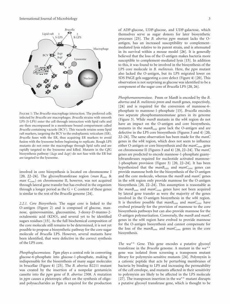
The infection process of Brucella has been subjectto intense research. The preferred cell types infected bybrucellae are phagocytic cells such as macrophages [14](Figure 1). Brucellae are taken up into a phagosome, whichis then targeted to the endoplasmic reticulum, the replicativeniche of the Brucella within the host [12, 14]. Evasion ofthe immune system of the host organism and targeting ofthe bacterium to its replicative niche are of key importancefor the infection process. The bacterial cell envelope is themajor point of interaction between brucellae and the hostand as such, molecules within the bacterial cell envelopeplay a significant part in the infection process. This paperwill focus on the role of two brucellae sugar-modifiedcell envelope components, lipopolysaccharide (LPS) andcyclic β-1,2-glucans (CβGs), in host interactions and vaccinedevelopment.
2. Lipopolysaccharide
Brucellae are Gram-negative bacteria and as such their cellenvelope is composed of two membranes (Figure 2). Theouter membrane plays a crucial role in the infection process,as it is the first point of interaction between the bacteriumand the host. The outer layer of the outer membrane iscomposed of LPS, which consists of three key components:(i) the lipid A, which forms the hydrophobic anchor of theLPS within the outer membrane, (ii) an inner and outercore composed of sugar molecules, and (iii) the O-antigen,a polymerized sugar chain extending into the extracellularenvironment (Figure 2). Brucellae occur naturally as smoothLPS (S-LPS) strains, which contain LPS that is modified withan O-antigen, and rough LPS (R-LPS) strains, which lack theO-antigen [15]. This paper will focus on the importance ofBrucella LPS on the host interaction and on how its structurecan influence the infection process.
2.1. Importance of LPS for the Interaction with Host Cells. Inmany bacterial infections, LPS is the major molecule thatis recognized by the innate immune system and can triggera severe immune response against the invading organism.The ability of brucellae to produce S-LPS with a completeO-antigen is crucial for its virulence in humans [2]. B.melitensis, B. suis, and B. abortus that express a completeO-antigen are the main species responsible for humaninfections [2]. Mutants of these Brucella species lacking theO-antigen modification are considerably less virulent thantheir respective parent strains [15]. Brucella species that arenaturally devoid of the O-antigen modification such as B.canis and B. ovis have either a low virulence or are avirulentin humans [2].
LPS is released by Gram-negative bacteria during theirgrowth and death and is the cause of endotoxic shock inseptic patients [16]. The LPS of Brucella is less toxic than thatof enterobacterial species and therefore plays a major role for
Brucella in the evasion of the host’s immune system and itssurvival thereafter [17, 18]. LPS is the major surface antigenand is recognized by the immune system by the Toll-likereceptor 4 (TLR4)/MD2 complex [19]. This complex bindsthe lipid A component of LPS and, by the recruitment ofadditional factors, initiates the innate immune response [19].Although Brucella LPS binds to TLR4, it does not inducethe production of cytokines and antimicrobial peptides[12]. Brucella LPS is several hundred times less effective atinducing the innate immunity than E. coli LPS, and thisis thought to be important for Brucella to evade immunedetection and to form a chronic intracellular infection [12,15]. The LPS O-antigen plays an important role in thedevelopment of an adaptive immune response to pathogenicbacteria [19] and is the major antigen that is presented bythe MHC II of B-cells [16]. However, Brucella LPS interactswith MHC II molecules in a way that prevents signaling andactivation of MHC II dependent T-cells [16, 18]. Therefore,Brucella LPS acts as an important virulence factor andprevents the initiation of an adaptive immune response. Themodification of Brucella LPS with its O-antigen also seems tobe essential for the entry of the bacterium into the host cells(Figure 1). Brucella abortus strains having S-LPS enter thehost cell via lipid rafts and thus the compartment in whichthey persist acquires lipid raft marker molecules that areimportant for the intracellular targeting of the compartmentto the endoplasmic reticulum (Figure 1) [12, 14]. R-LPSB. abortus mutants do not enter the host cells throughlipid rafts but through normal phagocytosis and are thentargeted to lysosomes (Figure 1) [12, 14]. Therefore, LPSplays a crucial role for Brucella in evading both innate andadaptive immunity and in enabling the bacterium to reachits intracellular niche.
2.2. Structure and Biosynthesis. Brucella only uses glucose asits carbon source and is therefore equipped with a set ofenzymes to convert glucose into the different types of sugarmolecules utilized within the organism (Figures 3 and 4)[20]. The biosynthesis of the Brucella O-antigen and coremolecule has not yet been fully characterized and much hasbeen derived from predicted protein functions and homolo-gies to other microorganisms and from the identification ofR-LPS mutant phenotypes (Figures 3 and 4) [20–24]. Thesedata have been used to propose the biosynthetic pathway forthe LPS O-antigen and core (Figure 4) [20].
The biosynthesis of the LPS core molecule and O-antigenis not located in a single region within the B. melitensisgenome [23]. The majority of the O-antigen biosynthesisis located in the wbk region and an additional set of twoglycosyltransferases in the wbo region on chromosome I(Figure 3) [23]. The wbk region has a lower G + C content(44%–49%) than the average G + C content of the entireB. melitensis genome (56%–58%) and is flanked by severalinsertion sequences indicating that it has been acquired bylateral gene transfer (Figure 3) [22]. The genes involved in thebiosynthesis of the LPS core molecule are distributed on bothBrucella chromosomes [23]. Two enzymes involved in theprovision on mannose for the LPS core molecule are locatedon chromosome II whereas a putative glycosyltransferase
International Journal of Microbiology 3
Lipid raft
Brucella
Macrophage
Lysosome
Nucleus
BCV
ER
ΔcgsΔcgt
1S-LPS
2R-LPS
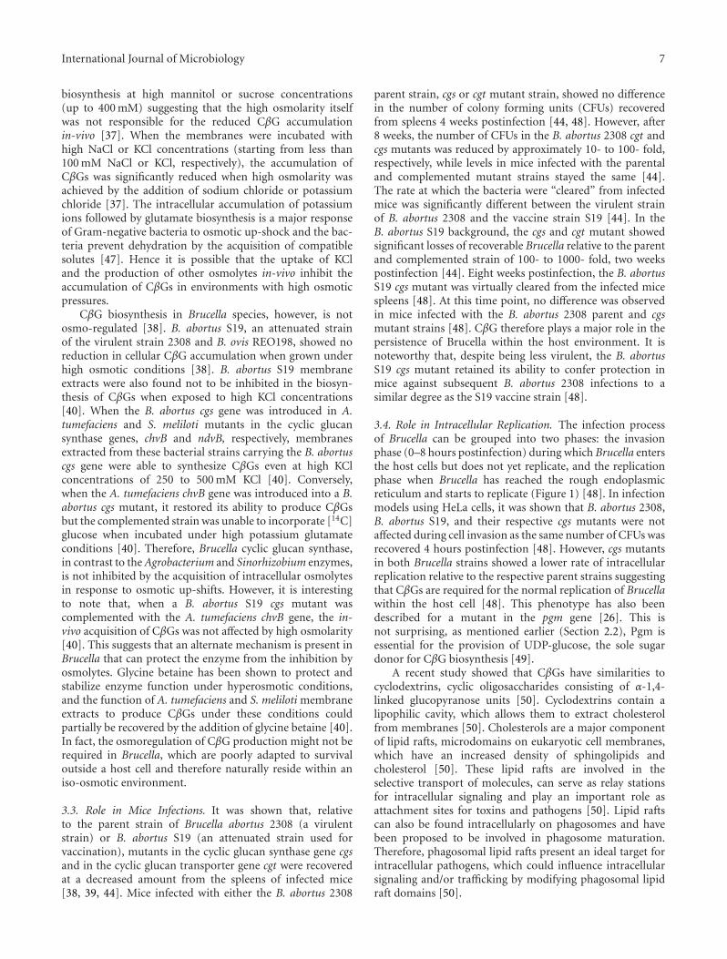
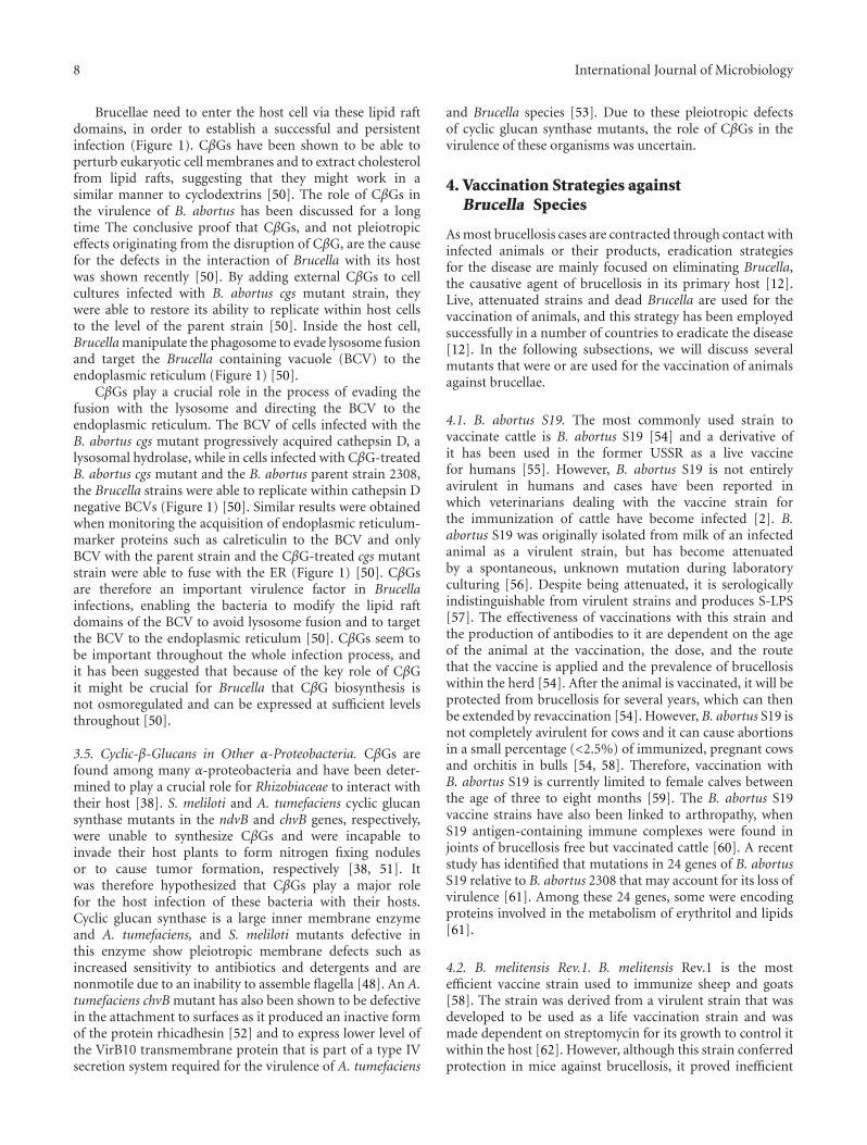
Figure 1: The Brucella-macrophage interaction. The preferred cellsinfected by Brucella are macrophages. Brucella strains with smoothLPS (S-LPS) enter the cell through interaction with lipid rafts andare then encompassed in a membrane bound compartment calledBrucella containing vacuole (BCV). This vacuole retains some lipidraft markers, targeting the BCV to the endoplasmic reticulum (ER).Brucella fuses with the ER, thus acquiring ER markers to avoidfusion with the lysosome before beginning to replicate. Rough LPSmutants do not enter the macrophage through lipid rafts and arerapidly targeted to the lysosome and killed. Mutants in the CβGbiosynthesis pathway (Δcgs and Δcgt) do not fuse with the ER butare targeted to the lysosome.
involved in core biosynthesis is located on chromosome I[20, 22–24]. The glycosyltransferase region (man Bcore &man Ccore) on chromosome II, however, was not acquiredthrough lateral gene transfer but has evolved in the organismthrough a longer period as the G + C content of these genesis similar to the rest of the Brucella genome [24].
2.2.1. Core Biosynthesis. The sugar core is linked to theO-antigen (Figure 2) and is composed of glucose, man-nose, quinovosamine, glucosamine, 3-deoxy-D-manno-2-octulosonic acid (KDO), and several yet to be identifiedsugars residues [15]. As the full biochemical composition ofthe core molecule still remains to be determined, it is not yetpossible to propose a biosynthetic pathway for the core sugarmolecule of Brucella LPS. However, several mutants havebeen identified, that were defective in the correct synthesisof the LPS core.
Phosphoglucomutase. Pgm plays a central role in convertingglucose-6-phosphate into glucose-1-phosphate, making itindispensable for the biosynthesis of many sugar moleculesin brucellae (Figure 4) [25]. The B. abortus B2211 mutantwas created by the insertion of a nonpolar gentamicincassette into the pgm gene of B. abortus 2308. A mutationin pgm causes a pleiotropic effect on the synthesis of oligo-and polysaccharides as Pgm is required for the production
of ADP-glucose, UDP-glucose, and UDP-galactose, whichthemselves serve as sugar donors for later biosyntheticprocesses [25]. The B. abortus pgm mutant lacks the O-antigen, has an increased susceptibility to complement-mediated lysis relative to its parent strain, and is attenuatedin its survival within a mouse model [26]. It is generallybelieved that the loss of the O-antigen makes bacteria moresusceptible to complement-mediated lysis [15]. In additionto this, it was found to be involved in the biosynthesis of theLPS core molecule in B. melitensis. Here, the pgm mutantalso lacked the O-antigen, but its LPS migrated lower onSDS PAGE gels suggesting a core defect (Figure 4) [20]. Thisobservation is not surprising as glucose was identified to be acomponent of the sugar core of Brucella LPS [20, 26].
Phosphomannomutase. Pmm or ManB is encoded by the B.abortus and B. melitensis pmm and manB genes, respectively,[24] and is required for the conversion of mannose-6-phosphate to mannose-1-phosphate [15]. Brucella encodestwo separate phosphomannomutase genes in its genome(Figure 3). While manB mutants in the wbk region do nothave an impact on the O-antigen and core biosynthesis,mutants in the manBcore gene lack the O-antigen and aredefective in the LPS core biosynthesis (Figures 3 and 4) [20,22–24]. The same observation has been made for the manCgenes in the wbk region, which does not seem to influenceeither O-antigen or core biosynthesis and the manCcore geneon chromosome II (Figures 3 and 4) [20, 22–24]. The manCgenes are predicted to encode mannose-1-phosphate guany-lyltransferases required for nucleotide activated mannose-1-phosphate provision (Figure 3) [20, 22–24]. It has beenhypothesized that the manBcore and manCcore genes canprovide mannose both for the biosynthesis of the O-antigenand the core molecule, whereas the manB and manC genesin the wbk region only provide mannose for the O-antigenbiosynthesis [20, 22–24]. This assumption is reasonable asthe manBcore and manCcore genes have not been acquiredby lateral gene transfer as were the majority of the genesinvolved in the O-antigen biosynthesis in the wbk region.It is therefore possible that manBcore and manCcore haveevolved primarily for the provision of mannose to the corebiosynthesis pathways but can also provide mannose for theO-antigen polymerization. Conversely, the manB and manCgenes in the wbk region have evolved to provide mannosefor the O-antigen biosynthesis and cannot compensate forthe loss of the manBcore and manCcore genes in the corebiosynthesis.
The wa∗∗ Gene. This gene encodes a putative glycosyltransferase in the Brucella genome. A mutant in the wa∗∗
gene was isolated from screening a transposon mutantlibrary for polymyxin-sensitive mutants [24]. Polymyxin isa cationic peptide that acts by perturbing membranes ofbacteria by binding to LPS and increasing the permeabilityof the cell envelope, and mutants affected in their sensitivityto polymyxin are likely to be affected in the LPS molecule[27]. The transposon insertion in the wa∗∗ mutant disruptsa putative glycosyl transferase gene, which is thought to be
4 International Journal of Microbiology
n n n n n n n
Lipopolysaccharide(LPS)
Outermembrane
Periplasm
Innermembrane
Cgm
Cgs
Cgt
O-antigen
Outer
Core
Inner
Lipid A
VLCFA
Peptidoglycan
Cyclic-β-glucan
Cyclic-β-glucan modified
with succinyl residues
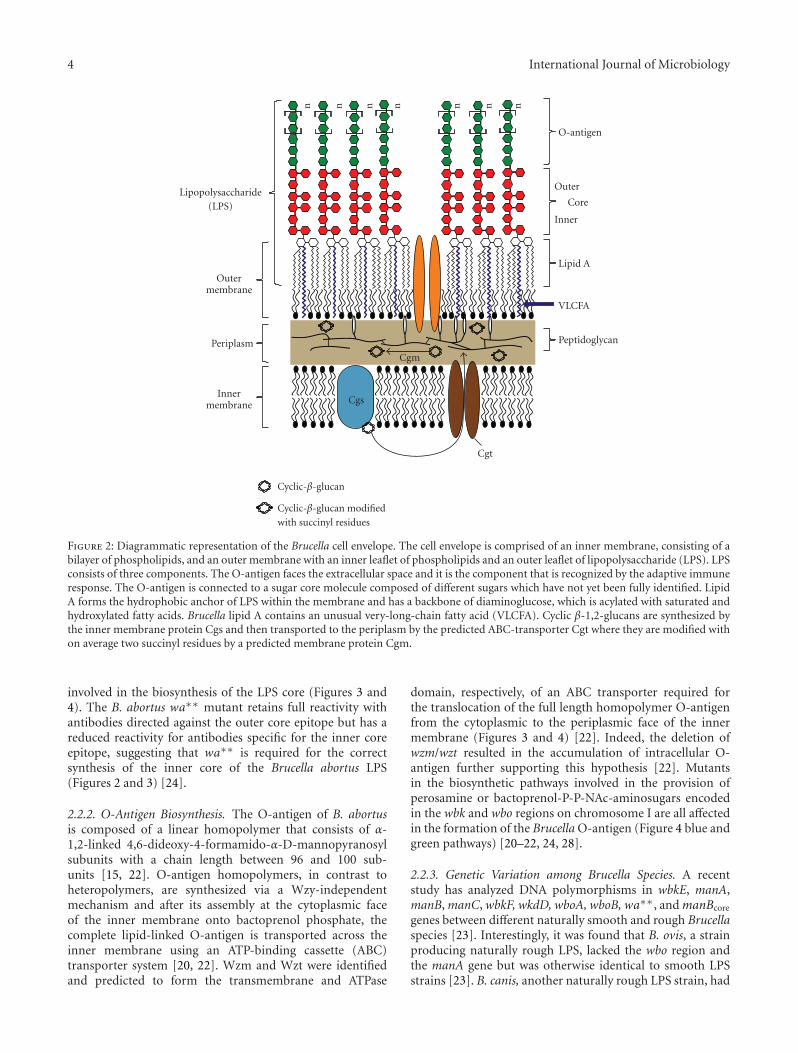
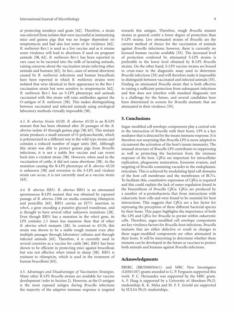
Figure 2: Diagrammatic representation of the Brucella cell envelope. The cell envelope is comprised of an inner membrane, consisting of abilayer of phospholipids, and an outer membrane with an inner leaflet of phospholipids and an outer leaflet of lipopolysaccharide (LPS). LPSconsists of three components. The O-antigen faces the extracellular space and it is the component that is recognized by the adaptive immuneresponse. The O-antigen is connected to a sugar core molecule composed of different sugars which have not yet been fully identified. LipidA forms the hydrophobic anchor of LPS within the membrane and has a backbone of diaminoglucose, which is acylated with saturated andhydroxylated fatty acids. Brucella lipid A contains an unusual very-long-chain fatty acid (VLCFA). Cyclic β-1,2-glucans are synthesized bythe inner membrane protein Cgs and then transported to the periplasm by the predicted ABC-transporter Cgt where they are modified withon average two succinyl residues by a predicted membrane protein Cgm.
involved in the biosynthesis of the LPS core (Figures 3 and4). The B. abortus wa∗∗ mutant retains full reactivity withantibodies directed against the outer core epitope but has areduced reactivity for antibodies specific for the inner coreepitope, suggesting that wa∗∗ is required for the correctsynthesis of the inner core of the Brucella abortus LPS(Figures 2 and 3) [24].
2.2.2. O-Antigen Biosynthesis. The O-antigen of B. abortusis composed of a linear homopolymer that consists of α-1,2-linked 4,6-dideoxy-4-formamido-α-D-mannopyranosylsubunits with a chain length between 96 and 100 sub-units [15, 22]. O-antigen homopolymers, in contrast toheteropolymers, are synthesized via a Wzy-independentmechanism and after its assembly at the cytoplasmic faceof the inner membrane onto bactoprenol phosphate, thecomplete lipid-linked O-antigen is transported across theinner membrane using an ATP-binding cassette (ABC)transporter system [20, 22]. Wzm and Wzt were identifiedand predicted to form the transmembrane and ATPase
domain, respectively, of an ABC transporter required forthe translocation of the full length homopolymer O-antigenfrom the cytoplasmic to the periplasmic face of the innermembrane (Figures 3 and 4) [22]. Indeed, the deletion ofwzm/wzt resulted in the accumulation of intracellular O-antigen further supporting this hypothesis [22]. Mutantsin the biosynthetic pathways involved in the provision ofperosamine or bactoprenol-P-P-NAc-aminosugars encodedin the wbk and wbo regions on chromosome I are all affectedin the formation of the Brucella O-antigen (Figure 4 blue andgreen pathways) [20–22, 24, 28].
2.2.3. Genetic Variation among Brucella Species. A recentstudy has analyzed DNA polymorphisms in wbkE, manA,manB, manC, wbkF, wkdD, wboA, wboB,wa∗∗, andmanBcore
genes between different naturally smooth and rough Brucellaspecies [23]. Interestingly, it was found that B. ovis, a strainproducing naturally rough LPS, lacked the wbo region andthe manA gene but was otherwise identical to smooth LPSstrains [23]. B. canis, another naturally rough LPS strain, had
International Journal of Microbiology 5
wbkB wbkC wbkF wbkDISs
Form
yltr
ansf
eras
e
Un
deca
pren
ylgl
ycos
yltr
ansf
eras
e
Epi
mer
ase/
deh
ydra
tase
Un
know
nfu
nct
ion
Man
nos
yltr
ansf
eras
e
wbkE
Man
nos
e-6-
phos
phat
eiso
mer
ase
wbkA gmdISsISs wzmper
Per
osam
ine
syn
thet
ase
Tra
nsm
embr
ane
com
pon
ent
wzt
AT
P-b
indi
ng
com
pon
ent
O-antigen ABC transporterWbk region
Gly
cosy
ltra
nsf
eras
e
wboAwboB
wbo region
Man
nos
yltr
ansf
eras
e
LPS core genes
Man
nos
e-1-
phos
phat
egu
anyl
yltr
ansf
eras
e
Ph
osph
oman
nom
uta
se
Man
nos
yltr
ansf
eras
e
GD
P m
ann
ose
deh
ydra
tase
Man
nos
yltr
ansf
eras
e
Ph
osph
oman
nom
uta
se
Man
nos
e-1-
phos
phat
egu
anyl
yltr
ansf
eras
e
manAOAg manCOAgmanBOAg
(pmm)
wa∗∗ manBcore manCcore
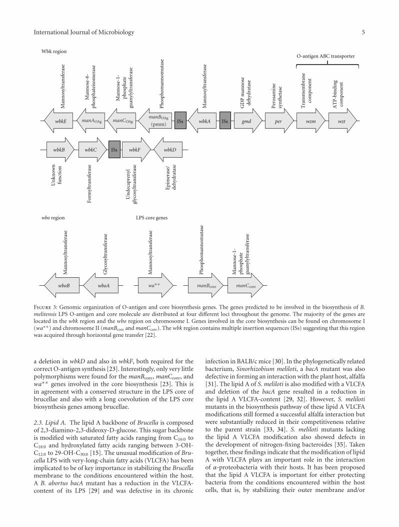
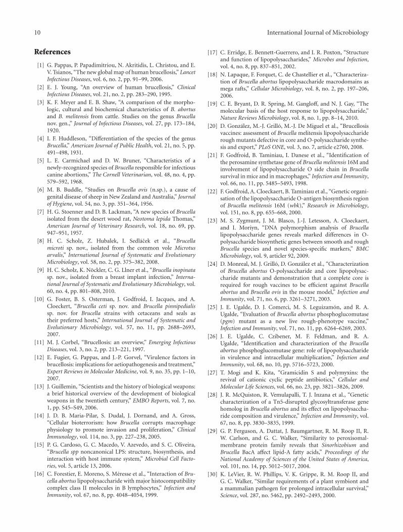
Figure 3: Genomic organization of O-antigen and core biosynthesis genes. The genes predicted to be involved in the biosynthesis of B.melitensis LPS O-antigen and core molecule are distributed at four different loci throughout the genome. The majority of the genes arelocated in the wbk region and the wbo region on chromosome I. Genes involved in the core biosynthesis can be found on chromosome I(wa∗∗) and chromosome II (manBcore and manCcore). The wbk region contains multiple insertion sequences (ISs) suggesting that this regionwas acquired through horizontal gene transfer [22].
a deletion in wbkD and also in wbkF, both required for thecorrect O-antigen synthesis [23]. Interestingly, only very littlepolymorphisms were found for the manBcore, manCcore, andwa∗∗ genes involved in the core biosynthesis [23]. This isin agreement with a conserved structure in the LPS core ofbrucellae and also with a long coevolution of the LPS corebiosynthesis genes among brucellae.
2.3. Lipid A. The lipid A backbone of Brucella is composedof 2,3-diamino-2,3-dideoxy-D-glucose. This sugar backboneis modified with saturated fatty acids ranging from C16:0 toC18:0 and hydroxylated fatty acids ranging between 3-OH-C12:0 to 29-OH-C30:0 [15]. The unusual modification of Bru-cella LPS with very-long-chain fatty acids (VLCFA) has beenimplicated to be of key importance in stabilizing the Brucellamembrane to the conditions encountered within the host.A B. abortus bacA mutant has a reduction in the VLCFA-content of its LPS [29] and was defective in its chronic
infection in BALB/c mice [30]. In the phylogenetically relatedbacterium, Sinorhizobium meliloti, a bacA mutant was alsodefective in forming an interaction with the plant host, alfalfa[31]. The lipid A of S. meliloti is also modified with a VLCFAand deletion of the bacA gene resulted in a reduction inthe lipid A VLCFA-content [29, 32]. However, S. melilotimutants in the biosynthesis pathway of these lipid A VLCFAmodifications still formed a successful alfalfa interaction butwere substantially reduced in their competitiveness relativeto the parent strain [33, 34]. S. meliloti mutants lackingthe lipid A VLCFA modification also showed defects inthe development of nitrogen-fixing bacteroides [35]. Takentogether, these findings indicate that the modification of lipidA with VLCFA plays an important role in the interactionof α-proteobacteria with their hosts. It has been proposedthat the lipid A VLCFA is important for either protectingbacteria from the conditions encountered within the hostcells, that is, by stabilizing their outer membrane and/or
6 International Journal of Microbiology
GlucoseGlycolysis
Mannose-6-phosphate
Mannose-1-phosphate
GDP-mannose
GDP-4-keto-6-deoxy-mannose
UDP-NAc-quinovosamine
Bactoprenol-P-P-NAc-aminosugar
UDP-glucose
Wzt
Wzm
ATP
ADP
Gmd Per
N-formylperosamine
WbkC
Fructose-6-phosphate
NAc-glucosamine
WbkD WbkF
Pgm
WaaL
OAg OAg
OAg OAg
OAg
OAg
OAg
MsbA
Core Core
Core
Core
Inner membrane
Outer membrane
Periplasm
Cytoplasm
WbkAWbkEWboAWboB
ManAOAg ManBOAg ManCOAg
Wa∗∗
ManBcore ManCcore
GDP-4-NH2-4,6-dideoxymannose
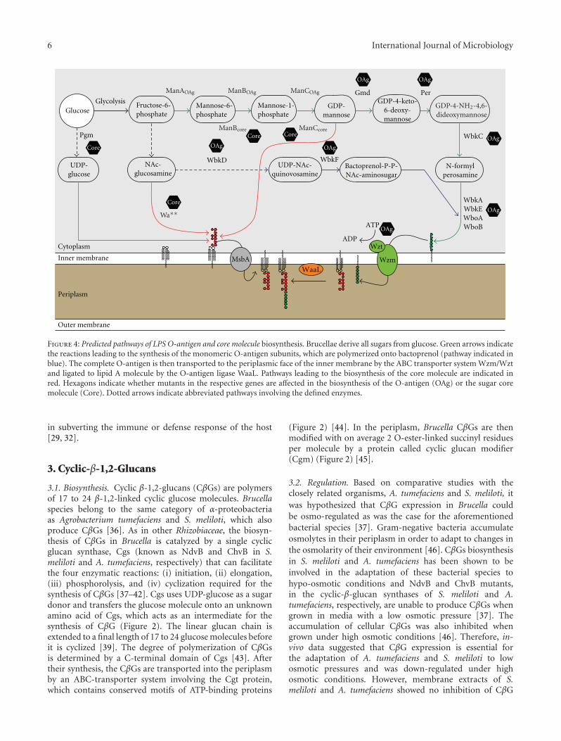
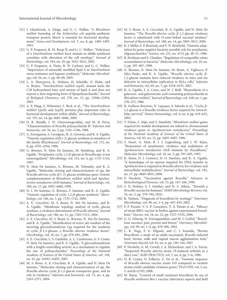
Figure 4: Predicted pathways of LPS O-antigen and core molecule biosynthesis. Brucellae derive all sugars from glucose. Green arrows indicatethe reactions leading to the synthesis of the monomeric O-antigen subunits, which are polymerized onto bactoprenol (pathway indicated inblue). The complete O-antigen is then transported to the periplasmic face of the inner membrane by the ABC transporter system Wzm/Wztand ligated to lipid A molecule by the O-antigen ligase WaaL. Pathways leading to the biosynthesis of the core molecule are indicated inred. Hexagons indicate whether mutants in the respective genes are affected in the biosynthesis of the O-antigen (OAg) or the sugar coremolecule (Core). Dotted arrows indicate abbreviated pathways involving the defined enzymes.
in subverting the immune or defense response of the host[29, 32].
3. Cyclic-β-1,2-Glucans
3.1. Biosynthesis. Cyclic β-1,2-glucans (CβGs) are polymersof 17 to 24 β-1,2-linked cyclic glucose molecules. Brucellaspecies belong to the same category of α-proteobacteriaas Agrobacterium tumefaciens and S. meliloti, which alsoproduce CβGs [36]. As in other Rhizobiaceae, the biosyn-thesis of CβGs in Brucella is catalyzed by a single cyclicglucan synthase, Cgs (known as NdvB and ChvB in S.meliloti and A. tumefaciens, respectively) that can facilitatethe four enzymatic reactions: (i) initiation, (ii) elongation,(iii) phosphorolysis, and (iv) cyclization required for thesynthesis of CβGs [37–42]. Cgs uses UDP-glucose as a sugardonor and transfers the glucose molecule onto an unknownamino acid of Cgs, which acts as an intermediate for thesynthesis of CβG (Figure 2). The linear glucan chain isextended to a final length of 17 to 24 glucose molecules beforeit is cyclized [39]. The degree of polymerization of CβGsis determined by a C-terminal domain of Cgs [43]. Aftertheir synthesis, the CβGs are transported into the periplasmby an ABC-transporter system involving the Cgt protein,which contains conserved motifs of ATP-binding proteins
(Figure 2) [44]. In the periplasm, Brucella CβGs are thenmodified with on average 2 O-ester-linked succinyl residuesper molecule by a protein called cyclic glucan modifier(Cgm) (Figure 2) [45].
3.2. Regulation. Based on comparative studies with theclosely related organisms, A. tumefaciens and S. meliloti, itwas hypothesized that CβG expression in Brucella couldbe osmo-regulated as was the case for the aforementionedbacterial species [37]. Gram-negative bacteria accumulateosmolytes in their periplasm in order to adapt to changes inthe osmolarity of their environment [46]. CβGs biosynthesisin S. meliloti and A. tumefaciens has been shown to beinvolved in the adaptation of these bacterial species tohypo-osmotic conditions and NdvB and ChvB mutants,in the cyclic-β-glucan synthases of S. meliloti and A.tumefaciens, respectively, are unable to produce CβGs whengrown in media with a low osmotic pressure [37]. Theaccumulation of cellular CβGs was also inhibited whengrown under high osmotic conditions [46]. Therefore, in-vivo data suggested that CβG expression is essential forthe adaptation of A. tumefaciens and S. meliloti to lowosmotic pressures and was down-regulated under highosmotic conditions. However, membrane extracts of S.meliloti and A. tumefaciens showed no inhibition of CβG
International Journal of Microbiology 7
biosynthesis at high mannitol or sucrose concentrations(up to 400 mM) suggesting that the high osmolarity itselfwas not responsible for the reduced CβG accumulationin-vivo [37]. When the membranes were incubated withhigh NaCl or KCl concentrations (starting from less than100 mM NaCl or KCl, respectively), the accumulation ofCβGs was significantly reduced when high osmolarity wasachieved by the addition of sodium chloride or potassiumchloride [37]. The intracellular accumulation of potassiumions followed by glutamate biosynthesis is a major responseof Gram-negative bacteria to osmotic up-shock and the bac-teria prevent dehydration by the acquisition of compatiblesolutes [47]. Hence it is possible that the uptake of KCland the production of other osmolytes in-vivo inhibit theaccumulation of CβGs in environments with high osmoticpressures.
CβG biosynthesis in Brucella species, however, is notosmo-regulated [38]. B. abortus S19, an attenuated strainof the virulent strain 2308 and B. ovis REO198, showed noreduction in cellular CβG accumulation when grown underhigh osmotic conditions [38]. B. abortus S19 membraneextracts were also found not to be inhibited in the biosyn-thesis of CβGs when exposed to high KCl concentrations[40]. When the B. abortus cgs gene was introduced in A.tumefaciens and S. meliloti mutants in the cyclic glucansynthase genes, chvB and ndvB, respectively, membranesextracted from these bacterial strains carrying the B. abortuscgs gene were able to synthesize CβGs even at high KClconcentrations of 250 to 500 mM KCl [40]. Conversely,when the A. tumefaciens chvB gene was introduced into a B.abortus cgs mutant, it restored its ability to produce CβGsbut the complemented strain was unable to incorporate [14C]glucose when incubated under high potassium glutamateconditions [40]. Therefore, Brucella cyclic glucan synthase,in contrast to the Agrobacterium and Sinorhizobium enzymes,is not inhibited by the acquisition of intracellular osmolytesin response to osmotic up-shifts. However, it is interestingto note that, when a B. abortus S19 cgs mutant wascomplemented with the A. tumefaciens chvB gene, the in-vivo acquisition of CβGs was not affected by high osmolarity[40]. This suggests that an alternate mechanism is present inBrucella that can protect the enzyme from the inhibition byosmolytes. Glycine betaine has been shown to protect andstabilize enzyme function under hyperosmotic conditions,and the function of A. tumefaciens and S. meliloti membraneextracts to produce CβGs under these conditions couldpartially be recovered by the addition of glycine betaine [40].In fact, the osmoregulation of CβG production might not berequired in Brucella, which are poorly adapted to survivaloutside a host cell and therefore naturally reside within aniso-osmotic environment.
3.3. Role in Mice Infections. It was shown that, relativeto the parent strain of Brucella abortus 2308 (a virulentstrain) or B. abortus S19 (an attenuated strain used forvaccination), mutants in the cyclic glucan synthase gene cgsand in the cyclic glucan transporter gene cgt were recoveredat a decreased amount from the spleens of infected mice[38, 39, 44]. Mice infected with either the B. abortus 2308
parent strain, cgs or cgt mutant strain, showed no differencein the number of colony forming units (CFUs) recoveredfrom spleens 4 weeks postinfection [44, 48]. However, after8 weeks, the number of CFUs in the B. abortus 2308 cgt andcgs mutants was reduced by approximately 10- to 100- fold,respectively, while levels in mice infected with the parentaland complemented mutant strains stayed the same [44].The rate at which the bacteria were “cleared” from infectedmice was significantly different between the virulent strainof B. abortus 2308 and the vaccine strain S19 [44]. In theB. abortus S19 background, the cgs and cgt mutant showedsignificant losses of recoverable Brucella relative to the parentand complemented strain of 100- to 1000- fold, two weekspostinfection [44]. Eight weeks postinfection, the B. abortusS19 cgs mutant was virtually cleared from the infected micespleens [48]. At this time point, no difference was observedin mice infected with the B. abortus 2308 parent and cgsmutant strains [48]. CβG therefore plays a major role in thepersistence of Brucella within the host environment. It isnoteworthy that, despite being less virulent, the B. abortusS19 cgs mutant retained its ability to confer protection inmice against subsequent B. abortus 2308 infections to asimilar degree as the S19 vaccine strain [48].
3.4. Role in Intracellular Replication. The infection processof Brucella can be grouped into two phases: the invasionphase (0–8 hours postinfection) during which Brucella entersthe host cells but does not yet replicate, and the replicationphase when Brucella has reached the rough endoplasmicreticulum and starts to replicate (Figure 1) [48]. In infectionmodels using HeLa cells, it was shown that B. abortus 2308,B. abortus S19, and their respective cgs mutants were notaffected during cell invasion as the same number of CFUs wasrecovered 4 hours postinfection [48]. However, cgs mutantsin both Brucella strains showed a lower rate of intracellularreplication relative to the respective parent strains suggestingthat CβGs are required for the normal replication of Brucellawithin the host cell [48]. This phenotype has also beendescribed for a mutant in the pgm gene [26]. This isnot surprising, as mentioned earlier (Section 2.2), Pgm isessential for the provision of UDP-glucose, the sole sugardonor for CβG biosynthesis [49].
A recent study showed that CβGs have similarities tocyclodextrins, cyclic oligosaccharides consisting of α-1,4-linked glucopyranose units [50]. Cyclodextrins contain alipophilic cavity, which allows them to extract cholesterolfrom membranes [50]. Cholesterols are a major componentof lipid rafts, microdomains on eukaryotic cell membranes,which have an increased density of sphingolipids andcholesterol [50]. These lipid rafts are involved in theselective transport of molecules, can serve as relay stationsfor intracellular signaling and play an important role asattachment sites for toxins and pathogens [50]. Lipid raftscan also be found intracellularly on phagosomes and havebeen proposed to be involved in phagosome maturation.Therefore, phagosomal lipid rafts present an ideal target forintracellular pathogens, which could influence intracellularsignaling and/or trafficking by modifying phagosomal lipidraft domains [50].
8 International Journal of Microbiology
Brucellae need to enter the host cell via these lipid raftdomains, in order to establish a successful and persistentinfection (Figure 1). CβGs have been shown to be able toperturb eukaryotic cell membranes and to extract cholesterolfrom lipid rafts, suggesting that they might work in asimilar manner to cyclodextrins [50]. The role of CβGs inthe virulence of B. abortus has been discussed for a longtime The conclusive proof that CβGs, and not pleiotropiceffects originating from the disruption of CβG, are the causefor the defects in the interaction of Brucella with its hostwas shown recently [50]. By adding external CβGs to cellcultures infected with B. abortus cgs mutant strain, theywere able to restore its ability to replicate within host cellsto the level of the parent strain [50]. Inside the host cell,Brucella manipulate the phagosome to evade lysosome fusionand target the Brucella containing vacuole (BCV) to theendoplasmic reticulum (Figure 1) [50].
CβGs play a crucial role in the process of evading thefusion with the lysosome and directing the BCV to theendoplasmic reticulum. The BCV of cells infected with theB. abortus cgs mutant progressively acquired cathepsin D, alysosomal hydrolase, while in cells infected with CβG-treatedB. abortus cgs mutant and the B. abortus parent strain 2308,the Brucella strains were able to replicate within cathepsin Dnegative BCVs (Figure 1) [50]. Similar results were obtainedwhen monitoring the acquisition of endoplasmic reticulum-marker proteins such as calreticulin to the BCV and onlyBCV with the parent strain and the CβG-treated cgs mutantstrain were able to fuse with the ER (Figure 1) [50]. CβGsare therefore an important virulence factor in Brucellainfections, enabling the bacteria to modify the lipid raftdomains of the BCV to avoid lysosome fusion and to targetthe BCV to the endoplasmic reticulum [50]. CβGs seem tobe important throughout the whole infection process, andit has been suggested that because of the key role of CβGit might be crucial for Brucella that CβG biosynthesis isnot osmoregulated and can be expressed at sufficient levelsthroughout [50].
3.5. Cyclic-β-Glucans in Other α-Proteobacteria. CβGs arefound among many α-proteobacteria and have been deter-mined to play a crucial role for Rhizobiaceae to interact withtheir host [38]. S. meliloti and A. tumefaciens cyclic glucansynthase mutants in the ndvB and chvB genes, respectively,were unable to synthesize CβGs and were incapable toinvade their host plants to form nitrogen fixing nodulesor to cause tumor formation, respectively [38, 51]. Itwas therefore hypothesized that CβGs play a major rolefor the host infection of these bacteria with their hosts.Cyclic glucan synthase is a large inner membrane enzymeand A. tumefaciens, and S. meliloti mutants defective inthis enzyme show pleiotropic membrane defects such asincreased sensitivity to antibiotics and detergents and arenonmotile due to an inability to assemble flagella [48]. An A.tumefaciens chvB mutant has also been shown to be defectivein the attachment to surfaces as it produced an inactive formof the protein rhicadhesin [52] and to express lower level ofthe VirB10 transmembrane protein that is part of a type IVsecretion system required for the virulence of A. tumefaciens
and Brucella species [53]. Due to these pleiotropic defectsof cyclic glucan synthase mutants, the role of CβGs in thevirulence of these organisms was uncertain.
4. Vaccination Strategies againstBrucella Species
As most brucellosis cases are contracted through contact withinfected animals or their products, eradication strategiesfor the disease are mainly focused on eliminating Brucella,the causative agent of brucellosis in its primary host [12].Live, attenuated strains and dead Brucella are used for thevaccination of animals, and this strategy has been employedsuccessfully in a number of countries to eradicate the disease[12]. In the following subsections, we will discuss severalmutants that were or are used for the vaccination of animalsagainst brucellae.
4.1. B. abortus S19. The most commonly used strain tovaccinate cattle is B. abortus S19 [54] and a derivative ofit has been used in the former USSR as a live vaccinefor humans [55]. However, B. abortus S19 is not entirelyavirulent in humans and cases have been reported inwhich veterinarians dealing with the vaccine strain forthe immunization of cattle have become infected [2]. B.abortus S19 was originally isolated from milk of an infectedanimal as a virulent strain, but has become attenuatedby a spontaneous, unknown mutation during laboratoryculturing [56]. Despite being attenuated, it is serologicallyindistinguishable from virulent strains and produces S-LPS[57]. The effectiveness of vaccinations with this strain andthe production of antibodies to it are dependent on the ageof the animal at the vaccination, the dose, and the routethat the vaccine is applied and the prevalence of brucellosiswithin the herd [54]. After the animal is vaccinated, it will beprotected from brucellosis for several years, which can thenbe extended by revaccination [54]. However, B. abortus S19 isnot completely avirulent for cows and it can cause abortionsin a small percentage (<2.5%) of immunized, pregnant cowsand orchitis in bulls [54, 58]. Therefore, vaccination withB. abortus S19 is currently limited to female calves betweenthe age of three to eight months [59]. The B. abortus S19vaccine strains have also been linked to arthropathy, whenS19 antigen-containing immune complexes were found injoints of brucellosis free but vaccinated cattle [60]. A recentstudy has identified that mutations in 24 genes of B. abortusS19 relative to B. abortus 2308 that may account for its loss ofvirulence [61]. Among these 24 genes, some were encodingproteins involved in the metabolism of erythritol and lipids[61].
4.2. B. melitensis Rev.1. B. melitensis Rev.1 is the mostefficient vaccine strain used to immunize sheep and goats[58]. The strain was derived from a virulent strain that wasdeveloped to be used as a life vaccination strain and wasmade dependent on streptomycin for its growth to control itwithin the host [62]. However, although this strain conferredprotection in mice against brucellosis, it proved inefficient
International Journal of Microbiology 9
at protecting monkeys and goats [62]. Therefore, a strainwas selected from isolates that were successful at immunizingmice and guinea pigs that was no longer dependent onstreptomycin and had also lost some of its virulence [62].B. melitensis Rev.1 is used as a live vaccine and as it retainssome virulence will lead to abortions if used on pregnantanimals [58, 62]. B. melitensis Rev.1 has been reported inrare cases to be excreted into the milk of lactating animals,raising concerns about the vaccination strain infecting otheranimals and humans [62]. In fact, cases of animal abortionscaused by B. melitensis infections and human brucellosishave been reported in which B. melitensis strains wereisolated that were identical in their appearance to the Rev.1vaccination strain but were sensitive to streptomycin [62].B. melitensis Rev.1 has an S-LPS phenotype and animalsvaccinated with this strain will raise antibodies against theO-antigen of B. melitensis [58]. This makes distinguishingbetween vaccinated and infected animals using serologicallaboratory methods virtually impossible [58].
4.3. B. abortus Strain 45/20. B. abortus 45/20 is an R-LPSmutant that has been obtained after 20 passages of the B.abortus isolate 45 through guinea pigs [58, 63]. This mutantstrain produces a small amount of O-polysaccharide, whichis polymerized in a different way to the wild type strains andcontains a reduced number of sugar units [64]. Althoughthis strain was able to protect guinea pigs from Brucellainfections, it is not a stable live vaccine and can revertback into a virulent strain [58]. However, when used in thevaccination of cattle, it did not cause abortions [58]. As themutation causing the R-LPS phenotype of B. abortus 45/20is unknown [58] and reversion to the S-LPS and virulentstrain can occur, it is not currently used as a vaccine strain[63].
4.4. B. abortus RB51. B. abortus RB51 is an attenuatedspontaneous R-LPS mutant that was obtained by repeatedpassage of B. abortus 2308 on media containing rifampicinand penicillin [65]. RB51 carries an IS771 insertion inwboA, a gene encoding a putative glycosyl transferase, andis thought to have several other unknown mutations [28].Even though RB51 has a mutation in the wboA gene, itsLPS contains 2.5 times less mannose than that of otherB. abortus wboA mutants [28]. In contrast to 45/20, thisstrain was shown to be a stable rough mutant even aftermultiple passages through laboratory cultures and throughinfected animals [65]. Therefore, it is currently used inseveral countries as a vaccine for cattle [66]. RB51 has beenshown to be efficient in protecting mice against brucellosisbut was not effective when tested in sheep [58]. RB51 isresistant to rifampicin, which is used in the treatment ofhuman brucellosis [63].
4.5. Advantages and Disadvantages of Vaccination Strategies.Many other R-LPS Brucella strains are available for vaccinedevelopment (refer to Section 2). However, as the O-antigenis the most exposed antigen during Brucella infectionsthe majority of the adaptive immune response is targeted
towards this antigen. Therefore, rough Brucella mutantstrains in general confer a lower degree of protection thanS-LPS strains. Live attenuated strains of Brucella are thecurrent method of choice for the vaccination of animalsagainst Brucella infections; however, there is currently nolicensed human vaccine available [55]. The increased levelof protection conferred by attenuated S-LPS strains ispreferable to the lower level obtained by R-LPS Brucellastrains. On the other hand, S-LPS vaccine strains are boundto cross-react in the diagnostic assay used to determineBrucella infections [55] and will therefore make it impossibleto distinguish between vaccinated and infected animals [55].Finding an attenuated Brucella strain that is both effectivein raising a sufficient protection from subsequent infectionsand that does not interfere with standard diagnostic testis a challenge for the future, and several candidates havebeen determined in screens for Brucella mutants that areattenuated in their virulence [55].
5. Conclusions
Sugar-modified cell envelope components play a central rolein the interaction of Brucella with their hosts. LPS is a keymediator that is detected by the innate immune response. It istherefore not surprising that Brucella LPS is more adapted tocircumvent the activation of the host’s innate immunity. Theunusual structure of Brucella LPS contributes to suppressingas well as protecting the bacterium from the immuneresponse of the host. CβGs are important for intracellularreplication, phagosome maturation, lysosome evasion, andtargeting of Brucella containing vacuoles to the endoplasmicreticulum. This is achieved by modulating lipid raft domainsof the host cell membrane and the membranes of BCVs.To facilitate this, constitutive expression of CβGs is requiredand this could explain the lack of osmo-regulation found inthe biosynthesis of Brucella CβGs. CβGs are produced bya number of α-proteobacteria that form interactions witheukaryotic host cells and were found to be essential for hostinteractions. This suggests that CβGs are a key factor forrepressing the perception of these different bacterial speciesby their hosts. This paper highlights the importance of boththe LPS and CβGs for Brucella to persist within eukaryoticcells. Therefore, sugar-modified cell envelope componentsare key virulence factors for Brucella-host infections. Brucellamutants that are either defective or result in changes tothese sugar-modified components are often attenuated intheir hosts. It will be interesting to determine whether thesemutants can be developed in the future as vaccines to protectboth animals and humans against Brucella infections.
Acknowledgments
BBSRC (BB/D000564/1) and MRC New Investigator(G0501107) grants awarded to G. P. Ferguson supported thiswork. P. C. Hernandez was supported by the MRC grant.A. F. Haag is supported by a University of Aberdeen Ph.D.studentship; K. K. Myka and M. F. F. Arnold are supportedby SULSA Ph.D. studentships.
10 International Journal of Microbiology
References
[1] G. Pappas, P. Papadimitriou, N. Akritidis, L. Christou, and E.V. Tsianos, “The new global map of human brucellosis,” LancetInfectious Diseases, vol. 6, no. 2, pp. 91–99, 2006.
[2] E. J. Young, “An overview of human brucellosis,” ClinicalInfectious Diseases, vol. 21, no. 2, pp. 283–290, 1995.
[3] K. F. Meyer and E. B. Shaw, “A comparison of the morpho-logic, cultural and biochemical characteristics of B. abortusand B. melitensis from cattle. Studies on the genus Brucellanov. gen.,” Journal of Infectious Diseases, vol. 27, pp. 173–184,1920.
[4] I. F. Huddleson, “Differentiation of the species of the genusBrucella,” American Journal of Public Health, vol. 21, no. 5, pp.491–498, 1931.
[5] L. E. Carmichael and D. W. Bruner, “Characteristics of anewly-recognized species of Brucella responsible for infectiouscanine abortions,” The Cornell Veterinarian, vol. 48, no. 4, pp.579–592, 1968.
[6] M. B. Buddle, “Studies on Brucella ovis (n.sp.), a cause ofgenital disease of sheep in New Zealand and Australia,” Journalof Hygiene, vol. 54, no. 3, pp. 351–364, 1956.
[7] H. G. Stoenner and D. B. Lackman, “A new species of Brucellaisolated from the desert wood rat, Neotoma lepida Thomas,”American Journal of Veterinary Research, vol. 18, no. 69, pp.947–951, 1957.
[8] H. C. Scholz, Z. Hubalek, I. Sedlacek et al., “Brucellamicroti sp. nov., isolated from the common vole Microtusarvalis,” International Journal of Systematic and EvolutionaryMicrobiology, vol. 58, no. 2, pp. 375–382, 2008.
[9] H. C. Scholz, K. Nockler, C. G. Llner et al., “Brucella inopinatasp. nov., isolated from a breast implant infection,” Interna-tional Journal of Systematic and Evolutionary Microbiology, vol.60, no. 4, pp. 801–808, 2010.
[10] G. Foster, B. S. Osterman, J. Godfroid, I. Jacques, and A.Cloeckert, “Brucella ceti sp. nov. and Brucella pinnipedialissp. nov. for Brucella strains with cetaceans and seals astheir preferred hosts,” International Journal of Systematic andEvolutionary Microbiology, vol. 57, no. 11, pp. 2688–2693,2007.
[11] M. J. Corbel, “Brucellosis: an overview,” Emerging InfectiousDiseases, vol. 3, no. 2, pp. 213–221, 1997.
[12] E. Fugier, G. Pappas, and J.-P. Gorvel, “Virulence factors inbrucellosis: implications for aetiopathogenesis and treatment,”Expert Reviews in Molecular Medicine, vol. 9, no. 35, pp. 1–10,2007.
[13] J. Guillemin, “Scientists and the history of biological weapons:a brief historical overview of the development of biologicalweapons in the twentieth century,” EMBO Reports, vol. 7, no.1, pp. S45–S49, 2006.
[14] J. D. B. Maria-Pilar, S. Dudal, J. Dornand, and A. Gross,“Cellular bioterrorism: how Brucella corrupts macrophagephysiology to promote invasion and proliferation,” ClinicalImmunology, vol. 114, no. 3, pp. 227–238, 2005.
[15] P. G. Cardoso, G. C. Macedo, V. Azevedo, and S. C. Oliveira,“Brucella spp noncanonical LPS: structure, biosynthesis, andinteraction with host immune system,” Microbial Cell Facto-ries, vol. 5, article 13, 2006.
[16] C. Forestier, E. Moreno, S. Meresse et al., “Interaction of Bru-cella abortus lipopolysaccharide with major histocompatibilitycomplex class II molecules in B lymphocytes,” Infection andImmunity, vol. 67, no. 8, pp. 4048–4054, 1999.
[17] C. Erridge, E. Bennett-Guerrero, and I. R. Poxton, “Structureand function of lipopolysaccharides,” Microbes and Infection,vol. 4, no. 8, pp. 837–851, 2002.
[18] N. Lapaque, F. Forquet, C. de Chastellier et al., “Characteriza-tion of Brucella abortus lipopolysaccharide macrodomains asmega rafts,” Cellular Microbiology, vol. 8, no. 2, pp. 197–206,2006.
[19] C. E. Bryant, D. R. Spring, M. Gangloff, and N. J. Gay, “Themolecular basis of the host response to lipopolysaccharide,”Nature Reviews Microbiology, vol. 8, no. 1, pp. 8–14, 2010.
[20] D. Gonzalez, M.-J. Grillo, M.-J. De Miguel et al., “Brucellosisvaccines: assessment of Brucella melitensis lipopolysacchariderough mutants defective in core and O-polysaccharide synthe-sis and export,” PLoS ONE, vol. 3, no. 7, article e2760, 2008.
[21] F. Godfroid, B. Taminiau, I. Danese et al., “Identification ofthe perosamine synthetase gene of Brucella melitensis 16M andinvolvement of lipopolysaccharide O side chain in Brucellasurvival in mice and in macrophages,” Infection and Immunity,vol. 66, no. 11, pp. 5485–5493, 1998.
[22] F. Godfroid, A. Cloeckaert, B. Taminiau et al., “Genetic organi-sation of the lipopolysaccharide O-antigen biosynthesis regionof Brucella melitensis 16M (wbk),” Research in Microbiology,vol. 151, no. 8, pp. 655–668, 2000.
[23] M. S. Zygmunt, J. M. Blasco, J.-J. Letesson, A. Cloeckaert,and I. Moriyn, “DNA polymorphism analysis of Brucellalipopolysaccharide genes reveals marked differences in O-polysaccharide biosynthetic genes between smooth and roughBrucella species and novel species-specific markers,” BMCMicrobiology, vol. 9, articler 92, 2009.
[24] D. Monreal, M. J. Grillo, D. Gonzalez et al., “Characterizationof Brucella abortus O-polysaccharide and core lipopolysac-charide mutants and demonstration that a complete core isrequired for rough vaccines to be efficient against Brucellaabortus and Brucella ovis in the mouse model,” Infection andImmunity, vol. 71, no. 6, pp. 3261–3271, 2003.
[25] J. E. Ugalde, D. J. Comerci, M. S. Leguizamon, and R. A.Ugalde, “Evaluation of Brucella abortus phosphoglucomutase(pgm) mutant as a new live rough-phenotype vaccine,”Infection and Immunity, vol. 71, no. 11, pp. 6264–6269, 2003.
[26] J. E. Ugalde, C. Czibener, M. F. Feldman, and R. A.Ugalde, “Identification and characterization of the Brucellaabortus phosphoglucomutase gene: role of lipopolysaccharidein virulence and intracellular multiplication,” Infection andImmunity, vol. 68, no. 10, pp. 5716–5723, 2000.
[27] T. Mogi and K. Kita, “Gramicidin S and polymyxins: therevival of cationic cyclic peptide antibiotics,” Cellular andMolecular Life Sciences, vol. 66, no. 23, pp. 3821–3826, 2009.
[28] J. R. McQuiston, R. Vemulapalli, T. J. Inzana et al., “Geneticcharacterization of a Tn5-disrupted glycosyltransferase genehomolog in Brucella abortus and its effect on lipopolysaccha-ride composition and virulence,” Infection and Immunity, vol.67, no. 8, pp. 3830–3835, 1999.
[29] G. P. Ferguson, A. Dattat, J. Baumgartner, R. M. Roop II, R.W. Carlson, and G. C. Walker, “Similarity to peroxisomal-membrane protein family reveals that Sinorhizobium andBrucella BacA affect lipid-A fatty acids,” Proceedings of theNational Academy of Sciences of the United States of America,vol. 101, no. 14, pp. 5012–5017, 2004.
[30] K. LeVier, R. W. Phillips, V. K. Grippe, R. M. Roop II, andG. C. Walker, “Similar requirements of a plant symbiont anda mammalian pathogen for prolonged intracellular survival,”Science, vol. 287, no. 5462, pp. 2492–2493, 2000.
International Journal of Microbiology 11
[31] J. Glazebrook, A. Ichige, and G. C. Walker, “A Rhizobiummeliloti homolog of the Escherichia coli peptide-antibiotictransport protein SbmA is essential for bacteroid develop-ment,” Genes and Development, vol. 7, no. 8, pp. 1485–1497,1993.
[32] G. P. Ferguson, R. M. Roop II, and G. C. Walker, “Deficiencyof a Sinorhizobium meliloti bacA mutant in alfalfa symbiosiscorrelates with alteration of the cell envelope,” Journal ofBacteriology, vol. 184, no. 20, pp. 5625–5632, 2002.
[33] G. P. Ferguson, A. Datta, R. W. Carlson, and G. C. Walker,“Importance of unusually modified lipid A in Sinorhizobiumstress resistance and legume symbiosis,” Molecular Microbiol-ogy, vol. 56, no. 1, pp. 68–80, 2005.
[34] L. A. Sharypova, K. Niehaus, H. Scheidle, O. Holst, andA. Becker, “Sinorhizobium meliloti acpXL mutant lacks theC28 hydroxylated fatty acid moiety of lipid A and does notexpress a slow migrating form of lipopolysaccharide,” Journalof Biological Chemistry, vol. 278, no. 15, pp. 12946–12954,2003.
[35] A. F. Haag, S. Wehmeier, S. Beck et al., “The Sinorhizobiummeliloti LpxXL and AcpXL proteins play important roles inbacteroid development within alfalfa,” Journal of Bacteriology,vol. 191, no. 14, pp. 4681–4686, 2009.
[36] D. R. Bundle, J. W. Cherwonogrodzky, and M. B. Perry,“Characterization of Brucella polysaccharide B,” Infection andImmunity, vol. 56, no. 5, pp. 1101–1106, 1988.
[37] A. Zorreguieta, S. Cavaignac, R. A. Geremia, and R. A. Ugalde,“Osmotic regulation of β(1-2) glucan synthesis in members ofthe family Rhizobiaceae,” Journal of Bacteriology, vol. 172, no.8, pp. 4701–4704, 1990.
[38] G. Briones, N. Inon De Iannino, M. Steinberg, and R. A.Ugalde, “Periplasmic cyclic 1,2-β-glucan in Brucella spp. is notosmoregulated,” Microbiology, vol. 143, no. 4, pp. 1115–1124,1997.
[39] N. Inon De Iannino, G. Briones, M. Tolmasky, and R. A.Ugalde, “Molecular cloning and characterization of cgs, theBrucella abortus cyclic β(1-2) glucan synthetase gene: Geneticcomplementation of Rhizobium meliloti ndvB and Agrobac-terium tumefaciens chvB mutants,” Journal of Bacteriology, vol.180, no. 17, pp. 4392–4400, 1998.
[40] N. I. De Iannino, G. Briones, F. Iannino, and R. A. Ugalde,“Osmotic regulation of cyclic 1,2-β-glucan synthesis,” Micro-biology, vol. 146, no. 7, pp. 1735–1742, 2000.
[41] A. E. Ciocchini, M. S. Roset, N. Ino De Iannino, and R.A. Ugalde, “Membrane topology analysis of cyclic glucansynthase, a virulence determinant of Brucella abortus,” Journalof Bacteriology, vol. 186, no. 21, pp. 7205–7213, 2004.
[42] A. E. Ciocchini, M. S. Roset, G. Briones, N. Ino De Iannino,and R. A. Ugalde, “Identification of active site residues of theinverting glycosyltransferase Cgs required for the synthesisof cyclic β-1,2-glucan, a Brucella abortus virulence factor,”Glycobiology, vol. 16, no. 7, pp. 679–691, 2006.
[43] A. E. Ciocchini, L. S. Guidolin, A. C. Casabuono, A. S. Couto,N. Inon De Iannino, and R. A. Ugalde, “A glycosyltransferasewith a length-controlling activity as a mechanism to regulatethe size of polysaccharides,” Proceedings of the NationalAcademy of Sciences of the United States of America, vol. 104,no. 42, pp. 16492–16497, 2007.
[44] M. S. Roset, A. E. Ciocchini, R. A. Ugalde, and N. Inon DeIannino, “Molecular cloning and characterization of cgt, theBrucella abortus cyclic β-1,2-glucan transporter gene, and itsrole in virulence,” Infection and Immunity, vol. 72, no. 4, pp.2263–2271, 2004.
[45] M. S. Roset, A. E. Ciocchini, R. A. Ugalde, and N. Inon DeIannino, “The Brucella abortus cyclic β-1,2-glucan virulencefactor is substituted with O-ester-linked succinyl residues,”Journal of Bacteriology, vol. 188, no. 14, pp. 5003–5013, 2006.
[46] K. J. Miller, E. P. Kennedy, and V. N. Reinhold, “Osmotic adap-tation by gram-negative bacteria: possible role for periplasmicoligosaccharides,” Science, vol. 231, no. 4733, pp. 48–51, 1986.
[47] B. Poolman and E. Glaasker, “Regulation of compatible soluteaccumulation in bacteria,” Molecular Microbiology, vol. 29, no.2, pp. 397–407, 1998.
[48] G. Briones, N. Inon De Iannino, M. Roset, A. Vigliocco, P.Silva Paulo, and R. A. Ugalde, “Brucella abortus cyclic β-1,2-glucan mutants have reduced virulence in mice and aredefective in intracellular replication in HeLa cells,” Infectionand Immunity, vol. 69, no. 7, pp. 4528–4535, 2001.
[49] R. A. Ugalde, J. A. Coira, and W. J. Brill, “Biosynthesis of agalactose- and galacturonic acid-containing polysaccharide inRhizobium meliloti,” Journal of Bacteriology, vol. 168, no. 1, pp.270–275, 1986.
[50] B. Arellano-Reynoso, N. Lapaque, S. Salcedo et al., “Cyclic β-1,2-glucan is a brucella virulence factor required for intracel-lular survival,” Nature Immunology, vol. 6, no. 6, pp. 618–625,2005.
[51] T. Dylan, L. Ielpi, and S. Stanfield, “Rhizobium meliloti genesrequired for nodule development are related to chromosomalvirulence genes in Agrobacterium tumefaciens,” Proceedingsof the National Academy of Sciences of the United States ofAmerica, vol. 83, no. 12, pp. 4403–4407, 1986.
[52] S. Swart, G. Smit, B. J. J. Lugtenberg, and J. W. Kijne,“Restoration of attachment, virulence and nodulation ofAgrobacterium tumefaciens chvB mutants by rhicadhesin,”Molecular Microbiology, vol. 10, no. 3, pp. 597–605, 1993.
[53] R. Sieira, D. J. Comerci, D. O. Sanchez, and R. A. Ugalde,“A homologue of an operon required for DNA transfer inAgrobacterium is required in Brucella abortus for virulence andintracellular multiplication,” Journal of Bacteriology, vol. 182,no. 17, pp. 4849–4855, 2000.
[54] P. Nicoletti, “Vaccination against Brucella,” Advances inBiotechnological Processes, vol. 13, pp. 147–168, 1990.
[55] S. D. Perkins, S. J. Smither, and H. S. Atkins, “Towards aBrucella vaccine for humans,” FEMS Microbiology Reviews, vol.34, no. 3, pp. 379–394, 2010.
[56] K. Nielsen, “Diagnosis of brucellosis by serology,” VeterinaryMicrobiology, vol. 90, no. 1–4, pp. 447–459, 2002.
[57] F. P. Poester, V. S. P. Goncalves, T. A. Paixao et al., “Efficacyof strain RB51 vaccine in heifers against experimental brucel-losis,” Vaccine, vol. 24, no. 25, pp. 5327–5334, 2006.
[58] G. G. Schurig, N. Sriranganathan, and M. J. Corbel, “Brucel-losis vaccines: past, present and future,” Veterinary Microbiol-ogy, vol. 90, no. 1–4, pp. 479–496, 2002.
[59] L. K. Nagy, P. G. Hignett, and C. J. Ironside, “BovineBrucellosis: a study of an adult-vaccinated, Brucella-infectedherd. Serum, milk and vaginal mucus agglutination tests,”Veterinary Record, vol. 81, no. 6, pp. 140–144, 1967.
[60] P. Nicoletti, A. M. Crowly, J. A. Richardson, and J. A. Farrar,“Suspected Brucella abortus strain 19-induced arthritis in adairy cow,” AGRI-PRACTICE, vol. 7, no. 6, pp. 5–6, 1986.
[61] O. R. Crasta, O. Folkerts, Z. Fei et al., “Genome sequenceof Brucella abortus vaccine strain S19 compared to virulentstrains yields candidate virulence genes,” PLoS ONE, vol. 3, no.5, article e2193, 2008.
[62] M. Banai, “Control of small ruminant brucellosis by use ofBrucella melitensis Rev.1 vaccine: laboratory aspects and field
12 International Journal of Microbiology
observations,” Veterinary Microbiology, vol. 90, no. 1–4, pp.497–519, 2002.
[63] I. Moriyon, M. J. Grillo, D. Monreal et al., “Rough vaccinesin animal brucellosis: structural and genetic basis and presentstatus,” Veterinary Research, vol. 35, no. 1, pp. 1–38, 2004.
[64] E. Freer, J. Pizarro-Cerda, A. Weintraub et al., “The outermembrane of Brucella ovis shows increased permeabilityto hydrophobic probes and is more susceptible to cationicpeptides than are the outer membranes of mutant roughBrucella abortus strains,” Infection and Immunity, vol. 67, no.11, pp. 6181–6186, 1999.
[65] G. G. Schurig, R. M. Roop II, T. Bagchi, S. Boyle, D.Buhrman, and N. Sriranganathan, “Biological properties ofRB51; a stable rough strain of Brucella abortus,” VeterinaryMicrobiology, vol. 28, no. 2, pp. 171–188, 1991.
[66] J. Ko and G. A. Splitter, “Molecular host-pathogen interactionin brucellosis: current understanding and future approachesto vaccine development for mice and humans,” ClinicalMicrobiology Reviews, vol. 16, no. 1, pp. 65–78, 2003.

Submit your manuscripts athttp://www.hindawi.com
Hindawi Publishing Corporationhttp://www.hindawi.com Volume 2014
Anatomy Research International
PeptidesInternational Journal of
Hindawi Publishing Corporationhttp://www.hindawi.com Volume 2014
Hindawi Publishing Corporation http://www.hindawi.com
International Journal of
Volume 2014
Zoology
Hindawi Publishing Corporationhttp://www.hindawi.com Volume 2014
Molecular Biology International
GenomicsInternational Journal of
Hindawi Publishing Corporationhttp://www.hindawi.com Volume 2014
The Scientific World JournalHindawi Publishing Corporation http://www.hindawi.com Volume 2014
Hindawi Publishing Corporationhttp://www.hindawi.com Volume 2014
BioinformaticsAdvances in
Marine BiologyJournal of
Hindawi Publishing Corporationhttp://www.hindawi.com Volume 2014
Hindawi Publishing Corporationhttp://www.hindawi.com Volume 2014
Signal TransductionJournal of
Hindawi Publishing Corporationhttp://www.hindawi.com Volume 2014
BioMed Research International
Evolutionary BiologyInternational Journal of
Hindawi Publishing Corporationhttp://www.hindawi.com Volume 2014
Hindawi Publishing Corporationhttp://www.hindawi.com Volume 2014
Biochemistry Research International
ArchaeaHindawi Publishing Corporationhttp://www.hindawi.com Volume 2014
Hindawi Publishing Corporationhttp://www.hindawi.com Volume 2014
Genetics Research International
Hindawi Publishing Corporationhttp://www.hindawi.com Volume 2014
Advances in
Virolog y
Hindawi Publishing Corporationhttp://www.hindawi.com
Nucleic AcidsJournal of
Volume 2014
Stem CellsInternational
Hindawi Publishing Corporationhttp://www.hindawi.com Volume 2014
Hindawi Publishing Corporationhttp://www.hindawi.com Volume 2014
Enzyme Research
Hindawi Publishing Corporationhttp://www.hindawi.com Volume 2014
International Journal of
Microbiology