Identification of α-Glucosidase Inhibitors From Psidium guajava … · · 2012-03-31Diabetes...
7
Identification of α-Glucosidase Inhibitors From Psidium guajava Leaves and Syzygium cumini Linn. Seeds Kathirvel Alagesan*, Prabhakar Thennarasu, Vinoth Kumar, Sadasivam Sankarnarayanan, and Thayumanavan Balsamy Department of Biotechnology, Kumaraguru college of Technology, Coimbatore, India# E-Mail: [email protected] Abstract We screened for α-glucosidase inhibitors from 12 traditional medicinal plants used for the treatment of diabetes. Potent α-glucosidase inhibitory activity was found in aqueous extract of Psidium guajava leaves and Syzygium cumini Linn seed extract. Active principles inhibiting α-glucosidase were isolated and identified as apigenin 7- O-glucoside in the S.cumini Linn seed extract and Dihydro-3,3’,4’,5,7 – pentahydroxyflavone glycoside in P.guajava leaf extract through FT-IR and LC-MS. The inhibitor present in both P.guajava leaves and S.cumini Linn seed extract was found to be reversible in nature. Keywords: Diabetes, α-glucosidase, Medicinal plants, P.guajava, Reversible inhibitor, S.cumini 1. Introduction Diabetes mellitus is a metabolic disorder of multiple etiologies characterized by chronic hyperglycemia with disturbances of carbohydrate, fat and protein metabolism resulting from defects in insulin secretion, insulin action, or both (Andrade-Cetto et al., 2008). Enzyme inhibitors have potential value in many areas of disease control and treatment. The control of kinetics of carbohydrate digestion and monosaccharide absorption could be of value in the prevention and control of conditions such as diabetes, obesity, hyperlipoproteinaemia and hyperlipidaemia, and in these respect inhibitors of glucosidase are of particular interest (Kim and Nho, 2004). Glucosidases (EC 3.2.1.20) are located in the intestinal brush-border surface of small intestinal cells and they catalyze the cleavage of glycosidic bonds in oligosaccharides or glycoconjugates. Therefore, an inhibitor of intestinal α-glucosidase could be expected to retard carbohydrate digestion and absorption (Ohta et al., 2002; Borges de Melo et al., 2006). Potent α-glucosidase inhibitor, acarbose, a tetrasaccharide synthesized by Actinomyces has been reported to blunt postprandial rise in plasma glucose. However, its use has been limited by the side effects such as flatulence and diarrhoea. The α-glucosidase inhibitors with molecular weights below 250, which can be absorbed appreciably from the gut into the bloodstream, are arousing great interest as therapeutic agents (Kim and Nho, 2004). Therefore, present study was targeted towards the identification of low molecular weight α-glucosidase inhibitor from medicinal plants and the hypoglycemic activity of the selected twelve medicinal plant were evaluated with respect to their ability to inhibit intestinal α-glucosidase. 2. Materials and Methods 2.1 Plant materials Medicinal plants Psidium guajava, Syzygium cumini, Butea frondosa, Nelumbo nucifera, Catharanthus roseus, Cuminum cyminum, Andrographis lineate, Abrus precatorius, Costus specious, Punica granatum, Artemisia pallens and Ocimum sanctum ( Subramoniam et al., 1996; Li et al., 2004; Bnouham et al., 2006; Rai et al., 2007; Ayyanar et al., 2008; Deore et al., 2008) were selected for the investigation because of their previously reported hypoglycemic effects and their usage in the treatment of diabetes. The different medicinal plant parts used for the present investigation were collected from botanical garden, Tamil Nadu Agricultural University (TNAU), Coimbatore, India and some were purchased from the medicinal herb market in Coimbatore city and all of them were botanically identified and authenticated. Voucher specimen of the plant materials were deposited at TNAU herbarium viz TNAU 1459 to TNAU 1470 respectively as in the order described above. Kathirvel Alagesan et al. / International Journal of Pharma Sciences and Research (IJPSR) ISSN : 0975-9492 Vol 3 No 2 February 2012 316
Transcript of Identification of α-Glucosidase Inhibitors From Psidium guajava … · · 2012-03-31Diabetes...
Identification of α-Glucosidase Inhibitors From Psidium guajava Leaves and
Syzygium cumini Linn. Seeds
Kathirvel Alagesan*, Prabhakar Thennarasu, Vinoth Kumar, Sadasivam Sankarnarayanan, and Thayumanavan Balsamy
Department of Biotechnology, Kumaraguru college of Technology, Coimbatore, India# E-Mail: [email protected]
Abstract We screened for α-glucosidase inhibitors from 12 traditional medicinal plants used for the treatment of diabetes. Potent α-glucosidase inhibitory activity was found in aqueous extract of Psidium guajava leaves and Syzygium cumini Linn seed extract. Active principles inhibiting α-glucosidase were isolated and identified as apigenin 7-O-glucoside in the S.cumini Linn seed extract and Dihydro-3,3’,4’,5,7 – pentahydroxyflavone glycoside in P.guajava leaf extract through FT-IR and LC-MS. The inhibitor present in both P.guajava leaves and S.cumini Linn seed extract was found to be reversible in nature. Keywords: Diabetes, α-glucosidase, Medicinal plants, P.guajava, Reversible inhibitor, S.cumini
1. Introduction
Diabetes mellitus is a metabolic disorder of multiple etiologies characterized by chronic hyperglycemia with disturbances of carbohydrate, fat and protein metabolism resulting from defects in insulin secretion, insulin action, or both (Andrade-Cetto et al., 2008). Enzyme inhibitors have potential value in many areas of disease control and treatment. The control of kinetics of carbohydrate digestion and monosaccharide absorption could be of value in the prevention and control of conditions such as diabetes, obesity, hyperlipoproteinaemia and hyperlipidaemia, and in these respect inhibitors of glucosidase are of particular interest (Kim and Nho, 2004). Glucosidases (EC 3.2.1.20) are located in the intestinal brush-border surface of small intestinal cells and they catalyze the cleavage of glycosidic bonds in oligosaccharides or glycoconjugates. Therefore, an inhibitor of intestinal α-glucosidase could be expected to retard carbohydrate digestion and absorption (Ohta et al., 2002; Borges de Melo et al., 2006). Potent α-glucosidase inhibitor, acarbose, a tetrasaccharide synthesized by Actinomyces has been reported to blunt postprandial rise in plasma glucose. However, its use has been limited by the side effects such as flatulence and diarrhoea. The α-glucosidase inhibitors with molecular weights below 250, which can be absorbed appreciably from the gut into the bloodstream, are arousing great interest as therapeutic agents (Kim and Nho, 2004). Therefore, present study was targeted towards the identification of low molecular weight α-glucosidase inhibitor from medicinal plants and the hypoglycemic activity of the selected twelve medicinal plant were evaluated with respect to their ability to inhibit intestinal α-glucosidase.
2. Materials and Methods
2.1 Plant materials
Medicinal plants Psidium guajava, Syzygium cumini, Butea frondosa, Nelumbo nucifera, Catharanthus roseus, Cuminum cyminum, Andrographis lineate, Abrus precatorius, Costus specious, Punica granatum, Artemisia pallens and Ocimum sanctum ( Subramoniam et al., 1996; Li et al., 2004; Bnouham et al., 2006; Rai et al., 2007; Ayyanar et al., 2008; Deore et al., 2008) were selected for the investigation because of their previously reported hypoglycemic effects and their usage in the treatment of diabetes. The different medicinal plant parts used for the present investigation were collected from botanical garden, Tamil Nadu Agricultural University (TNAU), Coimbatore, India and some were purchased from the medicinal herb market in Coimbatore city and all of them were botanically identified and authenticated. Voucher specimen of the plant materials were deposited at TNAU herbarium viz TNAU 1459 to TNAU 1470 respectively as in the order described above.
Kathirvel Alagesan et al. / International Journal of Pharma Sciences and Research (IJPSR)
ISSN : 0975-9492 Vol 3 No 2 February 2012 316
2.2 Preparation of plant extract
Five gram of the desired fresh plant parts were taken and washed well with distilled water and ground with 30ml of distilled water using ice cold mortar and pestle. Then the homogenates were centrifuged (Kubota 3700 model) at 10,000rpm for 20min and the supernatant were used as crude inhibitor source.
2.3 Preparation of crude α-glucosidase from rat small intestine
Rat small intestine homogenate was prepared according to the method described by Lossow et al.,(1964) with slight modifications. Rats were exsanguinated by withdrawal of blood from the heart (usually 8-10 ml) under chloroform anesthesia. Small intestine was removed and washed with 30ml of 0.9% NaCl and placed in ice cold 0.9% NaCl. The intestine was minced with a surgical knife and homogenized using Potter-Elvehjem type of homogenizer in 50 ml of 0.1 M potassium phosphate buffer of pH 6.8. After 30 min, the homogenates were centrifuged for 30 min at 10,000 rpm at 4˚C.The Supernatant was used as crude enzyme source.
2.4 α- Glucosidase inhibitor assay
The α-glucosidase inhibitory activity was determined by measuring the release of 4-nitrophenol from P-nitrophenyl α-D glucopyranoside. The assay mixtures for these experiments contained 0.3 ml of 10mM p-nitrophenyl α-glucopyranoside, 1.0ml of potassium phosphate (0.1M, pH: 6.8), 0.2 ml of crude enzyme solution and 0.2 ml of inhibitor extract , all in a final volume of 1.7 ml. Following an incubation time of 30 min at 37oC, the reaction was terminated by the addition of 2.0 ml of 100 mM sodium carbonate. The liberated p-nitrophenol was determined at 400nm using spectrophotometer (Beckman coulter DU 530 model). The % inhibition rates were calculated using the formula, % inhibition= 100 X (Absorption control- Absorption test) / Absorption control.
2.5 Ammonium sulphate precipitation
Ammonium sulphate precipitation (0-100% F) was carried out as described by Sadasivam and Manickam (2004). To 10ml of the extract, 7.07 gram of ammonium sulphate was added under constant stirring conditions, and then the contents were centrifuged at 6000rpm for 20 min. The supernatant and pellet were further checked for their inhibitory activity.
2.6 Phenol estimation
Total phenol content of the extract was determined according to the Folin-Ciocalteau colorimetric method as described by Maher et al (2006). Briefly, to 50 µl of each sample, 500 µl of the Folin-Ciocalteau reagent and 2ml of 10% sodium carbonate were added before incubating at room temperature for 3 min. The absorbance of each sample was measured spectrophotometrically at 650nm using spectrophotometer (Beckman coulter DU 530 model).
2.7 Separation and purification of compound
The aqueous extract was subjected to thin layer chromatography (using silica gel, G) to identify the chemical nature of the inhibitor compound. Different solvent systems were employed and best separation was observed with ethyl acetate: ethanol: water = 5:1:5. The plates were then developed by spraying 1% ethanolic solution of aluminium chloride and Dragendroff reagent to identify the presence of flavonoids and alkaloids respectively.
The fraction containing α-glucosidase inhibitory compounds were subjected to silica gel column chromatography. The active components were separated successively using the following solvent system: ethanol, ethanol:water (1:1 v/v) and water. The fractions collected were assayed for their flavonoid content & pooled together and checked for their inhibitory activity.
Flavonoid content was determined using aluminium chloride colorimetric method. Briefly, to 0.1ml of the sample, 75µl of 5% sodium nitrite was added and incubated at room temperature for 5min. Then, 150µl of 10% aluminium chloride were added and incubated at room temperature for 6min. The absorbance of each sample was measured spectrophotometrically at 510nm using spectrophotometer (Beckman coulter DU 530 model) after the addition of 0.5ml of 1M sodium hydroxide (Sathishkumar et al., 2008).
Kathirvel Alagesan et al. / International Journal of Pharma Sciences and Research (IJPSR)
ISSN : 0975-9492 Vol 3 No 2 February 2012 317
2.8 HPLC
The active fraction showing maximum inhibition towards α-glucosidase was checked for its purity by RP-HPLC using Schimadzu HPLC model RP4A, C18 5µ column (4.6 X 250mm) equipped with variable UV detector. The compound were eluted at the flow rate of 1ml/min using acetonitrile (A) and water containing 0.1% phosphoric acid (B) using the gradient program (0-5min- 5%A, 95%B; 6-10min- 10%A, 90%B; 11-25min- 15%A,85%B; 26-45min- 40%A, 60%B; 46-55min- 95%A, 5%B; 56-65min- 5%A, 95%B). Wavelength was set between 200-400nm, retention at 285 and 365nm.
2.9 Spectral analysis
The purified fractions which showed maximum inhibition for α-glucosidase, was further analyzed through FT-IR available at Indian Institute of Technology (IIT), Chennai, India and LC-MS available at Regional Sophisticated instrumentation Center, Central Drug Research Institute (CDRI), Lucknow, India.
2.10 Dialysis
The solution containing α-glucosidase (0.2ml), crude inhibitor extract (0.2ml) and potassium phosphate buffer (1ml) were incubated for 2hr at room temperature and then were dialyzed against potassium phosphate buffer (0.1M, pH: 6.8) at 40C for 12h. The buffer was changed at every 6hr. The aliquot of 0.5ml kept at 40C for 12hr without dialysis served as control.
3. Results and discussion
3.1 α- Glucosidase inhibitory fractions and their phenolic content
Pancreatic and intestinal glucosidases are the key enzymes of dietary carbohydrate digestion and inhibitors of these enzymes may be effective in retarding glucose absorption (Bhat et al., 2008). This is because only monosaccharides are readily taken up from the intestine and all other carbohydrates have to be broken-down enzymatically before they can be absorbed. Out of twelve medicinal plants screened for α-glucosidase inhibitor, the aqueous extract of Psidium guajava showed strong inhibition (99.5%) followed by Abrus precatorius (76.8%) whereas Butea frondosa (flower) showed least inhibition 6.22%. Among the plant extracts which showed inhibition against α-glucosidase, the total phenolic content were high in P. guajava leaves and Syzygium cumini seed. IC50 values were determined using dose response curves for the plant extracts which showed inhibitory activity and their total phenolic content were determined and are shown in Table 1.
Kathirvel Alagesan et al. / International Journal of Pharma Sciences and Research (IJPSR)
ISSN : 0975-9492 Vol 3 No 2 February 2012 318
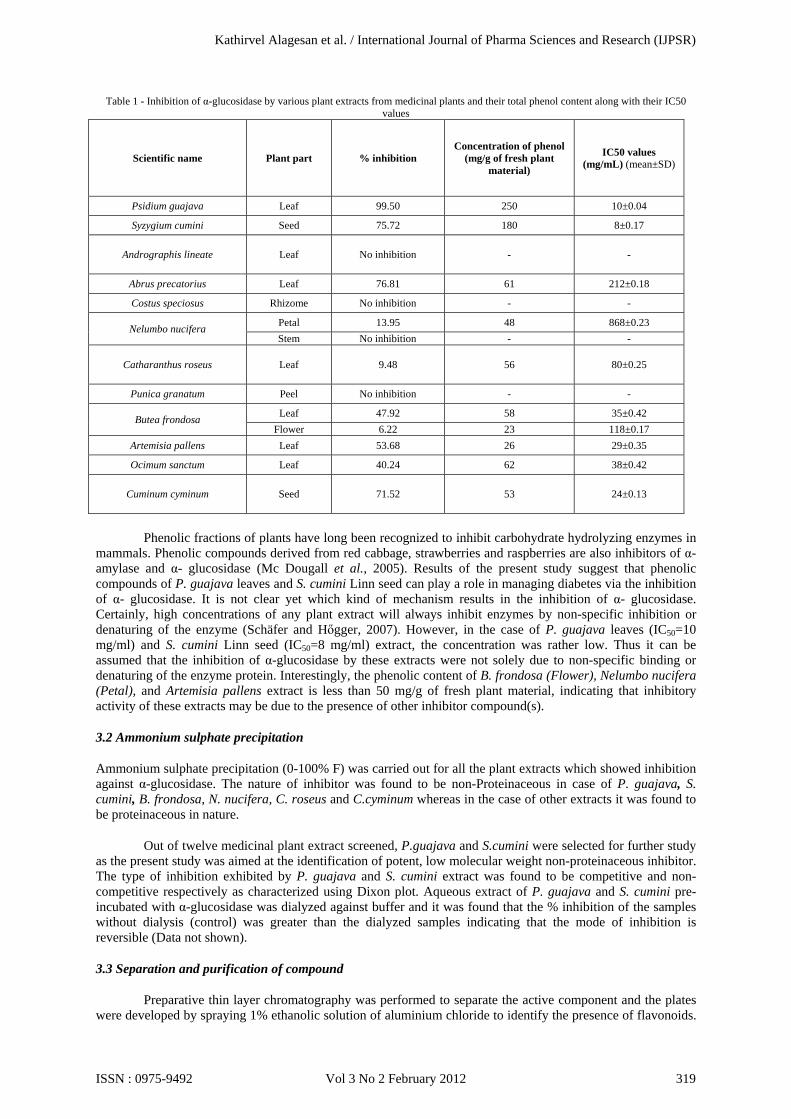
Table 1 - Inhibition of α-glucosidase by various plant extracts from medicinal plants and their total phenol content along with their IC50 values
Scientific name Plant part % inhibition Concentration of phenol
(mg/g of fresh plant material)
IC50 values (mg/mL) (mean±SD)
Psidium guajava Leaf 99.50 250 10±0.04
Syzygium cumini Seed 75.72 180 8±0.17
Andrographis lineate Leaf No inhibition - -
Abrus precatorius Leaf 76.81 61 212±0.18
Costus speciosus Rhizome No inhibition - -
Nelumbo nucifera Petal 13.95 48 868±0.23 Stem No inhibition - -
Catharanthus roseus Leaf 9.48 56 80±0.25
Punica granatum Peel No inhibition - -
Butea frondosa Leaf 47.92 58 35±0.42 Flower 6.22 23 118±0.17
Artemisia pallens Leaf 53.68 26 29±0.35
Ocimum sanctum Leaf 40.24 62 38±0.42
Cuminum cyminum Seed 71.52 53 24±0.13
Phenolic fractions of plants have long been recognized to inhibit carbohydrate hydrolyzing enzymes in mammals. Phenolic compounds derived from red cabbage, strawberries and raspberries are also inhibitors of α- amylase and α- glucosidase (Mc Dougall et al., 2005). Results of the present study suggest that phenolic compounds of P. guajava leaves and S. cumini Linn seed can play a role in managing diabetes via the inhibition of α- glucosidase. It is not clear yet which kind of mechanism results in the inhibition of α- glucosidase. Certainly, high concentrations of any plant extract will always inhibit enzymes by non-specific inhibition or denaturing of the enzyme (Schäfer and Hőgger, 2007). However, in the case of P. guajava leaves (IC50=10 mg/ml) and S. cumini Linn seed (IC50=8 mg/ml) extract, the concentration was rather low. Thus it can be assumed that the inhibition of α-glucosidase by these extracts were not solely due to non-specific binding or denaturing of the enzyme protein. Interestingly, the phenolic content of B. frondosa (Flower), Nelumbo nucifera (Petal), and Artemisia pallens extract is less than 50 mg/g of fresh plant material, indicating that inhibitory activity of these extracts may be due to the presence of other inhibitor compound(s).
3.2 Ammonium sulphate precipitation
Ammonium sulphate precipitation (0-100% F) was carried out for all the plant extracts which showed inhibition against α-glucosidase. The nature of inhibitor was found to be non-Proteinaceous in case of P. guajava, S. cumini, B. frondosa, N. nucifera, C. roseus and C.cyminum whereas in the case of other extracts it was found to be proteinaceous in nature.
Out of twelve medicinal plant extract screened, P.guajava and S.cumini were selected for further study as the present study was aimed at the identification of potent, low molecular weight non-proteinaceous inhibitor. The type of inhibition exhibited by P. guajava and S. cumini extract was found to be competitive and non-competitive respectively as characterized using Dixon plot. Aqueous extract of P. guajava and S. cumini pre-incubated with α-glucosidase was dialyzed against buffer and it was found that the % inhibition of the samples without dialysis (control) was greater than the dialyzed samples indicating that the mode of inhibition is reversible (Data not shown).
3.3 Separation and purification of compound
Preparative thin layer chromatography was performed to separate the active component and the plates were developed by spraying 1% ethanolic solution of aluminium chloride to identify the presence of flavonoids.
Kathirvel Alagesan et al. / International Journal of Pharma Sciences and Research (IJPSR)
ISSN : 0975-9492 Vol 3 No 2 February 2012 319
The fraction with Rf value 0.88 to 0.92 (P. guajava) & 0.86 to 1.00 (S. cumini) in preparative TLC, which showed maximum inhibition towards α-glucosidase were further separated by silica gel column chromatography. The collected fractions were pooled together based on their flavonoid content and checked for their purity by RP-HPLC.
3.4 Spectral analysis
The Mass spectral analysis of the purified sample obtained from P. guajava leaves revealed the presence of Dihydro- 3, 3, 4’, 5, 7 – pentahydroxyflavone glycoside (FT-IR: 1419 and 1382 – phenol OH, 1654 – unsaturation due to C=C; 1275- Phenol –CO stretch) & S. cumini Linn seed purified aqueous extract revealed the presence of apigenin 7-O-glucoside (FT-IR: 1332- Phenol; 1276- Phenol –CO stretch). The molecular ion peak of Dihydro- 3, 3, 4’, 5, 7 – pentahydroxyflavone glycoside was found at m/z 303, and apigenin 7-O-glucoside at m/z 433.
S. cumini seed powder has been reported to lower glucose level in various animal models (Sridhar et al., 2005; Bnouham et al., 2006). The acetone extract of S. cumini seed was found to be a potent inhibitor of α-glucosidase hydrolysis of maltose in in-vitro and in-vivo Gato-Kakazaki (GK) rats (Shinde et al., 2008). The aqueous extract of P. guajava leaves has a good effect to lower blood glucose and various parts have been used for the treatment of Diabetes mellitus (Li et al., 2004; Rai et al., 2007). Maruyama et al., (1985) have already reported the possible hypoglycemic activity of flavonoid glycosides present in P. guajava leaf extract.
The poly phenols in plants have been reported to play an important role in the regulation of carbohydrates hydrolyzing enzymes (Mai & Chuyen., 2007). Glycosides, flavanoids, tannins and alkaloids have been reported to posses hypoglycemic activities (Kumar et al., 2009). In the present investigation, flavanoid glycoside isolated from P. guajava and S. cumini was found to inhibit α-glucosidase which is in accordance with the findings of Jung et al., (2006) who have shown inhibition of α-glucosidase by flavanoid glycoside isolated from five different plants.
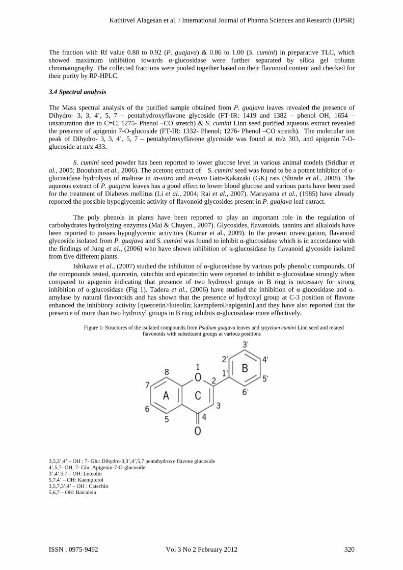
Ishikawa et al., (2007) studied the inhibition of α-glucosidase by various poly phenolic compounds. Of the compounds tested, quercetin, catechin and epicatechin were reported to inhibit α-glucosidase strongly when compared to apigenin indicating that presence of two hydroxyl groups in B ring is necessary for strong inhibition of α-glucosidase (Fig 1). Tadera et al., (2006) have studied the inhibition of α-glucosidase and α-amylase by natural flavonoids and has shown that the presence of hydroxyl group at C-3 position of flavone enhanced the inhibitory activity [quercetin>luteolin; kaempferol>apigenin] and they have also reported that the presence of more than two hydroxyl groups in B ring inhibits α-glucosidase more effectively.
Figure 1: Structures of the isolated compounds from Psidium guajava leaves and syzyzium cumini Linn seed and related
flavonoids with substituent groups at various positions
3,5,3’,4’ – OH ; 7- Glu: Dihydro-3,3’,4’,5,7 pentahydroxy flavone glucoside 4’,5,7- OH; 7- Glu: Apigenin-7-O-glucoside 3’,4’,5,7 – OH: Luteolin 5,7,4’ – OH: Kaempferol 3,5,7,3’,4’ – OH : Catechin 5,6,7 – OH: Baicalein
Kathirvel Alagesan et al. / International Journal of Pharma Sciences and Research (IJPSR)
ISSN : 0975-9492 Vol 3 No 2 February 2012 320
Effect of hydroxyl group at various positions at A ring has been studied by Gao et al., (2004). They showed that the presence of hydroxyl group at C-5, 6 & 7 positions alone has significant α-glucosidase inhibitory activity whereas others have little or no inhibition. It has been known that the substituent groups of the flavones have a great impact on the enzyme inhibition activity. C-3’OH of B ring of flavones in particular increase the α-glucosidase inhibitory activity whereas C-glycosylation at C-6 or C-8 of A-ring weakened the inhibition activity (Li et al., 2009).
In the present study, inhibition of α-glucosidase by dihydro-3,3’,4’,5,7-pentahyroxy flavone may be due to the presence of two OH groups in the B ring and the glucoside substitution might have also an positive effect on the α-glucosidase inhibition. Other compound, apigenin-7-O-glucoside identified in our study lack C-3’OH group however, it shows significant α-glucosidase inhibition indicating that the presence of glucoside substitution at C-7 position, have favoured the inhibition and might have played an significant role in inhibition of the α-glucosidase.
4. CONCLUSION
We screened for low molecular weight α-glucosidase inhibitors from various traditional plant parts used for the treatment of diabetes, of which maximum inhibition was exhibited by S.cumini seed extracts and P. guajava leaves and active component inhibiting α-glucosidase were found to be apigenin 7-O-glucoside and dihydro-3,3’,4’,5,7 – pentahydroxyflavone glycoside respectively. Since, the present investigation does not involve any in-vivo studies, the observed inhibitory activity towards α-glucosidase by these plant extracts (Table 1) might be one the possible reason for their hypoglycemic activity. Acknowledgements Authors thank Sugarcane Breeding Institute (SBI), Coimbatore, TamilNadu, India for the assistance in carrying out HPLC analysis, Sophisticated Analytical Instrument Facility, Indian Institute of Technology (IIT), Chennai, TamilNadu, India for recording FT-IR spectra and Sophisticated Analytical Instrument Facility, Central Drug Research Institute (CDRI), Lucknow, India for recording the mass spectra
References
[1] Andrade-Cetto, A., Jaime Becerra-Jim´enez., & Ren´e C´ardenas-V´azquez. (2008). Alfa- glucosidase -inhibiting activity of
some Mexican plants used in the treatment of type 2 diabetes. Journal of Ethnopharmacolgy, 116, 27-32. [2] Ayyanar, M., Sankarasivaraman, K., & Ignacimuthu, S. (2008). Traditional herbal medicines used for the treatment of diabetes
among two major groups in south Tamilnadu India. Ethnobotanical leaflets, 12, 276-280. [3] Bhat, M., Zinjarde, S.S., Bhargava, S.Y., Kumar, A.R., & Joshi, B,N. (2008). Antidiabetic Indian Plants: a Good Source of
potent amylase inhibitors. Evidence-Based Complementary and Alternative Medicine, 1, 324-328. [4] Bnouham, M., Ziyyat, A., Mekhfi, H., Tahri, A., & Legssyer, A. (2006). Medicinal plants with potential antidiabetic activity - A
review of ten years of herbal medicine research (1990-2000). International Journal of Diabetes & Metabolism, 14, 1-25. [5] Borges de Melo E., Adriane da Silveira Gomes., & Ivone Carvalho. (2006). α- and β Glucosidase inhibitors: chemical structure
and biological activity. Tetrahedron, 62, 10277-10302. [6] Deore, S.L., Khadabadi, S.S., Daulatkar V.D., Deokate U.A., & Farooqui IU, (2008). Evaluation of hypoglycemic and anti
diabetic activity of bark of Butea monosperma. Pharmacognosy Magazine, 4, 134-138. [7] Gao. H., Nishioka, T., Kawabata, J., & Kasai, T. (2004). Structure – activity relationship for α-glucosidase inhibition of
baicalein, 5,6,7-Trihydroxyflavone: the effect of A-ring substitution. Bioscience, Biotechnology, and Biochemistry, 68, 369-375.
[8] Ishikawa, A., Yamashita, H., Hiemori, M., Inagaki, E., Kimoto, M., Okamoto, M., Tsuji, H., Menon, A.N., Mohammadi, A., & Natori, Y. (2007). Characterization of inhibitors of postprandial hyperglycemia from the leaves of Nerium indicum. J Nutritional Science and Vitaminology, 53, 166-173.
[9] Jung, M., Park, M., Chul, H.L., Kang, Y., Kang, S.E., & Ki-Kim, S. (2006). Antidiabetic agents from medicinal plants. Current Medicinal Chemistry, 13, 1203-18.
[10] Kim, S.D., & Nho, H.J, (2004). Isolation and characterization of α-Glucosidase inhibitor from the fungus Ganoderma lucidum. Journal of Microbiology, 42, 223-227.
[11] Kumar, A., Illavarasan, R., Jayachandran, T., Deecaraman, M., Aravindan, P., Padmanabhan, N., & Krishnan, M.R.V., (2009) Phytochemicals investigation on tropical plant, Syzygium cumini from kattuppalayam, Erode district, Tamilnadu, South India. Pakistan Journal of Nutrition, 8, 83-85.
[12] Li, H., Song, F., Xing, J., Tsao. R., Liu. Z., & Liu S. (2009). Screening and structural characterization of α-glucosidase inhibitors from hawthorn leaf flavonoids extract by ultrafiltration LC-DAD-MSn and SORI-CID FTICR MS. Journal of the American Society for Mass Spectrometry, 20, 1496-1503.
[13] Li, W.L., Zheng, H.C., Bukuru, J., & De Kimpeb, N. (2004). Natural medicines used in the traditional Chinese medical system for therapy of diabetes mellitus. Journal of Ethnopharmacology, 92, 1-21.
[14] Lossow, W.J., Migliorini, R.H., Brot, N., & Chaikoff, I.L. (1964). Effect of total exclusion of the exocrine pancreas in the rat upon in vitro esterification of C14 – labeled cholesterol by the intestine and upon lymphatic absorption of C14 – labeled cholesterol. Journal of Lipid Research, 5,198-202.
Kathirvel Alagesan et al. / International Journal of Pharma Sciences and Research (IJPSR)
ISSN : 0975-9492 Vol 3 No 2 February 2012 321
[15] Maher, M.A., Kanefumi, K., Toshihika, S., Fumio, H., & Kenjiro, T. (2006). Antioxidant and α-amylase inhibitory compounds from Aerial Parts of Varthemia iphionoides. Bioscience, Biotechnology, and Biochemistry, 70, 2178-2184.
[16] Mai, T.T., & Chuyen, N.V., (2007). Anti-Hyperglycemic activity of an aqueous extract from flower buds of Cleistocalyx operculatus (Roxb)Merr and Perry. Bioscience, Biotechnology, and Biochemistry, 71, 69-76.
[17] Maruyama, Y., Matsuda, H., Matsuda, R., Kubo, M., Hatano, T., & Okuda, T. (1985). Study on Psidium guajava L. (I). Antidiabetic effect and effective components of the leaf of Psidium guajava L. (Part 1). Shoyakugaku Zasshi, 39, 261–269.
[18] McDougall, G.J., Shpiro, F., Dobson, P., Smith, P., Blake, A., & Stewart D. (2005). Different polyphenolic components of soft fruits inhibit alpha-amylase and alpha-glucosidase. Journal of Agricultural and Food Chemistry, 53, 2760-6.
[19] Ohta, T., Sasaki, S., Oohori, T., Kurihara, H., & Yoshikawa S. (2002). α- glucosidase inhibitory activity of a 70% methanol extract from Ezoishige (Pelvetia babingtonii de Toni) and its effects on the elevation of blood glucose level in rats .Bioscience, Biotechnology, and Biochemistry, 66, 1552 – 1554.
[20] Rai, P.K., Singh, S.K., Kesari, A.N., & watal. G. (2007). Glycaemic evaluation of Psidium guajava in rats. Indian Journal of medicinal research, 126, 224 -227.
[21] Sadasivam, S., & Manickam A. (2004). Biochemical methods, New age international publications, New Delhi, India. 96-97. [22] Sathishkumar, T., Baskar, R., Shanmugam, S., Rajasekaran, P., Sadasivam, S., & Manikandan V. (2008). Optimization of
flavonoids extraction from the leaves of Taberaemontana heyeana Wall using L16 orthogonal design. Nature and science, 6, 10-21.
[23] Schäfer, A., & Hőgger A. (2007). Oligomeric procyanidins of French maritime pine bark extract (Pycnogenol®) effectively inhibits α-glucosidase. Diabetes Research and Clinical Practice, 77, 41-46.
[24] Shinde, J., Taldone, T., Michael, M., Kunaparaju, N., Hu, B., Kumar, S., Placido, J., & Zito SW. (2008). α-Glucosidase inhibitory activity of Syzygium cumini (Linn) skeels seed kernel in vitro and in Goto- Kakizaki (GK) rats. Carbohydrates Research, 343, 1278-1281.
[25] Sridhar, S.B., Sheetal, U.D., Pai, M.R.S.M., & Shastri, M.S. (2005). Preclinical evaluation of the antidiabetic effect of Eugenia jambolana seed powder in streptozotocin-diabetic rats. Brazilian Journal of Medical and Biological Research, 38, 463-468.
[26] Subramoniam, A., Pushpangadan, P., Rajasekharan, S., Evans, D.A., Latha P.G., & Valsaraj R. (1996). Effects of Artemisia pallens Wall. on blood glucose levels in normal and alloxan-induced diabetic rats. Journal of Ethnopharmacology, 50, 13-17.
[27] Tadera, K., Minami, Y., Takamatsu, K., & Matsuoka T. (2006). Inhibition of α-glucosidase and α-amylase by flavonoids. Journal of Nutritional Science and Vitaminology, 52, 149-153.
Kathirvel Alagesan et al. / International Journal of Pharma Sciences and Research (IJPSR)
ISSN : 0975-9492 Vol 3 No 2 February 2012 322
![Clinical Characteristics to Differentiate · Asthma-COPD overlap syndrome (ACOS) [a description] Asthma-COPD overlap syndrome (ACOS) is characterized by persistent airflow limitation](https://static.fdocument.org/doc/165x107/5f0914d17e708231d4252460/clinical-characteristics-to-differentiate-asthma-copd-overlap-syndrome-acos-a.jpg)


![In silico analysis of compounds characterized from ethanolic ...eprints.covenantuniversity.edu.ng/10478/1/20420-74354-2...1 25.637 1.16 Bicyclo[3.1.1]heptane, 2,6,6-trimethyl-138.24992](https://static.fdocument.org/doc/165x107/60f88dd94a7e5669bd2167ee/in-silico-analysis-of-compounds-characterized-from-ethanolic-1-25637-116.jpg)