Hyaluronic acid and TGF-β1 in dogs with hepatobiliary diseases
10
Hyaluronic acid and TGF-β1 in dogs with hepatobiliary diseases Václav Ceplecha 1 , Kristína Řeháková 2 , Corinne Lendon 3 , Pavel Proks 1,4 , Miša Škorič 5 , Guy C.M. Grinwis 6 , Barbora Hřibová 1 , Miloš Vávra 1 , Jana Lorenzová 1,4 , Michal Crha 1 1 University of Veterinary and Pharmaceutical Sciences Brno, Faculty of Veterinary Medicine, Small Animal Clinic, Brno, Czech Republic 2 University of Veterinary and Pharmaceutical Sciences Brno, Faculty of Veterinary Medicine, Small Animal Clinical Laboratory, Brno, Czech Republic 3 Edith Cowan University, Research & Innovation, Joondalup, WA, Australia 4 CEITEC - Central European Institute of Technology, Brno, Czech Republic 5 University of Veterinary and Pharmaceutical Sciences Brno, Faculty of Veterinary Medicine, Department of Pathological Morphology and Parasitology, Brno, Czech Republic 6 University of Utrecht, Faculty of Veterinary Medicine, Department of Pathobiology, Utrecht, the Netherlands Received June 26, 2018 Accepted August 13, 2018 Abstract The aim of the study was to assess serum hyaluronic acid (HA) and transforming growth factor beta 1 (TGF-β1) concentrations: 1) to differentiate hepatic fibrosis from other forms of liver disease, and 2) for the non-invasive staging of canine liver fibrosis. We also evaluated the association between serum HA concentration and the size of the shunt vessel as an indirect marker of decreased liver clearance in patients with single congenital vascular anomaly. Forty-one healthy client-owned dogs and forty dogs diagnosed with hepatobiliary disease were enrolled in the prospective study. Patients were divided into 4 subgroups: 1) congenital portosystemic shunts (CPSS); 2) parenchymal diseases (a. mild and moderate fibrosis, b. advanced fibrosis and cirrhosis); 3) hepatic neoplasia; 4) biliary tract disorders based on thorough clinical, ultrasound and histopathological examination. Serum HA and TGF-β1 concentrations were measured using ELISA. The HA concentration was significantly increased in patients with advanced liver fibrosis/cirrhosis (P < 0.001) and CPSS (P < 0.001) compared to healthy dogs. Using a cut-off HA concentration of 135.94 ng/ml, the sensitivity and specificity for diagnosis for advanced liver fibrosis/cirrhosis was 100% (95% CI, 50.6–100) and 90.8% (95% CI, 81.6–95.7), respectively. The TGF-β1 levels did not significantly differ among groups (P = 0.180). Negligible correlation was found between serum HA concentration and the size of portosystemic shunt vessel (rs = 0.07; P = 0.831). These findings suggest that serum HA concentration is a potential non- invasive biomarker for advanced liver fibrosis and/or cirrhosis in dogs. The utility of measuring serum concentration of TGF-β1 for diagnosing canine liver fibrosis was not supported. Chronic hepatitis, cirrhosis, liver fibrosis, serum biomarkers, laparoscopy, canine Non-invasive detection of hepatic fibrosis is important for initiation of anti-fibrotic treatment to prevent progression to irreversible cirrhosis. Liver fibrosis is characterized by excessive accumulation of extracellular matrix (ECM) which results from both increased synthesis and decreased degradation of ECM and is observed in most types of chronic liver diseases (Liu et al. 2012). Histopathological examination of liver biopsy has been considered to be the gold standard in order to detect and stage liver fibrosis. However, because of the general anaesthesia, invasiveness and possible complications during sampling (bleeding, pain and financial issues) substituting biopsy procedure for minimally invasive specific blood tests would be preferrable, at least in the case of monitoring treatment efficacy in humans (Sebastiani and Alberti 2006). Hyaluronic acid (HA) is a glycosaminoglycan synthesized by hepatic stellate cells (HSCs) as a main component of the ECM (McGary et al. 1989). High serum levels of HA in humans correspond to increased production of ECM by HSCs (Rosenberg et al. ACTA VET. BRNO 2018, 87: 231-240; https://doi.org/10.2754/avb201887030231 Address for correspondence: MVDr. Václav Ceplecha Small Animal Clinic, Faculty of Veterinary Medicine University of Veterinary and Pharmaceutical Sciences Brno Palackeho tr. 1946/1, 61242 Brno, Czech Republic Phone: +420 721 850 370 E-mail: [email protected] http://actavet.vfu.cz/
Transcript of Hyaluronic acid and TGF-β1 in dogs with hepatobiliary diseases
Hyaluronic acid and TGF-β1 in dogs with hepatobiliary diseases
Václav Ceplecha1, Kristína Řeháková2, Corinne Lendon3, Pavel Proks1,4, Miša Škorič5, Guy C.M. Grinwis6, Barbora Hřibová1, Miloš Vávra1, Jana Lorenzová1,4, Michal Crha1
1University of Veterinary and Pharmaceutical Sciences Brno, Faculty of Veterinary Medicine,Small Animal Clinic, Brno, Czech Republic
2University of Veterinary and Pharmaceutical Sciences Brno, Faculty of Veterinary Medicine,Small Animal Clinical Laboratory, Brno, Czech Republic
3Edith Cowan University, Research & Innovation, Joondalup, WA, Australia4CEITEC - Central European Institute of Technology, Brno, Czech Republic
5University of Veterinary and Pharmaceutical Sciences Brno, Faculty of Veterinary Medicine,Department of Pathological Morphology and Parasitology, Brno, Czech Republic
6 University of Utrecht, Faculty of Veterinary Medicine, Department of Pathobiology, Utrecht, the Netherlands
Received June 26, 2018Accepted August 13, 2018
AbstractThe aim of the study was to assess serum hyaluronic acid (HA) and transforming growth
factor beta 1 (TGF-β1) concentrations: 1) to differentiate hepatic fibrosis from other forms of liver disease, and 2) for the non-invasive staging of canine liver fibrosis. We also evaluated the association between serum HA concentration and the size of the shunt vessel as an indirect marker of decreased liver clearance in patients with single congenital vascular anomaly. Forty-one healthy client-owned dogs and forty dogs diagnosed with hepatobiliary disease were enrolled in the prospective study. Patients were divided into 4 subgroups: 1) congenital portosystemic shunts (CPSS); 2) parenchymal diseases (a. mild and moderate fibrosis, b. advanced fibrosis and cirrhosis); 3) hepatic neoplasia; 4) biliary tract disorders based on thorough clinical, ultrasound and histopathological examination. Serum HA and TGF-β1 concentrations were measured using ELISA. The HA concentration was significantly increased in patients with advanced liver fibrosis/cirrhosis (P < 0.001) and CPSS (P < 0.001) compared to healthy dogs. Using a cut-off HA concentration of 135.94 ng/ml, the sensitivity and specificity for diagnosis for advanced liver fibrosis/cirrhosis was 100% (95% CI, 50.6–100) and 90.8% (95% CI, 81.6–95.7), respectively. The TGF-β1 levels did not significantly differ among groups (P = 0.180). Negligible correlation was found between serum HA concentration and the size of portosystemic shunt vessel (rs = 0.07; P = 0.831). These findings suggest that serum HA concentration is a potential non-invasive biomarker for advanced liver fibrosis and/or cirrhosis in dogs. The utility of measuring serum concentration of TGF-β1 for diagnosing canine liver fibrosis was not supported.
Chronic hepatitis, cirrhosis, liver fibrosis, serum biomarkers, laparoscopy, canine
Non-invasive detection of hepatic fibrosis is important for initiation of anti-fibrotic treatment to prevent progression to irreversible cirrhosis. Liver fibrosis is characterized by excessive accumulation of extracellular matrix (ECM) which results from both increased synthesis and decreased degradation of ECM and is observed in most types of chronic liver diseases (Liu et al. 2012). Histopathological examination of liver biopsy has been considered to be the gold standard in order to detect and stage liver fibrosis. However, because of the general anaesthesia, invasiveness and possible complications during sampling (bleeding, pain and financial issues) substituting biopsy procedure for minimally invasive specific blood tests would be preferrable, at least in the case of monitoring treatment efficacy in humans (Sebastiani and Alberti 2006).
Hyaluronic acid (HA) is a glycosaminoglycan synthesized by hepatic stellate cells (HSCs) as a main component of the ECM (McGary et al. 1989). High serum levels of HA in humans correspond to increased production of ECM by HSCs (Rosenberg et al.
ACTA VET. BRNO 2018, 87: 231-240; https://doi.org/10.2754/avb201887030231
Address for correspondence:MVDr. Václav CeplechaSmall Animal Clinic, Faculty of Veterinary MedicineUniversity of Veterinary and Pharmaceutical Sciences BrnoPalackeho tr. 1946/1, 61242 Brno, Czech Republic
Phone: +420 721 850 370 E-mail: [email protected] http://actavet.vfu.cz/
2004) and thus appear to be the most reliable individual test reflecting increased ECM in human patients with chronic hepatitis C and alcoholic liver disease (Wong et al. 1998; Stickel et al. 2003). Transforming growth factor β1 (TGF-β1) is the major mediator of fibrogenesis, thus being a potential biomarker for hepatic fibrosis. It favours the transition of HSCs to myofibroblast-like cells, stimulates the synthesis of ECM proteins and inhibits their degradation (Bataller and Brenner 2005). In human patients, serum TGF-β1 concentrations have a lower diagnostic accuracy for fibrosis than HA (67% and 86%, respectively) (Oberti et al. 1997).
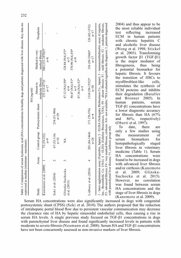
To date, there are only a few studies using the measurement of serum biomarkers for histopathologically staged liver fibrosis in veterinary medicine (Table 1). Serum HA concentrations were found to be increased in dogs with advanced liver fibrosis and/or cirrhosis (Kanemoto et al. 2009; Glinska-Suchocka et al. 2015). However, no correlation was found between serum HA concentrations and the stage of liver fibrosis in dogs (Kanemoto et al. 2009).
Serum HA concentrations were also significantly increased in dogs with congenital portosystemic shunt (CPSS) (Seki et al. 2010). The authors proposed that the reduction of intrahepatic portal blood flow due to persistent vascular communication may decrease the clearance rate of HA by hepatic sinusoidal endothelial cells, thus causing a rise in serum HA levels. A single previous study focused on TGF-β1 concentrations in dogs with parenchymal liver disease and found significantly increased levels in patients with moderate to severe fibrosis (Neumann et al. 2008). Serum HA and TGF-β1 concentrations have not been concurrently assessed as non-invasive markers of liver fibrosis.
232Ta
ble
1. C
ompa
rison
of s
erum
hya
luro
nic
acid
(HA
) con
cent
ratio
ns in
hea
lthy
dogs
and
pat
ient
s dia
gnos
ed w
ith li
ver d
isea
se. K
ey d
ata
are
expr
esse
d as
med
ian
(min
imum
- m
axim
um).
Stud
y A
ssay
C
ontro
l gro
up
Porto
syst
emic
shun
t A
bsen
t to
Mar
ked
to v
ery
Neo
plas
ia
m
oder
ate
fibro
sis
mar
ked
fibro
sis
Kan
emot
o et
al.
(200
9)
1 73
(19-
232)
-
153
(15-
477)
† 50
0 (1
51-1
970)
††
n=
9
n=17
n=
8 -
Seki
et a
l. (2
010)
2
67 (3
2-11
4)
234
(51-
904)
-
- -
n=10
n=
29
Gli
nska
-Suc
hock
a 3
NA
47.1
2 (N
A) F
0*
128.
84 (N
A) F
3*et
al.
(201
5)
n=
6 n=
5
- 50
.47
(NA
) F1*
37
0.90
(NA
) F4*
n=
5 n=
8
-
87
.36
(NA
) F2*
n=
5
Lid
bury
et a
l. (2
016)
4
201
(84-
1464
) 17
1 (7
0-57
6)
1310
(50-
1552
)*
156
(50-
3360
)*
126
(82-
1532
)
n=
24
n=4
n=30
n
=17
n=17
1 - l
atex
-agg
lutin
atio
n as
say
(Fuj
irebi
o, Ja
pan)
; 2 -
ELIS
A (S
eika
gaku
Bio
busi
ness
Co.
, Jap
an);
3 - i
mm
unot
urbi
dim
etric
ass
ay (C
orge
nix,
In
c., B
room
field
, Co,
USA
); 4
- EL
ISA
(Ec
helo
n B
iosc
ienc
es, U
SA).
F0 -
abs
ent
fibro
sis;
F1
- m
ild fi
bros
is;
F2 -
mod
erat
e fib
rosi
s;
F3 -
adva
nced
fibr
osis
; F4
- ver
y m
arke
d fib
rosi
s, ci
rrho
sis.
NA
- no
t ava
ilabl
e; *
HA
eval
uate
d re
gard
less
the d
iagn
osis
; †, p
atie
nts d
iagn
osed
w
ith p
aren
chym
al li
ver d
isea
se; †
†, c
irrho
sis g
roup
onl
y
HA
(ng/
ml)
The aim of the current study was to assess the correlation between two potential serum biomarkers – HA and TGF-β1 and histologically staged liver fibrosis in dogs with respect to the stage of fibrosis according to the METAVIR score (Desmet et al. 1994). We also speculated that with larger volumes of blood that bypass the liver if the shunt vessel has a larger diameter, it results in less HA being cleared by the liver and might cause higher serum HA concentrations in the absence of hepatic fibrosis. Thus, we hypothesized that serum HA concentration would be negatively correlated with the size of CPSS vessel. To the authors’ knowledge, the correlation between serum HA concentration and the size of CPSS vessel has not been evaluated before.
Materials and MethodsStudy design
The study was a prospective case series in dogs that assessed differences between serum HA and TGF-β1
concentrations and type of liver disease, and correlations with the stage of liver fibrosis (METAVIR score). The study was approved by the Ethical Committee for Animal Experiments, University of Veterinary and Pharmaceutical Sciences Brno, Czech Republic.
Thorough history evaluation and physical examination were carried out. Peripheral blood samples were collected for a complete blood count (Sysmex XT-2000iV haematology analyzer, Sysmex America Inc., IL, USA) and serum biochemical profile including concentrations of total protein, albumin, urea, creatinine, total bilirubin, cholesterol, calcium, phosphate, sodium, potassium, chlorides and activities of alanine aminotransferase, aspartate aminotransferase, alkaline phosphatase, and gamma glutamyl transferase (Abbott Architect c4000 clinical chemistry analyzer, Abbott Laboratories, IL, USA, data not shown). Urinalysis of voided samples and abdominal ultrasound were performed.
The control group comprised 41 healthy client-owned dogs. Results of all performed examinations were within normal range. Control dogs had not suffered from any known disease for three months prior to presentation nor received any medication. The patient group was represented by 41 dogs diagnosed with hepatobiliary disease at the Small Animal Clinic, University of Veterinary and Pharmaceutical Sciences, Brno, Czech Republic, from July 2014 to June 2017. Diagnosis of hepatobiliary disease was made based on clinical signs, alterations in the biochemistry profile and/or abnormal liver and gall bladder ultrasound finding (nodular lesions, hyper/hypoechoic liver parenchyma, microhepatica, hepatomegaly, biliary tract abnormalities). The patient group was divided into 4 subgroups according to the World Small Animal Veterinary Association (WSAVA) classification (Rothuizen 2006): 1) vascular disorders (with exclusive cases of congenital portosystemic shunts); 2) parenchymal diseases (a - mild and moderate fibrosis; b - advanced fibrosis and cirrhosis); 3) neoplasia; and 4) biliary tract disorders. Patients with any neoplastic liver disease were included in subgroup 3 regardless of other microscopic changes. In subgroup 1, patients with vascular anomalies, the maximal luminal diameter of the portosystemic shunt was measured in a transverse plane during abdominal ultrasound, where possible. If the vessel orientation did not allow the perpendicular cross-sectional approach, the luminal diameter was measured in the sagittal plane (Vivid 7, GE Medical Systems, Horten, Norway).
Dogs that were diagnosed with arthritis were excluded from the study because of previous reports of increased serum HA concentrations in animals with inflammatory changes in the joints (Arican et al. 1994). Also, dogs previously treated by corticosteroids in the last six months were excluded to avoid false negative results due to the anti-fibrotic effect of this group of drugs.
HA and TGF-β1 analysesBlood was collected by jugular venipuncture and placed into sterile anticoagulant-free tubes. Serum was
separated after centrifugation at 1000 g for 30 min and stored at -20 °C until analysis (maximum of 6 months).In all animals serum HA (Hyaluronan Quantikine ELISA Kit, R&D Systems, MN, USA) and TGF-β1
(Human TGF-beta 1 Quantikine ELISA Kit, R&D Systems, MN, USA) concentrations were measured using commercially available ELISA kits, which were both previously used for measuring HA and TGF in canine serum samples (Neumann et al. 2008; Kanemoto et al. 2009). Samples were run using materials provided by the respective manufacturers according to their instructions. Concentrations of HA were measured with ELISA assay validated by the manufacturer for canine samples. The TGF-β1 ELISA kit was used based on the shared sequence identity of human and canine TGF-β1. Assay precision for TGF-β1 was assessed by calculating intra-assay coefficients of variation (%CV) for 2 samples (low and high concentration) run 6 times on the same ELISA plate. Repeatability for TGF-β1 assay was assessed by calculating the inter-assay %CV for 2 other samples (low and high concentration) run 6 times on different days.
Laparoscopic liver biopsy Laparoscopic liver biopsies were taken from all dogs in the patient group, and in six healthy controls that underwent
general anaesthesia because of neutering or preventive gastropexy with the owner’s consent. Prior to biopsy specimen collection, measurement of prothrombin time (PT), activated partial thromboplastin time (APTT) and fibrinogen with buccal mucosal bleeding time were performed to avoid possible bleeding complications (data not shown).
233
All dogs were placed in dorsal recumbency and the ventral abdominal region was aseptically prepared. A 30° forward-oblique, 5-mm telescope (Hopkins II, Karl Storz SE & Co. KG, Germany) was inserted through the umbilical port with 5 mm cup biopsy forceps (CLICKline, Karl Storz SE & Co. KG, Germany) used for liver biopsy. Samples were collected from any macroscopic liver lesion, and at least three more samples from different liver lobes were obtained in each dog and submitted for histopathological analysis. After the surgical procedure the abdominal cavity was deflated and port incisions were closed in a routine manner.
Histopathological examination Histopathological examination is the gold standard for diagnosis of liver disease and staging of liver fibrosis.
Liver specimens were fixed in 10% neutral buffered formalin, routinely dehydrated and embedded in paraffin using standard procedures. The liver tissue sections (4 µm) were stained with haematoxylin and eosin (H&E) stain and Sirius red to evaluate the degree of fibrosis semiquantitatively with classification according to METAVIR score (Desmet et al. 1994): F0 – no fibrosis; F1 – fibrous expansion in some portal tracts; F2 – fibrous expansion in most portal tracts with occasionally portal-portal/portal-central bridging; F3 – intralobular degeneration with marked bridging; F4 – cirrhosis (Desmet et al. 1994). Samples were examined by an experienced veterinary anatomic pathologist (MS) blinded to group assignment except 1 case of neoplasia and 1 case of biliary tract disorder evaluated by board certified anatomic pathologist (GCMG).
Hepatic fibrosis in patients diagnosed with parenchymal liver disease was staged by a single pathologist (MS).
Statistical analysisFor all statistical tests, a receiver operating characteristic (ROC) curve and the area under the receiver
operating characteristic curve (AUROC) a PC-based software (MedCalc for Windows, version 14, MedCalc Software, Ostend, Belgium) was used. Descriptive statistics were generated to report baseline HA and TGF-β1 data (median, minimum, maximum). Based on assessment of data distribution normality (Shapiro-Wilk test) and homoscedasticity (Levene’s test), Kruskal-Wallis test followed by Dunn’s post hoc test were performed to compare HA and TGF-β1 concentrations between subgroups. Sensitivity and specificity with 95% confidence intervals (CIs) were calculated at various cut-off values. The optimal cut-off value was determined by the value with the highest Youden’s J index (sensitivity + specificity - 1). The relationship between serum HA concentrations and the stage of liver fibrosis in patients with parenchymal disorders or CPSS size were assessed using Spearman’s rank correlation (rs). Scatter plot investigation of HA concentrations among subgroups was created using commercial software (Prism, version 7, GraphPad, La Jolla, CA, USA). Statistical significance was set at P < 0.05.
Results
Serum samples from 82 animals were prospectively collected. Of the 41 healthy control dogs, 26 were female and 15 male, the median age was 6.8 years (age range: 12 months to 14 years). The control group was represented by 23 purebred dogs of 10 different breeds and 18 mixed-breed dogs. The patient group comprised 41 dogs. One case was excluded because of a concurrent presence of hepatitis, extrahepatic biliary duct obstruction, and acquired portosystemic shunts. Of the remaining 40 dogs with the hepatic disease, 24 were female and 16 male, the median age was 8.3 years (age range: 7 months to 16 years). The following breeds were included: Yorkshire terrier (n = 6), mixed-breed (n = 6), Labrador Retriever (n = 3), Golden Retriever (n = 3), Prague Ratter (n = 2), Border Collie (n = 2) and one of American Cockerspaniel, English Cockerspaniel, West Highland White Terrier, Australian Cattle Dog, Bernese Mountain Dog, Border Terrier, Dachshund, Doberman, Weimaraner, German Shepherd, Hovawart, Italian Greyhound, Beagle, Fox Terrier, Bichon Frise, Maltese Dog, Poodle, Schnauzer. Patients were grouped based on the WSAVA standards for clinical and histological diagnosis of canine liver diseases. Subgroup 1 vascular disorders: 11 dogs with extrahepatic CPSS, 1 dog with intrahepatic CPSS; subgroup 2 parenchymal diseases, further subdivided into 2a, 5 dogs with chronic hepatitis and mild to moderate fibrosis; and 2b, 5 dogs with chronic hepatitis and advanced fibrosis and cirrhosis; subgroup 3 hepatic neoplasia: 4 dogs with lymphoma, 3 with hepatocellular carcinoma, 1 with metastatic mast cell tumour, 1 with metastatic insulinoma, 1 with metastatic intestinal carcinoid; subgroup 4 biliary tract disorders: 3 dogs with acute pancreatitis leading to extrahepatic biliary duct obstruction (EHBDO), 3 with biliary mucocele and 2 with emphysematous cholecystitis.
234
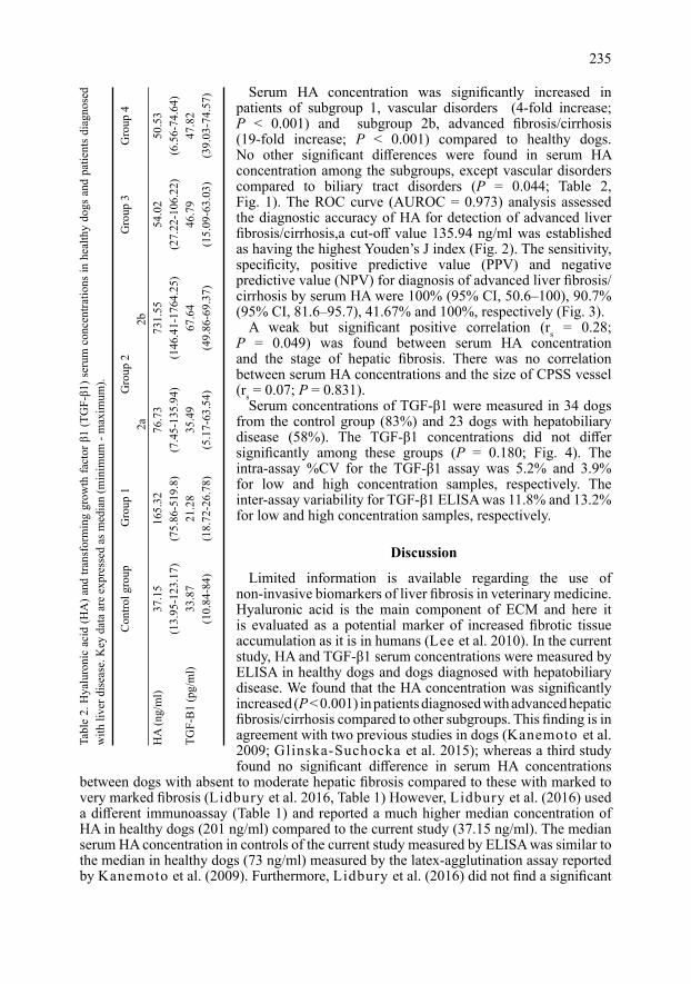
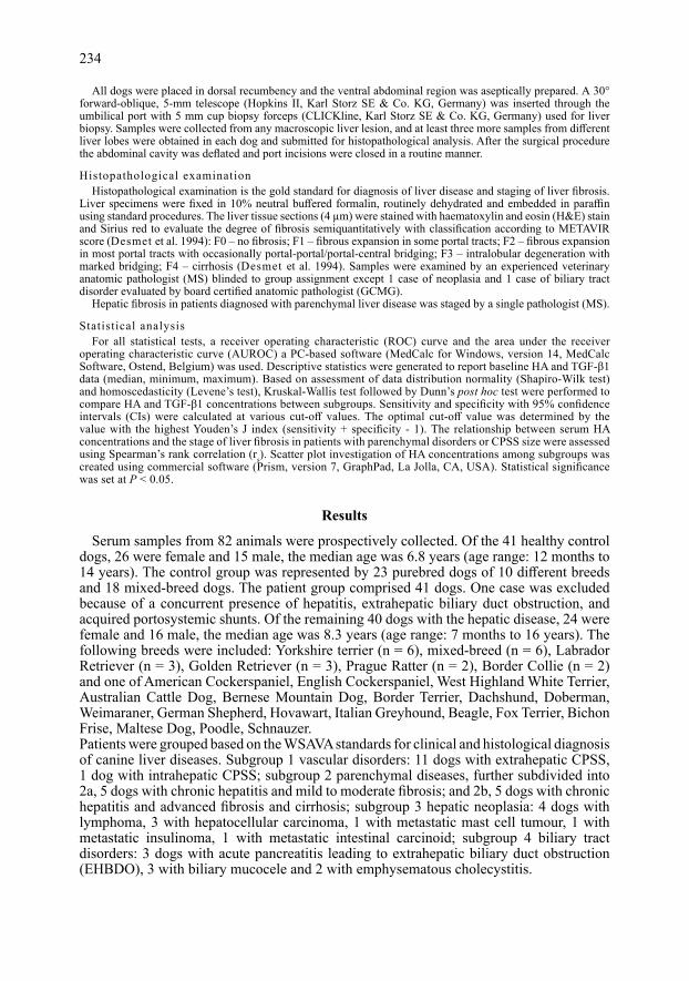
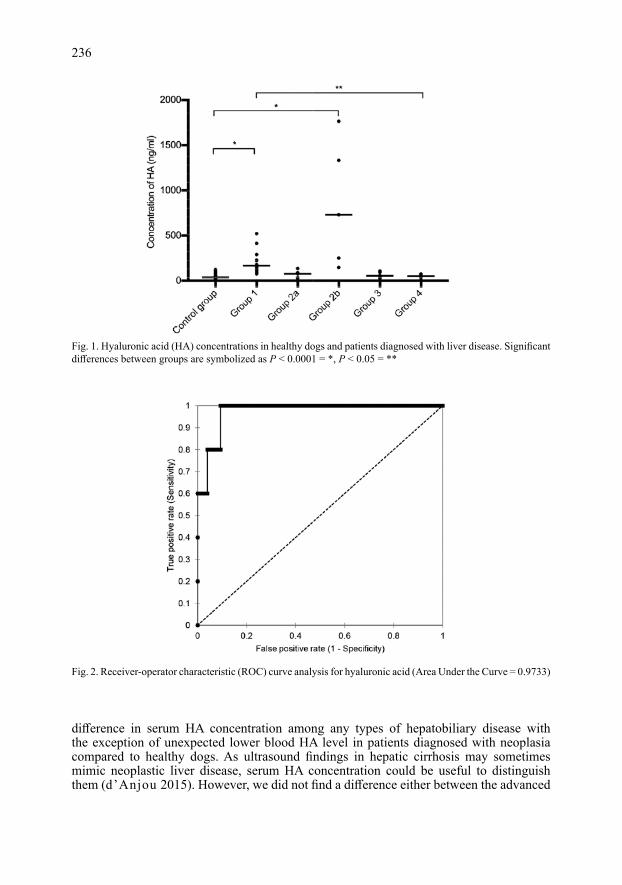
Serum HA concentration was significantly increased in patients of subgroup 1, vascular disorders (4-fold increase; P < 0.001) and subgroup 2b, advanced fibrosis/cirrhosis (19-fold increase; P < 0.001) compared to healthy dogs. No other significant differences were found in serum HA concentration among the subgroups, except vascular disorders compared to biliary tract disorders (P = 0.044; Table 2, Fig. 1). The ROC curve (AUROC = 0.973) analysis assessed the diagnostic accuracy of HA for detection of advanced liver fibrosis/cirrhosis,a cut-off value 135.94 ng/ml was established as having the highest Youden’s J index (Fig. 2). The sensitivity, specificity, positive predictive value (PPV) and negative predictive value (NPV) for diagnosis of advanced liver fibrosis/cirrhosis by serum HA were 100% (95% CI, 50.6–100), 90.7% (95% CI, 81.6–95.7), 41.67% and 100%, respectively (Fig. 3).
A weak but significant positive correlation (rs = 0.28; P = 0.049) was found between serum HA concentration and the stage of hepatic fibrosis. There was no correlation between serum HA concentrations and the size of CPSS vessel (rs = 0.07; P = 0.831).
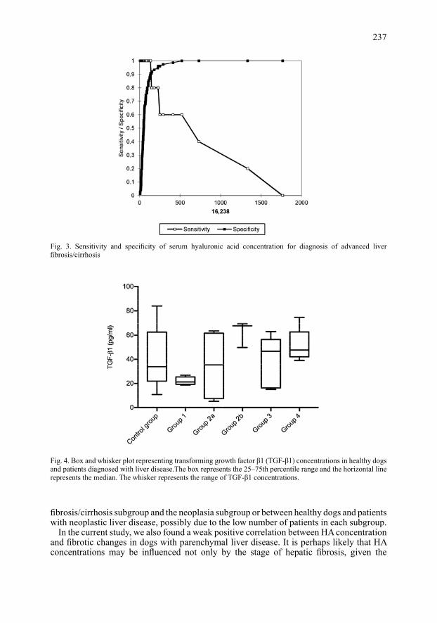
Serum concentrations of TGF-β1 were measured in 34 dogs from the control group (83%) and 23 dogs with hepatobiliary disease (58%). The TGF-β1 concentrations did not differ significantly among these groups (P = 0.180; Fig. 4). The intra-assay %CV for the TGF-β1 assay was 5.2% and 3.9% for low and high concentration samples, respectively. The inter-assay variability for TGF-β1 ELISA was 11.8% and 13.2% for low and high concentration samples, respectively.
Discussion
Limited information is available regarding the use of non-invasive biomarkers of liver fibrosis in veterinary medicine. Hyaluronic acid is the main component of ECM and here it is evaluated as a potential marker of increased fibrotic tissue accumulation as it is in humans (Lee et al. 2010). In the current study, HA and TGF-β1 serum concentrations were measured by ELISA in healthy dogs and dogs diagnosed with hepatobiliary disease. We found that the HA concentration was significantly increased (P < 0.001) in patients diagnosed with advanced hepatic fibrosis/cirrhosis compared to other subgroups. This finding is in agreement with two previous studies in dogs (Kanemoto et al. 2009; Glinska-Suchocka et al. 2015); whereas a third study found no significant difference in serum HA concentrations
between dogs with absent to moderate hepatic fibrosis compared to these with marked to very marked fibrosis (Lidbury et al. 2016, Table 1) However, Lidbury et al. (2016) used a different immunoassay (Table 1) and reported a much higher median concentration of HA in healthy dogs (201 ng/ml) compared to the current study (37.15 ng/ml). The median serum HA concentration in controls of the current study measured by ELISA was similar to the median in healthy dogs (73 ng/ml) measured by the latex-agglutination assay reported by Kanemoto et al. (2009). Furthermore, Lidbury et al. (2016) did not find a significant
235Ta
ble
2. H
yalu
roni
c ac
id (H
A) a
nd tr
ansf
orm
ing
grow
th fa
ctor
β1
(TG
F-β1
) ser
um c
once
ntra
tions
in h
ealth
y do
gs a
nd p
atie
nts
diag
nose
d w
ith li
ver d
isea
se. K
ey d
ata
are
expr
esse
d as
med
ian
(min
imum
- m
axim
um).
C
ontro
l gro
up
Gro
up 1
G
roup
2
Gro
up 3
G
roup
4
2a
2b
H
A (n
g/m
l) 37
.15
165.
32
76.7
3 73
1.55
54
.02
50.5
3
(13.
95-1
23.1
7)
(75.
86-5
19.8
) (7
.45-
135.
94)
(146
.41-
1764
.25)
(2
7.22
-106
.22)
(6
.56-
74.6
4)TG
F-B
1 (p
g/m
l) 33
.87
21.2
8 35
.49
67.6
4 46
.79
47.8
2
(10.
84-8
4)
(18.
72-2
6.78
) (5
.17-
63.5
4)
(49.
86-6
9.37
) (1
5.09
-63.
03)
(39.
03-7
4.57
)
difference in serum HA concentration among any types of hepatobiliary disease with the exception of unexpected lower blood HA level in patients diagnosed with neoplasia compared to healthy dogs. As ultrasound findings in hepatic cirrhosis may sometimes mimic neoplastic liver disease, serum HA concentration could be useful to distinguish them (d’Anjou 2015). However, we did not find a difference either between the advanced
236
Fig. 1. Hyaluronic acid (HA) concentrations in healthy dogs and patients diagnosed with liver disease. Significant differences between groups are symbolized as P < 0.0001 = *, P < 0.05 = **
Fig. 2. Receiver-operator characteristic (ROC) curve analysis for hyaluronic acid (Area Under the Curve = 0.9733)
fibrosis/cirrhosis subgroup and the neoplasia subgroup or between healthy dogs and patients with neoplastic liver disease, possibly due to the low number of patients in each subgroup.
In the current study, we also found a weak positive correlation between HA concentration and fibrotic changes in dogs with parenchymal liver disease. It is perhaps likely that HA concentrations may be influenced not only by the stage of hepatic fibrosis, given the
237
Fig. 3. Sensitivity and specificity of serum hyaluronic acid concentration for diagnosis of advanced liver fibrosis/cirrhosis
Fig. 4. Box and whisker plot representing transforming growth factor β1 (TGF-β1) concentrations in healthy dogs and patients diagnosed with liver disease.The box represents the 25–75th percentile range and the horizontal line represents the median. The whisker represents the range of TGF-β1 concentrations.
study by Glinska-Suchocka et al. (2015) reports a positive correlation between HA concentration and degree of inflammation.
In the current study, low PPV for liver fibrosis and only a weak positive correlation between serum HA levels and the stage of hepatic fibrosis indicates that serum HA is not a reliable marker of early fibrotic changes (METAVIR F1-F2), whereas the high NPV suggests measurement of HA may potentially rule out advanced liver fibrosis/cirrhosis. Thus, although liver biopsy remains the gold standard for the definitive diagnosis and for the institution of treatment, our results suggest that serum HA concentration may be useful for identification of ongoing advanced fibrosis and cirrhosis.
In agreement with the findings of Seki et al. (2010), the current study found a significant increase in serum HA concentration (P < 0.001) in patients diagnosed with CPSS compared to healthy dogs, whereas Lidbury et al. (2016) did not detect a difference between healthy dogs and those with PSS. Seki et al. (2010) proposed that the elevated serum HA levels in dogs with PSS without hepatic fibrosis may possibly arise due to reduced sinusoidal clearance of HA by endothelial cells.
We thus postulated that a greater shunt vessel diameter might enhance the reduction in HA clearance. However, we did not find a correlation between serum HA concentration and the size of the CPSS vessel to explain the findings.
Transforming growth factor β1 is a pleiotropic cytokine that regulates extracellular matrix production, wound healing, immune functions, cell proliferation and differentiation. It is also a major mediator of fibrogenesis. However, in the current study advanced fibrosis and cirrhosis was not associated with an increase in TGF-β1 concentration. This is in contrast with human studies (Nelson et al. 1997) and a previous report (Neumann et al. 2008) in dogs. The median serum TGF-β1 found in healthy dogs in the current study was similar to those previously reported for healthy dogs (Maiolini et al. 2013) (33.87 pg/ml and 23.84 pg/ml, respectively), whereas the median found in healthy dogs in the German study was more than × 10 higher (352 pg/ml) (Neumann et al. 2008). The reason for this difference is unclear because Neumann et al. (2008) used the same TGF-β1 assay as was used in the current study and in a study of canine arthropathies (Arican et al. 1994), and the assay had acceptable precision and repeatability.
Our study had several limitations: 1) The number of patients per subgroup, in particular dogs diagnosed with chronic
hepatitis, was small. This necessitated the formation of just two subgroups by collapsing the METAVIR scores for hepatic parenchymal disease (2a - mild and moderate fibrosis; 2b - advanced fibrosis and cirrhosis), instead of dividing it into four subgroups (F1–F4) according to the stage of liver fibrosis;
2) Liver biopsy samples were taken from all dogs in the patient group, but only 6 healthy dogs. Laboratory abnormalities may be missing in chronic liver disease (Poldervaart et al. 2009) and abdominal ultrasound has low sensitivity in detection of hepatic fibrosis of any extent (Kemp et al. 2013);
3) We did not evaluate the correlation between serum HA level and other histopathological findings (degree of inflammation or necrosis);
4) We cannot exclude the presence of fibrosis in another organ system due to subclinical disease (this is also a limitation of other studies).
These limitations have implications for further studies. Larger sample sizes are required to evaluate the correlation of serum HA concentration with respect to each stage of fibrosis (F1-F4).
In conclusion, liver biopsy and histopathology remain a gold standard for the diagnosis of liver disease and stage of fibrosis, however, the results of our study suggest that unlike TGF-β1, an above-threshold increase in serum HA concentration is a potential non-invasive marker in dogs that might differentiate early-moderate fibrosis (F1–F2) from advanced fibrosis and
238
cirrhosis (F3–F4) in combination with other diagnostic tests. Due to its low specificity and thus inability to differentiate from vascular liver disorders, further refinement necessitates additional diagnostic work-up. Our study confirms that serum HA concentration could serve as a clinically useful diagnostic tool particularly as a marker of progression of liver disease. The high negative predictive value makes it a reliable test to exclude advanced fibrosis/cirrhosis in canine patients with hepatobiliary diseases.
Conflict of Interest
The authors disclose no conflict of interest. None of the authors have any financial or personal relationships that could inappropriately influence or bias the content of the paper.
Off-label Antimicrobial Declaration
The authors declare that there is no off-label use of antimicrobials.
Acknowledgements
This study was supported by the Institutional Program IGA VFU Brno 129/2016/FVL.
References
Arican M, Carter SD, May C, Bennett D 1994: Hyaluronan in canine arthropathies. J Comp Pathol 111: 185-195
Bataller R, Brenner DA 2005: Liver fibrosis. J Clin Invest 115: 209-218d’Anjou MA 2015: Liver. In: Penninck D, d’Anjou MA (Eds): Atlas of Small Animal Ultrasonography, 2nd edn,
Blackwell Publishing, Ames, pp. 217-261Desmet VJ, Gerber M, Hoofnagle JH, Manns M, Scheuer PJ 1994: Classification of chronic hepatitis: Diagnosis,
grading and staging. Hepatology 19: 1513-1520Glinska-Suchocka K, Orlowska A, Spuzak J, Jankowski M, Kubiak K 2015: Suitability of using serum hialuronic
acid concentrations in the diagnosis of canine liver fibrosis. Pol J Vet Sci 18: 873-878Kanemoto H, Ohno K, Sakai M, Nakashima K, Takahashi M, Fujino Y, Tsujimoto H 2009: Blood hyaluronic acid
as a marker for canine cirrhosis. J Vet Med Sci 71: 1251-1254Kemp SD, Panciera DL, Larson MM, Saunders GK, Werre SR 2013: A comparison of hepatic sonographic
features and histopathologic diagnosis in canine liver disease: 138 cases. J Vet Intern Med 27: 806-813Lee HH, Seo YS, Um SH, Won NH, Yoo H, Jung ES, Kwon YD, Park S, Keum B, Kim YS, Yim HJ, Jeen YT,
Chun HJ, Kim CD, Ryu HS 2010: Usefulness of non-invasive markers for predicting significant fibrosis in patients with chronic liver disease. Journal of Korean Medical Science 25: 67-74
Lidbury JA, Hoffmann AR, Fry JK, Suchodolski JS, Steiner JM 2016: Evaluation of hyaluronic acid, procollagen type III N-terminal peptide, and tissue inhibitor of matrix metalloproteinase-1 as serum markers of canine hepatic fibrosis. Can J Vet Res 80: 302-308
Liu T, Wang X, Karsdal MA, Leeming DJ, Genovese F 2012: Molecular Serum Markers of Liver Fibrosis. Biomark Insights 7: 105-117
Maiolini A, Otten M, Hewicker-Trautwein M, Carlson R, Tipold A 2013: Interleukin-6, vascular endothelial growth factor and transforming growth factor beta 1 in canine steroid responsive meningitis-arteritis. BMC Vet Res 9: 23
McGary CT, Raja RH, Weigel PH 1989: Endocytosis of hyaluronic acid by rat liver endothelial cells. Evidence for receptor recycling. Biochem J 257: 875-884
Nelson DR, Gonzalez-Peralta RP, Qian K, Xu Y, Marousis CG, Davis GL, Lau JY 1997: Transforming growth factor-beta 1 in chronic hepatitis C. J Viral Hepat 4: 29-35
Neumann S, Kaup F-J, Beardi B 2008: Plasma concentration of transforming growth factor-β1 and hepatic fibrosis in dogs. Can J Vet Res 72: 428-431
Oberti F, Valsesia E, Pilette C, Rousselet MC, Bedossa P, Aube C, Gallois Y, Rifflet H, Maiga MY, Penneau-Fontbonne D, Cales P 1997: Noninvasive diagnosis of hepatic fibrosis or cirrhosis. Gastroenterology 113: 1609-1616
Poldervaart JH, Favier RP, Penning LC, Van Den Ingh TSGAM, Rothuizen J 2009: Primary Hepatitis in Dogs: A Retrospective Review (2002–2006). J Vet Intern Med 23: 72-80
Rosenberg WMC, Voelker M, Thiel R, Becka M, Burt A, Schuppan D, Hubscher S, Roskams T, Pinzani M, Arthur MJP 2004: Serum markers detect the presence of liver fibrosis: A cohort study. Gastroenterology 127: 1704-1713
Rothuizen J 2006: Chapter 1 - Introduction - background, aims and methods. In: Rothuizen J, Bunch SE, Charles JA, Cullen JM, Desmet VJ, Szatmári V, Twedt DC, van den Ingh TSGAM, Van Winkle T, Washabau
239

RJ (Eds): WSAVA Standards for Clinical and Histological Diagnosis of Canine and Feline Liver Diseases. W.B. Saunders, Edinburgh, pp. 1-4
Sebastiani G, Alberti A 2006: Non invasive fibrosis biomarkers reduce but not substitute the need for liver biopsy. World J Gastroenterol 12: 3682-3694
Seki M, Asano K, Sakai M, Kanno N, Teshima K, Edamura K, Tanaka S 2010: Serum hyaluronic acid in dogs with congenital portosystemic shunts. J Small Anim Pract 51: 260-263
Stickel F, Poeschl G, Schuppan D, Conradt C, Strenge-Hesse A, Fuchs FS, Hofmann WJ, Seitz HK 2003: Serum hyaluronate correlates with histological progression in alcoholic liver disease. Eur J Gastroenterol Hepatol 15: 945-950
Wong VS, Hughes V, Trull A, Wight DG, Petrik J, Alexander GJ 1998: Serum hyaluronic acid is a useful marker of liver fibrosis in chronic hepatitis C virus infection. J Viral Hepat 5: 187-192
240

![Supplementary Material: Unsupervised Learning of Probably ...openaccess.thecvf.com/content_CVPR_2020/...C. V. Jawahar. Cats and dogs. In Proc. CVPR, 2012.1 [8] Yuxin Wu and Kaiming](https://static.fdocument.org/doc/165x107/5f9e6e331fb6866d2166c552/supplementary-material-unsupervised-learning-of-probably-c-v-jawahar.jpg)