PURIFICATION OF THE ALPHA SUBUNIT OF THE EPITHELIAL SODIUM …
Histology, Epithelial Tissue
1.Tissues. Classification
2.General properties of basic tissues
3.Epithelial tissue – principalcharacteristics and functions
4.Classification of epithelium
5.Types of epithelia:
� covering epithelia – types
� glandular epithelia – types
Prof. Dr. Nikolai Lazarov 2
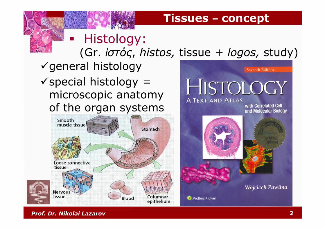
Tissues – concept
� Histology:(Gr. ἱστός, histos, tissue + logos, study)
�general histology
�special histology = microscopic anatomyof the organ systems
Prof. Dr. Nikolai Lazarov 3
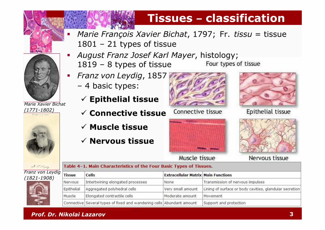
Tissues – classification
� Marie François Xavier Bichat, 1797; Fr. tissu = tissue
1801 – 21 types of tissue
� August Franz Josef Karl Mayer, histology; 1819 – 8 types of tissue
� Franz von Leydig, 1857
– 4 basic types:
� Epithelial tissue
� Connective tissue
� Muscle tissue
� Nervous tissue
Franz von Leydig
(1821-1908)
Marie Xavier Bichat
(1771-1802)
Prof. Dr. Nikolai Lazarov 4
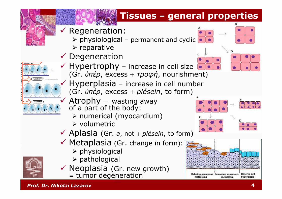
Tissues – general properties
� Regeneration:� physiological – permanent and cyclic
� reparative
� Degeneration� Hypertrophy – increase in cell size
(Gr. ὑπέρ, excess + τροφή, nourishment)
� Hyperplasia – increase in cell number (Gr. ὑπέρ, excess + plésein, to form)
� Atrophy – wasting away of a part of the body:� numerical (myocardium)� volumetric
� Aplasia (Gr. a, not + plésein, to form)
� Metaplasia (Gr. change in form):
� physiological� pathological
� Neoplasia (Gr. new growth)= tumor degeneration
Prof. Dr. Nikolai Lazarov 5
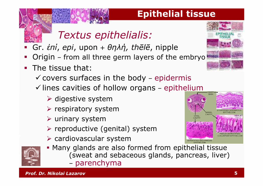
Epithelial tissue
� Gr. ἐπί, epi, upon + θηλή, thēlē, nipple
� Origin – from all three germ layers of the embryo
� The tissue that:
�covers surfaces in the body – epidermis
� lines cavities of hollow organs – epithelium
� digestive system
� respiratory system
� urinary system
� reproductive (genital) system
� cardiovascular system
� Many glands are also formed from epithelial tissue(sweat and sebaceous glands, pancreas, liver)– parenchyma
Textus epithelialis:
Prof. Dr. Nikolai Lazarov 6
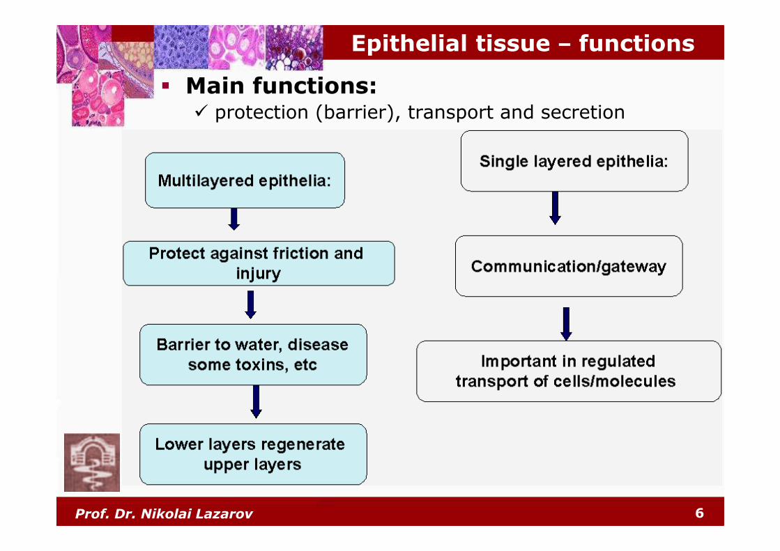
Epithelial tissue – functions
� Main functions:� protection (barrier), transport and secretion
Prof. Dr. Nikolai Lazarov 7
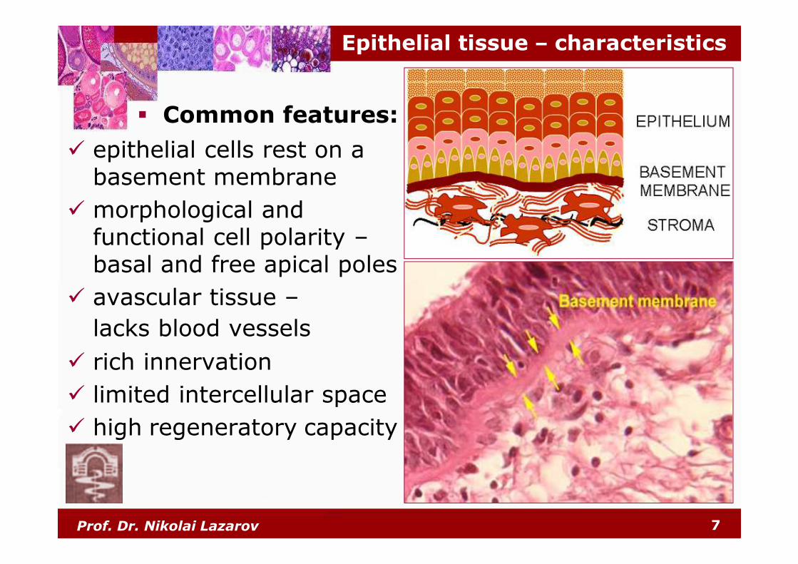
� epithelial cells rest on a basement membrane
� morphological and functional cell polarity –basal and free apical poles
� avascular tissue –
lacks blood vessels
� rich innervation
� limited intercellular space
� high regeneratory capacity
Epithelial tissue – characteristics
� Common features:
Prof. Dr. Nikolai Lazarov 8
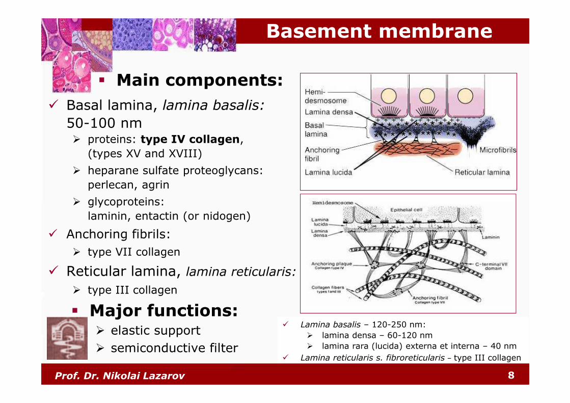
Basement membrane
� Basal lamina, lamina basalis:
50-100 nm� proteins: type IV collagen,
(types ХV and ХVІІІ)
� heparane sulfate proteoglycans:
perlecan, agrin
� glycoproteins:
laminin, entactin (or nidogen)
� Anchoring fibrils:
� type VII collagen
� Reticular lamina, lamina reticularis:
� type III collagen
� Major functions:� elastic support
� semiconductive filter
� Lamina basalis – 120-250 nm:
� lamina densa – 60-120 nm
� lamina rara (lucida) externa et interna – 40 nm
� Lamina reticularis s. fibroreticularis – type III collagen
� Main components:
Prof. Dr. Nikolai Lazarov 9
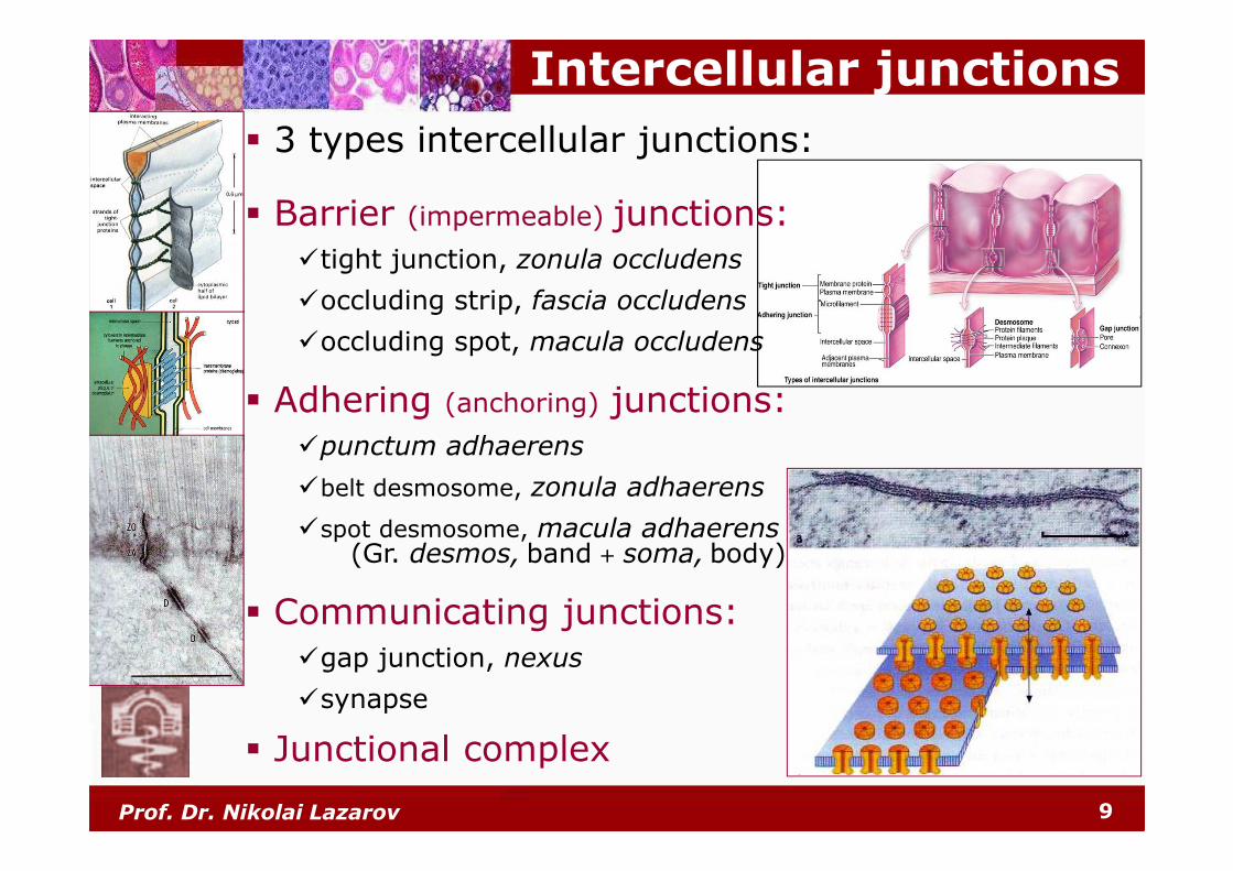
Intercellular junctions
� Barrier (impermeable) junctions:
�tight junction, zonula occludens
�occluding strip, fascia occludens
�occluding spot, macula occludens
� Adhering (anchoring) junctions:
�punctum adhaerens
�belt desmosome, zonula adhaerens
�spot desmosome, macula adhaerens(Gr. desmos, band + soma, body)
� Communicating junctions:
�gap junction, nexus
�synapse
� Junctional complex
� 3 types intercellular junctions:
Prof. Dr. Nikolai Lazarov 10
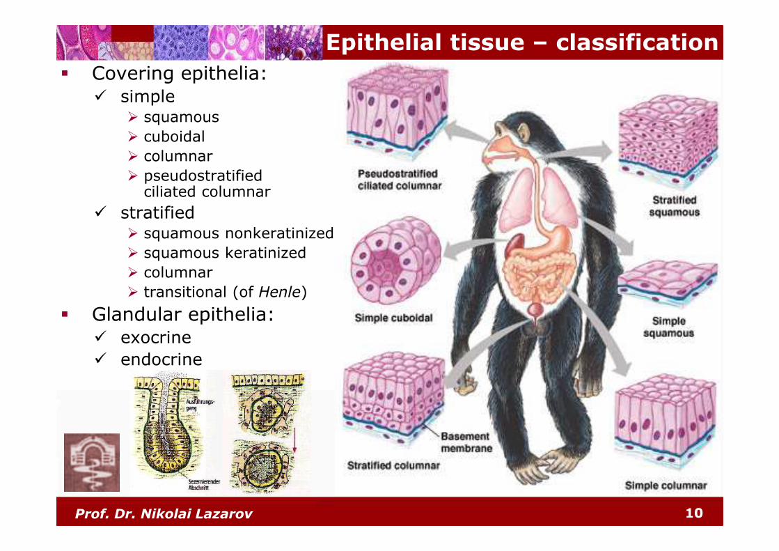
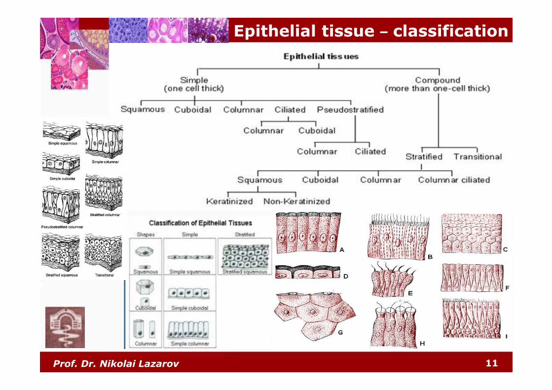
Epithelial tissue – classification
� Covering epithelia:� simple
� squamous
� cuboidal
� columnar
� pseudostratifiedciliated columnar
� stratified� squamous nonkeratinized
� squamous keratinized
� columnar
� transitional (of Henle)
� Glandular epithelia:� exocrine
� endocrine
Prof. Dr. Nikolai Lazarov 11
Epithelial tissue – classification
Prof. Dr. Nikolai Lazarov 12
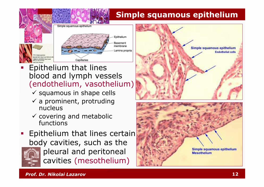
Simple squamous epithelium
� Epithelium that lines blood and lymph vessels (endothelium, vasothelium)� squamous in shape cells
� a prominent, protruding nucleus
� covering and metabolic functions
� Epithelium that lines certain body cavities, such as the
pleural and peritoneal cavities (mesothelium)
Prof. Dr. Nikolai Lazarov 13
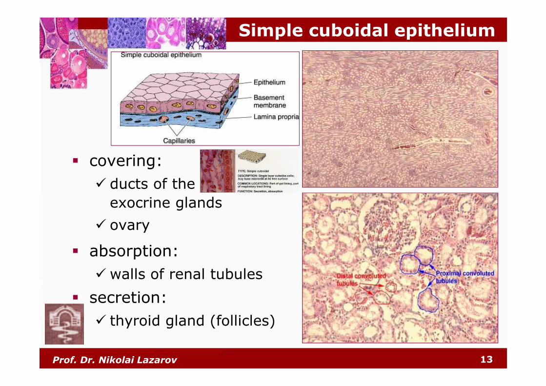
Simple cuboidal epithelium
� covering:
� ducts of the
exocrine glands
� ovary
� absorption:
� walls of renal tubules
� secretion:
� thyroid gland (follicles)
Prof. Dr. Nikolai Lazarov 14
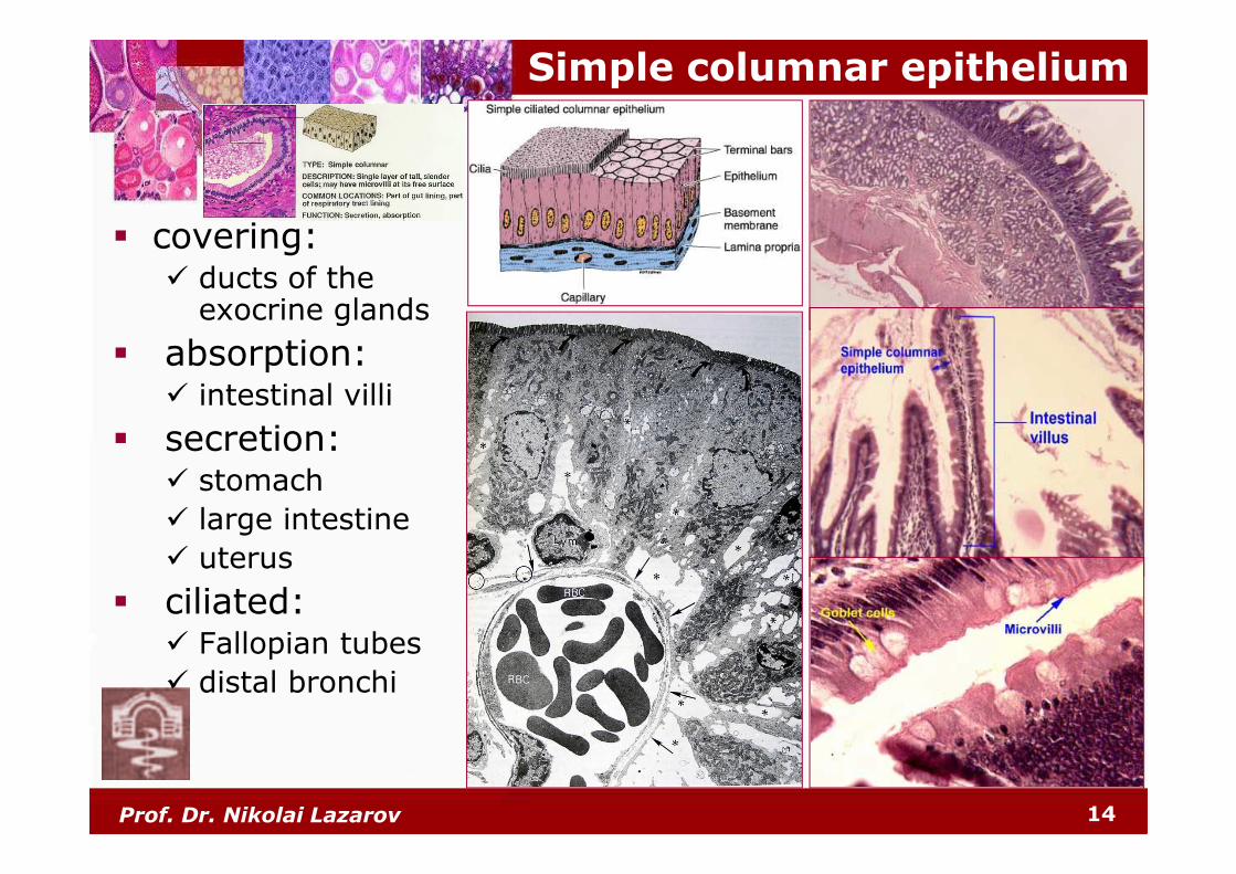
Simple columnar epithelium
� covering:� ducts of the
exocrine glands
� absorption:� intestinal villi
� secretion:� stomach
� large intestine
� uterus
� ciliated:� Fallopian tubes
� distal bronchi
Prof. Dr. Nikolai Lazarov 15
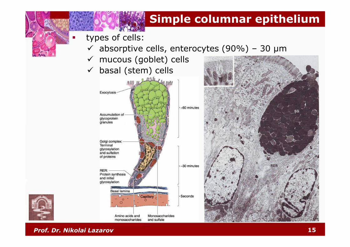
Simple columnar epithelium
� types of cells:
� absorptive cells, enterocytes (90%) – 30 µm
� mucous (goblet) cells
� basal (stem) cells
Prof. Dr. Nikolai Lazarov 16
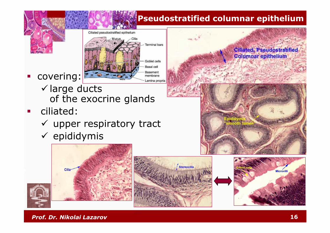
Pseudostratified columnar epithelium
� covering:
� large ducts of the exocrine glands
� ciliated:
� upper respiratory tract
� epididymis
Prof. Dr. Nikolai Lazarov 17
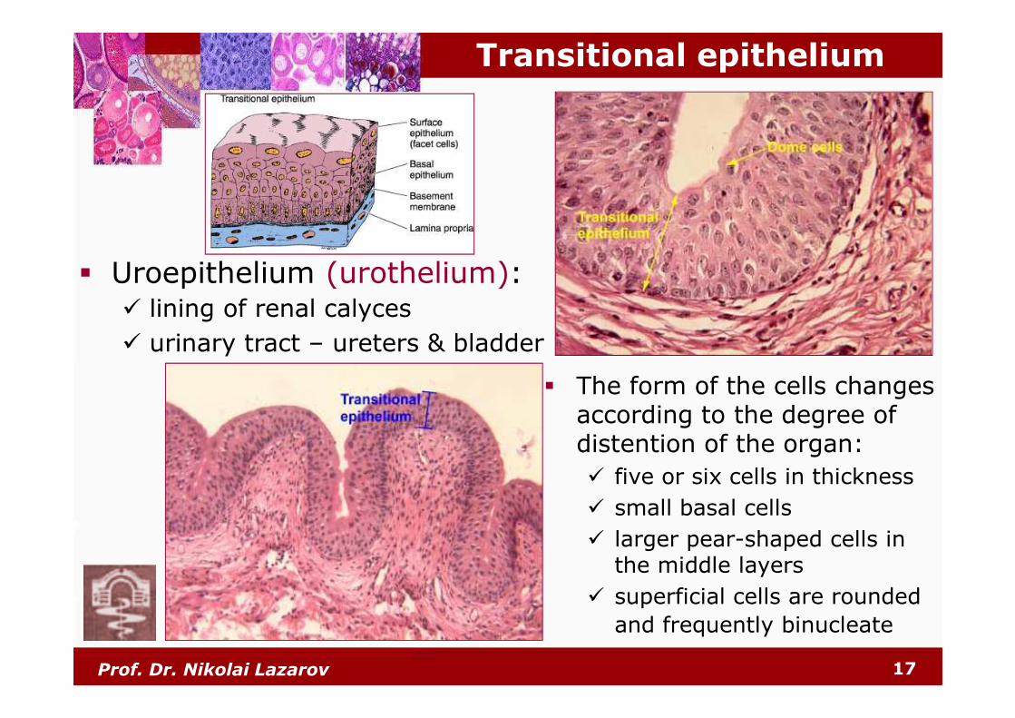
Transitional epithelium
� Uroepithelium (urothelium):� lining of renal calyces
� urinary tract – ureters & bladder
� The form of the cells changes according to the degree of distention of the organ:
� five or six cells in thickness
� small basal cells
� larger pear-shaped cells in the middle layers
� superficial cells are rounded
and frequently binucleate
Prof. Dr. Nikolai Lazarov 18
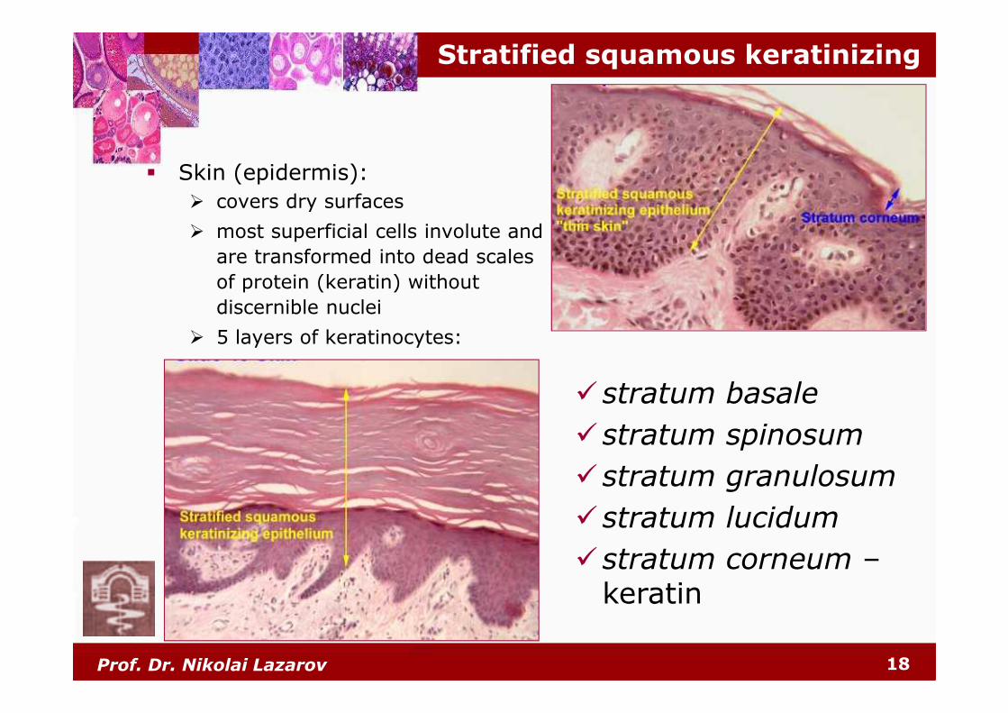
Stratified squamous keratinizing
� Skin (epidermis):
� covers dry surfaces
� most superficial cells involute and
are transformed into dead scales
of protein (keratin) without
discernible nuclei
� 5 layers of keratinocytes:
�stratum basale
�stratum spinosum
�stratum granulosum
�stratum lucidum
�stratum corneum –keratin

Prof. Dr. Nikolai Lazarov 19
Stratified squamous nonkeratinizing
� Mucous epithelium –
covers wet surfaces:
� oral cavity
� oropharynx
� esophagus
� anal canal
� vagina
� Metaplasia
� Corneal epithelium
Prof. Dr. Nikolai Lazarov 20
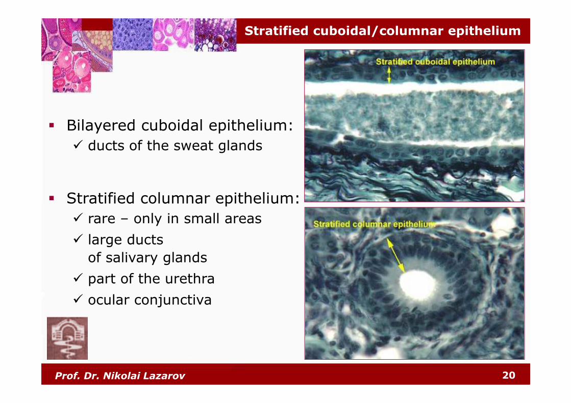
Stratified cuboidal/columnar epithelium
� Bilayered cuboidal epithelium:
� ducts of the sweat glands
� Stratified columnar epithelium:
� rare – only in small areas
� large ducts
of salivary glands
� part of the urethra
� ocular conjunctiva
Prof. Dr. Nikolai Lazarov 21
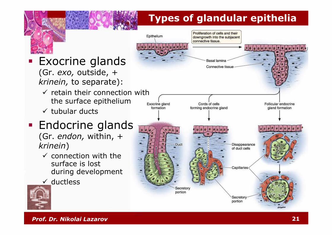
Types of glandular epithelia
� Exocrine glands (Gr. exo, outside, + krinein, to separate):
� retain their connection with the surface epithelium
� tubular ducts
� Endocrine glands (Gr. endon, within, + krinein)
� connection with the surface is lost during development
� ductless
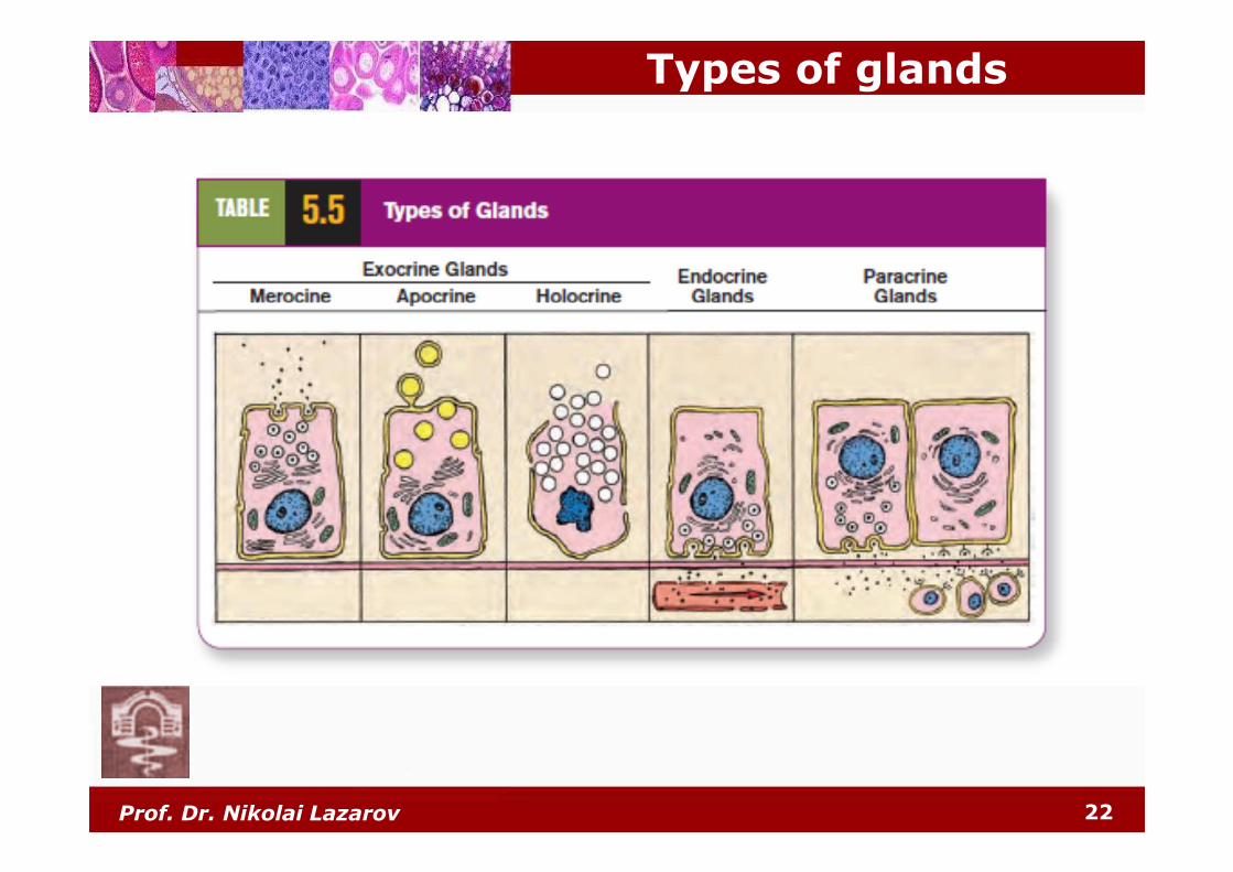
Types of glands
22Prof. Dr. Nikolai Lazarov
Prof. Dr. Nikolai Lazarov 23
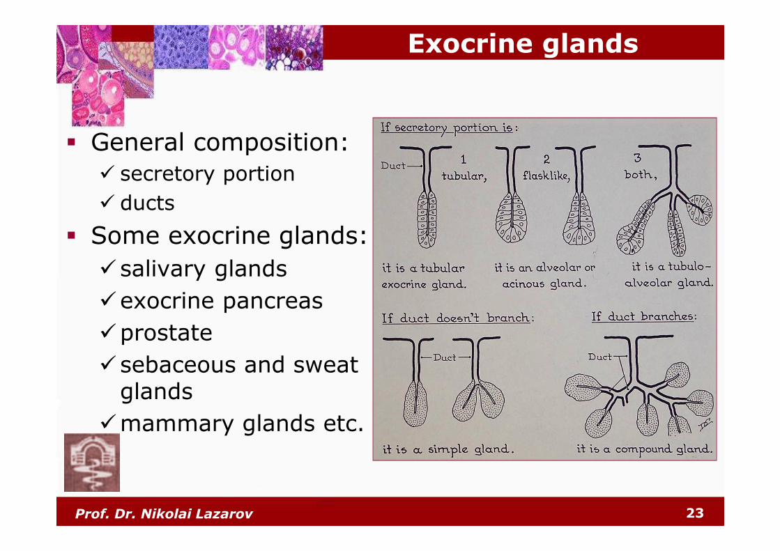
Exocrine glands
� General composition:
� secretory portion
� ducts
� Some exocrine glands:
�salivary glands
�exocrine pancreas
�prostate
�sebaceous and sweat glands
�mammary glands etc.
Prof. Dr. Nikolai Lazarov 24
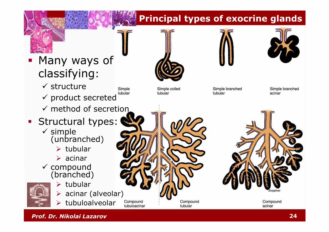
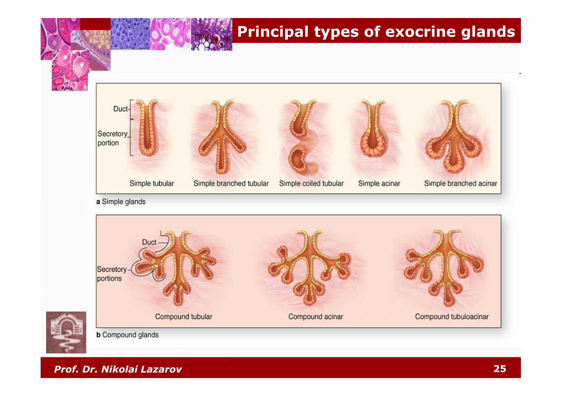
Principal types of exocrine glands
� Many ways of classifying:� structure
� product secreted
� method of secretion
� Structural types:� simple
(unbranched)� tubular
� acinar
� compound (branched)� tubular
� acinar (alveolar)
� tubuloalveolar
Prof. Dr. Nikolai Lazarov 25
Principal types of exocrine glands
Prof. Dr. Nikolai Lazarov 26
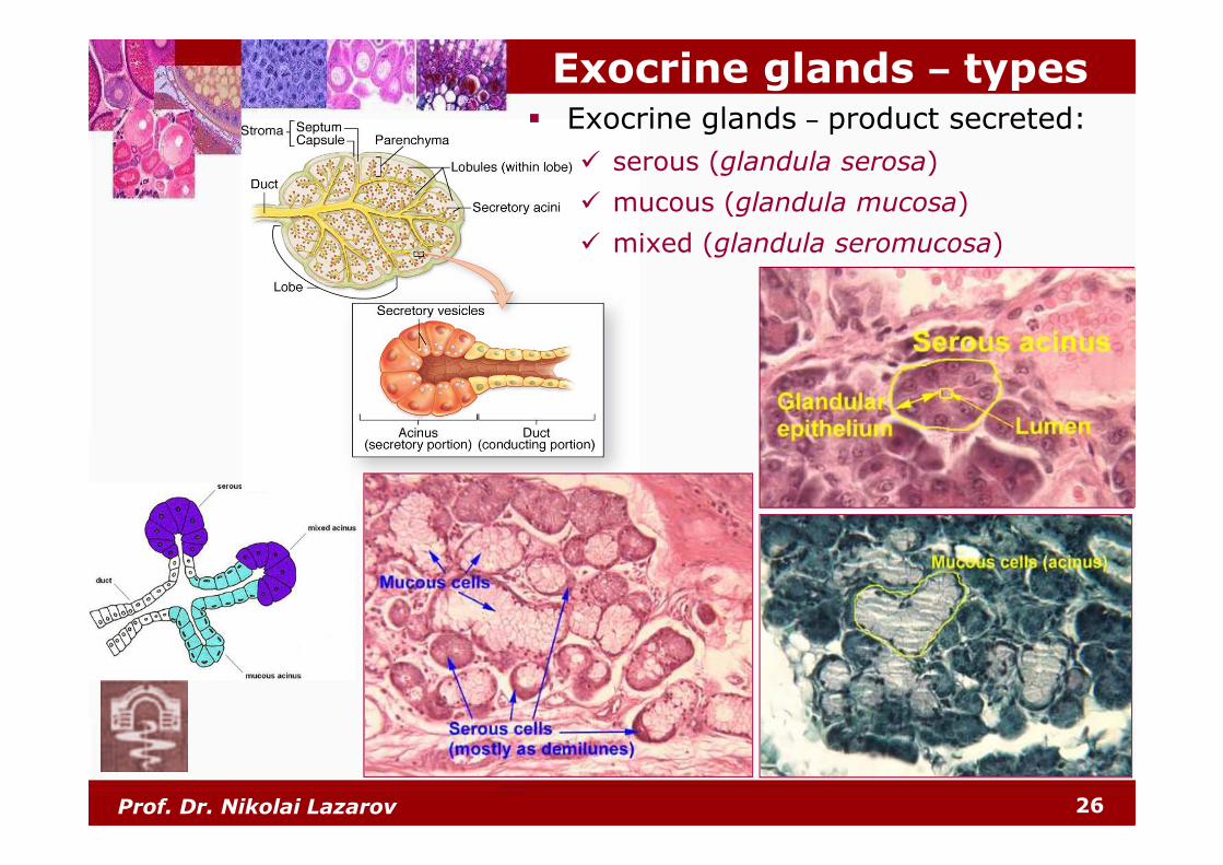
Exocrine glands – types� Exocrine glands – product secreted:
� serous (glandula serosa)
� mucous (glandula mucosa)
� mixed (glandula seromucosa)
Prof. Dr. Nikolai Lazarov 27
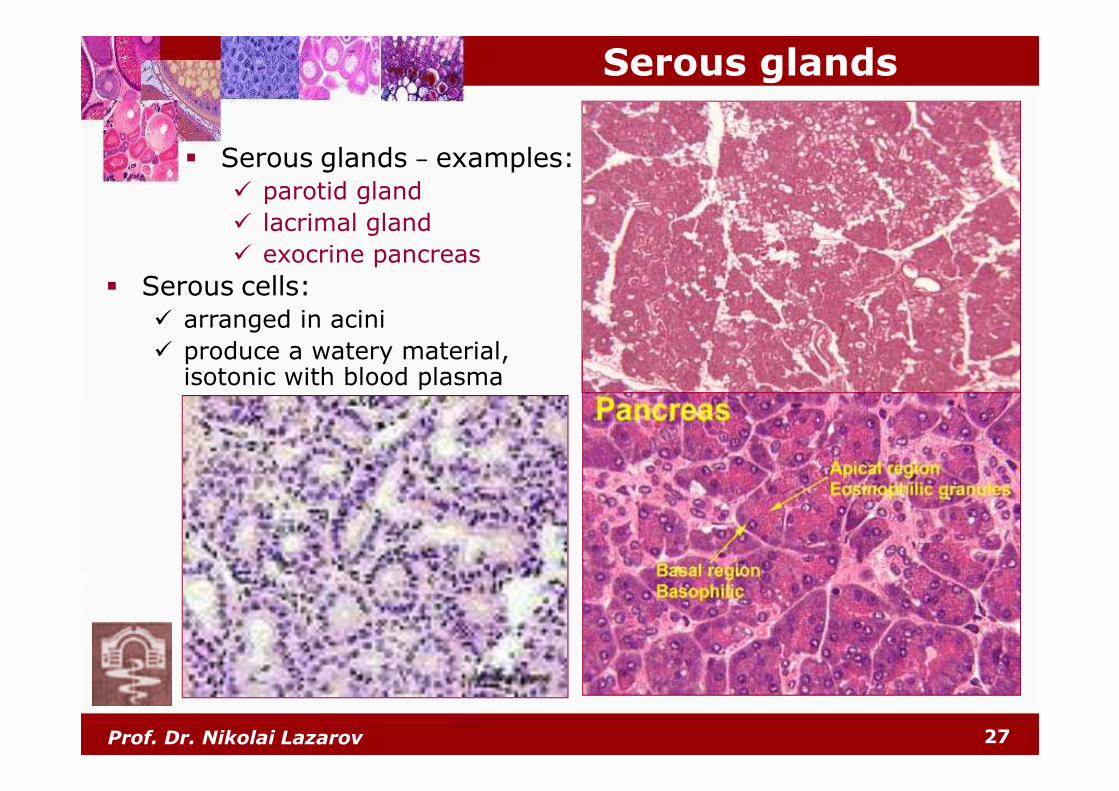
Serous glands
� Serous glands – examples:� parotid gland
� lacrimal gland
� exocrine pancreas
� Serous cells:� arranged in acini
� produce a watery material, isotonic with blood plasma
Prof. Dr. Nikolai Lazarov 28
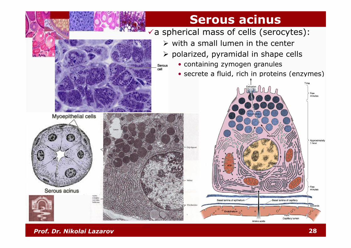
Serous acinus�a spherical mass of cells (serocytes):
� with a small lumen in the center
� polarized, pyramidal in shape cells
• containing zymogen granules
• secrete a fluid, rich in proteins (enzymes)
Prof. Dr. Nikolai Lazarov 29
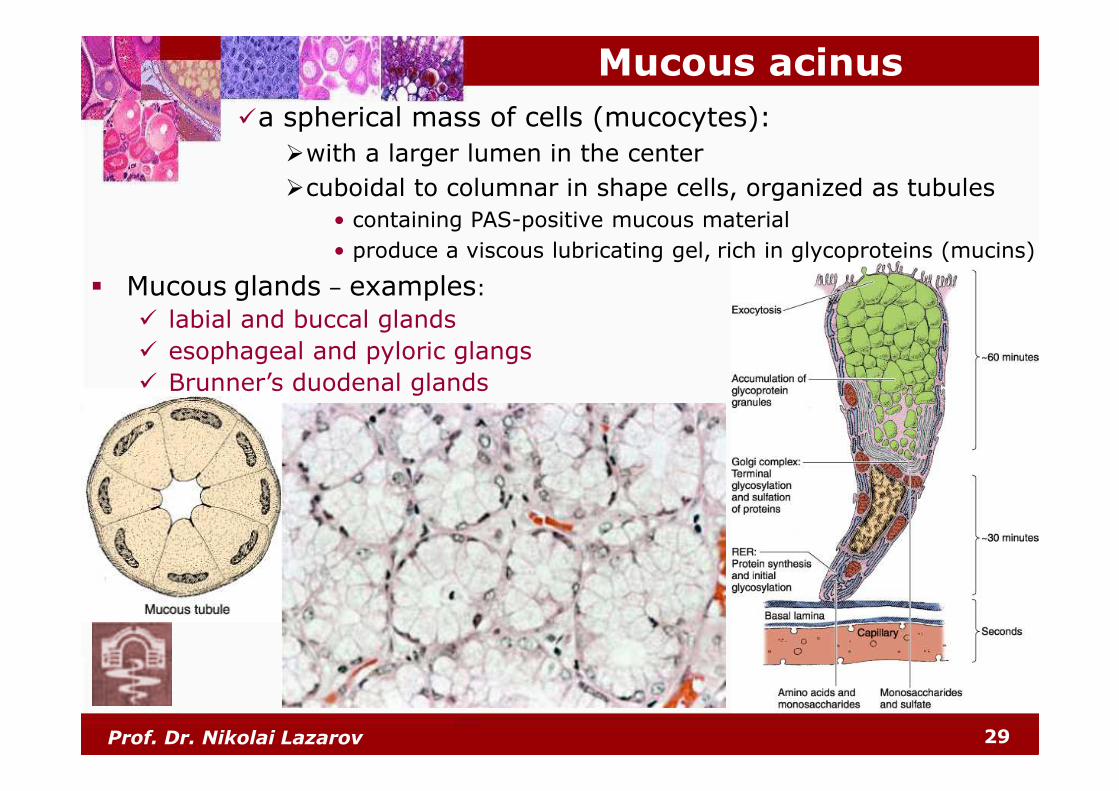
Mucous acinus
�a spherical mass of cells (mucocytes):
�with a larger lumen in the center
�cuboidal to columnar in shape cells, organized as tubules
• containing PAS-positive mucous material
• produce a viscous lubricating gel, rich in glycoproteins (mucins)
� Mucous glands – examples:
� labial and buccal glands
� esophageal and pyloric glangs
� Brunner’s duodenal glands
Prof. Dr. Nikolai Lazarov 30
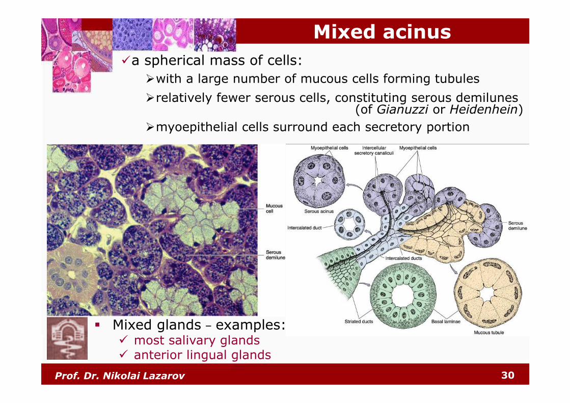
Mixed acinus
�a spherical mass of cells:
�with a large number of mucous cells forming tubules
�relatively fewer serous cells, constituting serous demilunes(of Gianuzzi or Heidenhein)
�myoepithelial cells surround each secretory portion
� Mixed glands – examples:� most salivary glands� anterior lingual glands
Prof. Dr. Nikolai Lazarov 31
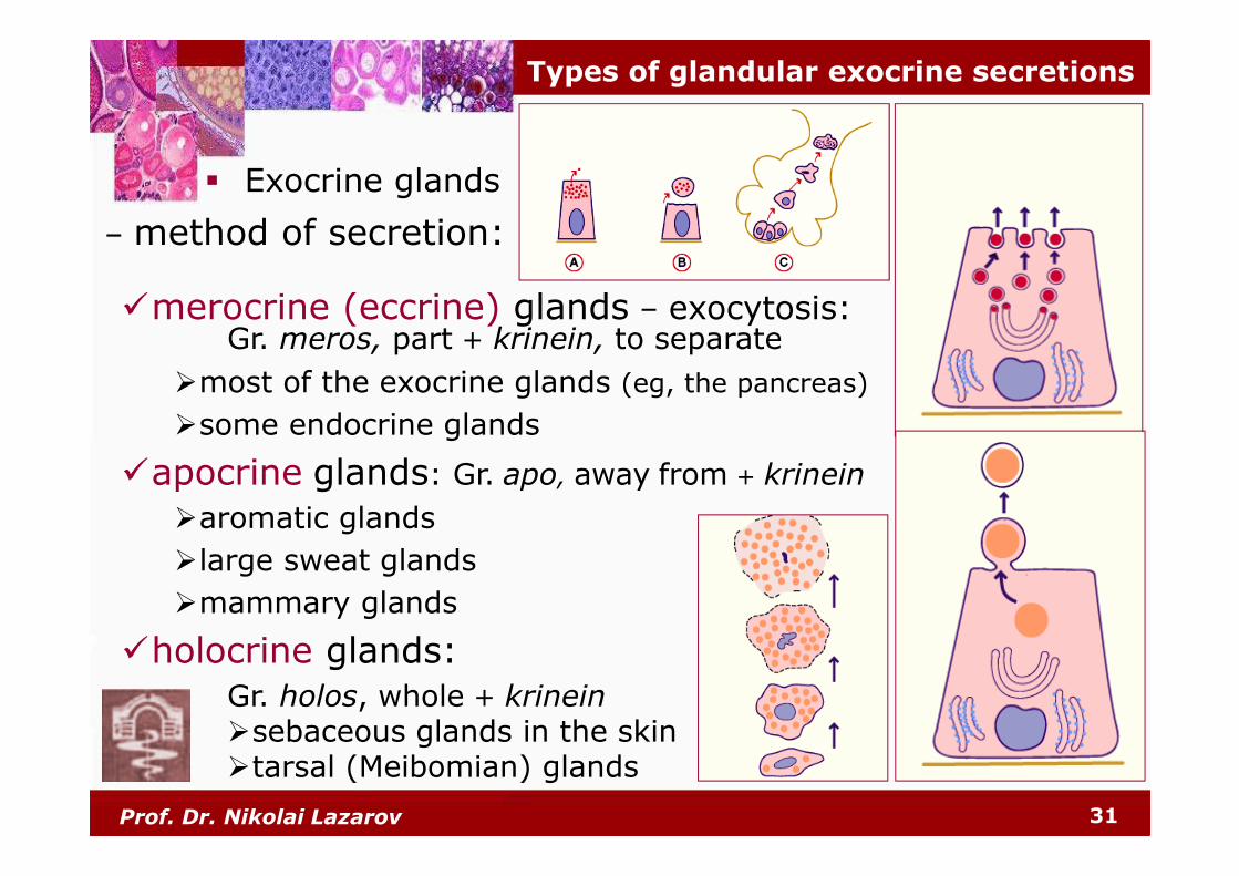
Types of glandular exocrine secretions
�merocrine (eccrine) glands – exocytosis:Gr. meros, part + krinein, to separate
�most of the exocrine glands (eg, the pancreas)
�some endocrine glands
�apocrine glands: Gr. apo, away from + krinein
�aromatic glands
�large sweat glands
�mammary glands
�holocrine glands:
Gr. holos, whole + krinein�sebaceous glands in the skin�tarsal (Meibomian) glands
� Exocrine glands
– method of secretion:
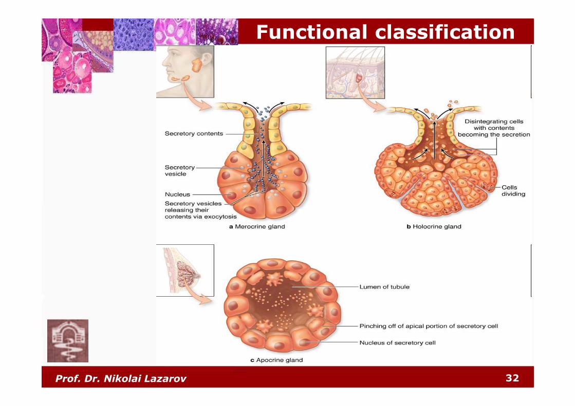
Functional classification
32Prof. Dr. Nikolai Lazarov
Prof. Dr. Nikolai Lazarov 33
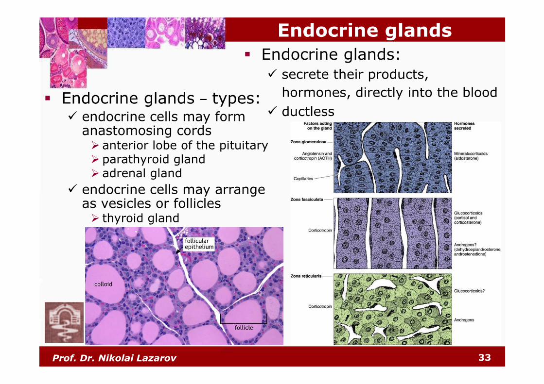
Endocrine glands
� Endocrine glands:
� secrete their products,
hormones, directly into the blood
� ductless� Endocrine glands – types:
� endocrine cells may form anastomosing cords� anterior lobe of the pituitary� parathyroid gland� adrenal gland
� endocrine cells may arrange as vesicles or follicles� thyroid gland
Prof. Dr. Nikolai Lazarov 34
Thank you ...


![Bone Tissue Mechanics - FenixEdu · Bone Tissue Mechanics João Folgado ... Introduction to linear elastic fracture mechanics ... Lesson_2016.03.14.ppt [Compatibility Mode]](https://static.fdocument.org/doc/165x107/5ae984637f8b9aee0790eb6e/bone-tissue-mechanics-tissue-mechanics-joo-folgado-introduction-to-linear.jpg)