High-Resolution Microtubule Structures Reveal the ...
13
High-Resolution Microtubule Structures Reveal the Structural Transitions in ab -Tubulin upon GTP Hydrolysis Gregory M. Alushin, 1,5,6 Gabriel C. Lander, 2,5,7 Elizabeth H. Kellogg, 3,5,8 Rui Zhang, 2 David Baker, 3 and Eva Nogales 2,4, * 1 Biophysics Graduate Program, University of California, Berkeley, Berkeley, CA 94720, USA 2 Life Sciences Division, Lawrence Berkeley National Lab, Berkeley, CA 94720, USA 3 Howard Hughes Medical Institute, Department of Biochemistry, University of Washington, Seattle, WA 98105, USA 4 Howard Hughes Medical Institute, Department of Molecular and Cell Biology, University of California, Berkeley, Berkeley, CA 94720, USA 5 Co-first author 6 Present address: Cell Biology and Physiology Center, National Heart Lung and Blood Institute, Bethesda, MD 20892, USA 7 Present address: Department of Integrative Structural and Computational Biology, The Scripps Research Institute, 10550 North Torrey Pines Road, La Jolla, CA 92037, USA 8 Present address: Howard Hughes Medical Institute, Department of Molecular and Cell Biology, University of California, Berkeley, Berkeley, CA 94720, USA *Correspondence: [email protected] http://dx.doi.org/10.1016/j.cell.2014.03.053 SUMMARY Dynamic instability, the stochastic switching between growth and shrinkage, is essential for microtubule function. This behavior is driven by GTP hydrolysis in the microtubule lattice and is inhibited by anti- cancer agents like Taxol. We provide insight into the mechanism of dynamic instability, based on high-res- olution cryo-EM structures (4.7–5.6 A ˚ ) of dynamic mi- crotubules and microtubules stabilized by GMPCPP or Taxol. We infer that hydrolysis leads to a compac- tion around the E-site nucleotide at longitudinal interfaces, as well as movement of the a-tubulin inter- mediate domain and H7 helix. Displacement of the C-terminal helices in both a- and b-tubulin subunits suggests an effect on interactions with binding part- ners that contact this region. Taxol inhibits most of these conformational changes, allosterically inducing a GMPCPP-like state. Lateral interactions are similar in all conditions we examined, suggesting that micro- tubule lattice stability is primarily modulated at longi- tudinal interfaces. INTRODUCTION Microtubules are ubiquitous cytoskeletal filaments critical for multiple cellular processes, including intracellular trafficking, establishment and maintenance of cell morphology, and cell di- vision (Hyams and Lloyd, 1993). For many microtubule-depen- dent processes, the underlying dynamics of the polymer play a pivotal role. Perhaps the most striking example is mitosis, when chromosome motions are driven by microtubule dynamics and segregation is primarily powered by microtubule depolymer- ization (Desai and Mitchison, 1997; McIntosh et al., 2010; Rieder and Salmon, 1994). Highlighting this fact, many successful anti- proliferative drugs bind to tubulin and interfere with microtubule dynamics (Dumontet and Jordan, 2010). Describing the confor- mational cycle accompanying tubulin polymerization, nucleotide hydrolysis, and microtubule depolymerization is essential for our understanding of microtubule dynamics and will significantly aid in improving existing anticancer drugs, as well as facilitating the development of novel agents. Dynamic instability, the stochastic switching between phases of microtubule growth and shrinkage, is driven by the binding and hydrolysis of GTP by the ab-tubulin dimer (Mitchison and Kirschner, 1984). Tubulin dimers associate longitudinally to form polar protofilaments, which associate laterally to form a tube. Subunit addition occurs preferentially at the end of the microtubule capped by b-tubulin subunits, termed the ‘‘plus end.’’ ab-tubulin contains two GTP-binding sites (Figure 1A). The N-site (nonexchangeable) in a-tubulin is buried within the tubulin dimer at a longitudinal monomer-monomer (or intra- dimer) interface (Nogales et al., 1998). This site is constitutively occupied by GTP and has been ascribed a structural role (Mene ´ ndez et al., 1998). The nucleotide at the E-site (exchangeable) in b-tubulin is exposed on the surface of an un- polymerized dimer and the terminal subunits of a microtubule plus end (Mitchison, 1993; Nogales, 2000). Free ab-tubulin dimers exchange bound guanosine diphosphate (GDP) for gua- nosine triphosphate (GTP) at the E-site, rendering them compe- tent for polymerization (Figure 1A). Upon addition of a tubulin dimer to a growing microtubule plus end, the a-tubulin subunit in the incoming dimer contacts the E-site GTP of the terminal b-tubulin subunit, completing the binding pocket that enables hydrolysis (Nogales et al., 1999). Thus, microtubule growth and GTP hydrolysis are coupled, giving rise to the metastable character of this polymer. Whereas a lattice of GTP-tubulin is stable and promotes polymerization, the GDP-tubulin lattice is Cell 157, 1117–1129, May 22, 2014 ª2014 Elsevier Inc. 1117
Transcript of High-Resolution Microtubule Structures Reveal the ...
High-Resolution Microtubule StructuresReveal the Structural Transitionsin ab-Tubulin upon GTP HydrolysisGregory M. Alushin,1,5,6 Gabriel C. Lander,2,5,7 Elizabeth H. Kellogg,3,5,8 Rui Zhang,2 David Baker,3 and Eva Nogales2,4,*1Biophysics Graduate Program, University of California, Berkeley, Berkeley, CA 94720, USA2Life Sciences Division, Lawrence Berkeley National Lab, Berkeley, CA 94720, USA3Howard Hughes Medical Institute, Department of Biochemistry, University of Washington, Seattle, WA 98105, USA4Howard Hughes Medical Institute, Department of Molecular and Cell Biology, University of California, Berkeley, Berkeley, CA 94720, USA5Co-first author6Present address: Cell Biology and Physiology Center, National Heart Lung and Blood Institute, Bethesda, MD 20892, USA7Present address: Department of Integrative Structural and Computational Biology, The Scripps Research Institute, 10550 North Torrey PinesRoad, La Jolla, CA 92037, USA8Present address: Howard Hughes Medical Institute, Department of Molecular and Cell Biology, University of California, Berkeley, Berkeley,
CA 94720, USA
*Correspondence: [email protected]://dx.doi.org/10.1016/j.cell.2014.03.053
SUMMARY
Dynamic instability, the stochastic switchingbetweengrowth and shrinkage, is essential for microtubulefunction. This behavior is driven by GTP hydrolysisin the microtubule lattice and is inhibited by anti-cancer agents like Taxol. We provide insight into themechanismof dynamic instability, based on high-res-olution cryo-EM structures (4.7–5.6 A) of dynamic mi-crotubules and microtubules stabilized by GMPCPPor Taxol. We infer that hydrolysis leads to a compac-tion around the E-site nucleotide at longitudinalinterfaces, aswell asmovement of thea-tubulin inter-mediate domain and H7 helix. Displacement of theC-terminal helices in both a- and b-tubulin subunitssuggests an effect on interactions with binding part-ners that contact this region. Taxol inhibits most ofthese conformational changes, allosterically inducinga GMPCPP-like state. Lateral interactions are similarin all conditions we examined, suggesting that micro-tubule lattice stability is primarily modulated at longi-tudinal interfaces.
INTRODUCTION
Microtubules are ubiquitous cytoskeletal filaments critical for
multiple cellular processes, including intracellular trafficking,
establishment and maintenance of cell morphology, and cell di-
vision (Hyams and Lloyd, 1993). For many microtubule-depen-
dent processes, the underlying dynamics of the polymer play a
pivotal role. Perhaps the most striking example is mitosis,
when chromosomemotions are driven by microtubule dynamics
and segregation is primarily powered bymicrotubule depolymer-
ization (Desai and Mitchison, 1997; McIntosh et al., 2010; Rieder
and Salmon, 1994). Highlighting this fact, many successful anti-
proliferative drugs bind to tubulin and interfere with microtubule
dynamics (Dumontet and Jordan, 2010). Describing the confor-
mational cycle accompanying tubulin polymerization, nucleotide
hydrolysis, and microtubule depolymerization is essential for our
understanding of microtubule dynamics and will significantly aid
in improving existing anticancer drugs, as well as facilitating the
development of novel agents.
Dynamic instability, the stochastic switching between phases
of microtubule growth and shrinkage, is driven by the binding
and hydrolysis of GTP by the ab-tubulin dimer (Mitchison and
Kirschner, 1984). Tubulin dimers associate longitudinally to
form polar protofilaments, which associate laterally to form a
tube. Subunit addition occurs preferentially at the end of the
microtubule capped by b-tubulin subunits, termed the ‘‘plus
end.’’ ab-tubulin contains two GTP-binding sites (Figure 1A).
The N-site (nonexchangeable) in a-tubulin is buried within the
tubulin dimer at a longitudinal monomer-monomer (or intra-
dimer) interface (Nogales et al., 1998). This site is constitutively
occupied by GTP and has been ascribed a structural role
(Menendez et al., 1998). The nucleotide at the E-site
(exchangeable) in b-tubulin is exposed on the surface of an un-
polymerized dimer and the terminal subunits of a microtubule
plus end (Mitchison, 1993; Nogales, 2000). Free ab-tubulin
dimers exchange bound guanosine diphosphate (GDP) for gua-
nosine triphosphate (GTP) at the E-site, rendering them compe-
tent for polymerization (Figure 1A). Upon addition of a tubulin
dimer to a growing microtubule plus end, the a-tubulin subunit
in the incoming dimer contacts the E-site GTP of the terminal
b-tubulin subunit, completing the binding pocket that enables
hydrolysis (Nogales et al., 1999). Thus, microtubule growth
and GTP hydrolysis are coupled, giving rise to the metastable
character of this polymer. Whereas a lattice of GTP-tubulin is
stable and promotes polymerization, the GDP-tubulin lattice is
Cell 157, 1117–1129, May 22, 2014 ª2014 Elsevier Inc. 1117
BA
β
α
GTP GDP
Exchange
E-site
N-site
Polymerizing
Depolymerizing
Stochastichydrolysis
β
α
E-site
N-site
Taxol
elbatSelbatsnUelbatS
P
GTPhydrolysis
Taxolbinding
C
-
+
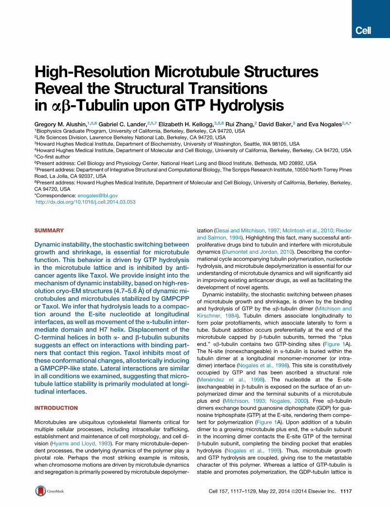
Figure 1. High-Resolution Cryo-EM Structures of Dynamic and Stabilized Microtubules
(A) Cartoon of the ab-tubulin dimer, which spontaneously exchanges bound GDP for GTP in solution.
(B) Cartoon illustrating structural intermediates of microtubule polymerization and depolymerization.
(C) Cryo-EMmaps of GMPCPP (left panel, 4.7 A resolution), GDP (middle panel, 4.9 A resolution), and Taxol-stabilized (right panel, 5.6 A resolution) microtubules,
viewed from inside the microtubule lumen. a-tubulin, green; b-tubulin, blue; GMPCPP/GTP; orange; GDP, pink; Taxol, yellow. Maps are contoured at 1.1 s.
See also Figures S1 and S2, Table S1A, and Movies S1, S2, and S3.
unstable and prone to depolymerization, or ‘‘catastrophe’’
(Desai and Mitchison, 1997). In the long-standing ‘‘GTP-cap’’
model (Mitchison and Kirschner, 1984), a microtubule will
continue to grow as long as it contains GTP-tubulin subunits
at its plus end (i.e., subunit addition outpaces hydrolysis).
When this GTP cap is lost, rapid depolymerization ensues
(Figure 1B).
The detailed molecular mechanism by which tubulin GTP
binding and hydrolysis controls microtubule dynamics remains
elusive despite decades of intensive study. Structural studies
1118 Cell 157, 1117–1129, May 22, 2014 ª2014 Elsevier Inc.
have led to the consensus view that conformational changes in
tubulin must be correlated with the transition from polymeriza-
tion to depolymerization. A straight tubulin conformation is found
within the body of the microtubule (Li et al., 2002; Nogales et al.,
1999), and all high-resolution structural analyses of this state
to date have been limited to electron crystallography of zinc-
induced two-dimensional (2D) sheets, which contain protofila-
ment-like head-to-tail assemblies of straight ab-tubulin (Nettles
et al., 2004; Nogales et al., 1998). A curved conformation is found
in microtubule depolymerization peels (Mandelkow et al., 1991),

head-to-tail arrays of bent tubulin heterodimers wherein longitu-
dinal contacts are maintained after lateral contacts are broken
(Figure 1B). The existence of peels as a depolymerization inter-
mediate, together with the fact that contacts between protofila-
ments aremediated by a series of loop-loop interactions (Li et al.,
2002; Sui and Downing, 2010), has led to the assumption that
lateral contacts are the most labile of the microtubule lattice
interactions. In this model, hydrolysis-induced destabilization
of the distant lateral contacts would occur through an unknown
allosteric mechanism.
Despite the apparently clear link between tubulin conforma-
tion and polymerization state, a robust connection between
tubulin nucleotide state and tubulin dimer conformation has
been difficult to establish. There is a continuing controversy
over whether different nucleotide-dependent curvature states
exist for tubulin dimers in solution (Barbier et al., 2010; Elie-Caille
et al., 2007; Muller-Reichert et al., 1998; Nogales and Wang,
2006). An intermediate curvature state in alternative polymers
formed at low temperatures with the nonhydrolyzable analog
GMPCPP (Wang and Nogales, 2005) has been proposed to
correspond structurally to an assembly sheet intermediate
observed during fast microtubule growth (Figure 1B) (Chretien
et al., 1995). This led to the speculation that exchange of
GDP for GTP causes unassembled tubulin to adopt a straighter
conformation, compatible with polymerization. However, all
high-resolution X-ray crystal structures of tubulin to date have
captured the molecule in a bent conformation, regardless of
nucleotide state (Nawrotek et al., 2011), supporting a model
wherein lattice contacts, rather than nucleotide, controls tubulin
conformation (Aldaz et al., 2005). However, in all such structures,
polymerization was suppressed by binding tubulin to a microtu-
bule depolymerizer (Ayaz et al., 2012; Gigant et al., 2000; Prota
et al., 2013) and/or to microtubule-destabilizing drugs (Gigant
et al., 2005; Ravelli et al., 2004), which are likely to intrinsically
bias tubulin toward a bent conformation. The most direct infor-
mation on unassembled tubulin in different nucleotide states
has come from low-resolution small-angle X-ray scattering
data obtained under extremely low magnesium conditions
to inhibit polymerization, which was most consistent with a
constitutively bent conformation (Rice et al., 2008). Otherwise,
the solution conformation of individual tubulin dimers has been
extrapolated from that of individual protofilaments (Hyman
et al., 1992; Vale et al., 1994), aberrant assemblies, or inhibited
states, none of which is devoid of shortcomings, highlighting
the need for improved techniques to probe native tubulin and
microtubule structure.
In order to probe the link between nucleotide state, tubulin
conformation, and the stability of the microtubule lattice, we
have generated cryoelectron microscopy (cryo-EM) reconstruc-
tions of microtubules composed of tubulin bound to GMPCPP,
GDP, and GDP in the presence of Taxol, at around 5 A
resolution, sufficient to generate pseudoatomic structures in
conjunction with Rosetta modeling. In addition to providing
insight into how GTP hydrolysis leads to microtubule destabili-
zation, our methodology should be broadly applicable to future
structural studies of microtubule-drug complexes and the inter-
actions between microtubule-associated proteins (MAPs) and
microtubules.
RESULTS
High-Resolution Cryo-EM Structures of Dynamic andStabilized MicrotubulesIn order to gain insight into the role of nucleotide state in micro-
tubule structure and stability, we used cryo-EM (Figures S1 and
S2 available online) to obtain structures of microtubules with
significantly improved resolution from those previously reported.
Prior cryo-EM studies of microtubules have been unable to
achieve better than �8 A resolution, probably because of a
mixture of technical issues related to the quality of the micro-
scope, limitations in the image processing, or difficulties with
microtubule structure preservation. To improve data quality,
we collected our images on a latest-generation FEI 300 kV Titan
electron microscope, where 3.7 A resolution diffraction rings
were visible in the power spectra of micrographs containing
ice contamination (Figure S1B), and where reference-free class
averages of microtubule segments showed signal to �6.7 A
resolution (Figure S1C).
An additional limitation in prior cryo-EM reconstructions of
naked microtubules (Sui and Downing, 2010), or microtubules
decorated with the kinetochore complex Ndc80 (Alushin et al.,
2010), which binds microtubules with a tubulin monomer repeat,
was the inability of the image processing algorithms to distin-
guish the highly similar a- and b-tubulin subunits. This resulted
in reconstructions stalling at about 8 A resolution, at which point
a- and b-tubulin become structurally distinct. To address this
problem, we decoratedmicrotubules with kinesin motor domain,
which provides a low-resolution feature marking each tubulin
dimer and aids in identifying the microtubule seam (the special
lateral contact breaking helical symmetry with heterotypic inter-
actions). Although the kinesin signal drove initial alignments
for our modified iterative helical real space refinement (IHRSR)
approach (Experimental Procedures; Figure S2), the kinesin den-
sity was ultimately less ordered than tubulin and is excluded from
display items.
Using this experimental strategy, we have obtained signifi-
cantly improved reconstructions of microtubules in two distinct
nucleotide states: GTP-like (bound to GMPCPP) (Figure 1, left)
and GDP (dynamicmicrotubules in which GTP is hydrolyzed dur-
ing assembly) (Figure 1, center). Both structures are resolved to
better than 5 A (Fourier shell correlation [FSC] 0.143 criterion;
Figure S2B), sufficient to visualize the bound nucleotides, all sec-
ondary structural elements (including individual b strands), and
large aromatic side chains (Figures 2B, 2C, and S3). We also
report the structure of microtubules stabilized with Taxol at
5.6 A resolution (Figure 1, right).
Comparison of the GMPCPP and GDP reconstructions indi-
cates a remodeling of the longitudinal interface between dimers
corresponding to an axial dimer repeat change of 2.4% (from
83 to 81 A) (Movie S1). A longer monomer repeat (averaging
together a- and b-tubulin) for the GMPCPP versus GDP lattice
in the context of naked microtubules was reported almost 20
years ago (Hyman et al., 1995). Our study now demonstrates
that the change corresponds to a compaction at the dimer
interface, rather than a compression of the structure of each
monomer. Importantly, we do not observe this dimer interface
compaction when comparing the GMPCPP and GDP-Taxol
Cell 157, 1117–1129, May 22, 2014 ª2014 Elsevier Inc. 1119

160º
BA
C
β-H11
β-H11’
β-H12
α-S3
α-S2
α-S1
α-S4
Figure 2. Rosetta Modeling of the GMPCPP
Microtubule
(A) The low-energy 1% GMPCPP ensemble is
shown in cartoon representation and colored as
in Figure 1. Bound nucleotides are shown in stick
representation and colored by heteroatom, as are
magnesium ions (green) and coordinating water
molecules. The map is displayed as a transparent
gray isosurface. Regions of high variability in the
Rosetta ensemble (above an rmsf threshold of 0.89)
are colored in shades of purple.
(B) b-tubulin C-terminal helices from the energy-
minimized consensus, all-atom Rosetta model,
colored as in Figure 1. Map is displayed as in (A).
(C) Individual beta strands in the a-tubulin interme-
diate domain.
See also Figures S3 and S4 and Tables S1B
and S1C.
reconstructions (Movie S2), suggesting that Taxol binding
allosterically affects remodeling of the interdimer interface. This
finding is again in agreement with early studies showing that
the monomer axial repeat in microtubules formed in the pres-
ence of Taxol is increased with respect to GDP microtubules
(Arnal and Wade, 1995). Reconstructions of kinesin-free micro-
tubules demonstrated that the effect we see for GMPCPP is
also present in the absence of kinesin (although in this case, a-
and b-tubulin are indistinguishable) (Movie S3), in accord with
the earlier results (Arnal and Wade, 1995; Hyman et al., 1995).
Rosetta Full-Atom Modeling of the Microtubule LatticePrevious atomistic models of microtubule structure have gener-
ally relied on rigid-body docking of the electron-crystallographic
structure of tubulin into cryo-EM maps of the microtubule lattice
that never surpassed 8 A resolution (Li et al., 2002; Maurer et al.,
2012; Sindelar and Downing, 2010). Although the structures re-
ported here (4.7–5.6 A) still fall short of true atomic resolution,
tubulin features appear in much greater detail than ever re-
ported before within a microtubule. To take full advantage of
these features and produce a biochemically consistent atomic
model of microtubule structure, we used a Rosetta protocol (Di-
Maio et al., 2009; Song et al., 2013) that utilizes the cryo-EM
density map in structure refinement through the addition of an
energy term representing model-map agreement to the stan-
dard Rosetta energy function (see Extended Experimental Pro-
cedures). We used symmetry in energy calculations to model
neighboring interactions in a 3 3 3 lattice segment. The final
ensemble of microtubule models for each state, consisting of
the 1% lowest energy models, is well converged in terms of to-
tal energy and fit to the map (Figures 2A and S4A). For example,
the average pairwise root-mean-square deviation (rmsd) within
the GMPCPP ensemble is 0.52 A. Regions of higher variability
within any given ensemble (>0.89 A root-mean-square fluctua-
tion [rmsf] purple in Figure 2) correspond to loops or regions
in which density is weak and thus indicative of flexibility. The en-
sembles for the three microtubule states are distinct from each
other, with average pairwise rmsds ranging from 0.86 to 1 A
(Figure S4B). To facilitate analysis, ‘‘consensus’’ models for
each ensemble were computed by averaging the coordinates
1120 Cell 157, 1117–1129, May 22, 2014 ª2014 Elsevier Inc.
of ensemble members, followed by side-chain optimization
and energy minimization.
Cross-correlation scores for each consensus model against
each map had the expected pattern, with each model fitting its
associated map best (Table S1B). We also compared each
consensus model to both the 14 pf GMPCPP map and an inde-
pendently determined 13 pf map of GMPCPP not used in the
Rosetta refinement (Table S1C). The GMPCPP model fits both
the 14 pf GMPCPP map used for refinement and the 13 pf
GMPCPP map better than the consensus models for the other
states (Table S1C), providing additional support that the refine-
ment procedure captures structural features corresponding to
nucleotide state rather than to noise in the maps.
Remodeling of E-site Structural ElementsTo gain further insight into the conformational differences be-
tween the GMPCPP and GDP models, and by extension, a
GTP-like and GDP state of the microtubule lattice, we analyzed
a single protofilament of two dimers from each model, superim-
posed at the indicated b-tubulin subunit (Figure 3A). The overall
rmsd between the GMPCPP and GDP models, computed from
optimally aligned dimers, is 0.75 A. Larger rmsd changes are
observed within the a subunit (0.85 A) than within the b subunit
(0.63 A). Interestingly, larger structural changes occur in the
alpha subunit C-terminal half compared to the N-terminal half:
1 A versus 0.48 A rmsd, whereas the difference between the
N- and C-terminal portions of the beta subunit exhibit similar dif-
ferences of 0.62 A rmsd. We next calculated difference vectors
for the Ca positions, which represent the average differences be-
tween the two ensembles (Figure 3B). Calculation of control vec-
tors demonstrates that the pattern of displacements we observe
are robust to the chosen frame of reference for superposition
(i.e., a- or b-tubulin) (Figure S5). The pattern of conformational
changes is subtle but complex and is most clearly visualized in
morphs between the representative models of each state
(Movies S4, S5, S6, and S7).
The overall compaction of the interdimer interface is clearly
illustrated by the displacement vectors of the a-tubulin subunit
at that interface (Figure 3B). Although there are few internal re-
arrangements within the b subunit, we observe a substantial

BA C
D
Figure 3. Hydrolysis Results in a Compression of the E-site at the Interdimer Interface
(A) Ca traces of two adjacent tubulin dimers from the GMPCPP (gold) and GDP (light purple) consensus models, superimposed at the underlined b-tubulin. The
view is tangential to the microtubule lumen. Nucleotides from the consensus models are shown in orange (GTP) and pink (GDP).
(B) Displacement vectors between Ca coordinates from the consensus models of the GMPCPP state to the GDP state, superimposed as in (A), are displayed as
narrow cylinders. The chain-trace displayed corresponds to the GMPCPP consensus model: a-tubulin is light gray and b-tubulin is dark gray. For clarity, vectors
are only displayed for every other Ca pair. Vectors are colored by subdomain unless otherwise noted: N-terminal domain, light blue; intermediate domain, purple;
C-terminal domain, red; vector length has been scaled 1.5-fold to aid visualization throughout. Selected structural elements along with associated vectors
are highlighted: b-tubulin nucleotide binding loops, dark blue; a-tubulin T7-H8, green; a-tubulin intermediate domain beta sheet, purple; a-tubulin H7, yellow.
Nucleotides are displayed as in (A).
(C) View of the E-site structural unit, colored as in (B). The RGB values of vector colors correspond to angular displacements relative to a Cartesian coordinate
system, i.e., vectors of similar color point in a similar direction.
(D) View of the a-tubulin intermediate domain and H7 in the GMPCPP model (gold), GDP model (light purple), and 1SA0 (dark red), superimposed on the beta
sheet of the a-tubulin N-terminal domain.
See also Figure S5 and Movies S1, S3, S4, and S5.
remodeling of the T3 and T5 loops, which engage the E-site
nucleotide at the interdimer interface (Figure 3C; Movie S4).
These b-tubulin loops move downward toward the rest of
the subunit, as well as inward toward the microtubule lumen.
Such motions had been predicted to accompany repositioning
of the nucleotide following hydrolysis, based on comparisons of
available tubulin and FtsZ structures (Aylett et al., 2011).
Notably, our modeling shows that loop T3 fills in the cavity
created upon loss of the g-phosphate. We infer that this struc-
tural transition is coupled to the movement we observe for the
T7 loop and helix H8 of the adjacent a subunit, which contains
the presumed catalytic residue Glu254 (Lowe et al., 2001), as
this region translates nearly 2 A in a direction that matches
the motion of the a-tubulin loops (Figure 3C; Movie S4). Our
vector analysis suggests that this a-T7-H8 region moves fairly
independently from the remainder of the a subunit (Fig-
ure 3B), which, nevertheless, shows an overall translation to-
ward the minus end of the lattice. We therefore propose that
the b-tubulin T3 and T5 loops and the a-tubulin T7-H8 region
act as a cohesive structural unit across the interdimer inter-
face (Figure 3B, blue), presumably coordinated by the E-site
nucleotide.
Cell 157, 1117–1129, May 22, 2014 ª2014 Elsevier Inc. 1121
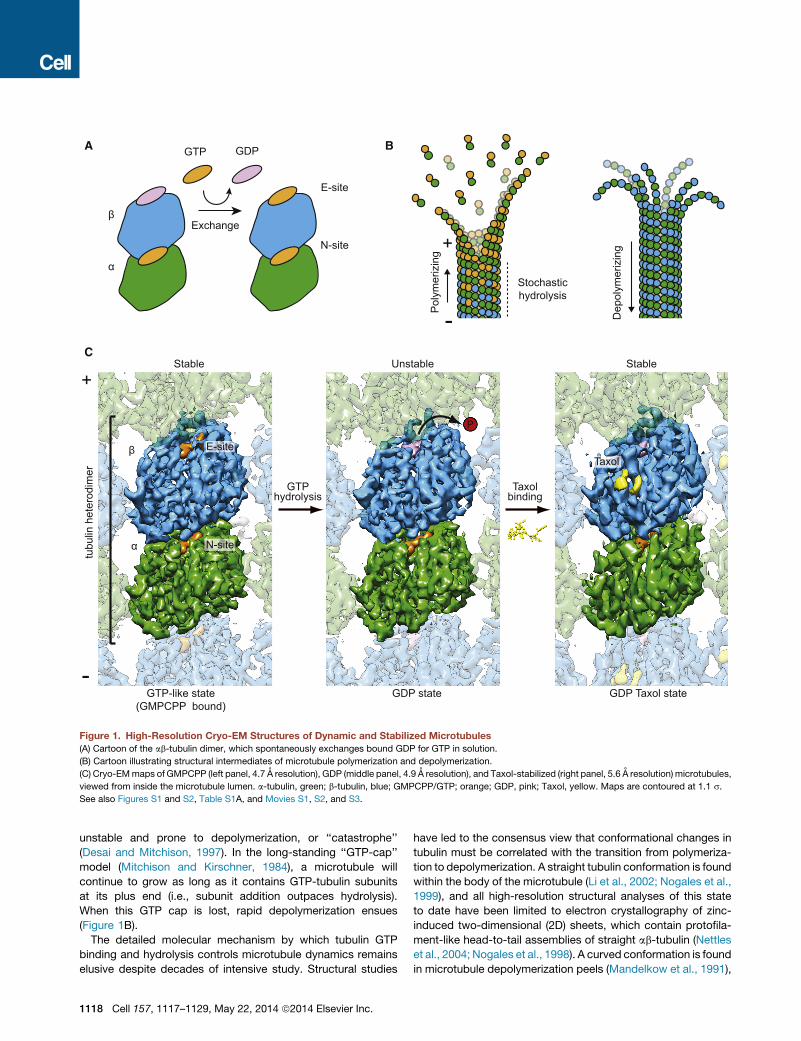
Figure 4. Rearrangements upon Hydrolysis Alter MAP Binding Sites
on the Microtubule Surface
Rearrangements on the microtubule surface. Vectors are colored by sub-
domain as in Figure 3. Binding sites for key MAPs are indicated.
See also Movie S6.
Remodeling of Longitudinal Dimer Contacts Is Coupledto Conformational Changes in a-TubulinThe observed changes at the interdimer contact, around the
E-site, are accompanied by internal rearrangements of the
tubulin dimer involving the intermediate and C-terminal domains
of a-tubulin (Figure 3B, bottom subunit). When aligned on the
N-terminal strands (S1–S5), the rmsd between the intermediate
domains from the GMPCPP and GDP consensus models
(comprising strands S7–S10 and helices H9–H10) shows a sig-
nificant difference of 1.64 A. The structural change is dominated
by a translation of 1.28 A toward the plus end, moving as a unit
with the C-terminal base of H7, effectively tucking up into the
dimer (Figure 3D).
Conformational differences between the electron crystallo-
graphic structure of tubulin, corresponding to a straight, pro-
tofilament-like state, and X-ray crystallographic structures of
curved, peel-like tubulin states, differ most significantly by a
rotation of the intermediate domain and a translation of H7 (Rav-
elli et al., 2004; Gigant et al., 2000; Nogales and Wang, 2006).
Comparing a-tubulin from the crystal structure of bent tubulin
bound to a stathmin-like domain (Protein Data Bank [PDB] ID
code 1SA0; Ravelli et al., 2004) with the GMPCPP and GDP
models, indicates that upon hydrolysis the a-tubulin intermedi-
1122 Cell 157, 1117–1129, May 22, 2014 ª2014 Elsevier Inc.
ate domain within the microtubule undergoes a shift similar to
that reported for the straight to bent transition, but no rotation,
likely because of the lattice constraints (Figure 3D; Movie S5).
This result is consistent with a model wherein reorganization
of the E-site following GTP hydrolysis generates internal strain
within the tubulin dimer that would subsequently relax during
depolymerization, a long-standing concept in the tubulin field
(Caplow et al., 1994). Interestingly, whereas in the stathmin-
bound structure the structural changes within the b subunit
are even larger than those observed in the a-subunit, within
the microtubule the nucleotide has a much smaller effect on
this region of b-tubulin, with no appreciable translation or
rotation.
Interplay between Nucleotide State and MAP BindingSitesWe additionally observe hydrolysis-induced changes that prop-
agate outward to the microtubule surface, where the vast major-
ity of MAPs and motors interact with tubulin (Al-Bassam et al.,
2002; Alushin et al., 2010, 2012; Redwine et al., 2012; Sindelar
and Downing, 2010) (Figure 4; Movie S6). Helices H11 and
H12 in a and b subunits translate 1.1 and 0.53 A, respectively,
away from the lumen upon GTP hydrolysis. For the a subunit,
where changes are most substantial, vector analysis demon-
strates that the movement of the C-terminal helices parallels
the rearrangement we observe in the intermediate domain, sug-
gesting the intermediate domain transmits rearrangements
in the E-site to the microtubule surface (Figure 3B; Movie S6).
The rearrangement of a-H12 is of particular importance, as
this helix constitutes part of the binding site for the kinesin and
cytoplasmic dynein motors (Figure 4) (Al-Bassam et al., 2002;
Redwine et al., 2012; Sindelar and Downing, 2010). This finding
may explain the observation that kinesin preferentially binds
to GMPCPP microtubules over destabilized GDP microtubules
(Nakata et al., 2011).
Recent reports indicate that GTPgS-stabilized microtubules
are an optimal structural mimic of the cap recognized by the
plus-end tracking factor end-binding protein 1 (EB1, Mal3p in
fission yeast) (Maurer et al., 2011; Maurer et al., 2012). Maurer
et al. (2012) reported an additional lateral contact present
in the GTPgS microtubule with respect to others previously
visualized, involving the b-tubulin H3 helix, a region closely
apposed to the E-site g-phosphate. As this helix was found
to be a component of the Mal3p binding site on the micro-
tubule, they proposed that this extra contact could be a key
mechanism for plus-tip recognition (Maurer et al., 2012). In
our studies, we observe minimal rearrangements in this region
(Figure 4B, green), which is never involved in lateral contacts.
We do not believe that this structural disparity is due to a
difference in nonhydrolyzable nucleotide but is likely due to
the difference in resolution of the two studies. On the other
hand, we observe a substantial nucleotide state-dependent
displacement of a-tubulin H10 and the S9-H10 loop in the in-
termediate domain (Figure 4B, purple; Movie S6), which were
also identified as a major component of the Mal3p binding
site. We therefore propose that rearrangement of the a-tubulin
intermediate domain could play a significant role in plus-tip
recognition.

BA Figure 5. Taxol Binding Restores the GDP
Lattice to a GMPCPP-like Extended State
(A) Analogous to Figure 2A, but comparing the
GMPCPP (gold) to the GDP-Taxol (light blue) state.
(B) View from the microtubule lumen of the su-
perimposed GDP (light purple) and GDP-Taxol
(light blue) models. Note the swelling of the Taxol
binding site (1), opening of the E-site interface (2),
and reversal of the longitudinal displacement of the
a-tubulin intermediate domain (3). Nucleotides
from the GDP-Taxol consensus models are dis-
played and colored as in Figure 3. Taxol is yellow.
See also Figure S6 and Movies S2 and S7.
Taxol Binding Restores the GDP Lattice to aGMPCPP-like StateSuperposition of our GMPCPP and GDP-Taxol models demon-
strates that, remarkably, Taxol binding globally reverses the
majority of the conformational changes we observe when
comparing the GMPCPP and GDP states (Figure 5A). We do
not observe any major conformational differences between
the GMPCPP and GDP-Taxol states, such as rearrangement
of tubulin subdomains or formation of a second layer of lateral
contacts in the GMPCPP state, as was reported in a recent
cryo-EM study (Yajima et al., 2012). Possible explanations for
these discrepancies include the different symmetries of the
microtubules chosen for processing (15 PF four-start helical
microtubules versus 14 PF three-start microtubules in this
study), image analysis procedures, the presence of bound kine-
sin in our work, and the different resolutions. The restoration of a
GMPCPP-like state by Taxol extends to the MAP-interacting
regions of the C-terminal domain (Figure 5A), suggesting that
caution should be employed when considering Taxol-bound mi-
crotubules to be in a GDP-like state in studies of MAP-microtu-
bule interactions.
Taxol binds distal to the E-site nucleotide, at the interface be-
tween the nucleotide binding and intermediate domains on the
luminal face of b-tubulin (Nogales et al., 1998) (Figure 1C). The
global remodeling we observe with respect to the GDP structure
suggests that Taxol is capable of allosterically reversing the
Cell 157, 1117–11
conformational transition accompanying
GTP hydrolysis, thus leading to an
extended state at the E-site and a reversal
in the movement of the intermediate
domain in a-tubulin (Figure 5B; Movie
S7). The T3 and T5 nucleotide binding
loops do not return to a GTP-like con-
formation, suggesting that the E-site is
opened by an alternative mechanism. In
order to be accommodated in the b sub-
unit, Taxol acts as a wedge to increase
the volume of the Taxol-binding pocket,
which swells from 883 A3 in the GDP
model to 996 A3 in the GDP-Taxol model
(Figure 5B). The volume of the pocket is
914 A3 in the GMPCPPmodel, suggesting
that most of the volume increase is a
specific effect of Taxol binding. This expansion results from
slight rearrangements around the Taxol binding site (Figure 5B),
which we infer to be allosterically linked to expansion of the
E-site and reversal of the longitudinal displacement of the
a-tubulin intermediate domain. Vector analysis comparing GDP
versus GDP-Taxol and GMPCPP versus GDP-Taxol suggests
that these rearrangements are specifically generated by Taxol
binding (Figure S6).
The electron crystallographic structure of zinc-induced
sheets, 1JFF, was obtained in the stabilizing presence of Taxol.
Intriguingly, the axial repeat in 1JFF is similar to that of the GDP
microtubule (81.2 versus 81.3 A). On the other hand, comparison
of the tubulin dimer structures shows that both a- and b-tubulin
subunits in 1JFF are more dissimilar to any of the three micro-
tubule-based structures we described here, than are the three
structures to each other (Table S1D). The volume of the Taxol
binding pocket is also slightly smaller in the zinc sheets
(956 A3), an observation that, taken together with the different
lateral contacts present in this polymer, suggests that Taxol
may stabilize sheets by an alternative mechanism (there are no
available sheet structures in the absence of a stabilizing drug).
These differences indicate that physiological lateral contacts
are necessary for the allosteric effect of Taxol at the E-site, high-
lighting the importance of studying the mechanisms of microtu-
bule-stabilizing agents in a microtubule context. Future studies
of microtubules at higher resolution will be required to uncover
29, May 22, 2014 ª2014 Elsevier Inc. 1123
A
B
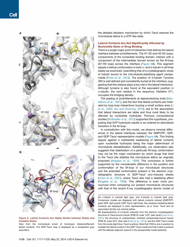
Figure 6. Lateral Contacts Are Highly Similar between Stable and
Unstable StatesView from the microtubule lumen of homotypic interprotofilament
lateral contacts. The GDP-Taxol map is displayed as a transparent gray
isosurface.
1124 Cell 157, 1117–1129, May 22, 2014 ª2014 Elsevier Inc.
the detailed allosteric mechanism by which Taxol restores the
microtubule lattice to a GTP-like state.
Lateral Contacts Are Not Significantly Affected byNucleotide State or Drug BindingThere is a single major point of interaction that defines the lateral
interface between protofilaments. The H1-S2 and H2-S3 loops,
components of the nucleotide binding domain, interact with a
component of the intermediate domain known as the M-loop
(S7-H9 loop) across the interface (Figure 6A). This segment
adopts a helical conformation in both a- and b-tubulin in all three
states we examined, resembling that of a crystallographic study
of tubulin bound to the microtubule-stabilizing agent zampa-
nolide (Prota et al., 2013). The position of b-tubulin Tyrosine
283 is well defined and consistently buried at the interface, sug-
gesting that this residue plays a key role in the lateral interaction.
Although tyrosine is also found at the equivalent position in
a-tubulin, the next residue in the sequence, Histidine 271,
occupies the bridging density.
The peeling of protofilaments at depolymerizing ends (Man-
delkow et al., 1991), and the fact that lateral contacts are medi-
ated by loop-loop interactions burying a small surface area (Li
et al., 2002; Sui and Downing, 2010), led to the assumption
that lateral interactions are labile and thus most likely to be
affected by nucleotide hydrolysis. Previous computational
studies (Grafmuller et al., 2013) supported this hypothesis, pro-
posing that GTP hydrolysis results in an ordered-to-disordered
transition in the M-loop.
In contradiction with this model, we observe minimal differ-
ences in the lateral interfaces between the GMPCPP, GDP,
and GDP-Taxol representative models (Figure 6A). This finding
argues against a substantial weakening of lateral contacts
upon nucleotide hydrolysis being the major determinant of
microtubule destabilization. Additionally, our observation also
suggests that stabilization of a particular M-loop conformation
may not be the major mechanism by which drugs that bind
to the Taxol site stabilize the microtubule lattice as originally
proposed (Nogales et al., 1999). This conclusion is further
supported by the considerable difference in the position and
conformation of the M-loop in our microtubule structures
and the extended conformation present in the electron crys-
tallographic structure of GDP-Taxol zinc-induced sheets
(Lowe et al., 2001), where Taxol also had a stabilizing effect
(Nogales et al., 1995). This difference is even more pro-
nounced when comparing our present microtubule structures
with that of the recent X-ray crystallography atomic model of
(A) a-tubulin is colored light gray, and b-tubulin is colored dark gray.
Consensus models are displayed with lateral contacts colored (GMPCPP,
gold; GDP, light purple; GDP-Taxol, light blue). Key residues mediating lateral
contacts are displayed in stick representation, as is Taxol. Taxol and its
associated density are colored yellow.
(B) Superpositions of b-tubulin subunits from the electron crystallographic
structure of Taxol-bound tubulin (PDB ID code 1JFF; dark blue) (Lowe et al.,
2001), the structure of unassembled, inhibited zampanolide-bound tubulin
(PDB ID code 4I4T; dark red) (Prota et al., 2013), and the GDP-Taxol model
from this study (light blue; Taxol is yellow). Note that Y283 is only in position to
mediate the lateral contact in the GDP-Taxol model and that a clash is present
with the laterally adjacent subunit in the zampanolide model (asterisk).

Figure 7. Proposed Model of Destabilizing
and Stabilizing Structural Transitions in the
Microtubule Lattice
Cartoon of conformational transitions colored as in
Figure 1, except the a-tubulin intermediate domain
is purple. Left: nucleotide hydrolysis and phos-
phate release leads to compaction of the E-site
and rearrangement of the a-tubulin intermediate
domain (middle), generating destabilizing strain,
while tubulin remains within the constraints of the
microtubule lattice. Taxol binding (right, top) allo-
sterically leads to a reversal of E-site compaction
and the a-tubulin rearrangement; unraveling the
detailed mechanism of this transition will require
structural analysis at near-atomic resolution.
Subtle structural changes could be propagated
across the E-site interdimer interface (up arrow),
within the dimer (down arrow) or both. In the
absence of binding by a stabilizing agent, strain
would be dissipated by tubulin bending during
catastrophe (right, bottom), when the a-tubulin-
intermediate domain (and b-tubulin intermediate
domain, dark blue) is capable of undergoing rota-
tion due to the relief of steric constraints imposed
by lateral contacts.
zampanolide-bound, unassembled tubulin (PDB ID code 4I4T;
Prota et al., 2013), where the helical M-loop is in a position
that would clash with the laterally adjacent protofilament (Fig-
ure 6B). Rather, our data are most consistent with a model
wherein the microtubule-stabilizing effect of Taxol concerns
the structure and strength of longitudinal interfaces linked to a
GTP-like conformation of the tubulin dimer, as originally envi-
sioned by Amos and Lowe (1999).
DISCUSSION
Our results suggest a mechanism by which nucleotide hydroly-
sis is linked to destabilization of the microtubule lattice (Fig-
ure 7). We propose that upon hydrolysis and phosphate release,
compaction of the E-site leads to an energetically unfavorable
longitudinal translation of the a-tubulin intermediate domain,
including the H7 helix. The observed rearrangements of tubulin
upon hydrolysis are reminiscent of the conformational trajectory
toward a depolymerized, bent tubulin structure (Ravelli et al.,
2004). In the context of the microtubule, only the translational
component of that trajectory is present, whereas the rotational
component is not apparent. This difference is likely because
of lattice constraints that maintain a strained, straight conforma-
tion compatible with the microtubule lattice. Thus, we propose
that hydrolysis leads to conformational strain that would be
released by bending during depolymerization. This model is
Cell 157, 1117–11
consistent with the changes we observe
upon Taxol binding, a stabilizer of the
GDP lattice. The predominant effect
of Taxol is to restore the longitudinal
interface and the a-tubulin-intermediate
domain to a GTP-like state, suggesting
that these structural transitions are
responsible for destabilization of the microtubule lattice upon
nucleotide hydrolysis.
The substantial changes we have visualized by comparing
GMPCPP, GDP, and GDP-Taxol microtubules contrast with
the minimal effect of nucleotide state on the inhibited form of
tubulin previously described by X-ray crystallographic studies.
In the context of an RB3-bound, bent tubulin dimer, the presence
of a gamma phosphate at the E-site affected the position of
merely two amino acids adjacent to the site, rearrangements
which were not further propagated throughout the structure
(Nawrotek et al., 2011). This suggests that the inhibition of tubulin
for crystallization purposes by cellular factors that bind and
stabilize its bent conformation can override the conformational
effects of nucleotide hydrolysis and stabilizing drugs that other-
wise occur within a microtubule.
More recently, the structure of the RB3-inhibited tubulin bound
to epothilone A or zampanolide showed the ordering of the
adjacent M-loop region (otherwise unresolved in previous crys-
tal structures) into a small helix, without any other significant
changes. This structure led to the proposal that inducing the
folded conformation of the b-tubulin M-loop was the primary
mechanism by which antimitotic agents that bind at the Taxol
site stabilize the microtubule lattice, i.e., by facilitating lateral
contacts (Prota et al., 2013). However, the position of the
M-loop region in the context of a rotated intermediate domain
clashes with the neighboring lateral subunit when this crystal
29, May 22, 2014 ª2014 Elsevier Inc. 1125

structure is placed in the microtubule lattice. Our finding that
lateral contacts are not substantially altered in different nucleo-
tide states or upon Taxol binding suggests that this set of tubulin
polymerization interfaces plays a more passive role in microtu-
bule stability than anticipated. Our results instead support a
model in which the energy contribution of lateral contacts may
be essentially constant, whereas longitudinal contacts and
conformational strain within the tubulin monomers (particularly
a-tubulin) are modulated.
It is possible that the folding of the M-loop observed in the
crystal structure of zampanolide/epothilone A-bound tubulin
could facilitate the incorporation of tubulin dimers into themicro-
tubule and thus be a component of the assembly-driving action
of Taxol-like compounds. Once assembled, however, the stabil-
ity of the microtubule lattice is likely governed by the total energy
of lateral and longitudinal contacts in the GTP state versus the
GDP state. Our analysis supports a model in which micro-
tubule-stabilizing agents like Taxol primarily exert a stabilizing
influence on the microtubule lattice by affecting internal rear-
rangements of the tubulin dimer that modulate conformational
strain and longitudinal contacts. This concept is consistent
with a study demonstrating that Taxol induces the formation of
straight individual protofilaments from GDP-tubulin peels (Elie-
Caille et al., 2007). In particular, our studies suggest that modu-
lating longitudinal interfaces by altering the conformation of
tubulin is a robust mechanism for affecting microtubule stability,
as previously predicted (Amos and Lowe, 1999), a model that
should aid future design efforts for drugs that target the Taxol
binding site.
Future ProspectsOur study demonstrates that microtubules are structurally trac-
table by cryo-EM at resolutions approaching those obtained by
X-ray crystallography, thus enabling the detailed study of nucle-
otide and drug effects and the interaction of microtubule binding
proteins in the context of the native tubulin polymer. The three
microtubule models we present here should provide an impor-
tant resource for computational modeling studies of dynamic
instability, effects of drug binding, and the physical properties
of microtubules.
Several outstanding questions and concerns remain to be
addressed. The presence of kinesin, currently a technical neces-
sity, may alter certain structural transitions occurring between
different states. Indeed, our reconstructions of kinesin-free mi-
crotubules suggest that the presence of kinesin slightly dampens
the magnitude of E-site remodeling (Movie S3). We optimistically
anticipate that future methodological advances in image pro-
cessing and instrumentation will allow similar and hopefully
even superior resolution to be achieved for undecorated micro-
tubule specimens.
Additionally, caution must be employed when interpreting the
structural details of GMPCPP-bound microtubules as the bona
fide ‘‘GTP state’’ of the microtubule lattice, in light of the dif-
ferences observed with different GTP analogs. One potential
explanation for such differences is that there are additional
structural stages in the microtubule polymerization-depolymer-
ization cycle, such as one corresponding to a post-hydrolysis
state, but before Pi release, that may be mimicked by one of
1126 Cell 157, 1117–1129, May 22, 2014 ª2014 Elsevier Inc.
these GTP analogs. Tubulin could also adopt several distinct
conformations prior to GTP hydrolysis (related perhaps to the
transition from an open growth sheet to the closed microtubule
cylinder), which different analogs may selectively stabilize.
Future high-resolution studies of microtubules bound to GTPgS
and other nucleotide state mimics, like the GDP-Pi analog
GDP-BeF3, will be important to investigate and address these
issues.
Finally, although our present structural analysis leads to the
unexpected conclusion that lateral contacts are not substan-
tially altered upon nucleotide hydrolysis, we cannot rule out
the possibility of more subtle effects that could nevertheless
contribute significantly to energetics and which would only be
revealed at higher, i.e., atomic, resolution. Additionally, hetero-
typic lateral contacts across the seam, where tubulin subunits
within a single dimer on one side of the interface are contacting
subunits contributed by two dimers on the other, are more
likely to be affected by the change in dimer repeat upon remod-
eling of the E-site with GTP hydrolysis. The seam could there-
fore constitute a ‘‘weak point’’ to initiate disruption of the lattice
at the onset of catastrophe, consistent with early observations
of microtubule conversion to sheets when depolymerization
is induced by cold treatment (Simon and Salmon, 1990). A
mismatch of axial repeats will not only occur at the seam of
microtubule segments with a homogeneous nucleotide state
but would also be present at homotypic lateral contacts in
a transition zone along the microtubule lattice, where hydroly-
sis is taking place and therefore containing a mixture of GTP
and GDP states. Although a mixed nucleotide lattice is
not amenable to experimental analysis by current structural
methods, which require averaging of microtubule segments,
computational modeling of these transitions, guided by the
structural findings described here, could contribute to our mo-
lecular understanding of the catastrophe process. In particular,
it would be interesting to explore potential cooperativity in the
structural transitions we have described, which seems likely
given the steric constraints imposed by the tightly packed
microtubule lattice.
EXPERIMENTAL PROCEDURES
Full details of the experimental procedures are presented in the Extended
Experimental Procedures.
Kinesin and Microtubule Preparation for Cryo-EM
Kinesin was diluted into EM buffer (for buffer compositions, see the Extended
Experimental Procedures) and desalted. ATP was added, and aggregates
were removed by ultracentrifugation. Taxol-stabilized GDP microtubules
(MTs) were prepared as previously described (Alushin et al., 2010). GMPCPP
microtubuleswere prepared by polymerizing porcine tubulin in CB1 buffer. The
MTswere pelleted and resuspended in cold EMbuffer, generating a solution of
GDP-tubulin. After clarification, GMPCPP was exchanged for bound GDP
for 1 hr on ice. GMPCPP MTs were then polymerized in EM buffer at 37�C.Dynamic (primarily GDP) microtubules were polymerized in CB1 buffer at
37�C for 30 min and then pelleted.
All cryo-EM samples were prepared on glow-discharged C-flat grids (Proto-
chips) using a Vitrobot (Maastricht Instruments). Taxol and GMPCPP microtu-
bules were diluted to 0.25 mg/ml in EM buffer, and 4 ml was applied to the grid.
TheMTs were allowed to adsorb, and kinesin was then added to the grid. After
a short incubation, the sample was blotted and plunged into liquid ethane.

Pelleted dynamic GDP microtubules were resuspended into warm kinesin
solution. From this MT-kinesin mixture, 4 ml was added to the EM grid and
allowed to incubate for 1 min before blotting and vitrification.
Electron Microscopy and Image Processing
Cryo-grids were loaded into a 626 single-tilt cryo-transfer system (Gatan) and
imaged in a Titan electron microscope (FEI) operated at 300 keV. Data were
collected using the Leginon software (Suloway et al., 2005) for exposure tar-
geting, and the FEI Low-Dose Kit was used for focusing and image acquisition.
Low-dose exposures (25 e�/A2) were acquired at 72,0003 magnification on
Kodak SO163 film, with underfocus ranging from 1.4 to 3.5 mm. Films were
digitized at 0.87 A/pixel using a Nikon CoolScan 8000 (see Table S1A for the
number of films digitized for each sample). Image processing was performed
within the Appion processing environment (Lander et al., 2009). Contrast trans-
fer function (CTF) was estimated, and the best-quality micrographs selected
for further processing. Microtubules were manually selected, and overlapping
segments were extracted with a spacing of 80 A, phase flipped, and binned by
a factor of two. The particle stacks were subjected to iterative multivariate
statistical analysis (MSA) and multireference alignment (MRA). Particles in
classes that did not clearly show kinesin density (for kinesin-bound samples),
or whose power spectra did not exhibit layer lines beyond 10 A, were excluded
from further processing, as were classes that did not have either 13 or 14
protofilaments.
3D Reconstruction
Undecorated 13 and 14 pf MT densities were used as initial models for a
preliminary reconstruction of the kinesin-bound Taxol MT data set. The proto-
filament with the best apparent kinesin density was extracted and used to
generate 13 and 14 pf microtubule maps with seams. These preliminary den-
sities served as starting models for refinement of complete data sets using
IHRSR (Egelman, 2007). Our modified IHRSR reconstruction schema (Fig-
ure S2) allows us to take advantage of the helical character of the microtubule
lattice, without artifacts due to seam symmetrization. A final refinement of
the alignment parameters was performed only for the 14 pf MT particles using
FREALIGN (Grigorieff, 2007), with a modified script that incorporated the sym-
metrization and MT seam regeneration after each round. The final resolution
for each reconstruction was estimated by calculating the Fourier shell corre-
lation (FSC) of a single dimer (Figure S2B). Resolution-dependent negative
b-factors were applied to the three reconstructions using BFACTOR (http://
grigoriefflab.janelia.org/bfactor).
Atomic Model Building and Refinement with Rosetta
Initial models of the microtubule lattice were obtained through rigid-body
docking the electron crystallographic structure of tubulin (PDB ID code
1JFF; Lowe et al., 2001) into the cryo-EM density maps using Chimera. Ligand
conformations were copied from the corresponding crystal structures and
were held fixed during refinement. The lattice was modeled in a 33 3 arrange-
ment using symmetry operations as previously described (DiMaio et al., 2011).
Structure refinement was carried out using the RosettaCM protocol (Song
et al., 2013) supplemented with an electron density term (DiMaio et al.,
2009). Regions in the starting 1JFF structure that fit the density poorly were
rebuilt using short fragments fromproteins of known structure with similar local
sequences, and recombination with X-ray crystallographic tubulin structures
at higher resolution in different nucleotide states was used to increase accu-
racy in the vicinity of the nucleotide binding sites (see the Extended Experi-
mental Procedures for details). Following loop region rebuilding and structure
hybridization, models were relaxed in the Rosetta all-atom force field supple-
mented with the electron density term. Analyses in the text are based on
the lowest 1% total energy (Rosetta energy + electron density fit) models for
each map. The spread of energies and rmsds of these low-energy ensembles
was small (average 0.56 A rmsd).
Molecular Graphics
All structural figures andmovies were generated with UCSFChimera (Goddard
et al., 2007; Pettersen et al., 2004). Displacement vectors were generated
with a Python program that creates marker (.cmm) files that are viewable
and editable in Chimera (available upon request).
ACCESSION NUMBERS
The cryo-EM maps and coordinates of the energy-minimized consensus
Rosetta models have been deposited for the three microtubule studies
we describe: GMPCPP-stabilized (Electron Microscopy Data Bank [EMDB]
accession number 5895; PDB ID code 3J6E), GDP, dynamic (EMDB accession
number 5896; PDB ID code 36JF), and Taxol-stabilized microtubules (EMDB
accession number 5897; PDB ID code 3J6G).
SUPPLEMENTAL INFORMATION
Supplemental Information includes Extended Experimental Procedures, one
table, seven movies, and six figures and can be found with this article online
at http://dx.doi.org/10.1016/j.cell.2014.03.053.
AUTHOR CONTRIBUTIONS
G.M.A. andG.C.L. performed sample preparation, electronmicroscopy exper-
iments, and data processing. E.H.K. performed Rosetta modeling analysis.
R.Z. performed data processing. G.M.A. wrote the initial draft of the paper,
and E.H.K. generated supplemental movies. All authors contributed to data
analysis and to assembling the manuscript.
ACKNOWLEDGMENTS
We thank Patricia Grob and TomHouweling for electronmicroscopy and com-
puter support, respectively, Tom Goddard for help with UCSF Chimera, Yifan
Song and Frank DiMaio for making code available for use prior to publication,
and Stuart Howes for assistance with kinesin purification. We are grateful to
Robert Glaeser for assistance with data collection and many discussions.
The kinesin expression construct was the gift of Erik Jonsson and Ron Vale.
This work was funded by the National Institute of General Medical Sciences
(GM051487) (to E.N.) and a Damon Runyon Cancer Research Foundation
fellowship (DRG 2055-10) (to G.C.L.). E.N. and D.B. are Howard Hughes Med-
ical Institute Investigators.
Received: December 18, 2013
Revised: January 17, 2014
Accepted: March 18, 2014
Published: May 22, 2014
REFERENCES
Al-Bassam, J., Ozer, R.S., Safer, D., Halpain, S., and Milligan, R.A. (2002).
MAP2 and tau bind longitudinally along the outer ridges of microtubule proto-
filaments. J. Cell Biol. 157, 1187–1196.
Aldaz, H., Rice, L.M., Stearns, T., and Agard, D.A. (2005). Insights into micro-
tubule nucleation from the crystal structure of human gamma-tubulin. Nature
435, 523–527.
Alushin, G.M., Ramey, V.H., Pasqualato, S., Ball, D.A., Grigorieff, N., Musac-
chio, A., and Nogales, E. (2010). The Ndc80 kinetochore complex forms olig-
omeric arrays along microtubules. Nature 467, 805–810.
Alushin, G.M., Musinipally, V., Matson, D., Tooley, J., Stukenberg, P.T., and
Nogales, E. (2012). Multimodal microtubule binding by the Ndc80 kinetochore
complex. Nat. Struct. Mol. Biol. 19, 1161–1167.
Amos, L.A., and Lowe, J. (1999). How taxol stabilises microtubule structure.
Chem. Biol. 6, R65–R69.
Arnal, I., and Wade, R.H. (1995). How does taxol stabilize microtubules? Curr.
Biol. 5, 900–908.
Ayaz, P., Ye, X., Huddleston, P., Brautigam, C.A., and Rice, L.M. (2012). A
TOG:ab-tubulin complex structure reveals conformation-based mechanisms
for a microtubule polymerase. Science 337, 857–860.
Aylett, C.H., Lowe, J., and Amos, L.A. (2011). New insights into the mecha-
nisms of cytomotive actin and tubulin filaments. Int. Rev. Cell Mol. Biol. 292,
1–71.
Cell 157, 1117–1129, May 22, 2014 ª2014 Elsevier Inc. 1127
Barbier, P., Dorleans, A., Devred, F., Sanz, L., Allegro, D., Alfonso, C., Knos-
sow, M., Peyrot, V., and Andreu, J.M. (2010). Stathmin and interfacial microtu-
bule inhibitors recognize a naturally curved conformation of tubulin dimers.
J. Biol. Chem. 285, 31672–31681.
Caplow, M., Ruhlen, R.L., and Shanks, J. (1994). The free energy for hydrolysis
of a microtubule-bound nucleotide triphosphate is near zero: all of the free
energy for hydrolysis is stored in the microtubule lattice. J. Cell Biol. 127,
779–788.
Chretien, D., Fuller, S.D., and Karsenti, E. (1995). Structure of growing micro-
tubule ends: two-dimensional sheets close into tubes at variable rates. J. Cell
Biol. 129, 1311–1328.
Desai, A., and Mitchison, T.J. (1997). Microtubule polymerization dynamics.
Annu. Rev. Cell Dev. Biol. 13, 83–117.
DiMaio, F., Tyka, M.D., Baker, M.L., Chiu, W., and Baker, D. (2009). Refine-
ment of protein structures into low-resolution density maps using rosetta.
J. Mol. Biol. 392, 181–190.
DiMaio, F., Leaver-Fay, A., Bradley, P., Baker, D., and Andre, I. (2011).
Modeling symmetric macromolecular structures in Rosetta3. PLoS ONE 6,
e20450.
Dumontet, C., and Jordan, M.A. (2010). Microtubule-binding agents: a
dynamic field of cancer therapeutics. Nat. Rev. Drug Discov. 9, 790–803.
Egelman, E.H. (2007). The iterative helical real space reconstruction method:
surmounting the problems posed by real polymers. J. Struct. Biol. 157, 83–94.
Elie-Caille, C., Severin, F., Helenius, J., Howard, J., Muller, D.J., and Hyman,
A.A. (2007). Straight GDP-tubulin protofilaments form in the presence of taxol.
Curr. Biol. 17, 1765–1770.
Gigant, B., Curmi, P.A., Martin-Barbey, C., Charbaut, E., Lachkar, S., Lebeau,
L., Siavoshian, S., Sobel, A., and Knossow, M. (2000). The 4 A X-ray structure
of a tubulin:stathmin-like domain complex. Cell 102, 809–816.
Gigant, B., Wang, C., Ravelli, R.B., Roussi, F., Steinmetz, M.O., Curmi, P.A.,
Sobel, A., and Knossow,M. (2005). Structural basis for the regulation of tubulin
by vinblastine. Nature 435, 519–522.
Goddard, T.D., Huang, C.C., and Ferrin, T.E. (2007). Visualizing density maps
with UCSF Chimera. J. Struct. Biol. 157, 281–287.
Grafmuller, A., Noya, E.G., and Voth, G.A. (2013). Nucleotide-dependent
lateral and longitudinal interactions in microtubules. J. Mol. Biol. 425, 2232–
2246.
Grigorieff, N. (2007). FREALIGN: high-resolution refinement of single particle
structures. J. Struct. Biol. 157, 117–125.
Hyams, J.S., and Lloyd, C.W. (1993). Microtubules (New York: Wiley-Liss).
Hyman, A.A., Salser, S., Drechsel, D.N., Unwin, N., and Mitchison, T.J. (1992).
Role of GTP hydrolysis in microtubule dynamics: information from a slowly
hydrolyzable analogue, GMPCPP. Mol. Biol. Cell 3, 1155–1167.
Hyman, A.A., Chretien, D., Arnal, I., andWade, R.H. (1995). Structural changes
accompanying GTP hydrolysis in microtubules: information from a slowly hy-
drolyzable analogue guanylyl-(alpha,beta)-methylene-diphosphonate. J. Cell
Biol. 128, 117–125.
Lander, G.C., Stagg, S.M., Voss, N.R., Cheng, A., Fellmann, D., Pulokas, J.,
Yoshioka, C., Irving, C., Mulder, A., Lau, P.W., et al. (2009). Appion: an inte-
grated, database-driven pipeline to facilitate EM image processing.
J. Struct. Biol. 166, 95–102.
Li, H., DeRosier, D.J., Nicholson, W.V., Nogales, E., and Downing, K.H. (2002).
Microtubule structure at 8 A resolution. Structure 10, 1317–1328.
Lowe, J., Li, H., Downing, K.H., and Nogales, E. (2001). Refined structure of
alpha beta-tubulin at 3.5 A resolution. J. Mol. Biol. 313, 1045–1057.
Mandelkow, E.-M., Mandelkow, E., and Milligan, R.A. (1991). Microtubule
dynamics and microtubule caps: a time-resolved cryo-electron microscopy
study. J. Cell Biol. 114, 977–991.
Maurer, S.P., Bieling, P., Cope, J., Hoenger, A., and Surrey, T. (2011).
GTPgammaS microtubules mimic the growing microtubule end structure
recognized by end-binding proteins (EBs). Proc. Natl. Acad. Sci. USA 108,
3988–3993.
1128 Cell 157, 1117–1129, May 22, 2014 ª2014 Elsevier Inc.
Maurer, S.P., Fourniol, F.J., Bohner, G., Moores, C.A., and Surrey, T. (2012).
EBs recognize a nucleotide-dependent structural cap at growing microtubule
ends. Cell 149, 371–382.
McIntosh, J.R., Volkov, V., Ataullakhanov, F.I., and Grishchuk, E.L. (2010).
Tubulin depolymerization may be an ancient biological motor. J. Cell Sci.
123, 3425–3434.
Menendez, M., Rivas, G., Dıaz, J.F., and Andreu, J.M. (1998). Control of the
structural stability of the tubulin dimer by one high affinity bound magnesium
ion at nucleotide N-site. J. Biol. Chem. 273, 167–176.
Mitchison, T.J. (1993). Localization of an exchangeable GTP binding site at the
plus end of microtubules. Science 261, 1044–1047.
Mitchison, T., and Kirschner, M. (1984). Dynamic instability of microtubule
growth. Nature 312, 237–242.
Muller-Reichert, T., Chretien, D., Severin, F., and Hyman, A.A. (1998). Struc-
tural changes at microtubule ends accompanying GTP hydrolysis: information
from a slowly hydrolyzable analogue of GTP, guanylyl (a,b)methylenedi-
phosphonate. Proc. Natl. Acad. Sci. USA 95, 3661–3666.
Nakata, T., Niwa, S., Okada, Y., Perez, F., and Hirokawa, N. (2011). Preferential
binding of a kinesin-1 motor to GTP-tubulin-rich microtubules underlies polar-
ized vesicle transport. J. Cell Biol. 194, 245–255.
Nawrotek, A., Knossow, M., and Gigant, B. (2011). The determinants that
govern microtubule assembly from the atomic structure of GTP-tubulin.
J. Mol. Biol. 412, 35–42.
Nettles, J.H., Li, H., Cornett, B., Krahn, J.M., Snyder, J.P., and Downing, K.H.
(2004). The binding mode of epothilone A on alpha,beta-tubulin by electron
crystallography. Science 305, 866–869.
Nogales, E. (2000). Structural insights into microtubule function. Annu. Rev.
Biochem. 69, 277–302.
Nogales, E., andWang, H.W. (2006). Structural mechanisms underlying nucle-
otide-dependent self-assembly of tubulin and its relatives. Curr. Opin. Struct.
Biol. 16, 221–229.
Nogales, E., Wolf, S.G., Zhang, S.X., and Downing, K.H. (1995). Preservation
of 2-D crystals of tubulin for electron crystallography. J. Struct. Biol. 115,
199–208.
Nogales, E., Wolf, S.G., and Downing, K.H. (1998). Structure of the a b tubulin
dimer by electron crystallography. Nature 391, 199–203.
Nogales, E., Whittaker, M., Milligan, R.A., and Downing, K.H. (1999). High-res-
olution model of the microtubule. Cell 96, 79–88.
Pettersen, E.F., Goddard, T.D., Huang, C.C., Couch, G.S., Greenblatt, D.M.,
Meng, E.C., and Ferrin, T.E. (2004). UCSF Chimera—a visualization system
for exploratory research and analysis. J. Comput. Chem. 25, 1605–1612.
Prota, A.E., Bargsten, K., Zurwerra, D., Field, J.J., Dıaz, J.F., Altmann, K.H.,
and Steinmetz, M.O. (2013). Molecular mechanism of action of microtubule-
stabilizing anticancer agents. Science 339, 587–590.
Ravelli, R.B., Gigant, B., Curmi, P.A., Jourdain, I., Lachkar, S., Sobel, A., and
Knossow, M. (2004). Insight into tubulin regulation from a complex with colchi-
cine and a stathmin-like domain. Nature 428, 198–202.
Redwine, W.B., Hernandez-Lopez, R., Zou, S., Huang, J., Reck-Peterson,
S.L., and Leschziner, A.E. (2012). Structural basis for microtubule binding
and release by dynein. Science 337, 1532–1536.
Rice, L.M., Montabana, E.A., and Agard, D.A. (2008). The lattice as allosteric
effector: structural studies of alphabeta- and gamma-tubulin clarify the role
of GTP in microtubule assembly. Proc. Natl. Acad. Sci. USA 105, 5378–5383.
Rieder, C.L., and Salmon, E.D. (1994). Motile kinetochores and polar ejection
forces dictate chromosome position on the vertebrate mitotic spindle. J. Cell
Biol. 124, 223–233.
Simon, J.R., and Salmon, E.D. (1990). The structure ofmicrotubule ends during
the elongation and shortening phases of dynamic instability examined by
negative-stain electron microscopy. J. Cell Sci. 96, 571–582.
Sindelar, C.V., and Downing, K.H. (2010). An atomic-level mechanism for
activation of the kinesin molecular motors. Proc. Natl. Acad. Sci. USA 107,
4111–4116.
Song, Y., DiMaio, F., Wang, R.Y., Kim, D., Miles, C., Brunette, T., Thomp-
son, J., and Baker, D. (2013). High-resolution comparative modeling with
RosettaCM. Structure 21, 1735–1742.
Sui, H., and Downing, K.H. (2010). Structural basis of interprotofilament inter-
action and lateral deformation of microtubules. Structure 18, 1022–1031.
Suloway, C., Pulokas, J., Fellmann, D., Cheng, A., Guerra, F., Quispe, J.,
Stagg, S., Potter, C.S., and Carragher, B. (2005). Automated molecular micro-
scopy: the new Leginon system. J. Struct. Biol. 151, 41–60.
Vale, R.D., Coppin, C.M., Malik, F., Kull, F.J., and Milligan, R.A. (1994). Tubulin
GTP hydrolysis influences the structure, mechanical properties, and kinesin-
driven transport of microtubules. J. Biol. Chem. 269, 23769–23775.
Wang, H.W., and Nogales, E. (2005). Nucleotide-dependent bending flexibility
of tubulin regulates microtubule assembly. Nature 435, 911–915.
Yajima, H., Ogura, T., Nitta, R., Okada, Y., Sato, C., and Hirokawa, N. (2012).
Conformational changes in tubulin in GMPCPP and GDP-taxol microtubules
observed by cryoelectron microscopy. J. Cell Biol. 198, 315–322.
Cell 157, 1117–1129, May 22, 2014 ª2014 Elsevier Inc. 1129