GSK3β and β-Catenin Modulate Radiation Cytotoxicity in Pancreatic Cancer
9
GSK3β and β-Catenin Modulate Radiation Cytotoxicity in Pancreatic Cancer 1 Richard L. Watson * ,2 , Aaron C. Spalding * ,†,2 , Steven P. Zielske * , Meredith Morgan * , Alex C. Kim ‡ , Guido T. Bommer §,¶ , Hagit Eldar-Finkelman # , Thomas Giordano ** , Eric R. Fearon ‡,§,¶,# , Gary D. Hammer ‡ , Theodore S. Lawrence * and Edgar Ben-Josef * *Department of Radiation Oncology, University of Michigan Medical School, Ann Arbor, MI, USA; † The Norton Cancer Institute Radiation Center and Kosair Children’s Hospital, Louisville, KY, USA; ‡ Department of Internal Medicine—Metabolism, Endocrinology & Diabetes, University of Michigan Medical School, Ann Arbor, MI, USA; § Department of Internal Medicine, University of Michigan Medical School, Ann Arbor, MI, USA; ¶ Department of Internal Medicine—Molecular Medicine and Genetics, University of Michigan Medical School, Ann Arbor, MI, USA; # Department of Human Genetics, Tel Aviv University, Ramat Aviv, Israel; **Department of Pathology, University of Michigan Medical School, Ann Arbor, MI, USA Abstract BACKGROUND: Knowledge of factors and mechanisms contributing to the inherent radioresistance of pancreatic cancer may improve cancer treatment. Irradiation inhibits glycogen synthase kinase 3β (GSK3β) by phosphorylation at serine 9. In turn, release of cytosolic membrane β-catenin with subsequent nuclear translocation promotes survival. Both GSK3β and β-catenin have been implicated in cancer cell proliferation and resistance to death. METHODS: We investigated pancreatic cancer cell survival after radiation in vitro and in vivo, with a particular focus on the role of the function of the GSK3β/β-catenin axis. RESULTS: Lithium chloride, RNAi-medicated silencing of GSK3β, or the expression of a kinase dead mutant GSK3β resulted in radioresistance of Panc1 and BxPC3 pancreatic cancer cells. Conversely, ectopic expression of a constitutively active form of GSK3β resulted in radio- sensitization of Panc1 cells. GSK3β silencing increased radiation-induced β-catenin target gene expression as measured by studies of AXIN2 and LEF1 transcript levels. Western blot analysis of total and phosphorylated levels of GSK3β and β-catenin showed that GSK3β inhibition resulted in stabilization of β-catenin. Xenografts of both BxPC3 and Panc1 with targeted silencing of GSK3β exhibited radioresistance in vivo. Silencing of β-catenin resulted in radiosensitization, whereas a nondegradable β-catenin construct induced radioresistance. CONCLUSIONS: These data support the hypothesis that GSK3β modulates the cellular response to radiation in a β-catenin–dependent mechanism. Further understanding of this pathway may enhance the development of clinical trials combining drugs inhibiting β-catenin activation with radiation and chemotherapy in locally advanced pancreatic cancer. Neoplasia (2010) 12, 357–365 Address all correspondence to: Aaron C. Spalding, MD, PhD, The Norton Cancer Institute Radiation Center, 4001 Dutchmans Ln, Louisville, KY 40207. E-mail: [email protected] 1 These studies were supported by an American Society of Therapeutic Radiology and Oncology Resident Seed Grant (A.S.), and National Institutes ofHealth R03 CA127050-01 (E.B.J.). These studies were conducted in partial fulfillment for the requirements (R.W.) for graduation with Honors, College of Literature, Science, and the Arts, from the University of Michigan. A. Spalding has been designated a B. Leonard Holman Pathway Fellow by the American Board of Radiology, and this work was presented in part at the 2008 Gastrointestinal Malignancies Symposium. 2 These authors contributed equally to this work. Received 30 December 2009; Revised 15 February 2010; Accepted 16 February 2010 Copyright © 2010 Neoplasia Press, Inc. All rights reserved 1522-8002/10/$25.00 DOI 10.1593/neo.92112 www.neoplasia.com Volume 12 Number 5 May 2010 pp. 357–365 357
Transcript of GSK3β and β-Catenin Modulate Radiation Cytotoxicity in Pancreatic Cancer

GSK3β and β-Catenin ModulateRadiation Cytotoxicity inPancreatic Cancer1
Richard L. Watson*,2, Aaron C. Spalding*,†,2,Steven P. Zielske*, Meredith Morgan*,Alex C. Kim‡, Guido T. Bommer§,¶,Hagit Eldar-Finkelman#, Thomas Giordano**,Eric R. Fearon‡,§,¶,#, Gary D. Hammer‡,Theodore S. Lawrence* and Edgar Ben-Josef*
*Department of Radiation Oncology, University ofMichigan Medical School, Ann Arbor, MI, USA; †The NortonCancer Institute Radiation Center and Kosair Children’sHospital, Louisville, KY, USA; ‡Department of InternalMedicine—Metabolism, Endocrinology & Diabetes,University of Michigan Medical School, Ann Arbor, MI, USA;§Department of Internal Medicine, University of MichiganMedical School, Ann Arbor, MI, USA; ¶Department ofInternal Medicine—Molecular Medicine and Genetics,University of Michigan Medical School, Ann Arbor, MI, USA;#Department of Human Genetics, Tel Aviv University, RamatAviv, Israel; **Department of Pathology, University ofMichigan Medical School, Ann Arbor, MI, USA
AbstractBACKGROUND: Knowledge of factors and mechanisms contributing to the inherent radioresistance of pancreatic cancer mayimprove cancer treatment. Irradiation inhibits glycogen synthase kinase 3β (GSK3β) by phosphorylation at serine 9. In turn, releaseof cytosolic membrane β-catenin with subsequent nuclear translocation promotes survival. Both GSK3β and β-catenin have beenimplicated in cancer cell proliferation and resistance to death. METHODS: We investigated pancreatic cancer cell survival afterradiation in vitro and in vivo, with a particular focus on the role of the function of the GSK3β/β-catenin axis. RESULTS: Lithiumchloride, RNAi-medicated silencing of GSK3β, or the expression of a kinase dead mutant GSK3β resulted in radioresistance ofPanc1 and BxPC3 pancreatic cancer cells. Conversely, ectopic expression of a constitutively active form of GSK3β resulted in radio-sensitization of Panc1 cells. GSK3β silencing increased radiation-induced β-catenin target gene expression asmeasured by studiesof AXIN2 and LEF1 transcript levels. Western blot analysis of total and phosphorylated levels of GSK3β and β-catenin showed thatGSK3β inhibition resulted in stabilization of β-catenin. Xenografts of both BxPC3 and Panc1 with targeted silencing of GSK3βexhibited radioresistance in vivo. Silencing of β-catenin resulted in radiosensitization, whereas a nondegradable β-catenin constructinduced radioresistance. CONCLUSIONS: These data support the hypothesis that GSK3β modulates the cellular response toradiation in a β-catenin–dependent mechanism. Further understanding of this pathway may enhance the development of clinicaltrials combining drugs inhibiting β-catenin activation with radiation and chemotherapy in locally advanced pancreatic cancer.
Neoplasia (2010) 12, 357–365
Address all correspondence to: Aaron C. Spalding, MD, PhD, The Norton Cancer Institute Radiation Center, 4001 Dutchmans Ln, Louisville, KY 40207.E-mail: [email protected] studies were supported by an American Society of Therapeutic Radiology and Oncology Resident Seed Grant (A.S.), andNational Institutes ofHealth R03 CA127050-01(E.B.J.). These studies were conducted in partial fulfillment for the requirements (R.W.) for graduation withHonors, College of Literature, Science, and the Arts, from the University ofMichigan. A. Spalding has been designated a B. Leonard Holman Pathway Fellow by the American Board of Radiology, and this work was presented in part at the 2008GastrointestinalMalignancies Symposium.2These authors contributed equally to this work.Received 30 December 2009; Revised 15 February 2010; Accepted 16 February 2010
Copyright © 2010 Neoplasia Press, Inc. All rights reserved 1522-8002/10/$25.00DOI 10.1593/neo.92112
www.neoplasia.com
Volume 12 Number 5 May 2010 pp. 357–365 357

IntroductionRoughly 37,000 US patients are diagnosed annually with pancreaticcancer, with the annual death rate from pancreatic cancer approachingits incidence. Aggressive treatment of locally advanced pancreatic can-cer patients with highly conformal radiation and chemotherapy pro-duces a moderate beneficial effect on local control of the cancer, albeitwith possible increases in toxicity and a minor impact on patient sur-vival. Unfortunately, the addition of agents targeting either Ras [1](zarnestra, tipifarnib), matrix metalloproteases [2] (marimistat), orEGFR [3] (erlotinib) has not dramatically increased overall survivaleither. Thus, elucidation of the mechanisms underlying pancreaticcancer radioresistance may lead to improved targeted therapies, whichmay improve clinical outcomes.Depending on the context and cell type under study, glycogen
synthase kinase 3β (GSK3β) can promote cell survival or apoptosisafter cytotoxic insults [4]. Active GSK3β can induce mitochondrialrelease of cytochrome c, leading to the activation of the intrinsic apop-tosis pathway [5]. Active GSK3β phosphorylates β-catenin, a tran-scription factor involved in survival and proliferation, at serine 33,which primes β-catenin for ubiquitination and subsequent proteosome-mediated degradation [6]. Survival signals such as binding ofWnt ligandsor growth factors to their respective receptors leads to inhibition ofGSK3βthrough phosphorylation at serine 9. Inhibition of GSK3β then leads tothe stabilization of β-catenin resulting in nuclear translocation and hetero-dimerization of β-catenin with T-cell factor family members thus pro-moting transcription of β-catenin target genes and cell survival [7].We previously conducted a study that showed that the protein kinase
Cβ inhibitor enzastaurin prevents radiation-induced phosphorylationof protein kinase Cβ and leads to radiosensitization through persistentactivation of GSK3β in pancreatic cancer cells. In addition, we demon-strated that radiation induces phosphorylation of GSK3β at serine 9, asite known to inhibit GSK3β activity [8,9]. Our current study expandson that observation to test the hypothesis that GSK3β mediates theradiation resistance of pancreatic cancer by suppression of β-catenin.Our findings establish that the GSK3β/β-catenin pathway modulatesradiation resistance of pancreatic cancer and suggest potential targetsto increase efficacy of radiation therapy in pancreatic cancer.
Materials and Methods
Cell Line GenerationPanc-1 and BxPc3 human pancreatic cancer cells were obtained from
the American Type Culture Collection (Manassas, VA) and were main-tained according to standard tissue culture conditions. We generatedlentivirus particles for transduction of shRNA to silence GSK3β orβ-catenin. Mission short hairpin RNA (shRNA) lentiviral plasmids(Sigma, St Louis, MO) contain a U6 promoter transcribing nonspecific(NS), GSK3β, or β-catenin shRNA along with a puromyocin resistancegene for selection. We collected supernatants after cotransfection ofHEK293T cells with mission shRNA and packaging plasmids [10].BxPC3 or Panc1 cells were then transduced with NS, GSK3β, or β-catenin shRNA particles and selected under 2 μg/ml puromycin.We used lentivirus transduction to express kinase inactive GSK3β
[11]. We subcloned the GSK3βKK(85,86)MA KKMA insert using KpnI(5′ end) and XbaI (3′ end) from the pCMV4 vector to pLentilox-IRES-GFP. The resulting pLL− GSK3βKK(85,86)MA-IGFP lentiviralplasmid uses a CMV promoter to drive expression of a single messengerRNA with both insert and GFP for the identification of transduced
cells. We collected supernatants after cotransfection of HEK293T cellswith empty vector control or pLL− GSK3βKK(85,86)MA-IGFP and pack-aging plasmids. BxPC3 or Panc1 cells were then transduced with emptyvector control or pLL− GSK3βKK(85,86)MA-IGFP particles and analyzedby flow cytometry. Only cell populations with more than 90% GFPexpression were used.
We used stable transfection to generate cells expressing nondegrad-able β-catenin [12]. We subcloned the β-cateninS33Y-FLAG insert usingBamHI (5′ end) andXbaI (3′ end) from the pCMV4 vector to pcDNA3.1(+). Cells were transfectedwith 1μg of empty vector control or pcDNA3.1(+)β-cateninS33Y-FLAG and then selected with G418. Stable pooled pop-ulations of individual clones were verified by Western blot analysisfor FLAG.
Colony Formation AssaysAfter irradiation, cells were trypsinized, counted, and plated at pre-
determined clonal densities. Two weeks later, cells were fixed with amethanol/acetic acid mixture (7:1) and stained with crystal violet.Colony counting was done using an automated counter. Data werethen analyzed by determining the surviving fraction at each dose ofradiation. Cell survival curves were fit using the linear-quadraticequation. Radiation sensitivity is expressed in mean inactivation dose(MID), which represents the area under the cell survival curve [13].MID was calculated for control and each experimental manipulation,and the enhancement ratio was calculated as the MID in the controlcurve divided by the MID in the experimental curve.
Reverse Transcription–Polymerase Chain ReactionA Qiagen RNeasy RNA extraction kit was used to collect RNA for
reverse transcription–polymerase chain reaction (RT-PCR). RT-PCRwas performed in duplicate using a Qiagen Quantitect Syber Green RT-PCR kit on GAPDH [14], AXIN2 [15], and Lef1 [16] using previouslypublished primer sequences. CT values for each unknown were com-pared with a standard curve made of serially diluted RNA from wild-type BxPC3 and Panc1 cells in logarithmic phase growth. AXIN2 andLef1 values were normalized to the level of GAPDH in each sample.
Antibodies and Immunoblot AnalysisAntibodies to GSK3β (Cell Signaling, Danvers, MA), phospho-
Ser9 GSK3β (Cell Signaling), β-catenin (Cell Signaling), phospho-Ser33 β-catenin (Cell Signaling), and β-actin (Sigma) were used atdilutions per the manufacturer. Cell lysate productionwithRIPAbufferand immunoblot analysis were performed using detailed protocols fromCell Signaling. Xenograph samples were taken after treatment andfrozen in a dry ice bath. A mortar and pestle was then used to grindthe xenograph samples. β-Actin was used as a control to show that totalprotein quantities were equal among the groups. EachWestern blot wasperformed three independent times from unique lysates; representativefilms are shown in Figures 1A, 2A, and 3A.
XenograftsAnimals used in this study were maintained in facilities approved by
the American Association for Accreditation of Laboratory Animal Carein accordance with current regulations and standards of the UnitedStates Department of Agriculture and Department of Health and Hu-man Services. Under an institutionally approved protocol, 4-week-oldfemale athymic nude mice were implanted with 5 × 10⁷ BxPC3 or
358 Wnt Pathway Modulates Radiosensitivity Watson et al. Neoplasia Vol. 12, No. 5, 2010

Panc1 cells subcutaneously. Tumor volume (TV) was calculated accord-ing to the following equation: TV =Π/6 × a × b2, where a and b are thelonger and shorter dimensions of the tumor, respectively. When theaverage tumor volume achieved 100 mm3, mice were randomized totreatment groups.
IrradiationCells or xenografts were irradiated using a Phillips 250 orthovoltage
unit at approximately 2 Gy/min for cells or 1.4 Gy/min for mice in theIrradiation Core of the University of Michigan Cancer Center. Dosim-etry is carried out using an ionization chamber connected to an elec-trometer system, which is directly traceable to a National Institute ofStandards and Technology calibration. Mice were anesthetized with amixture of ketamine 60 mg/kg and xylazine 3 mg/kg and positionedsuch that the apex of each flank tumor was at the center of a 2.4-cmaperture in the secondary collimator and irradiated with the rest ofthe mouse being shielded from radiation.
Statistical AnalysisThe clonogenic assays were conducted on three independent occa-
sions in triplicate. Mean and SD from the three independent experi-ments are displayed in Figures 1A, 2, B and C , and 6. A two-tailedt-test was used to analyze differences between mean values of in vitroassays, with α values less than 0.05 considered significant. The radia-tion enhancement factor (REF) was calculated as previously described[17], with numbers less than 1 indicating radioprotection and numbersgreater than 1 indicating radiosensitization.
The RT-PCR data in Figure 5A represent the mean and SD valuesof three independent experiments performed in triplicate after irradi-ation. A two-tailed t-test was used to analyze differences between
mean values at each time point, with α values less than 0.05 consid-ered significant.
The in vivo experiments were designed with a power of 80% todetect a 20% difference in tumor growth delay between the controlversus irradiated tumors, resulting in a sample size of 10 tumors pergroup. Tumor volumes are plotted relative to the pretreatment volumein Figure 3, B and C . A two-tailed t-test was used to analyze differencesbetweenmean values at each measurement, with α values less than 0.05considered significant.
Results
GSK3β Signaling Modulates Radiation Resistance In VitroInhibition of GSK3β by phosphorylation at Ser9 has been pre-
viously observed after irradiation of pancreatic cancer cells [17], po-tentially underscoring their observed radioresistance. We examinedthe phenotypic effects of GSK3β modulation on radiation responsein vitro. Previous studies have shown that lithium chloride (LiCl) is apharmacological inhibitor of GSK3β, with inhibition correlating withincreased phosphorylation of GSK3β at serine 9 [18]. We determinedthe concentration of LiCl needed to increase GSK3β phosphorylationand found that 30 mM was associated with phosphorylation in BxPC3and Panc1 cells (Figure 1A). Inhibition of GSK3β by a 6-hour exposureto LiCl before radiation led to an increase in survival in response toradiation in both BxPC3 and Panc1 cells (REFs: 0.78 and 0.79, respec-tively, P < .05; Figure 1B). Because pharmacologic inhibition such asLiCl treatment may have unintended off-target effects, we also usedgenetic approaches to test our hypothesis that GSK3β inhibition pro-motes radioresistance in pancreatic cancer.
To further characterize the role of GSK3β in radiation survival, wetransduced BxPC3 and Panc1 cells with a lentivirus construct expressing
Figure 1. (A) BxPC3 and Panc1 cells were treated with LiCl for 6 hours, and Western blot analysis for total and phosphorylated GSK3βwas performed. (B) Clonogenic survival of control (○) or LiCl-pretreated (•) BxPC3 and Panc1 cells. *P ≤ 0.05. Error bars are SD of threeindependent experiments performed in triplicate and are smaller than the symbols at some data points.
Neoplasia Vol. 12, No. 5, 2010 Wnt Pathway Modulates Radiosensitivity Watson et al. 359
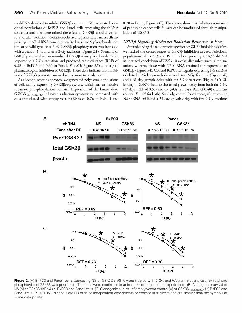
an shRNA designed to inhibit GSK3β expression. We generated poly-clonal populations of BxPC3 and Panc1 cells expressing the shRNAconstruct and then determined the effect of GSK3β knockdown onsurvival after radiation. Radiation delivered to pancreatic cancer cells ex-pressing an NS shRNA construct resulted in serine 9 phosphorylation,similar to wild-type cells. Ser9 GSK3β phosphorylation was increasedwith a peak at 1 hour after a 2-Gy radiation (Figure 2A). Silencing ofGSK3β prevented radiation-induced GSK3β serine phosphorylation inresponse to a 2-Gy radiation and produced radioresistance (REFs of0.82 in BxPC3 and 0.60 in Panc1, P < .05; Figure 2B ) similarly topharmacological inhibition of GSK3β. These data indicate that inhibi-tion of GSK3β promotes survival in response to irradiation.As a second genetic approach, we generated polyclonal populations
of cells stably expressing GSK3βKK(85,86)MA, which has an inactivesubstrate phosphorylation domain. Expression of the kinase deadGSK3βKK(85,86)MA inhibited radiation cytotoxicity compared withcells transduced with empty vector (REFs of 0.76 in BxPC3 and
0.70 in Panc1; Figure 2C ). These data show that radiation resistanceof pancreatic cancer cells in vitro can be modulated through manipu-lation of GSK3β.
GSK3β Signaling Modulates Radiation Resistance In VivoAfter observing the radioprotective effect of GSK3β inhibition in vitro,
we studied the consequences of GSK3β inhibition in vivo. Polyclonalpopulations of BxPC3 and Panc1 cells expressing GSK3β shRNAmaintained knockdown of GSK3 10 weeks after subcutaneous implan-tation, whereas those with NS shRNA retained the expression ofGSK3β (Figure 3A). Control BxPC3 xenografts expressing NS shRNAexhibited a 26-day growth delay with ten 2-Gy fractions (Figure 3B)and a 61-day growth delay with ten 3-Gy fractions (Figure 3C ). Si-lencing of GSK3β leads to shortened growth delay from both the 2-Gy(17 days, REF of 0.65) and the 3-Gy (25 days, REF of 0.40) treatmentcourses (P < .05 for both). Similarly, control Panc1 xenografts expressingNS shRNA exhibited a 24-day growth delay with five 2-Gy fractions
Figure 2. (A) BxPC3 and Panc1 cells expressing NS or GSK3β shRNA were treated with 2 Gy, and Western blot analysis for total andphosphorylated GSK3β was performed. The blots were confirmed in at least three independent experiments. (B) Clonogenic survival ofNS (○) or GSK3β shRNA (•) BxPC3 and Panc1 cells. (C) Clonogenic survival of empty vector control (○) or GSK3βKK(85,86)MA (•) BxPC3 andPanc1 cells. *P ≤ 0.05. Error bars are SD of three independent experiments performed in triplicate and are smaller than the symbols atsome data points.
360 Wnt Pathway Modulates Radiosensitivity Watson et al. Neoplasia Vol. 12, No. 5, 2010
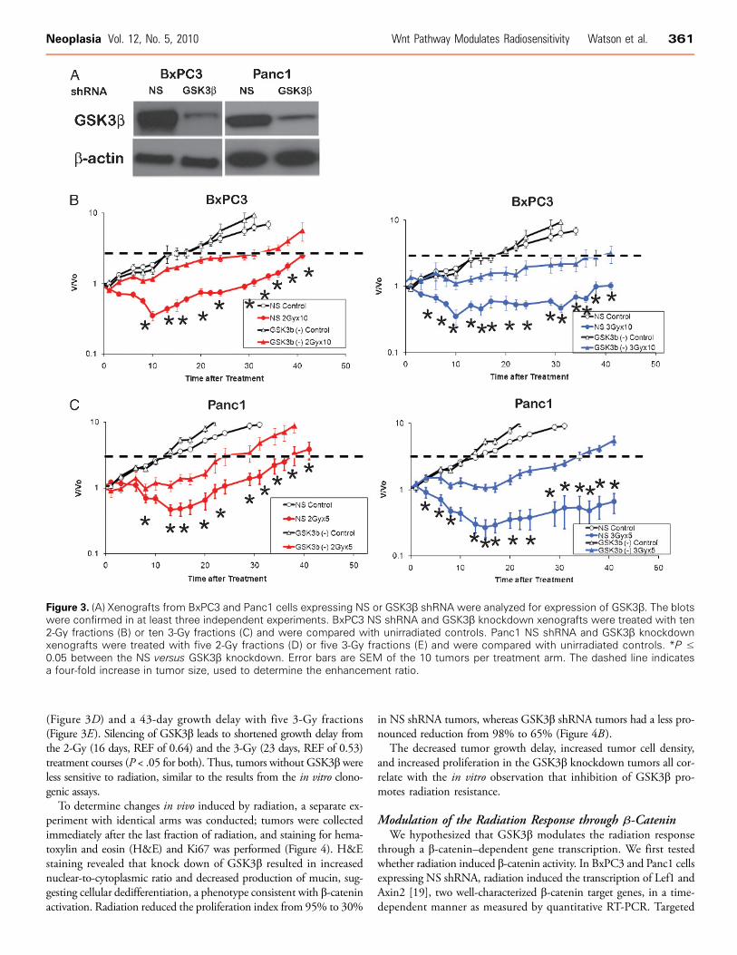
(Figure 3D) and a 43-day growth delay with five 3-Gy fractions(Figure 3E). Silencing of GSK3β leads to shortened growth delay fromthe 2-Gy (16 days, REF of 0.64) and the 3-Gy (23 days, REF of 0.53)treatment courses (P < .05 for both). Thus, tumors without GSK3β wereless sensitive to radiation, similar to the results from the in vitro clono-genic assays.
To determine changes in vivo induced by radiation, a separate ex-periment with identical arms was conducted; tumors were collectedimmediately after the last fraction of radiation, and staining for hema-toxylin and eosin (H&E) and Ki67 was performed (Figure 4). H&Estaining revealed that knock down of GSK3β resulted in increasednuclear-to-cytoplasmic ratio and decreased production of mucin, sug-gesting cellular dedifferentiation, a phenotype consistent with β-cateninactivation. Radiation reduced the proliferation index from 95% to 30%
in NS shRNA tumors, whereas GSK3β shRNA tumors had a less pro-nounced reduction from 98% to 65% (Figure 4B ).
The decreased tumor growth delay, increased tumor cell density,and increased proliferation in the GSK3β knockdown tumors all cor-relate with the in vitro observation that inhibition of GSK3β pro-motes radiation resistance.
Modulation of the Radiation Response through β-CateninWe hypothesized that GSK3β modulates the radiation response
through a β-catenin–dependent gene transcription. We first testedwhether radiation induced β-catenin activity. In BxPC3 and Panc1 cellsexpressing NS shRNA, radiation induced the transcription of Lef1 andAxin2 [19], two well-characterized β-catenin target genes, in a time-dependent manner as measured by quantitative RT-PCR. Targeted
Figure 3. (A) Xenografts from BxPC3 and Panc1 cells expressing NS or GSK3β shRNA were analyzed for expression of GSK3β. The blotswere confirmed in at least three independent experiments. BxPC3 NS shRNA and GSK3β knockdown xenografts were treated with ten2-Gy fractions (B) or ten 3-Gy fractions (C) and were compared with unirradiated controls. Panc1 NS shRNA and GSK3β knockdownxenografts were treated with five 2-Gy fractions (D) or five 3-Gy fractions (E) and were compared with unirradiated controls. *P ≤0.05 between the NS versus GSK3β knockdown. Error bars are SEM of the 10 tumors per treatment arm. The dashed line indicatesa four-fold increase in tumor size, used to determine the enhancement ratio.
Neoplasia Vol. 12, No. 5, 2010 Wnt Pathway Modulates Radiosensitivity Watson et al. 361

silencing of GSK3β resulted in both higher basal and radiation-inducedlevels of Lef1 or Axin2 gene transcription (Figure 5A). Because we ob-served that radiation affects β-catenin transcriptional activity in vitrothrough GSK3β, we hypothesized that radiation would have similareffects in vivo (Figure 5B). Before irradiation, β-catenin localized to thecytosolic membrane. After radiation of xenografts, β-catenin translocatedto the nucleus, suggesting induction of β-catenin signaling. Radiationinduction of β-catenin nuclear translocation correlates with the obser-vation in vitro that GSK3β phosphorylation modulates β-catenin–dependent gene transcription.If GSK3β modulates pancreatic cancer cell response to radiation
through β-catenin, then modulation of β-catenin activity may influ-ence cell survival after radiation. Therefore, we transduced BxPC3and Panc1 cells with lentivirus encoding shRNA targeting β-catenin.Compared with cells transduced with NS shRNA, cells with silencedβ-catenin were more sensitive to radiation as shown by reduced clono-genic survival (REFs of 1.4 in BxPC3 and 1.25 in Panc1; Figure 6).On the basis of these experiments, constitutive activation of β-
catenin would be predicted to render pancreatic cancer cells resistantto radiation. Therefore, a β-cateninS33Y-FLAG vector was used to createcells expressing constituently active β-catenin. The S33Y mutationprevents GSK3β-mediated phosphorylation at Ser33, thus preventingubiquitination and subsequent degradation [20]. Cells expressing con-stituently active β-cateninS33Y showed increased clonogenic survival(REF for Panc1 cells was 0.8; Figure 6C). The effects of nondegradableβ-cateninS33Y were analogous to those resulting fromGSK3β inhibitionor silencing because both showed an increased resistance to radiationtogether with an increased level of β-catenin activity. Thus, increasedβ-catenin activity results in greater radiation resistance of pancreatic
cancer cells, whereas loss of β-catenin through RNAi-mediated silenc-ing results in increased radiation sensitivity.
DiscussionIn this study, we found that inhibition of GSK3β, by either geneticor pharmacological methods, induces radiation resistance of pancre-atic cancer cells in vitro, reduces the duration of radiation-inducedtumor growth delay, and leads to increased cell proliferation in vivo.Similarly, the expression of a constituently active β-catenin in pancreaticcancer cells increases the resistance to radiation. Our results reinforceand expand on previous studies of radiation effects on GSK3β.
We have previously demonstrated that radiation induces phos-phorylation of GSK3β at Ser9, an event known to inhibit GSK3βkinase activity [17], and that abrogation of this phosphorylation re-sulted in radiosensitization. Radiation was also shown to inhibitGSK3β activity in SAOS-2 cells [21], although the phenotypic con-sequences in sensitivity to radiation were not investigated. Irradiationof A549 cells induced phosphorylation of GSK3β at Ser9, and thiseffect was reduced when cells were plated on fibronectin [22]. Theauthors suggested that GSK3β is involved in the interaction of cellswith the extracellular matrix after radiation to modulate the cytotox-icity of radiation. These studies implicate GSK3β as a mediator ofradiation sensitivity.
We hypothesized that GSK3β modulates radiation cytotoxicity, atleast in part, through its downstream effector β-catenin. Herein, weshow that radiation induces the transcription of Lef1 and Axin2, twowell-characterized β-catenin target genes, and targeted silencing ofGSK3β results in both higher basal and radiation-induced levels of
Figure 4. Xenografts from BxPC3 and Panc1 cells expressing NS or GSK3β shRNA were analyzed by H&E (A). Black arrows indicateglandular structures present in the NS shRNA xenografts, which are absent in GSK3β shRNA xenografts. Panc1 xenografts with andwithout radiation were analyzed for proliferation by Ki67 (B). Original magnification, ×400.
362 Wnt Pathway Modulates Radiosensitivity Watson et al. Neoplasia Vol. 12, No. 5, 2010

Lef1 or Axin2 gene transcription. Furthermore, we show that radia-tion induces translocation of cytosolic β-catenin to the nucleus inPanc1 and BxPC3 xenographs, an observation consistent with thein vitro induction of transcription of Lef1 and Axin2. Finally, we showthat cells with silenced β-catenin aremore sensitive, whereas cells expres-sing constituently active β-cateninS33Y are more resistant to radia-tion. β-Catenin has been shown to prevent epithelial cell deathafter radiation or anoikis [23]. These findings suggest that β-cateninis involved in determining clonogenic survival of pancreatic cancercells after irradiation.
Our studies potentially explain the relationship between Wnt sig-naling and radiation cytotoxicity in other tumor sites. Activation of theWnt signaling pathway resulted in β-catenin cytoplasmic accumulationwith translocation to the nucleus in head and neck cancer cell linesexpressing COX-2 [24]. In turn, up-regulation of Ku expression leadsto increased radioresistance. Blocking COX-2 signaling led to the sup-pression of β-catenin–induced Ku expression and consequent radiationsensitivity. Others have suggested that the radioresistance observed clin-
ically in glioblastoma depends in part on the activation of β-catenin inputative cancer stem cells [25]. In a mouse model of breast develop-ment, radiation selectively enriched for mammary epithelial progenitorsisolated from transgenic mice with activated Wnt/β-catenin signalingbut not for background-matched controls [26]. We conversely showedthat suppressing β-catenin using shRNA correlated with an increase inradiation sensitivity.
Our data reinforce observations from others that GSK3β inhibitionprotects normal tissue from radiation toxicity. Radiation-inducedGSK3β activation results in mouse hippocampal neuronal apoptosisand subsequent neurocognitive decline. The expression of kinase-inactiveGSK3β or pharmacologic inhibition before irradiation significantlyattenuated radiation-induced apoptosis in hippocampal neurons, lead-ing to improved cognitive function in irradiated animals [27]. Micetreated with lithium chloride, a known GSK3β inhibitor, had decreasedneurocognitive impairment after irradiation as well [28]. Akt serves toinhibit GSK3β after irradiation in normal vascular endothelium [29],and administration of recombinant growth factors known to activate
Figure 5. (A) Time course of Lef1 and Axin2 levels in NS (○) or GSK3β shRNA (•) BxPC3 and Panc1 cells subjected to 2-Gy radiation.Mean of three experiments with SDs, *P≤ 0.05. (B) BxPC3 or Panc1 xenografts were treated with 2-Gy radiation and were stained forβ-catenin (green) and propidium idodide (red). Yellow indicates overlap of red and green, consistent with nuclear β-catenin.
Neoplasia Vol. 12, No. 5, 2010 Wnt Pathway Modulates Radiosensitivity Watson et al. 363

Akt may prevent normal tissue toxicity. However, any pharmacologicstrategy to reduce normal tissue damage must be carefully weighedagainst the risk of tumor protection.Our results are consistent with radioprotection caused by active
β-catenin. A reporter mouse model demonstrated that ionizing radia-tion activates β-catenin–mediated, T-cell factor–dependent transcrip-tion both in vitro and in vivo. Mouse-derived fibroblast culturesexpressing stabilized β-catenin formed more colony-forming units thanwild-type or null cells after irradiation. β-Catenin levels in irradiatedwounds correlated with tensile strength of the wound, and lithiumchloride treatment also increased β-catenin levels and increased woundstrength [30]. The newly identified R-Spondin1 augments canonicalWnt/β-catenin signaling and causes nuclear translocation of β-catenin.
R-Spondin1 reduced mucosal ulceration after whole-body or snout-only irradiation inmousemodels [31]. Therefore, in normal cells, GSK3βinhibition with β-catenin activation may be a radioprotective mecha-nism. Pancreatic cancer cells potentially invoke a similar mechanismto evade the cytotoxic effects of radiation.
Our results help explain an apparent contradiction present in theliterature regarding pancreatic cancer and β-catenin. Mutations inAPC leading to β-catenin nuclear accumulation have been well char-acterized to play a role in colon cancer. However, mutations in APC[32] or β-catenin [33] have not been found in pancreatic cancer. Thepublished literature suggests that constitutive activation of β-catenindoes not play a role in pancreatic cancer development. In fact, ourresults also demonstrate similar findings, as unirradiated tumorslacked nuclear β-catenin, and we did not find evidence of increasedβ-catenin target gene expression without irradiation. However, wedid find that pancreatic cancer cells activate β-catenin in responseto radiation to promote survival. Our results may therefore explainin part the clinically observed radioresistance of pancreatic cancer;specifically, it may not be the basal level of β-catenin but rather the in-duction of β-catenin by radiation that promotes pancreatic cancer cellsurvival. We plan to test this hypothesis by immunoflorescence of pan-creatic cancer specimens treated with neoadjuvant radiation to deter-mine whether activation of β-catenin occurs in patients.
The implications of this work identify a link between radiationand a pathway central to tumor growth, invasion, and metastasisof pancreatic cancer. By further discovering the molecular signalingcascades upstream and downstream of GSK3β, we will also start togain insight into the potential interactions with other signaling path-ways that are known to be involved in radioresistance. Further under-standing of this pathway will also help develop clinical trials combiningdrugs inhibiting β-catenin activation with radiation and cytotoxicagents in locally advanced pancreatic cancer.
AcknowledgmentThe authors thank Steven Kronenberg for his graphical expertise.
References[1] Macdonald JS, McCoy S, Whitehead RP, Iqbal S, Wade JL III, Giguere JK, and
Abbruzzese JL (2005). A phase II study of farnesyl transferase inhibitorR115777 in pancreatic cancer: a Southwest oncology group (SWOG 9924)study. Invest New Drugs 23, 485–487.
[2] Jones L, Ghaneh P, Humphreys M, and Neoptolemos JP (1999). The matrixmetalloproteinases and their inhibitors in the treatment of pancreatic cancer.Ann N Y Acad Sci 880, 288–307.
[3] Moore MJ, Goldstein D, Hamm J, Figer A, Hecht JR, Gallinger S, Au HJ,Murawa P, Walde D, Wolff RA, et al. (2007). Erlotinib plus gemcitabine com-pared with gemcitabine alone in patients with advanced pancreatic cancer: aphase III trial of the National Cancer Institute of Canada Clinical Trials Group.J Clin Oncol 25, 1960–1966.
[4] Beurel E and Jope RS (2006). The paradoxical pro- and anti-apoptotic actions ofGSK3 in the intrinsic and extrinsic apoptosis signaling pathways. Prog Neurobiol79, 173–189.
[5] Juhaszova M, Zorov DB, Kim SH, Pepe S, Fu Q, Fishbein KW, Ziman BD,Wang S, Ytrehus K, Antos CL, et al. (2004). Glycogen synthase kinase-3β medi-ates convergence of protection signaling to inhibit the mitochondrial permeabil-ity transition pore. J Clin Invest 113, 1535–1549.
[6] Korinek V, Barker N, Morin PJ, van Wichen D, de Weger R, Kinzler KW,Vogelstein B, and Clevers H (1997). Constitutive transcriptional activation by aβ-catenin–Tcf complex in APC−/− colon carcinoma. Science 275, 1784–1787.
[7] Chan TA, Wang Z, Dang LH, Vogelstein B, and Kinzler KW (2002). Targetedinactivation of CTNNB1 reveals unexpected effects of β-catenin mutation. ProcNatl Acad Sci USA 99, 8265–8270.
Figure 6. Clonogenic survival of NS (○) or β-catenin shRNA (•)BxPC3 (A) and Panc1 (B) cells. Clonogenic survival of empty vectorcontrol (○) or β-cateninS33YFLAG (•) Panc1 cells (C). Error bars areSD of three independent experiments performed in triplicate andare smaller than the symbols at some data points.
364 Wnt Pathway Modulates Radiosensitivity Watson et al. Neoplasia Vol. 12, No. 5, 2010

[8] Sutherland C, Leighton IA, and Cohen P (1993). Inactivation of glycogensynthase kinase-3 β by phosphorylation: new kinase connections in insulinand growth-factor signalling. Biochem J 296(pt 1), 15–19.
[9] Welsh GI and Proud CG (1993). Glycogen synthase kinase-3 is rapidly inacti-vated in response to insulin and phosphorylates eukaryotic initiation factor eIF-2B. Biochem J 294(pt 3), 625–629.
[10] Zufferey R, Dull T, Mandel RJ, Bukovsky A, Quiroz D, Naldini L, and Trono D(1998). Self-inactivating lentivirus vector for safe and efficient in vivo gene delivery.J Virol 72, 9873–9880.
[11] Eldar-FinkelmanH,ArgastGM, FoordO, Fischer EH, andKrebs EG (1996). Expres-sion and characterization of glycogen synthase kinase-3 mutants and their effect onglycogen synthase activity in intact cells. Proc Natl Acad Sci USA 93, 10228–10233.
[12] Kolligs FT, Hu G, Dang CV, and Fearon ER (1999). Neoplastic transformationof RK3E by mutant β-catenin requires deregulation of Tcf/Lef transcription butnot activation of c-myc expression. Mol Cell Biol 19, 5696–5706.
[13] Fertil B, Dertinger H, Courdi A, and Malaise EP (1984). Mean inactivationdose: a useful concept for intercomparison of human cell survival curves. RadiatRes 99, 73–84.
[14] Simpson DA, Feeney S, Boyle C, and Stitt AW (2000). Retinal VEGF mRNAmeasured by SYBR green I fluorescence: a versatile approach to quantitativePCR. Mol Vis 6, 178–183.
[15] Koinuma K, Yamashita Y, Liu W, Hatanaka H, Kurashina K, Wada T, Takada S,Kaneda R, Choi YL, Fujiwara SI, et al. (2006). Epigenetic silencing of AXIN2in colorectal carcinoma with microsatellite instability. Oncogene 25, 139–146.
[16] Kahler RA, Galindo M, Lian J, Stein GS, van Wijnen AJ, and Westendorf JJ(2006). Lymphocyte enhancer-binding factor 1 (Lef1) inhibits terminal differ-entiation of osteoblasts. J Cell Biochem 97, 969–983.
[17] Spalding AC, Watson R, Davis ME, Kim AC, Lawrence TS, and Ben-Josef E(2007). Inhibition of protein kinase C{beta} by enzastaurin enhances radiationcytotoxicity in pancreatic cancer. Clin Cancer Res 13, 6827–6833.
[18] De Sarno P, Li X, and Jope RS (2002). Regulation of Akt and glycogen synthasekinase-3 β phosphorylation by sodium valproate and lithium. Neuropharmacology43, 1158–1164.
[19] Leung JY, Kolligs FT, Wu R, Zhai Y, Kuick R, Hanash S, Cho KR, and Fearon ER(2002). Activation of AXIN2 expression by β-catenin–T cell factor. A feedbackrepressor pathway regulating Wnt signaling. J Biol Chem 277, 21657–21665.
[20] Sun Y, Kolligs FT, Hottiger MO, Mosavin R, Fearon ER, and Nabel GJ (2000).Regulation of β-catenin transformation by the p300 transcriptional coactivator.Proc Natl Acad Sci USA 97, 12613–12618.
[21] Turenne GA and Price BD (2001). Glycogen synthase kinase3 β phosphorylatesserine 33 of p53 and activates p53’s transcriptional activity. BMC Cell Biol 2, 12.
[22] Cordes N, Beinke C, Plasswilm L, and van Beuningen D (2004). Irradiationand various cytotoxic drugs enhance tyrosine phosphorylation and β(1)-integrinclustering in human A549 lung cancer cells in a substratum-dependent mannerin vitro. Strahlenther Onkol 180, 157–164.
[23] Orford K, Orford CC, and Byers SW (1999). Exogenous expression of β-cateninregulates contact inhibition, anchorage-independent growth, anoikis, andradiation-induced cell cycle arrest. J Cell Biol 146, 855–868.
[24] Chang HW, Roh JL, Jeong EJ, Lee SW, Kim SW, Choi SH, Park SK, and Kim SY(2008). Wnt signaling controls radiosensitivity via cyclooxygenase-2–mediated Kuexpression in head and neck cancer. Int J Cancer 122, 100–107.
[25] Rich JN (2007). Cancer stem cells in radiation resistance. Cancer Res 67,8980–8984.
[26] Woodward WA, Chen MS, Behbod F, Alfaro MP, Buchholz TA, and Rosen JM(2007). WNT/β-catenin mediates radiation resistance of mouse mammary pro-genitor cells. Proc Natl Acad Sci USA 104, 618–623.
[27] Thotala DK, Hallahan DE, and Yazlovitskaya EM (2008). Inhibition of glyco-gen synthase kinase 3 β attenuates neurocognitive dysfunction resulting fromcranial irradiation. Cancer Res 68, 5859–5868.
[28] Yazlovitskaya EM, Edwards E, Thotala D, Fu A, Osusky KL, Whetsell WO Jr,Boone B, Shinohara ET, and Hallahan DE (2006). Lithium treatment pre-vents neurocognitive deficit resulting from cranial irradiation. Cancer Res 66,11179–11186.
[29] Tan J, Geng L, Yazlovitskaya EM, and Hallahan DE (2006). Protein kinase B/Akt–dependent phosphorylation of glycogen synthase kinase-3β in irradiatedvascular endothelium. Cancer Res 66, 2320–2327.
[30] Gurung A, Uddin F, Hill RP, Ferguson PC, and Alman BA (2009). β-Catenin is amediator of the response of fibroblasts to irradiation. Am J Pathol 174, 248–255.
[31] Zhao J, Kim KA, De Vera J, Palencia S, Wagle M, and Abo A (2009). R-Spondin1protects mice from chemotherapy or radiation-induced oral mucositis through thecanonical Wnt/β-catenin pathway. Proc Natl Acad Sci USA 106, 2331–2336.
[32] Caca K, Kolligs FT, Ji X, Hayes M, Qian J, Yahanda A, Rimm DL, Costa J, andFearon ER (1999). β- and γ-catenin mutations, but not E-cadherin inactivation,underlie T-cell factor/lymphoid enhancer factor transcriptional deregulation ingastric and pancreatic cancer. Cell Growth Differ 10, 369–376.
[33] Gerdes B, Ramaswamy A, Simon B, Pietsch T, Bastian D, Kersting M, Moll R,and Bartsch D (1999). Analysis of β-catenin gene mutations in pancreatic tu-mors. Digestion 60, 544–548.
Neoplasia Vol. 12, No. 5, 2010 Wnt Pathway Modulates Radiosensitivity Watson et al. 365