Gene Regulation: Transcription Factors Sp1 and Hif2α...
24
Liwei Xie and James F. Collins Hypoxia Gene in Intestinal Epithelial Cells During Copper-Transporting ATPase (Atp7a) Mediate Induction of the α Transcription Factors Sp1 and Hif2 Gene Regulation: published online June 28, 2013 J. Biol. Chem. 10.1074/jbc.M113.489500 Access the most updated version of this article at doi: . JBC Affinity Sites Find articles, minireviews, Reflections and Classics on similar topics on the Alerts: When a correction for this article is posted • When this article is cited • to choose from all of JBC's e-mail alerts Click here Supplemental material: http://www.jbc.org/content/suppl/2013/06/28/M113.489500.DC1.html http://www.jbc.org/content/early/2013/06/28/jbc.M113.489500.full.html#ref-list-1 This article cites 0 references, 0 of which can be accessed free at at University of Florida on July 8, 2013 http://www.jbc.org/ Downloaded from
Transcript of Gene Regulation: Transcription Factors Sp1 and Hif2α...

Liwei Xie and James F. Collins HypoxiaGene in Intestinal Epithelial Cells During Copper-Transporting ATPase (Atp7a)Mediate Induction of the
αTranscription Factors Sp1 and Hif2Gene Regulation:
published online June 28, 2013J. Biol. Chem.
10.1074/jbc.M113.489500Access the most updated version of this article at doi:
.JBC Affinity SitesFind articles, minireviews, Reflections and Classics on similar topics on the
Alerts:
When a correction for this article is posted•
When this article is cited•
to choose from all of JBC's e-mail alertsClick here
Supplemental material:
http://www.jbc.org/content/suppl/2013/06/28/M113.489500.DC1.html
http://www.jbc.org/content/early/2013/06/28/jbc.M113.489500.full.html#ref-list-1
This article cites 0 references, 0 of which can be accessed free at
at University of Florida on July 8, 2013http://www.jbc.org/Downloaded from
Sp1 and Hif2α regulate Atp7a transcription during hypoxia
1
Transcription Factors Sp1 and Hif2α Mediate Induction of the Copper-Transporting ATPase (Atp7a)
Gene in Intestinal Epithelial Cells During Hypoxia*
Liwei Xie1 and James F. Collins
1,2,3
1From the Food Science and Human Nutrition Department
University of Florida, Gainesville, FL 32611
* Running title: Sp1 and Hif2α regulate Atp7a transcription during hypoxia
2To whom correspondence should be addressed: James F. Collins, Food Science and Human Nutrition
Department, University of Florida, 572 Newell Dr., FSN Bldg., #441, Gainesville, FL USA, Tel.: (352)
392-1991, ext. 289; Fax: (352) 392-9467; E-mail: [email protected]
Keywords: Iron; chromatin immunoprecipitation (ChIP); hypoxia-inducible factor (HIF); intestine;
transcription coactivators; IEC-6 cells; cobalt chloride
Background: A hypoxia-inducible transcription
factor (Hif2α) mediates induction of intestinal iron
and copper transporters during iron deficiency.
Results: Specificity factor 1 (Sp1) is required for
transcriptional induction of an intestinal copper
transporter (Atp7a) by Hif2α.
Conclusions: Sp1 and Hif2α may synergistically
mediate the genetic response to iron deficiency.
Significance: Understanding molecular
mechanisms governing iron absorption may allow
modulation of this process during disease states.
ABSTRACT
Genes with G/C-rich promoters were
upregulated in the duodenal epithelium of iron-
deficient rats, including those encoding iron
(e.g. Dmt1, and Dcytb) and copper (e.g. Atp7a,
and Mt1) metabolism-related proteins. It was
previously shown that an intestinal copper
transporter (Atp7a) was co-regulated with iron
transport-related genes by a hypoxia-inducible
transcription factor, Hif2α. In the current
study, we sought to test the role of Sp1 in
transcriptional regulation of Atp7a expression
during iron deprivation/hypoxia. Initial studies
in IEC-6 cells showed that mithramycin, an Sp1
inhibitor, reduced expression of Atp7a and iron
transport-related genes (Dmt1, Dcytb, Fpn1),
and also blocked their induction by CoCl2, a
hypoxia mimetic. Consistent with this,
overexpression of Sp1 increased endogenous
Atp7a mRNA and protein expression, and also
stimulated Atp7a, Dmt1, and Dcytb promoter
activity. Site-directed mutagenesis and
functional analysis of a basal Atp7a promoter
construct revealed 4 functional Sp1 binding
sites, which were necessary for Hif2α-mediated
induction of promoter activity. Furthermore,
chromatin immunoprecipitation (ChIP) assays
confirmed that Sp1 specifically interacts with
the Atp7a promoter in IEC-6 cells and in rat
duodenal enterocytes. This investigation has
thus revealed a novel aspect of Atp7a gene
regulation in which Sp1 may be necessary for
the HIF-mediated induction of gene
transcription during iron deficiency/hypoxia.
Understanding regulation of Atp7a expression
may help further clarify the physiological role
of copper in the maintenance of iron
homeostasis. Furthermore, this Sp1/Hif2α
regulatory mechanism may have broader
implications for understanding the genetic
response of the intestinal epithelium to
maintain whole-body iron homeostasis during
states of deficiency.
Iron is essential for life, as it plays critical
roles in biological systems, including those related
to mitochondrial electron transport and energy
production, enzyme activity, oxygen transport, and
regulation of gene expression (1). Systemic iron
levels are maintained by intestinal absorption,
which is precisely controlled, as there is no active
excretory mechanism in mammals. Iron absorption
is enhanced during iron deprivation, as reflected
by increased expression of iron transport-related
genes including divalent metal transporter 1 (Dmt1;
an iron importer)4, duodenal cytochrome B (Dcytb;
a brush-border membrane ferrireductase) and
http://www.jbc.org/cgi/doi/10.1074/jbc.M113.489500The latest version is at JBC Papers in Press. Published on June 28, 2013 as Manuscript M113.489500
Copyright 2013 by The American Society for Biochemistry and Molecular Biology, Inc. at University of Florida on July 8, 2013http://www.jbc.org/Downloaded from
Sp1 and Hif2α regulate Atp7a transcription during hypoxia
2
ferroportin 1 (Fpn1; an iron exporter) in duodenal
enterocytes (2). Studies also found that the
Menkes copper-transporting ATPase (Atp7a), an
enterocyte copper exporter, was upregulated in the
rat duodenal epithelium during iron deficiency,
consistent with noted increases in copper content
of the intestinal mucosa, liver, and serum (2,3).
Similar perturbations in tissue copper levels have
been noted in other mammalian species during
states of iron deficiency (4-6). It has thus been
hypothesized that copper plays a role in the
maintenance of iron homeostasis (7). Importantly,
two multi-copper ferroxidases, one expressed in
enterocytes of the small intestine (hephaestin) and
one produced in liver and secreted into the blood
(ceruloplasmin), provide key links between iron
and copper homeostasis (8).
Depletion of body iron stores leads to
decreased red blood cell hemoglobin levels
causing tissue hypoxia. Low tissue oxygen tension
in turn, results in stabilization of hypoxia-
inducible trans-acting factors (HIFs). The HIFs
function as heterodimers, containing a
constitutively expressed β subunit and a hypoxia-
responsive alpha subunit (one of 3 known Hifα
subunits). The increase in intestinal iron
absorption when body iron stores are depleted has
in fact been shown to be mediated via activation of
Hif2α. This regulatory mechanism was revealed
by two recent studies in which the alpha subunits
(Hif1α and Hif2α) of the functional HIF protein
complexes were specifically inactivated in the
intestinal epithelium of mice (9,10). Results
showed that regulation of iron absorption was
defective in mice lacking intestinal Hif2α, while
induction of iron absorption during iron
deprivation was maintained in mice lacking Hif1α.
It was further shown that the Dmt1, Dcytb and
Fpn1 promoters contained functional hypoxia-
responsive elements (HREs) which specifically
interacted with Hif2α, explaining their induction
during iron deficiency (and tissue hypoxia) (9-11).
Hif2α is thus critical to maintain intestinal iron
homeostasis in mice.
Interestingly, our previous studies in iron-
deficient rats showed that Atp7a was upregulated
in the duodenal epithelium similar to Dmt1, Dcytb
and Fpn1 (2), and we thus hypothesized that Atp7a
was coordinately regulated with these iron
transport-related genes. Subsequently, it was
demonstrated that the Atp7a promoter was indeed
a direct Hif2α target in rat intestinal epithelial
(IEC-6) cells (12). Furthermore, in a previous
investigation, it was noted that promoters of genes
induced in the duodenal epithelium of iron-
deficient rats contained a statistical
overrepresentation of G/C-rich sequences (13). It
was also shown that an abundance of genes
upregulated in differentiated Caco-2 cells (human
intestinal adenocarcinoma cells) in response to
iron chelation contained G/C-rich promoter
sequences as well as putative HREs (14).
Importantly, many of the iron and copper
homeostasis-related genes induced in both models
of intestinal iron transport contained G/C-rich
promoters and putative HREs. These observations
led us to hypothesize that a G/C-binding protein
(e.g. Sp1 or a related trans-acting factor) was
important for the transcriptional response of the
intestinal epithelium to iron deprivation (14). To
test this hypothesis, in the current investigation,
we performed a series of experiments to determine
if Sp1 is important for the Hif2α-mediated
transactivation of Atp7a gene expression using an
in vitro model of the intestinal epithelium (IEC-6
cells) and iron-deprived rats. Results of this
investigation showed that Sp1 specifically
interacts with cis-elements in the Atp7a promoter,
and further that Sp1 binding is necessary for
Hif2α-mediated induction of Atp7a transcription
during hypoxia.
EXPERIMENTAL PROCEDURES
Cell culture - Rat intestinal epithelial
(IEC-6) cells were obtained from the American
Type Culture Collection (Manassas, VA) and
cultured as described previously (12,15). For
hypoxia experiments, IEC-6 cells at ~85%
confluence were cultured in a hypoxia chamber
with 1% O2 and 5% CO2 (with the balance being
nitrogen). To mimic hypoxia, 200 μM CoCl2 was
added to the culture medium when the cells were
~85% confluent and cells were then cultured for
60 hours. To interrupt Sp1 binding, fully
differentiated IEC-6 cells (i.e. 7 days post-
confluence) were treated with mithramycin (a G/C
base pair-specific, DNA-binding antibiotic) (16,17)
at various concentrations for 24 hours.
Animals and diets- Weanling Sprague-
Dawley rats (male) were purchased from Harlan
(n=12), raised in overhanging, wire mesh-
bottomed cages in a room with 12-hour light/dark
at University of Florida on July 8, 2013http://www.jbc.org/Downloaded from
Sp1 and Hif2α regulate Atp7a transcription during hypoxia
3
cycles and sacrificed at 10 am. Rats were fed
custom AIN93G-based diets (Dyets; Bethlehem,
PA) that varied only in iron content for 5 weeks;
the control (Ctrl) diet contained 198 ppm iron,
while the iron-deficient (FeD) diet contained 3
ppm iron. Animals were weighed weekly.
Subsequently, rats were anesthetized by CO2
exposure and killed by cervical dislocation. Blood
was collected by cardiac puncture and hemoglobin
and hematocrit were measured by routine methods
(8). The duodenum was excised and inverted on a
wooden stick, followed by isolating enterocytes
using a well-established, previously published
method (8,18). Duodenal enterocytes were used
for mRNA isolation, western blot analysis, and
chromatin immunoprecipitation experiments. All
animal studies were approved by the Institutional
Animal Care and Use Committee of the University
of Florida.
RNA isolation and real-time quantitative
RT-PCR- Total RNA was isolated from IEC-6
cells or duodenal enterocytes using TRIzol®
reagent (Life Technologies; Grand Island, NY),
following a standard protocol. RNA was reverse
transcribed using the iScript cDNA Synthesis Kit
(BioRad; Hercules, CA) and the resulting cDNA
was utilized for qRT-PCR reactions with SYBR
Green PCR Master Mix (BioRad). Primers, listed
in Table S1, were designed to span large introns to
avoid amplification from genomic DNA. Standard
curve reactions and melt curves were routinely run
to validate primer pairs and PCR reactions.
Experimental genes were normalized to 18S
rRNA, and relative gene expression was quantified
using routine methods.
Plasmid construction- The rat Sp1 open
reading frame (ORF) (Genbank accession #
D12768) was cloned by PCR from cDNA derived
from IEC-6 cells using Phusion High Fidelity
DNA Polymerase® (Thermo Scientific; Pittsburgh,
PA). The forward primer contained the
translational start codon and the reverse primer
ended just 5’ of the stop codon. Primers were
designed with overhanging KpnI (forward) and
EcoRV (reverse) restriction-enzyme cutting sites.
PCR-amplified The Sp1 ORF amplicon and
pcDNA 3.1 expression vector (Invitrogen) were
double digested with KpnI and EcoRV, followed
by column purification. Sp1 ORF was then sub-
cloned into double digested pcDNA 3.1 with
LigaFast™
Rapid DNA Ligation System (Promega;
Madison, WI). An HA-tag was inserted into the 3’
end of the Sp1 ORF by PCR amplifying the entire
pcDNA-Sp1 plasmid with primers containing
overhanging sequences containing an HA
sequence tag and a stop codon. Primers were
designed with the forward primer at the 3’ end of
the Sp1 ORF, and the reverse primer at the 5’ end
of the EcoRV site on the pcDNA 3.1 vector. Each
primer was phosphorylated at the 5’ end, which
allowed ligation of the PCR amplicons to reform
the intact plasmid. Primer sequences are provided
in Table S1.
Mutant Atp7a promoter constructs were
prepared by PCR-amplifying the entire WT
promoter fragment (-224/+88) in the pGL4.18
vector (Promega) with QuickChange® Lightning
Site-Directed Mutagenesis Kit (Agilent
Technologies; Santa Clara, CA). Primers
contained mutations in putative Sp1 binding sites,
with amplification reactions proceeding in
opposite directions. PCR products were digested
with DpnI restriction enzyme (Agilent
Technologies) to remove the template DNA
(which was methylated during replication in
bacteria). All DNA constructs were sequenced to
confirm that promoter amplicons did not contain
random mutations. Primer sequences are listed in
Table S1.
Transfection and luciferase assays- WT or
mutated Atp7a promoter constructs in the pGL4.18
vector (1 μg) were transiently transfected into
IEC-6 cells at ~60% confluence and cultured in
24-well plates. For Sp1 and Hif2α overexpression
experiments, 1 μg of Atp7a promoter construct
(WT or mutated) was co-transfected with 1 μg of
either Sp1 (described above) or Hif2α
overexpression vector (described previously) (12).
Other constructs used were pGL4.18 plasmids
containing ~1 kb mouse Dcytb and Dmt1 promoter
fragments (kindly provided by Dr. Yatrik Shah,
University of Michigan). Co-transfected pRL
CMV plasmid (Invitrogen) expressing renilla
luciferase was used to normalize expression of
firefly luciferase driven by experimental
promoters. 36 hours after transfection, luciferase
activity was measured with the Dual-Luciferase®
Reporter Assay System (Promega) according to
the manufacturer’s instructions.
Stable Sp1 overexpression- IEC-6 cells
were grown in 6-well plates and transfected with
pcDNA 3.1 (empty vector) or pcDNA-Sp1-HA
at University of Florida on July 8, 2013http://www.jbc.org/Downloaded from
Sp1 and Hif2α regulate Atp7a transcription during hypoxia
4
vector with TurboFect® in vitro Transfection Kit
(Thermo Scientific). 60 hours after transfection,
cells were treated with G418 (at a predetermined
concentration) to kill non-transfected cells,
allowing transfected cells expressing the neomycin
gene in the pcDNA3.1 or pcDNA-Sp1-HA vectors
to survive. IEC-6 cells with stable overexpression
of Sp1 were used to analyze Atp7a and Sp1
mRNA expression using qRT-PCR and protein
expression by western blotting, and to determine
the effect of forced Sp1 expression on Atp7a
promoter activity.
Protein isolation and western blot
analysis- Cytosolic and nuclear proteins were
isolated from IEC-6 cells cultured in 10 cm cell
culture dishes using a kit from Active Motif, as
described previously (15). Protein concentrations
were determined by BCA Protein Assay (Pierce).
30 μg cytosolic or 50 μg nuclear proteins were
resolved by 7.5% SDS-PAGE, followed by
transfer to PVDF membranes. The membranes
were blocked with 5% non-fat milk (w/v) and
then incubated with one of the following primary
antibodies: Atp7a (called 54-10) (3), Sp1 (cat.
#07-645; Millipore), phosphorylated-Sp1 (cat.
#ab37707; Abcam; Cambridge, MA), Hif1α (cat.
#NB100-105; Novus Biologicals; Littleton, CO) or
Hif2α (cat. #NB100-122; Novus Biologicals).
Subsequently, membranes were reacted with an
anti-rabbit IgG secondary antibody. Antibody
binding was visualized using home-made ECL
reagent (8) followed by exposure to X-ray film.
For quantification, protein expression was
normalized to total proteins on stained blots, as
this method does not rely on the expression level
of any individual protein which may or may not be
affected by various treatments (and as used
extensively by us in the past).
Chromatin immunoprecipitation (ChIP)
assay- Assays were performed as described
previously (12). Briefly, IEC-6 cells or rat
duodenal enterocytes were cross-linked with 1.1%
chloroform (v/v) for 10 minutes, followed by
quenching with 0.3 M glycine. Cells were
subsequently lysed with hypotonic buffer (Active
Motif) and homogenized. Nuclei were collected
and resuspended in nuclear lysis buffer, followed
by sonication with a BioRuptor (Diagenode;
Liege, Belgium) for 30 cycles with 30 seconds on
and 30 seconds off. Target DNA with bound
protein was pulled down with anti-Sp1 (Millipore)
or anti-Hif2α (Novus) antibodies. After removing
cross-links, DNA samples were analyzed by PCR
with primer sets listed in Table S1. Primers were
designed to amplify regions of the Atp7a promoter
containing putative Sp1 or Hif2α binding sites or
other up- or downstream regions that did not
contain predicted Sp- or HIF-like sites.
Statistical analysis- One-way ANOVA
(with Tukey’s post hoc test) and paired Student’s
t-test (Graphpad; La Jolla, CA) were used to
statistically compare data across groups. p<0.05
was considered statistically significant.
RESULTS
Mithramycin inhibits expression of iron
and copper transport-related genes-Expression of
Atp7a and other genes was analyzed by qRT-PCR
after mithramycin treatment of differentiated IEC-
6 cells (Table 1). Mithramycin reduced expression
of all experimental genes tested (Atp7a, Dmt1,
Dcytb and Fpn1), as well as positive control genes
including ankyrin repeat domain 37 (Ankrd37),
Hif2α, and Sp1. The inhibition was most
significant for all tested genes with 500 nM
mithramycin; higher concentrations were without
additional effect (data not shown), although
cellular toxicity was not noted with concentrations
up to 1 μM. Ankrd37, which was strongly induced
by iron deprivation (2), is a known Sp1 target gene
(19), as is Hif2α (20). Interestingly, Sp1 self-
regulates via a positive feedback loop (21). Sp6
and transferrin receptor 1 (Tfr1) were selected as
negative controls, as neither gene is known to be
regulated by Sp-like factors. Expression of Sp6
was unaffected by mithramycin treatment, while
for unknown reasons, Tfr1 expression was induced.
Inhibition of Sp1 binding blocks hypoxia-
mediated gene expression- Under normoxic
conditions, the Hifα subunits are hydroxylated on
conserved proline residues and subsequently
targeted for proteosomal degradation. Hypoxia
stabilizes the Hifα subunits by inhibiting the HIF
prolyl-hydroxylase enzymes that mediate this
hydroxylation reaction (22). Hypoxia can be
mimicked by treating cells with cobalt chloride,
which effectively inhibits proteosomal degradation
of the HIF alpha subunits under normoxic
conditions (23,24). Here, CoCl2 was utilized to
mimic hypoxia in IEC-6 cells. Results showed that
expression of experimental (Atp7a, Dcytb, Dmt1,
Fpn1) and positive control (Ankrd37, Vegf) genes
at University of Florida on July 8, 2013http://www.jbc.org/Downloaded from
Sp1 and Hif2α regulate Atp7a transcription during hypoxia
5
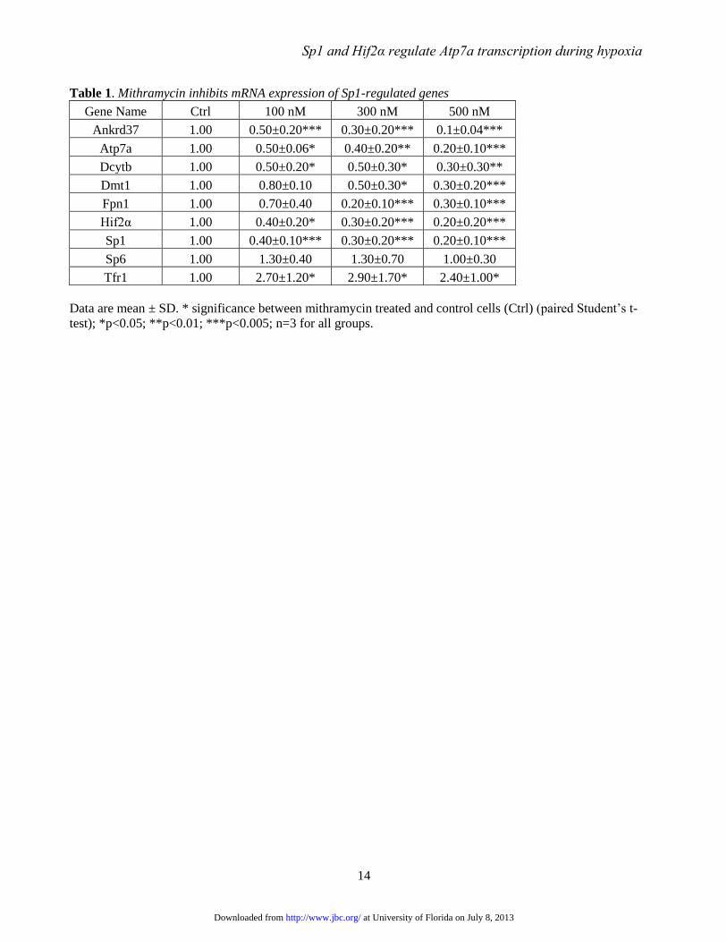
was increased by CoCl2 exposure (Fig. 1). The
Ankrd37 and vascular endothelial growth factor
(Vegf) genes are known Sp1 targets (19).
Moreover, mithramycin decreased basal
expression of all tested genes, and it also inhibited
the induction of Atp7a, Dcytb, Dmt1 and Fpn1 by
CoCl2. Conversely however, mithramycin did not
affect the induction of Ankrd37 or Vegf
expression by CoCl2.
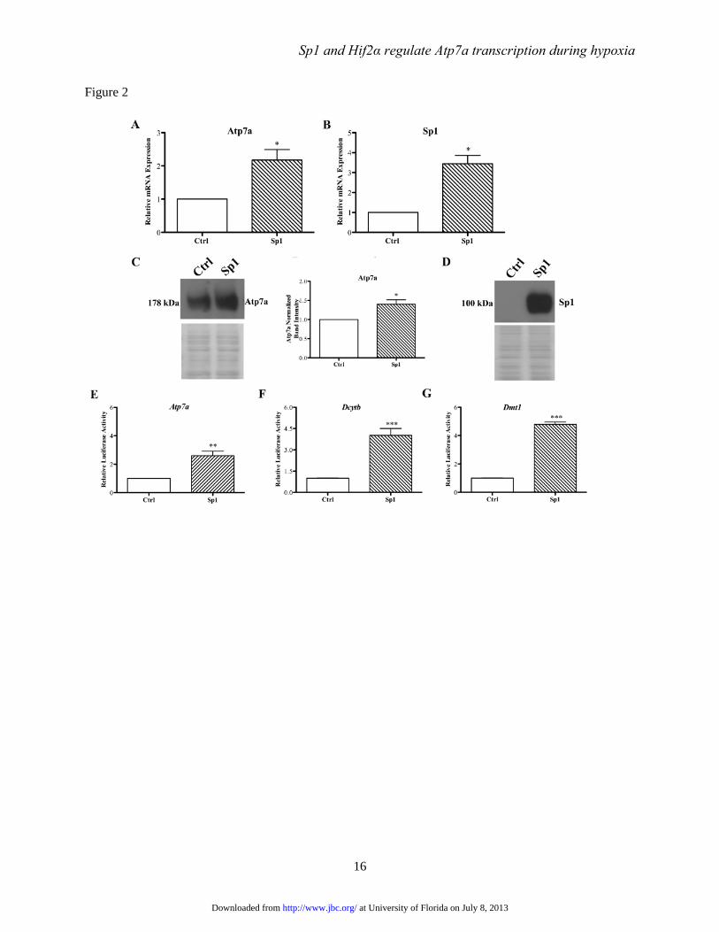
Regulation of Atp7a expression by Sp1-
IEC-6 cells stably transfected with an Sp1
overexpression plasmid showed significant
increases in Sp1 mRNA and protein expression, as
expected (Fig. 2). Sp1 overexpression also induced
Atp7a mRNA and immunoreactive protein
expression. Additionally, the Atp7a, Dcytb, and
Dmt1 promoters were transactivated by Sp1
overexpression (Fig. 2).
Sp1-like cis-elements are required for
basal Atp7a promoter activity- Phylogenetic
footprinting analysis showed that multiple, G/C-
rich sequences in the basal Atp7a promoter region
(-224/+88) were conserved among rats, mice and
humans (data not shown). Moreover, TFSEARCH
(http://www.cbrc.jp/research/db/TFSEARCH.html
) was utilized to predict putative Sp1-binding sites;
9 potential sites were identified. Initially, all 9
sites were individually mutated in the basal Atp7a
promoter and promoter activity was assessed in
IEC-6 cells. These experiments (n=3) showed that
4 putative Sp1 binding sites had the most
significant effects on basal promoter activity (data
not shown), so these 4 sites (called S1-S4) were
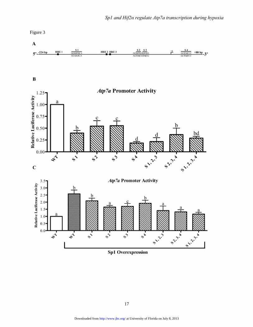
selected for further analysis. Mutation of each site
individually significantly reduced basal promoter
activity (Fig. 3). Combinatorial mutations (i.e.
triple and quadruple mutations) however, had little
additional effect on basal promoter activity.
Additionally, the effect of Sp1 overexpression on
Atp7a promoter activity was assessed to consider
the functional role of the putative Sp1 binding
sites. Sp1 overexpression induced activity of the
WT promoter (~2.5-fold), while individual and
combinatorial mutations had varying effects on
promoter activity, with some mutations (e.g. S1-3,
S2-4) abolishing the increase caused by Sp1
overexpression (Fig. 3).
Sp1 physically interacts with the Atp7a
promoter- To assess potential interactions between
Sp1 and the Atp7a promoter, chromatin
immunoprecipitation (ChIP) assays were
employed. Chromosomal DNA containing cross-
linked proteins was isolated from IEC-6 cells and
sheared to ~200 bp, then DNA samples were
pulled down with a ChIP-grade, anti-Sp1 antibody.
After reversing cross-links and purifying DNA,
PCR analysis was utilized to determine if specific
regions of the Atp7a promoter were present in the
IP’d samples. Results showed that all 4 putative
Sp1-binding site regions were present, while up-
or downstream Atp7a promoter regions lacking
putative Sp1-like binding sites were not detected
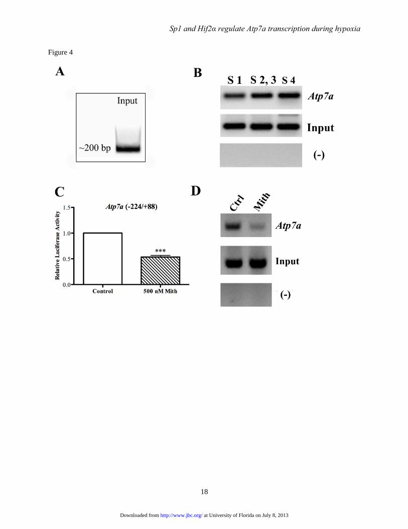
(Fig. 4). It was further shown that mithramycin
significantly reduced the amount of Atp7a
promoter DNA pulled down (containing all 4
putative Sp1 binding sites). In this experiment
(and others), there was no apparent difference in
the amount of input DNA among different
reactions.
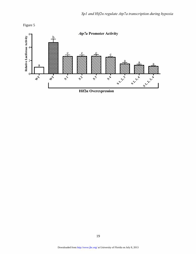
Sp1 binding is required for Hif2α-
mediated upregulation of Atp7a expression- We
next sought to determine if the putative Sp1
binding sites were required for Hif2α-mediated
induction of Atp7a promoter activity. As shown
previously (12), Hif2α overexpression induced
Atp7a promoter activity ~5-fold in IEC-6 cells
(Fig. 5). This induction was blunted by mutation
of each Sp1 binding site individually and
combinatorial mutations abolished transactivation
by Hif2α.
Sp1 and Hif2α interact with the Atp7a
promoter in vivo- As all experiments reported so
far were from an in vitro model of the mammalian
intestinal epithelium, it was important to confirm
these observations in vivo. Accordingly, studies
were performed in rats that were deprived of
dietary iron for 5 weeks after weaning. The intent
was to determine if Sp1 and Hif2α bound to the
Atp7a promoter in rat intestine and whether iron
deprivation altered DNA-protein interactions.
Iron-deprived rats had significantly decreased
hemoglobin and hematocrit levels (both
reduced >75%), indicative of iron-deficiency
anemia, consistent with previous observations (2,8)
(data not shown). Body weights were also lower
(~19%) in the rats fed the low-iron diet. Dcytb,
Dmt1, and Atp7a mRNA expression increased in
duodenal enterocytes isolated from the FeD rats,
as expected, and serum ceruloplasmin (Cp) protein
expression increased consistent with previous
observations (Fig. 6) (8). Moreover, Atp7a protein
expression increased, and although inconsistent
at University of Florida on July 8, 2013http://www.jbc.org/Downloaded from
Sp1 and Hif2α regulate Atp7a transcription during hypoxia
6
between animals, Hif2α protein levels were higher
in the iron-deficient rats. Lack of Atp7a detection
in one sample and Hif2α in 2 samples may relate
to delays in sample processing (and subsequent
partial protein degradation) since RNA
purification was undertaken first. Degradation was
not however observed upon visual inspection of
the stained blots. Furthermore, Hif1α was
undetectable under these conditions (but the
antibody had been validated by us using other
nuclear protein samples).
ChIP experiments confirmed that Sp1 and
Hif2α specifically interacted with the Atp7a
promoter in vivo (Fig. 6). There was a trend
towards increased Hif2α binding in samples
derived from FeD rats, while conversely, no
differences in Sp1 binding were noted among
groups (data not shown). The detection of Hif2
binding in control rat samples was unexpected as
the protein is normally degraded during normoxia.
The intestinal epithelium however exists in a
natural state of mild hypoxia (25), particularly in
epithelial cells at the villus tip which is furthest
from the blood supply (and where iron and copper
transporters are expressed). This may explain the
stabilization of the Hif2 protein in control
duodenum.
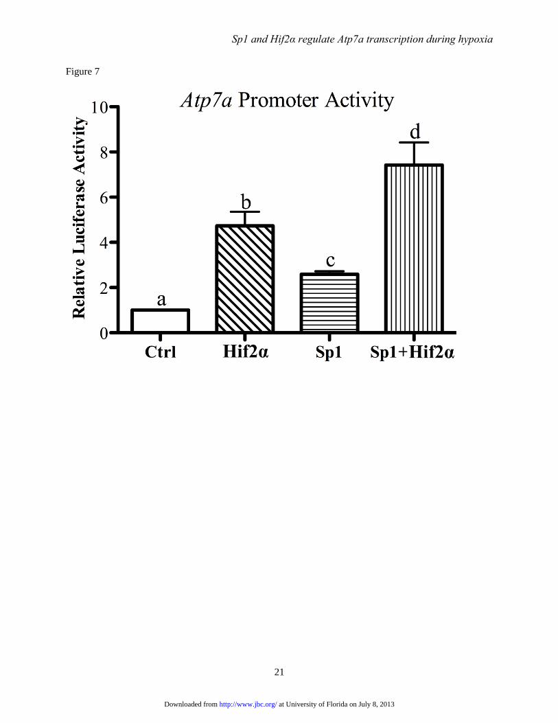
Sp1 and Hif2α synergistically activate the
Atp7a promoter- Forced expression of Sp1 or
Hif2α activates the Atp7a promoter in IEC-6 cells.
To determine if these two trans-acting factors can
further enhance the promoter response when co-
overexpressed, IEC-6 cells were transfected with
Sp1 and Hif2α expression vectors individually or
together, along with the basal Atp7a promoter
construct, and reporter gene assays were
performed. Forced Sp1 expression increased
activity ~3-fold while Hif2α overexpression
increased activity ~5-fold (Fig. 7). When both
were overexpressed together, Atp7a promoter
activity was further trans-activated to ~8-fold over
control (empty vector transfected) cells.
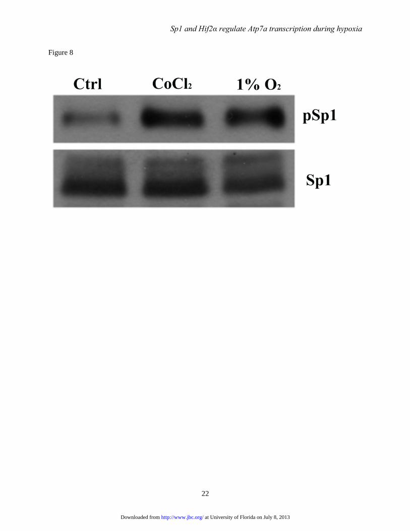
CoCl2 and low oxygen enhance
phosphorylation of Sp1- Sp1 activity is regulated
by phosphorylation. We thus sought to determine
if the level of phospho-Sp1 was altered by hypoxia.
Accordingly, IEC-6 cells were treated with CoCl2
to mimic hypoxia or grown in a hypoxia chamber
(12) and Sp1/phospho-Sp1 proteins levels were
determined by immunoblot analysis. Results
showed significantly higher levels of phospho-Sp1
in treated cells, while total immunoreactive Sp1
levels were relatively constant when comparing
the treatment groups to control (untreated) cells
(Fig. 8).
DISCUSSION During iron deficiency, hemoglobin levels
fall decreasing oxygen delivery to tissues and
cells, leading to a hypoxic response. At the
molecular level, this causes stabilization of the
HIFα subunits which promotes nuclear
localization and interaction with a constitutively
expressed HIFβ subunit followed by DNA binding
and activation of genes related to energy
metabolism (glycolysis), angiogenesis and iron
homeostasis. In the intestinal mucosa, during iron
deficiency/hypoxia, a Hif2α-specific
transcriptional response enhances absorption of
dietary iron by transactivating genes encoding
proteins which mediate iron transport.
Interestingly, Hif2α may also modulate intestinal
copper absorption, as reflected by induction of
Atp7a and metallothionein in duodenal enterocytes
during iron deprivation. The co-regulation of iron
and copper transport during iron deficiency
supports the concept that copper plays an
important physiologic role in the maintenance of
iron homeostasis. The current studies aimed to
further evaluate mechanistic aspects of the Hif2α
transcriptional response.
It was previously noted that many genes
induced by iron deprivation in the rat intestine and
in Caco-2 cells have G/C-rich promoters (14),
suggesting regulation by a trans-acting factor with
an affinity for G-C base pairs. The classic example
of such a transcription factor is specificity protein
1 (Sp1). This widely expressed protein is a
member of the Sp1-like and Krüppel-like factor
family of DNA-binding proteins, which are
integral parts of the transcriptional machinery of
eukaryotic cells (26,27). Sp1/KLFs have highly
conserved carboxy-terminal DNA-binding
domains containing three tandem zinc-finger
motifs. The amino terminal regions are variable
and contain transcription regulatory domains that
interact with co-regulators. By regulating the
expression of a large number of genes containing
GC-rich promoters, Sp1/KLF proteins are
involved in many biological processes, including
cell proliferation, differentiation, apoptosis and
neoplastic transformation (26). Few studies to date
at University of Florida on July 8, 2013http://www.jbc.org/Downloaded from
Sp1 and Hif2α regulate Atp7a transcription during hypoxia
7
have investigated Sp1-like factors in the regulation
of iron homeostasis-related genes. One recent
study suggested that the age-related decline in
hepatic transferrin gene expression may relate in
part to Sp1-like DNA binding activity (28). The
hepatic Hfe gene, which is mutated in some types
of hereditary hemochromatosis in humans, also
has apparent Sp1-like binding sites (29). Sp1 and
Sp3 were also shown to bind to an enhancer in the
ferritin H gene and activate expression in
fibroblast and liver cell lines (30). It was
additionally previously reported that the Dmt1
gene has three predicted Sp1-like binding sites, but
these were not experimentally verified (31).
Moreover, although a role for Sp1-like factors in
mediating the transcriptional response of intestinal
epithelial cells to iron deprivation was previously
postulated (13), this possibility has not been
experimentally tested to date.
To elucidate a potential role for Sp1 in the
Hif2α-mediated transcriptional response to iron
deprivation, we performed initial studies on the
Atp7a gene, which is coordinately regulated with
iron transport-related genes during iron
deprivation. Atp7a-mediated regulation of copper
absorption may play an important physiologic role
in the maintenance of intestinal iron transport,
possibly by enhancing activity of a multi-copper
ferroxidase (hephaestin), which couples iron
oxidation to efflux via Fpn1 (32,33). We
previously evaluated the rat Atp7a promoter
(12,34), including mapping the transcriptional start
site and defining the basal promoter region (-
224/+88). The role of Sp1 in basal and Hif2α-
stimulated Atp7a transcription has not however
been examined. This investigation was thus
undertaken to test the hypothesis that Sp1 (or an
Sp1-like factor) is necessary for the Hif2α-
mediated induction of gene expression in the
duodenal mucosa during iron deficiency.
Whenever possible, whether Atp7a-specific
regulatory mechanisms were conserved among
iron homeostasis-related genes (e.g. Dmt1, Dctyb,
Fpn1) was assessed to broaden the scope of this
experimental analysis.
Initial experiments utilized a drug that
blocks Sp1 binding to DNA (mithramycin) to
assess a possible role for Sp1 in Atp7a gene
transcription in IEC-6 cells. Mithramycin is a
DNA-binding antibiotic that binds to the minor
groove of G-C base pairs (35,36). This interaction
with DNA blocks trans-acting factor binding to
G/C-rich regions. Although initial studies showed
specific inhibition of Sp1 binding (16,17,37),
mithramycin could theoretically block binding of
any protein with an affinity for G-C base pairs. In
the current study, mithramycin was utilized to
show that Atp7a and other iron homeostasis-
related genes were potentially regulated by Sp1, as
mRNA expression was significantly inhibited. A
G/C-binding protein was however not absolutely
required for basal transcriptional activation of
these genes as expression was not abolished.
These observations provide preliminary evidence
that intestinal genes induced by Hif2α during iron
deprivation/hypoxia may be regulated by Sp1.
CoCl2 chemically mimics hypoxia (under
normoxic conditions) by stabilizing the HIFα
subunits; Hif1α and Hif2α are both stabilized via
inhibition of oxygen-dependent degradation.
Expression of Atp7a and iron transport-related
genes increased with CoCl2 treatment, consistent
with their known regulation by Hif2α. Ankrd37
and Vegf were also upregulated, likely reflecting
regulation by Hif1α (19,38). Interestingly,
mithramycin had differing effects on the induction
of mRNA expression by CoCl2; it blocked the
increase of some genes while other genes were
unaffected. This exemplified two modes of
regulation, one in which Sp1 (or a related G/C-
binding protein) is necessary for the HIF response
(e.g. for Atp7a and Dmt1), and another in which
Sp1 is not required (e.g. for Ankrd37 and Vegf).
These opposing regulatory mechanisms may relate
to distinct transactivation properties of the
different HIFα subunits. A trans-acting factor with
affinity for G/C-rich DNA regions may thus be
required for the Hif2α-mediated increase in gene
expression which ultimately promotes iron
absorption during hypoxia.
Several experimental observations
presented herein suggest that Atp7a gene
transcription is regulated by Sp1, including: 1) Sp1
overexpression increased endogenous Atp7a
mRNA and protein expression in IEC-6 cells and
also stimulated the exogenously expressed Atp7a
promoter; 2) Putative Sp1 binding sites were
shown to be required for full transactivation of
Atp7a gene expression; 3) ChIP assays showed
that Sp1 directly interacts with the Atp7a gene in
IEC-6 cells and in rat duodenal enterocytes; and 4)
Mithramycin significantly decreased pull down of
at University of Florida on July 8, 2013http://www.jbc.org/Downloaded from
Sp1 and Hif2α regulate Atp7a transcription during hypoxia
8
Atp7a promoter DNA containing the putative Sp1
binding sites from IEC-6 cells, consistent with the
documented decrease in Atp7a promoter activity in
the presence of mithramycin. Furthermore, in the
current investigation, the previously reported
binding of Hif2α to the Atp7a promoter in IEC-6
cells (12) was confirmed in rat duodenal
enterocytes. Atp7a is thus a bona fide Sp1 and
Hif2α target gene.
A final series of experiments was designed
to determine if putative Sp1 binding sites were
necessary for Hif2α-mediated induction of Atp7a
promoter activity. Forced Hif2α expression
increased promoter activity ~5-fold while
individual Sp1 binding site mutations attenuated
this increase to ~3-fold. Combinatorial Sp1 site
mutations abolished transactivation by Hif2α
overexpression. Interestingly, Hif2α
overexpression maintained basal Atp7a promoter
activity at WT levels, even when multiple Sp1
sites were mutated (in contrast to decreases in
basal activity without forced Hif2α expression).
Putative Sp1 binding sites are thus necessary for
transactivation of the Atp7a gene by Hif2α.
Data presented here show that the HIF-
mediated induction of Atp7a expression during
iron deficiency/hypoxia involves Sp1. Sp1-
dependent, Hif2α transactivation of gene
expression has not been reported in the scientific
literature (to our knowledge), suggesting that this
is a novel regulatory mechanism. Hif2α is
preferentially stabilized during iron deprivation in
the intestine of mice (9,10) and rats (this study),
and in Caco-2 cells (14). Hif2α protein levels
likely increase due to tissue hypoxia in iron-
deprived mice and rats, and as a result of
inhibition of the iron-dependent HIF prolyl
hydroxylases in Caco-2 cells treated with
deferroxamine (an iron chelator). What is not clear
is the specific molecular mechanism by which Sp1
potentiates the HIF-mediated induction of Atp7a
gene transcription. Sp1 is known to be regulated
by phosphorylation (39-41), which alters its DNA-
binding affinity and/or transactivation capabilities.
As such, we quantified Sp1/phosph-Sp1 levels in
CoCl2-treated IEC-6 cells and in cells grown in 1%
O2. Phosphorylation of Sp1 increased
dramatically in treated cells, suggesting that
posttranslational modification of the protein may
play a role in induction of Atp7a expression during
iron deprivation/hypoxia. Since ChIP assays
showed no difference in the amount of Atp7a
promoter DNA pulled down with Sp1 antibody
from enterocytes isolated from control or iron-
deficient rats, we speculate that Sp1
phosphorylation increases transactivation of Atp7a
gene expression.
This investigation focused on the gene
encoding the primary enterocyte copper exporter,
Atp7a. Lack of fully functional Atp7a is the
underlying cause of Menkes’ disease in humans, a
Mendelian disorder in which inefficient absorption
of dietary copper leads to systemic copper
deficiency and the dire physiologic consequences
of copper depletion (e.g. neurological damage,
hypopigmentation, etc.) (42,43). During iron
deficiency/hypoxia, Atp7a expression increases
dramatically, implicating copper in control of iron
homeostasis. In fact, copper increases in tissues
and cells important for homeostatic control of iron
homeostasis (e.g. enterocytes, hepatocytes) during
iron deficiency (3, 8). Given that Atp7a represents
the rate-limiting step in acquisition of dietary
copper, it may then play a key role in the
compensatory response to iron deficiency. Thus, a
detailed mechanistic understanding of Atp7a gene
regulation may increase knowledge of regulatory
aspects of whole-body iron homeostasis.
In summary, Sp1 binding is necessary for
the hypoxia-mediated induction of Atp7a promoter
activity in IEC-6 cells. Whether this mechanism is
also true of in vivo regulation of Atp7a gene
expression during iron deprivation is unknown,
but we provide evidence that the Atp7a gene is a
direct Hif2α and Sp1 target in rat duodenal
enterocytes. Three lines of evidence suggest that
these observations may have importance beyond
understanding Atp7a gene regulation: 1) Atp7a is
coordinately regulated by Hif2α along with genes
encoding proteins required for iron absorption
(Dcytb, Dmt1, Fpn1); 2) Many genes upregulated
by iron deficiency in the mammalian duodenum
have G/C-rich promoters and evolutionarily
conserved HREs; and 3) Hypoxia resulted in
increased phosphorylation of Sp1, likely altering
its’ transactivation properties. Sp1-dependent,
Hif2α-mediated induction of gene expression may
thus have broader implications for understanding
additional mechanistic aspects of intestinal iron
homeostasis.
at University of Florida on July 8, 2013http://www.jbc.org/Downloaded from
Sp1 and Hif2α regulate Atp7a transcription during hypoxia
9
REFERENCES
1. Aisen, P., Enns, C., and Wessling-Resnick, M. (2001) Chemistry and biology of eukaryotic iron
metabolism. Int. J. Biochem. Cell. Biol. 33, 940-959
2. Collins, J. F., Franck, C. A., Kowdley, K. V., and Ghishan, F. K. (2005) Identification of
differentially expressed genes in response to dietary iron deprivation in rat duodenum. Am. J.
Physiol. Gastrointest. Liver Physiol. 288, G964-971
3. Ravia, J. J., Stephen, R. M., Ghishan, F. K., and Collins, J. F. (2005) Menkes Copper ATPase
(Atp7a) is a novel metal-responsive gene in rat duodenum, and immunoreactive protein is present
on brush-border and basolateral membrane domains. J. Biol. Chem. 280, 36221-36227
4. Lee, G. R., Nacht, S., Lukens, J. N., and Cartwright, G. E. (1968) Iron metabolism in copper-
deficient swine. J. Clin. Invest. 47, 2058-2069
5. Naveh, Y., Hazani, A., and Berant, M. (1981) Copper deficiency with cow's milk diet. Pediatrics
68, 397-400
6. Prohaska, J. R., and Broderius, M. (2006) Plasma peptidylglycine alpha-amidating
monooxygenase (PAM) and ceruloplasmin are affected by age and copper status in rats and mice.
Comp. Biochem. Physiol. 143, 360-366
7. Gulec, S., and Collins, J. F. (2013) Investigation of Iron Metabolism in Mice Expressing a Mutant
Menke's Copper Transporting ATPase (Atp7a) Protein with Diminished Activity (Brindled;
MoBr/y). PloS One 8(6), e66010. doi:66010.61371/journal.pone.0066010
8. Ranganathan, P. N., Lu, Y., Jiang, L., Kim, C., and Collins, J. F. (2011) Serum ceruloplasmin
protein expression and activity increases in iron-deficient rats and is further enhanced by higher
dietary copper intake. Blood 118, 3146-3153
9. Mastrogiannaki, M., Matak, P., Keith, B., Simon, M. C., Vaulont, S., and Peyssonnaux, C. (2009)
HIF-2alpha, but not HIF-1alpha, promotes iron absorption in mice. J. Clin. Invest. 119, 1159-
1166
10. Shah, Y. M., Matsubara, T., Ito, S., Yim, S. H., and Gonzalez, F. J. (2009) Intestinal hypoxia-
inducible transcription factors are essential for iron absorption following iron deficiency. Cell
Metab. 9, 152-164
11. Taylor, M., Qu, A., Anderson, E. R., Matsubara, T., Martin, A., Gonzalez, F. J., and Shah, Y. M.
(2011) Hypoxia-Inducible Factor-2alpha Mediates the Adaptive Increase of Intestinal Ferroportin
During Iron Deficiency in Mice. Gastroenterology 140, 2044-2055
12. Xie, L., and Collins, J. F. (2011) Transcriptional regulation of the Menkes copper ATPase
(Atp7a) gene by hypoxia-inducible factor (HIF2alpha) in intestinal epithelial cells. Am. J.
Physiol. Cell Physiol. 300, C1298-1305
13. Collins, J. F., and Hu, Z. (2007) Promoter analysis of intestinal genes induced during iron-
deprivation reveals enrichment of conserved SP1-like binding sites. BMC Genomics 8, 420
14. Hu, Z., Gulec, S., and Collins, J. F. (2010) Cross-species comparison of genomewide gene
expression profiles reveals induction of hypoxia-inducible factor-responsive genes in iron-
deprived intestinal epithelial cells. Am. J. Physiol. Cell Physiol. 299, C930-938
15. Xie, L., and Collins, J. F. (2013) Copper stabilizes the Menkes copper-transporting ATPase
(Atp7a) protein expressed in rat intestinal epithelial cells. Am. J. Physiol. Cell Physiol. 304,
C257-262
16. Blume, S. W., Snyder, R. C., Ray, R., Thomas, S., Koller, C. A., and Miller, D. M. (1991)
Mithramycin inhibits SP1 binding and selectively inhibits transcriptional activity of the
dihydrofolate reductase gene in vitro and in vivo. J. Clin. Invest. 88, 1613-1621
17. Ray, R., Snyder, R. C., Thomas, S., Koller, C. A., and Miller, D. M. (1989) Mithramycin blocks
protein binding and function of the SV40 early promoter. J. Clin. Invest. 83, 2003-2007
18. Jiang, L., Ranganathan, P. N., Lu, Y., Kim, C., and Collins, J. F. (2011) Exploration of the copper
related compensatory response in the Belgrade rat model of genetic iron deficiency. Am. J.
Physiol. Gastrointest. Liver Physiol. 301, G877-886
at University of Florida on July 8, 2013http://www.jbc.org/Downloaded from
Sp1 and Hif2α regulate Atp7a transcription during hypoxia
10
19. Benita, Y., Kikuchi, H., Smith, A. D., Zhang, M. Q., Chung, D. C., and Xavier, R. J. (2009) An
integrative genomics approach identifies Hypoxia Inducible Factor-1 (HIF-1)-target genes that
form the core response to hypoxia. Nucleic Acids Res. 37, 4587-4602
20. Wada, T., Shimba, S., and Tezuka, M. (2006) Transcriptional regulation of the hypoxia inducible
factor-2alpha (HIF-2alpha) gene during adipose differentiation in 3T3-L1 cells. Biol. Pharm.
Bulletin 29, 49-54
21. Liang, Z. D., Tsai, W. B., Lee, M. Y., Savaraj, N., and Kuo, M. T. (2012) Specificity protein 1
(Sp1) oscillation is involved in copper homeostasis maintenance by regulating human high-
affinity copper transporter 1 expression. Mol. Pharm. 81, 455-464
22. Salceda, S., and Caro, J. (1997) Hypoxia-inducible factor 1alpha (HIF-1alpha) protein is rapidly
degraded by the ubiquitin-proteasome system under normoxic conditions. Its stabilization by
hypoxia depends on redox-induced changes. J. Biol. Chem. 272, 22642-22647
23. Yuan, Y., Beitner-Johnson, D., and Millhorn, D. E. (2001) Hypoxia-inducible factor 2alpha binds
to cobalt in vitro. Biochem. Biophys. Res. Comm. 288, 849-854
24. Yuan, Y., Hilliard, G., Ferguson, T., and Millhorn, D. E. (2003) Cobalt inhibits the interaction
between hypoxia-inducible factor-alpha and von Hippel-Lindau protein by direct binding to
hypoxia-inducible factor-alpha. J. Biol. Chem. 278, 15911-15916
25. Taylor, C. T., and Colgan, S. P. (2007) Hypoxia and gastrointestinal disease. J. Mol. Med. 85,
1295-1300
26. Kaczynski, J., Cook, T., and Urrutia, R. (2003) Sp1- and Kruppel-like transcription factors.
Genome Biol. 4, 206
27. Black, A. R., Black, J. D., and Azizkhan-Clifford, J. (2001) Sp1 and Kruppel-like factor family of
transcription factors in cell growth regulation and cancer. J. Cell. Physiol. 188, 143-160
28. Adrian, G. S., Seto, E., Fischbach, K. S., Rivera, E. V., Adrian, E. K., Herbert, D. C., Walter, C.
A., Weaker, F. J., and Bowman, B. H. (1996) YY1 and Sp1 transcription factors bind the human
transferrin gene in an age-related manner. J. Gerentol. A Biol. Sci. Med. Sci. 51, B66-75
29. Mura, C., Le Gac, G., Jacolot, S., and Ferec, C. (2004) Transcriptional regulation of the human
HFE gene indicates high liver expression and erythropoiesis coregulation. FASEB J. 18, 1922-
1924
30. Tsuji, Y., Torti, S. V., and Torti, F. M. (1998) Activation of the ferritin H enhancer, FER-1, by
the cooperative action of members of the AP1 and Sp1 transcription factor families. J. Biol.
Chem. 273, 2984-2992
31. Lee, P. L., Gelbart, T., West, C., Halloran, C., and Beutler, E. (1998) The human Nramp2 gene:
characterization of the gene structure, alternative splicing, promoter region and polymorphisms.
Blood Cells Mol. Dis. 24, 199-215
32. Fuqua, B. K., Vulpe, C. D., and Anderson, G. J. (2012) Intestinal iron absorption. J. Trace Elem.
Med. Biol. 26, 115-119
33. Vulpe, C. D., Kuo, Y. M., Murphy, T. L., Cowley, L., Askwith, C., Libina, N., Gitschier, J., and
Anderson, G. J. (1999) Hephaestin, a ceruloplasmin homologue implicated in intestinal iron
transport, is defective in the sla mouse. Nat. Genet. 21, 195-199
34. Collins, J. F., Hua, P., Lu, Y., and Ranganathan, P. N. (2009) Alternative splicing of the Menkes
copper Atpase (Atp7a) transcript in the rat intestinal epithelium. Am. J. Physiol. Gastrointest.
Liver Physiol. 297, G695-707
35. Schnedl, W., Breitenbach, M., and Stranzinger, G. (1977) Mithramycin and DIPI: a pair of
fluorochromes specific for GC-and AT-rich DNA respectively. Hum. Genet. 36, 299-305
36. Ward, D. C., Reich, E., and Goldberg, I. H. (1965) Base specificity in the interaction of
polynucleotides with antibiotic drugs. Science 149, 1259-1263
37. Snyder, R. C., Ray, R., Blume, S., and Miller, D. M. (1991) Mithramycin blocks transcriptional
initiation of the c-myc P1 and P2 promoters. Biochemistry 30, 4290-4297
at University of Florida on July 8, 2013http://www.jbc.org/Downloaded from
Sp1 and Hif2α regulate Atp7a transcription during hypoxia
11
38. Dvorak, H. F. (2002) Vascular permeability factor/vascular endothelial growth factor: a critical
cytokine in tumor angiogenesis and a potential target for diagnosis and therapy. J. Clin. Oncol.
20, 4368-4380
39. Daniel, S., Zhang, S., DePaoli-Roach, A. A., and Kim, K. H. (1996) Dephosphorylation of Sp1
by protein phosphatase 1 is involved in the glucose-mediated activation of the acetyl-CoA
carboxylase gene. J. Biol. Chem. 271, 14692-14697
40. Rohlff, C., and Glazer, R. I. (1998) Regulation of the MDR1 promoter by cyclic AMP-dependent
protein kinase and transcription factor Sp1. Int. J. Oncol. 12, 383-386
41. Rohlff, C., Ahmad, S., Borellini, F., Lei, J., and Glazer, R. I. (1997) Modulation of transcription
factor Sp1 by cAMP-dependent protein kinase. J. Biol. Chem. 272, 21137-21141
42. Tumer, Z., and Moller, L. B. (2010) Menkes disease. Eur. J. Hum. Genet.18, 511-518
43. Kaler, S. G. (1994) Menkes disease. Adv. Peds. 41, 263-304
at University of Florida on July 8, 2013http://www.jbc.org/Downloaded from
Sp1 and Hif2α regulate Atp7a transcription during hypoxia
12
FOOTNOTES
2To whom correspondence should be addressed: James F. Collins, Food Science and Human Nutrition
Department, University of Florida, 572 Newell Dr., FSN Bldg., #441, Gainesville, FL 32611 USA, Tel.:
(352) 392-1991, ext. 289; Fax: (352) 392-9467; E-mail: [email protected] 3These studies were supported by NIH grant 1R01-DK074867 to JFC.
4The abbreviations used are: Ankrd37, ankyrin repeat domain 37; Dmt1, divalent metal transporter 1;
Dcytb, doudenal cytochrome B; Fpn1, ferroportin 1; HA, hemagglutinin; HRE, hypoxia-response
element; HIF, hypoxia-inducible factor; ORF, open reading frame; Sp1, specificity factor 1; Sp6,
specificity factor 6; Tfr1, transferrin receptor 1; Vegf, vascular endothelial growth factor
FIGURE LEGENDS
FIGURE 1. Effect of mithramycin on CoCl2-mediated transcriptional induction. Post-confluent IEC-6
cells were cultured for 60 hours in the presence or absence (Ctrl) of 200 μM CoCl2. Mithramycin (Mith)
(500 nM) was added to one set of culture dishes from each treatment group for the last 24 hours (Mith).
Gene expression levels were subsequently determined by qRT-PCR. Gene names are shown in each panel.
Each bar represents mean ± SD (n=3). Different letters above each bar (a, b, c) indicate significant
differences between groups within each panel (p<0.05, one-way ANOVA).
FIGURE 2. Effect of Sp1 overexpression on Atp7a expression and Atp7a, Dmt1, and Dcytb promoter
activity. IEC-6 cells were transfected with HA-tagged Sp1 expression vector (Sp1) or empty expression
vector (Ctrl; pcDNA-3.1), and Atp7a (panels A & C) and Sp1 (panels B & D) mRNA and protein
expression was determined. Western blots in panels C and D are representative of 3 experiments with
similar results. Panel C also shows quantitative data for Atp7a protein expression (*p<0.05). Atp7a (panel
E), Dmt1 (panel F), and Dcytb (panel G) promoter constructs were co-transfected along with Sp1
overexpression vector into cells and luciferase activity was measured as an indicator of promoter
transactivation. Each bar represents the mean value ± SD. *P<0.05, **P<0.01, ***P<0.005; paired
Student’s t-test; n=3-4 for all experiments presented in this figure.
FIGURE 3. Functional analysis of putative Sp1 binding sites on the Atp7a promoter. Panel A shows a
schematic representation of the Atp7a promoter (-224 to +88). The 5’ most transcriptional start site
identified previously is marked as +1. Also shown are the previously identified hypoxia-response
elements (HREs) and the putative Sp1 binding sites (designated as S1-S4) with the mutated bases shown
in bold font. These putative Sp1 binding sites were mutated individually or in combination in the basal
Atp7a promoter construct and were subsequently transfected into IEC-6 cells. Panel B shows the activity
of mutated promoters in relation to the activity of the wild-type (WT) promoter. Panel C shows the effect
of forced Sp1 expression on WT and mutated Atp7a promoter activity. In panels C, D & E, each bar
represents the mean value ± SD. Different letters above bars indicate significant differences among
groups (unpaired Student’s t-test; n=3-4).
FIGURE 4. Chromatin immunoprecipitation (ChIP) analysis of Sp1 binding to the Atp7a promoter.
Cross-linked chromosomal DNA was immunoprecipitated from IEC-6 cell nuclear extracts using a ChIP-
grade Sp1 antibody. Subsequent PCR analysis was utilized to determine whether certain regions of the
Atp7a promoter were pulled down by the antibody. Panel A shows the typical size of DNA fragments
after sonication. Panel B depicts PCR analysis of recovered DNA. Results showed that all 4 putative Sp1
binding sites were present in the IP’d sample (Atp7a), but other regions of the promoter not containing
putative Sp1 binding sites (-) were not detected. Also shown is amplification of the Sp1 binding-site
regions from the input DNA. ChIP analysis was also performed with nuclear extracts derived from control
or mithromyin (Mith)-treated IEC-6 cells (Panels C & D). Panel C shows the effect of 500 nM Mith on
at University of Florida on July 8, 2013http://www.jbc.org/Downloaded from
Sp1 and Hif2α regulate Atp7a transcription during hypoxia
13
the activity of the WT Atp7a promoter transfected into cells. Each bar represents mean value ± SD (n=3;
***p<0.005, paired Student’s t-test). Mithramycin also decreased the amount of Atp7a promoter DNA
containing the putative Sp1 binding sites detected by PCR after ChIP (Atp7a) (panel D). Amplification
from input DNA samples was similar, indicating that equal amounts of starting material were used. Again,
other unrelated promoter regions were not detected (-). ChIP experiments depicted here are typical of
three independent experiments performed with similar results.
FIGURE 5. Effect of Hif2α overexpression on Atp7a promoter activity. Hif2α expression vector was co-
transfected into IEC-6 cells along with wild-type (WT) or mutant Atp7a promoter constructs and
luciferase assays were performed. The effect of Hif2α overexpression is shown relative to activity of the
WT promoter without Hif2α overexpression. The specific Sp1 sites (S) mutated are indicated below each
bar and Hif2α overexpression is indicated further below. Each bar represents the mean value ± SD.
Different letters on top of bars indicate statistical differences between groups (p<0.05; paired Student’s t-
test; n=3-4).
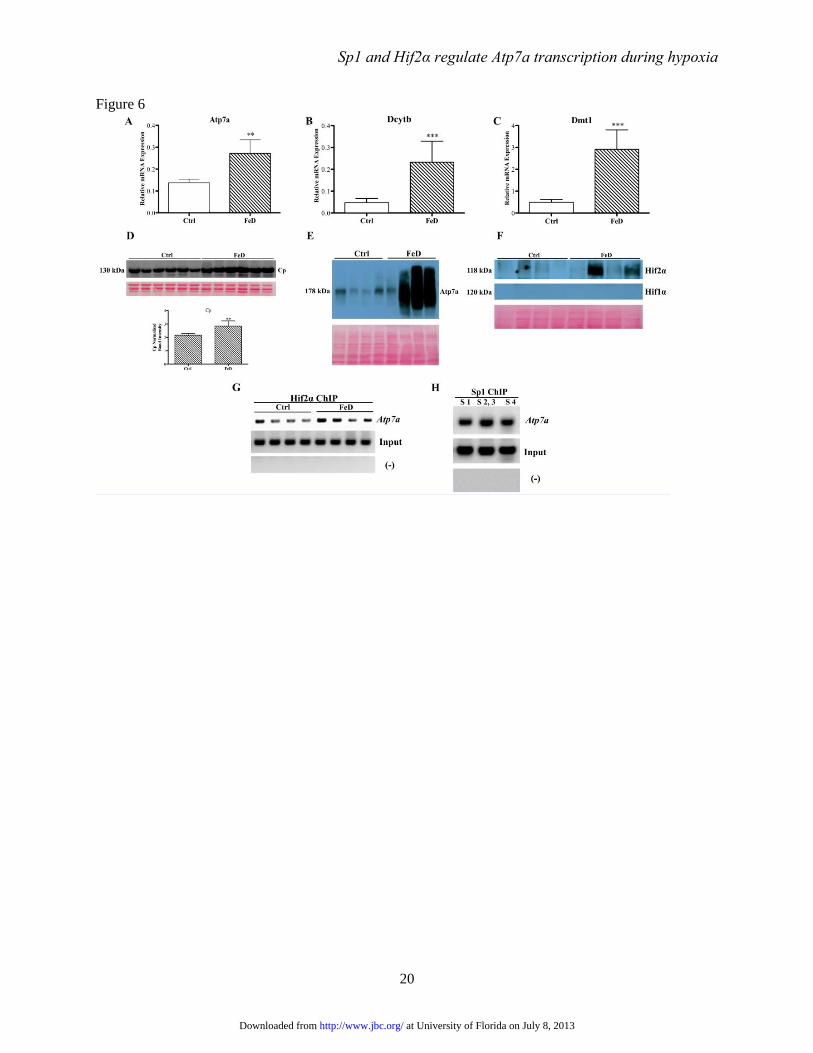
FIGURE 6. Molecular analysis of control and iron-deficient rats. Weanling rats consumed control or
low-iron diets for 5 weeks and were then sacrificed. Expression of known iron-responsive genes was
analyzed in isolated duodenal enterocytes by qRT-PCR (panels A-C). Each bar represents mean value ±
SD. **P<0.01, ***P<0.005, paired Student’s t-test, n=6. Ceruloplasmin (in serum; panel D), Atp7a (in
enterocytes; panel E) and Hif1/2α (in enterocytes; panel F) protein expression was assessed by western
blotting. Shown below the blots are total stained proteins exemplifying equal loading of the gels and
efficient transfer of proteins to membranes. ChIP experiments were also performed using cross-linked
chromosomal DNA isolated from duodenal enterocytes and ChIP-grade Hif2a (panel G) or Sp1 (panel H)
antibodies. For Hif2a ChIP (G), primers were used that covered the region containing the 3 HREs on the
Atp7a promoter. In panel H, the primers encompassed the Sp1 binding sites on the promoter. In panels G
and H, (-) indicates the use of primers from unrelated up-or downstream sites within the Atp7a promoter
and ‘Input’ indicates amplification from the DNA samples prior to immunoprecipitation.
FIGURE 7. Effect of Hif2α and/or Sp1 overexpression on Atp7a promoter activity. The basal Atp7a
promoter construct (-224/+88) was co-transfected along with Hif2α and/or Sp1 expression plasmids into
IEC-6 cells. Subsequent luciferase assays indicated promoter activity, which are shown in relation to
promoter activity in the absence of Sp1 or Hif2α overexpression (Ctrl). Each bar represents mean ± SD.
Different letters above each bar (a, b, c, d) indicate significant differences (p<0.05, one-way ANOVA; n =
3-4).
FIGURE 8. Immunoblot analysis of phosphorylated Sp1 protein expression. IEC-6 cells at 85%
confluence were either untreated and grown under control conditions (Ctrl), or treated with 200 μM CoCl2
or cultured in a hypoxia chamber (with 1% O2) for 60 hours. Nuclear proteins were then isolated and
immunoblots were run for detection of Sp1 and phosphorylated Sp1 (pSp1). The pSp1 band was detected
at ~120 kDa, while the total Sp1 protein band was detected at ~108 kDa. The blots shown are
representative of three independent experiments with comparable results.
at University of Florida on July 8, 2013http://www.jbc.org/Downloaded from
Sp1 and Hif2α regulate Atp7a transcription during hypoxia
14
Table 1. Mithramycin inhibits mRNA expression of Sp1-regulated genes
Gene Name Ctrl 100 nM 300 nM 500 nM
Ankrd37 1.00 0.50±0.20*** 0.30±0.20*** 0.1±0.04***
Atp7a 1.00 0.50±0.06* 0.40±0.20** 0.20±0.10***
Dcytb 1.00 0.50±0.20* 0.50±0.30* 0.30±0.30**
Dmt1 1.00 0.80±0.10 0.50±0.30* 0.30±0.20***
Fpn1 1.00 0.70±0.40 0.20±0.10*** 0.30±0.10***
Hif2α 1.00 0.40±0.20* 0.30±0.20*** 0.20±0.20***
Sp1 1.00 0.40±0.10*** 0.30±0.20*** 0.20±0.10***
Sp6 1.00 1.30±0.40 1.30±0.70 1.00±0.30
Tfr1 1.00 2.70±1.20* 2.90±1.70* 2.40±1.00*
Data are mean ± SD. * significance between mithramycin treated and control cells (Ctrl) (paired Student’s t-
test); *p<0.05; **p<0.01; ***p<0.005; n=3 for all groups.
at University of Florida on July 8, 2013http://www.jbc.org/Downloaded from
Sp1 and Hif2α regulate Atp7a transcription during hypoxia
15
Figure 1
at University of Florida on July 8, 2013http://www.jbc.org/Downloaded from
Sp1 and Hif2α regulate Atp7a transcription during hypoxia
16
Figure 2
at University of Florida on July 8, 2013http://www.jbc.org/Downloaded from
Sp1 and Hif2α regulate Atp7a transcription during hypoxia
17
Figure 3
at University of Florida on July 8, 2013http://www.jbc.org/Downloaded from
Sp1 and Hif2α regulate Atp7a transcription during hypoxia
18
Figure 4
at University of Florida on July 8, 2013http://www.jbc.org/Downloaded from
Sp1 and Hif2α regulate Atp7a transcription during hypoxia
19
Figure 5
at University of Florida on July 8, 2013http://www.jbc.org/Downloaded from
Sp1 and Hif2α regulate Atp7a transcription during hypoxia
20
Figure 6
at University of Florida on July 8, 2013http://www.jbc.org/Downloaded from
Sp1 and Hif2α regulate Atp7a transcription during hypoxia
21
Figure 7
at University of Florida on July 8, 2013http://www.jbc.org/Downloaded from
Sp1 and Hif2α regulate Atp7a transcription during hypoxia
22
Figure 8
at University of Florida on July 8, 2013http://www.jbc.org/Downloaded from

Supplementary Material http://www.jbc.org/content/suppl/2013/06/28/M113.489500.DC1.html
Supplementary material can be found at:
at University of Florida on July 8, 2013http://www.jbc.org/Downloaded from


![Original Article Pokemon Inhibits Transforming Growth ... · (SP1) has been found to be an important potentiator in the TGFβ/Smad4 signaling pathway [25,26], in this study, we attempted](https://static.fdocument.org/doc/165x107/5e0e08789413ab632f1d5c93/original-article-pokemon-inhibits-transforming-growth-sp1-has-been-found-to.jpg)
![Original Article EMMPRIN, SP1 and microRNA-27a mediate ... · pathways and mechanisms, including loss of function of the tumor suppressor p53 [10], upregulated vascular endothelial](https://static.fdocument.org/doc/165x107/5e22380d3a89c23c53196456/original-article-emmprin-sp1-and-microrna-27a-mediate-pathways-and-mechanisms.jpg)