Treatment With Lopinavir/Ritonavir or Interferon-β1b ...€¦ · which best represents severe...
10
MAJOR ARTICLE Treatment With Lopinavir/Ritonavir or Interferon-β1b Improves Outcome of MERS- CoV Infection in a Nonhuman Primate Model of Common Marmoset Jasper Fuk-Woo Chan, 1,2,3,4,a Yanfeng Yao, 5,a Man-Lung Yeung, 2 Wei Deng, 5 Linlin Bao, 5 Lilong Jia, 2 Fengdi Li, 5 Chong Xiao, 5 Hong Gao, 5 Pin Yu, 5 Jian-Piao Cai, 2 Hin Chu, 2 Jie Zhou, 2 Honglin Chen, 1,2,3,4 Chuan Qin, 5,b and Kwok-Yung Yuen 1,2,3,4,b 1 State Key Laboratory of Emerging Infectious Diseases, 2 Department of Microbiology, 3 Research Centre of Infection and Immunology, 4 Carol Yu Centre for Infection, The University of Hong Kong, Hong Kong Special Administrative Region, and 5 Institute of Laboratory Animal Sciences, Chinese Academy of Medical Sciences, Beijing, China Middle East respiratory syndrome coronavirus (MERS-CoV) causes severe disease in human with an overall case-fatality rate of >35%. Effective antivirals are crucial for improving the clinical outcome of MERS. Although a number of repurposed drugs, convalescent-phase plasma, antiviral peptides, and neutralizing antibodies ex- hibit anti-MERS-CoV activity in vitro, most are not readily available or have not been evaluated in nonhuman primates. We assessed 3 repurposed drugs with potent in vitro anti-MERS-CoV activity (mycophenolate mofetil [MMF], lopinavir/ritonavir, and interferon-β1b) in common marmosets with severe disease resembling MERS in humans. The lopinavir/ritonavir-treated and interferon-β1b-treated animals had better outcome than the untreated animals, with improved clinical (mean clinical scores ↓50.9%–95.0% and ↓weight loss than the un- treated animals), radiological (minimal pulmonary infiltrates), and pathological (mild bronchointerstitial pneumonia) findings, and lower mean viral loads in necropsied lung (↓0.59–1.06 log 10 copies/glyceraldehyde 3-phosphate dehydrogenase [GAPDH]; P < .050) and extrapulmonary (↓0.11–1.29 log 10 copies/GAPDH; P < .050 in kidney) tissues. In contrast, all MMF-treated animals developed severe and/or fatal disease with high- er mean viral loads (↑0.15–0.54 log 10 copies/GAPDH) than the untreated animals. The mortality rate at 36 hours postinoculation was 67% (untreated and MMF-treated) versus 0–33% (lopinavir/ritonavir-treated and in- terferon-β1b-treated). Lopinavir/ritonavir and interferon-β1b alone or in combination should be evaluated in clinical trials. MMF alone may worsen MERS and should not be used. Keywords. animal; common marmoset; coronavirus; interferon; Kaletra; lopinavir; MERS; mycophenolate; primate; treatment. Coronaviruses (CoVs) have repeatedly crossed species barriers and caused epidemics with significant socio- economic impact [1]. Middle East respiratory syndrome coronavirus (MERS-CoV) is a novel zoonotic lineage C βCoV discovered in 2012 [2]. MERS-CoV infection in human, or MERS, commonly manifests as a severe acute respiratory disease, which is often complicated by acute respiratory distress syndrome and multiorgan failure [3, 4]. Clinical deterioration and death occur rap- idly and at a few days earlier in MERS than in severe acute respiratory syndrome (SARS) [3]. The case-fatality rate of MERS is the highest among all human CoV in- fections and exceeds 35% [3, 5]. Effective antiviral treat- ment, especially when given early to patients at risk of developing severe disease, may improve the clinical out- come of MERS. A number of new drug compounds, such as humanized or human monoclonal antibodies against the S1 subunit receptor-binding domain and Received 15 June 2015; accepted 6 July 2015; electronically published 21 July 2015. a J. F.-W. C. and Y. Y. contributed equally to this work as co-first authors. b C. Q. and K.-Y. Y. contributed equally to this work as co-corresponding authors. Correspondence: Kwok-Yung Yuen, MD, State Key Laboratory of Emerging Infec- tious Diseases, Carol Yu Centre for Infection, Department of Microbiology, The Uni- versity of Hong Kong, Queen Mary Hospital, 102 Pokfulam Road, Pokfulam, Hong Kong Special Administrative Region, China ([email protected]). The Journal of Infectious Diseases ® 2015;212:1904–13 © The Author 2015. Published by Oxford University Press on behalf of the Infectious Diseases Society of America. All rights reserved. For Permissions, please e-mail: [email protected]. DOI: 10.1093/infdis/jiv392 1904 • JID 2015:212 (15 December) • Chan et al
Transcript of Treatment With Lopinavir/Ritonavir or Interferon-β1b ...€¦ · which best represents severe...
M A J O R A R T I C L E
Treatment With Lopinavir/Ritonavir orInterferon-β1b Improves Outcome of MERS-CoV Infection in a Nonhuman Primate Model ofCommon Marmoset
Jasper Fuk-Woo Chan,1,2,3,4,a Yanfeng Yao,5,a Man-Lung Yeung,2 Wei Deng,5 Linlin Bao,5 Lilong Jia,2 Fengdi Li,5 Chong Xiao,5
Hong Gao,5 Pin Yu,5 Jian-Piao Cai,2 Hin Chu,2 Jie Zhou,2 Honglin Chen,1,2,3,4 Chuan Qin,5,b and Kwok-Yung Yuen1,2,3,4,b
1State Key Laboratory of Emerging Infectious Diseases, 2Department of Microbiology, 3Research Centre of Infection and Immunology, 4Carol Yu Centre forInfection, The University of Hong Kong, Hong Kong Special Administrative Region, and 5Institute of Laboratory Animal Sciences, Chinese Academy ofMedical Sciences, Beijing, China
Middle East respiratory syndrome coronavirus (MERS-CoV) causes severe disease in human with an overallcase-fatality rate of >35%. Effective antivirals are crucial for improving the clinical outcome of MERS. Althougha number of repurposed drugs, convalescent-phase plasma, antiviral peptides, and neutralizing antibodies ex-hibit anti-MERS-CoV activity in vitro, most are not readily available or have not been evaluated in nonhumanprimates. We assessed 3 repurposed drugs with potent in vitro anti-MERS-CoV activity (mycophenolate mofetil[MMF], lopinavir/ritonavir, and interferon-β1b) in common marmosets with severe disease resembling MERSin humans. The lopinavir/ritonavir-treated and interferon-β1b-treated animals had better outcome than theuntreated animals, with improved clinical (mean clinical scores ↓50.9%–95.0% and ↓weight loss than the un-treated animals), radiological (minimal pulmonary infiltrates), and pathological (mild bronchointerstitialpneumonia) findings, and lower mean viral loads in necropsied lung (↓0.59–1.06 log10 copies/glyceraldehyde3-phosphate dehydrogenase [GAPDH]; P < .050) and extrapulmonary (↓0.11–1.29 log10 copies/GAPDH;P < .050 in kidney) tissues. In contrast, all MMF-treated animals developed severe and/or fatal disease with high-er mean viral loads (↑0.15–0.54 log10 copies/GAPDH) than the untreated animals. The mortality rate at 36hours postinoculation was 67% (untreated and MMF-treated) versus 0–33% (lopinavir/ritonavir-treated and in-terferon-β1b-treated). Lopinavir/ritonavir and interferon-β1b alone or in combination should be evaluated inclinical trials. MMF alone may worsen MERS and should not be used.
Keywords. animal; common marmoset; coronavirus; interferon; Kaletra; lopinavir; MERS; mycophenolate;primate; treatment.
Coronaviruses (CoVs) have repeatedly crossed speciesbarriers and caused epidemics with significant socio-economic impact [1].Middle East respiratory syndrome
coronavirus (MERS-CoV) is a novel zoonotic lineageC βCoV discovered in 2012 [2]. MERS-CoV infectionin human, or MERS, commonly manifests as a severeacute respiratory disease, which is often complicatedby acute respiratory distress syndrome and multiorganfailure [3, 4]. Clinical deterioration and death occur rap-idly and at a few days earlier in MERS than in severeacute respiratory syndrome (SARS) [3]. The case-fatalityrate of MERS is the highest among all human CoV in-fections and exceeds 35% [3, 5]. Effective antiviral treat-ment, especially when given early to patients at risk ofdeveloping severe disease, may improve the clinical out-come of MERS. A number of new drug compounds,such as humanized or human monoclonal antibodiesagainst the S1 subunit receptor-binding domain and
Received 15 June 2015; accepted 6 July 2015; electronically published 21 July2015.
aJ. F.-W. C. and Y. Y. contributed equally to this work as co-first authors.bC. Q. and K.-Y. Y. contributed equally to this work as co-corresponding authors.Correspondence: Kwok-Yung Yuen, MD, State Key Laboratory of Emerging Infec-
tious Diseases, Carol Yu Centre for Infection, Department of Microbiology, The Uni-versity of Hong Kong, Queen Mary Hospital, 102 Pokfulam Road, Pokfulam, HongKong Special Administrative Region, China ([email protected]).
The Journal of Infectious Diseases® 2015;212:1904–13© The Author 2015. Published by Oxford University Press on behalf of the InfectiousDiseases Society of America. All rights reserved. For Permissions, please e-mail:[email protected]: 10.1093/infdis/jiv392
1904 • JID 2015:212 (15 December) • Chan et al
antiviral peptides targeting the S2 subunit heptad repeat 2 do-main of the MERS-CoV spike glycoprotein, have recently beendeveloped and showed potent anti-MERS-CoV activity in vitro[6–9]. However, their developments are still in the preclinicalphase. Another possible treatment option for MERS is conva-lescent-phase plasma therapy. This has been previously usedto treat other severe viral respiratory infections, includingSARS and pandemic influenza, with good clinical and virolog-ical response [10–12].However, this is also not readily available,as its preparation requires the recruitment of a large number ofconvalescent patients with high serum antibody titers. Conva-lescent-phase plasma with low serum antibody titers may beassociated with immune enhancement, which could lead toworsened outcome as observed in in vitro and animal experi-ments on SARS [13, 14].
In order to find immediately available treatment for MERSbefore these novel agents and convalescent-phase plasmatherapy become available, we and others have applied high-throughput screening to identify existing drugs that may berepurposed to treat patients with MERS [15–17]. Examples ofdrugs identified using this drug discovery approach includedinhibitors of virus entry, clathrin-mediated endocytosis, neuro-transmitters, estrogen receptor, kinase signaling, lipid metabo-lism, protein processing, and DNA synthesis/repair [3, 15–19].Although all of them exhibit in vitro anti-MERS-CoV activity,only a few are likely clinically relevant, as most are either asso-ciated with severe adverse effects or have anti-MERS-CoV half-maximal effective concentrations (EC50) that are not clinicallyachievable at therapeutic dosages. Previous studies have identi-fied mycophenolate mofetil (MMF), ribavirin, lopinavir, inter-feron-α, and interferon-β as potential candidates for further invivo evaluation [15–17, 20]. Treatment with ribavirin and inter-feron-α has shown some effects in vitro and in MERS-CoV-infected rhesus macaques with mild disease, but the benefits inMERS patients were not obvious [20–22]. In this study, we
evaluated the treatment effects of MMF, lopinavir, and interfer-on-β in MERS-CoV-infected common marmosets. This recent-ly described nonhuman primate model is the animal modelwhich best represents severe human disease among all existinganimal models for MERS [3, 23]. Importantly, we found that lo-pinavir/ritonavir and interferon-β1b, but not MMF, improvedthe outcome of MERS-CoV-infected common marmosets.The findings provide the basis for future clinical trials for thishighly fatal disease.
METHODS
Virus Strain and TitrationMERS-CoV (EMC/2012 strain) was kindly provided by RonFouchier (Erasmus Medical Center). The virus was propagatedin VeroE6 cells (ATCC) in Dulbecco’s modified Eagle’s medium(DMEM) supplemented with 10% fetal calf serum and 100units/mL penicillin plus 100 µg/mL streptomycin. All experi-ments involving live MERS-CoV followed the approved stan-dard operating procedures of the biosafety level 3 facility aspreviously described [24–28]. The 50% tissue culture infectiousdose (TCID50) per mL was determined for MERS-CoV inVeroE6 cells as previously described [25].
Common Marmoset Infection Model and TreatmentAll experiments involving common marmosets were performedas previously described with slight modifications and approvedby the Institutional Animal Care and Use Committee [23, 26,29]. Briefly, 12 healthy male common marmosets (Callithrix jac-chus; 3 years old; 230–395 g) were inoculated with 5 × 10⁶
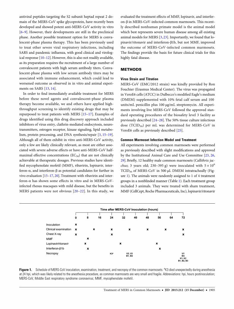
TCID50 of MERS-CoV in 500 µL DMEM intratracheally (Fig-ure 1). The animals were randomly assigned to 1 of 4 treatmentgroups in a nonblinded manner (Table 1). Each treatment groupincluded 3 animals. They were treated with sham treatment,MMF (CellCept, Roche Pharmaceuticals, Inc), lopinavir/ritonavir
Figure 1. Schedule of MERS-CoV inoculation, examination, treatment, and necropsy of the common marmosets. aK3 died unexpectedly during anesthesiaat 24 hpi, which was likely related to the anesthesia procedure, as common marmosets are very small and fragile. Abbreviations: hpi, hours postinoculation;MERS-CoV, Middle East respiratory syndrome coronavirus; MMF, mycophenolate mofetil.
Treatment of MERS in Common Marmosets • JID 2015:212 (15 December) • 1905
(4:1) (Kaletra, Abbot Laboratories), or recombinant interferon-β1b (Betaferon, Bayer Schering Pharma, Germany) using previ-ously described dosages [30–32]. We administered MMF intra-peritoneally and interferon-β1b subcutaneously instead ofintravenously or orally, respectively, because the small veins ofthe commonmarmosets prevented reliable drug delivery throughthe intravenous route and because critically ill MERS patientsusually require parenteral treatment. Lopinavir/ritonavir wasonly available in oral preparation. We initiated treatment withMMF and interferon-β1b at 8 hours postinoculation (hpi)according to a previously described treatment model usingMERS-CoV-infected rhesus macaques [21].Treatment with lopi-navir/ritonavir was started at 6 hpi as lopinavir/ritonavir takeslonger to achieve its peak serum concentration (Cmax) comparedwith MMF and interferon-β1b. The animals were observed twicedaily for clinical signs and scored using a previously describedclinical scoring system, which included the evaluation of the ge-neral appearance, skin and fur appearance, discharge (oral, nasal,and/or ocular), respiratory rate, and food consumption [23].Other clinical examinations in this study included measurementof body temperature (twice daily) and body weight (0, 24, and 72hpi), and blood collection for hematology tests (0 hpi andnecropsy). Dorsal-ventral and lateral chest X-rays (CXR) wereperformed on anesthetized animals (0, 24, and 72 hpi) as previ-ously described [29]. Necropsies were scheduled at 72 hpi orwhen the clinical score was ≥35 as previously described [21,23]. The necropsied tissues were collected for histopathology, im-munohistochemistry, and viral load studies.
Histopathology and ImmunohistochemistryThe sections of the necropsied tissues were stained with hematox-ylin and eosin for light microscopic examination and immunohis-tochemically stained with mouse anti-MERS-CoV nucleocapsidprotein (1:200) antibody overnight at 4°C for immunofluorescentexamination of MERS-CoV nucleocapsid protein expression asdescribed previously [27, 29, 33].
Viral Load StudiesRNA extraction and quantitative real-time reverse transcription–polymerase chain reaction (RT-PCR) were performed on necrop-sied lung, kidney, liver, spleen, and heart tissues as we previouslydescribed [25–27]. Three areas with the most severe macroscopiclesions were obtained from each necropsied organ for viral loadstudies. Briefly, total RNA and viral RNA were isolated usingTrizol (Life Technologies) and Viral RNA Mini kit (QIAGEN),respectively. After RNA quantification, 1 µg RNA was reversetranscribed into complementary DNA (cDNA) using randomhexamers. Total viral transcripts were detected using Novel Coro-navirus 2012 Real-Time RT-PCR assay (CDC; Catalog #KT0136).The viral RNAwere normalized to themessenger RNA expressionlevel of glyceraldehyde 3-phosphate dehydrogenase (GAPDH)and a spike-in control, enterovirus 71 (strain SZ/HK08–5),using primers 5′-GCTCACTGGCATGGCCTTCCGTGT-3′ and5′-TGGAGGAGTGGGTGTCGCTGTTGA-3′ (GAPDH), and5′-CCCCTGAATGCGGCTAATCC-3′ and 5′- ACACGGACACCCAAAGTAGT -3′ (enterovirus 71). For each reaction, equalamounts of cDNA were mixed with FS Universal SYBR GreenMaster Rox (Roche) plus 5 pmol each of forward and reverseprimers. Amplification was done under the condition of 15 sec-onds at 95°C and 1minute at 60°C for 55 cycles in a 7900 real-timePCR detection system (ABI).
Statistical AnalysisStatistical comparison between different groups was performedby the Student t test using GraphPad Prism 6. A P value <.05was considered statistically significant.
RESULTS
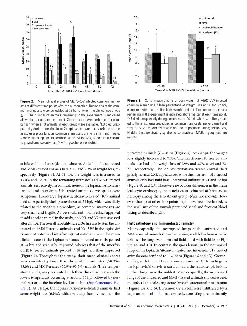
Clinical and Radiological FindingsThe untreated and MMF-treated animals developed severe dis-ease with increased respiratory rates, reduced movement, andloss of appetite soon after MERS-CoV inoculation. At 12 hpi,the untreated andMMF-treated animals both had a mean clinicalscore of 18.3 (Figure 2). Their mean clinical scores peaked at 36hpi, at which point 2 untreated (U2 and U3) and 2 MMF-treated(M1 and M2) animals developed severe dyspnea with cyanosis,barely moved, and had blood-stained oral discharge. They wereeuthanized at 36 hpi as their clinical scores exceeded 35. The eu-thanized animals had severe hypothermia (≤34.5°C) at necropsy,which was suggestive of severe disease with shock (SupplementaryFigure 1). CXR performed on these 4 animals prior to necropsyshowed bilateral interstitial infiltrates, which were indicative ofextensive pneumonia (Figure 4A and 4B). The remaining un-treated (U1) and MMF-treated (M3) animals survived, but con-tinued to have severe symptoms with clinical scores ≥15(Figure 2) and temperature ≤37.5°C, which was below their base-line levels, throughout the remaining study period (Supplemen-tary Figure 1). CXR at 24 and 72 hpi showed interstitial infiltrates
Table 1. Treatment Groups and Drug Regimens Used in ThisStudy
GroupCommonMarmoset Treatment Regimen
1 U1, U2, U3 Untreated (sham treatment with comparablevolume per kg body weight of sterile saline)
2 M1, M2, M3 CellCept (25 mg/kg of MMF given ip once at8 hpi)
3 K1, K2, K3 Kaletra (12 mg/kg/day of lopinavir + 3 mg/kg/day of ritonavir
given orally once daily at 6, 30, and 54 hpi)
4 B1, B2, B3 Betaferon (0.267million IU/kg of interferon-β1bgiven sc at 8 hpi and at 56 hpi)
Abbreviations: hpi, hours postinoculation; ip, intraperitoneal; MMF,mycophenolate mofetil; sc, subcutaneous.
1906 • JID 2015:212 (15 December) • Chan et al
at bilateral lung bases (data not shown). At 24 hpi, the untreatedand MMF-treated animals had 9.0% and 9.3% of weight loss, re-spectively (Figure 3). At 72 hpi, the weight loss increased to15.0% and 12.9% in the remaining untreated and MMF-treatedanimals, respectively. In contrast, none of the lopinavir/ritonavir-treated and interferon-β1b-treated animals developed severesymptoms. However, 1 lopinavir/ritonavir-treated (K3) animaldied unexpectedly during anesthesia at 24 hpi, which was likelyrelated to the anesthesia procedure, as common marmosets arevery small and fragile. As we could not obtain ethics approvalto add another animal in the study, only K1 and K2 were assessedafter 24 hpi. The overallmortality rate at 36 hpiwas 67% in the un-treated and MMF-treated animals, and 0%–33% in the lopinavir/ritonavir-treated and interferon-β1b-treated animals. The meanclinical score of the lopinavir/ritonavir-treated animals peakedat 24 hpi and gradually improved, whereas that of the interfer-on-β1b-treated animals peaked at 36 hpi and then improved(Figure 2). Throughout the study, their mean clinical scoreswere consistently lower than those of the untreated (50.9%–
95.0%) and MMF-treated (50.9%–93.3%) animals. Their temper-ature trend grossly correlated with their clinical scores, with thelowest temperature occurring at around 36 hpi, followed by nor-malization to the baseline level at 72 hpi (Supplementary Fig-ure 1). At 24 hpi, the lopinavir/ritonavir-treated animals hadsome weight loss (6.0%), which was significantly less than the
untreated animals (P = .038) (Figure 3). At 72 hpi, the weightloss slightly increased to 7.5%. The interferon-β1b-treated ani-mals also had mild weight loss of 7.9% and 8.7% at 24 and 72hpi, respectively. The lopinavir/ritonavir-treated animals hadgrossly normal CXR appearances, while the interferon-β1b-treatedanimals only had mild basal interstitial infiltrate at 24 and 72 hpi(Figure 4C and 4D). Therewere no obvious differences in themeanleukocyte, erythrocyte, and platelet counts obtained at 0 hpi and atnecropsy among the 4 treatment groups (data not shown). How-ever, changes at other time points might have been overlooked, asthe small size of the animals prevented serial and frequent bloodtaking as described [23].
Histopathology and ImmunohistochemistryMacroscopically, the necropsied lungs of the untreated andMMF-treated animals showed extensive, multilobar hemorrhagiclesions. The lungs were firm and fluid-filled with fluid leak (Fig-ure 4A and 4B). In contrast, the gross lesions in the necropsiedlungs of the lopinavir/ritonavir-treated and interferon-β1b-treatedanimals were confined to 1–2 lobes (Figure 4C and 4D). Corrob-orating with the mild symptoms and normal CXR findings inthe lopinavir/ritonavir-treated animals, the macroscopic lesionsin their lungs were the mildest. Microscopically, the necropsiedlungs of the untreated andMMF-treated animals showed severe,multifocal to coalescing acute bronchointerstitial pneumonia(Figure 5A and 5C). Pulmonary alveoli were infiltrated by alarge amount of inflammatory cells, consisting predominantly
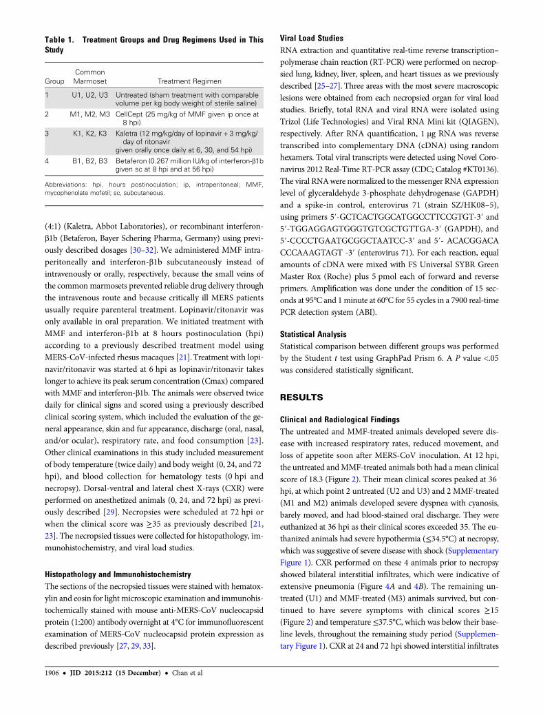
Figure 2. Mean clinical scores of MERS-CoV-infected common marmo-sets at different time points after virus inoculation. Necropsies of the com-mon marmosets were scheduled at 72 hpi or when the clinical score was≥35. The number of animals remaining in the experiment is indicatedabove the bar at each time point. Student t test was performed for com-parison when all 3 animals in each group were available. aK3 died unex-pectedly during anesthesia at 24 hpi, which was likely related to theanesthesia procedure, as common marmosets are very small and fragile.Abbreviations: hpi, hours postinoculation; MERS-CoV, Middle East respira-tory syndrome coronavirus; MMF, mycophenolate mofetil.
Figure 3. Serial measurements of body weight of MERS-CoV-infectedcommon marmosets. Mean percentage of weight loss at 24 and 72 hpi,compared with the baseline body weight at 0 hpi. The number of animalsremaining in the experiment is indicated above the bar at each time point.aK3 died unexpectedly during anesthesia at 24 hpi, which was likely relat-ed to the anesthesia procedure, as common marmosets are very small andfragile. *P < .05. Abbreviations: hpi, hours postinoculation; MERS-CoV,Middle East respiratory syndrome coronavirus; MMF, mycophenolatemofetil.
Treatment of MERS in Common Marmosets • JID 2015:212 (15 December) • 1907
of macrophages, neutrophils, and lymphocytes. The alveolar in-terstitium was thickened with edema, fibrin, and hemorrhage.Abundant MERS-CoV nucleocapsid protein expression was de-tected in immunohistochemical staining (Figure 5B and 5D). Incontrast, the necropsied lungs of the lopinavir/ritonavir-treatedand interferon-β1b-treated animals showed mild to moderatebronchointerstitial pneumonia confined to 1–2 lobes, with asmall amount of inflammatory cell infiltrates, edema, and hem-orrhage (Figure 5E and 5G). Immunohistochemical stainingshowed scarce and focal MERS-CoV nucleocapsid protein ex-pression (Figure 5F and 5H). The histopathological changes
in the necropsied extrapulmonary tissues of the 4 treatmentgroups were similar and considered to be nonspecific changesassociated with hypoxic damage or incidental findings in com-mon marmosets as described (data not shown) [23].
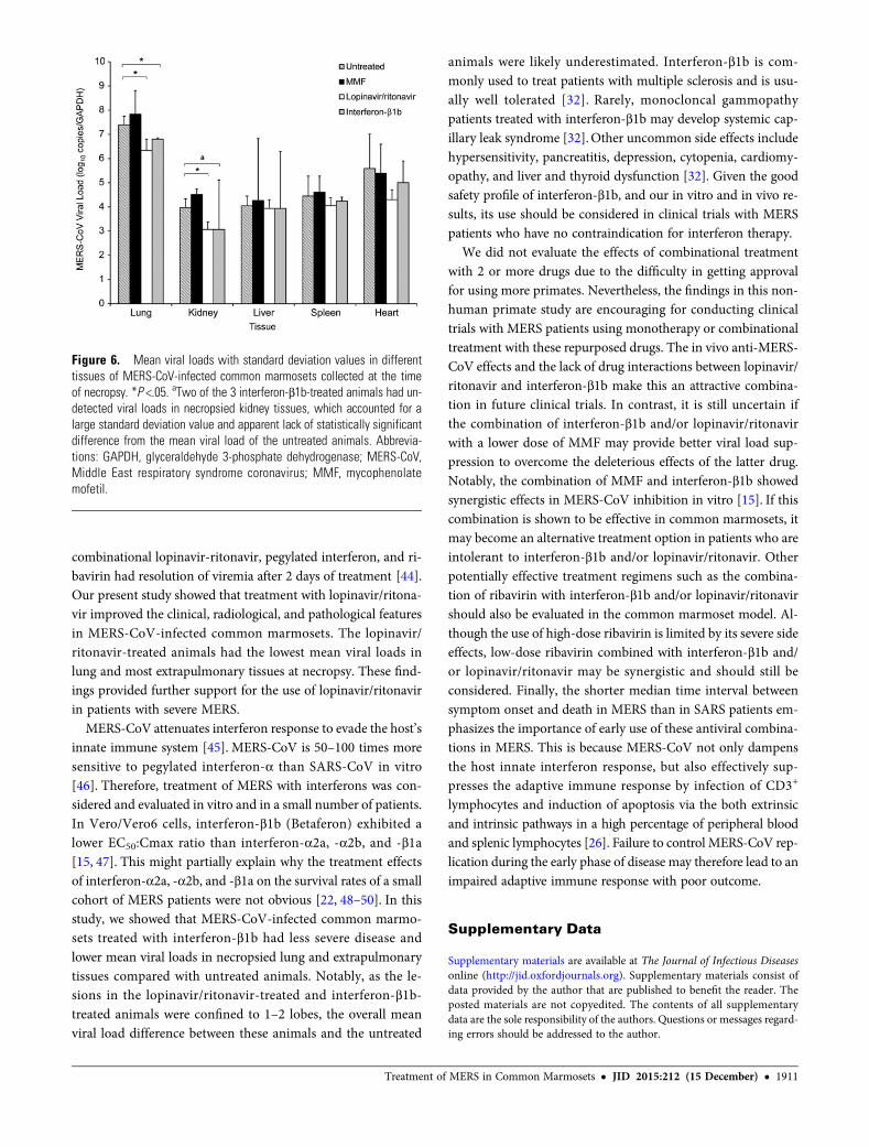
Viral Load StudiesThe mean viral load of necropsied lungs was highest in theMMF-treated animals and lowest in the lopinavir/ritonavir-treated animals (Figure 6). The mean viral load of the necrop-sied lungs of the MMF-treated animals was 0.44 log10 copies/GAPDH higher than that of the untreated animals. The mean
Figure 4. Representative radiological findings and macroscopic pathology of the lungs of MERS-CoV-infected common marmosets. Dorsal-ventral andlateral chest X-rays were performed immediately prior to necropsy. The circles represent areas of interstitial infiltration indicative of pneumonia. The arrowsrepresent gross lesions in the necropsied lungs. A, Untreated common marmoset (U2) at 36 hpi. B, MMF-treated common marmoset (M2) at 36 hpi. C,Lopinavir/ritonavir-treated common marmoset (K1) at 72 hpi. D, Interferon-β1b-treated common marmoset (B2) at 72 hpi. Abbreviations: D, dorsal side; hpi;hours postinoculation; L, left side; MERS-CoV, Middle East respiratory syndrome coronavirus; MMF, mycophenolate mofetil.
1908 • JID 2015:212 (15 December) • Chan et al
viral loads of the necropsied lungs of lopinavir/ritonavir-treatedand interferon-β1b-treated animals were significantly lowerthan that of the untreated animals by 1.06 (P = .036) and 0.59(P = .048) log10 copies/GAPDH, respectively. The MMF-treatedanimals also had the highest mean viral loads in the kidney,liver, and spleen, which were 0.15–0.54 log10 copies/GAPDHhigher than those of the untreated animals. In contrast, themean viral loads in all extrapulmonary tissues of the lopina-vir/ritonavir-treated and interferon-β1b-treated animals wereconsistently lower than those of the untreated animals by0.11–1.29 log10 copies/GAPDH. The mean viral load in thenecropsied kidney tissues of the lopinavir/ritonavir-treated an-imals was significantly lower than that of the untreated animalsby 0.90 log10 copies/GAPDH (P = .032). Notably, 2 of the 3 in-terferon-β1b-treated animals actually had undetected viral loadsin necropsied kidney tissues, which accounted for a large stan-dard deviation value and apparent lack of statistically significantdifference, despite a 0.90 log10 copies/GAPDH reduction ascompared to the untreated animals.
DISCUSSION
As in most other epidemics caused by emerging viruses, the denovo development of novel anti-MERS-CoV drugs has lagged be-hind the rapid expansion of the MERS epidemic [3]. The recentlarge healthcare-associated outbreak involving more than 180infected patients and over 5000 close contacts in South Korea re-emphasizes the urgent need to find effective anti-MERS-CoVtreatment. Drug repurposing is an especially advantageous ap-proach, as repurposed drugs usually have well-known pharmaco-kinetics, pharmacodynamics, side effects, and dosing regimens.These data would facilitate drug use in patients with severeMERS, who often have comorbidities or are complicated by mul-tiorgan dysfunction. However, the lack of suitable animal modelshas been a major obstacle to evaluating countermeasures forMERS in the past 3 years. BALB/c mice, Syrian hamsters, and fer-rets are not susceptible to MERS-CoV infection, while MERS-CoV-infected rhesus macaques only develop very mild andself-limiting disease [3, 21, 34–36]. Intranasal administration ofadenoviral vectors encoding the human dipeptidyl peptidase-4(DPP4) receptor has been used to transduce and render BALB/c and C57BL/6 mice susceptible to MERS-CoV infection [37].Using this animal model, the treatment effects of polyinosinic-polycytidylic acid and adoptive transfer of serum samplescontaining anti-MERS-CoV spike glycoprotein antibodies inaccelerating virus clearance from the lungs were demonstrated[37]. However, the infection was relatively mild and confined tothe respiratory tract. The DPP4-transgenic mouse model withmore severe pulmonary and disseminated extrapulmonary dis-ease was definitely better, but it was still not a nonhuman primatemodel [38]. Recently, the common marmoset model, whichmimicked severe, disseminated MERS-CoV human infection,
was established [23]. This is the first study to report on the useof this new nonhuman primate model for evaluating the treat-ment effects of repurposed drugs for MERS.
Consistent with the findings in the original description of thecommon marmoset model for MERS, the untreated animals inour study developed severe disease after MERS-CoV inoculation[23]. Comparatively, our untreated animals developed symptomsand died slightly earlier than those described in Falzarano’s report[23]. This might be related to the different ages of the animals(our study, 3 years; Falzarano’s study, 2–6 years) and routes of virusinoculation. Instead of combined intranasal (8 × 10⁵ TCID50),intratracheal (2 × 10⁶ TCID50), conjunctival (4 × 10⁵ TCID50),and oral (2 × 10⁶ TCID50) inoculation (total inoculum: 5.2 × 10⁶
TCID50), we used intratracheal inoculation (5 × 10⁶ TCID50),which likely resulted in more direct delivery of the virus to thelower respiratory tract with faster disease onset. Nonetheless, theoverall clinical, radiological, pathological, and virological findingsof our untreated animals were otherwise similar to those describedpreviously and mimicked severe MERS in humans.
We have previously demonstrated the potent in vitro anti-MERS-CoV activity of MMF [15]. However, the immuno-suppressive effects of MMF have so far limited its use in MERSpatients. Compared to ribavirin, which has doubtful effects inMERS patients, MMF exhibited a much lower EC50 (MMF,0.17 µg/mL; ribavirin, 10–40 µg/mL) that was markedly belowits peak serum level (10–50 µg/mL) at routine clinical dosages[15]. It was therefore postulated that a less immunosuppressivedose of MMF could be used to treat MERS. Our primate studyshowed that a single dose of MMF with long half-life did notimprove and might have worsened MERS-CoV infection incommon marmosets. This may partially explain why renaltransplant recipients on MMF still developed severe or fatalMERS [39, 40].
Lopinavir is a protease inhibitor, which may inhibit the 3C-like protease of MERS-CoV and modulate apoptosis in humancells [17, 41]. Kaletra is a ritonavir-boosted lopinavir prepara-tion, which is commonly used as an anti–human immunodefi-ciency virus medication. Lopinavir exhibits anti-MERS-CoVactivity with an EC50 of 8.0 µM in vitro, which is below itsCmax achieved after a single 500 mg oral dose of Kaletra(400 mg lopinavir/100 mg ritonavir) [17, 42]. We have previ-ously demonstrated the in vivo treatment effects of lopinavir/ri-tonavir in SARS patients. Compared to control SARS patientswho received ribavirin for 21 days, patients who additionally re-ceived lopinavir/ritonavir 500 mg twice daily for 14 days hadmilder disease with less diarrhea, recurrence of fever, lympho-penia, nosocomial infections, nasopharyngeal viral load, fecalRT-PCR positivity rate, and 21-day adverse outcome [43].Most patients tolerated a short course of lopinavir/ritonavirwell, although some experienced gastrointestinal upset, de-ranged liver function, headache, blurred vision, anemia, andasymptomatic bradycardia [43]. A MERS patient who received
Treatment of MERS in Common Marmosets • JID 2015:212 (15 December) • 1909
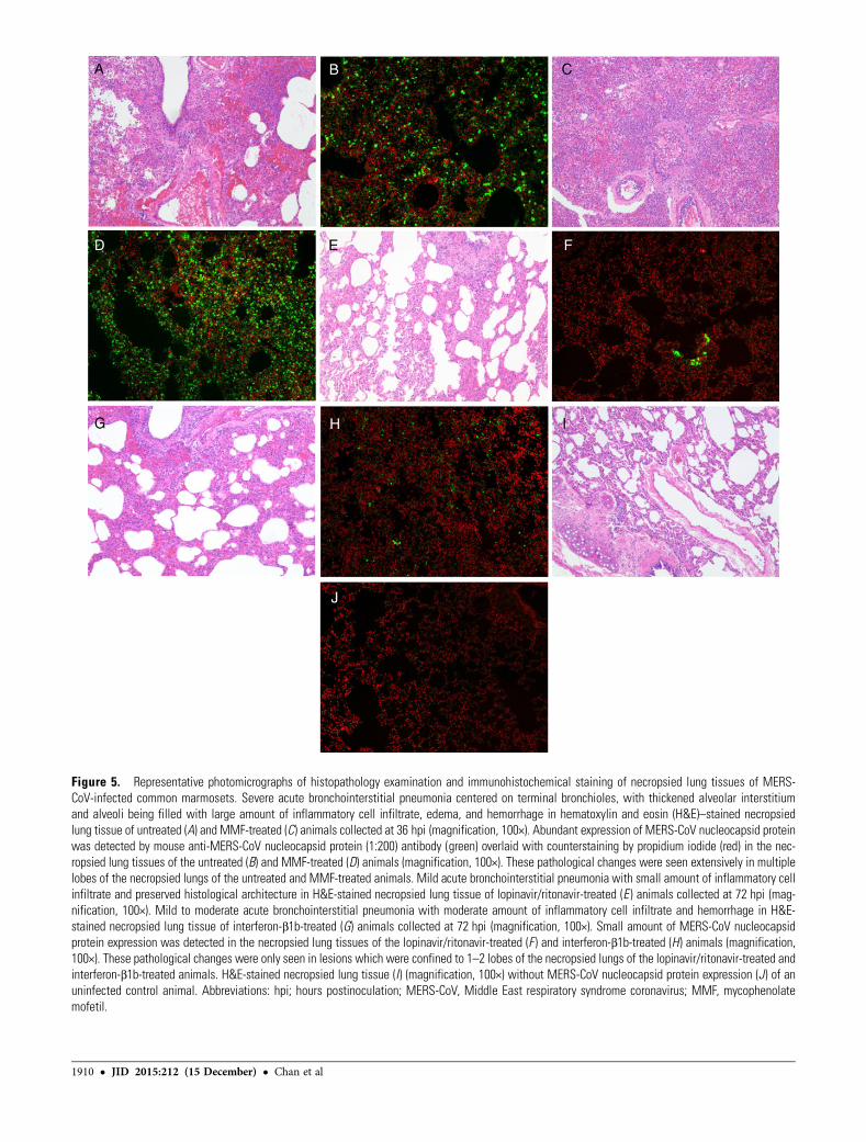
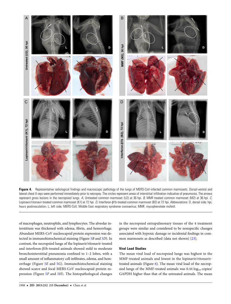
Figure 5. Representative photomicrographs of histopathology examination and immunohistochemical staining of necropsied lung tissues of MERS-CoV-infected common marmosets. Severe acute bronchointerstitial pneumonia centered on terminal bronchioles, with thickened alveolar interstitiumand alveoli being filled with large amount of inflammatory cell infiltrate, edema, and hemorrhage in hematoxylin and eosin (H&E)–stained necropsiedlung tissue of untreated (A) and MMF-treated (C) animals collected at 36 hpi (magnification, 100×). Abundant expression of MERS-CoV nucleocapsid proteinwas detected by mouse anti-MERS-CoV nucleocapsid protein (1:200) antibody (green) overlaid with counterstaining by propidium iodide (red) in the nec-ropsied lung tissues of the untreated (B) and MMF-treated (D) animals (magnification, 100×). These pathological changes were seen extensively in multiplelobes of the necropsied lungs of the untreated and MMF-treated animals. Mild acute bronchointerstitial pneumonia with small amount of inflammatory cellinfiltrate and preserved histological architecture in H&E-stained necropsied lung tissue of lopinavir/ritonavir-treated (E ) animals collected at 72 hpi (mag-nification, 100×). Mild to moderate acute bronchointerstitial pneumonia with moderate amount of inflammatory cell infiltrate and hemorrhage in H&E-stained necropsied lung tissue of interferon-β1b-treated (G) animals collected at 72 hpi (magnification, 100×). Small amount of MERS-CoV nucleocapsidprotein expression was detected in the necropsied lung tissues of the lopinavir/ritonavir-treated (F ) and interferon-β1b-treated (H) animals (magnification,100×). These pathological changes were only seen in lesions which were confined to 1–2 lobes of the necropsied lungs of the lopinavir/ritonavir-treated andinterferon-β1b-treated animals. H&E-stained necropsied lung tissue (I) (magnification, 100×) without MERS-CoV nucleocapsid protein expression (J) of anuninfected control animal. Abbreviations: hpi; hours postinoculation; MERS-CoV, Middle East respiratory syndrome coronavirus; MMF, mycophenolatemofetil.
1910 • JID 2015:212 (15 December) • Chan et al
combinational lopinavir-ritonavir, pegylated interferon, and ri-bavirin had resolution of viremia after 2 days of treatment [44].Our present study showed that treatment with lopinavir/ritona-vir improved the clinical, radiological, and pathological featuresin MERS-CoV-infected common marmosets. The lopinavir/ritonavir-treated animals had the lowest mean viral loads inlung and most extrapulmonary tissues at necropsy. These find-ings provided further support for the use of lopinavir/ritonavirin patients with severe MERS.
MERS-CoV attenuates interferon response to evade the host’sinnate immune system [45]. MERS-CoV is 50–100 times moresensitive to pegylated interferon-α than SARS-CoV in vitro[46]. Therefore, treatment of MERS with interferons was con-sidered and evaluated in vitro and in a small number of patients.In Vero/Vero6 cells, interferon-β1b (Betaferon) exhibited alower EC50:Cmax ratio than interferon-α2a, -α2b, and -β1a[15, 47]. This might partially explain why the treatment effectsof interferon-α2a, -α2b, and -β1a on the survival rates of a smallcohort of MERS patients were not obvious [22, 48–50]. In thisstudy, we showed that MERS-CoV-infected common marmo-sets treated with interferon-β1b had less severe disease andlower mean viral loads in necropsied lung and extrapulmonarytissues compared with untreated animals. Notably, as the le-sions in the lopinavir/ritonavir-treated and interferon-β1b-treated animals were confined to 1–2 lobes, the overall meanviral load difference between these animals and the untreated
animals were likely underestimated. Interferon-β1b is com-monly used to treat patients with multiple sclerosis and is usu-ally well tolerated [32]. Rarely, monocloncal gammopathypatients treated with interferon-β1b may develop systemic cap-illary leak syndrome [32].Other uncommon side effects includehypersensitivity, pancreatitis, depression, cytopenia, cardiomy-opathy, and liver and thyroid dysfunction [32]. Given the goodsafety profile of interferon-β1b, and our in vitro and in vivo re-sults, its use should be considered in clinical trials with MERSpatients who have no contraindication for interferon therapy.
We did not evaluate the effects of combinational treatmentwith 2 or more drugs due to the difficulty in getting approvalfor using more primates. Nevertheless, the findings in this non-human primate study are encouraging for conducting clinicaltrials with MERS patients using monotherapy or combinationaltreatment with these repurposed drugs. The in vivo anti-MERS-CoV effects and the lack of drug interactions between lopinavir/ritonavir and interferon-β1b make this an attractive combina-tion in future clinical trials. In contrast, it is still uncertain ifthe combination of interferon-β1b and/or lopinavir/ritonavirwith a lower dose of MMF may provide better viral load sup-pression to overcome the deleterious effects of the latter drug.Notably, the combination of MMF and interferon-β1b showedsynergistic effects in MERS-CoV inhibition in vitro [15]. If thiscombination is shown to be effective in common marmosets, itmay become an alternative treatment option in patients who areintolerant to interferon-β1b and/or lopinavir/ritonavir. Otherpotentially effective treatment regimens such as the combina-tion of ribavirin with interferon-β1b and/or lopinavir/ritonavirshould also be evaluated in the common marmoset model. Al-though the use of high-dose ribavirin is limited by its severe sideeffects, low-dose ribavirin combined with interferon-β1b and/or lopinavir/ritonavir may be synergistic and should still beconsidered. Finally, the shorter median time interval betweensymptom onset and death in MERS than in SARS patients em-phasizes the importance of early use of these antiviral combina-tions in MERS. This is because MERS-CoV not only dampensthe host innate interferon response, but also effectively sup-presses the adaptive immune response by infection of CD3+
lymphocytes and induction of apoptosis via the both extrinsicand intrinsic pathways in a high percentage of peripheral bloodand splenic lymphocytes [26]. Failure to control MERS-CoV rep-lication during the early phase of disease may therefore lead to animpaired adaptive immune response with poor outcome.
Supplementary Data
Supplementary materials are available at The Journal of Infectious Diseasesonline (http://jid.oxfordjournals.org). Supplementary materials consist ofdata provided by the author that are published to benefit the reader. Theposted materials are not copyedited. The contents of all supplementarydata are the sole responsibility of the authors. Questions or messages regard-ing errors should be addressed to the author.
Figure 6. Mean viral loads with standard deviation values in differenttissues of MERS-CoV-infected common marmosets collected at the timeof necropsy. *P <.05. aTwo of the 3 interferon-β1b-treated animals had un-detected viral loads in necropsied kidney tissues, which accounted for alarge standard deviation value and apparent lack of statistically significantdifference from the mean viral load of the untreated animals. Abbrevia-tions: GAPDH, glyceraldehyde 3-phosphate dehydrogenase; MERS-CoV,Middle East respiratory syndrome coronavirus; MMF, mycophenolatemofetil.
Treatment of MERS in Common Marmosets • JID 2015:212 (15 December) • 1911
Notes
Acknowledgments. We are grateful to Hua Zhu and Yanfeng Xu fortheir technical support.Financial support. This work was supported by the donations of Respi-
ratory Virus Research Foundation Limited; Larry Chi-Kin Yung; the HuiHoy and Chow Sin Lan Charity Fund Limited; and the Providence Founda-tion Limited in memory of the late Dr Lui Hac Minh; and funding from theConsultancy Service for Enhancing Laboratory Surveillance of Emerging In-fectious Disease of the Department of Health, the Hong Kong Health andMedical Research Fund (14131392); Hong Kong Research Grants Council(N_HKU728/14); the Theme-based Research Scheme (T11/707/15) of theResearch Grants Council, Hong Kong Special Administrative Region; andthe National Science and Technology Major Projects of Infectious Diseases,China (2012ZX10004501-004). The funding sources had no role in studydesign; data collection, analysis, or interpretation; or writing of the report.Potential conflicts of interest. All authors: No reported conflicts.All authors have submitted the ICMJE Form for Disclosure of Potential
Conflicts of Interest. Conflicts that the editors consider relevant to the con-tent of the manuscript have been disclosed.
References
1. Chan JF, To KK, Tse H, Jin DY, Yuen KY. Interspecies transmission andemergence of novel viruses: lessons from bats and birds. Trends Micro-biol 2013; 21:544–55.
2. Zaki AM, van Boheemen S, Bestebroer TM, Osterhaus AD, FouchierRA. Isolation of a novel coronavirus from a man with pneumonia inSaudi Arabia. N Engl J Med 2012; 367:1814–20.
3. Chan JF, Lau SK, To KK, Cheng VC, Woo PC, Yuen KY. Middle Eastrespiratory syndrome coronavirus: another zoonotic betacoronaviruscausing SARS-like disease. Clin Microbiol Rev 2015; 28:465–522.
4. Assiri A, Al-Tawfiq JA, Al-Rabeeah AA, et al. Epidemiological, demo-graphic, and clinical characteristics of 47 cases of Middle East respira-tory syndrome coronavirus disease from Saudi Arabia: a descriptivestudy. Lancet Infect Dis 2013; 13:752–61.
5. Chan JF, Li KS, To KK, Cheng VC, Chen H, Yuen KY. Is the discovery ofthe novel human betacoronavirus 2c EMC/2012 (HCoV-EMC) the be-ginning of another SARS-like pandemic? J Infect 2012; 65:477–89.
6. Jiang L, Wang N, Zuo T, et al. Potent neutralization of MERS-CoV byhuman neutralizing monoclonal antibodies to the viral spike glycopro-tein. Sci Transl Med 2014; 6:234ra59.
7. Lu L, Liu Q, Zhu Y, et al. Structure-based discovery of Middle East re-spiratory syndrome coronavirus fusion inhibitor. Nat Commun 2014;5:3067.
8. Gao J, Lu G, Qi J, et al. Structure of the fusion core and inhibition offusion by a heptad repeat peptide derived from the S protein of MiddleEast respiratory syndrome coronavirus. J Virol 2013; 87:13134–40.
9. Du L, Zhao G, Yang Y, et al. A conformation-dependent neutralizingmonoclonal antibody specifically targeting receptor-binding domainin Middle East respiratory syndrome coronavirus spike protein. JVirol 2014; 88:7045–53.
10. Soo YO, Cheng Y, Wong R, et al. Retrospective comparison of conva-lescent plasma with continuing high-dose methylprednisolone treat-ment in SARS patients. Clin Microbiol Infect 2004; 10:676–8.
11. Cheng Y, Wong R, Soo YO, et al. Use of convalescent plasma therapy inSARS patients in Hong Kong. Eur J Clin Microbiol Infect Dis 2005;24:44–6.
12. Hung IF, To KK, Lee CK, et al. Convalescent plasma treatment reducedmortality in patients with severe pandemic influenza A (H1N1) 2009virus infection. Clin Infect Dis 2011; 52:447–56.
13. Yang ZY, Werner HC, Kong WP, et al. Evasion of antibody neutraliza-tion in emerging severe acute respiratory syndrome coronaviruses. ProcNatl Acad Sci USA 2005; 102:797–801.
14. Weingartl H, Czub M, Czub S, et al. Immunization with modified vac-cinia virus Ankara-based recombinant vaccine against severe acute
respiratory syndrome is associated with enhanced hepatitis in ferrets.J Virol 2004; 78:12672–6.
15. Chan JF, Chan KH, Kao RY, et al. Broad-spectrum antivirals for theemerging Middle East respiratory syndrome coronavirus. J Infect2013; 67:606–16.
16. Dyall J, Coleman CM, Hart BJ, et al. Repurposing of clinically developeddrugs for treatment of Middle East respiratory syndrome coronavirusinfection. Antimicrob Agents Chemother 2014; 58:4885–93.
17. de Wilde AH, Jochmans D, Posthuma CC, et al. Screening of an FDA-approved compound library identifies four small-molecule inhibitors ofMiddle East respiratory syndrome coronavirus replication in cell cul-ture. Antimicrob Agents Chemother 2014; 58:4875–84.
18. Kindrachuk J, Ork B, Hart BJ, et al. Antiviral potential of ERK/MAPKand PI3K/AKT/mTOR signaling modulation for Middle East respirato-ry syndrome coronavirus infection as identified by temporal kinomeanalysis. Antimicrob Agents Chemother 2015; 59:1088–99.
19. Liu Q, Xia S, Sun Z, et al. Testing of Middle East respiratory syndromecoronavirus replication inhibitors for the ability to block viral entry.Antimicrob Agents Chemother 2015; 59:742–4.
20. Falzarano D, de Wit E, Martellaro C, Callison J, Munster VJ, FeldmannH. Inhibition of novel beta coronavirus replication by a combination ofinterferon-alpha2b and ribavirin. Sci Rep 2013; 3:1686.
21. Falzarano D, de Wit E, Rasmussen AL, et al. Treatment with interferon-alpha2b and ribavirin improves outcome in MERS-CoV-infected rhesusmacaques. Nat Med 2013; 19:1313–7.
22. Omrani AS, Saad MM, Baig K, et al. Ribavirin and interferon alfa-2a forsevere Middle East respiratory syndrome coronavirus infection: a retro-spective cohort study. Lancet Infect Dis 2014; 14:1090–5.
23. Falzarano D, de Wit E, Feldmann F, et al. Infection with MERS-CoVcauses lethal pneumonia in the common marmoset. PLOS Pathog2014; 10:e1004250.
24. Chan JF, Choi GK, Tsang AK, et al. Development and evaluation ofnovel real-time RT-PCR assays with locked nucleic acid probes target-ing the leader sequences of human pathogenic coronaviruses. J Clin Mi-crobiol 2015; 53:2722–6.
25. Zhou J, Chu H, Li C, et al. Active replication of Middle East respiratorysyndrome coronavirus and aberrant induction of inflammatory cyto-kines and chemokines in human macrophages: implications for patho-genesis. J Infect Dis 2014; 209:1331–42.
26. Chu H, Zhou J, Wong BH, et al. Middle East respiratory syndrome co-ronavirus efficiently infects human primary T lymphocytes and acti-vates both the extrinsic and intrinsic apoptosis pathways [Epub aheadof print]. J Infect Dis 2015; doi:10.1093/infdis/jiv380.
27. Chan JF, Chan KH, Choi GK, et al. Differential cell line susceptibility tothe emerging novel human betacoronavirus 2c EMC/2012: implicationsfor disease pathogenesis and clinical manifestation. J Infect Dis 2013;207:1743–52.
28. Chan KH, Chan JF, Tse H, et al. Cross-reactive antibodies in convales-cent SARS patients’ sera against the emerging novel human coronavirusEMC (2012) by both immunofluorescent and neutralizing antibodytests. J Infect 2013; 67:130–40.
29. Yao Y, Bao L, Deng W, et al. An animal model of MERS produced byinfection of rhesus macaques with MERS coronavirus. J Infect Dis 2014;209:236–42.
30. Ishimatsu M, Suzuki H, Akiyama H, Miura T, Hayami M, Ido E. Con-struction of a novel SHIV having an HIV-1-derived protease gene andits infection to rhesus macaques: a useful tool for in vivo efficacy tests ofprotease inhibitors. Microbes Infect 2007; 9:475–82.
31. Genentech USA, Inc. Product Information: CellCept®. http://www.gene.com/download/pdf/cellcept_prescribing.pdf. Accessed 10 June 2015.
32. Bayer Australia Limited. Product Information: Betaferon® Single usepack drug. http://www.bayerresources.com.au/resources/uploads/PI/file9314.pdf. Accessed 10 June 2015.
33. Chu H, Zhou J, Wong BH, et al. Productive replication of Mid-dle East respiratory syndrome coronavirus in monocyte-deriveddendritic cells modulates innate immune response. Virology 2014;454–455:197–205.
1912 • JID 2015:212 (15 December) • Chan et al
34. Raj VS, Smits SL, Provacia LB, et al. Adenosine deaminase acts as a nat-ural antagonist for dipeptidyl peptidase 4-mediated entry of the MiddleEast respiratory syndrome coronavirus. J Virol 2014; 88:1834–8.
35. de Wit E, Prescott J, Baseler L, et al. The Middle East respiratory syn-drome coronavirus (MERS-CoV) does not replicate in Syrian hamsters.PLOS One 2013; 8:e69127.
36. Coleman CM, Matthews KL, Goicochea L, Frieman MB. Wild-type andinnate immune-deficient mice are not susceptible to the Middle East re-spiratory syndrome coronavirus. J Gen Virol 2014; 95(Pt 2):408–12.
37. Zhao J, Li K, Wohlford-Lenane C, et al. Rapid generation of a mousemodel for Middle East respiratory syndrome. Proc Natl Acad Sci USA2014; 111:4970–5.
38. Agrawal AS, Garron T, Tao X, et al. Generation of a transgenic mousemodel of Middle East respiratory syndrome coronavirus infection anddisease. J Virol 2015; 89:3659–70.
39. Faure E, Poissy J, Goffard A, et al. Distinct immune response in twoMERS-CoV-infected patients: can we go from bench to bedside?PLOS One 2014; 9:e88716.
40. AlGhamdiM,Mushtaq F, AwnN, Shalhoub S. MERS CoV infection in tworenal transplant recipients: case report. Am J Transplant 2015; 15:1101–4.
41. Rizza SA, Badley AD. HIV protease inhibitors impact on apoptosis.Med Chem 2008; 4:75–9.
42. Abbott Laboratories. Product Information: Kaletra®. http://hivdb.stanford.edu/pages/linksPages/LPV_RTV_PI.pdf. Accessed 10 June 1015.
43. Chu CM, Cheng VC, Hung IF, et al. Role of lopinavir/ritonavir in thetreatment of SARS: initial virological and clinical findings. Thorax 2004;59:252–6.
44. Spanakis N, Tsiodras S, Haagmans BL, et al. Virological and serologicalanalysis of a recent Middle East respiratory syndrome coronavirus infec-tion case on a triple combination antiviral regimen. Int J AntimicrobAgents 2014; 44:528–32.
45. Lau SK, Lau CC, Chan KH, et al. Delayed induction of proinflammatorycytokines and suppression of innate antiviral response by the novelMiddle East respiratory syndrome coronavirus: implications for patho-genesis and treatment. J Gen Virol 2013; 94:2679–90.
46. de Wilde AH, Raj VS, Oudshoorn D, et al. MERS-coronavirus replica-tion induces severe in vitro cytopathology and is strongly inhibited bycyclosporin A or interferon-alpha treatment. J Gen Virol 2013;94:1749–60.
47. Hart BJ, Dyall J, Postnikova E, et al. Interferon-beta and mycophenolicacid are potent inhibitors of Middle East respiratory syndrome corona-virus in cell-based assays. J Gen Virol 2014; 95:571–7.
48. Al-Tawfiq JA, Momattin H, Dib J, Memish ZA. Ribavirin and inter-feron therapy in patients infected with the Middle East respiratory syn-drome coronavirus: an observational study. Int J Infect Dis 2014;20:42–6.
49. Khalid M, Al Rabiah F, Khan B, Al Mobeireek A, Butt TS, Al Mutairy E.Ribavirin and interferon (IFN)-alpha-2b as primary and preventivetreatment for Middle East respiratory syndrome coronavirus (MERS-CoV): a preliminary report of two cases. Antivir Ther 2015; 20:87–91.
50. Shalhoub S, Farahat F, Al-Jiffri A, et al. IFN-alpha2a or IFN-beta1a incombination with ribavirin to treat Middle East respiratory syndromecoronavirus pneumonia: a retrospective study. J Antimicrob Chemother2015; 70:2129–32.
Treatment of MERS in Common Marmosets • JID 2015:212 (15 December) • 1913