Foundation Storti ©Ferrata - ICGEB Source/Efremov1112.pdf · Germany) and mixed with 2.5 μg of...
9
1246 haematologica | 2012; 97(8) Articles and Brief Reports Chronic Lymphocytic Leukemia Funding: this work was supported by grants from the Italian Association for Cancer Research (AIRC, Milan, Italy, n. 5917) and The Leukemia & Lymphoma Society (LLS, White Plains, NY, n. R6170- 10) to DGE and grants from the State of São Paulo Research Foundation (FAPESP, Brazil) and the National Council for Scientific and Technological Development (CNPq, Brazil) to MAZ. FPC was supported by a fellowship from FAPESP. Manuscript received on September 2, 2011. Revised version arrived on January 11, 2012. Manuscript accepted February 13, 2012. Correspondence: Dimitar Efremov, Molecular Hematology, International Centre for Genetic Engineering & Biotechnology, “Adriano Buzzati- Traverso” Campus, Via E. Ramarini 32, I-00016 Monterotondo Scalo, Rome, Italy. Phone: international +39.06.90091300. E-mail: [email protected] The online version of this article has a Supplementary Appendix. Background The malignant B cells in chronic lymphocytic leukemia receive signals from the bone marrow and lymph node microenvironments which regulate their survival and proliferation. Characterization of these signals and the pathways that propagate them to the interior of the cell is important for the identification of novel potential targets for therapeutic intervention. Design and Methods We compared the gene expression profiles of chronic lymphocytic leukemia B cells purified from bone marrow and peripheral blood to identify genes that are induced by the bone marrow microenvironment. Two of the differentially expressed genes were further studied in cell culture experiments and in an animal model to determine whether they could represent appropriate ther- apeutic targets in chronic lymphocytic leukemia. Results Functional classification analysis revealed that the majority of differentially expressed genes belong to gene ontology categories related to cell cycle and mitosis. Significantly up-regulated genes in bone marrow-derived tumor cells included important cell cycle regulators, such as Aurora A and B, survivin and CDK6. Down-regulation of Aurora A and B by RNA interference inhibited proliferation of chronic lymphocytic leukemia-derived cell lines and induced low levels of apop- tosis. A similar effect was observed with the Aurora kinase inhibitor VX-680 in primary chronic lymphocytic leukemia cells that were induced to proliferate by CpG-oligonucleotides and inter- leukin-2. Moreover, VX-680 significantly blocked leukemia growth in a mouse model of chronic lymphocytic leukemia. Conclusions Aurora A and B are up-regulated in proliferating chronic lymphocytic leukemia cells and repre- sent potential therapeutic targets in this disease. Key words: chronic lymphocytic leukemia, microenvironment, Aurora kinase, novel therapeutic targets. Citation: de Paula Careta F, Gobessi S, Panepucci RA, Bojnik E, Morato de Oliveira F, Mazza Matos D, Falcão RP, Laurenti L, Zago MA, and Efremov DG. The Aurora A and B kinases are up-regulated in bone marrow-derived chronic lymphocytic leukemia cells and represent potential therapeutic targets. Haematologica 2012;97(8):1246-1254. doi:10.3324/haematol.2011.054668 ©2012 Ferrata Storti Foundation. This is an open-access paper. The Aurora A and B kinases are up-regulated in bone marrow-derived chronic lymphocytic leukemia cells and represent potential therapeutic targets Francisco de Paula Careta, 1,2 Stefania Gobessi, 2 Rodrigo Alexandre Panepucci, 1 Engin Bojnik, 2 Fabio Morato de Oliveira, 1 Daniel Mazza Matos, 1 Roberto P. Falcão, 1 Luca Laurenti, 3 Marco A. Zago, 1 and Dimitar G. Efremov 2 1 Hematology Division and Center for Cell-Based Therapy, Faculty of Medicine of Ribeirão Preto, University of São Paulo, São Paulo, Brazil; 2 Molecular Hematology, International Centre for Genetic Engineering & Biotechnology, Campus "A. Buzzati-Traverso", Rome, Italy, and 3 Department of Hematology, “A. Gemelli” Catholic University Hospital, Rome, Italy ABSTRACT ©Ferrata Storti Foundation
Transcript of Foundation Storti ©Ferrata - ICGEB Source/Efremov1112.pdf · Germany) and mixed with 2.5 μg of...
1246 haematologica | 2012; 97(8)
Articles and Brief Reports Chronic Lymphocytic Leukemia
Funding: this work was supportedby grants from the ItalianAssociation for Cancer Research(AIRC, Milan, Italy, n. 5917) andThe Leukemia & Lymphoma Society(LLS, White Plains, NY, n. R6170-10) to DGE and grants from theState of São Paulo ResearchFoundation (FAPESP, Brazil) and theNational Council for Scientific andTechnological Development (CNPq,Brazil) to MAZ. FPC was supportedby a fellowship from FAPESP.
Manuscript received onSeptember 2, 2011. Revisedversion arrived on January 11,2012. Manuscript accepted February 13, 2012.
Correspondence: Dimitar Efremov, MolecularHematology, International Centrefor Genetic Engineering &Biotechnology, “Adriano Buzzati-Traverso” Campus, Via E. Ramarini32, I-00016 Monterotondo Scalo,Rome, Italy.Phone: international+39.06.90091300.E-mail: [email protected]
The online version of this articlehas a Supplementary Appendix.
BackgroundThe malignant B cells in chronic lymphocytic leukemia receive signals from the bone marrow andlymph node microenvironments which regulate their survival and proliferation. Characterizationof these signals and the pathways that propagate them to the interior of the cell is important forthe identification of novel potential targets for therapeutic intervention.
Design and MethodsWe compared the gene expression profiles of chronic lymphocytic leukemia B cells purified frombone marrow and peripheral blood to identify genes that are induced by the bone marrowmicroenvironment. Two of the differentially expressed genes were further studied in cell cultureexperiments and in an animal model to determine whether they could represent appropriate ther-apeutic targets in chronic lymphocytic leukemia.
ResultsFunctional classification analysis revealed that the majority of differentially expressed genesbelong to gene ontology categories related to cell cycle and mitosis. Significantly up-regulatedgenes in bone marrow-derived tumor cells included important cell cycle regulators, such as AuroraA and B, survivin and CDK6. Down-regulation of Aurora A and B by RNA interference inhibitedproliferation of chronic lymphocytic leukemia-derived cell lines and induced low levels of apop-tosis. A similar effect was observed with the Aurora kinase inhibitor VX-680 in primary chroniclymphocytic leukemia cells that were induced to proliferate by CpG-oligonucleotides and inter-leukin-2. Moreover, VX-680 significantly blocked leukemia growth in a mouse model of chroniclymphocytic leukemia.
ConclusionsAurora A and B are up-regulated in proliferating chronic lymphocytic leukemia cells and repre-sent potential therapeutic targets in this disease.
Key words: chronic lymphocytic leukemia, microenvironment, Aurora kinase, novel therapeutictargets.
Citation: de Paula Careta F, Gobessi S, Panepucci RA, Bojnik E, Morato de Oliveira F, Mazza MatosD, Falcão RP, Laurenti L, Zago MA, and Efremov DG. The Aurora A and B kinases are up-regulatedin bone marrow-derived chronic lymphocytic leukemia cells and represent potential therapeutic targets.Haematologica 2012;97(8):1246-1254. doi:10.3324/haematol.2011.054668
©2012 Ferrata Storti Foundation. This is an open-access paper.
The Aurora A and B kinases are up-regulated in bone marrow-derived chroniclymphocytic leukemia cells and represent potential therapeutic targetsFrancisco de Paula Careta,1,2 Stefania Gobessi,2 Rodrigo Alexandre Panepucci,1 Engin Bojnik,2 Fabio Morato de Oliveira,1 Daniel Mazza Matos,1 Roberto P. Falcão,1 Luca Laurenti,3 Marco A. Zago,1 and Dimitar G. Efremov2
1Hematology Division and Center for Cell-Based Therapy, Faculty of Medicine of Ribeirão Preto, University of São Paulo, São Paulo,Brazil; 2Molecular Hematology, International Centre for Genetic Engineering & Biotechnology, Campus "A. Buzzati-Traverso", Rome, Italy,and 3Department of Hematology, “A. Gemelli” Catholic University Hospital, Rome, Italy
ABSTRACT
©Ferrata
Stor
ti Fou
ndati
on
Introduction
Chronic lymphocytic leukemia (CLL) is characterized bythe progressive accumulation of mature, monoclonal, CD5-positive B cells in the peripheral blood (PB), bone marrow(BM) and secondary lymphoid organs, such as lymph nodesand spleen.1,2 Traditionally, the disease has been considereda disorder of cells with defective apoptosis that accumulatebecause of extended survival. Consistent with this view, themajority of CLL cells in the PB are arrested in the G0/G1phase of the cell cycle and show a gene expression profile ofresting B cells.3 However, more recent studies using deuter-ated water have shown that a notable proliferative compart-ment exists, which continuously replenishes the bulk popu-lation of non-cycling CLL cells.4 This proliferative compart-ment contributes to disease progression not only by increas-ing the leukemic cell burden but also by facilitating the accu-mulation of new mutations that increase the aggressivenessand chemoresistance of the malignant clone. The CLL proliferating compartment is located in imper-
fectly defined structures in the lymph nodes and BM,named proliferation centers or pseudofollicles. These struc-tures are composed of focal aggregates of neoplastic pro-lymphocytes and paraimmunoblasts that are interspersedwith auxiliary cells, such as CD4+ T cells, follicular dendriticcells, stromal cells and nurse-like cells.2,5 In these structures,CLL cells are believed to receive various signals that stimu-late their proliferation and survival. Examples of suchmicroenvironmental signals include interleukin (IL)-4,6CD40 ligand,7,8 CXCL12,9 BAFF,10 CD31,11 VCAM-1,12 CpG-DNA,13,14 antigen,15,16 and contact with stromal,17 nurse-like,10,12 and follicular dendritic cells.18 In vitro, most of thesesignals only increase the resistance of CLL cells to sponta-neous and chemotherapy-induce apoptosis. However, cer-tain combinations can induce relatively efficient prolifera-tion, such as unmethylated CpG oligonucleotides (CpG-ODN) with IL-2,13,14 or CD40L/IL-2/IL-10 in co-culture withstromal cells.8Despite the progress in identifying potential microenvi-
ronmental signals that can induce the proliferation orincrease the survival of CLL cells in vitro, the microenviron-mental signals that sustain the growth of the malignantclone in vivo remain poorly defined. In addition, the intracel-lular pathways that transduce the proliferation signal in themalignant B cells have not been fully characterized.Identification of the signals and pathways that operate invivo is important for the development of strategies to blockthe microenvironmental interactions that regulate the prolif-eration of the malignant clone. Such strategies could be par-ticularly important in patients with CLL with high prolifer-ation rates, in whom they could prevent the development ofdangerous clonal variants or eliminate them as they occur.To further define the pathways that transduce the
microenvironmental signals in CLL cells in vivo and to iden-tify novel potential targets for therapeutic intervention, weinvestigated the gene expression profiles of purified CLLcells from paired BM and PB samples by microarray andreal-time reverse transcriptase polymerase chain reaction(RT-PCR) analysis. A number of genes involved in cell cycleand mitosis were found to be significantly up-regulated inBM-CLL cells. Among these, the Aurora A (AURKA) andAurora B (AURKB) kinases appeared as particularly interest-ing molecules for further study, considering that selectivesmall molecule inhibitors of these kinases have been devel-oped and have already shown promising activity against a
wide range of other malignant diseases.19,20 To determinewhether these kinases could also represent potential thera-peutic targets in CLL, we investigated the effects of down-regulation or inhibition of Aurora A and Aurora B in CLLcell lines, primary CLL cells induced to proliferate by CpG-ODN/IL-2, and murine leukemias that develop in the Eμ-TCL1 transgenic model of CLL.
Design and Methods
Chronic lymphocytic leukemia and normal B-cell samplesBlood samples satisfying standard morphological and
immunophenotypic criteria for B-cell CLL were collected frompatients. Informed consent was obtained from all patients accord-ing to the Declaration of Helsinki and approval for the study wasobtained from the Ethical Committees at the "A. Gemelli" CatholicUniversity Hospital, Rome (Italy) and the University ClinicalHospital of the Faculty of Medicine, Ribeirão Preto (Brazil). CLL B cells from PB and BM that were used for the microarray
and real-time RT-PCR experiments were purified by negative selec-tion using the RosetteSep human B cell-enrichment cocktail(StemCell Technologies, Vancouver, Canada). For all other experi-ments, CLL cells were first isolated by Ficoll gradient centrifugationand then further purified by negative selection with anti-CD3, anti-CD14 and anti-CD16 mouse monoclonal antibodies andDynabeads coated with pan-anti-mouse IgG antibody (DynalBiotech, Oslo, Norway). The purity of the selected B-cell popula-tions was >95%, as determined by staining with anti-CD5 andanti-CD19 antibodies and analysis on a FACSCalibur flow cytome-ter (BD Biosciences, Franklin Lakes, NJ, USA).
Gene expression profiling and real-time reversetranscriptase polymerase chain reaction analysisMicroarray profiling of total RNA extracted from purified BM
and PB CLL cells was performed using Whole Human GenomeOligo microarrays (Agilent, Palo Alto, CA, USA) containing 41,000distinct probes. Complete microarray data sets have been deposit-ed in NCBI Gene Expression Omnibus and are accessible throughGEO Series accession number GSE30896. Differentially expressedtranscripts were identified by a two-tailed paired t-test, consideringP values of less than 0.05 as statistically significant. Functional clas-sification analysis was performed using the online Database forAnnotation, Visualization and Integrated Discovery (DAVID), ver-sion 6.7. Expression of AURKA, AURKB, BIRC5 and CDK6was fur-ther quantified by RT-PCR analysis with pre-designed TaqManprobes in a 7300 Real-Time PCR System (Applied Biosystems).Details on the gene expression profiling and quantitative RT-PCRanalysis are provided in the Online Supplementary Design andMethods.
Cell culture, RNA interference and immunoblotting experimentsFreshly isolated CLL B cells were cultured in the presence of CpG
oligonucleotide 2006 (Microsynth, Balgach, Switzerland), IL-2(R&D Systems, Minneapolis, MN, USA), goat anti-human IgM(Southern Biotechnology Associates, Birmingham, AL, USA) coat-ed on Dynabeads M-450 Epoxy (Dynal Biotech) or 3T40L cells asdescribed elsewhere.14,15 The pan-Aurora kinase inhibitor VX-680(Cayman Chemical, Ann Arbor, Michigan) was used as indicated. Stealth Select small interfering (si) RNA (Invitrogen) were used to
silence the expression of Aurora A and B in MEC1 and EHEB cells(DSMZ, Braunschweig, Germany), as previously described.14
Briefly, 0.6x10⁷cells were resuspended in 100 μL of Cell LineNucleofector Solution L (Amaxa Biosystems GmbH, Cologne,
Targeting Aurora kinases in CLL
haematologica | 2012; 97(8) 1247
©Ferrata
Stor
ti Fou
ndati
on
Germany) and mixed with 2.5 μg of siRNA. Nucleofections wereperformed on an Amaxa Nucleofector II device (Amaxa BiosystemsGmbH) using the C-005 program. Cells were collected andprocessed for subsequent analysis at the indicated times.Immunoblotting analysis was performed as described else-
where.14 The following antibodies were used: Aurora A(Invitrogen), Aurora B, phospho-AURKAT288, phospho-histoneH3S10, rabbit IgG-horseradish peroxidase (HRP), mouse IgG HRP-linked (Cell Signaling Technology, Danvers, MA, USA), and b-actin(Sigma-Aldrich).
Cell cycle, apoptosis and proliferation assaysThe cell cycle was analyzed by propidium iodide staining and
flow cytometry. Annexin-A5-fluorescein isothiocyanate (FITC)(Nexins Research, Kattendijke, The Netherlands) was used to eval-uate the percentage of viable and apoptotic cells. For analysis ofproliferation, 5-bromo-2-deoxyuridine (BrdU) 10 μM was added tosynchronized or unsynchronized MEC1 or EHEB cells for 1 h priorto harvesting. In experiments with primary CLL cells, BrdU wasadded after 48 h of stimulation with CpG-ODN and IL-2. Cellswere cultured for another 18 h prior to harvesting. IncorporatedBrdU was detected with anti-BrdU-FITC antibody (BDBiosciences). Further details are provided in the OnlineSupplementary Design and Methods.
Treatment of mice with adoptively transferred Eμ-TCL1 leukemias For adoptive transfer, 1.2x10⁷ TCL1 leukemia cells were thawed,
resuspended in 500 μL of phosphate-buffered saline and injectedintraperitoneally into B6/C3H F1 female mice (6-8 weeks old;Harlan Laboratories B.V., Horst, The Netherlands). The VX-680solution was first dissolved in PEG400 (Sigma-Aldrich) and thendiluted 2-fold with 50 mM sodium phosphate pH 4.0 (the pH wasadjusted to 7.1 before injection). Five days after adoptive transfer,treatment was started in groups of seven to ten mice with VX-68040mg/kg bid or vehicle control, intraperitoneally. Leukemia devel-opment was monitored by measuring white blood cell counts on aHemavet HV950FS hematology analyzer (Drew Scientific, Inc.,Dallas, TX, USA) and flow-cytometry analysis of CD5+/B220+ cells.All animal procedures were performed in accordance with Italiannational law (Italian legislative decree 116/92 and European direc-tive 8/609) and ICGEB institutional guidelines.
Statistical analysis Student’s t tests and Mann-Whitney rank sum tests were per-
formed to determine the significance of the differences betweenmean and median values as appropriate. Fisher’s exact test wasused to investigate the correlation between response to CpG-ODN/IL-2 stimulation and IGHV mutation status or ZAP-70expression. The survival curves and medians were calculated with-in subgroups with the Kaplan-Meier method. The log-rank testwas used to compare differences between estimated survivalcurves. All statistical analyses were performed using the SigmaStat3.1 program (Systat Software, Richmond, CA, USA).
Results
Genes involved in cell cycle and mitosis are significantlyup-regulated in chronic lymphocytic leukemia cells from bone marrow
To identify the pathways that are activated by microenvi-ronmental stimuli in CLL cells in vivo, we performed geneexpression profiling of purified BM and PB CLL cells. Six
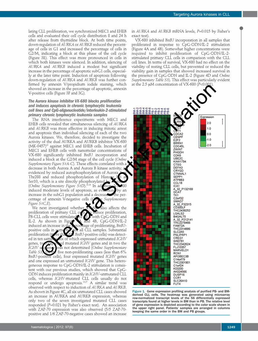
paired BM and PB samples were used for this analysis. Atotal of 272 genes were identified as differentially expressedbetween BM and PB CLL cells, with expression levels thatwere at least two-fold different (Online Supplementary TableS1). Of these, 56 were up-regulated and 216 were down-regulated in BM-CLL cells. Among the significantly up-reg-ulated genes in BM-CLL cells several important regulators ofthe cell cycle were identified, such as survivin (BIRC5),AURKA, E2F2, CDK6, CDC45L, and CDCA2 (Figure 1). Inaddition, functional annotation analysis with the DAVIDtool revealed that many of the other up-regulated genes alsobelong to gene ontology categories that are related to cellu-lar proliferation, including the categories cell cycle(GO:0007049), mitosis (GO:0007067), nuclear division(GO:0000280) and M phase of mitotic cell cycle(GO:0000087)(Online Supplementary Table S2).In order to validate the gene expression data, we reana-
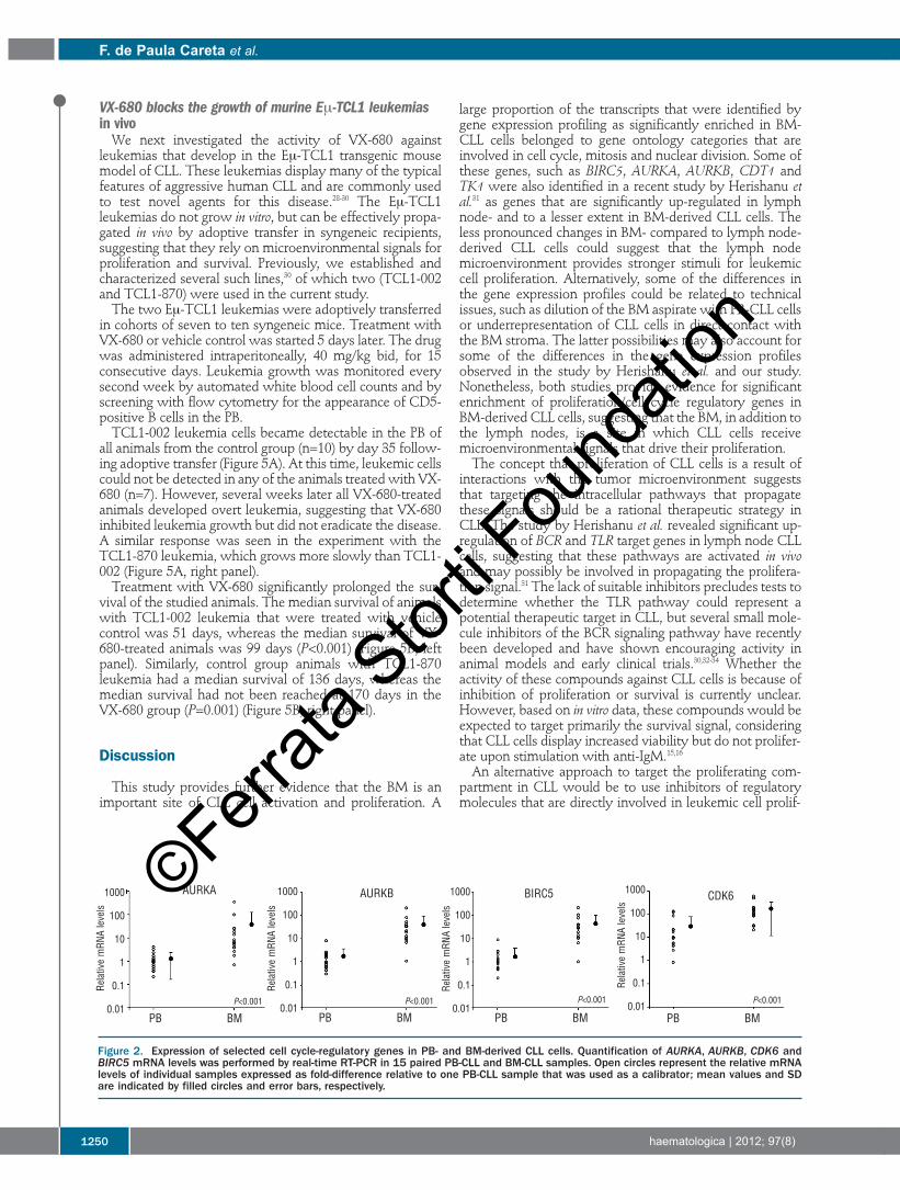
lyzed the expression of AURKA, CDK6 and survivin by real-time RT-PCR in the six samples used for the microarrayanalysis and in an additional set of nine paired BM and PBCLL samples. AURKB, an AURKA homologue that was notidentified as differentially expressed by the microarray pro-filing, was also included in this analysis because it is inducedin other cell types during cell cycle progression and is alsoinvolved in mitosis regulation.21 As shown in Figure 2, allfour genes, including AURKB, were found to be expressed atsignificantly higher levels in BM-CLL cells, with mean val-ues exceeding the levels in PB-CLL cells by 10-fold.Collectively, these data show that genes involved in cellcycle control and proliferation are up-regulated in BM-CLLcells.
Down-regulation of AURKA and AURKB induces growtharrest and apoptosis in chronic lymphocytic leukemia cell lines in vitroAmong the various proliferation/cell cycle regulatory
genes that were up-regulated in BM-CLL cells, the Aurorakinases appeared as particularly interesting for further study,as they have recently emerged as promising therapeutic tar-gets in a broad range of malignant diseases. These kinasesplay critical roles in chromosome segregation and cytokine-sis during mitosis and are frequently deregulated in cancer,which has led to the development of several pharmacologi-cally active small-molecule inhibitors that selectively inhibitAURKA, AURKB or both. Preclinical studies with several ofthese agents have shown promising activity, resulting ininhibition of tumor growth in a variety of in vivo xenograftmodels at well-tolerated doses.22-24To determine whether the Aurora kinases could be appro-
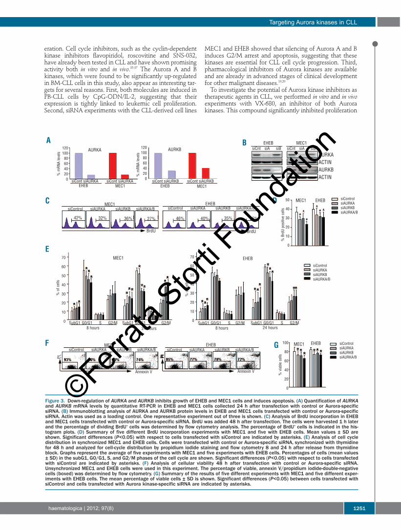
priate therapeutic targets in CLL, we first evaluated thefunctional consequences of AURKA and AURKB knock-down in the CLL-derived cell lines MEC1 and EHEB. Asshown in Figure 3A and 3B, both Aurora kinases were effi-ciently down-regulated by RNA interference, with anapproximately 70-80% reduction in mRNA and protein lev-els with respect to cells transfected with control siRNA. Toevaluate the effect of Aurora kinase knockdown on prolifer-ation, the percentage of dividing cells was determined byBrdU incorporation analysis 48 h after siRNA transfection.In both cell lines, down-regulation of AURKA or AURKB sig-nificantly reduced the percentage of BrdU-positive cells(Figure 3C and 3D). Inhibition of proliferation appearedeven greater when AURKA and AURKBwere simultaneous-ly silenced, which was particularly evident in MEC1 cells. To further clarify the role of AURKA and AURKB in regu-
F. de Paula Careta et al.
1248 haematologica | 2012; 97(8)
©Ferrata
Stor
ti Fou
ndati
on
lating CLL proliferation, we synchronized MEC1 and EHEBcells and evaluated their cell cycle distribution 8 and 24 hafter release from thymidine block. At both time points,down-regulation of AURKA or AURKB reduced the percent-age of cells in G1 and increased the percentage of cells inG2/M, indicating a block at this phase of the cell cycle(Figure 3E). This effect was more pronounced in cells inwhich both kinases were silenced. In addition, silencing ofAURKA and AURKB induced a modest but significantincrease in the percentage of apoptotic subG1 cells, especial-ly at the later time point. Induction of apoptosis followingdown-regulation of AURKA and AURKB was further con-firmed by annexin V/propidium iodide staining, whichshowed an increase in the percentage of apoptotic, annexinV-positive cells (Figure 3F and 3G).
The Aurora kinase inhibitor VX-680 blocks proliferationand induces apoptosis in chronic lymphocytic leukemiacell lines and CpG-oligonucleotide/interleukin-2-stimulatedprimary chronic lymphocytic leukemia samples The RNA interference experiments with MEC1 and
EHEB cells revealed that simultaneous silencing of AURKAand AURKB was more effective in inducing mitotic arrestand apoptosis than individual silencing of each of the twoAurora kinases. We, therefore, decided to investigate theactivity of the dual AURKA and AURKB inhibitor VX-680(MK-0457)22 against MEC1 and EHEB cells. Incubation ofMEC1 and EHEB cells with nanomolar concentrations ofVX-680 significantly inhibited BrdU incorporation andinduced a block at the G2/M stage of the cell cycle (OnlineSupplementary Figure S1A-C). These effects correlated with adecrease in both Aurora A and Aurora B kinase activity, asevidenced by reduced autophosphorylation of Aurora A atThr288 and reduced phosphorylation of Histone H3 atSer10, which is a site directly phosphorylated by Aurora B(Online Supplementary Figure S1D).25,26 In addition, VX-680induced moderate levels of apoptosis, as evidenced by anincrease in the subG1 population and a decrease in the per-centage of annexin V-negative cells (Online SupplementaryFigure S1C,E). We next investigated whether VX-680 also affects the
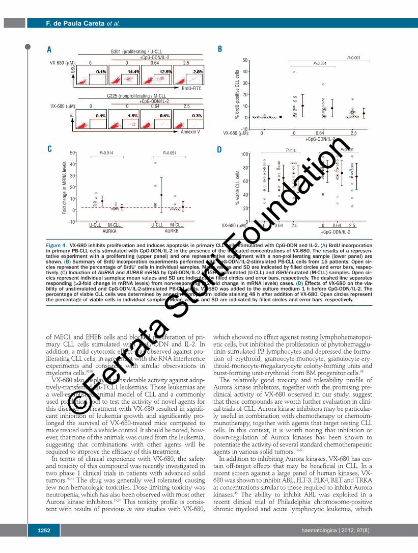
proliferation of primary CLL cells. To induce proliferation,PB-CLL cells were stimulated for 48 h with CpG-ODN andIL-2. As shown in Figure 4A and 4B, CpG-ODN/IL-2induced an increase in the percentage of proliferating, BrdU-positive cells in some but not all CLL samples. Substantialproliferation (more than 6% BrdU-positive cells) was detect-ed in ten samples, six of which expressed unmutated IGHVgenes, two expressed mutated IGHV genes and in two theIGHV sequence was not determined (Online SupplementaryTable S3). Of the five non-proliferating cases (less than 6%BrdU-positive cells), four expressed mutated IGHV genesand one expressed an unmutated IGHV gene. This hetero-geneous response to CpG-ODN/IL-2 stimulation is consis-tent with our previous studies, which showed that CpG-ODN induces proliferation mainly in IGHV-unmutated CLLcells, whereas IGHV-mutated CLL cells usually do notrespond or undergo apoptosis.14,27 A similar trend wasobserved with respect to induction of AURKA and AURKB.As shown in Figure 4C, all six unmutated CLL cases showedan increase in AURKA and AURKB expression, whereasonly two of the seven investigated mutated CLL casesresponded (P=0.021 by Fisher’s exact test). An associationwith ZAP-70 expression was also observed (5/5 ZAP-70-positive and 1/6 ZAP-70-negative cases showed an increase
in AURKA and AURKB mRNA levels, P=0.015 by Fisher’sexact test).VX-680 inhibited BrdU incorporation in all samples that
proliferated in response to CpG-ODN/IL-2 stimulation(Figure 4A and 4B). Somewhat higher concentrations wererequired to inhibit proliferation of CpG-ODN/IL-2-stimulated primary CLL cells in comparison with the CLLcell lines. In terms of survival, VX-680 had no effect on theviability of resting CLL cells, but prevented or reduced theviability gain in samples that showed increased survival inthe presence of CpG-ODN and IL-2 (Figure 4D and OnlineSupplementary Table S3). This effect was particularly evidentat the 2.5 μM concentration of VX-680 (P<0.001).
Targeting Aurora kinases in CLL
haematologica | 2012; 97(8) 1249
Figure 1. Gene expression profiling analysis of purified PB- and BM-derived CLL cells. The heatmap was generated using microarrayrow-normalized transcript levels of the 56 differentially expressedtranscripts found at higher levels in BM than in PB. The relative levelof gene expression is depicted according to the color scale shown inthe upper right panel. Patients’ samples are arranged in columnskeeping the same order in the BM and PB groups.
©Ferrata
Stor
ti Fou
ndati
on
VX-680 blocks the growth of murine Eμ-TCL1 leukemiasin vivoWe next investigated the activity of VX-680 against
leukemias that develop in the Eμ-TCL1 transgenic mousemodel of CLL. These leukemias display many of the typicalfeatures of aggressive human CLL and are commonly usedto test novel agents for this disease.28-30 The Eμ-TCL1leukemias do not grow in vitro, but can be effectively propa-gated in vivo by adoptive transfer in syngeneic recipients,suggesting that they rely on microenvironmental signals forproliferation and survival. Previously, we established andcharacterized several such lines,30 of which two (TCL1-002and TCL1-870) were used in the current study.The two Eμ-TCL1 leukemias were adoptively transferred
in cohorts of seven to ten syngeneic mice. Treatment withVX-680 or vehicle control was started 5 days later. The drugwas administered intraperitoneally, 40 mg/kg bid, for 15consecutive days. Leukemia growth was monitored everysecond week by automated white blood cell counts and byscreening with flow cytometry for the appearance of CD5-positive B cells in the PB. TCL1-002 leukemia cells became detectable in the PB of
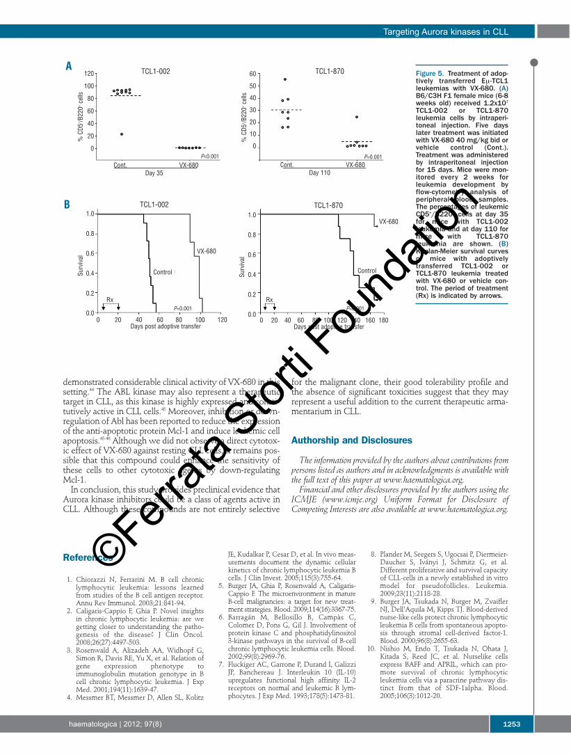
all animals from the control group (n=10) by day 35 follow-ing adoptive transfer (Figure 5A). At this time, leukemic cellscould not be detected in any of the animals treated with VX-680 (n=7). However, several weeks later all VX-680-treatedanimals developed overt leukemia, suggesting that VX-680inhibited leukemia growth but did not eradicate the disease.A similar response was seen in the experiment with theTCL1-870 leukemia, which grows more slowly than TCL1-002 (Figure 5A, right panel). Treatment with VX-680 significantly prolonged the sur-
vival of the studied animals. The median survival of animalswith TCL1-002 leukemia that were treated with vehiclecontrol was 51 days, whereas the median survival of VX-680-treated animals was 99 days (P<0.001) (Figure 5B, leftpanel). Similarly, control group animals with TCL1-870leukemia had a median survival of 136 days, whereas themedian survival had not been reached at 170 days in theVX-680 group (P=0.001) (Figure 5B, right panel).
Discussion
This study provides further evidence that the BM is animportant site of CLL cell activation and proliferation. A
large proportion of the transcripts that were identified bygene expression profiling as significantly enriched in BM-CLL cells belonged to gene ontology categories that areinvolved in cell cycle, mitosis and nuclear division. Some ofthese genes, such as BIRC5, AURKA, AURKB, CDT1 andTK1 were also identified in a recent study by Herishanu etal.31 as genes that are significantly up-regulated in lymphnode- and to a lesser extent in BM-derived CLL cells. Theless pronounced changes in BM- compared to lymph node-derived CLL cells could suggest that the lymph nodemicroenvironment provides stronger stimuli for leukemiccell proliferation. Alternatively, some of the differences inthe gene expression profiles could be related to technicalissues, such as dilution of the BM aspirate with PB-CLL cellsor underrepresentation of CLL cells in direct contact withthe BM stroma. The latter possibilities may also account forsome of the differences in the gene expression profilesobserved in the study by Herishanu et al. and our study.Nonetheless, both studies provide evidence for significantenrichment of proliferation/cell cycle regulatory genes inBM-derived CLL cells, suggesting that the BM, in addition tothe lymph nodes, is a site in which CLL cells receivemicroenvironmental signals that drive their proliferation.The concept that proliferation of CLL cells is a result of
interactions with the tumor microenvironment suggeststhat targeting the intracellular pathways that propagatethese signals should be a rational therapeutic strategy inCLL. The study by Herishanu et al. revealed significant up-regulation of BCR and TLR target genes in lymph node CLLcells, suggesting that these pathways are activated in vivoand may possibly be involved in propagating the prolifera-tion signal.31 The lack of suitable inhibitors precludes tests todetermine whether the TLR pathway could represent apotential therapeutic target in CLL, but several small mole-cule inhibitors of the BCR signaling pathway have recentlybeen developed and have shown encouraging activity inanimal models and early clinical trials.30,32-34 Whether theactivity of these compounds against CLL cells is because ofinhibition of proliferation or survival is currently unclear.However, based on in vitro data, these compounds would beexpected to target primarily the survival signal, consideringthat CLL cells display increased viability but do not prolifer-ate upon stimulation with anti-IgM.15,16An alternative approach to target the proliferating com-
partment in CLL would be to use inhibitors of regulatorymolecules that are directly involved in leukemic cell prolif-
F. de Paula Careta et al.
1250 haematologica | 2012; 97(8)
Figure 2. Expression of selected cell cycle-regulatory genes in PB- and BM-derived CLL cells. Quantification of AURKA, AURKB, CDK6 andBIRC5 mRNA levels was performed by real-time RT-PCR in 15 paired PB-CLL and BM-CLL samples. Open circles represent the relative mRNAlevels of individual samples expressed as fold-difference relative to one PB-CLL sample that was used as a calibrator; mean values and SDare indicated by filled circles and error bars, respectively.
AURKA AURKB BIRC51000
100
10
1
0.1
0.01
1000
100
10
1
0.1
0.01
1000
100
10
1
0.1
0.01
1000
100
10
1
0.1
0.01
CDK6
PB BM
Relative mRNA levels
Relative mRNA levels
Relative mRNA levels
Relative mRNA levels
PB BM PB BM PB BM
P<0.001P<0.001P<0.001P<0.001
©Ferrata
Stor
ti Fou
ndati
on
eration. Cell cycle inhibitors, such as the cyclin-dependentkinase inhibitors flavopiridol, roscovitine and SNS-032,have already been tested in CLL and have shown promisingactivity both in vitro and in vivo.35-37 The Aurora A and Bkinases, which were found to be significantly up-regulatedin BM-CLL cells in this study, also appear as interesting tar-gets for several reasons. First, both molecules are induced inPB-CLL cells by CpG-ODN/IL-2, suggesting that theirexpression is tightly linked to leukemic cell proliferation.Second, siRNA experiments with the CLL-derived cell lines
MEC1 and EHEB showed that silencing of Aurora A and Binduces G2/M arrest and apoptosis, suggesting that thesekinases are essential for CLL cell cycle progression. Third,pharmacological inhibitors of Aurora kinases are availableand are already in advanced stages of clinical developmentfor other malignant diseases.19,20To investigate the potential of Aurora kinase inhibitors as
therapeutic agents in CLL, we performed in vitro and in vivoexperiments with VX-680, an inhibitor of both Aurorakinases. This compound significantly inhibited proliferation
Targeting Aurora kinases in CLL
haematologica | 2012; 97(8) 1251
Figure 3. Down-regulation of AURKA and AURKB inhibits growth of EHEB and MEC1 cells and induces apoptosis. (A) Quantification of AURKAand AURKB mRNA levels by quantitative RT-PCR in EHEB and MEC1 cells collected 24 h after transfection with control or Aurora-specificsiRNA. (B) Immunoblotting analysis of AURKA and AURKB protein levels in EHEB and MEC1 cells transfected with control or Aurora-specificsiRNA. Actin was used as a loading control. One representative experiment out of three is shown. (C) Analysis of BrdU incorporation in EHEBand MEC1 cells transfected with control or Aurora-specific siRNA. BrdU was added 48 h after transfection. The cells were harvested 1 h laterand the percentage of dividing BrdU+ cells was determined by flow cytometry analysis. The percentage of BrdU+ cells is indicated in the his-togram plots. (D) Summary of five different BrdU incorporation experiments with MEC1 and five with EHEB cells. Mean values ± SD areshown. Significant differences (P<0.05) with respect to cells transfected with siControl are indicated by asterisks. (E) Analysis of cell cycledistribution in synchronized MEC1 and EHEB cells. Cells were transfected with control or Aurora-specific siRNA, synchronized with thymidinefor 48 h and analyzed for cell-cycle distribution by propidium iodide staining and flow cytometry 8 and 24 h after release from thymidineblock. Graphs represent the average of five experiments with MEC1 and five experiments with EHEB cells. Percentages of cells (mean values± SD) in the subG1, G0/G1, S, and G2/M phases of the cell cycle are shown. Significant differences (P<0.05) with respect to cells transfectedwith siControl are indicated by asterisks. (F) Analysis of cellular viability 48 h after transfection with control or Aurora-specific siRNA.Unsynchronized MEC1 and EHEB cells were used in this experiment. The percentage of viable, annexin V/propidium iodide-double-negativecells (boxed) was determined by flow cytometry. (G) Summary of the results of five different experiments with MEC1 and five different exper-iments with EHEB cells. The mean percentage of viable cells ± SD is shown. Significant differences (P<0.05) between cells transfected withsiControl and cells transfected with Aurora kinase-specific siRNA are indicated by asterisks.
A B
DC
E
F G
AURKA
siCont siAURKA
siControl siAURKA
SubG1 G0/G1 S G2/M SubG1 G0/G1 S G2/M24 hours
SubG1 G0/G1 S G2/M24 hours8 hours
SubG1 G0/G1 S G2/M8 hours
Annexin VAnnexin V
PI PI
siAURKB siAURKA/B
siControl siAURKA siAURKB siAURKA/B siControl siAURKA siAURKB siAURKA/B
siControl siAURKA siAURKB siAURKA/B
siCont siAURKB siCont siAURKB
siCnt siA siB siCnt siA siB
AURKAACTINAURKBACTIN
siControlsiAURKAsiAURKBsiAURKA/B
siControlsiAURKAsiAURKBsiAURKA/B
siControlsiAURKAsiAURKBsiAURKA/B
siCont siAURKAMEC1EHEB MEC1EHEB
AURKB
% mRNA levels
% BrdU positive cells
% of cells
% viable cells
% of cells
% mRNA levels
120100806040200
120100806040200
70
60
50
40
30
20
10
0
70
60
50
40
30
20
10
0
100
80
60
40
20
0
50
40
30
20
10
0
EHEB MEC1
MEC1 EHEB
MEC1
MEC1 MEC1
BrdU BrdU
42% 32% 36% 27% 46% 40% 35% 35%
MEC1
EHEB
EHEB EHEB
EHEB
©Ferrata
Stor
ti Fou
ndati
on
of MEC1 and EHEB cells and blocked proliferation of pri-mary CLL cells stimulated with CpG-ODN and IL-2. Inaddition, a mild cytotoxic effect was observed against pro-liferating CLL cells, in agreement with the RNA interferenceexperiments and consistent with similar observations inmyeloma cells.38,39VX-680 also displayed considerable activity against adop-
tively-transferred Eμ-TCL1 leukemias. These leukemias area well-established animal model of CLL and a commonlyused preclinical tool to test the activity of novel agents forthis disease.28-30 Treatment with VX-680 resulted in signifi-cant inhibition of leukemia growth and significantly pro-longed the survival of VX-680-treated mice compared tomice treated with a vehicle control. It should be noted, how-ever, that none of the animals was cured from the leukemia,suggesting that combinations with other agents will berequired to improve the efficacy of this treatment.In terms of clinical experience with VX-680, the safety
and toxicity of this compound was recently investigated intwo phase 1 clinical trials in patients with advanced solidtumors.40,41 The drug was generally well tolerated, causingfew non-hematologic toxicities. Dose-limiting toxicity wasneutropenia, which has also been observed with most otherAurora kinase inhibitors.19,20 This toxicity profile is consis-tent with results of previous in vitro studies with VX-680,
which showed no effect against resting lymphohematopoi-etic cells, but inhibited the proliferation of phytohemagglu-tinin-stimulated PB lymphocytes and depressed the forma-tion of erythroid, granuocyte-monocyte, granulocyte-ery-throid-monocyte-megakaryocyte colony-forming units andburst-forming unit-erythroid from BM progenitor cells.38The relatively good toxicity and tolerability profile of
Aurora kinase inhibitors, together with the promising pre-clinical activity of VX-680 observed in our study, suggestthat these compounds are worth further evaluation in clini-cal trials of CLL. Aurora kinase inhibitors may be particular-ly useful in combination with chemotherapy or chemoim-munotherapy, together with agents that target resting CLLcells. In this context, it is worth noting that inhibition ordown-regulation of Aurora kinases has been shown topotentiate the activity of several standard chemotherapeuticagents in various solid tumors.19,42In addition to inhibiting Aurora kinases, VX-680 has cer-
tain off-target effects that may be beneficial in CLL. In arecent screen against a large panel of human kinases, VX-680 was shown to inhibit ABL, FLT-3, PLK4, RET and TRKAat concentrations similar to those required to inhibit Aurorakinases.43 The ability to inhibit ABL was exploited in arecent clinical trial of Philadelphia chromosome-positivechronic myeloid and acute lymphocytic leukemia, which
F. de Paula Careta et al.
1252 haematologica | 2012; 97(8)
Figure 4. VX-680 inhibits proliferation and induces apoptosis in primary CLL cells stimulated with CpG-ODN and IL-2. (A) BrdU incorporationin primary PB-CLL cells stimulated with CpG-ODN/IL-2 in the presence of the indicated concentrations of VX-680. The results of a represen-tative experiment with a proliferating (upper panel) and one representative experiment with a non-proliferating sample (lower panel) areshown. (B) Summary of BrdU incorporation experiments performed with CpG-ODN/IL-2-stimulated PB-CLL cells from 15 patients. Open cir-cles represent the percentage of BrdU+ cells in individual samples. Mean values and SD are indicated by filled circles and error bars, respec-tively. (C) Induction of AURKA and AURKB mRNA by CpG-ODN/IL-2 in IGHV-unmutated (U-CLL) and IGHV-mutated (M-CLL) samples. Open cir-cles represent individual samples; mean values and SD are indicated by filled circles and error bars, respectively. The dashed line separatesresponding (≥2-fold change in mRNA levels) from non-responding (<2-fold change in mRNA levels) cases. (D) Effects of VX-680 on the via-bility of unstimulated and CpG-ODN/IL-2-stimulated PB-CLL cells. VX-680 was added to the culture medium 1 h before CpG-ODN/IL-2. Thepercentage of viable CLL cells was determined by annexin V/propidium iodide staining 48 h after addition of VX-680. Open circles representthe percentage of viable cells in individual samples. Mean values and SD are indicated by filled circles and error bars, respectively.
A
C D
B
VX-680 (μM): 0 0 0.64 2.5
VX-680 (μM): 0 0.64 2.5 0 0.64 2.5
+CpG-ODN/IL-2Annexin V
PISSC
BrdU-FITC
VX-680 (μM): 0 0 0.64 2.5
VX-680 (μM): 0 0 0.64 2.5
G225 (nonproliferating / M-CLL
G301 (proliferating / U-CLL
+CpG-ODN/IL-2
+CpG-ODN/IL-2
+CpG-ODN/IL-2U-CLL M-CLL
AURKBU-CLL M-CLL
AURKA% brdU-positive CLL cells
% viable CLL cells
Fold change in MRNA levels
P<0.001P<0.001
P<0.001P=n.s.P=0.014 P=0.051
50
40
30
20
10
0
-10
100
80
60
40
20
0
50
40
30
20
10
0
-10
©Ferrata
Stor
ti Fou
ndati
on
demonstrated considerable clinical activity of VX-680 in thissetting.44 The ABL kinase may also represent a therapeutictarget in CLL, as this kinase is highly expressed and consti-tutively active in CLL cells.45 Moreover, inhibition or down-regulation of Abl has been reported to reduce the expressionof the anti-apoptotic protein Mcl-1 and induce leukemic cellapoptosis.45-46 Although we did not observe a direct cytotox-ic effect of VX-680 against resting CLL cells, it remains pos-sible that this compound could enhance the sensitivity ofthese cells to other cytotoxic agents by down-regulatingMcl-1. In conclusion, this study provides preclinical evidence that
Aurora kinase inhibitors could be a class of agents active inCLL. Although these compounds are not entirely selective
for the malignant clone, their good tolerability profile andthe absence of significant toxicities suggest that they mayrepresent a useful addition to the current therapeutic arma-mentarium in CLL.
Authorship and Disclosures
The information provided by the authors about contributions frompersons listed as authors and in acknowledgments is available withthe full text of this paper at www.haematologica.org.Financial and other disclosures provided by the authors using the
ICMJE (www.icmje.org) Uniform Format for Disclosure ofCompeting Interests are also available at www.haematologica.org.
Targeting Aurora kinases in CLL
haematologica | 2012; 97(8) 1253
Figure 5. Treatment of adop-tively transferred Eμ-TCL1leukemias with VX-680. (A)B6/C3H F1 female mice (6-8weeks old) received 1.2x10⁷
TCL1-002 or TCL1-870leukemia cells by intraperi-toneal injection. Five dayslater treatment was initiatedwith VX-680 40 mg/kg bid orvehicle control (Cont.).Treatment was administeredby intraperitoneal injectionfor 15 days. Mice were mon-itored every 2 weeks forleukemia development byflow-cytometry analysis ofperipheral blood samples.The percentages of leukemicCD5+/B220+ cells at day 35for mice with TCL1-002leukemia and at day 110 formice with TCL1-870leukemia are shown. (B)Kaplan-Meier survival curvesof mice with adoptivelytransferred TCL1-002 orTCL1-870 leukemia treatedwith VX-680 or vehicle con-trol. The period of treatment(Rx) is indicated by arrows.
References
1. Chiorazzi N, Ferrarini M. B cell chroniclymphocytic leukemia: lessons learnedfrom studies of the B cell antigen receptor.Annu Rev Immunol. 2003;21:841-94.
2. Caligaris-Cappio F, Ghia P. Novel insightsin chronic lymphocytic leukemia: are wegetting closer to understanding the patho-genesis of the disease? J Clin Oncol.2008;26(27):4497-503.
3. Rosenwald A, Alizadeh AA, Widhopf G,Simon R, Davis RE, Yu X, et al. Relation ofgene expression phenotype toimmunoglobulin mutation genotype in Bcell chronic lymphocytic leukemia. J ExpMed. 2001;194(11):1639-47.
4. Messmer BT, Messmer D, Allen SL, Kolitz
JE, Kudalkar P, Cesar D, et al. In vivo meas-urements document the dynamic cellularkinetics of chronic lymphocytic leukemia Bcells. J Clin Invest. 2005;115(3):755-64.
5. Burger JA, Ghia P, Rosenwald A, Caligaris-Cappio F. The microenvironment in matureB-cell malignancies: a target for new treat-ment strategies. Blood. 2009;114(16):3367-75.
6. Barragán M, Bellosillo B, Campàs C,Colomer D, Pons G, Gil J. Involvement ofprotein kinase C and phosphatidylinositol3-kinase pathways in the survival of B-cellchronic lymphocytic leukemia cells. Blood.2002;99(8):2969-76.
7. Fluckiger AC, Garrone P, Durand I, GalizziJP, Banchereau J. Interleukin 10 (IL-10)upregulates functional high affinity IL-2receptors on normal and leukemic B lym-phocytes. J Exp Med. 1993;178(5):1473-81.
8. Plander M, Seegers S, Ugocsai P, Diermeier-Daucher S, Iványi J, Schmitz G, et al.Different proliferative and survival capacityof CLL-cells in a newly established in vitromodel for pseudofollicles. Leukemia.2009;23(11):2118-28.
9. Burger JA, Tsukada N, Burger M, ZvaiflerNJ, Dell'Aquila M, Kipps TJ. Blood-derivednurse-like cells protect chronic lymphocyticleukemia B cells from spontaneous apopto-sis through stromal cell-derived factor-1.Blood. 2000;96(8):2655-63.
10. Nishio M, Endo T, Tsukada N, Ohata J,Kitada S, Reed JC, et al. Nurselike cellsexpress BAFF and APRIL, which can pro-mote survival of chronic lymphocyticleukemia cells via a paracrine pathway dis-tinct from that of SDF-1alpha. Blood.2005;106(3):1012-20.
A
B
TCL1-002% CD5
+ /B220+cells
Survival
Survival
% CD5
+ /B220+cells
TCL1-002
TCL1-870
P=0.001P<0.001
Cont. VX-680
VX-680
VX-680
ControlControl
0 20 40 60 80 100 120Days post adoptive transfer
0 20 40 60 80 100 120 140 160 180Days post adoptive transfer
Rx RxP=0.001P=0.001
Cont. VX-680Day 110Day 35
60
50
40
30
20
10
0
120
100
80
60
40
20
0
1.0
0.8
0.6
0.4
0.2
0.0
1.0
0.8
0.6
0.4
0.2
0.0
TCL1-870
©Ferrata
Stor
ti Fou
ndati
on
11. Deaglio S, Vaisitti T, Bergui L, Bonello L,Horenstein AL, Tamagnone L, et al. CD38and CD100 lead a network of surfacereceptors relaying positive signals for B-CLL growth and survival. Blood. 2005;105(8):3042-50.
12. Zucchetto A, Benedetti D, Tripodo C,Bomben R, Dal Bo M, Marconi D, et al.CD38/CD31, the CCL3 and CCL4chemokines, and CD49d/vascular celladhesion molecule-1 are interchained bysequential events sustaining chronic lym-phocytic leukemia cell survival. CancerRes. 2009;69(9):4001-9.
13. Decker T, Schneller F, Sparwasser T, TretterT, Lipford GB, Wagner H, et al.Immunostimulatory CpG-oligonucleotidescause proliferation, cytokine production,and an immunogenic phenotype in chroniclymphocytic leukemia B cells. Blood.2000;95(3):999-1006.
14. Longo PG, Laurenti L, Gobessi S,Petlickovski A, Pelosi M, Chiusolo P, et al.The Akt signaling pathway determines thedifferent proliferative capacity of chroniclymphocytic leukemia B-cells from patientswith progressive and stable disease.Leukemia. 2007;21(1):110-20.
15. Petlickovski A, Laurenti L, Li X, Marietti S,Chiusolo P, Sica S, et al. Sustained signalingthrough the B-cell receptor induces Mcl-1and promotes survival of chronic lympho-cytic leukemia B-cells. Blood. 2005;105(12):4820-7.
16. Efremov DG, Gobessi S, Longo PG.Signaling pathways activated by antigen-receptor engagement in chronic lympho-cytic leukemia B-cells. Autoimmun Rev.2007;7(2):102-8.
17. Kurtova AV, Balakrishnan K, Chen R, DingW, Schnabl S, Quiroga MP, et al. Diversemarrow stromal cells protect CLL cellsfrom spontaneous and drug-induced apop-tosis: development of a reliable and repro-ducible system to assess stromal cell adhe-sion-mediated drug resistance. Blood.2009;114(20):4441-50.
18. Pedersen IM, Kitada S, Leoni LM, ZapataJM, Karras JG, Tsukada N, et al. Protectionof CLL B cells by a follicular dendritic cellline is dependent on induction of Mcl-1.Blood. 2002;100(5):1795-801.
19. Gautschi O, Heighway J, Mack PC, PurnellPR, Lara PN Jr, Gandara DR. Aurora kinasesas anticancer drug targets. Clin Cancer Res.2008;14(6):1639-48.
20. Keen N, Taylor S. Mitotic drivers-inhibitors of the Aurora B kinase. CancerMetastasis Rev. 2009;28(1-2):185-95.
21. Andrews PD, Knatko E, Moore WJ,Swedlow JR. Mitotic mechanics: the auro-ras come into view. Curr Opin Cell Biol.2003;15(6):672-83.
22. Harrington EA, Bebbington D, Moore J,Rasmussen RK, Ajose-Adeogun AO,Nakayama T, et al. VX-680, a potent andselective small-molecule inhibitor of theAurora kinases, suppresses tumor growthin vivo. Nat Med. 2004;10(3):262-7.
23. Soncini C, Carpinelli P, Gianellini L,Fancelli D, Vianello P, Rusconi L, et al. PHA-680632, a novel Aurora kinase inhibitorwith potent antitumoral activity. ClinCancer Res. 2006;12(13):4080-9.
24. Manfredi MG, Ecsedy JA, Meetze KA,Balani SK, Burenkova O, Chen W, et al.Antitumor activity of MLN8054, an orallyactive small-molecule inhibitor of Aurora Akinase. Proc Natl Acad Sci USA. 2007;104(10):4106-11.
25. Ferrari S, Marin O, Pagano MA, Meggio F,Hess D, El-Shemerly M, Krystyniak A,Pinna LA. Aurora-A site specificity: a studywith synthetic peptide substrates. BiochemJ. 2005;390(Pt 1):293-302.
26. Hirota T, Lipp JJ, Toh BH, Peters JM.Histone H3 serine 10 phosphorylation byAurora B causes HP1 dissociation from het-erochromatin. Nature. 2005;438(7071):1176-80.
27. Tarnani M, Laurenti L, Longo PG, PiccirilloN, Gobessi S, Mannocci A, et al. The prolif-erative response to CpG-ODN stimulationpredicts PFS, TTT and OS in patients withchronic lymphocytic leukemia. Leuk Res.2010;34(9):1189-94.
28. Zanesi N, Aqeilan R, Drusco A, Kaou M,Sevignani C, Costinean S, et al. Effect ofrapamycin on mouse chronic lymphocyticleukemia and the development of non-hematopoietic malignancies in Emu-TCL1transgenic mice. Cancer Res. 2006;66(2):915-20.
29. Johnson AJ, Lucas DM, Muthusamy N,Smith LL, Edwards RB, De Lay MD, et al.Characterization of the TCL-1 transgenicmouse as a preclinical drug developmenttool for human chronic lymphocyticleukemia. Blood. 2006;108(4):1334-8.
30. Suljagic M, Longo PG, Bennardo S, Perlas E,Leone G, Laurenti L, Efremov DG. The Sykinhibitor fostamatinib disodium (R788)inhibits tumor growth in the Eμ-TCL1transgenic mouse model of CLL by block-ing antigen-dependent B-cell receptor sig-naling. Blood. 2010;116(23):4894-905.
31. Herishanu Y, Pérez-Galán P, Liu D,Biancotto A, Pittaluga S, Vire B, et al. Thelymph node microenvironment promotesB-cell receptor signaling, NF-kappaB activa-tion, and tumor proliferation in chroniclymphocytic leukemia. Blood. 2011;117(2):563-74.
32. Friedberg JW, Sharman J, Sweetenham J,Johnston PB, Vose JM, Lacasce A, et al.Inhibition of Syk with fostamatinib disodi-um has significant clinical activity in nonHodgkin's lymphoma and chronic lympho-cytic leukemia. Blood. 2010;115(13):2578-85.
33. Herman SE, Gordon AL, Hertlein E,Ramanunni A, Zhang X, Jaglowski S, et al.Bruton's tyrosine kinase represents a prom-ising therapeutic target for treatment ofchronic lymphocytic leukemia and is effec-tively targeted by PCI-32765. Blood.2011;117(23):6287-96.
34. Herman SE, Gordon AL, Wagner AJ,Heerema NA, Zhao W, Flynn JM, et al.
Phosphatidylinositol 3-kinase-δ inhibitorCAL-101 shows promising preclinicalactivity in chronic lymphocytic leukemiaby antagonizing intrinsic and extrinsic cel-lular survival signals. Blood. 2010;116(12):2078-88.
35. Christian BA, Grever MR, Byrd JC, Lin TS.Flavopiridol in the treatment of chroniclymphocytic leukemia. Curr Opin Oncol.2007;19(6):573-8.
36. Hahntow IN, Schneller F, Oelsner M, WeickK, Ringshausen I, Fend F, et al. Cyclin-dependent kinase inhibitor roscovitineinduces apoptosis in chronic lymphocyticleukemia cells. Leukemia. 2004;18(4):747-55.
37. Chen R, Wierda WG, Chubb S, Hawtin RE,Fox JA, Keating MJ, et al. Mechanism ofaction of SNS-032, a novel cyclin-depen-dent kinase inhibitor, in chronic lympho-cytic leukemia. Blood. 2009;113(19):4637-45.
38. Shi Y, Reiman T, Li W, Maxwell CA, Sen S,Pilarski L, et al. Targeting aurora kinases astherapy in multiple myeloma. Blood.2007;109(9):3915-21.
39. Hose D, Rème T, Meissner T, Moreaux J,Seckinger A, Lewis J, et al. Inhibition ofaurora kinases for tailored risk-adaptedtreatment of multiple myeloma. Blood.2009;113(18):4331-40.
40. Rubin EH, Shapiro GI, Stein MN, Watson P,Bergstrom D, Xiao A, et al. A phase I clini-cal and pharmacokinetic (PK) trial of theaurora kinase (AK) inhibitor MK-0457 incancer patients. J Clin Oncol 2006;24(18S):3009.
41. Traynor AM, Hewitt M, Liu G, FlahertyKT, Clark J, Freedman SJ, et al. Phase I doseescalation study of MK-0457, a novelAurora kinase inhibitor, in adult patientswith advanced solid tumors. CancerChemother Pharmacol. 2011;67(2):305-14.
42. Sun C, Chan F, Briassouli P, LinardopoulosS. Aurora kinase inhibition downregulatesNF-kappaB and sensitises tumour cells tochemotherapeutic agents. BiochemBiophys Res Commun. 2007;352(1):220-5.
43. Carter TA, Wodicka LM, Shah NP, VelascoAM, Fabian MA, Treiber DK, et al.Inhibition of drug-resistant mutants ofABL, KIT, and EGF receptor kinases. ProcNatl Acad Sci USA. 2005;102(31):11011-6.
44. Giles FJ, Cortes J, Jones D, Bergstrom D,Kantarjian H, Freedman SJ. MK-0457, anovel kinase inhibitor, is active in patientswith chronic myeloid leukemia or acutelymphocytic leukemia with the T315IBCR-ABL mutation. Blood. 2007;109(2):500-2.
45. Lin K, Glenn MA, Harris RJ, Duckworth AD,Dennett S, Cawley JC, et al. c-Abl expres-sion in chronic lymphocytic leukemia cells:clinical and therapeutic implications. CancerRes. 2006;66(15): 7801-9.
46. Allen JC, Talab F, Zuzel M, Lin K, SlupskyJR. c-Abl regulates Mcl-1 gene expression inchronic lymphocytic leukemia cells. Blood.2011;117(8):2414-22.
F. de Paula Careta et al.
1254 haematologica | 2012; 97(8)
©Ferrata
Stor
ti Fou
ndati
on