
Expression of AP-2α, AP-2γ and ESDN in primary melanomas: Correlation with histopathological...
3
[10] Gisondi P, Tessari G, Conti A, Piaserico S, Schianchi S, Peserico A, et al. Prevalence of metabolic syndrome in patients with psoriasis: a hospital-based case–control study. Br J Dermatol 2007;157:68–73. Tomomi Miyake a , Eisaku Ogawa a , Asuka Mikoshiba a , Aya Kobayashi a , Hiroaki Hosoe b , Shoji Kashiwabara b , Hisashi Uhara a , Yuji Owada c , Ryuhei Okuyama a, * a Department of Dermatology, Shinshu University School of Medicine, 3-1-1 Asahi, Matsumoto 390-8621, Japan; b Research and Development Division, DS Pharma Biomedical Co., Ltd., Suita, Osaka 564-0053, Japan; c Department of Organ Anatomy, Yamaguchi University Graduate School of Medicine, 1-1-1 Minami Kogushi, Ube 755-8505, Japan *Corresponding author. Tel.: +81 263 37 2645; fax: +81 263 37 2646 E-mail address: [email protected] (R. Okuyama) 9 July 2012 http://dx.doi.org/10.1016/j.jdermsci.2012.09.002 Letter to the Editor Expression of AP-2a, AP-2g and ESDN in primary melanomas: Correlation with histopathological features and potential prognostic value To the Editor Melanoma is characterized by increasing trends of incidence and high metastatic potential [1]. The transition from non-invasive to invasive and metastatic stage is characterized by a multi-step process by which tumour cells acquire the ability to detach and invade adjacent tissues. Loss of the transcription factor activator protein-2a (AP-2a) is an important molecular event in melanoma progression resulting in deregulation of AP-2 target genes involved in tumour growth and metastasis and associated with short Disease-Free-Survival (DFS) [2,3]. Our recent investigations on different tumour cells including melanoma [4–6] provided evidence that not only AP-2a but also AP-2g play an essential role in tumorigenesis by modulating specific genes such as Endothelial and Smooth muscle cell Derived Neuropilin-like molecule (ESDN) [7]. So far, no data are available regarding AP- 2g and ESDN expression in primary melanoma lesions. Therefore, we analysed the expression of these two genes in primary melanomas, to understand their relationships as well as their connection with AP-2a expression and melanoma clinico-patho- logical features. We included 67 patients stage 0 to II (AJCC classification); patients with evidence of metastatic involvement at first diagnosis, including positive sentinel lymph node biopsy (SLNB) were excluded. The study was approved by the Hospital Ethical Committee. Melanoma samples were subdivided according to Breslow thickness based on AJCC classification: 20 in situ and 47 invasive (1 mm: 25; >1 – 2 mm: 9; >2 – 4 mm: 10; >4 mm: 3). All patients with invasive melanomas had regular follow-up visits (median follow-up 4.9 years, range: 1.1–11.1). One progres- sion localised to regional lymph nodes occurred in a patient with melanoma <1 mm not previously treated by SLNB; 13 progres- sions occurred in patients with melanoma >1 mm (6 nodes, 5 ‘‘in transit’’ and 2 distant). Four out of 6 patients developed nodal recurrence in the same basin of previously negative SLNB (‘‘false- negative’’) and 2 in another regional basin. AP-2a, AP-2g and ESDN expression was analysed immunohis- tochemically on paraffin tumour sections (AP-2a, C-18; 1:150; Santa Cruz; DCBLD2-ESDN, 1:50; Sigma; AP-2g, 1:50; Bioworld Technology). The number of positive melanoma cells were scored in two categories: score 0 30% or score 1 > 30% positive nuclei. Univariate/multivariate logistic regression were used to evaluate the significance of the predictor variables’ association with DFS. In situ melanomas were excluded from DFS analyses. Significant differences were found for marker expression according to tumor thickness. In situ melanomas had a high expression of all markers. In invasive melanomas, a progressive down-regulation of AP-2a, AP-2g and ESDN expression was observed, which inversely correlated with increase thickness. Indeed, the large majority of thin melanomas (1 mm) expressed the three markers on more than 30% of tumour cells (AP-2a: 22/2; AP-2g: 21/25; ESDN: 22/25); on the contrary, the majority of thick melanomas showed significantly lower AP-2a, AP-2g and ESDN expression (x 2 : p = 0.022 for AP-2a, p < 0.001 for AP-2g and p < 0.001 for ESDN). (Table 1) AP-2a, AP-2g and ESDN expression was also related to other histopathological unfavourable features. Univariate logistic regression showed that a low expression of AP- 2a, AP-2g and ESDN was associated with increased vascularity A R T I C L E I N F O Keywords: ESDN; AP-2alpha; AP-2gamma; Melanoma; DFS Table 1 AP-2a, AP-2g and ESDN expression in primary melanoma and skin metastasis. Protein analysed % Positive cells In situ tumours Invasive tumours Breslow thickness (mm) P value Skin metastases 1 1.01–2 2.01–4 >4 AP-2a <30% 0 3 3 6 2 0.0022 8 30% 20 22 6 4 1 2 AP-2g <30% 3 4 7 10 3 <0.001 8 30% 17 21 2 0 0 2 ESDN <30% 5 3 4 9 3 <0.001 6 30% 15 22 5 1 0 4 Letters to the Editor / Journal of Dermatological Science 68 (2012) 194–204 202
Transcript of Expression of AP-2α, AP-2γ and ESDN in primary melanomas: Correlation with histopathological...
Letters to the Editor / Journal of Dermatological Science 68 (2012) 194–204202
[10] Gisondi P, Tessari G, Conti A, Piaserico S, Schianchi S, Peserico A, et al.Prevalence of metabolic syndrome in patients with psoriasis: a hospital-basedcase–control study. Br J Dermatol 2007;157:68–73.
Tomomi Miyakea, Eisaku Ogawaa, Asuka Mikoshibaa,Aya Kobayashia, Hiroaki Hosoeb, Shoji Kashiwabarab,Hisashi Uharaa, Yuji Owadac, Ryuhei Okuyamaa,*
aDepartment of Dermatology, Shinshu University School of Medicine,
3-1-1 Asahi, Matsumoto 390-8621, Japan;bResearch and Development Division, DS Pharma Biomedical Co., Ltd.,
cDepartment of Organ Anatomy, Yamaguchi University Graduate
School of Medicine, 1-1-1 Minami Kogushi, Ube 755-8505, Japan
*Corresponding author. Tel.: +81 263 37 2645;fax: +81 263 37 2646E-mail address: [email protected] (R. Okuyama)
9 July 2012
http://dx.doi.org/10.1016/j.jdermsci.2012.09.002
t
Suita, Osaka 564-0053, Japan;
Letter to the Editor
Expression of AP-2a, AP-2g and ESDN in primary melanomas:Correlation with histopathological features and potentialprognostic value
To the Editor
Melanoma is characterized by increasing trends of incidenceand high metastatic potential [1]. The transition from non-invasiveto invasive and metastatic stage is characterized by a multi-stepprocess by which tumour cells acquire the ability to detach andinvade adjacent tissues. Loss of the transcription factor activatorprotein-2a (AP-2a) is an important molecular event in melanomaprogression resulting in deregulation of AP-2 target genes involvedin tumour growth and metastasis and associated with shortDisease-Free-Survival (DFS) [2,3]. Our recent investigations ondifferent tumour cells including melanoma [4–6] providedevidence that not only AP-2a but also AP-2g play an essentialrole in tumorigenesis by modulating specific genes such asEndothelial and Smooth muscle cell Derived Neuropilin-likemolecule (ESDN) [7]. So far, no data are available regarding AP-2g and ESDN expression in primary melanoma lesions. Therefore,we analysed the expression of these two genes in primarymelanomas, to understand their relationships as well as theirconnection with AP-2a expression and melanoma clinico-patho-logical features.
We included 67 patients stage 0 to II (AJCC classification);patients with evidence of metastatic involvement at first diagnosis,
A R T I C L E I N F O
Keywords:
ESDN; AP-2alpha; AP-2gamma; Melanoma;
DFS
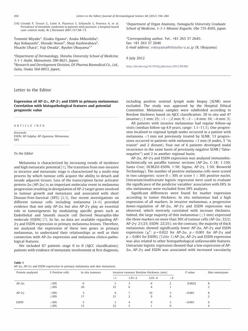
Table 1AP-2a, AP-2g and ESDN expression in primary melanoma and skin metastasis.
Protein analysed % Positive cells In situ tumours Invasive
�1
AP-2a <30% 0 3
�30% 20 22
AP-2g <30% 3 4
�30% 17 21
ESDN <30% 5 3
�30% 15 22
including positive sentinel lymph node biopsy (SLNB) wereexcluded. The study was approved by the Hospital EthicalCommittee. Melanoma samples were subdivided according toBreslow thickness based on AJCC classification: 20 in situ and 47invasive (�1 mm: 25; >1 – �2 mm: 9; >2 – �4 mm: 10; >4 mm: 3).
All patients with invasive melanomas had regular follow-upvisits (median follow-up 4.9 years, range: 1.1–11.1). One progres-sion localised to regional lymph nodes occurred in a patient withmelanoma <1 mm not previously treated by SLNB; 13 progres-sions occurred in patients with melanoma >1 mm (6 nodes, 5 ‘‘intransit’’ and 2 distant). Four out of 6 patients developed nodalrecurrence in the same basin of previously negative SLNB (‘‘false-negative’’) and 2 in another regional basin.
AP-2a, AP-2g and ESDN expression was analysed immunohis-tochemically on paraffin tumour sections (AP-2a, C-18; 1:150;Santa Cruz; DCBLD2-ESDN, 1:50; Sigma; AP-2g, 1:50; BioworldTechnology). The number of positive melanoma cells were scoredin two categories: score 0 � 30% or score 1 > 30% positive nuclei.Univariate/multivariate logistic regression were used to evaluatethe significance of the predictor variables’ association with DFS. Insitu melanomas were excluded from DFS analyses.
Significant differences were found for marker expressionaccording to tumor thickness. In situ melanomas had a highexpression of all markers. In invasive melanomas, a progressivedown-regulation of AP-2a, AP-2g and ESDN expression wasobserved, which inversely correlated with increase thickness.Indeed, the large majority of thin melanomas (�1 mm) expressedthe three markers on more than 30% of tumour cells (AP-2a: 22/2;AP-2g: 21/25; ESDN: 22/25); on the contrary, the majority of thickmelanomas showed significantly lower AP-2a, AP-2g and ESDNexpression (x2: p = 0.022 for AP-2a, p < 0.001 for AP-2g andp < 0.001 for ESDN). (Table 1) AP-2a, AP-2g and ESDN expressionwas also related to other histopathological unfavourable features.Univariate logistic regression showed that a low expression of AP-2a, AP-2g and ESDN was associated with increased vascularity
umours Breslow thickness (mm) P value Skin metastases
1.01–2 2.01–4 >4
3 6 2 0.0022 8
6 4 1 2
7 10 3 <0.001 8
2 0 0 2
4 9 3 <0.001 6
5 1 0 4
(odds-ratio (OR) 5.77 for AP-2a, 5.14 for AP-2g and 3.14 for ESDNexpression <30% positive melanoma cells), vascular invasion (OR12.8 for AP-2a, 17.34 for ESDN and 52.8 for AP-2g) and mitotic rate(OR 16.33 for AP-2a, 17.9 for AP-2g and 14.64 for ESDNexpression).
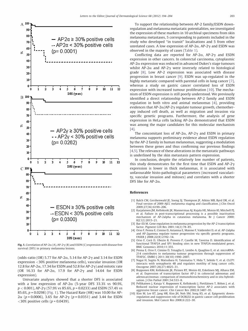
Univariate analyses showed that a shorter DFS is associatedwith a low expression of AP-2a (5-year DFS 33.3% vs 90.9%,p < 0.001), AP-2g (57.9% vs 85.6%, p = 0.0233) and ESDN (57.4% vs85.6%, p = 0.0298) (Fig. 1). The Hazard Rate (HR) was 10.00 for AP-2a (p = 0.0006), 3.65 for AP-2g (p = 0.0351) and 3.44 for ESDN<30% positive cells (p = 0.0439).
To support the relationship between AP-2 family/ESDN down-regulation and melanoma metastatic potentialities, we investigatedthe expression of these markers in 10 archival specimens from skinmelanoma metastases, 5 corresponding to patients included in thestudy who developed ‘‘in transit’’ localisations and 5 from otherunrelated cases. A low expression of AP-2a, AP-2g and ESDN wasobserved in the majority of cases (Table 1).
Conflicting data are reported for AP-2a, AP-2g and ESDNexpression in other cancers. In colorectal carcinoma, cytoplasmicAP-2a expression was reduced in advanced Dukes’s stage tumourswhilst AP-2a and AP-2g were inversely related to histologicalgrade [8]. Low AP-2 expression was associated with diseaseprogression in breast cancer [9]. ESDN was up-regulated in thehighly metastatic compared with parental cells in lung cancer [7],whereas a study on gastric cancer correlated loss of ESDNexpression with increased tumour proliferation [10]. The mecha-nism of ESDN expression is still poorly understood. We previouslyidentified a direct relationship between AP-2 family and ESDNregulation in both vitro and animal melanomas [4], providingevidences that AP-2a/AP-2g regulate tumour growth, chemother-apy induced cell death, as well as migration and invasion viaspecific genetic programs. Furthermore, the analysis of geneexpression in HeLa cells lacking AP-2a demonstrated that ESDNwas among the major candidates for this molecular mechanism[4].
The concomitant loss of AP-2a, AP-2g and ESDN in primarymelanoma supports preliminary evidence about ESDN regulationby the AP-2 family in human melanomas, suggesting a modulationbetween these genes and thus confirming our previous findings[4,5]. The relevance of these alterations in the metastatic pathwaysis underlined by the skin metastasis pattern expression.
In conclusion, despite the relatively low number of patients,this study demonstrates for the first time that ESDN and AP-2gexpression is lower in thick melanomas, it is associated withunfavourable histo-pathological parameters (increased vasculari-ty, vascular invasion and mitoses) and correlates with a shorterDFS like for AP-2a.
References
[1] Balch CM, Gershenwald JE, Soong SJ, Thompson JF, Atkins MB, Byrd DR, et al.Final version of 2009 AJCC melanoma staging and classification. J Clin Oncol2009;27(36):6199–206.
[2] Karjalainen JM, Kellokoski JK, Mannermaa AJ, Kujala HE, Moisio KI, Mitchell PJ,et al. Failure in post-transcriptional processing is a possible inactivationmechanism of AP-2alpha in cutaneous melanoma. Br J Cancer 2000;82(12):2015–21.
[3] Bar-Eli M. Gene regulation in melanoma progression by the AP-2 transcriptionfactor. Pigment Cell Res 2001;14(2):78–85.
[4] Orso F, Penna E, Cimino D, Astanina E, Maione F, Valdembri D, et al. AP-2alphaand AP-2gamma regulate tumor progression via specific genetic programs.FASEB J 2008;22(8):2702–14.
[5] Orso F, Cora D, Ubezio B, Provero P, Caselle M, Taverna D. Identification offunctional TFAP2A and SP1 binding sites in new TFAP2A-modulated genes.BMC Genomics 2010;11:355.
[6] Penna E, Orso F, Cimino D, Tenaglia E, Lembo A, Quaglino E, et al. microRNA-214 contributes to melanoma tumour progression through suppression ofTFAP2C. EMBO J 2011;30(10):1990–2007.
[7] Nagai H, Sugito N, Matsubara H, Tatematsu Y, Hida T, Sekido Y, et al. CLCP1interacts with semaphorin 4B and regulates motility of lung cancer cells.Oncogene 2007;26(27):4025–31.
[8] Ropponen KM, Kellokoski JK, Pirinen RT, Moisio KI, Eskelinen MJ, Alhava EM,et al. Expression of transcription factor AP-2 in colorectal adenomas andadenocarcinomas; comparison of immunohistochemistry and in situ hybridi-sation. J Clin Pathol 2001;54:533–8.
[9] Pellikainen J, Kataja V, Ropponen K, Kellokoski J, Pietilainen T, Bohm J, et al.Reduced nuclear expression of transcription factor AP-2 associates withaggressive breast cancer. Clin Cancer Res 2002;8:3487–95.
[10] Kim M, Lee KT, Jang HR, Kim JH, Noh SM, Song KS, et al. Epigenetic down-regulation and suppressive role of DCBLD2 in gastric cancer cell proliferationand invasion. Mol Cancer Res 2008;6:222–30.
Fig. 1. Correlation of AP-2a (A), AP-2g (B) and ESDN (C) expression with disease free
survival (DFS) in primary melanoma lesions.
Letters to the Editor / Journal of Dermatological Science 68 (2012) 194–204 203

Simona Osella-Abatea,*, Mauro Novellia, Pietro Quaglinoa,
Francesca Orsob, Benedetta Ubeziob, Carlo Tomasinic,Ester Berardengoc, Maria Grazia Bernengoa, Daniela Tavernab
aDepartment of Medical Sciences and Human Oncology,
Dermatologic Clinic, University of Turin, Turin, Italy; bMolecular
Biotechnology Center (MBC) and Department of Oncological Sciences,
University of Turin, Italy; cDivision of Anatomic Pathology IV, San
Giovanni Battista Hospital, Turin, Italy
*Corresponding author at: Department of Medical Sciences andHuman Oncology, Dermatologic Clinic, University of Turin, ViaCherasco 23, 10126 Turin, Italy. Tel.: +39 011 6335816;fax: +39 011 674034E-mail address: [email protected] (S. Osella-Abate)
22 May 20124 September 20125 September 2012
http://dx.doi.org/10.1016/j.jdermsci.2012.09.008
Letters to the Editor / Journal of Dermatological Science 68 (2012) 194–204204