EXPERIMENTAL VARICOCELE INDUCES HYPOXIA INDUCIBLE FACTOR-1α, VASCULAR ENDOTHELIAL GROWTH FACTOR...
4
EXPERIMENTAL VARICOCELE INDUCES HYPOXIA INDUCIBLE FACTOR-1, VASCULAR ENDOTHELIAL GROWTH FACTOR EXPRESSION AND ANGIOGENESIS IN THE RAT TESTIS FERHAT KILINC ¸ ,* FAZILET KAYASELCUK, CEM AYGUN, SEZGIN GUVEL, TULGA EGILMEZ AND HAKAN OZKARDES From the Department of Urology, Baskent University Faculty of Medicine (FK, FK, CA, SG, TE, HO) and Adana Teaching and Medical Research Center (FK, FK, SG, TE), Adana, Turkey ABSTRACT Purpose: In this study we investigated hypoxia inducible factor-1 (HIF-1) and vascular endothelial growth factor (VEGF) expression, and angiogenesis in an experimental model of varicocele in the rat testis. Materials and Methods: A total of 30 adult male Sprague-Dawley rats were investigated in 3 groups, namely varicocele group 1 (13), sham operated group 2 (9) and control group 3 (8). At 30 days after surgery was completed in groups 1 and 2 orchiectomy was performed in all rats. Histological findings in the left testicles of rats from each group were compared. HIF-1 and VEGF expression was immunohistochemically studied and CD31 panendothelial antigen was used to identify the number of microvessels, that is microvessel density (MVD), in paraffin embedded sections of testis tissue. Data were analyzed using the chi-square test, Fisher’s exact test, 1-way ANOVA and the Tukey HSD test for post hoc comparison. Results: HIF-1 expression was detected in 12 specimens (92.3%) in group 1, 4 (44.4%) in group 2 and 2 (25%) in group 3. The frequency of HIF-1 positivity in group 1 was significantly higher than the rates in groups 2 (p 0.023) and 3 (p 0.003). VEGF expression was detected in 8 specimens (61.5%) in group 1 but none of the group 2 or 3 specimens were VEGF positive. The frequency of VEGF positivity in group 1 was significantly higher than that in groups 2 (p 0.006) and 3 (p 0.007). Mean MVD SD in group 1 was 7.53 1.50 (range 6 to 12), and findings in groups 2 and 3 were 5.88 1.45 (range 4 to 8) and 5.12 1.12 (range 4 to 7), respectively. Mean MVD in group 2 was higher than in group 3 but this difference was not significant (p 0.509). Mean MVD in group 1 was significantly higher than the mean values in groups 2 (p 0.030) and 3 (p 0.002). Conclusions: Previous study of experimental varicocele models in rats documented HIF-1 and VEGF expression combined with angiogenesis in the testis. The results of this study show that varicocele can lead to tissue hypoxia and related pathophysiological events, such as angiogenesis. KEY WORDS: testis, varicocele, HIF-1 protein, vascular endothelial growth factor, angiogenesis Varicoceles have long been recognized as a cause of male infertility. It is estimated that approximately a third of the men evaluated for infertility have this condition. 1 The liter- ature indicates that 8% to 23% of men have clinically evident varicocele 2 but not all men with varicocele have abnormal semen parameters. The question of why approximately 15% to 20% of men with varicocele are infertile, whereas the rest are not infertile remains unanswered. 3 The reason probably lies in the pathophysiology of varicocele, although this is still unclear and requires more in-depth study. Over the years researchers have proposed a number of theories for the mech- anisms by which varicocele may impair male infertility. They include scrotal hyperthermia, 4 hypoxia, 5–7 retrograde flow of adrenal blood, 8 endocrine and testicular paracrine imbalanc- es, 3 and most recently apoptosis. 9 Hypoxia is known to be a key factor in the regulation of various physiological responses, including erythropoiesis, glycolysis, angiogenesis and vasodilatation. This regulation is partially mediated by transcription factors of the hypoxia inducible factor (HIF) family. HIF-1 is a heterodimer that con- sists of the hypoxic response factors HIF-1 and HIF-1. In the absence of oxygen HIF-1 binds to hypoxia response genes, such as the gene for pro-angiogenic growth factor vascular endothe- lial growth factor (VEGF). 10 HIF-1 is a putative marker of tumor hypoxia and it can be used as an immunohistochemical marker of hypoxia in paraffin embedded tissue specimens. 11 Angiogenesis is defined as the growth and proliferation of blood vessels from existing vasculature that allows developing tissue to obtain the nutrients necessary for maintaining growth. It is also one of the important adaptive mechanisms that occur in tissues subjected to hypoxic conditions. In this study we used an experimental model of varicocele to investigate how varicocele affects HIF-1 expression, VEGF expression and angiogenesis in the rat testis. Based on these findings we studied whether tissue hypoxia and related pathophysiological events such as angiogenesis occur in var- icocele. The aim was to gain new insight into the pathophys- iological mechanisms of varicocele related male infertility. MATERIALS AND METHODS The study included 36 male Sprague-Dawley rats of wean- ing age (12 to 14 weeks). All animals were fed the same food Accepted for publication March 5, 2004. Supported by the Education Foundation of Baskent University, Turkey. * Correspondence: Department of Urology, Baskent University Faculty of Medicine, Adana Teaching and Medical Research Center, Dadaloglu Mah. 39 sk. 01250*, Adana, Turkey (telephone: 90 322 3272727(1463); FAX: 90 322 3271274; e-mail: ferhatkilinc@ hotmail.com). 0022-5347/04/1723-1188/0 Vol. 172, 1188 –1191, September 2004 THE JOURNAL OF UROLOGY ® Printed in U.S.A. Copyright © 2004 by AMERICAN UROLOGICAL ASSOCIATION DOI: 10.1097/01.ju.0000135455.97627.15 1188
Transcript of EXPERIMENTAL VARICOCELE INDUCES HYPOXIA INDUCIBLE FACTOR-1α, VASCULAR ENDOTHELIAL GROWTH FACTOR...

EXPERIMENTAL VARICOCELE INDUCES HYPOXIA INDUCIBLEFACTOR-1�, VASCULAR ENDOTHELIAL GROWTH FACTOREXPRESSION AND ANGIOGENESIS IN THE RAT TESTIS
FERHAT KILINC,* FAZILET KAYASELCUK, CEM AYGUN, SEZGIN GUVEL, TULGA EGILMEZAND HAKAN OZKARDES
From the Department of Urology, Baskent University Faculty of Medicine (FK, FK, CA, SG, TE, HO) and Adana Teaching and MedicalResearch Center (FK, FK, SG, TE), Adana, Turkey
ABSTRACT
Purpose: In this study we investigated hypoxia inducible factor-1� (HIF-1�) and vascularendothelial growth factor (VEGF) expression, and angiogenesis in an experimental model ofvaricocele in the rat testis.Materials and Methods: A total of 30 adult male Sprague-Dawley rats were investigated in 3
groups, namely varicocele group 1 (13), sham operated group 2 (9) and control group 3 (8). At 30days after surgery was completed in groups 1 and 2 orchiectomy was performed in all rats.Histological findings in the left testicles of rats from each group were compared. HIF-1� andVEGF expression was immunohistochemically studied and CD31 panendothelial antigen wasused to identify the number of microvessels, that is microvessel density (MVD), in paraffinembedded sections of testis tissue. Data were analyzed using the chi-square test, Fisher’s exacttest, 1-way ANOVA and the Tukey HSD test for post hoc comparison.Results: HIF-1� expression was detected in 12 specimens (92.3%) in group 1, 4 (44.4%) in group
2 and 2 (25%) in group 3. The frequency of HIF-1� positivity in group 1 was significantly higherthan the rates in groups 2 (p � 0.023) and 3 (p � 0.003). VEGF expression was detected in 8specimens (61.5%) in group 1 but none of the group 2 or 3 specimens were VEGF positive. Thefrequency of VEGF positivity in group 1 was significantly higher than that in groups 2 (p � 0.006)and 3 (p � 0.007). Mean MVD � SD in group 1 was 7.53 � 1.50 (range 6 to 12), and findings ingroups 2 and 3 were 5.88 � 1.45 (range 4 to 8) and 5.12 � 1.12 (range 4 to 7), respectively. MeanMVD in group 2 was higher than in group 3 but this difference was not significant (p � 0.509).Mean MVD in group 1 was significantly higher than the mean values in groups 2 (p � 0.030) and3 (p � 0.002).Conclusions: Previous study of experimental varicocele models in rats documented HIF-1� and
VEGF expression combined with angiogenesis in the testis. The results of this study show thatvaricocele can lead to tissue hypoxia and related pathophysiological events, such as angiogenesis.
KEY WORDS: testis, varicocele, HIF-1 protein, vascular endothelial growth factor, angiogenesis
Varicoceles have long been recognized as a cause of maleinfertility. It is estimated that approximately a third of themen evaluated for infertility have this condition.1 The liter-ature indicates that 8% to 23% of men have clinically evidentvaricocele2 but not all men with varicocele have abnormalsemen parameters. The question of why approximately 15%to 20% of men with varicocele are infertile, whereas the restare not infertile remains unanswered.3 The reason probablylies in the pathophysiology of varicocele, although this is stillunclear and requires more in-depth study. Over the yearsresearchers have proposed a number of theories for the mech-anisms by which varicocele may impair male infertility. Theyinclude scrotal hyperthermia,4 hypoxia,5–7 retrograde flow ofadrenal blood,8 endocrine and testicular paracrine imbalanc-es,3 and most recently apoptosis.9
Hypoxia is known to be a key factor in the regulation ofvarious physiological responses, including erythropoiesis,glycolysis, angiogenesis and vasodilatation. This regulation
is partially mediated by transcription factors of the hypoxiainducible factor (HIF) family. HIF-1 is a heterodimer that con-sists of the hypoxic response factors HIF-1� and HIF-1�. In theabsence of oxygen HIF-1 binds to hypoxia response genes, suchas the gene for pro-angiogenic growth factor vascular endothe-lial growth factor (VEGF).10 HIF-1� is a putative marker oftumor hypoxia and it can be used as an immunohistochemicalmarker of hypoxia in paraffin embedded tissue specimens.11
Angiogenesis is defined as the growth and proliferation of bloodvessels from existing vasculature that allows developing tissueto obtain the nutrients necessary for maintaining growth. It isalso one of the important adaptive mechanisms that occur intissues subjected to hypoxic conditions.In this study we used an experimental model of varicocele
to investigate how varicocele affects HIF-1� expression,VEGF expression and angiogenesis in the rat testis. Based onthese findings we studied whether tissue hypoxia and relatedpathophysiological events such as angiogenesis occur in var-icocele. The aim was to gain new insight into the pathophys-iological mechanisms of varicocele related male infertility.
MATERIALS AND METHODS
The study included 36 male Sprague-Dawley rats of wean-ing age (12 to 14 weeks). All animals were fed the same food
Accepted for publication March 5, 2004.Supported by the Education Foundation of Baskent University,
Turkey.* Correspondence: Department of Urology, Baskent University
Faculty of Medicine, Adana Teaching and Medical Research Center,Dadaloglu Mah. 39 sk. 01250*, Adana, Turkey (telephone: � 90 3223272727(1463); FAX: � 90 322 3271274; e-mail: [email protected]).
0022-5347/04/1723-1188/0 Vol. 172, 1188–1191, September 2004THE JOURNAL OF UROLOGY® Printed in U.S.A.Copyright © 2004 by AMERICAN UROLOGICAL ASSOCIATION DOI: 10.1097/01.ju.0000135455.97627.15
1188

and maintained in a constant environment with a 12:12-hourlight-dark cycle. The rats were assigned to 1 of 3 groups.Group 1 animals (varicocele group, 16) underwent surgery inwhich varicocele was produced. Group 2 rats (sham surgerygroup, 10) underwent the same operation except ligatureswere only placed in position and not tied. Group 3 (controlgroup, 10) underwent no surgery. Six of the initial 36 ratswere excluded for different reasons. Three of the 16 rats ingroup 1 were excluded due to death or unsuccessful surgery.One of the 10 animals in group 2 was excluded due to death.Two of the 10 rats in group 3 were excluded due to inciden-tally detected spermatic vein dilatation.Technique of experimental varicocele. Each animal was
anesthetized with an intraperitoneal injection of 5% ket-amine hydrochloride (60 mg/kg) and xylazine (7 mg/kg). Anabdominal midline incision was made. The left renal vein,inferior vena cava and left spermatic vein were identified anda clamp was passed behind the left renal vein just distal tothe spermatic vein insertion. A 4-zero silk ligature wasloosely placed around the left renal vein at this site and arigid hydrophilic guide wire of 0.64 mm in diameter wasplaced on the left renal vein. The ligature was tied around thevein over the top of the guide wire. The guide wire was thenwithdrawn and the vein was allowed to expand to the limitsof the ligature, which caused vein diameter to be decreased toapproximately half of its original diameter. The renal veinand spermatic vein in each animal dilated immediately. Themidline incision was closed in 2 layers with 3-zero silk su-ture.Histology. All rats underwent orchiectomy 30 days after
the operations were performed in groups 1 and 2 animals.The testes were fixed in Bouin’s solution and embedded inparaffin. Histological sections of the left testis of each animalwere prepared and stained with hematoxylin and eosin forhistopathological examination.Immunohistochemistry for HIF-1�, VEGF and CD31. Im-
munohistochemical staining was performed using the stan-dard avidin-biotin-peroxidase complex technique with anL. V. Dako LSAB kit (Dako, Copenhagen, Denmark). Briefly,sections were deparaffinized, dehydrated and then immersedin 10-3 M sodium citrate buffer (pH 6.0). To detect HIF-1�and VEGF sections were heated in a microwave oven 3 timesat 900 W for a total of 20 minutes. Endogenous peroxidasewas inactivated by incubating sections with 3% hydrogenperoxide. Nonspecific reactions were blocked by incubatingsections in a solution containing 5% normal horse serum and1% normal goat serum. Sections were incubated with pri-mary antibody overnight at 4C. HIF-1� expression was as-sessed using goat polyclonal antibody IgG (catalogue No.SC-8711, Santa Cruz Biotechnology, Santa Cruz, California)(dilution 1:20). VEGF expression was assessed using rabbitmonoclonal antiVEGF-C (catalogue No. 18–2255, ZymedCorp., South San Francisco, California) (dilution 1:1000).After incubation with antibody sections were incubated for30 minutes at room temperature with biotinylated antimouseIg and then with streptavidin conjugated to horseradish per-oxidase. After 3 rinses with phosphate buffered saline thesections were incubated with diaminobenzidine substrate for5 minutes. Finally, they were rinsed with distilled water andcounterstained with hematoxylin. Breast cancer and coloncancer sections with strong nuclear expression were used aspositive controls for HIF-1� and VEGF, respectively. Foreach animal immunostaining for HIF-1� and VEGF wasassessed as present or absent. Group frequencies of positivity(expression) were compared.To assess angiogenesis we used JC70 monoclonal antibody
(Dako), which binds to CD31 panendothelial antigen(platelet-endothelial cell adhesion molecule). Paraffin em-bedded sections were processed with this antibody using thealkaline phosphatase/anti-alkaline phosphatase procedure
with resultant staining of microvessels and single endothe-lial cells.Angiogenesis assessment. The degree of angiogenesis in
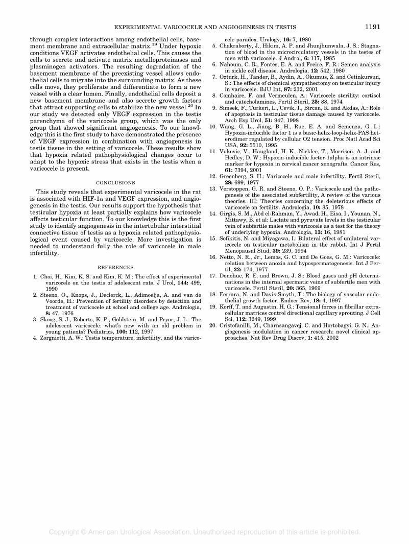
each specimen was assessed by counting stained microves-sels. In each specimen the 3 areas with the highest degree ofvascularization were identified at low power (�100) and thenthe number of vessels in the intertubular interstitial connec-tive tissue in the selected fields was counted at high power(�400) (part A of figure). For each testis the highest of these3 vessel counts was recorded as microvessel density (MVD).Only vessels with a clearly defined lumen or well-definedlinear vessel shape were counted as microvessels. Newlyforming vessels that consisted of only 1 layer of endothelialcells were ignored. Mean MVD values � SD in the 3 groupswere compared.Statistical analysis. Data were statistically analyzed with
the chi-square test, Fisher’s exact test, 1-way ANOVA andthe Tukey HSD test for post hoc comparison.
RESULTS
Left testis sections from groups 2 (sham surgery) and 3(controls) showed no significant histological abnormalities.Specimens from group 1 (varicocele) showed more germ celldesquamation, more congestion and more edema than spec-imens from groups 2 and 3. These findings were focal in someareas and diffuse in others but focal changes were mostprevalent. Some group 1 specimens also showed less sper-matogenesis than specimens from the sham and controlgroups (part B of figure).HIF-1� expression was detected in germ cell cytoplasm
and vascular endothelium (parts C and D of figure). Noneof the specimens showed nuclear staining for HIF-1�. Atotal of 12 of the 13 specimens (92.3%) in group 1 werepositive for HIF-1�, of which 2 showed positivity in therete testis (table 1). Four specimens in group 2 (44.4%) and2 in group 3 (25%) showed HIF-1� expression. The fre-quency of HIF-1� positivity in group 1 was significantlyhigher than in groups 2 (p � 0.023) and 3 (p � 0.003)Concerning VEGF expression, none of the specimens in
groups 2 and 3 stained positive in the testis parenchyma.Two group 2 specimens showed immunostaining for VEGF inthe epididymis and rete testis. Eight of the 13 group 1 spec-imens (61.5%) showed VEGF expression in germ cell cyto-plasm (part E of figure). The frequency of VEGF positivity ingroup 1 was significantly higher than in groups 2 (p � 0.006)and 3 (p � 0.007).The number of microvessels counted in each of the 3 se-
lected fields in group 1 specimens was 3 to 12 (mean 6.02 �1.69). The corresponding range in group 2 was 3 to 8 (mean4.55 � 1.55) and in group 3 it was 2 to 7 (mean 3.95 � 1.23).Mean MVD in group 1 was 7.53 � 1.50 and the values ingroups 2 and 3 were 5.88 � 1.45 and 5.12 � 1.12, respectively(table 2). Mean MVD in the varicocele group was signifi-cantly higher than in the sham operated (p � 0.030) andcontrol (p � 0.002) groups. Although mean MVD in the shamoperated group was higher than that in the control group,this difference was not statistically significant (p � 0.509).
DISCUSSION
The prevalence of varicocele in the general male populationis approximately 15% but the rate is higher than 35% ininfertile men.12 These lesions are common and varicoceletreatment is a traditional part of therapy for male infertility.This explains why numerous studies have been done of thepathophysiology of this condition. Varicocele is associatedwith phlebectasia, retrograde blood flow into the plexus pam-piniformis and limited capacity of the available channels forvenous return. Researchers have hypothesized that thesefactors increase the volume of blood in the testis and result in
EXPERIMENTAL VARICOCELE AND ANGIOGENESIS IN TESTIS 1189

venous stasis, which means decreased testicular oxygen lev-els.13
Hypoxia is known to initiate a cascade of molecular path-ways, including those involved in angiogenesis, glycolysisand changes in microenvironmental pH. Reports have docu-mented low serum lactate and pyruvate levels in the testic-ular veins of humans14 and animals15 with varicocele, andthese findings are not compatible with testicular hypoxia. Inaddition, studies of the possibility that altered gas contentcauses spermatogenic depression have failed to show evi-dence of hypoxia in patients with varicocele.16,17 However,Chakraborty et al documented blood stagnation within themicrocirculatory vessels of the testes in patients with vari-
cocele, which could cause local hypoxic or ischemic shock tothe cells.5 Furthermore, after analyzing semen in patientswith sickle cell disease Nahoum et al noted decreased sper-matozoa forward progression and a high incidence of thestress pattern during morphological analysis, which closelyresembled the types of seminal changes observed in patientswith varicocele.6 These groups proposed that these findingscould be considered indirect evidence in favor of testicularhypoxia.5,6 In another study Ozturk et al found that bio-chemical indicators of tissue hypoxia are significantly in-creased in experimental unilateral varicocele in the rat testisand chemical sympathectomy can prevent such increases.7 Inour study we observed HIF-1� expression in germ cells underconditions of experimental varicocele in the rat testis. To ourknowledge this is the first documentation of direct evidenceof tissue hypoxia in varicocele.HIF-1 binds to the VEGF gene and it is required for VEGF
transactivation in response to hypoxia. VEGF is a potent andspecific mitogen for endothelial cells derived from arteries,veins and lymphatics. Vasculogenesis and physiological orpathological angiogenesis depend on VEGF activity.18 There-fore, HIF-1 is a key transcriptional mediator of metabolicadaptation and of VEGF mediated angiogenesis in responseto hypoxia.Angiogenesis, which is the growth of new blood vessels
from existing vasculature, occurs in various physiologicaland pathophysiological conditions. The development of newblood vessels from the preexisting vasculature occurs
Sections of rat testis from group 1 with experimental varicocele. A, histological appearance of intertubular interstitial connective tissue.CD31 immunostain, reduced from �200. B, germ cell desquamation and decreased spermatogenesis. H & E, reduced from �100. C, HIF-1�expression in germ cells. Reduced from �200. D, HIF-1� expression in endothelial cells (arrow). Reduced from �400. E, VEGF expressionin germ cells. reduced from �100.
TABLE 1. HIF-1� and VEGF positive specimens in 3 study groups
Group No. Rats % HIF-1� % VEGF
Varicocele 13 12 (92.3) 8 (61.5)Sham operated 9 4 (44.4) 0Control 8 2 (25) 0
TABLE 2. MVD in 3 groups
MVD Varicocele ShamOperated Control
Min 6 4 4Max 12 8 7Mean 7.53 � 1.50 5.88 � 1.45 5.12 � 1.12
EXPERIMENTAL VARICOCELE AND ANGIOGENESIS IN TESTIS1190
through complex interactions among endothelial cells, base-ment membrane and extracellular matrix.19 Under hypoxicconditions VEGF activates endothelial cells. This causes thecells to secrete and activate matrix metalloproteinases andplasminogen activators. The resulting degradation of thebasement membrane of the preexisting vessel allows endo-thelial cells to migrate into the surrounding matrix. As thesecells move, they proliferate and differentiate to form a newvessel with a clear lumen. Finally, endothelial cells deposit anew basement membrane and also secrete growth factorsthat attract supporting cells to stabilize the new vessel.20 Inour study we detected only VEGF expression in the testisparenchyma of the varicocele group, which was the onlygroup that showed significant angiogenesis. To our knowl-edge this is the first study to have demonstrated the presenceof VEGF expression in combination with angiogenesis intestis tissue in the setting of varicocele. These results showthat hypoxia related pathophysiological changes occur toadapt to the hypoxic stress that exists in the testis when avaricocele is present.
CONCLUSIONS
This study reveals that experimental varicocele in the ratis associated with HIF-1� and VEGF expression, and angio-genesis in the testis. Our results support the hypothesis thattesticular hypoxia at least partially explains how varicoceleaffects testicular function. To our knowledge this is the firststudy to identify angiogenesis in the intertubular interstitialconnective tissue of testis as a hypoxia related pathophysio-logical event caused by varicocele. More investigation isneeded to understand fully the role of varicocele in maleinfertility.
REFERENCES
1. Choi, H., Kim, K. S. and Kim, K. M.: The effect of experimentalvaricocele on the testis of adolescent rats. J Urol, 144: 499,1990
2. Steeno, O., Knops, J., Declerck, L., Adimoelja, A. and van deVoorde, H.: Prevention of fertility disorders by detection andtreatment of varicocele at school and college age. Andrologia,8: 47, 1976
3. Skoog, S. J., Roberts, K. P., Goldstein, M. and Pryor, J. L.: Theadolescent varicocele: what’s new with an old problem inyoung patients? Pediatrics, 100: 112, 1997
4. Zorgniotti, A. W.: Testis temperature, infertility, and the varico-
cele paradox. Urology, 16: 7, 19805. Chakraborty, J., Hikim, A. P. and Jhunjhunwala, J. S.: Stagna-
tion of blood in the microcirculatory vessels in the testes ofmen with varicocele. J Androl, 6: 117, 1985
6. Nahoum, C. R., Fontes, E. A. and Freire, F. R.: Semen analysisin sickle cell disease. Andrologia, 12: 542, 1980
7. Ozturk, H., Tander, B., Aydin, A., Okumus, Z. and Cetinkursun,S.: The effects of chemical sympathectomy on testicular injuryin varicocele. BJU Int, 87: 232, 2001
8. Comhaire, F. and Vermeulen, A.: Varicocele sterility: cortisoland catecholamines. Fertil Steril, 25: 88, 1974
9. Simsek, F., Turkeri, L., Cevik, I., Bircan, K. and Akdas, A.: Roleof apoptosis in testicular tissue damage caused by varicocele.Arch Esp Urol, 51: 947, 1998
10. Wang, G. L., Jiang, B. H., Rue, E. A. and Semenza, G. L.:Hypoxia-inducible factor 1 is a basic-helix-loop-helix-PAS het-erodimer regulated by cellular O2 tension. Proc Natl Acad SciUSA, 92: 5510, 1995
11. Vukovic, V., Haugland, H. K., Nicklee, T., Morrison, A. J. andHedley, D. W.: Hypoxia-inducible factor-1alpha is an intrinsicmarker for hypoxia in cervical cancer xenografts. Cancer Res,61: 7394, 2001
12. Greenberg, S. H.: Varicocele and male infertility. Fertil Steril,28: 699, 1977
13. Verstoppen, G. R. and Steeno, O. P.: Varicocele and the patho-genesis of the associated subfertility, A review of the varioustheories. III: Theories concerning the deleterious effects ofvaricocele on fertility. Andrologia, 10: 85, 1978
14. Girgis, S. M., Abd el-Rahman, Y., Awad, H., Eisa, I., Younan, N.,Mittawy, B. et al: Lactate and pyruvate levels in the testicularvein of subfertile males with varicocele as a test for the theoryof underlying hypoxia. Andrologia, 13: 16, 1981
15. Sofikitis, N. and Miyagawa, I.: Bilateral effect of unilateral var-icocele on testicular metabolism in the rabbit. Int J FertilMenopausal Stud, 39: 239, 1994
16. Netto, N. R., Jr., Lemos, G. C. and De Goes, G. M.: Varicocele:relation between anoxia and hypospermatogenesis. Int J Fer-til, 22: 174, 1977
17. Donohue, R. E. and Brown, J. S.: Blood gases and pH determi-nations in the internal spermatic veins of subfertile men withvaricocele. Fertil Steril, 20: 365, 1969
18. Ferrara, N. and Davis-Smyth, T.: The biology of vascular endo-thelial growth factor. Endocr Rev, 18: 4, 1997
19. Korff, T. and Augustin, H. G.: Tensional forces in fibrillar extra-cellular matrices control directional capillary sprouting. J CellSci, 112: 3249, 1999
20. Cristofanilli, M., Charnsangavej, C. and Hortobagyi, G. N.: An-giogenesis modulation in cancer research: novel clinical ap-proaches. Nat Rev Drug Discov, 1: 415, 2002
EXPERIMENTAL VARICOCELE AND ANGIOGENESIS IN TESTIS 1191


![GPER mediates the angiocrine actions induced by IGF1 ... · tor hypoxia inducible factor-1 (HIF-1) and stimulated by low oxygen, hormones, cytokines and growth factors [8, 9]. Insulin-like](https://static.fdocument.org/doc/165x107/5f9d8b501ba938172359165c/gper-mediates-the-angiocrine-actions-induced-by-igf1-tor-hypoxia-inducible-factor-1.jpg)