Evidence From Reduced α2-6 Sialylation and Impaired - Diabetes
9
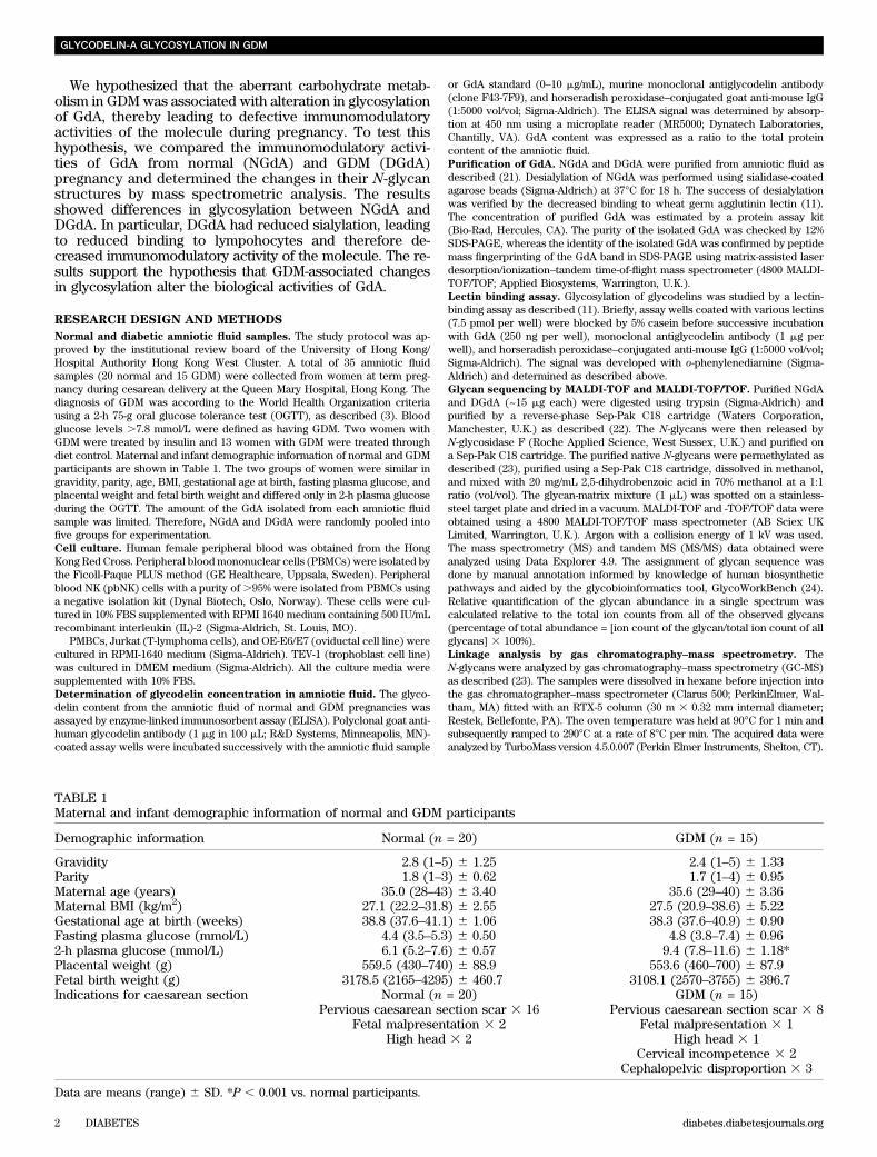
Glycosylation Failure Extends to Glycoproteins in Gestational Diabetes Mellitus Evidence From Reduced a2-6 Sialylation and Impaired Immunomodulatory Activities of Pregnancy-Related Glycodelin-A Cheuk-Lun Lee, 1,2,3 Philip C.N. Chiu, 1,3 Poh-Choo Pang, 4 Ivan K. Chu, 2 Kai-Fai Lee, 1,3 Riitta Koistinen, 5 Hannu Koistinen, 5 Markku Seppälä, 5 Howard R. Morris, 4 Bérangère Tissot, 4 Maria Panico, 4 Anne Dell, 4 and William S.B. Yeung 1,3 OBJECTIVE—Gestational diabetes mellitus (GDM) is a common metabolic disorder of pregnancy. Patients with GDM are at risk for high fetal mortality and gestational complications associated with reduced immune tolerance and abnormal carbohydrate metabolism. Glycodelin-A (GdA) is an abundant decidual glyco- protein with glycosylation-dependent immunomodulatory activi- ties. We hypothesized that aberrant carbohydrate metabolism in GDM was associated with changes in glycosylation of GdA, leading to defective immunomodulatory activities. RESEARCH DESIGN AND METHODS—GdA in the amniotic fluid from women with normal (NGdA) and GDM (DGdA) pregnancies was purified by affinity chromatography. Structural analysis of protein glycosylation was preformed by lectin-binding assay and mass spectrometry. Cytotoxicity, cell death, cytokine secretion, and GdA binding of the GdA-treated lymphocytes and natural killer (NK) cells were determined. The sialidase ac- tivity in the placental tissue from normal and GDM patients was measured. RESULTS—GDM affected the glycosylation but not the protein core of GdA. Specifically, DGdA had a lower abundance of a2-6– sialylated and high-mannose glycans and a higher abundance of glycans with Sda (NeuAca2-3[GalNAcb1-4]Gal) epitopes com- pared with NGdA. DGdA had reduced immuosuppressive activi- ties in terms of cytotoxicity on lymphocytes, inhibitory activities on interleukin (IL)-2 secretion by lymphocytes, stimulatory activ- ities on IL-6 secretion by NK cells, and binding to these cells. Desialylation abolished the immunomodulation and binding of NGdA. Placental sialidase activity was increased in GDM patients, which may account for the reduced sialic acid content of DGdA. CONCLUSIONS—Taken together, this study provides the first direct evidence for altered enzymatic glycosylation and impaired bioactivity of GdA in GDM patients. G estational diabetes mellitus (GDM) is a meta- bolic disorder manifested as glucose intolerance with an onset during pregnancy (1,2). It occurs in ~7% of pregnancies in the U.S. (1) and in 14% of pregnancies in Hong Kong (3). Compared with normal pregnancy, GDM is associated with a higher risk of preg- nancy complications such as macrosomia, preeclampsia, fetal mortality, placental alterations, and increased risk of diabetes of the mother and the offspring later in life (2,4,5). The underlying pathophysiology of GDM has been associ- ated with dysregulated immune responses, as demonstrated by changes in the immune cell subpopulations and the cy- tokine profile in women with GDM (6,7). Glycodelin-A (GdA) is an abundant secretory glycopro- tein of the pregnancy decidua (8). It is proposed to be involved in fetomaternal defense (9,10) through its im- munomodulatory activities. These include induction of apoptosis of T-cells (11), skewing of T-cell response to- ward the Th-2 phenotype (12), and modulation of the ac- tivities of natural killer (NK) cells (13), B-cells (14), and dendritic cells (15). Decreased secretion of glycodelin is associated with recurrent spontaneous abortion and un- explained infertility (8,10). Glycosylation is crucial to the biological activities of glycodelins (8,10). Its importance is shown by the lack of immunomodulatory activity in two other glycodelin isoforms with different glycosylation, glycodelin-S and glycodelin-C (11). Although there is no difference in the glycodelin concentration of the maternal serum in the first trimester (16) and of the cord serum (17) between GDM and normal pregnancy, the glycosylation of glycodelin in GDM pregnancies is unknown. Hyperglycemia in diabetes causes abnormal carbohydrate metabolism and the pro- duction of advanced glycation end products, leading to alteration of gene expression and activities of the cellular glycosyltransferases and glycosidases (18). Diabetic preg- nancy has been associated with changes in the glycosyla- tion and subsequently the biological activities of human chorionic gonadotrophin (19) and the placental transferrin receptor (20). However, because of the lack of advanced methodological procedures, these studies did not provide any detailed information of the glycan structures and the resultant changes in biological activities. From the 1 Department of Obstetrics and Gynecology, University of Hong Kong, Hong Kong, China; the 2 Department of Chemistry, University of Hong Kong, Hong Kong, China; the 3 Centre for Reproduction, Development, and Growth, University of Hong Kong, Hong Kong, China; the 4 Division of Mo- lecular Biosciences, Faculty of Natural Sciences, Imperial College London, London, U.K.; and the 5 Department of Clinical Chemistry, University of Helsinki and Helsinki University Central Hospital, Helsinki, Finland. Corresponding author: Philip C.N. Chiu, [email protected]. Received 19 August 2010 and accepted 7 December 2010. DOI: 10.2337/db10-1186 This article contains Supplementary Data online at http://diabetes. diabetesjournals.org/lookup/suppl/doi:10.2337/db10-1186/-/DC1. C.-L.L. and P.C.N.C. contributed equally to this work. Ó 2011 by the American Diabetes Association. Readers may use this article as long as the work is properly cited, the use is educational and not for profit, and the work is not altered. See http://creativecommons.org/licenses/by -nc-nd/3.0/ for details. diabetes.diabetesjournals.org DIABETES 1 ORIGINAL ARTICLE Diabetes Publish Ahead of Print, published online February 7, 2011 Copyright American Diabetes Association, Inc., 2011
Transcript of Evidence From Reduced α2-6 Sialylation and Impaired - Diabetes
Glycosylation Failure Extends to Glycoproteins inGestational Diabetes MellitusEvidence From Reduced a2-6 Sialylation and ImpairedImmunomodulatory Activities of Pregnancy-RelatedGlycodelin-ACheuk-Lun Lee,
1,2,3Philip C.N. Chiu,
1,3Poh-Choo Pang,
4Ivan K. Chu,
2Kai-Fai Lee,
1,3
Riitta Koistinen,5Hannu Koistinen,
5Markku Seppälä,
5Howard R. Morris,
4Bérangère Tissot,
4
Maria Panico,4Anne Dell,
4and William S.B. Yeung
1,3
OBJECTIVE—Gestational diabetes mellitus (GDM) is a commonmetabolic disorder of pregnancy. Patients with GDM are at riskfor high fetal mortality and gestational complications associatedwith reduced immune tolerance and abnormal carbohydratemetabolism. Glycodelin-A (GdA) is an abundant decidual glyco-protein with glycosylation-dependent immunomodulatory activi-ties. We hypothesized that aberrant carbohydrate metabolism inGDM was associated with changes in glycosylation of GdA,leading to defective immunomodulatory activities.
RESEARCH DESIGN AND METHODS—GdA in the amnioticfluid from women with normal (NGdA) and GDM (DGdA)pregnancies was purified by affinity chromatography. Structuralanalysis of protein glycosylation was preformed by lectin-bindingassay and mass spectrometry. Cytotoxicity, cell death, cytokinesecretion, and GdA binding of the GdA-treated lymphocytesand natural killer (NK) cells were determined. The sialidase ac-tivity in the placental tissue from normal and GDM patients wasmeasured.
RESULTS—GDM affected the glycosylation but not the proteincore of GdA. Specifically, DGdA had a lower abundance of a2-6–sialylated and high-mannose glycans and a higher abundance ofglycans with Sda (NeuAca2-3[GalNAcb1-4]Gal) epitopes com-pared with NGdA. DGdA had reduced immuosuppressive activi-ties in terms of cytotoxicity on lymphocytes, inhibitory activitieson interleukin (IL)-2 secretion by lymphocytes, stimulatory activ-ities on IL-6 secretion by NK cells, and binding to these cells.Desialylation abolished the immunomodulation and binding ofNGdA. Placental sialidase activity was increased in GDM patients,which may account for the reduced sialic acid content of DGdA.
CONCLUSIONS—Taken together, this study provides the firstdirect evidence for altered enzymatic glycosylation and impairedbioactivity of GdA in GDM patients.
Gestational diabetes mellitus (GDM) is a meta-bolic disorder manifested as glucose intolerancewith an onset during pregnancy (1,2). It occursin ~7% of pregnancies in the U.S. (1) and in 14%
of pregnancies in Hong Kong (3). Compared with normalpregnancy, GDM is associated with a higher risk of preg-nancy complications such as macrosomia, preeclampsia,fetal mortality, placental alterations, and increased risk ofdiabetes of the mother and the offspring later in life (2,4,5).The underlying pathophysiology of GDM has been associ-ated with dysregulated immune responses, as demonstratedby changes in the immune cell subpopulations and the cy-tokine profile in women with GDM (6,7).
Glycodelin-A (GdA) is an abundant secretory glycopro-tein of the pregnancy decidua (8). It is proposed to beinvolved in fetomaternal defense (9,10) through its im-munomodulatory activities. These include induction ofapoptosis of T-cells (11), skewing of T-cell response to-ward the Th-2 phenotype (12), and modulation of the ac-tivities of natural killer (NK) cells (13), B-cells (14), anddendritic cells (15). Decreased secretion of glycodelin isassociated with recurrent spontaneous abortion and un-explained infertility (8,10).
Glycosylation is crucial to the biological activities ofglycodelins (8,10). Its importance is shown by the lackof immunomodulatory activity in two other glycodelinisoforms with different glycosylation, glycodelin-S andglycodelin-C (11). Although there is no difference in theglycodelin concentration of the maternal serum in the firsttrimester (16) and of the cord serum (17) between GDMand normal pregnancy, the glycosylation of glycodelin inGDM pregnancies is unknown. Hyperglycemia in diabetescauses abnormal carbohydrate metabolism and the pro-duction of advanced glycation end products, leading toalteration of gene expression and activities of the cellularglycosyltransferases and glycosidases (18). Diabetic preg-nancy has been associated with changes in the glycosyla-tion and subsequently the biological activities of humanchorionic gonadotrophin (19) and the placental transferrinreceptor (20). However, because of the lack of advancedmethodological procedures, these studies did not provideany detailed information of the glycan structures and theresultant changes in biological activities.
From the 1Department of Obstetrics and Gynecology, University of HongKong, Hong Kong, China; the 2Department of Chemistry, University of HongKong, Hong Kong, China; the 3Centre for Reproduction, Development, andGrowth, University of Hong Kong, Hong Kong, China; the 4Division of Mo-lecular Biosciences, Faculty of Natural Sciences, Imperial College London,London, U.K.; and the 5Department of Clinical Chemistry, University ofHelsinki and Helsinki University Central Hospital, Helsinki, Finland.
Corresponding author: Philip C.N. Chiu, [email protected] 19 August 2010 and accepted 7 December 2010.DOI: 10.2337/db10-1186This article contains Supplementary Data online at http://diabetes.
diabetesjournals.org/lookup/suppl/doi:10.2337/db10-1186/-/DC1.C.-L.L. and P.C.N.C. contributed equally to this work.� 2011 by the American Diabetes Association. Readers may use this article as
long as the work is properly cited, the use is educational and not for profit,and the work is not altered. See http://creativecommons.org/licenses/by-nc-nd/3.0/ for details.
diabetes.diabetesjournals.org DIABETES 1
ORIGINAL ARTICLE Diabetes Publish Ahead of Print, published online February 7, 2011
Copyright American Diabetes Association, Inc., 2011
We hypothesized that the aberrant carbohydrate metab-olism in GDMwas associated with alteration in glycosylationof GdA, thereby leading to defective immunomodulatoryactivities of the molecule during pregnancy. To test thishypothesis, we compared the immunomodulatory activi-ties of GdA from normal (NGdA) and GDM (DGdA)pregnancy and determined the changes in their N-glycanstructures by mass spectrometric analysis. The resultsshowed differences in glycosylation between NGdA andDGdA. In particular, DGdA had reduced sialylation, leadingto reduced binding to lympohocytes and therefore de-creased immunomodulatory activity of the molecule. The re-sults support the hypothesis that GDM-associated changesin glycosylation alter the biological activities of GdA.
RESEARCH DESIGN AND METHODS
Normal and diabetic amniotic fluid samples. The study protocol was ap-proved by the institutional review board of the University of Hong Kong/Hospital Authority Hong Kong West Cluster. A total of 35 amniotic fluidsamples (20 normal and 15 GDM) were collected from women at term preg-nancy during cesarean delivery at the Queen Mary Hospital, Hong Kong. Thediagnosis of GDM was according to the World Health Organization criteriausing a 2-h 75-g oral glucose tolerance test (OGTT), as described (3). Bloodglucose levels .7.8 mmol/L were defined as having GDM. Two women withGDM were treated by insulin and 13 women with GDM were treated throughdiet control. Maternal and infant demographic information of normal and GDMparticipants are shown in Table 1. The two groups of women were similar ingravidity, parity, age, BMI, gestational age at birth, fasting plasma glucose, andplacental weight and fetal birth weight and differed only in 2-h plasma glucoseduring the OGTT. The amount of the GdA isolated from each amniotic fluidsample was limited. Therefore, NGdA and DGdA were randomly pooled intofive groups for experimentation.Cell culture. Human female peripheral blood was obtained from the HongKong Red Cross. Peripheral bloodmononuclear cells (PBMCs) were isolated bythe Ficoll-Paque PLUS method (GE Healthcare, Uppsala, Sweden). Peripheralblood NK (pbNK) cells with a purity of .95% were isolated from PBMCs usinga negative isolation kit (Dynal Biotech, Oslo, Norway). These cells were cul-tured in 10% FBS supplemented with RPMI 1640 medium containing 500 IU/mLrecombinant interleukin (IL)-2 (Sigma-Aldrich, St. Louis, MO).
PMBCs, Jurkat (T-lymphoma cells), and OE-E6/E7 (oviductal cell line) werecultured in RPMI-1640 medium (Sigma-Aldrich). TEV-1 (trophoblast cell line)was cultured in DMEM medium (Sigma-Aldrich). All the culture media weresupplemented with 10% FBS.Determination of glycodelin concentration in amniotic fluid. The glyco-delin content from the amniotic fluid of normal and GDM pregnancies wasassayed by enzyme-linked immunosorbent assay (ELISA). Polyclonal goat anti-human glycodelin antibody (1 mg in 100 mL; R&D Systems, Minneapolis, MN)-coated assay wells were incubated successively with the amniotic fluid sample
or GdA standard (0–10 mg/mL), murine monoclonal antiglycodelin antibody(clone F43-7F9), and horseradish peroxidase–conjugated goat anti-mouse IgG(1:5000 vol/vol; Sigma-Aldrich). The ELISA signal was determined by absorp-tion at 450 nm using a microplate reader (MR5000; Dynatech Laboratories,Chantilly, VA). GdA content was expressed as a ratio to the total proteincontent of the amniotic fluid.Purification of GdA. NGdA and DGdA were purified from amniotic fluid asdescribed (21). Desialylation of NGdA was performed using sialidase-coatedagarose beads (Sigma-Aldrich) at 37°C for 18 h. The success of desialylationwas verified by the decreased binding to wheat germ agglutinin lectin (11).The concentration of purified GdA was estimated by a protein assay kit(Bio-Rad, Hercules, CA). The purity of the isolated GdA was checked by 12%SDS-PAGE, whereas the identity of the isolated GdA was confirmed by peptidemass fingerprinting of the GdA band in SDS-PAGE using matrix-assisted laserdesorption/ionization–tandem time-of-flight mass spectrometer (4800 MALDI-TOF/TOF; Applied Biosystems, Warrington, U.K.).Lectin binding assay. Glycosylation of glycodelins was studied by a lectin-binding assay as described (11). Briefly, assay wells coated with various lectins(7.5 pmol per well) were blocked by 5% casein before successive incubationwith GdA (250 ng per well), monoclonal antiglycodelin antibody (1 mg perwell), and horseradish peroxidase–conjugated anti-mouse IgG (1:5000 vol/vol;Sigma-Aldrich). The signal was developed with o-phenylenediamine (Sigma-Aldrich) and determined as described above.Glycan sequencing by MALDI-TOF and MALDI-TOF/TOF. Purified NGdAand DGdA (~15 mg each) were digested using trypsin (Sigma-Aldrich) andpurified by a reverse-phase Sep-Pak C18 cartridge (Waters Corporation,Manchester, U.K.) as described (22). The N-glycans were then released byN-glycosidase F (Roche Applied Science, West Sussex, U.K.) and purified ona Sep-Pak C18 cartridge. The purified native N-glycans were permethylated asdescribed (23), purified using a Sep-Pak C18 cartridge, dissolved in methanol,and mixed with 20 mg/mL 2,5-dihydrobenzoic acid in 70% methanol at a 1:1ratio (vol/vol). The glycan-matrix mixture (1 mL) was spotted on a stainless-steel target plate and dried in a vacuum. MALDI-TOF and -TOF/TOF data wereobtained using a 4800 MALDI-TOF/TOF mass spectrometer (AB Sciex UKLimited, Warrington, U.K.). Argon with a collision energy of 1 kV was used.The mass spectrometry (MS) and tandem MS (MS/MS) data obtained wereanalyzed using Data Explorer 4.9. The assignment of glycan sequence wasdone by manual annotation informed by knowledge of human biosyntheticpathways and aided by the glycobioinformatics tool, GlycoWorkBench (24).Relative quantification of the glycan abundance in a single spectrum wascalculated relative to the total ion counts from all of the observed glycans(percentage of total abundance = [ion count of the glycan/total ion count of allglycans] 3 100%).Linkage analysis by gas chromatography–mass spectrometry. TheN-glycans were analyzed by gas chromatography–mass spectrometry (GC-MS)as described (23). The samples were dissolved in hexane before injection intothe gas chromatographer–mass spectrometer (Clarus 500; PerkinElmer, Wal-tham, MA) fitted with an RTX-5 column (30 m 3 0.32 mm internal diameter;Restek, Bellefonte, PA). The oven temperature was held at 90°C for 1 min andsubsequently ramped to 290°C at a rate of 8°C per min. The acquired data wereanalyzed by TurboMass version 4.5.0.007 (Perkin Elmer Instruments, Shelton, CT).
TABLE 1Maternal and infant demographic information of normal and GDM participants
Demographic information Normal (n = 20) GDM (n = 15)
Gravidity 2.8 (1–5) 6 1.25 2.4 (1–5) 6 1.33Parity 1.8 (1–3) 6 0.62 1.7 (1–4) 6 0.95Maternal age (years) 35.0 (28–43) 6 3.40 35.6 (29–40) 6 3.36Maternal BMI (kg/m2) 27.1 (22.2–31.8) 6 2.55 27.5 (20.9–38.6) 6 5.22Gestational age at birth (weeks) 38.8 (37.6–41.1) 6 1.06 38.3 (37.6–40.9) 6 0.90Fasting plasma glucose (mmol/L) 4.4 (3.5–5.3) 6 0.50 4.8 (3.8–7.4) 6 0.962-h plasma glucose (mmol/L) 6.1 (5.2–7.6) 6 0.57 9.4 (7.8–11.6) 6 1.18*Placental weight (g) 559.5 (430–740) 6 88.9 553.6 (460–700) 6 87.9Fetal birth weight (g) 3178.5 (2165–4295) 6 460.7 3108.1 (2570–3755) 6 396.7Indications for caesarean section Normal (n = 20) GDM (n = 15)
Pervious caesarean section scar 3 16 Pervious caesarean section scar 3 8Fetal malpresentation 3 2 Fetal malpresentation 3 1
High head 3 2 High head 3 1Cervical incompetence 3 2
Cephalopelvic disproportion 3 3
Data are means (range) 6 SD. *P , 0.001 vs. normal participants.
GLYCODELIN-A GLYCOSYLATION IN GDM
2 DIABETES diabetes.diabetesjournals.org
XTT cell viability assay. Cell viability was determined by the XTT assay(Roche Diagnostics, Basel, Switzerland), according to the manufacturer’sprotocol. The absorbance of the resulting color product was measured at450 nm with a l correction at 595 nm. The changes in cell viability wereexpressed as the suppression index (SI) using the following equation: sup-pression index (%) = (Abs GdA 2 Abs blank) 3 100%.Cell death analysis by flow cytometry. Apoptotic and necrotic cell deathwere determined by flow cytometry using Yo-Pro-1 and propidium iodide dye(Invitrogen, Carlsbad, CA) and analyzed with a flow cytometer (BeckmanCoulter, Fullerton, CA) equipped with a 488-nm argon laser. The fluorescencesignal was measured using the 525-nm and 610-nm band pass filters and wasanalyzed by the Winlist software (Verity Software House, Topsham, ME).Determination of cytokine production by ELISA. The levels of IL-2 and IL-6in the conditioned media were measured by ELISA (IL-2, BD Biosciences,Franklin Lakes, NJ; IL-6, Invitrogen, Carlsbad, CA), according to themanufacturer’sprocedure. The absorbance derived from 3.39,5.59-tetramethylbenzidine wasmeasured at 450 nm with a l correction of 595 nm as above.Glycodelin binding assay. The binding of GdA was visualized by flow cyto-metric analysis. In brief, GdA was fluorescence labeled using the Alexa Fluor488 microscale fluorescence labeling kit (Invitrogen) as described (25). Thecells (5 3 105) were incubated with 1 mg of Alexa Fluor 488–labeled GdA for2 h, washed with PBS twice, and analyzed by flow cytometry as above.Determination of placental sialidase activity. Human placentae wereobtained from 20 singleton pregnancies (10 normal pregnancies and 10 GDMpregnancies) after elective cesarean section at term before the onset of labor atthe Queen Mary Hospital, Hong Kong. The tissues were dissected, washed withPBS, and homogenized in the presence of protease inhibitors and phosphataseinhibitor. The sialidase activity in the total cell lysates (50 mg) was determinedby a fluorimetric assay using an artificial substrate, 4-methylumbelliferylN-acetylneuraminic acid (Sigma-Aldrich) as described (11). Fluorescenceemission was measured by a fluorometer with excitation at 340 nm andemission at 505 nm (Infinite F200; Tecan, Männedorf, Switzerland).Data analysis. All values were expressed as means 6 SEM. The data werecompared by ANOVA to discern differences between groups. ParametricStudent t test or a nonparametric Mann-Whitney U test were used where ap-propriate as the posttest. A P value ,0.05 was considered significant.
RESULTS
Purification and identification of NGdA and DGdA.There was no significant difference in the amount of GdAin the amniotic fluid from normal (0.90 6 0.32 mg/mg totalprotein; n = 20) and GDM (0.71 6 0.56 mg/mg; n = 15)pregnancies. Purified NGdA and DGdA had a similar mo-lecular size in SDS-PAGE (~30 kDa, Supplementary Fig. 1)and peptide mass fingerprinting in MS/MS (SupplementaryFig. 1). The peptide mass fingerprints of both NGdA andDGdA were significantly matched to the product of theprogesterone-associated endometrial protein gene (proteinscore = 162 for NGdA, P , 0.001; protein score = 120 forDGdA, P , 0.001), a gene-encoding glycodelin.NGdA and DGdA have different lectin-binding affinities.DGdA reacted weakly to concanavalin A (ConA), suggest-ing a low abundance of mannose/glucose in its glycans.DGdA also had reduced affinity to sialic acid (N-acetyl-5-neuraminic acid) and N-acetylglucosamine (GlcNAc)-binding lectin and wheat germ agglutinin (WGA) (Table 2).Because NGdA and DGdA had similar affinity to succinylatedwheat germ agglutinin (S-WGA), a lectin that binds to N-acetylglucosamine, the reduced binding affinity of DGdA toWGA reflected a lower amount of sialylated glycans in themolecule.Differential glycomics between NGdA and DGdA.Glycomics analysis was performed using strategies pre-viously optimized for GdA characterization (11). The per-methylated N-glycans were subjected to MALDI-MSprofiling and MALDI-MS/MS sequencing. Linkage analysisusing GC-MS was subsequently carried out on the re-maining samples. The complete MALDI spectra of NGdAand DGdA glycans are shown in single panels in Fig. 1 tofacilitate visual comparison. For clarity, only the most
informative molecular ions are annotated with glycanstructures in this figure. Comprehensive annotations areshown on the magnified spectra, which are reproduced inSupplementary Fig. 2. Because the amounts of purifiedNGdA and DGdA were limited, only strong signals in thespectra could be sequenced by MALDI-TOF/TOF MS/MS.These components are flagged in Supplementary Fig. 2.Combining information on the glycan compositions, struc-ture, and linkage, the structures were assigned manually,based on knowledge of human N-glycan biosynthetic path-ways.
The N-glycans were highly complex, and most of theabundant glycans were biantennary and triantennary gly-cans. Some mass peaks of the same m/z value containedmixtures of glycans (e.g., glycans of m/z of 1,662, 1,836,1,866, 2,070, 2,244, 2,285, 2,489, 2,693, 2,717, 2,850, and3,253). The glycans of both NGdA and DGdA comprisedhigh-mannose, hybrid, and abundant complex structures.NGdA from term pregnancy shared the same glycosylationpattern as that reported for GdA from midtrimester preg-nancy (11); both had heavily sialylated glycans, LewisX glycans, the Sda (NeuAca2-3[GalNAcb1-4]Gal) epitopein the high–molecular weight glycans and bisecting N-acetylglucosamine on some of the biantennary glycans.Some common characteristics of N-glycans in many glyco-proteins were observed, such as lacNAc and lacdiNAc asantenna backbones, sialylated lacNAc or lacdiNAc anten-nae, fucosylated lacNAc or lacdiNAc, and fucosylated coreGlcNAc. Linkage analysis by GC-MS (Supplementary Table 1)showed that DGdA contained terminal fucose, mannose,galactose, N-acetylglucosamine and N-acetylgalactosamine,2-linked mannose, 6-linked galactose, 3,4-linked galactose,2,4-linked mannose, 3,6-linked mannose, 3,4,6-linked man-nose, 4-linked GlcNAc, and 4,6-linked GlcNAc. In addi-tion, linkage analysis gave evidence for the specific glycanstructures of GdA, such as Sda (3,4-linked galactose),bisecting GlcNAc (3,4,6-linked mannose), core fucose (4,6-linked GlcNAc), and terminal sialic acid (6-linked galactose).Comparison of these DGdA linkage data with publishedlinkage data for NGdA (11) reveals substantially lower levelsof 6-linked galactose and 6-linked N-acetylgalactosamine inthe former compared with the latter. This is indicative oflower levels of a2-6 sialylation in DGdA compared withNGdA.
The relative abundances of the glycans of NGdA andDGdA were compared (Fig. 1). Some important differenceswere found: 1) Of 147 glycoforms identified, .55% were
TABLE 2Binding of lectins with NGdA and DGdA
Lectin (specificity)
Lectin immunoassayat OD450
NGdA DGdA
Wisteria floribunda agglutinin(GalNAc) 0.55 6 0.09 0.53 6 0.02
Sambucus nigra bark agglutinin(2NeuNAc[2-6]Gal/GalNAc) 0.63 6 0.03 0.58 6 0.11
Concanavalin A (ConA)(2Man, 2Glc) 0.59 6 0.04 0.42 6 0.05*
WGA ([GlcNAc]2, NeuNAc) 0.42 6 0.05 0.04 6 0.02*S-WGA (GlcNAc or itsoligomer) 0.11 6 0.05 0.17 6 0.04
Data are means 6 SEM, n = 5. *P , 0.05 vs. NGdA at the sameconcentration.
C.-L. LEE AND ASSOCIATES
diabetes.diabetesjournals.org DIABETES 3
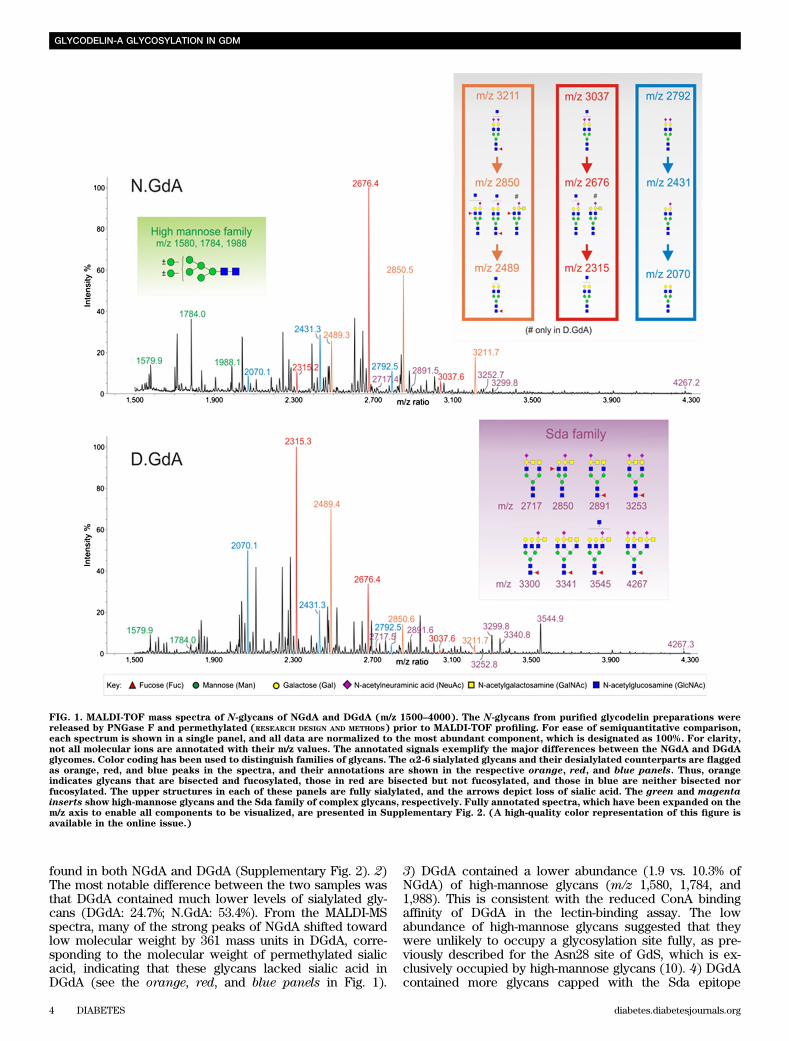
found in both NGdA and DGdA (Supplementary Fig. 2). 2)The most notable difference between the two samples wasthat DGdA contained much lower levels of sialylated gly-cans (DGdA: 24.7%; N.GdA: 53.4%). From the MALDI-MSspectra, many of the strong peaks of NGdA shifted towardlow molecular weight by 361 mass units in DGdA, corre-sponding to the molecular weight of permethylated sialicacid, indicating that these glycans lacked sialic acid inDGdA (see the orange, red, and blue panels in Fig. 1).
3) DGdA contained a lower abundance (1.9 vs. 10.3% ofNGdA) of high-mannose glycans (m/z 1,580, 1,784, and1,988). This is consistent with the reduced ConA bindingaffinity of DGdA in the lectin-binding assay. The lowabundance of high-mannose glycans suggested that theywere unlikely to occupy a glycosylation site fully, as pre-viously described for the Asn28 site of GdS, which is ex-clusively occupied by high-mannose glycans (10). 4) DGdAcontained more glycans capped with the Sda epitope
FIG. 1. MALDI-TOF mass spectra of N-glycans of NGdA and DGdA (m/z 1500–4000). The N-glycans from purified glycodelin preparations werereleased by PNGase F and permethylated (RESEARCH DESIGN AND METHODS) prior to MALDI-TOF profiling. For ease of semiquantitative comparison,each spectrum is shown in a single panel, and all data are normalized to the most abundant component, which is designated as 100%. For clarity,not all molecular ions are annotated with their m/z values. The annotated signals exemplify the major differences between the NGdA and DGdAglycomes. Color coding has been used to distinguish families of glycans. The a2-6 sialylated glycans and their desialylated counterparts are flaggedas orange, red, and blue peaks in the spectra, and their annotations are shown in the respective orange, red, and blue panels. Thus, orangeindicates glycans that are bisected and fucosylated, those in red are bisected but not fucosylated, and those in blue are neither bisected norfucosylated. The upper structures in each of these panels are fully sialylated, and the arrows depict loss of sialic acid. The green and magentainserts show high-mannose glycans and the Sda family of complex glycans, respectively. Fully annotated spectra, which have been expanded on them/z axis to enable all components to be visualized, are presented in Supplementary Fig. 2. (A high-quality color representation of this figure isavailable in the online issue.)
GLYCODELIN-A GLYCOSYLATION IN GDM
4 DIABETES diabetes.diabetesjournals.org
(e.g., m/z 2,717, 2,850, 2,891, 3,253, 3,300, 3,341, 3,545, and4,267; see the magenta panel in Fig. 1), which was a re-cently identified characteristic of the female glycodelins(11). The smaller amount of sialylated glycans in DGdAcompared with NGdA was further confirmed by the de-creased WGA-binding of DGdA in a second population ofnormal and GDM patients (Supplementary Table 2).Reduced cytotoxicity of DGdA and desialylated NGdAon human lymphocytes. Treatment with both NGdA andDGdA at concentrations of $0.01 mg/mL for 36 h signifi-cantly (P , 0.05) decreased the viability of PBMCs, thecytotoxicity of the latter was significantly (P , 0.05) lowerthan that of the former at concentrations of 0.01 and 0.1mg/mL (Table 3). NGdA, but not DGdA, at a concentrationof 0.1 mg/mL, significantly (P , 0.05) decreased the viabilityof Jurkat cells. At 1 mg/mL, the cytotoxic effect of NGdA onJurkat cells was also significantly (P , 0.05) higher thanthat of DGdA (viability: NGdA, 41.7 6 4.9%; DGdA, 79.4 68.2%). At the tested concentrations, neither NGdA norDGdA affected the viability of TEV-1 and OE-E6/E7 cells.
Compared with NGdA, the cytotoxic effect of desialylatedNGdA could only be observed at higher concentrations(PBMCs: $0.1 mg/mL; Jurkat cells: $1 mg/mL). In ad-dition, the suppression index of desialylated NGdA washigher than that of NGdA when tested at the same con-centrations.DGdA and desialylated NGdA have impaired ability toinduce cell death of lymphocytes. Treatment with 0.1mg/mL NGdA significantly (P , 0.01) decreased the viablepopulation of PBMCs from 87.0 6 5.7% to 26.9 6 2.9% inthe YoPro-PI assay (Table 4 and Supplementary Fig. 3).The corresponding decrease by DGdA was significantly(P , 0.05) smaller (from 87.0 6 5.7% to 42.4 6 15.5%),indicating a lower cytotoxic activity of DGdA on PBMCs.
Treatment with 0.1 mg/mL NGdA for 48 h significantly(P , 0.05) decreased the percentage of viable Jurkat cellsfrom 86.66 2.4% to 77.26 3.5% (Table 4 and SupplementaryFig. 3). By contrast, DGdA at the same concentration had nosignificant effect on the viability of these cells (82.96 1.8%).
A differential response (P , 0.05) of Jurkat cells to NGdAand DGdA was also observed at the concentration of1 mg/mL.
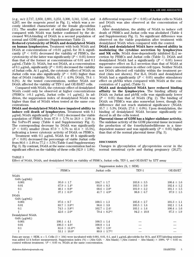
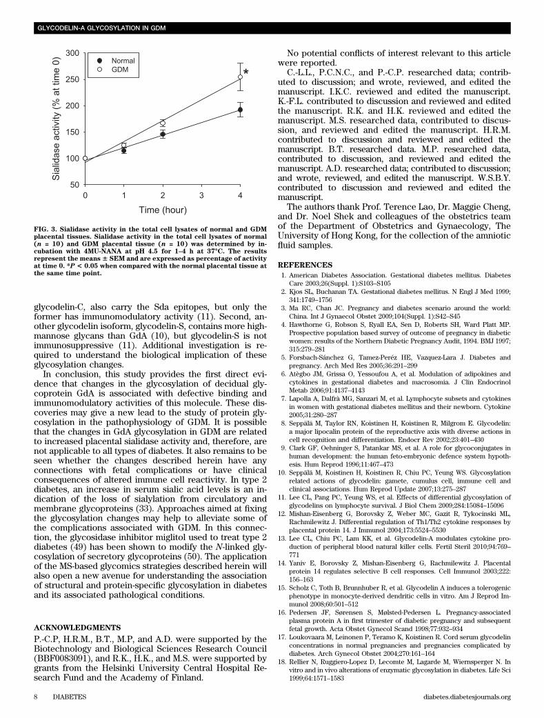
After desialylation, the ability of NGdA to induce celldeath of PBMCs and Jurkat cells was abolished (Table 4and Supplementary Fig. 3). No significant difference wasobserved on the viable population after treatment withdesialylated NGdA when compared with the control.DGdA and desialylated NGdA have reduced ability inmodulating the cytokine secretion by lymphocytesand NK cells. NGdA dose-dependently inhibited IL-2 se-cretion by PBMCs and Jurkat cells (Table 5). DGdA anddesialylated NGdA had a significantly (P , 0.05) lowersuppressive effect on IL-2 secretion than that of NGdA atthe same concentration in either cell types. Neither NGdAnor DGdA affected cell viability within the treatment pe-riod (data not shown). For IL-6, DGdA and desialylatedNGdA had a significantly (P , 0.05) smaller stimulatoryeffect on pbNKs when compared with NGdA at the con-centration of 1 mg/mL (Table 5).DGdA and desialylated NGdA have reduced bindingaffinity to the lymphocytes. The binding affinity ofDGdA on Jurkat and pbNK cells was significantly lower(P , 0.05) than that of NGdA (Fig. 2). The binding ofDGdA on PBMCs was also somewhat lower, though thedifference did not reach statistical significance (NGdA:35.7 6 5.3%; DGdA: 27.6 6 6.0%). Upon desialylation, thebinding of desialylated NGdA became significantly re-duced in all the cells tested.Placental tissue of GDM has a higher sialidase activity.The sialidase activity of the GDM placental tissue increasedthe production of the 4-methylumbelliferone in a time-dependent manner and was significantly (P , 0.05) higherthan that of the normal placental tissue (Fig. 3).
DISCUSSION
Changes in glycosylation of glycoproteins occur in thenormal menstrual cycle and during pregnancy (26,27).
TABLE 3Effect of NGdA, DGdA, and desialylated NGdA on viability of PBMCs, Jurkat cells, TEV-1, and OE-E6/E7 by XTT assay
Suppression index (SI 6 SEM)
PBMCs Jurkat cells TEV-1 OE-E6/E7
NGdAGdA (mg/mL)0.001 95.0 6 1.7 104.7 6 1.7 103.8 6 3.5 100.4 6 1.60.01 67.9 6 1.4* 93.0 6 8.3 103.5 6 3.0 102.4 6 1.20.1 40.1 6 0.8* 68.6 6 2.8* 101.0 6 2.2 101.1 6 1.51 37.1 6 0.5* 41.7 6 4.9* 98.8 6 6.7 97.9 6 1.5
DGdAGdA (mg/mL)0.001 97.6 6 8.7 100.5 6 1.3 105.8 6 2.7 101.3 6 1.10.01 83.7 6 3.8*† 96.6 6 3.8 105.5 6 1.6 102.1 6 1.40.1 74.5 6 3.8*† 102.7 6 2.4† 103.1 6 1.6 100.4 6 1.61 42.6 6 2.0* 79.4 6 8.2*† 94.2 6 10.8 97.3 6 1.9
Desialylated NGdAGdA (mg/mL)0.001 100.1 6 4.1 100.5 6 1.4 — —
0.01 100.3 6 4.0† 100.8 6 1.4 — —
0.1 81.6 6 11.6*† 98.7 6 1.9† — —
1 53.1 6 10.0* 89.8 6 3.7*† — —
Data are mean 6 SEM, n = 5. Cells (3 3 104) were incubated with 0.001, 0.01, 0.1, and 1 mg/mL glycodelin for 36 h, and XTT-labeling mixturewas added 12 h before measurement. Suppression index (%) = (Abs GdA 2 Abs blank) / (Abs control 2 Abs blank) 3 100%. *P , 0.05 vs.control without treatment. †P , 0.05 vs. NGdA at the same concentration.
C.-L. LEE AND ASSOCIATES
diabetes.diabetesjournals.org DIABETES 5

Altered glycosylation of glycoproteins and glycolipids oc-cur in diabetes, cancer, AIDS, Alzheimer’s disease, andinflammatory diseases (28,29). Two observations in thisstudy demonstrate for the first time changes in glycosyla-tion of GdA in GDM. First, the binding affinities of DGdA toConA and WGA were lower than that of NGdA. Second,glycomics analyses of the N-glycans revealed substantivequantitative and qualitative differences in the glycan struc-tures between NGdA and DGdA. The main quantitative dif-ference is the smaller amount of a2-6 sialylated glycansin DGdA. An interesting qualitative difference is that mostof the major sialylated glycans in NGdA appear as non-sialylated in DGdA.
Sialic acid levels on glycoproteins are regulated bysialyltransferases during their cellular biosynthesis and,in some instances, by sialidase(s) after secretion fromcells. Decreases in sialyltransferase (30) and increases insialidase activities (30,31) as well as changes in other gly-cosidase activities (32) have been reported in humans andanimals suffering from diabetes. These findings are in ac-cordance with the increased free sialic acid level in theserum of type 2 diabetes (33). Human endometrial tissuesexpresses both sialidase and sialyltransferase (27,34). Inthis study, placental sialidase activity is higher in GDM thanin normal pregnancy, consistent with the reported abnormalcarbohydrate metabolism in the placental-decidual unit ofGDM pregnancy (35). Therefore, it is not surprising that thealtered carbohydrate metabolism in GDM leads to the pro-duction of GdA with reduced sialic acid content.
Sialic acid is usually the terminal monosaccharide inhuman N-glycans, and it affects the conformation, binding,and biological activities of glycoproteins (29). The relativeamount of some sialic acid–containing glycoproteins inamniotic fluid (36) and maternal plasma (37) is elevatedduring pregnancy and increases with advancing gestation.The results of this study emphasize the role of sialylationin pregnancy; a decrease in sialic acid content reduces theimmunomodulatory activities of DGdA. Indeed, the abun-dance of sialic acid in different glycodelin isoforms corre-lates with their apoptosis-inducing activity on lymphocytes(11). Consistently, DGdA with less sialylated glycans hasreduced apoptosis-inducing activity on lymphocytes, sup-porting the importance of sialic acid in mediating the im-munomodulatory function of GdA.
In a normal pregnancy, selective deletion of T-cellsoccurs at the fetomaternal interface throughout gestation(38). Suppression of the response of maternal lymphocytesto fetal alloantigen is necessary for fetal survival (39). GdAmodulates the T-cell population by inducing apoptosisof T-cells (11) and expression of Fas in Th-1 lymphocytes(40). The reduced ability of DGdA to induce T-cell apo-ptosis could be, at least in part, responsible for the ob-served increase of lymphocytes in the GDM patients (7,41).The involvement of carbohydrate metabolism in alterationof the T-cell population in GDM is reflected by the re-duction of T-cells after insulin treatment of these women(7,41).
Changes in GdA glycosylation may also lead to in-appropriate cytokine profiles in GDM. A shift in cytokineprofile in women with GDM has been documented (6,7).Whether this is related to an increased risk of complica-tions in GDM remains to be investigated. Significantly,T-cells treated with DGdA produce more IL-2 than thosetreated with NGdA. Excessive production of Th-1 cyto-kines including IL-2 would mediate rejection of the fetalsemiallograft (42). On the other hand, DGdA has impairedT
ABLE
4Effec
tof
NGdA
,DGdA
,an
dde
sialylated
NGdA
onthece
llde
athof
PBMCsan
dJurkat
cells
Con
trol
NGdA
(mg/mL)
DGdA
(mg/mL)
DesialylatedNGdA
(mg/mL)
0.01
0.1
10.01
0.1
10.01
0.1
1
PBMCs(%
)Viable
87.0
65.7
44.3
615
.4*
26.9
62.9*
20.8
63.2*
57.1
61.3*†
42.4
615
.5*†
23.7
65.4*
76.7
67.7
75.7
68.4†
71.3
68.3†
Nec
rosis
8.06
4.0
42.5
613
.7*
59.9
61.8*
64.7
63.4*
25.7
61.4*
41.2
616
.2*
62.0
65.5*
15.0
64.7†
15.8
65.6†
19.6
65.7†
Apo
ptosis
4.86
1.3
12.8
61.8*
12.9
61.3*
14.1
60.6*
16.5
60.3*†
15.8
61.6*
13.8
61.2*
7.46
2.7†
7.66
2.7†
8.16
2.5†
Jurkat
cells
(%)
Viable
86.6
62.4
77.0
63.3*
77.2
63.5*
61.9
611
.8*
82.0
62.6
82.9
61.8†
77.0
64.1*†
83.9
61.5
83.0
61.4†
83.9
61.3†
Nec
rosis
6.66
1.8
13.2
62.2*
16.7
62.8*
34.9
611
.5*
10.2
60.9*
9.26
0.7
15.8
62.4*†
9.76
1.2*
10.1
60.5*
9.06
1.2†
Apo
ptosis
4.26
1.5
8.76
1.2*
5.56
0.8
2.76
0.7
7.16
1.8
7.26
1.2*
6.66
1.9††
6.36
0.4
6.86
1.1
7.06
1.0*†
Dataaremea
ns6
SEM,n
=5.
PBMCan
dJurkat
cells
(53
105)wereincu
batedwith0–
1mg/mLglyc
odelinsfor48
h.Viable,
necrotic,a
ndap
optoticce
llswereiden
tified
andqu
antified
bybiva
riateYo-Pro-1/PIflow
cytometry.C
ellswitho
utstainwereco
untedas
viab
lece
lls.C
ellslabe
ledwithYo-Pro-1
only
wereco
untedas
apop
toticce
lls.C
ellslabe
ledwithbo
thYo-Pro-1
and
PIwas
coun
tedas
necrotic
cells.*P
,0.05
vs.co
ntrolwitho
uttrea
tmen
t.†P,
0.05
vs.NGdA
atthesameco
ncen
tration.
GLYCODELIN-A GLYCOSYLATION IN GDM
6 DIABETES diabetes.diabetesjournals.org
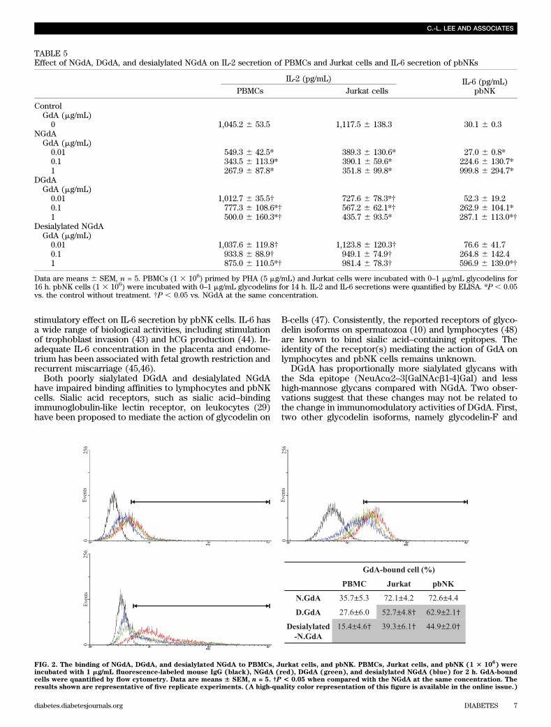
stimulatory effect on IL-6 secretion by pbNK cells. IL-6 hasa wide range of biological activities, including stimulationof trophoblast invasion (43) and hCG production (44). In-adequate IL-6 concentration in the placenta and endome-trium has been associated with fetal growth restriction andrecurrent miscarriage (45,46).
Both poorly sialylated DGdA and desialylated NGdAhave impaired binding affinities to lymphocytes and pbNKcells. Sialic acid receptors, such as sialic acid–bindingimmunoglobulin-like lectin receptor, on leukocytes (29)have been proposed to mediate the action of glycodelin on
B-cells (47). Consistently, the reported receptors of glyco-delin isoforms on spermatozoa (10) and lymphocytes (48)are known to bind sialic acid–containing epitopes. Theidentity of the receptor(s) mediating the action of GdA onlymphocytes and pbNK cells remains unknown.
DGdA has proportionally more sialylated glycans withthe Sda epitope (NeuAca2–3[GalNAcb1-4]Gal) and lesshigh-mannose glycans compared with NGdA. Two obser-vations suggest that these changes may not be related tothe change in immunomodulatory activities of DGdA. First,two other glycodelin isoforms, namely glycodelin-F and
FIG. 2. The binding of NGdA, DGdA, and desialylated NGdA to PBMCs, Jurkat cells, and pbNK. PBMCs, Jurkat cells, and pbNK (1 3 106) were
incubated with 1 mg/mL fluorescence-labeled mouse IgG (black), NGdA (red), DGdA (green), and desialylated NGdA (blue) for 2 h. GdA-boundcells were quantified by flow cytometry. Data are means 6 SEM, n = 5. †P < 0.05 when compared with the NGdA at the same concentration. Theresults shown are representative of five replicate experiments. (A high-quality color representation of this figure is available in the online issue.)
TABLE 5Effect of NGdA, DGdA, and desialylated NGdA on IL-2 secretion of PBMCs and Jurkat cells and IL-6 secretion of pbNKs
IL-2 (pg/mL) IL-6 (pg/mL)PBMCs Jurkat cells pbNK
ControlGdA (mg/mL)0 1,045.2 6 53.5 1,117.5 6 138.3 30.1 6 0.3
NGdAGdA (mg/mL)0.01 549.3 6 42.5* 389.3 6 130.6* 27.0 6 0.8*0.1 343.5 6 113.9* 390.1 6 59.6* 224.6 6 130.7*1 267.9 6 87.8* 351.8 6 99.8* 999.8 6 294.7*
DGdAGdA (mg/mL)0.01 1,012.7 6 35.5† 727.6 6 78.3*† 52.3 6 19.20.1 777.3 6 108.6*† 567.2 6 62.1*† 262.9 6 104.1*1 500.0 6 160.3*† 435.7 6 93.5* 287.1 6 113.0*†
Desialylated NGdAGdA (mg/mL)0.01 1,037.6 6 119.8† 1,123.8 6 120.3† 76.6 6 41.70.1 933.8 6 88.9† 949.1 6 74.9† 264.8 6 142.41 875.0 6 110.5*† 981.4 6 78.3† 596.9 6 139.0*†
Data are means 6 SEM, n = 5. PBMCs (1 3 106) primed by PHA (5 mg/mL) and Jurkat cells were incubated with 0–1 mg/mL glycodelins for16 h. pbNK cells (1 3 106) were incubated with 0–1 mg/mL glycodelins for 14 h. IL-2 and IL-6 secretions were quantified by ELISA. *P , 0.05vs. the control without treatment. †P , 0.05 vs. NGdA at the same concentration.
C.-L. LEE AND ASSOCIATES
diabetes.diabetesjournals.org DIABETES 7
glycodelin-C, also carry the Sda epitopes, but only theformer has immunomodulatory activity (11). Second, an-other glycodelin isoform, glycodelin-S, contains more high-mannose glycans than GdA (10), but glycodelin-S is notimmunosuppressive (11). Additional investigation is re-quired to understand the biological implication of theseglycosylation changes.
In conclusion, this study provides the first direct evi-dence that changes in the glycosylation of decidual gly-coprotein GdA is associated with defective binding andimmunomodulatory activities of this molecule. These dis-coveries may give a new lead to the study of protein gly-cosylation in the pathophysiology of GDM. It is possiblethat the changes in GdA glycosylation in GDM are relatedto increased placental sialidase activity and, therefore, arenot applicable to all types of diabetes. It also remains to beseen whether the changes described herein have anyconnections with fetal complications or have clinicalconsequences of altered immune cell reactivity. In type 2diabetes, an increase in serum sialic acid levels is an in-dication of the loss of sialylation from circulatory andmembrane glycoproteins (33). Approaches aimed at fixingthe glycosylation changes may help to alleviate some ofthe complications associated with GDM. In this connec-tion, the glycosidase inhibitor miglitol used to treat type 2diabetes (49) has been shown to modify the N-linked gly-cosylation of secretory glycoproteins (50). The applicationof the MS-based glycomics strategies described herein willalso open a new avenue for understanding the associationof structural and protein-specific glycosylation in diabetesand its associated pathological conditions.
ACKNOWLEDGMENTS
P.-C.P, H.R.M., B.T., M.P, and A.D. were supported by theBiotechnology and Biological Sciences Research Council(BBF0083091), and R.K., H.K., and M.S. were supported bygrants from the Helsinki University Central Hospital Re-search Fund and the Academy of Finland.
No potential conflicts of interest relevant to this articlewere reported.
C.-L.L., P.C.N.C., and P.-C.P. researched data; contrib-uted to discussion; and wrote, reviewed, and edited themanuscript. I.K.C. reviewed and edited the manuscript.K.-F.L. contributed to discussion and reviewed and editedthe manuscript. R.K. and H.K. reviewed and edited themanuscript. M.S. researched data, contributed to discus-sion, and reviewed and edited the manuscript. H.R.M.contributed to discussion and reviewed and edited themanuscript. B.T. researched data. M.P. researched data,contributed to discussion, and reviewed and edited themanuscript. A.D. researched data; contributed to discussion;and wrote, reviewed, and edited the manuscript. W.S.B.Y.contributed to discussion and reviewed and edited themanuscript.
The authors thank Prof. Terence Lao, Dr. Maggie Cheng,and Dr. Noel Shek and colleagues of the obstetrics teamof the Department of Obstetrics and Gynaecology, TheUniversity of Hong Kong, for the collection of the amnioticfluid samples.
REFERENCES
1. American Diabetes Association. Gestational diabetes mellitus. DiabetesCare 2003;26(Suppl. 1):S103–S105
2. Kjos SL, Buchanan TA. Gestational diabetes mellitus. N Engl J Med 1999;341:1749–1756
3. Ma RC, Chan JC. Pregnancy and diabetes scenario around the world:China. Int J Gynaecol Obstet 2009;104(Suppl. 1):S42–S45
4. Hawthorne G, Robson S, Ryall EA, Sen D, Roberts SH, Ward Platt MP.Prospective population based survey of outcome of pregnancy in diabeticwomen: results of the Northern Diabetic Pregnancy Audit, 1994. BMJ 1997;315:279–281
5. Forsbach-Sánchez G, Tamez-Peréz HE, Vazquez-Lara J. Diabetes andpregnancy. Arch Med Res 2005;36:291–299
6. Atègbo JM, Grissa O, Yessoufou A, et al. Modulation of adipokines andcytokines in gestational diabetes and macrosomia. J Clin EndocrinolMetab 2006;91:4137–4143
7. Lapolla A, Dalfrà MG, Sanzari M, et al. Lymphocyte subsets and cytokinesin women with gestational diabetes mellitus and their newborn. Cytokine2005;31:280–287
8. Seppälä M, Taylor RN, Koistinen H, Koistinen R, Milgrom E. Glycodelin:a major lipocalin protein of the reproductive axis with diverse actions incell recognition and differentiation. Endocr Rev 2002;23:401–430
9. Clark GF, Oehninger S, Patankar MS, et al. A role for glycoconjugates inhuman development: the human feto-embryonic defence system hypoth-esis. Hum Reprod 1996;11:467–473
10. Seppälä M, Koistinen H, Koistinen R, Chiu PC, Yeung WS. Glycosylationrelated actions of glycodelin: gamete, cumulus cell, immune cell andclinical associations. Hum Reprod Update 2007;13:275–287
11. Lee CL, Pang PC, Yeung WS, et al. Effects of differential glycosylation ofglycodelins on lymphocyte survival. J Biol Chem 2009;284:15084–15096
12. Mishan-Eisenberg G, Borovsky Z, Weber MC, Gazit R, Tykocinski ML,Rachmilewitz J. Differential regulation of Th1/Th2 cytokine responses byplacental protein 14. J Immunol 2004;173:5524–5530
13. Lee CL, Chiu PC, Lam KK, et al. Glycodelin-A modulates cytokine pro-duction of peripheral blood natural killer cells. Fertil Steril 2010;94:769–771
14. Yaniv E, Borovsky Z, Mishan-Eisenberg G, Rachmilewitz J. Placentalprotein 14 regulates selective B cell responses. Cell Immunol 2003;222:156–163
15. Scholz C, Toth B, Brunnhuber R, et al. Glycodelin A induces a tolerogenicphenotype in monocyte-derived dendritic cells in vitro. Am J Reprod Im-munol 2008;60:501–512
16. Pedersen JF, Sørensen S, Mølsted-Pedersen L. Pregnancy-associatedplasma protein A in first trimester of diabetic pregnancy and subsequentfetal growth. Acta Obstet Gynecol Scand 1998;77:932–934
17. Loukovaara M, Leinonen P, Teramo K, Koistinen R. Cord serum glycodelinconcentrations in normal pregnancies and pregnancies complicated bydiabetes. Arch Gynecol Obstet 2004;270:161–164
18. Rellier N, Ruggiero-Lopez D, Lecomte M, Lagarde M, Wiernsperger N. Invitro and in vivo alterations of enzymatic glycosylation in diabetes. Life Sci1999;64:1571–1583
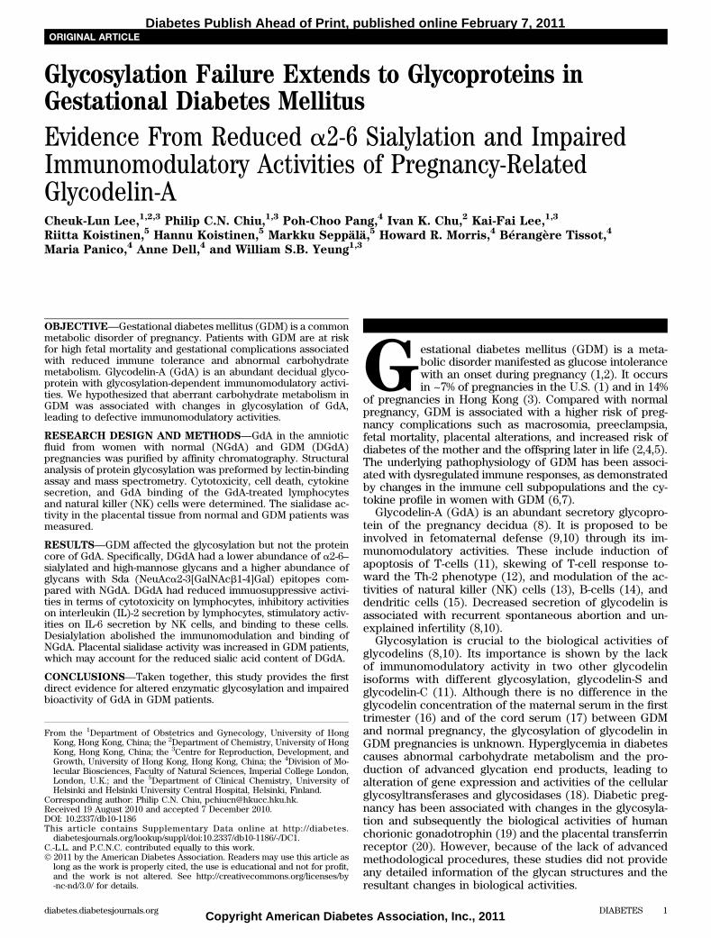
FIG. 3. Sialidase activity in the total cell lysates of normal and GDMplacental tissues. Sialidase activity in the total cell lysates of normal(n = 10) and GDM placental tissue (n = 10) was determined by in-cubation with 4MU-NANA at pH 4.5 for 1–4 h at 37°C. The resultsrepresent the means6 SEM and are expressed as percentage of activityat time 0. *P < 0.05 when compared with the normal placental tissue atthe same time point.
GLYCODELIN-A GLYCOSYLATION IN GDM
8 DIABETES diabetes.diabetesjournals.org
19. Elliott MM, Kardana A, Lustbader JW, Cole LA. Carbohydrate and peptidestructure of the alpha- and beta-subunits of human chorionic gonadotropinfrom normal and aberrant pregnancy and choriocarcinoma. Endocrine1997;7:15–32
20. Georgieff MK, Petry CD, Mills MM, McKay H, Wobken JD. IncreasedN-glycosylation and reduced transferrin-binding capacity of transferrinreceptor isolated from placentae of diabetic women. Placenta 1997;18:563–568
21. Riittinen L, Närvänen O, Virtanen I, Seppälä M. Monoclonal antibodiesagainst endometrial protein PP14 and their use for purification and ra-dioimmunoassay of PP14. J Immunol Methods 1991;136:85–90
22. Jang-Lee J, North SJ, Sutton-Smith M, et al. Glycomic profiling of cells andtissues by mass spectrometry: fingerprinting and sequencing methodolo-gies. Methods Enzymol 2006;415:59–86
23. Sutton-Smith M, Dell A. Analysis of carbohydrates/glycoproteins by massspectrometry. In Cell Biology: A Laboratory Handbook. 3rd ed. Celis JE,Ed. Boston, Elsevier Academic, 2006, p. 415–425
24. Ceroni A, Maass K, Geyer H, Geyer R, Dell A, Haslam SM. Glyco-Workbench: a tool for the computer-assisted annotation of mass spectra ofglycans. J Proteome Res 2008;7:1650–1659
25. Lam KK, Chiu PC, Chung MK, et al. Glycodelin-A as a modulator of tro-phoblast invasion. Hum Reprod 2009;24:2093–2103
26. Carson DD, Farrar JD, Laidlaw J, Wright DA. Selective activation of theN-glycosylation apparatus in uteri by estrogen. J Biol Chem 1990;265:2947–2955
27. Kaneko Y, Yamamoto H, Colley KJ, Moskal JR. Expression of Gal beta1,4GlcNAc alpha 2,6-sialyltransferase and alpha 2,6-linked sialoglyco-conjugates in normal human and rat tissues. J Histochem Cytochem 1995;43:945–954
28. Oppenheimer SB, Alvarez M, Nnoli J. Carbohydrate-based experimentaltherapeutics for cancer, HIV/AIDS and other diseases. Acta Histochem2008;110:6–13
29. Varki A. Essentials of Glycobiology. Cold Spring Harbor, NY, Cold SpringHarbor Laboratory Press, 2009
30. Cohen-Forterre L, Andre J, Mozere G, Peyroux J, Sternberg M. Kidneysialidase and sialyltransferase activities in spontaneously and experimen-tally diabetic rats. Influence of insulin and sorbinil treatments. BiochemPharmacol 1990;40:507–513
31. Chari SN, Nath N. Sialic acid content and sialidase activity of poly-morphonuclear leucocytes in diabetes mellitus. Am J Med Sci 1984;288:18–20
32. Serrano MA, Reglero A, Cabezas JA, et al. Serum glycosidases in diabetesmellitus in relation to the retinopathy and to the length of the disease. ClinChim Acta 1983;132:23–27
33. Chen J, Gall MA, Yokoyama H, Jensen JS, Deckert M, Parving HH. Raisedserum sialic acid concentration in NIDDM patients with and without di-abetic nephropathy. Diabetes Care 1996;19:130–134
34. Ganguly S, Sarkar D, Ghosh JJ. Sialic acid and sialidase activity in humanendometrial tissue, uterine fluid and plasma under different conditions ofuterine dysfunction. Acta Endocrinol (Copenh) 1976;81:574–579
35. Jawerbaum A, Roselló Catafau J, Gonzalez ET, et al. Glucose metabolism,triglyceride and glycogen levels, as well as eicosanoid production in
isolated uterine strips and in embryos in a rat model of non-insulin-dependent diabetes mellitus during pregnancy. Prostaglandins 1994;47:81–96
36. Orczyk-Pawiłowicz M, Floria�nski J, Zalewski J, Katnik-Prastowska I. Rel-ative amounts of sialic acid and fucose of amniotic fluid glycoconjugates inrelation to pregnancy age. Glycoconj J 2005;22:433–442
37. Rajan R, Sheth AR, Rao SS. Sialic acid, sialyltransferase and neuramini-dase levels in maternal plasma, urine and lymphocytes during pregnancyand post-partum period—a longitudinal study in women. Eur J ObstetGynecol Reprod Biol 1983;16:37–46
38. Jiang SP, Vacchio MS. Multiple mechanisms of peripheral T cell toleranceto the fetal “allograft”. J Immunol 1998;160:3086–3090
39. Koch CA, Platt JL. T cell recognition and immunity in the fetus and mother.Cell Immunol 2007;248:12–17
40. Lee CL, Chiu PC, Lam KK, et al. Differential actions of glycodelin-A on Th-1and Th-2 cells: a paracrine mechanism that could produce the Th-2 dom-inant environment during pregnancy. Hum Reprod. In press.
41. Lapolla A, Betterle C, Sanzari M, et al. An immunological and genetic studyof patients with gestational diabetes mellitus. Acta Diabetol 1996;33:139–144
42. Saito S. Cytokine cross-talk between mother and the embryo/placenta.J Reprod Immunol 2001;52:15–33
43. Fitzgerald JS, Poehlmann TG, Schleussner E, Markert UR. Trophoblastinvasion: the role of intracellular cytokine signalling via signal transducerand activator of transcription 3 (STAT3). Hum Reprod Update 2008;14:335–344
44. Nishino E, Matsuzaki N, Masuhiro K, et al. Trophoblast-derivedinterleukin-6 (IL-6) regulates human chorionic gonadotropin releasethrough IL-6 receptor on human trophoblasts. J Clin Endocrinol Metab1990;71:436–441
45. Street ME, Seghini P, Fieni S, et al. Changes in interleukin-6 and IGFsystem and their relationships in placenta and cord blood in newbornswith fetal growth restriction compared with controls. Eur J Endocrinol2006;155:567–574
46. Jasper MJ, Tremellen KP, Robertson SA. Reduced expression of IL-6 andIL-1alpha mRNAs in secretory phase endometrium of women with re-current miscarriage. J Reprod Immunol 2007;73:74–84
47. Dell A, Morris HR, Easton RL, et al. Structural analysis of the oligo-saccharides derived from glycodelin, a human glycoprotein with potentimmunosuppressive and contraceptive activities. J Biol Chem 1995;270:24116–24126
48. Ish-Shalom E, Gargir A, André S, et al. alpha2,6-Sialylation promotesbinding of placental protein 14 via its Ca2+-dependent lectin activity: in-sights into differential effects on CD45RO and CD45RA T cells. Glycobi-ology 2006;16:173–183
49. Dwek RA, Butters TD, Platt FM, Zitzmann N. Targeting glycosylation asa therapeutic approach. Nat Rev Drug Discov 2002;1:65–75
50. Ludolph D, Gross V, Katz NR, et al. Effect of the alpha-glucosidase in-hibitor N-hydroxyethyl-1-deoxynojirimycin (Bay m 1099) on the bio-synthesis of liver secretory glycoproteins. Biochem Pharmacol 1989;38:2479–2486
C.-L. LEE AND ASSOCIATES
diabetes.diabetesjournals.org DIABETES 9