European Journal of Pharmacology - COnnecting … · [email protected] (C. de Pablo),...
11
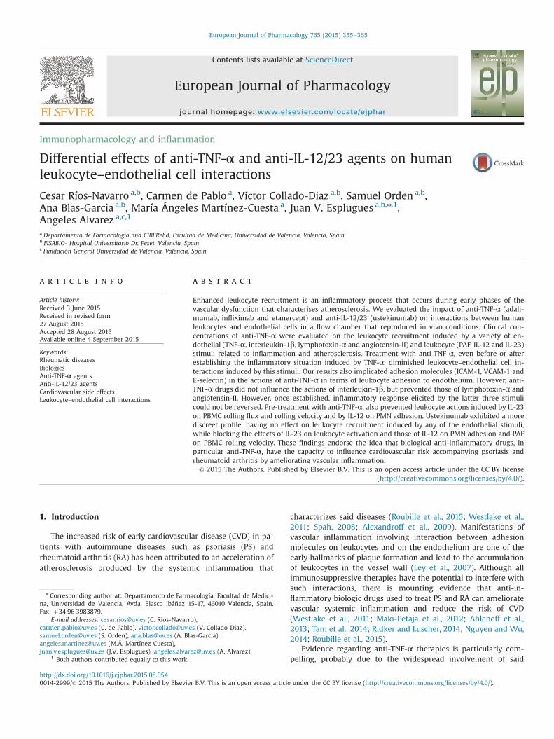
Immunopharmacology and inflammation Differential effects of anti-TNF-α and anti-IL-12/23 agents on human leukocyte–endothelial cell interactions Cesar Ríos-Navarro a,b , Carmen de Pablo a , Víctor Collado-Diaz a,b , Samuel Orden a,b , Ana Blas-Garcia a,b , María Ángeles Martínez-Cuesta a , Juan V. Esplugues a,b,n,1 , Angeles Alvarez a,c,1 a Departamento de Farmacología and CIBERehd, Facultad de Medicina, Universidad de Valencia, Valencia, Spain b FISABIO- Hospital Universitario Dr. Peset, Valencia, Spain c Fundación General Universidad de Valencia, Valencia, Spain article info Article history: Received 3 June 2015 Received in revised form 27 August 2015 Accepted 28 August 2015 Available online 4 September 2015 Keywords: Rheumatic diseases Biologics Anti-TNF-α agents Anti-IL-12/23 agents Cardiovascular side effects Leukocyte–endothelial cell interactions abstract Enhanced leukocyte recruitment is an inflammatory process that occurs during early phases of the vascular dysfunction that characterises atherosclerosis. We evaluated the impact of anti-TNF-α (adali- mumab, infliximab and etanercept) and anti-IL-12/23 (ustekinumab) on interactions between human leukocytes and endothelial cells in a flow chamber that reproduced in vivo conditions. Clinical con- centrations of anti-TNF-α were evaluated on the leukocyte recruitment induced by a variety of en- dothelial (TNF-α, interleukin-1β, lymphotoxin-α and angiotensin-II) and leukocyte (PAF, IL-12 and IL-23) stimuli related to inflammation and atherosclerosis. Treatment with anti-TNF-α, even before or after establishing the inflammatory situation induced by TNF-α, diminished leukocyte–endothelial cell in- teractions induced by this stimuli. Our results also implicated adhesion molecules (ICAM-1, VCAM-1 and E-selectin) in the actions of anti-TNF-α in terms of leukocyte adhesion to endothelium. However, anti- TNF-α drugs did not influence the actions of interleukin-1β, but prevented those of lymphotoxin-α and angiotensin-II. However, once established, inflammatory response elicited by the latter three stimuli could not be reversed. Pre-treatment with anti-TNF-α, also prevented leukocyte actions induced by IL-23 on PBMC rolling flux and rolling velocity and by IL-12 on PMN adhesion. Ustekinumab exhibited a more discreet profile, having no effect on leukocyte recruitment induced by any of the endothelial stimuli, while blocking the effects of IL-23 on leukocyte activation and those of IL-12 on PMN adhesion and PAF on PBMC rolling velocity. These findings endorse the idea that biological anti-inflammatory drugs, in particular anti-TNF-α, have the capacity to influence cardiovascular risk accompanying psoriasis and rheumatoid arthritis by ameliorating vascular inflammation. & 2015 The Authors. Published by Elsevier B.V. This is an open access article under the CC BY license (http://creativecommons.org/licenses/by/4.0/). 1. Introduction The increased risk of early cardiovascular disease (CVD) in pa- tients with autoimmune diseases such as psoriasis (PS) and rheumatoid arthritis (RA) has been attributed to an acceleration of atherosclerosis produced by the systemic inflammation that characterizes said diseases (Roubille et al., 2015; Westlake et al., 2011; Spah, 2008; Alexandroff et al., 2009). Manifestations of vascular inflammation involving interaction between adhesion molecules on leukocytes and on the endothelium are one of the early hallmarks of plaque formation and lead to the accumulation of leukocytes in the vessel wall (Ley et al., 2007). Although all immunosuppressive therapies have the potential to interfere with such interactions, there is mounting evidence that anti-in- flammatory biologic drugs used to treat PS and RA can ameliorate vascular systemic inflammation and reduce the risk of CVD (Westlake et al., 2011; Maki-Petaja et al., 2012; Ahlehoff et al., 2013; Tam et al., 2014; Ridker and Luscher, 2014; Nguyen and Wu, 2014; Roubille et al., 2015). Evidence regarding anti-TNF-α therapies is particularly com- pelling, probably due to the widespread involvement of said Contents lists available at ScienceDirect journal homepage: www.elsevier.com/locate/ejphar European Journal of Pharmacology http://dx.doi.org/10.1016/j.ejphar.2015.08.054 0014-2999/& 2015 The Authors. Published by Elsevier B.V. This is an open access article under the CC BY license (http://creativecommons.org/licenses/by/4.0/). n Corresponding author at: Departamento de Farmacología, Facultad de Medici- na, Universidad de Valencia, Avda. Blasco Ibáñez 15-17, 46010 Valencia, Spain. Fax: þ34 96 3983879. E-mail addresses: [email protected] (C. Ríos-Navarro), [email protected] (C. de Pablo), [email protected] (V. Collado-Diaz), [email protected] (S. Orden), [email protected] (A. Blas-Garcia), [email protected] (M.Á. Martínez-Cuesta), [email protected] (J.V. Esplugues), [email protected] (A. Alvarez). 1 Both authors contributed equally to this work. European Journal of Pharmacology 765 (2015) 355–365
-
Upload
hoangtuyen -
Category
Documents
-
view
216 -
download
0
Transcript of European Journal of Pharmacology - COnnecting … · [email protected] (C. de Pablo),...
European Journal of Pharmacology 765 (2015) 355–365
Contents lists available at ScienceDirect
European Journal of Pharmacology
http://d0014-29
n Corrna, UniFax: þ3
E-mcarmensamuel.angelesjuan.v.e
1 Bo
journal homepage: www.elsevier.com/locate/ejphar
Immunopharmacology and inflammation
Differential effects of anti-TNF-α and anti-IL-12/23 agents on humanleukocyte–endothelial cell interactions
Cesar Ríos-Navarro a,b, Carmen de Pablo a, Víctor Collado-Diaz a,b, Samuel Orden a,b,Ana Blas-Garcia a,b, María Ángeles Martínez-Cuesta a, Juan V. Esplugues a,b,n,1,Angeles Alvarez a,c,1
a Departamento de Farmacología and CIBERehd, Facultad de Medicina, Universidad de Valencia, Valencia, Spainb FISABIO- Hospital Universitario Dr. Peset, Valencia, Spainc Fundación General Universidad de Valencia, Valencia, Spain
a r t i c l e i n f o
Article history:Received 3 June 2015Received in revised form27 August 2015Accepted 28 August 2015Available online 4 September 2015
Keywords:Rheumatic diseasesBiologicsAnti-TNF-α agentsAnti-IL-12/23 agentsCardiovascular side effectsLeukocyte–endothelial cell interactions
x.doi.org/10.1016/j.ejphar.2015.08.05499/& 2015 The Authors. Published by Elsevie
esponding author at: Departamento de Farmversidad de Valencia, Avda. Blasco Ibáñez 14 96 3983879.ail addresses: [email protected] (C. Rí[email protected] (C. de Pablo), [email protected]@uv.es (S. Orden), [email protected] (A. [email protected] (M.Á. Martínez-Cuesta),[email protected] (J.V. Esplugues), angeles.alvarth authors contributed equally to this work.
a b s t r a c t
Enhanced leukocyte recruitment is an inflammatory process that occurs during early phases of thevascular dysfunction that characterises atherosclerosis. We evaluated the impact of anti-TNF-α (adali-mumab, infliximab and etanercept) and anti-IL-12/23 (ustekinumab) on interactions between humanleukocytes and endothelial cells in a flow chamber that reproduced in vivo conditions. Clinical con-centrations of anti-TNF-α were evaluated on the leukocyte recruitment induced by a variety of en-dothelial (TNF-α, interleukin-1β, lymphotoxin-α and angiotensin-II) and leukocyte (PAF, IL-12 and IL-23)stimuli related to inflammation and atherosclerosis. Treatment with anti-TNF-α, even before or afterestablishing the inflammatory situation induced by TNF-α, diminished leukocyte–endothelial cell in-teractions induced by this stimuli. Our results also implicated adhesion molecules (ICAM-1, VCAM-1 andE-selectin) in the actions of anti-TNF-α in terms of leukocyte adhesion to endothelium. However, anti-TNF-α drugs did not influence the actions of interleukin-1β, but prevented those of lymphotoxin-α andangiotensin-II. However, once established, inflammatory response elicited by the latter three stimulicould not be reversed. Pre-treatment with anti-TNF-α, also prevented leukocyte actions induced by IL-23on PBMC rolling flux and rolling velocity and by IL-12 on PMN adhesion. Ustekinumab exhibited a morediscreet profile, having no effect on leukocyte recruitment induced by any of the endothelial stimuli,while blocking the effects of IL-23 on leukocyte activation and those of IL-12 on PMN adhesion and PAFon PBMC rolling velocity. These findings endorse the idea that biological anti-inflammatory drugs, inparticular anti-TNF-α, have the capacity to influence cardiovascular risk accompanying psoriasis andrheumatoid arthritis by ameliorating vascular inflammation.& 2015 The Authors. Published by Elsevier B.V. This is an open access article under the CC BY license
(http://creativecommons.org/licenses/by/4.0/).
1. Introduction
The increased risk of early cardiovascular disease (CVD) in pa-tients with autoimmune diseases such as psoriasis (PS) andrheumatoid arthritis (RA) has been attributed to an acceleration ofatherosclerosis produced by the systemic inflammation that
r B.V. This is an open access article
acología, Facultad de Medici-5-17, 46010 Valencia, Spain.
o),s (V. Collado-Diaz),s-Garcia),
[email protected] (A. Alvarez).
characterizes said diseases (Roubille et al., 2015; Westlake et al.,2011; Spah, 2008; Alexandroff et al., 2009). Manifestations ofvascular inflammation involving interaction between adhesionmolecules on leukocytes and on the endothelium are one of theearly hallmarks of plaque formation and lead to the accumulationof leukocytes in the vessel wall (Ley et al., 2007). Although allimmunosuppressive therapies have the potential to interfere withsuch interactions, there is mounting evidence that anti-in-flammatory biologic drugs used to treat PS and RA can amelioratevascular systemic inflammation and reduce the risk of CVD(Westlake et al., 2011; Maki-Petaja et al., 2012; Ahlehoff et al.,2013; Tam et al., 2014; Ridker and Luscher, 2014; Nguyen and Wu,2014; Roubille et al., 2015).
Evidence regarding anti-TNF-α therapies is particularly com-pelling, probably due to the widespread involvement of said
under the CC BY license (http://creativecommons.org/licenses/by/4.0/).
C. Ríos-Navarro et al. / European Journal of Pharmacology 765 (2015) 355–365356
cytokine in a wide variety of vascular inflammatory responses(Elliott et al., 1994; Westlake et al., 2011; Roubille et al., 2015).However, theories concerning the mechanisms implicated are ra-ther spurious, as these drugs have been related both, with heartfailure and a reduction in inflammation and plaque formation.There is also a case for analyzing anti-IL-12/23 agents given thepotential implication of both these cytokines in plaque formationand their frequent use in the treatment of PS as alternatives toanti-TNF-α; however, some reports have related them with anincrease in adverse cardiovascular events, which calls into ques-tion their validity (Ahlehoff et al., 2013; Alexandroff et al., 2009).
The aim of the present study was to characterize the effects ofthe most widely used anti-TNF-α-adalimumab (ADA, fully human),infliximab (INF, chimeric origin with human constant and murinevariable regions) and etanercept [ETA, human p75 region of tumornecrosis factor-α (TNF-α) receptor linked to the fragment crys-tallisable (Fc) region of human IgG1]-on leukocyte–endothelial cellinteractions and endothelial adhesion molecules induced by var-ious stimuli implicated in PS and RA, and to compare the effectswith those of the anti-p40 subunit of IL-12/23 ustekinumab (UST)(Nestorov, 2005; Tracey et al., 2008; Koutruba et al., 2010; Priceet al., 2007; Singh et al., 2011).
2. Materials and methods
2.1. Human umbilical vein endothelial cells (HUVEC) cell culture
HUVEC were harvested from freshly obtained umbilical cordsby collagenase treatment, as previously described (De Pablo et al.,2010). In short, umbilical cord veins were rinsed of blood productswith warm phosphate-buffered saline (PBS), after which the veinwas filled with collagenase (1 mg/ml) for 17 min at 37 °C. Thecords were then gently massaged to ensure detachment of en-dothelial cells from the vessel wall. The digest was collected,centrifuged and pelleted. The pellet was resuspended in en-dothelial cell growth medium (EGM-2) inside T25 culture flasks inwhich cells were cultured until confluence. After reaching con-fluence, primary cultures were detached with trypsin and trans-ferred to 6-well plate culture dishes. Passage 1 of these primarycultures was subsequently employed. For adhesion studies, HUVECwere cultured on fibronectin (5 μg/ml)-coated 25-mm plasticcoverslips until confluent (∼48 h).
2.2. Leukocyte isolation
Polymorphonuclear (PMN) or peripheral blood mononuclear(PBMC) cells were isolated from whole blood drawn from healthyvolunteers and anticoagulated with sodium citrate (De Pablo et al.,2010). Samples were incubated with dextran (3%) for 45 min.PBMC and PMN in the supernatant were separated by gradientdensity centrifugation (250 g, 25 min) with Ficoll-Paque™ Plus.After red blood cell lysis, leukocytes were washed (HBSS withoutCa2þ or Mg2þ) and resuspended in complete RPMI media. Themedical ethical committee of the Hospital Clínico Universitario deValencia approved the study and all participating patients pro-vided written informed consent.
2.3. Adhesion assay under flow conditions
The parallel plate flow chamber in vitro model has been de-scribed in detail previously (De Pablo et al., 2012; Cai et al., 2006).For adhesion assays, coverslips containing confluent HUVECmonolayers were placed in a circular recess in the bottom plate ofthe flow chamber (maintained at 37 °C), where a portion (5mm × 25 mm) of the monolayer was exposed to the flow. The
entire chamber was mounted on an inverted microscope (NikonEclipse TE 2000-S) connected to a video camera (Sony ExwareHAD). Experiments were conducted using a 40� objective lens.PMN or PBMC were resuspended in flow buffer (DPBSþ containing20 mM HEPES and 0.1% HSA) at 1�106 or 0.5�106 cells/ml re-spectively and were drawn across the HUVEC monolayer at acontrolled flow rate of 0.36 ml/min (estimated shear stress of0.7 dyne/cm2). A circular glass window in the top plate of thechamber allowed real-time microscopic examination of themonolayer exposed to the flow. Images in a single field of viewwere recorded over a 5-min period during which leukocyteparameters were determined. Leukocyte rolling was calculated bycounting the number of leukocytes passing a reference point in themonolayer during a period of 1 min. The velocities of 20 con-secutive leukocytes in the field of focus were determined bymeasuring the time required to travel a distance of 100 μm.Leukocyte adhesion was determined by counting the numberof leukocytes that maintained stable contact with the monolayerfor 30 s.
Two series of experiments were carried out in order to performa comprehensive evaluation of the mechanisms and cell popula-tions implicated and the pharmacological features of the drugsunder assay. Initially, we focused on the role of the endotheliumwhen activated by stimuli at doses that have been correlated withthe onset of the inflammatory process in atherosclerosis (Libby,2012; Alvarez et al., 2004): TNF-α (25 ng/ml, 4 h), IL-1β (inter-leukin-1β, 80 IU/ml, 4 h), LT-α (lymphotoxin-α, 3 ng/ml, 4 h) andAng-II (angiotensin-II, 10 nM, 4 h) (Nakada et al., 1998; Yamagataet al., 2012; Suna et al., 2008; Mateo et al., 2006). When necessary,HUVEC were pre-treated (0.5 h before) or post-treated (0.5, 4 or24 h after activation of the endothelium) with clinically relevantconcentrations of ADA (3–11 mg/ml), INF (200 mg/ml), ETA (5 mg/ml) or UST (3–15 mg/ml) (Rigby, 2007; Nestorov, 2005; Furst et al.,2006; Gottlieb et al., 2007).
The second group of experiments was designed to explore therole of leukocytes. Hence, PMN or PBMC were stimulated with thegeneral leukocyte activator Platelet-activating factor (PAF; 1-O-alkyl-2-acetyl-sn-glycero-3-phosphocholine, 10 mM, 1 h) (Mon-trucchio et al., 2000) or with stimuli specifically known to provokeleukocyte activation in psoriatic arthritis (Koutruba et al., 2010;Suzuki et al., 2014; Lowes et al., 2013): IL-12 (interleukin-12, 5 ng/ml, 1 h) and IL-23 (interleukin-23, 50 ng/ml, 1 h) (Allavena et al.,1994). When necessary, PMN or PBMC were pre-treated (0.5 hbefore activation of leukocytes) with clinically relevant con-centrations of ADA and UST. Post-activation treatment could notbe performed because of limits in the leukocytes' integrity.
2.4. Expression of adhesion molecules in HUVEC
Endothelial adhesion molecules [ICAM (intercellular adhesionmolecule)-1, VCAM (vascular adhesion molecule)-1 and E-selectin]were analyzed as described previously (Alvarez et al., 2004; Ibizaet al., 2009). HUVEC were grown to confluence in 6-well platesand thereafter stimulated with TNF-α (25 ng/ml) for 4 h at 37 °C.Some cells were treated with ADA (0.01–11 mg/ml) 0.5 h before or24 h after activation of the endothelium. Cells were detached withtrypsin, placed in suspension, incubated with the correspondingantibody (20 min, on ice, in darkness), fixed (in formaline 10%) andanalyzed for protein expression according to forward- and side-scatter characteristics in a FACS Calibur cytometer (BD, FranklinLakes, NJ, USA). In each case, 10,000 cells were analyzed and themean of the specific fluorescence intensity was employed as amarker of the expression of the respective epitope. All sampleswere compensated using the appropriate isotype-matched nega-tive control.
C. Ríos-Navarro et al. / European Journal of Pharmacology 765 (2015) 355–365 357
2.5. Materials
Dulbecco's PBS, with (DPBSþ) or without (DPBS�) Ca2þ andMg2þ , EGM-2 culture media, HBSS and fetal bovine serum wereacquired from LONZA (Verviers, Belgium). Recombinant TNF-α,human serum albumine (HSA, Albuminate 25%), RPMI1640 sup-plemented with 20 mM HEPES, fibronectin, formaline, dextran, IL-1β, IL-12, PAF and Ang-II were supplied by Sigma Chemical Co (St.Louis, MO, USA). LT-α and IL-23 were acquired from Prospecbio(supplied by Deltaclon, Madrid, Spain). Ficoll-Paque TM Plus wassupplied by GE Healthcare Life Sciences (Amersham, UK). Plasticcoverslips with a diameter of 25 mm were obtained from Nunc,supplied by Thermo Fisher Scientific (Walthman, MA, USA). PBS,
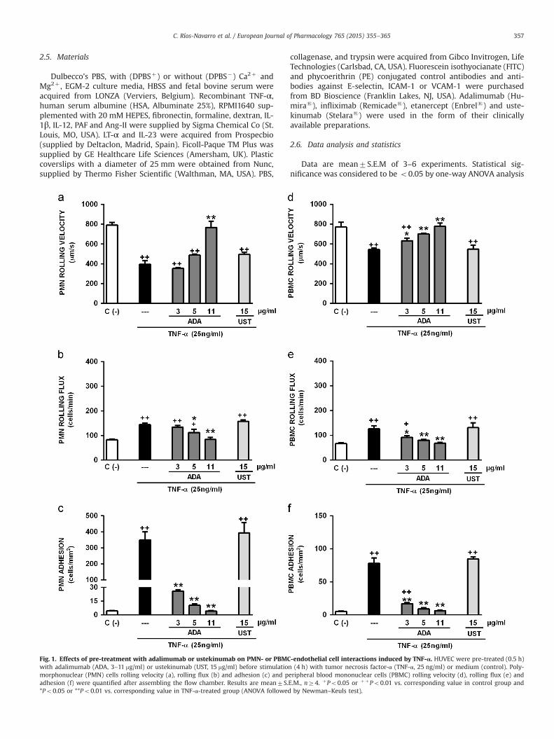
Fig. 1. Effects of pre-treatment with adalimumab or ustekinumab on PMN- or PBMCwith adalimumab (ADA, 3–11 mg/ml) or ustekinumab (UST, 15 mg/ml) before stimulatiomorphonuclear (PMN) cells rolling velocity (a), rolling flux (b) and adhesion (c) and padhesion (f) were quantified after assembling the flow chamber. Results are mean7S.*Po0.05 or **Po0.01 vs. corresponding value in TNF-α-treated group (ANOVA followed
collagenase, and trypsin were acquired from Gibco Invitrogen, LifeTechnologies (Carlsbad, CA, USA). Fluorescein isothyocianate (FITC)and phycoerithrin (PE) conjugated control antibodies and anti-bodies against E-selectin, ICAM-1 or VCAM-1 were purchasedfrom BD Bioscience (Franklin Lakes, NJ, USA). Adalimumab (Hu-miras), infliximab (Remicades), etanercept (Enbrels) and uste-kinumab (Stelaras) were used in the form of their clinicallyavailable preparations.
2.6. Data analysis and statistics
Data are mean7S.E.M of 3–6 experiments. Statistical sig-nificance was considered to be o0.05 by one-way ANOVA analysis
-endothelial cell interactions induced by TNF-α. HUVEC were pre-treated (0.5 h)n (4 h) with tumor necrosis factor-α (TNF-α, 25 ng/ml) or medium (control). Poly-eripheral blood mononuclear cells (PBMC) rolling velocity (d), rolling flux (e) andE.M., nZ4. þPo0.05 or þ þPo0.01 vs. corresponding value in control group andby Newman–Keuls test).
C. Ríos-Navarro et al. / European Journal of Pharmacology 765 (2015) 355–365358
of variance, with Newman–Keuls post-test correction to comparemultiple variances.
3. Results
3.1. Effects on cell interactions induced by endothelial stimuli
As expected, only a small number of PMN or PBMC cells ad-hered to unstimulated HUVEC (Figs. 1, 2, 4, 5 and 6), thus re-producing the conditions of a normal non-inflamed vessel (Sup-plementary material S1). In order to mimic the typical scenario of
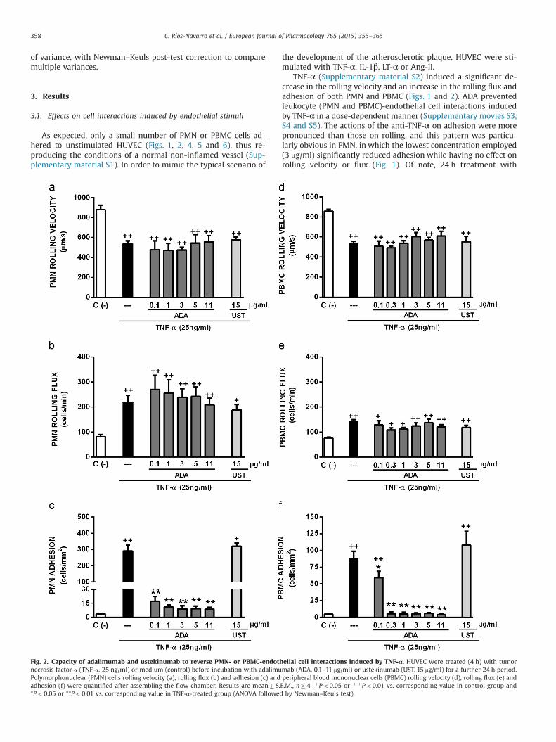
Fig. 2. Capacity of adalimumab and ustekinumab to reverse PMN- or PBMC-endothnecrosis factor-α (TNF-α, 25 ng/ml) or medium (control) before incubation with adalimuPolymorphonuclear (PMN) cells rolling velocity (a), rolling flux (b) and adhesion (c) andadhesion (f) were quantified after assembling the flow chamber. Results are mean7S.*Po0.05 or **Po0.01 vs. corresponding value in TNF-α-treated group (ANOVA followed
the development of the atherosclerotic plaque, HUVEC were sti-mulated with TNF-α, IL-1β, LT-α or Ang-II.
TNF-α (Supplementary material S2) induced a significant de-crease in the rolling velocity and an increase in the rolling flux andadhesion of both PMN and PBMC (Figs. 1 and 2). ADA preventedleukocyte (PMN and PBMC)-endothelial cell interactions inducedby TNF-α in a dose-dependent manner (Supplementary movies S3,S4 and S5). The actions of the anti-TNF-α on adhesion were morepronounced than those on rolling, and this pattern was particu-larly obvious in PMN, in which the lowest concentration employed(3 mg/ml) significantly reduced adhesion while having no effect onrolling velocity or flux (Fig. 1). Of note, 24 h treatment with
elial cell interactions induced by TNF-α. HUVEC were treated (4 h) with tumormab (ADA, 0.1–11 mg/ml) or ustekinumab (UST, 15 mg/ml) for a further 24 h period.peripheral blood mononuclear cells (PBMC) rolling velocity (d), rolling flux (e) andE.M., nZ4. þPo0.05 or þ þPo0.01 vs. corresponding value in control group andby Newman–Keuls test).
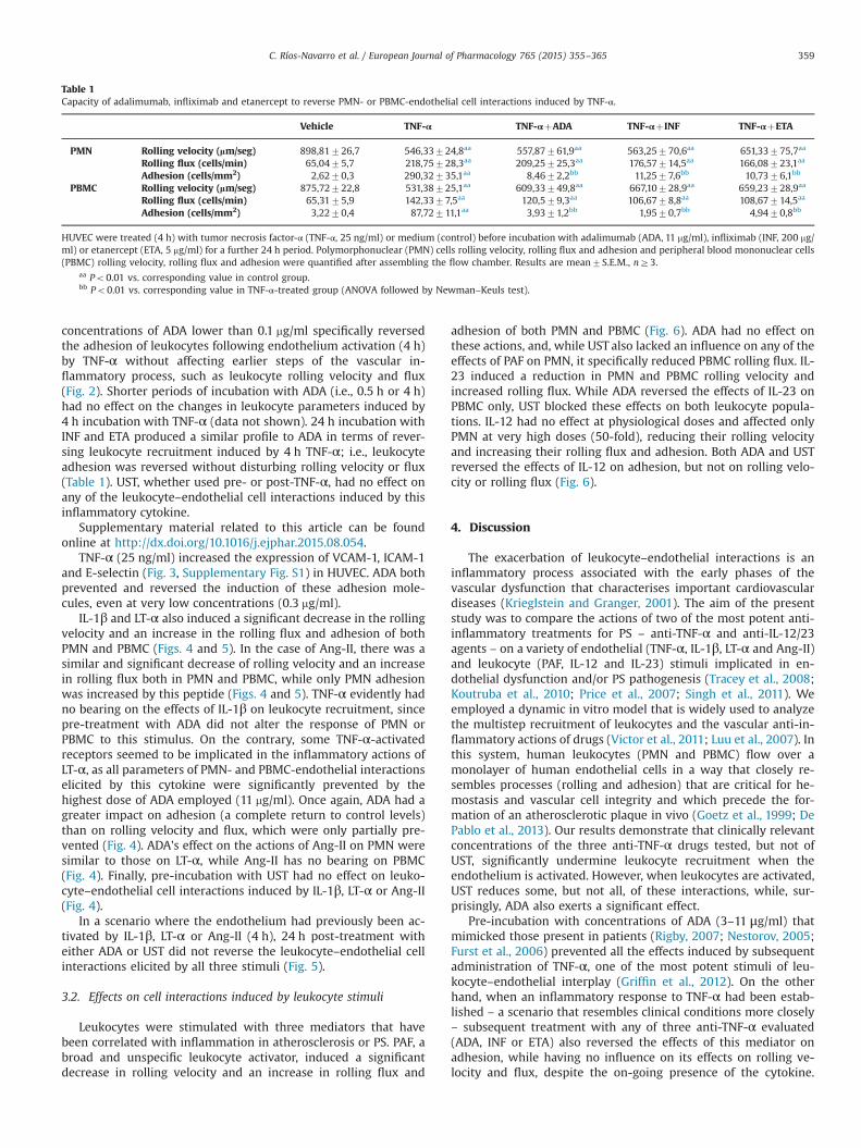
Table 1Capacity of adalimumab, infliximab and etanercept to reverse PMN- or PBMC-endothelial cell interactions induced by TNF-α.
Vehicle TNF-α TNF-αþADA TNF-αþINF TNF-αþETA
PMN Rolling velocity (lm/seg) 898,81726,7 546,33724,8aa 557,87761,9aa 563,25770,6aa 651,33775,7aa
Rolling flux (cells/min) 65,0475,7 218,75728,3aa 209,25725,3aa 176,57714,5aa 166,08723,1aa
Adhesion (cells/mm2) 2,6270,3 290,32735,1aa 8,4672,2bb 11,2577,6bb 10,7376,1bb
PBMC Rolling velocity (lm/seg) 875,72722,8 531,38725,1aa 609,33749,8aa 667,10728,9aa 659,23728,9aa
Rolling flux (cells/min) 65,3175,9 142,3377,5aa 120,579,3aa 106,6778,8aa 108,67714,5aa
Adhesion (cells/mm2) 3,2270,4 87,72711,1aa 3,9371,2bb 1,9570,7bb 4,9470,8bb
HUVEC were treated (4 h) with tumor necrosis factor-α (TNF-α, 25 ng/ml) or medium (control) before incubation with adalimumab (ADA, 11 mg/ml), infliximab (INF, 200 mg/ml) or etanercept (ETA, 5 mg/ml) for a further 24 h period. Polymorphonuclear (PMN) cells rolling velocity, rolling flux and adhesion and peripheral blood mononuclear cells(PBMC) rolling velocity, rolling flux and adhesion were quantified after assembling the flow chamber. Results are mean7S.E.M., nZ3.
aa Po0.01 vs. corresponding value in control group.bb Po0.01 vs. corresponding value in TNF-α-treated group (ANOVA followed by Newman–Keuls test).
C. Ríos-Navarro et al. / European Journal of Pharmacology 765 (2015) 355–365 359
concentrations of ADA lower than 0.1 mg/ml specifically reversedthe adhesion of leukocytes following endothelium activation (4 h)by TNF-α without affecting earlier steps of the vascular in-flammatory process, such as leukocyte rolling velocity and flux(Fig. 2). Shorter periods of incubation with ADA (i.e., 0.5 h or 4 h)had no effect on the changes in leukocyte parameters induced by4 h incubation with TNF-α (data not shown). 24 h incubation withINF and ETA produced a similar profile to ADA in terms of rever-sing leukocyte recruitment induced by 4 h TNF-α; i.e., leukocyteadhesion was reversed without disturbing rolling velocity or flux(Table 1). UST, whether used pre- or post-TNF-α, had no effect onany of the leukocyte–endothelial cell interactions induced by thisinflammatory cytokine.
Supplementary material related to this article can be foundonline at http://dx.doi.org/10.1016/j.ejphar.2015.08.054.
TNF-α (25 ng/ml) increased the expression of VCAM-1, ICAM-1and E-selectin (Fig. 3, Supplementary Fig. S1) in HUVEC. ADA bothprevented and reversed the induction of these adhesion mole-cules, even at very low concentrations (0.3 mg/ml).
IL-1β and LT-α also induced a significant decrease in the rollingvelocity and an increase in the rolling flux and adhesion of bothPMN and PBMC (Figs. 4 and 5). In the case of Ang-II, there was asimilar and significant decrease of rolling velocity and an increasein rolling flux both in PMN and PBMC, while only PMN adhesionwas increased by this peptide (Figs. 4 and 5). TNF-α evidently hadno bearing on the effects of IL-1β on leukocyte recruitment, sincepre-treatment with ADA did not alter the response of PMN orPBMC to this stimulus. On the contrary, some TNF-α-activatedreceptors seemed to be implicated in the inflammatory actions ofLT-α, as all parameters of PMN- and PBMC-endothelial interactionselicited by this cytokine were significantly prevented by thehighest dose of ADA employed (11 mg/ml). Once again, ADA had agreater impact on adhesion (a complete return to control levels)than on rolling velocity and flux, which were only partially pre-vented (Fig. 4). ADA's effect on the actions of Ang-II on PMN weresimilar to those on LT-α, while Ang-II has no bearing on PBMC(Fig. 4). Finally, pre-incubation with UST had no effect on leuko-cyte–endothelial cell interactions induced by IL-1β, LT-α or Ang-II(Fig. 4).
In a scenario where the endothelium had previously been ac-tivated by IL-1β, LT-α or Ang-II (4 h), 24 h post-treatment witheither ADA or UST did not reverse the leukocyte–endothelial cellinteractions elicited by all three stimuli (Fig. 5).
3.2. Effects on cell interactions induced by leukocyte stimuli
Leukocytes were stimulated with three mediators that havebeen correlated with inflammation in atherosclerosis or PS. PAF, abroad and unspecific leukocyte activator, induced a significantdecrease in rolling velocity and an increase in rolling flux and
adhesion of both PMN and PBMC (Fig. 6). ADA had no effect onthese actions, and, while UST also lacked an influence on any of theeffects of PAF on PMN, it specifically reduced PBMC rolling flux. IL-23 induced a reduction in PMN and PBMC rolling velocity andincreased rolling flux. While ADA reversed the effects of IL-23 onPBMC only, UST blocked these effects on both leukocyte popula-tions. IL-12 had no effect at physiological doses and affected onlyPMN at very high doses (50-fold), reducing their rolling velocityand increasing their rolling flux and adhesion. Both ADA and USTreversed the effects of IL-12 on adhesion, but not on rolling velo-city or rolling flux (Fig. 6).
4. Discussion
The exacerbation of leukocyte–endothelial interactions is aninflammatory process associated with the early phases of thevascular dysfunction that characterises important cardiovasculardiseases (Krieglstein and Granger, 2001). The aim of the presentstudy was to compare the actions of two of the most potent anti-inflammatory treatments for PS – anti-TNF-α and anti-IL-12/23agents – on a variety of endothelial (TNF-α, IL-1β, LT-α and Ang-II)and leukocyte (PAF, IL-12 and IL-23) stimuli implicated in en-dothelial dysfunction and/or PS pathogenesis (Tracey et al., 2008;Koutruba et al., 2010; Price et al., 2007; Singh et al., 2011). Weemployed a dynamic in vitro model that is widely used to analyzethe multistep recruitment of leukocytes and the vascular anti-in-flammatory actions of drugs (Victor et al., 2011; Luu et al., 2007). Inthis system, human leukocytes (PMN and PBMC) flow over amonolayer of human endothelial cells in a way that closely re-sembles processes (rolling and adhesion) that are critical for he-mostasis and vascular cell integrity and which precede the for-mation of an atherosclerotic plaque in vivo (Goetz et al., 1999; DePablo et al., 2013). Our results demonstrate that clinically relevantconcentrations of the three anti-TNF-α drugs tested, but not ofUST, significantly undermine leukocyte recruitment when theendothelium is activated. However, when leukocytes are activated,UST reduces some, but not all, of these interactions, while, sur-prisingly, ADA also exerts a significant effect.
Pre-incubation with concentrations of ADA (3–11 μg/ml) thatmimicked those present in patients (Rigby, 2007; Nestorov, 2005;Furst et al., 2006) prevented all the effects induced by subsequentadministration of TNF-α, one of the most potent stimuli of leu-kocyte–endothelial interplay (Griffin et al., 2012). On the otherhand, when an inflammatory response to TNF-α had been estab-lished – a scenario that resembles clinical conditions more closely– subsequent treatment with any of three anti-TNF-α evaluated(ADA, INF or ETA) also reversed the effects of this mediator onadhesion, while having no influence on its effects on rolling ve-locity and flux, despite the on-going presence of the cytokine.
Fig. 3. Effects of adalimumab on the expression of endothelial adhesion molecules induced by TNF-α. HUVEC were treated with adalimumab (ADA, 0.01–11 mg/ml) 0.5 hbefore (a–c) or 24 h after (d-f) stimulation (4 h) with tumor necrosis factor-α (TNF-α, 25 ng/ml) or medium (control). Expression of vascular cell adhesion molecule-1 (VCAM-1) (a and d), intracellular adhesion molecule-1 (ICAM-1) (b and e) and E-selectin (c and f) was analyzed by flow cytometry. Fluorescein isothyocianate (FITC) or phycoerithrin(PE)-fluorescence values are expressed as a percentage of the mean fluorescence intensities of control cells (100%). Results are mean7S.E.M., nZ3. þPo0.05 or þ þPo0.01vs. corresponding values in control group and **Po0.01 vs. corresponding value in TNF-α-treated group (ANOVA followed by Newman–Keuls test).
C. Ríos-Navarro et al. / European Journal of Pharmacology 765 (2015) 355–365360
Furthermore, the effects arising from deferred blockade of TNFreceptors were not noticeable after short periods (0.5 h or 4 h) ofanti-TNF-α incubation and reached significance only after 24 h,which is evidence of the multiple inflammatory pathways trig-gered following administration of the cytokine and its reducedrole in subsequent responses. In addition, ADA had no influence onthe actions of IL-1β, but prevented the actions of LT-α and Ang-IIwhen pre-administered. However, ADA failed to reverse the in-flammatory response elicited by the latter two stimuli once it hadbeen established. The effects described with ADA, which binds toTNF-α and avoids the interaction of this cytokine with its tworeceptors (TNF receptor 1 and TNF receptor 2) (Van et al., 2011),
are generally in line with published evidence implicating TNF-α inthe actions of the stimuli we have evaluated in this study. Thus,whereas IL-1β exerts its action through receptors unrelated toTNF-α (Dinarello, 2011), LT-α exhibits certain similarity with thetertiary and quaternary structure of TNF-α (Buhrmann et al., 2013),which probably explains its susceptibility to TNF-α blockade. In-deed, its actions are a result of its binding to a variety of receptors,including TNF receptor 1 and TNF receptor 2 and the more specificLT-β receptor (Etemadi et al., 2013). Lastly, Ang-II binds to angio-tensin-II receptor type I, which activates signal pathways shown toupregulate TNF-α production (Abadir et al., 2011).
The interaction of leukocytes with endothelial cells is
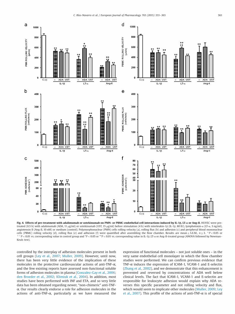
Fig. 4. Effects of pre-treatment with adalimumab or ustekinumab on PMN- or PBMC-endothelial cell interactions induced by IL-1β, LT-α or Ang-II. HUVEC were pre-treated (0.5 h) with adalimumab (ADA, 11 mg/ml) or ustekinumab (UST, 15 mg/ml) before stimulation (4 h) with interleukin-1β (IL-1β, 80 IU), lymphotoxin-α (LT-α, 3 ng/ml),angiotensin II (Ang-II, 10 nM) or medium (control). Polymorphonuclear (PMN) cells rolling velocity (a), rolling flux (b) and adhesion (c) and peripheral blood mononuclearcells (PBMC) rolling velocity (d), rolling flux (e) and adhesion (f) were quantified after assembling the flow chamber. Results are mean7S.E.M., nZ3. þPo0.05 orþ þPo0.01 vs. corresponding value in control group and *Po0.05 or **Po0.01 vs. corresponding value in IL-1β, LT-α or Ang-II-treated group (ANOVA followed by Newman–Keuls test).
C. Ríos-Navarro et al. / European Journal of Pharmacology 765 (2015) 355–365 361
controlled by the interplay of adhesion molecules present in bothcell groups (Ley et al., 2007; Muller, 2009). However, until now,there has been very little evidence of the implication of thesemolecules in the protective cardiovascular actions of anti-TNF-α,and the few existing reports have assessed non-functional solubleforms of adhesion molecules in plasma (Gonzalez-Gay et al., 2006;den Broeder et al., 2002; Klimiuk et al., 2004). In addition, moststudies have been performed with INF and ETA, and so very littledata has been obtained regarding newer, “non-chimeric” anti-TNF-α. Our results clearly endorse a role for adhesion molecules in theactions of anti-TNF-α, particularly as we have measured the
expression of functional molecules – not just soluble ones – in thevery same endothelial cell monolayer in which the flow chamberstudies were performed. We can confirm previous evidence thatTNF-α induces the expression of ICAM-1, VCAM-1 and E-selectin(Zhang et al., 2002), and we demonstrate that this enhancement isprevented and reversed by concentrations of ADA well belowclinical levels. The fact that ICAM-1, VCAM-1 and E-selectin areresponsible for leukocyte adhesion would explain why ADA re-verses this specific parameter and not rolling velocity and flux,which would seem to implicate other molecules (Muller, 2009; Leyet al., 2007). This profile of the actions of anti-TNF-α is of special
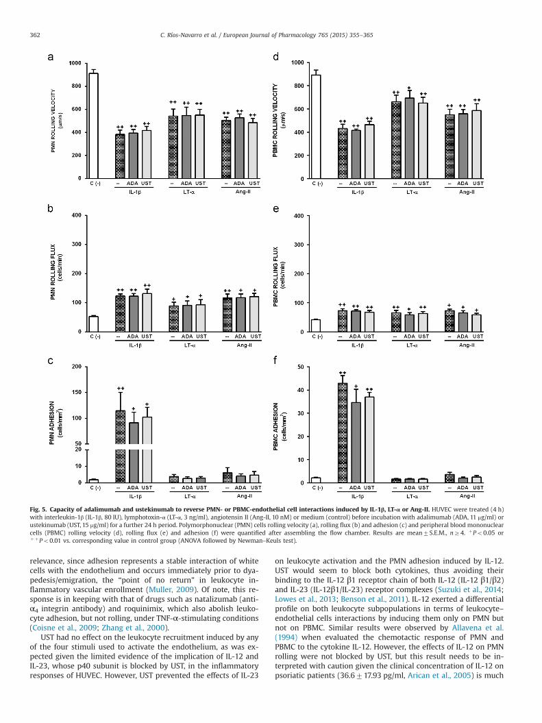
Fig. 5. Capacity of adalimumab and ustekinumab to reverse PMN- or PBMC-endothelial cell interactions induced by IL-1β, LT-α or Ang-II. HUVEC were treated (4 h)with interleukin-1β (IL-1β, 80 IU), lymphotoxin-α (LT-α, 3 ng/ml), angiotensin II (Ang-II, 10 nM) or medium (control) before incubation with adalimumab (ADA, 11 mg/ml) orustekinumab (UST, 15 mg/ml) for a further 24 h period. Polymorphonuclear (PMN) cells rolling velocity (a), rolling flux (b) and adhesion (c) and peripheral blood mononuclearcells (PBMC) rolling velocity (d), rolling flux (e) and adhesion (f) were quantified after assembling the flow chamber. Results are mean7S.E.M., nZ4. þPo0.05 orþ þPo0.01 vs. corresponding value in control group (ANOVA followed by Newman–Keuls test).
C. Ríos-Navarro et al. / European Journal of Pharmacology 765 (2015) 355–365362
relevance, since adhesion represents a stable interaction of whitecells with the endothelium and occurs immediately prior to dya-pedesis/emigration, the “point of no return” in leukocyte in-flammatory vascular enrollment (Muller, 2009). Of note, this re-sponse is in keeping with that of drugs such as natalizumab (anti-α4 integrin antibody) and roquinimix, which also abolish leuko-cyte adhesion, but not rolling, under TNF-α-stimulating conditions(Coisne et al., 2009; Zhang et al., 2000).
UST had no effect on the leukocyte recruitment induced by anyof the four stimuli used to activate the endothelium, as was ex-pected given the limited evidence of the implication of IL-12 andIL-23, whose p40 subunit is blocked by UST, in the inflammatoryresponses of HUVEC. However, UST prevented the effects of IL-23
on leukocyte activation and the PMN adhesion induced by IL-12.UST would seem to block both cytokines, thus avoiding theirbinding to the IL-12 β1 receptor chain of both IL-12 (IL-12 β1/β2)and IL-23 (IL-12β1/IL-23) receptor complexes (Suzuki et al., 2014;Lowes et al., 2013; Benson et al., 2011). IL-12 exerted a differentialprofile on both leukocyte subpopulations in terms of leukocyte–endothelial cells interactions by inducing them only on PMN butnot on PBMC. Similar results were observed by Allavena et al.(1994) when evaluated the chemotactic response of PMN andPBMC to the cytokine IL-12. However, the effects of IL-12 on PMNrolling were not blocked by UST, but this result needs to be in-terpreted with caution given the clinical concentration of IL-12 onpsoriatic patients (36.6717.93 pg/ml, Arican et al., 2005) is much
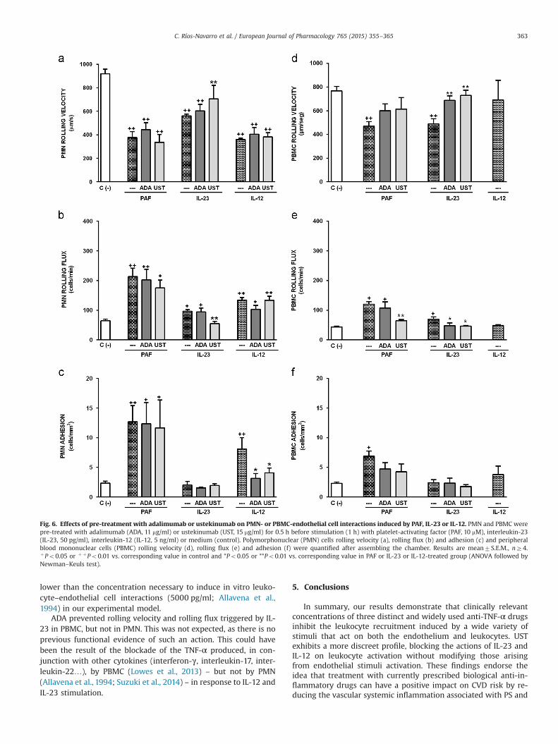
Fig. 6. Effects of pre-treatment with adalimumab or ustekinumab on PMN- or PBMC-endothelial cell interactions induced by PAF, IL-23 or IL-12. PMN and PBMC werepre-treated with adalimumab (ADA, 11 mg/ml) or ustekinumab (UST, 15 mg/ml) for 0.5 h before stimulation (1 h) with platelet-activating factor (PAF, 10 mM), interleukin-23(IL-23, 50 pg/ml), interleukin-12 (IL-12, 5 ng/ml) or medium (control). Polymorphonuclear (PMN) cells rolling velocity (a), rolling flux (b) and adhesion (c) and peripheralblood mononuclear cells (PBMC) rolling velocity (d), rolling flux (e) and adhesion (f) were quantified after assembling the chamber. Results are mean7S.E.M., nZ4.þPo0.05 or þ þPo0.01 vs. corresponding value in control and *Po0.05 or **Po0.01 vs. corresponding value in PAF or IL-23 or IL-12-treated group (ANOVA followed byNewman–Keuls test).
C. Ríos-Navarro et al. / European Journal of Pharmacology 765 (2015) 355–365 363
lower than the concentration necessary to induce in vitro leuko-cyte–endothelial cell interactions (5000 pg/ml; Allavena et al.,1994) in our experimental model.
ADA prevented rolling velocity and rolling flux triggered by IL-23 in PBMC, but not in PMN. This was not expected, as there is noprevious functional evidence of such an action. This could havebeen the result of the blockade of the TNF-α produced, in con-junction with other cytokines (interferon-γ, interleukin-17, inter-leukin-22…), by PBMC (Lowes et al., 2013) – but not by PMN(Allavena et al., 1994; Suzuki et al., 2014) – in response to IL-12 andIL-23 stimulation.
5. Conclusions
In summary, our results demonstrate that clinically relevantconcentrations of three distinct and widely used anti-TNF-α drugsinhibit the leukocyte recruitment induced by a wide variety ofstimuli that act on both the endothelium and leukocytes. USTexhibits a more discreet profile, blocking the actions of IL-23 andIL-12 on leukocyte activation without modifying those arisingfrom endothelial stimuli activation. These findings endorse theidea that treatment with currently prescribed biological anti-in-flammatory drugs can have a positive impact on CVD risk by re-ducing the vascular systemic inflammation associated with PS and
C. Ríos-Navarro et al. / European Journal of Pharmacology 765 (2015) 355–365364
RA. However, the degree of such effects varies considerably; inlight of our in vitro results obtained in human cells, the widerprofile of actions of anti-TNF-α – owing to the ubiquity of themediator in the inflammatory cascade – seems to promise a morepotent, and thus more clinically relevant effect. Importantly, thispotential CV effect is in addition to these drugs’ ability to coun-teract the inflammatory conditions that characterize the derma-tologic and rheumatic diseases for whose treatment they weredesigned, and obviously requires clinical corroboration.
Conflict of interest
JVE has given conferences and/or participated in advisoryboards for AstraZeneca, Gilead, Abbvie, MSD and Pfizer. JVE and AAhave received research funding from Gilead and Abbvie. None ofthe other authors report any potential conflicts.
Acknowledgments
CRN and CDP performed the research, MAMC, ABG, VCD and SOhelped perform the research, CRN, CDP and AA analyzed the dataand JVE and AA designed the research and wrote the paper. Wewould like to thank Nicole Roupain, Dora Martí and Fernando Sa-bater for their technical support to perform the experiments of thismanuscript. This work was supported by Ministerio de Ciencia eInnovación [Grant SAF2010-16030], Ministerio de Sanidad y Con-sumo [Grant PI11/00327 and PI14/00312], CIBERehd [Grant CB06/04/0071] and Generalitat Valenciana [Grant PROMETEOII/2014/035,PROMETEO/2010/060, ACOMP2013/147, ACOMP2013/236 and Ger-onimo Forteza FPA/2013/A/051]. CDP was funded by Ministerio deCiencia e Innovación [FPI Grant BES-2008-004338] and by Funda-ción Juan Esplugues. SO was funded by Universidad de Valencia[VLC CAMPUS Grant] and Fundación Fisabio Grant 2015/01. CRN andVCD were funded by Fundación Fisabio. Grants 2014/22 and 2014/23 respectively. ABG is recipient of a Juan de la Cierva contract (ref.JCI-2012-15124, Ministerio de Economía y Competitividad). AA wassupported by Ministerio de Ciencia e Innovación [Ramón y Cajalprogram RYC2005-002295 and I3 program].
Appendix A. Supplementary information
Supplementary data associated with this article can be found inthe online version at http://dx.doi.org/10.1016/j.ejphar.2015.08.054.
References
Abadir, P.M., Walston, J.D., Carey, R.M., Siragy, H.M., 2011. Angiotensin II type-2receptors modulate inflammation through signal transducer and activator oftranscription proteins 3 phosphorylation and TNFalpha production. J. InterferonCytokine Res. 31, 471–474.
Ahlehoff, O., Skov, L., Gislason, G., Lindhardsen, J., Kristensen, S.L., Iversen, L., Las-thein, S., Gniadecki, R., Dam, T.N., Torp-Pedersen, C., Hansen, P.R., 2013. Cardi-ovascular disease event rates in patients with severe psoriasis treated withsystemic anti-inflammatory drugs: a Danish real-world cohort study. J. Intern.Med. 273, 197–204.
Alexandroff, A.B., Pauriah, M., Camp, R.D., Lang, C.C., Struthers, A.D., Armstrong, D.J.,2009. More than skin deep: atherosclerosis as a systemic manifestation ofpsoriasis. Br. J. Dermatol. 161, 1–7.
Allavena, P., Paganin, C., Zhou, D., Bianchi, G., Sozzani, S., Mantovani, A., 1994. In-terleukin-12 is chemotactic for natural killer cells and stimulates their inter-action with vascular endothelium. Blood 84, 2261–2268.
Alvarez, A., Cerda-Nicolas, M., Naim Abu, N.Y., Mata, M., Issekutz, A.C., Panes, J.,Lobb, R.R., Sanz, M.J., 2004. Direct evidence of leukocyte adhesion in arteriolesby angiotensin II. Blood 104, 402–408.
Arican, O., Aral, M., Sasmaz, S., Ciragil, P., 2005. Serum levels of TNF-α, IFN-γ, IL-6,
IL-8, IL-12, IL-17 and IL-18 in patients with active psoriasis and correlation withdisease severity. Mediat. Inflamm. 5, 273–279.
Benson, J.M., Peritt, D., Scallon, B.J., Heavner, G.A., Shealy, D.J., Giles-Komar, J.M.,Mascelli, M.A., 2011. Discovery and mechanism of ustekinumab: a humanmonoclonal antibody targeting interleukin-12 and interleukin-23 for treatmentof immune-mediated disorders. MAbs 3, 535–545.
Buhrmann, C., Shayan, P., Aggarwal, B.B., Shakibaei, M., 2013. Evidence that TNF-beta (lymphotoxin alpha) can activate the inflammatory environment in hu-man chondrocytes. Arthritis Res. Ther. 15, R202.
Cai, Y.H., Alvarez, A., Alcaide, P., Duramad, P., Lim, Y.C., Jarolim, P., Lowe, J.B., Lus-cinskas, F.W., Lichtman, A.H., 2006. Abrogation of functional selectin-ligandexpression reduces migration of pathogenic CD8þ T cells into heart. J. Im-munol. 176, 6568–6575.
Coisne, C., Mao, W., Engelhardt, B., 2009. Cutting edge: natalizumab blocks adhe-sion but not initial contact of human T cells to the blood-brain barrier in vivo inan animal model of multiple sclerosis. J. Immunol. 182, 5909–5913.
De Pablo, C., Orden, S., Apostolova, N., Blanquer, A., Esplugues, J.V., Alvarez, A., 2010.Abacavir and didanosine induce the interaction between human leukocytesand endothelial cells through Mac-1 upregulation. AIDS 24, 1259–1266.
De Pablo, C., Orden, S., Calatayud, S., Marti-Cabrera, M., Esplugues, J.V., Alvarez, A.,2012. Differential effects of Tenofovir/Emtricitabine and Abacavir/Lamivudineon human leukocyte recruitment. Antivir. Ther. 17, 1615–1619.
De Pablo, C., Orden, S., Peris, J.E., Barrachina, M.D., Esplugues, J.V., Alvarez, A., 2013.Profile of leukocyte–endothelial cell interactions induced in venules and ar-terioles by nucleoside reverse-transcriptase inhibitors in vivo. J. Infect. Dis. 208,1448–1453.
den Broeder, A.A., Joosten, L.A., Saxne, T., Heinegard, D., Fenner, H., Miltenburg, A.M., Frasa, W.L., van Tits, L.J., Buurman, W.A., van Riel, P.L., van de Putte, L.B.,Barrera, P., 2002. Long term anti-tumour necrosis factor alpha monotherapy inrheumatoid arthritis: effect on radiological course and prognostic value ofmarkers of cartilage turnover and endothelial activation. Ann. Rheum. Dis. 61,311–318.
Dinarello, C.A., 2011. Interleukin-1 in the pathogenesis and treatment of in-flammatory diseases. Blood 117, 3720–3732.
Elliott, M.J., Maini, R.N., Feldmann, M., Kalden, J.R., Antoni, C., Smolen, J.S., Leeb, B.,Breedveld, F.C., Macfarlane, J.D., Bijl, H., 1994. Randomised double-blind com-parison of chimeric monoclonal antibody to tumour necrosis factor alpha (cA2)versus placebo in rheumatoid arthritis. Lancet 344, 1105–1110.
Etemadi, N., Holien, J.K., Chau, D., Dewson, G., Murphy, J.M., Alexander, W.S., Parker,M.W., Silke, J., Nachbur, U., 2013. Lymphotoxin alpha induces apoptosis, ne-croptosis and inflammatory signals with the same potency as tumour necrosisfactor. FEBS J. 280, 5283–5297.
Furst, D.E., Wallis, R., Broder, M., Beenhouwer, D.O., 2006. Tumor necrosis factorantagonists: different kinetics and/or mechanisms of action may explain dif-ferences in the risk for developing granulomatous infection. Semin. ArthritisRheum. 36, 159–167.
Goetz, D.J., Greif, D.M., Shen, J., Luscinskas, F.W., 1999. Cell–cell adhesive interac-tions in an in vitro flow chamber. Methods Mol. Biol. 96, 137–145.
Gonzalez-Gay, M.A., Garcia-Unzueta, M.T., de Matias, J.M., Gonzalez-Juanatey, C.,Garcia-Porrua, C., Sanchez-Andrade, A., Martin, J., Llorca, J., 2006. Influence ofanti-TNF-alpha infliximab therapy on adhesion molecules associated withatherogenesis in patients with rheumatoid arthritis. Clin. Exp. Rheumatol. 24,373–379.
Gottlieb, A.B., Cooper, K.D., McCormick, T.S., Toichi, E., Everitt, D.E., Frederick, B.,Zhu, Y., Pendley, C.E., Graham, M.A., Mascelli, M.A., 2007. A phase 1, double-blind, placebo-controlled study evaluating single subcutaneous administrationsof a human interleukin-12/23 monoclonal antibody in subjects with plaquepsoriasis. Curr. Med. Res. Opin. 23, 1081–1092.
Griffin, G.K., Newton, G., Tarrio, M.L., Bu, D.X., Maganto-Garcia, E., Azcutia, V., Al-caide, P., Grabie, N., Luscinskas, F.W., Croce, K.J., Lichtman, A.H., 2012. IL-17 andTNF-alpha sustain neutrophil recruitment during inflammation through sy-nergistic effects on endothelial activation. J. Immunol. 188, 6287–6299.
Ibiza, S., Alvarez, A., Romero, W., Barrachina, M.D., Esplugues, J.V., Calatayud, S.,2009. Gastrin induces the interaction between human mononuclear leukocytesand endothelial cells through the endothelial expression of P-selectin andVCAM-1. Am. J. Physiol.: Cell Physiol. 297, C1588–C1595.
Klimiuk, P.A., Sierakowski, S., Domyslawska, I., Fiedorczyk, M., Chwiecko, J., 2004.Reduction of soluble adhesion molecules (sICAM-1, sVCAM-1, and sE-selectin)and vascular endothelial growth factor levels in serum of rheumatoid arthritispatients following multiple intravenous infusions of infliximab. Arch. Immunol.Ther. Exp. (Warsz ) 52, 36–42.
Koutruba, N., Emer, J., Lebwohl, M., 2010. Review of ustekinumab, an interleukin-12and interleukin-23 inhibitor used for the treatment of plaque psoriasis. Ther.Clin. Risk Manag. 6, 123–141.
Krieglstein, C.F., Granger, D.N., 2001. Adhesion molecules and their role in vasculardisease. Am. J. Hypertens. 14, 44S–54S.
Ley, K., Laudanna, C., Cybulsky, M.I., Nourshargh, S., 2007. Getting to the site ofinflammation: the leukocyte adhesion cascade updated. Nat. Rev. Immunol. 7,678–689.
Libby, P., 2012. Inflammation in atherosclerosis. Arterioscler. Thromb. Vasc. Biol. 32,2045–2051.
Lowes, M.A., Russell, C.B., Martin, D.A., Towne, J.E., Krueger, J.G., 2013. The IL-23/T17pathogenic axis in psoriasis is amplified by keratinocyte responses. TrendsImmunol. 34, 174–181.
Luu, N.T., Madden, J., Calder, P.C., Grimble, R.F., Shearman, C.P., Chan, T., Tull, S.P.,Dastur, N., Rainger, G.E., Nash, G.B., 2007. Comparison of the pro-inflammatory
C. Ríos-Navarro et al. / European Journal of Pharmacology 765 (2015) 355–365 365
potential of monocytes from healthy adults and those with peripheral arterialdisease using an in vitro culture model. Atherosclerosis 193, 259–268.
Maki-Petaja, K.M., Elkhawad, M., Cheriyan, J., Joshi, F.R., Ostor, A.J., Hall, F.C., Rudd, J.H., Wilkinson, I.B., 2012. Anti-tumor necrosis factor-alpha therapy reducesaortic inflammation and stiffness in patients with rheumatoid arthritis. Circu-lation 126, 2473–2480.
Mateo, T., Abu Nabah, Y.N., Abu, T.M., Mata, M., Cerda-Nicolas, M., Proudfoot, A.E.,Stahl, R.A., Issekutz, A.C., Cortijo, J., Morcillo, E.J., Jose, P.J., Sanz, M.J., 2006.Angiotensin II-induced mononuclear leukocyte interactions with arteriolar andvenular endothelium are mediated by the release of different CC chemokines. J.Immunol. 176, 5577–5586.
Montrucchio, G., Alloatti, G., Camussi, G., 2000. Role of platelet-activating factor incardiovascular pathophysiology. Physiol. Rev. 80, 1669–1699.
Muller, W.A., 2009. Mechanisms of transendothelial migration of leukocytes. Circ.Res. 105, 223–230.
Nakada, M.T., Tam, S.H., Woulfe, D.S., Casper, K.A., Swerlick, R.A., Ghrayeb, J., 1998.Neutralization of TNF by the antibody cA2 reveals differential regulation ofadhesion molecule expression on TNF-activated endothelial cells. Cell Adhes.Commun. 5, 491–503.
Nestorov, I., 2005. Clinical pharmacokinetics of TNF antagonists: how do they dif-fer? Semin. Arthritis Rheum. 34, 12–18.
Nguyen, T., Wu, J.J., 2014. Relationship between tumor necrosis factor-alpha in-hibitors and cardiovascular disease in psoriasis: a review. Perm. J. 18, 49–54.
Price, A., Lockhart, J.C., Ferrell, W.R., Gsell, W., McLean, S., Sturrock, R.D., 2007.Angiotensin II type 1 receptor as a novel therapeutic target in rheumatoid ar-thritis: in vivo analyses in rodent models of arthritis and ex vivo analyses inhuman inflammatory synovitis. Arthritis Rheum. 56, 441–447.
Ridker, P.M., Luscher, T.F., 2014. Anti-inflammatory therapies for cardiovasculardisease. Eur. Heart J. 35, 1782–1791.
Rigby, W.F., 2007. Drug insight: different mechanisms of action of tumor necrosisfactor antagonists-passive-aggressive behavior? Nat. Clin. Pract. Rheumatol. 3,227–233.
Roubille, C., Richer, V., Starnino, T., McCourt, C., McFarlane, A., Fleming, P., Siu, S.,Kraft, J., Lynde, C., Pope, J., Gulliver, W., Keeling, S., Dutz, J., Bessette, L., Bis-sonnette, R., Haraoui, B., 2015. The effects of tumour necrosis factor inhibitors,methotrexate, non-steroidal anti-inflammatory drugs and corticosteroids oncardiovascular events in rheumatoid arthritis, psoriasis and psoriatic arthritis: asystematic review and meta-analysis. Ann. Rheum. Dis. 74, 480–489.
Singh, T.P., Huettner, B., Koefeler, H., Mayer, G., Bambach, I., Wallbrecht, K., Schon,
M.P., Wolf, P., 2011. Platelet-activating factor blockade inhibits the T-helper type17 cell pathway and suppresses psoriasis-like skin disease in K5.hTGF-beta1transgenic mice. Am. J. Pathol. 178, 699–708.
Spah, F., 2008. Inflammation in atherosclerosis and psoriasis: common pathogenicmechanisms and the potential for an integrated treatment approach. Br. J.Dermatol. 159 (Suppl. 2), S10–S17.
Suna, S., Sakata, Y., Sato, H., Mizuno, H., Nakatani, D., Shimizu, M., Usami, M., Ta-kashima, S., Takeda, H., Hori, M., 2008. Up-regulation of cell adhesion moleculegenes in human endothelial cells stimulated by lymphotoxin alpha: DNA mi-croarray analysis. J. Atheroscler. Thromb. 15, 160–165.
Suzuki, E., Mellins, E.D., Gershwin, M.E., Nestle, F.O., Adamopoulos, I.E., 2014. The IL-23/IL-17 axis in psoriatic arthritis. Autoimmun. Rev. 13, 496–502.
Tam, L.S., Kitas, G.D., Gonzalez-Gay, M.A., 2014. Can suppression of inflammation byanti-TNF prevent progression of subclinical atherosclerosis in inflammatoryarthritis? Rheumatol.: Oxf. 53, 1108–1119.
Tracey, D., Klareskog, L., Sasso, E.H., Salfeld, J.G., Tak, P.P., 2008. Tumor necrosisfactor antagonist mechanisms of action: a comprehensive review. Pharmacol.Ther. 117, 244–279.
Van, H.F., Vandenbroucke, R.E., Libert, C., 2011. Treatment of TNF mediated diseasesby selective inhibition of soluble TNF or TNFR1. Cytokine Growth Factor Rev. 22,311–319.
Victor, V.M., Rocha, M., Banuls, C., Alvarez, A., De Pablo, C., Sanchez-Serrano, M.,Gomez, M., Hernandez-Mijares, A., 2011. Induction of oxidative stress and hu-man leukocyte/endothelial cell interactions in polycystic ovary syndrome pa-tients with insulin resistance. J. Clin. Endocrinol. Metab. 96, 3115–3122.
Westlake, S.L., Colebatch, A.N., Baird, J., Curzen, N., Kiely, P., Quinn, M., Choy, E.,Ostor, A.J., Edwards, C.J., 2011. Tumour necrosis factor antagonists and the riskof cardiovascular disease in patients with rheumatoid arthritis: a systematicliterature review. Rheumatol.: Oxf. 50, 518–531.
Yamagata, K., Tanaka, N., Matsufuji, H., Chino, M., 2012. Beta-carotene reverses theIL-1beta-mediated reduction in paraoxonase-1 expression via induction of theCaMKKII pathway in human endothelial cells. Microvasc. Res. 84, 297–305.
Zhang, F., Yu, W., Hargrove, J.L., Greenspan, P., Dean, R.G., Taylor, E.W., Hartle, D.K.,2002. Inhibition of TNF-alpha induced ICAM-1, VCAM-1 and E-selectin ex-pression by selenium. Atherosclerosis 161, 381–386.
Zhang, X.W., Hedlund, G., Borgstrom, P., Arfors, K.E., Thorlacius, H., 2000. Linomideabolishes leukocyte adhesion and extravascular recruitment induced by tumornecrosis factor alpha in vivo. J. Leukoc. Biol. 68, 621–626.
ES/H
UD/091
5/08
50
![problemas tsup [Modo de compatibilidad] - uv.es · PDF fileFS3. A 20 °C, el ascenso capilar a nivel del mar del metanol en contacto con aire en un tubo de diámetro interno de 0.350](https://static.fdocument.org/doc/165x107/5a94f3547f8b9aba4a8c2b2b/problemas-tsup-modo-de-compatibilidad-uves-a-20-c-el-ascenso-capilar-a-nivel.jpg)