
Energy transfer processes in electronically coupled porphyrin hetero-dyads connected at the β...
11
Energy transfer processes in electronically coupled porphyrin hetero-dyads connected at the b position Barbara Ventura, a Francesco Barigelletti, a Fabio Lodato, b David L. Officer bc and Lucia Flamigni* a Received 28th October 2008, Accepted 18th December 2008 First published as an Advance Article on the web 6th February 2009 DOI: 10.1039/b819138g Energy transfer in a series of hetero-dyads of zinc porphyrin and free-base porphyrin connected at the b position by p conjugated bridges has been determined. The dyads have been characterized and compared with the homo dyads, excellent models for the donor and the acceptor porphyrins in the electronically conjugated system. The homo dyads provide reliable parameters for the determination of the energy transfer rate calculated according to the Fo¨rster theory. This model was inadequate to account for the experimental findings and an electron exchange mechanism was shown to contribute. A favorable coplanar arrangement of the bridge and the tetrapyrroles facilitates the energy transfer process, which displays a very low distance dependence and an efficiency 498%. Introduction Porphyrin-type components have been extensively used to construct dyads or higher homologues designed to achieve useful photoinduced intramolecular processes. In particular the study of energy transfer has been often performed on arrays based on these chromophores. In fact they are well characterized from the spectroscopic/photophysical viewpoint and are known to dis- play a wide tunability of energy levels which can be achieved by simple substitution or metallation. The use of components with different excited state energies has allowed us to detect, by means of time-resolved techniques, the decay of the donor’s and/or the rise of the acceptor’s excited state populations. 1–17 This was done by interrogating both the luminescence and the transient absorbance signals typical of the excited state. This type of methodology was extensively used to determine the rate of the energy transfer processes as a function of thermodynamic para- meters and, keeping the latter constant, as a function of distance, type of connecting bridge, orientation, solvent etc. This method has been mostly used for singlet energy migration, but examples of triplet energy transfer studies are present in the literature. 18–21 Knowledge of the reaction parameters has allowed a thorough discussion on the mechanism of energy transfer in the frame of current theories, and has shed light on this process which is of very high relevance to the issues of conversion of light energy and of molecular electronics. Whereas photoinduced processes in a wide variety of meso–meso bridged porphyrin dyads (or higher homologues) are well documented in the literature, very few examples of reports on photoinduced processes in porphyrin dyads connected at the b-pyrrole are reported. 22,23 Actually, the b position of porphyrins has seldom been used as connection point in the construction of porphyrin based photoactive arrays. 24–26 The position of the bridge connection is not a trivial issue, since the electronic distribution at the periphery of a porphyrin depends both on the nature and on the location of the substituents. In fact, the a 1u and a 2u HOMO orbitals of porphyrins have a different electron density at the b (-pyrrole) and at the meso carbons: whereas the a 1u orbital puts charge on the b carbons and has nodes at the meso positions the a 2u orbital has electron density at the meso positions and at the central nitrogens and nodes at the pyrrole carbons. Substi- tuents with different electron donating or withdrawing ability can modulate in different ways the energetic distribution of the porphyrin frontier orbitals (i.e. the a 2u /a 1u energies) according to their position and can even reverse the normal ordering a 2u 4 a 1u . 27–29 Thus, in a porphyrin supramolecular structure the connection point between components can greatly affect the electronic coupling between the component units and result in processes with different mechanisms and rates. The effect of orbital inversion on the rates of energy transfer in a porphyrin dyad was elegantly addressed by Lindsey, Holten, Bocian et al. for a series of porphyrinic dyads connected at either the b-pyrrole or the meso carbons position by a diaryl- ethyne linker. 22 In those systems the stabilization of the a 2u orbital of the donor by the presence of meso strong electron withdrawing groups induces the orbital ordering to reverse and the electronic communication between the units is enhanced in case of a b-linkage compared to a meso-linkage. For the monomer models of the present dyads, however, simple MO calculations could exclude an inversion of the orbital ordering of the porphyrin core; 30 therefore in the components of the dyads a normal a 1u o a 2u ordering should be considered. Our aim in this work is to study the photoinduced energy transfer process in a series of porphyrinic dyads where the individual components, a free-base and a zinc porphyrin, are connected at the b-pyrrole position by highly conjugated a Istituto per la Sintesi Organica e la Fotoreattivita ` (ISOF), CNR, Via P. Gobetti 101, 40129 Bologna, Italy. E-mail: fl[email protected]; Fax: +39 051 6399844; Tel: +39 051 6399812 b Nanomaterials Research Centre, Massey University, Palmerston North, Private Bag 11222, New Zealand c MacDiarmid Institute for Advanced Materials and Nanotechnology, New Zealand 2166 | Phys. Chem. Chem. Phys., 2009, 11, 2166–2176 This journal is c the Owner Societies 2009 PAPER www.rsc.org/pccp | Physical Chemistry Chemical Physics Published on 06 February 2009. Downloaded by St. Petersburg State University on 23/12/2013 22:26:01. View Article Online / Journal Homepage / Table of Contents for this issue
Transcript of Energy transfer processes in electronically coupled porphyrin hetero-dyads connected at the β...
Energy transfer processes in electronically coupled porphyrin
hetero-dyads connected at the b position
Barbara Ventura,a Francesco Barigelletti,a Fabio Lodato,b David L. Officerbc
and Lucia Flamigni*a
Received 28th October 2008, Accepted 18th December 2008
First published as an Advance Article on the web 6th February 2009
DOI: 10.1039/b819138g
Energy transfer in a series of hetero-dyads of zinc porphyrin and free-base porphyrin connected
at the b position by p conjugated bridges has been determined. The dyads have been
characterized and compared with the homo dyads, excellent models for the donor and the
acceptor porphyrins in the electronically conjugated system. The homo dyads provide reliable
parameters for the determination of the energy transfer rate calculated according to the Forster
theory. This model was inadequate to account for the experimental findings and an electron
exchange mechanism was shown to contribute. A favorable coplanar arrangement of the bridge
and the tetrapyrroles facilitates the energy transfer process, which displays a very low distance
dependence and an efficiency 498%.
Introduction
Porphyrin-type components have been extensively used to
construct dyads or higher homologues designed to achieve useful
photoinduced intramolecular processes. In particular the study
of energy transfer has been often performed on arrays based on
these chromophores. In fact they are well characterized from the
spectroscopic/photophysical viewpoint and are known to dis-
play a wide tunability of energy levels which can be achieved by
simple substitution or metallation. The use of components with
different excited state energies has allowed us to detect, by means
of time-resolved techniques, the decay of the donor’s and/or the
rise of the acceptor’s excited state populations.1–17 This was
done by interrogating both the luminescence and the transient
absorbance signals typical of the excited state. This type of
methodology was extensively used to determine the rate of the
energy transfer processes as a function of thermodynamic para-
meters and, keeping the latter constant, as a function of distance,
type of connecting bridge, orientation, solvent etc. This method
has been mostly used for singlet energy migration, but examples
of triplet energy transfer studies are present in the literature.18–21
Knowledge of the reaction parameters has allowed a thorough
discussion on the mechanism of energy transfer in the frame of
current theories, and has shed light on this process which is of
very high relevance to the issues of conversion of light energy
and of molecular electronics.
Whereas photoinduced processes in a wide variety of
meso–meso bridged porphyrin dyads (or higher homologues)
are well documented in the literature, very few examples
of reports on photoinduced processes in porphyrin dyads
connected at the b-pyrrole are reported.22,23 Actually, the
b position of porphyrins has seldom been used as connection
point in the construction of porphyrin based photoactive
arrays.24–26 The position of the bridge connection is not a
trivial issue, since the electronic distribution at the periphery
of a porphyrin depends both on the nature and on the location
of the substituents. In fact, the a1u and a2u HOMO orbitals of
porphyrins have a different electron density at the b (-pyrrole)
and at the meso carbons: whereas the a1u orbital puts charge
on the b carbons and has nodes at the meso positions the a2uorbital has electron density at the meso positions and at the
central nitrogens and nodes at the pyrrole carbons. Substi-
tuents with different electron donating or withdrawing ability
can modulate in different ways the energetic distribution of the
porphyrin frontier orbitals (i.e. the a2u/a1u energies) according
to their position and can even reverse the normal ordering
a2u 4 a1u.27–29 Thus, in a porphyrin supramolecular structure
the connection point between components can greatly affect
the electronic coupling between the component units and
result in processes with different mechanisms and rates. The
effect of orbital inversion on the rates of energy transfer in a
porphyrin dyad was elegantly addressed by Lindsey, Holten,
Bocian et al. for a series of porphyrinic dyads connected at
either the b-pyrrole or the meso carbons position by a diaryl-
ethyne linker.22 In those systems the stabilization of the a2uorbital of the donor by the presence of meso strong electron
withdrawing groups induces the orbital ordering to reverse
and the electronic communication between the units is enhanced
in case of a b-linkage compared to a meso-linkage. For the
monomer models of the present dyads, however, simple MO
calculations could exclude an inversion of the orbital ordering of
the porphyrin core;30 therefore in the components of the dyads
a normal a1u o a2u ordering should be considered.
Our aim in this work is to study the photoinduced energy
transfer process in a series of porphyrinic dyads where the
individual components, a free-base and a zinc porphyrin, are
connected at the b-pyrrole position by highly conjugated
a Istituto per la Sintesi Organica e la Fotoreattivita (ISOF), CNR,Via P. Gobetti 101, 40129 Bologna, Italy.E-mail: [email protected]; Fax: +39 051 6399844;Tel: +39 051 6399812
bNanomaterials Research Centre, Massey University, PalmerstonNorth, Private Bag 11222, New Zealand
cMacDiarmid Institute for Advanced Materials and Nanotechnology,New Zealand
2166 | Phys. Chem. Chem. Phys., 2009, 11, 2166–2176 This journal is �c the Owner Societies 2009
PAPER www.rsc.org/pccp | Physical Chemistry Chemical Physics
Publ
ishe
d on
06
Febr
uary
200
9. D
ownl
oade
d by
St.
Pete
rsbu
rg S
tate
Uni
vers
ity o
n 23
/12/
2013
22:
26:0
1.
View Article Online / Journal Homepage / Table of Contents for this issue

bridges, namely a divinyl, 1FB-PZn, and two p-phenyl-
enevinylene groups with increasing length, 2FB-PZn and
3FB-PZn in Scheme 1. The pertinent monomer models with
the conjugated substituent at the b-pyrrole positions, 1FB,
2FB, 3FB and 1PZn, 2PZn, 3PZn have been recently
reported30 and an electronic delocalization along the bridge
was shown. On this basis, a sizeable coupling between the
porphyrin components connected by such bridges is expected.
One of the problems which can arise in the determination of
photoinduced processes in electronically coupled arrays is the
difficulty to find suitable models for the individual components
to represent the unit in the specific chemical surroundings. In
fact, it is the comparison between the behavior of isolated
suitable models and the behavior of the corresponding unit in
the arrays which allows identification of the intra-molecular
reactivity. However, in a strongly coupled structure, the
components are expected to behave quite differently from
simple models because of the intrinsic perturbation of their
properties rather than because of intra-molecular reactivity. In
these conditions the identification of intramolecular processes
is rather difficult to assess. In order to overcome this difficulty
and compare the properties of the free-base porphyrin and the
zinc porphyrin units in an identical chemical surrounding, we
synthesized the homo-dyads with the same type of connections
1PZn2, 2PZn2, 3PZn2 and 1FB2, 2FB2, 3FB2 reported in
Scheme 1. They were found to be excellent models for the
above hetero-dyads 1FB-PZn, 2FB-PZn, and 3FB-PZn. This
allowed us to have reliable models which permitted the
determination of the rates of photoinduced intramolecular
processes with some accuracy.
Therefore, this paper reports on the spectroscopic and
photophysical characterization of a series of porphyrin
homo-dimers made of either aryl free-base porphyrin or aryl
zinc porphyrin connected at the b-pyrrole positions by diffe-
rent conjugated chains, 1PZn2, 2PZn2, 3PZn2 and 1FB2, 2FB2,
3FB2 and of the corresponding hetero-dyads 1FB-PZn,
2FB-PZn, and 3FB-PZn. The properties of the pertinent
monomer models with the conjugated substituent at the
b-pyrrole positions, 1FB, 2FB, 3FB and 1PZn, 2PZn, 3PZn
(Scheme 1) previously reported,30 will be here reviewed for
comparison purposes. A thorough discussion on the energy
transfer mechanism in the frame of current theories is con-
ducted and comparison with energy and related electron
transfer related parameters is performed.
Results and discussion
Absorption spectroscopy
The absorption spectra of 1FB-PZn, 2FB-PZn, and 3FB-PZn
in toluene are reported in Fig. 1, together with the absorption
spectra of the constituent monomer models, i.e. 1FB and 1PZn
for 1FB-PZn, 2FB and 2PZn for 2FB-PZn and 3FB and 3PZn
for 3FB-PZn. This comparison shows that the spectroscopic
properties of the dyads are far from being additive with respect
to the model components. Indeed, in the dyads a strong
perturbation of the property of the constituent components
appears from the absorption spectra, which is more evident
for the first term of the series connected by a divinyl chain,
1FB-PZn, and tends to decrease for the higher homologues
with p-phenylenevinylene groups. In 1FB-PZn, in fact, with
respect to the superposition of the component models there is
a decrease of the absorbance on the Soret band to about half,
the appearance of a wide shoulder around 480 nm and a
remarkable bathochromic shift on the Q bands, except the Qx
(0,0) of the free base component, which quite remarkably
keeps the same maximum at 659 nm as the model 1FBScheme 1 Homo- and hetero-dyads and monomer models.
This journal is �c the Owner Societies 2009 Phys. Chem. Chem. Phys., 2009, 11, 2166–2176 | 2167
Publ
ishe
d on
06
Febr
uary
200
9. D
ownl
oade
d by
St.
Pete
rsbu
rg S
tate
Uni
vers
ity o
n 23
/12/
2013
22:
26:0
1.
View Article Online

(Fig. 1, top panel). In 2FB-PZn and 3FB-PZn the behavior is
similar, except for the reduction in the absorption of the Soret
band and the shift to lower energies of the Q bands that,
compared to the models, is less important than in 1FB-PZn.
It should be noticed that in 3FB-PZn the bands of the bis-
para-phenylene vinylene unit, around 330–380 nm in the
models 3FB and 3PZn (already red-shifted by ca. 20 nm with
respect to the parent compounds30) is further broadened and
overlaps with the porphyrin Soret bands. As already
mentioned for 1FB-PZn, also for the other dyads the lowest
energy band of the free-base porphyrin, Qx (0,0), is unaltered
with respect to the model, at l= 659 nm. It is difficult to say if
it is the same for the zinc porphyrin component due to the
massive overlap with the free base porphyrin bands in the
dyads. The behavior of the lowest energy band is rather
important in determining the luminescence properties of the
system, which will be discussed below. The data are supportive
of some electronic coupling between the porphyrin termini, in
agreement with the nature of the electronically delocalized
connectors, in particular a stronger coupling reflected by a
larger perturbation is displayed by the dyad connected by the
shorter divinyl chain.
The absorption spectra of the homo-dyads, 1FB2, 2FB2,
3FB2 and 1PZn2, 2PZn2, 3FB2, reported in Fig. 2, display
features which are very similar to those of the hetero-dyads. In
fact, in this case the spectrum is also not a simple super-
position of the spectra of the relative monomer models
(comparison of Fig. 1 and 2) but displays a noticeable
perturbation. This is in agreement with previous reports on
several bis zinc porphyrins connected either at the b or at the
meso position by a vinyl or ethynyl spacer.31–33 It has been
shown that the electronic coupling between zinc porphyrins
linked by either ethyne or butadiyne groups decreases in the
ordermeso-to-meso4meso-to-b4 b-to-b and with increasing
bridge length for a specific linkage topology.25,31 Very similar
absorption spectra to the present ones have been reported for
Fig. 1 Absorption spectra of toluene solutions of: (upper panel)
1FB-PZn (dot), 1FB (dash) and 1PZn (line); (middle panel)
2FB-PZn (dot), 2FB (dash) and 2PZn (line); (lower panel) 3FB-PZn
(dot), 3FB (dash) and 3PZn (line).
Fig. 2 Absorption spectra of toluene solutions of dyads and the
relevant homo-dimers models: (upper panel) 1FB-PZn (dot), 1/2 1FB2
(dash) and 1/2 1PZn2 (line); (middle panel) 2FB-PZn (dot), 1/2
2FB2 (dash) and 1/2 2PZn2 (line); (lower panel) 3FB-PZn (dot), 1/2
3FB2 (dash) and 1/2 3PZn2 (line). As a gray line is also shown the
superposition of the models spectra.
2168 | Phys. Chem. Chem. Phys., 2009, 11, 2166–2176 This journal is �c the Owner Societies 2009
Publ
ishe
d on
06
Febr
uary
200
9. D
ownl
oade
d by
St.
Pete
rsbu
rg S
tate
Uni
vers
ity o
n 23
/12/
2013
22:
26:0
1.
View Article Online

bis-zinc and bis free-base porphyrin dyads connected at the
meso position by a tetraenic chain.34 It is worth noticing that
the divinyl bridged dyad 1FB2, behaves quite differently from a
monovinyl meso linked free-base dyad, trans-1,2-bis(meso-
octaethylporphyrinyl). The latter, in fact, shows an absorption
band in the near IR region, 600–900 nm, assigned to a minor
conformer with peculiar luminescence and photophysical
properties.35,36 The existence of two conformations was also
put forward for a b–b vinylene linked chlorophyll dimer
serving as a model of the photosyntetic bacterial special pair.37
Indeed in the present case we did not have any evidence of
different conformations; whereas the connection point, either
at the meso carbon or at the b-pyrrole, does not seem to make
a difference, the increase in chain length, from mono-vinyl to
di-vinyl can make this dyad less liable to form conformational
isomers.
For homo-dyads 2FB2, 3FB2, 2PZn2, 3PZn2 and hetero-
dyads 2FB-PZn and 3FB-PZn, containing p-phenylenevinylene
linkers, the extension of the p-conjugation over the bridge is
predicted considering that the whole molecule can assume a
planar conformation. Reports on b-linked dyads with different
isomeric phenylenevinylene connectors, in fact, show that, in
the para isomer, a planar conformation and a consequent
conjugation between the porphyrins and the bridge occurs.23,38
The coplanar geometry of the phenylene rings with the
porphyrin ones is permitted by the fact that in this case
the b-pyrrole carbons are connected to the vinyl group of
the oligophenylenevinylene chain, rather than directly to the
phenyl unit as is the case in most of the previously reported
dyads containing both oligophenylenevinylene and oligo-
phenylethynyl spacers.2,22,39 The molar absorption coefficients
of 1FB-PZn, 2FB-PZn, and 3FB-PZn, Fig. 2, can be
reproduced perfectly by the superposition of half of the molar
absorption coefficients of the corresponding free-base homo-
dyad and half of the molar absorption coefficients of the
corresponding zinc homo-dyad (gray line in Fig. 2). This
means that the single units of 1FB2, 2FB2, 3FB2 are good
models of the free-base unit and that the single units of 1PZn2,
2PZn2, 3PZn2 are good models for the zinc porphyrin com-
ponents in 1FB-PZn, 2FB-PZn, and 3FB-PZn. Therefore the
homo-dyads, or better one unit of the homo-dyads, will be
used as model for the components of the hetero-dyads in order
to establish their reactivity upon light absorption. The maxima
of the bands and the molar absorption coefficients are
collected in Table 1.
Luminescence spectroscopy
Homo-dyads. The luminescence spectra of the series 1FB2,
2FB2, 3FB2 and 1PZn2, 2PZn2, 3PZn2, detected in toluene
after excitation at 565 nm are reported in Fig. 3. The lumines-
cence parameters are reported in Table 2 together with the
parameters of the parent TPP and ZnTPP and, for com-
parison purposes, also the properties of the model compounds
1FB, 2FB, 3FB and 1PZn, 2PZn, 3PZn previously reported30
are listed in the Table. As a general remark, the change in
luminescence properties of the homo dyads with respect to the
models 1FB, 2FB, 3FB and 1PZn, 2PZn, 3PZn is smaller than
the change in absorption properties, and this has to be related
to the above noticed small perturbation of the Qx (0,0) lowest
energy band in the absorption spectra. However, the lumines-
cence quantum yield in the homo-dimers decreases by increas-
ing the length of the bridge, i.e. by decreasing the coupling.
Whereas the free-base homo-dimers display an emission quan-
tum yield similar to the TPP simple compound, the zinc
porphyrin homo-dimers have higher emission intensities than
the model ZnTPP, Table 2. The shape of the spectra and the
value of the maximum is rather homogeneous within each
series and only slightly red shifted with respect to the parent
TPP and ZnTPP with only one noticeable exception, 1PZn2.
This in fact not only displays a remarkable red shift with
respect to the parent compound and the other members of the
series, 47 nm with respect to ZnTPP, but it also displays an
emission quantum yield of the order of 0.3, six times higher
than ZnTPP, on the whole quite remarkable luminescence
properties. To our knowledge, this is one of the highest
fluorescence yield ever reported for zinc porphyrin dimers;
Table 1 Absorption maxima and molar absorption coefficients intoluene at 295 K
lmax/nm e/105 M�1 cm�1
1FB2 422 2.19483 (sh) 0.55522 0.45581 0.23607 0.24661 (sh) 0.05
2FB2 427 2.20484 (sh) 0.57522 0.40573 0.29603 0.19658 0.05
3FB2 428 1.76526 0.43571 0.30606 0.20665 0.07
1PZn2 427 3.19505 (sh) 0.75566 0.49614 0.40
2PZn2 434 3.36498 (sh) 0.74568 0.49607 0.37
3PZn2 435 3.56495 (sh) 0.89566 0.52603 0.34
1FB-PZn 424 2.53493 (sh) 0.70522 (sh) 0.50566 0.36609 0.35661 (sh) 0.04
2FB-PZn 429 2.92492 (sh) 0.65523 (sh) 0.37566 0.37601 0.30659 0.02
3FB-PZn 432 2.46525 (sh) 0.29569 0.36603 0.21659 0.02
This journal is �c the Owner Societies 2009 Phys. Chem. Chem. Phys., 2009, 11, 2166–2176 | 2169
Publ
ishe
d on
06
Febr
uary
200
9. D
ownl
oade
d by
St.
Pete
rsbu
rg S
tate
Uni
vers
ity o
n 23
/12/
2013
22:
26:0
1.
View Article Online

published Ffl values range between 0.018 40 and 0.18 1 but in
general they are of the same order or lower than the corres-
ponding monomer.4,32,33,41,42 The luminescence lifetimes,
collected in Table 2, are rather homogeneous within each
series but slightly lower than the parent TPP and ZnTPP, of
the order of 7.6 ns vs. 9.3 ns for the free-base derivatives and of
ca. 1.5 ns vs. 1.9 ns for the zinc porphyrin derivatives. Taken
together with the luminescence yield data discussed above, this
implies an higher radiative rate constant kr (kr = Ffl/t) for
each member of the zinc porphyrin series with respect to
ZnTPP, which is particularly noticeable for 1PZn2. For the
latter a kr = 2 � 108 s�1 can be calculated, one order of
magnitude higher than that of the simple models.
The 77 K behavior is in line with the behavior at room
temperature, with the emission slightly shifted to lower energy
for the homo-dimers with respect to the parent models,
resulting in a lower energy of the singlet excited state, by
ca. 0.03 eV–0.05 eV for the free-bases, and from 0.06 eV to
0.13 eV for the zinc derivatives, with the highest difference for
1PZn2. Phosphorescence bands at 805 nm, a slightly lower
energy with respect to the parent models, could be detected for
2PZn2 and 3PZn2, but not for 1PZn2. No phosphorescence
could be detected for the free-base series, but this is quite
expected since the simple TPP has a phosphorescence in the
near IR region, at ca. 840 nm hardly detectable by conven-
tional spectrofluorometers.43 A further bathochromic shift
would make the emission undetectable. As far as the lifetimes
are concerned, at variance with room temperature data, the
fluorescence lifetimes are essentially the same as TPP and
ZnTPP, i.e. ca. 13 ns for the free-bases homo-dimers and
ca. 2.5 ns for the zinc porphyrin homo-dimers, except for
1PZn2 which is still shorter, 1.7 ns. This seems to indicate that
at 295 K a temperature activated process affects the lifetime
of the singlet excited states in all homo-dimers which is
suppressed at low temperatures except for 1PZn2, confirming
an anomalous behavior for this dyad.
Hetero-dyads. Steady state experiments have been carried
out in order to detect sensitization or quenching of the
component units in the dyads. Since selective excitation of
Fig. 3 Luminescence spectra in toluene of optically matched solu-
tions of 1FB2, 2FB2, 3FB2 and 1PZn2, 2PZn2, 3PZn2 (lexc = 565 nm,
A = 0.14).
Table 2 Luminescence properties of homo-dyads 1FB2, 2FB2, 3FB2, 1Zn2, 2Zn2, 3Zn2, of the models 1FB, 2FB, 3FB and 1Zn, 2Zn, 3Zn and ofthe parent molecules TPP and ZnTPP in toluene
295 K 77 K
lmax/nm Ffa tb/ns lmax/nm tb/ns Ec/eV
TPP 650, 715 0.11 9.3 643, 712 13.4 1.921FB 662, 726 0.091 8.5 652, 721 12.4 1.902FB 663, 729 0.090 8.1 655, 724 12.2 1.893FB 662, 730 0.091 9.0 657, 726 13.7 1.891FB2 667, 733 0.13 7.4 664, 733 12.7 1.872FB2 663, 731 0.10 7.8 660, 730 13.0 1.883FB2 663, 731 0.06 7.9 660, 730 13.8 1.88ZnTPP 594, 642 0.047 1.9 598, 654 2.7 2.07
780d 1.591PZn 605, 654 0.050 1.8 607, 663 2.6 2.04
785d 1.582PZn 603, 659 0.063 1.9 611, 667 2.6 2.03
789d 1.573PZn 603, 660 0.073 1.8 613, 669 2.4 2.02
792d 1.571PZn2 641, 691 0.29 1.4 640, 698 1.7 1.94
—2PZn2 615, 670 0.09 1.6 617, 675 2.5 2.01
805d 1.543PZn2 610, 667 0.07 1.7 618, 674 2.5 2.01
805d 1.54
a Excitation at 565 nm. Emission maxima derived from non-corrected emission spectra. Absolute quantum yields determined comparing corrected
emission spectra to the emission of TPP (tetraphenylporphyrin) in aerated toluene as a standard (ffl = 0.11).27 b Excitation at 465 nm. c Derived
from the emission maxima at 77 K. d Phosphorescence bands.
2170 | Phys. Chem. Chem. Phys., 2009, 11, 2166–2176 This journal is �c the Owner Societies 2009
Publ
ishe
d on
06
Febr
uary
200
9. D
ownl
oade
d by
St.
Pete
rsbu
rg S
tate
Uni
vers
ity o
n 23
/12/
2013
22:
26:0
1.
View Article Online

each component is not possible, Fig. 2, a wavelength has been
chosen with equal absorbance of the zinc and free-base
component. Iso-absorbing wavelengths are 530 nm for
1FB-PZn, 520 nm for 2FB-PZn and 510 nm for 3FB-PZn.
The luminescence spectra upon excitation at these wavelength
of 1FB-PZn, 2FB-PZn and 3FB-PZn are reported in Fig. 4; in
these Figures are also shown the emission spectra of optically
matched solutions of the models 1PZn2 and 1FB2 (upper
panel), 2PZn2 and 2FB2 (middle panel) and 3PZn2 and 3FB2
(lower panel). It is evident that the emission spectrum of
the hetero-dyads is essentially identical within experimental
errors, both in shape and in intensity, to the emission spectrum
of the optically matched corresponding free-base homo-dyads,
except for a small luminescence residue around 620 nm–630 nm.
The latter, typical of zinc porphyrins, could be due both
to a residual luminescence of the zinc porphyrin component
after quenching in the hetero-dyads and/or to a minor
zinc porphyrin-type contaminant (see below). The results
indicate that a very efficient and complete transfer of energy
from the zinc porphyrin unit to the free-base unit takes place
and that the free-base unit in each dyad exhibit the same
luminescent energy of the free-base unit in the homo-dyad,
confirming the good choice of the models. A further confirma-
tion of the energy transfer process both at room temperature
and at 77 K comes from excitation spectra; the excitation
spectrum detected at lem = 730 nm, where only the free base
component emits is superimposable to the absorption spec-
trum of the dyad, confirming that the photons absorbed by the
dyad, irrespective of the absorbing unit, are re-emitted from
the free-base component (data not shown). The lumines-
cence properties of 1FB-PZn, 2FB-PZn and 3FB-PZn are
summarized in Table 3.
Nanosecond time resolved luminescence experiments after
excitation at 465 nm in toluene solutions indicate the existence
of a strong emission in the range 650–750 nm with lifetimes
of 8.5 ns for 1FB-PZn, 9.0 ns for 2FB-PZn and 8.7 ns for
3FB-PZn, respectively. This is ascribed to the fluorescence of
the free-base porphyrin component either formed directly by
light absorbance or upon energy transfer from the zinc
porphyrin unit of the dyad. Similar nanosecond experiments
performed at 77 K in a toluene glass indicate an increase of the
lifetimes with respect to room temperature, 13.6–14.8 ns for
the free-base porphyrin emission at 650–750 nm. The lifetime
of the free-base unit in 1FB-PZn, 2FB-PZn and 3FB-PZn
(Table 3) is slightly longer both at room temperature and at
77 K than that of the corresponding free-base homo-dimers
(Table 2), but within typical values for free-base porphyrins. A
very weak luminescence was detected in the range 610–630 nm
with lifetime of 1.4, 1.7 and 1.7 ns for 1FB-PZn, 2FB-PZn and
3FB-PZn, respectively, very likely due to some zinc porphyrin
homo-dimer present as a contaminant.
Further insight can be gained by an experiment with a
picosecond time-resolved streak camera after excitation with
a 35 ps laser at 532 nm, where excitation of both components
in the dyad takes place, see Fig. 2. These experiments evidence
spectral changes in the first 100 ps; the time-resolved spectra
taken at the end of the laser pulse and with a delay after the
end of the pulse for 1FB-PZn, 2FB-PZn and 3FB-PZn are
compared in Fig. 5. Whereas the spectral changes are hardly
noticeable in 1FB-PZn, in 2FB-PZn and 3FB-PZn a fast decay
around 620 nm, typical and exclusive range of the zinc
porphyrin emission can be detected with a concomitant small
but noticeable increase of the band around 730 nm, typical
and specific for the free-base component. This is the demon-
stration of a fast energy transfer process occurring within the
hetero-dyads from the zinc porphyrin moiety to the free-base
porphyrin unit. In the case of 1FB-PZn the evolution can
hardly be detected (Fig. 5, upper panel) because of a process
faster than the instrumental resolution (10 ps) and of an
extensive emission overlap between the zinc porphyrin com-
ponent and the free-base porphyrin component. This can be
easily seen from the comparison of the emission spectra of
models 1FB2 and 1Zn2 and is due to the anomalous shift of the
latter, see Fig. 3 and Table 2. The time evolution for 2FB-PZn
and 3FB-PZn at 620 nm are reported in Fig. 6. Both lumines-
cence decays can be fitted by two exponentials, a major
component (ca. 90%) with lifetimes of 14 ps for 2FB-PZn
Fig. 4 Luminescence spectra of optically matched solutions of
hetero-dyads 1FB-PZn (upper panel), 2FB-PZn (middle panel), and
3FB-PZn (lower panel) in toluene (line) compared to the luminescence
of the pertinent homo-dyads 1FB2, 2FB2, 3FB2 (dots) and 1PZn2,
2PZn2, 3PZn2 (dash). Excitation at the iso-absorbing point between
the zinc and the free-base porphyrin (see text).
This journal is �c the Owner Societies 2009 Phys. Chem. Chem. Phys., 2009, 11, 2166–2176 | 2171
Publ
ishe
d on
06
Febr
uary
200
9. D
ownl
oade
d by
St.
Pete
rsbu
rg S
tate
Uni
vers
ity o
n 23
/12/
2013
22:
26:0
1.
View Article Online

and 25 ps for 3FB-PZn and a slower, minor (ca. 10%)
component of ca. 1.7 ns for both 2FB-PZn and 3FB-PZn, in
agreement with the one detected in this spectral range by
nanosecond luminescence experiment and ascribed to zinc
porphyrin homo-dimer contaminant. Picosecond experiments
were also performed at 77 K in a toluene glass and the detected
lifetime at 615 nm is 80 ps for 2FB-PZn and 230 ps for
3FB-PZn, 3FB-PZn, in addition to a very minor component
of ca. 2.7 ns. As for the ambient temperature data, we interpret
the fast and predominant component as due to quenching by
energy transfer and the slow minor lifetime as due to traces of
zinc homo-dyad. Therefore energy transfer occurs still rather
efficiently at 77 K. The data at 295 K and 77 K discussed
above for 1FB-PZn, 2FB-PZn and 3FB-PZn are collected in
Table 3.
Photoinduced processes in the hetero-dyads
In order to discuss the photoinduced processes occurring in
1FB-PZn, 2FB-PZn and 3FB-PZn we introduce Scheme 2, an
energy level diagram for the dyads reporting the energy
levels for the excited states localized on either porphyrin
Table 3 Luminescence properties of hetero-dyads 1FB-PZn, 2FB-PZn and 3FB-PZn in toluene
295 K 77 K
State lmax/nm Ffla tb/ns lmax/nm tb/ns Ec/eV
1FB-PZn 1 FB-1PZn 640 (sh) o0.01d — o0.01d —11FB-PZn 666, 733 0.11 8.5 661, 730 14.7 1.87
2FB-PZn 2 FB-1PZn 615 0.014d 618 0.080d 2.0121FB-PZn 663, 731 0.10 9.0 660, 728 14.7 1.88
3FB-PZn 3 FB-1PZn 612 0.025d 620 0.230d 2.0031FB-PZn 663, 731 0.06 8.7 660, 728 13.6 1.88
a Excitation at the iso-absorbing point between the free-base and the zinc porphyrin component, i.e. at 530 nm for 1FB-PZn, at 520 nm for
2FB-PZn and at 510 nm and for 3FB-PZn. b Excitation at 465 nm for the nanosecond range and at 532 nm for the picosecond range. c Derived
from the maximum of the 77 K emission. d In the range 610 nm–630 nm a minor component (ca. 10%) with lifetimes of 1.4 ns, 1.7 ns, 1.7 ns at
295 K and of 1.6 ns, 2.8 ns and 2.7 ns at 77 K for 1FB-PZn, 2FB-PZn and 3FB-PZn, respectively, was found (see text).
Fig. 5 Time resolved spectra over a 30 ps time window at the end of
the pulse (black) and 160 ps after the end of pulse (gray). Excitation at
532 nm, 1 mJ/pulse.
Fig. 6 Time evolution of the luminescence in toluene (295 K) of
2FB-PZn and 3FB-PZn at 615 nm is reported with the bi-exponential
fitting. The exciting profile, 532 nm, 1 mJ/pulse, is also reported as a
continuous line.
2172 | Phys. Chem. Chem. Phys., 2009, 11, 2166–2176 This journal is �c the Owner Societies 2009
Publ
ishe
d on
06
Febr
uary
200
9. D
ownl
oade
d by
St.
Pete
rsbu
rg S
tate
Uni
vers
ity o
n 23
/12/
2013
22:
26:0
1.
View Article Online
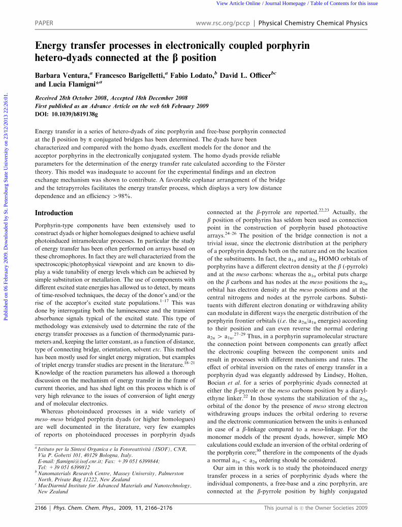
components. These are derived from the luminescence maxima
at 77 K of Table 3 and when data are not available, from the
corresponding model homo-dimers in Table 2. It has in fact
been shown that 1FB2, 2FB2, 3FB2 and 1PZn2, 2PZn2, 3PZn2are excellent models for the corresponding units in the hetero-
dyads. Energy transfer is predicted in 1FB-PZn, 2FB-PZn and
3FB-PZn from the singlet excited states localized on zinc
porphyrin (FB-1PZn) to the singlet excited state localized on
the free-base component (1FB-PZn). The DG0 for the reaction
step is �0.07 eV for 1FB-PZn, �0.13 eV for 2FB-PZn and
�0.12 eV for 3FB-PZn. A quenching of the zinc porphyrin
luminescence in 2FB-PZn and 3FB-PZn was determined, with
lifetimes of 14 ps and 25 ps, respectively. The rate constant ken,
with ken = 1/t-1/t0 where t is the experimental quenched
lifetime and t0 is the lifetime in absence of quenching, i.e. that
of the model homo-dimer (1PZn2-3PZn2), can be calculated.
They are, respectively 7.1 � 1010 s�1 for 2FB-PZn and
3.9 � 1010 s�1 for 3FB-PZn. The rates are in line with those
reported for similar porphyrinic dyads using phenylethynyl
spacers22,44 but higher than those recently reported by
Albinsson for the same phenylethynyl bridges.45 The diffe-
rence in the above literature data was ascribed to the different
nature of the HOMO orbital, respectively an a1u in the latter
case and an a2u in the former. As already discussed above,
whereas a2u is characterized by an electron density at the meso
carbon, the attach position of the bridge, a1u has a node at the
same carbon. However, in the present case where the HOMO
was shown to be an a2u30 and the bridge is attached at the
b pyrrole carbon, where a2u has a node, the rate is still very
fast. This seems to confirm the explanation proposed by
Albinsson and related to the role of the dihedral angle at the
connection point between the porphyrin and the bridge.45 The
perpendicular orientation decreases the electronic coupling
and consequently the rate whereas nearly coplanar conforma-
tion, as in the present case, enhances the coupling and the
rate.45 The energy transfer rate process within the components
in 1FB-PZn is very fast and occurs within the laser pulse, so we
can set a lower limit of 1011 s�1, i.e. a lifetime of 10 ps,
corresponding to the instrumental resolution. It should how-
ever be stressed that in 1FB-PZn the large overlap of the
emission of the zinc porphyrin donor unit (remarkably red-
shifted with respect to the other zinc porphyrin components)
with that of the free-base porphyrin acceptor makes this
determination liable to some uncertainty. The energy transfer
efficiency Z, Z = ken/(ken + 1/t0), is 100% for 1FB-PZn, 99%
for 2FB-PZn and 98% for 3FB-PZn, respectively. Energy
transfer takes also place at 77 K in a toluene glass with rates
of 1.2� 1010 s�1 and 3.9� 109 s�1 for 2FB-PZn and 3FB-PZn,
corresponding to an efficiency Z = 97% and Z = 91%,
respectively.
In order to discuss the process we have to take into account
that singlet-singlet energy transfer can take place via two
different mechanisms: a Coulombic interaction and an
electron exchange interaction. We will first discuss the
Coulombic interaction, also known in its simplest form as a
dipole–dipole interaction, described by Forster.46 The rate of
the process can be estimated from eqn (1).
kFen ¼8:8� 10�25k2F
n4td6DA
JF ð1Þ
F and t are the emission quantum yield and lifetime of the
donor, dDA is the donor–acceptor center-to-center distance, n
is the refractive index of toluene (1.4961) and JF is the Forster
overlap integral which can be calculated from the emission
spectrum of the donor, 1PZn2, 2PZn2, 3PZn2, and the absorp-
tion spectrum of the acceptor, i.e. half of the absorption
spectrum of 1FB2, 2FB2, 3FB2, according to eqn (2).
JF ¼RFð�uÞeð�uÞ=�u4d�uRFð�uÞd�u
ð2Þ
An assumption regarding the geometrical factor K2, which
depends on the relative orientation of the transition dipoles of
the donor and acceptor, is necessary in order to use eqn (1).47
K2, which is 2/3 when there is a random orientation of the
transition dipoles of the units as occurs in freely diffusing
components, can assume quite different values for units
engaged in supramolecular structures, where the orientation
depends on the architecture of the array. In addition, a
detailed knowledge of the minimized geometry of the array
should be known. K2 has been calculated for similar systems
and was found to range between 5/6 and 1.25, therefore we use
an average value of 1.1.22,44,45 The parameters used to derive
the Forster energy transfer rate constant, kFen, are reported in
Table 4 with the results and the experimental values ken. The
calculated values are lower than the experimental data, at least
in the cases where the rate could be experimentally deter-
mined. This indicate that dipole–dipole mechanism by itself is
inadequate to describe in full the energy transfer occurring in
the present case. Similar unsatisfactory results by a dipole–
dipole interpretation for dyads based on a Zn porphyrin donor
and a free-base porphyrin acceptor connected at either the
meso or the b positions by phenylethyne bridges have been
reported previously by other groups.22,44,45
We will therefore consider the exchange mechanism: this has
been introduced by Dexter48 and can be described as a double
electron exchange involving the transfer of the electron from
the LUMO of the excited donor to the empty LUMO of the
acceptor with a concomitant transfer of an electron from the
HOMO of the acceptor to the HOMO of the donor. Since it
requires contact of the orbitals of the donor and of the
acceptor it is considered a short distance mechanism. Sub-
sequent development of the superexchange theory49 has allowed
to extend the validity of the Dexter theory to the exchange of
electrons mediated by the connecting bridge. The rate of
Scheme 2 Energy level diagram of 1FB-PZn, 2FB-PZn, and
3FB-PZn.
This journal is �c the Owner Societies 2009 Phys. Chem. Chem. Phys., 2009, 11, 2166–2176 | 2173
Publ
ishe
d on
06
Febr
uary
200
9. D
ownl
oade
d by
St.
Pete
rsbu
rg S
tate
Uni
vers
ity o
n 23
/12/
2013
22:
26:0
1.
View Article Online

energy transfer by electron exchange mechanism in a classical
formulation can be expressed by the following equation:48
kDen ¼4p2H2
hJD ð3Þ
where JD, the Dexter integral overlap, can be evaluated
from the absorption spectrum of the acceptor and from
the luminescence spectrum of the donors according to the
following equation:
JD ¼RFð�uÞeð�uÞd�u
RFð�uÞd�u
Reð�uÞd�u
ð4Þ
The value of JD for the various cases are collected with the
other parameters used for the calculation of the energy
transfer according to the Dexter theory in Table 5. The
contribution of the exchange mechanism to the overall,
experimentally determined rate constant ken, was determined
by the difference with the calculated Forster energy transfer,
i.e. kDen = ken � kFen. By introducing the kDen calculated values
(Table 4) and JD in eqn (3), an inter-component electronic
interaction H of the order of 10.0 cm�1 for 2FB-PZn and
3FB-PZn can be derived. The order of magnitude of such
parameters indicate a moderate coupling between the model
components, in agreement with previously discussed
spectroscopic data.
It could be interesting to address the issue of the distance
dependence of energy transfer even if only two terms of the
series can be considered. In the frame of the superexchange
mechanism, H follows an approximate exponential law with
increasing distance of the donor and the acceptor and can be
expressed as:50
H = H0 exp [�0.5br] (5)
where H0 is the intercomponent electronic interaction at the
contact distance, b is the attenuation factor and r can be taken
as the edge to edge distance between the donor and acceptor.
From eqn (3) and (5), the rate constant can be expressed as a
function of distance between reacting partners, with A being a
pre-exponential factor:51,52
kDen = A exp(�br) (6)
In the present case, where two p-phenylenevinylene bridges of
increasing length are present in 2FB-PZn and 3FB-PZn, an
attenuation factor b of the order of 0.06 A�1 can be calculated.
This treatment is highly simplified; in fact not only uses only two
terms of a series, but also regards b as a constant. It has actually
been shown by Albinsson and coworkers40,47,48 that the pre-
exponential factor A is a function of the energy gap between the
donor and the bridge and therefore b, in addition to be a system
specific parameter (and not a bridge specific parameter) might be
non exponential for conjugated bridges. For these type of con-
nectors, in fact, the energy is strongly affected by the bridge length.
In spite of this approximation, the extracted b can be compared
with literature values for other types of bridges. For singlet energy
transfer attenuation factors b = 1.33 A�1 for rigid hydrocarbon
bridges53 and b = 0.2 A�1 for poly-phenyleneethynylene45 have
been reported and values of the order of 0.06–0.08 A�1 can be
calculated from the data reported for polyene and poly-yne
linkages.39 For triplet energy transfer were reported values of
b = 0.17 A�1 for poly-yne bridges,54 and of b = 0.33 A�1 or
b = 0.32 A�1 or b = 0.65 A�1 for oligophenylene bridges.55,56
In conclusion the present b value places these p-phenylene-
vinylene bridged porphyrin systems as extremely effective
energy transducers, and this fact should be ascribed not only
to the intrinsic high electronic conductance of the p-phenylene-
vinylene bridge but also to the favorable coplanar arrangement
of the bridge and the chromophores.
Conclusions
Determination of energy transfer in a series of free-base
porphyrin/zinc porphyrin dyads connected by p-delocalizedbridges has been accurately performed with respect to models,
the corresponding free-base porphyrin and zinc porphyrin
homo-dyads, which perfectly represent the units in the same
molecular environment of the dyads. This has allowed us to
compare the experimental rates with those calculated accord-
ing to current models with more confidence than has been ever
done before. The mechanism cannot be accounted for entirely
by a dipole–dipole (Forster) scheme, and the inclusion of a
contribution by electron exchange (Dexter) in the p-phenylene-
vinylene containing dyads has led us to derive a very weak
distance-dependence of the rate of the exchange energy trans-
fer. This is assigned to the nature of the bridge and to the
highly coplanar arrangements of the bridge with respect to the
donor, which enhances the coupling. The overall result is a
very high (498%) energy transfer efficiency.
Experimental
The synthesis of dyads 1FB2 and 1FB-Zn were performed
according to the procedures described by Bonfantini and
Table 4 Energy transfer; parameters used for the calculation (according to the Forster theory) and experimental data
dDA/Aa K2 F t/ns JF/cm3 M�1 kFen/s
�1b ken/s�1c
1FB-PZn 14.0 1.1 0.29 1.4 5.69 � 10�14 3.0 � 1011 41011
2FB-PZn 18.3 1.1 0.09 1.6 6.07 � 10�14 1.8 � 1010 7.1 � 1010
3FB-PZn 24.5 1.1 0.07 1.7 8.43 � 10�14 3.1 � 109 3.9 � 1010
a Center to center donor–acceptor distance. b Calculated value. c Experimental value.
Table 5 Energy transfer; parameters used for the calculation (accordingto the electron exchange mechanism) and experimental data
r/Aa JD/cm ken/s�1 kDen/s
�1
1FB-PZn 6.3 2.42 � 10�4 41011 —2FB-PZn 10.5 4.35 � 10�4 7.1 � 1010 5.3 � 1010
3FB-PZn 17.1 2.97 � 10�4 3.9 � 1010 3.6 � 1010
a Edge-to-edge donor–acceptor distance.
2174 | Phys. Chem. Chem. Phys., 2009, 11, 2166–2176 This journal is �c the Owner Societies 2009
Publ
ishe
d on
06
Febr
uary
200
9. D
ownl
oade
d by
St.
Pete
rsbu
rg S
tate
Uni
vers
ity o
n 23
/12/
2013
22:
26:0
1.
View Article Online

Officer.57 The synthesis of other dyads will be reported
separately.58 The solvent used was spectroscopic grade toluene
(C. Erba). Spectroscopic grade tetrahydrofurane (C. Erba)
was added in concentration ranging from 5% to 10% to the
free-base and zinc homo-dyads exhibiting some difficulty to
dissolve. Absorption spectra were recorded with a Perkin-
Elmer Lambda 9 spectrophotometer and emission spectra,
uncorrected if not otherwise specified, were detected by a Spex
Fluorolog II spectrofluorimeter equipped with a Hamamatsu
R928 photomultiplier. Relative luminescence intensities were
evaluated from the area of the luminescence spectra corrected
for the photomultiplier response. Emission quantum yields
were determined with reference to TPP in aerated toluene
(ffl = 0.1127). Luminescence lifetimes in the nanosecond range
were obtained with an IBH single photon counting equipment
with excitation at 465 nm from a pulsed diode source
(0.5 ns resolution). Luminescence lifetimes and spectra in the
picosecond range were determined by an apparatus based
on a Nd:YAG laser (Continuum PY62-10) with a 35 ps
pulse duration, 532 nm, 1 mJ/pulse and a Streak Camera
(Hamamatsu C1587 equipped with M1952). The luminescence
images from 500 laser shots were averaged and the time profile
was measured from the resulting streak image in a wavelength
range of ca. 20 nm around the selected wavelength. The fitting
of the luminescence decays were performed by standard
iterative non linear programs taking into consideration the
instrumental response. Time resolved spectra were taken over
a time window of 30 ps at the specified delays. More details for
the streak camera experiment can be found elsewhere.59
Standard 10 mm fluorescence cells were used at 295 K
whereas experiments at 77 K made use of capillary tubes
dipped in an home made quartz Dewar filled with liquid
nitrogen. Estimated errors are 10% on lifetimes and 20%
on quantum yields. Working temperature, if not otherwise
specified, was 295 � 2 K.
Computation of the integral overlap and of the rate for the
energy transfer processes according to Forster mechanism
were performed with the use of Matlab 5.2.60 Molecular
dimensions and distances were estimated after minimization
(MM2) by CS Chem 3D Ultra 6.0.61
Acknowledgements
We thank CNR of Italy (PM.P04.010 MACOL) for financial
support.
References
1 F. Odobel, S. Suresh, E. Blart, Y. Nicolas, J. P. Quintard,P. Janvier, J. Y. Le Questel, B. Illien, D. Rondeau,P. Richomme, T. Haupl, S. Wallin and L. Hammarstrom,Chem.–Eur. J., 2002, 8, 3027–3046.
2 D. Holten, D. F. Bocian and J. S. Lindsey, Acc. Chem. Res., 2002,35, 57–69.
3 I. V. Sazanovich, A. Balakumar, K. Muthukumaran, E. Hindin,C. Kirmaier, J. R. Diers, J. S. Lindsey, D. F. Bocian andD. Holten, Inorg. Chem., 2003, 42, 6616–6628.
4 N. Aratani, A. Osuka, H. S. Cho and D. Kim, J. Photochem.Photobiol. C, 2002, 3, 25–52.
5 M. S. Choi, T. Aida, T. Yamazaki and I. Yamazaki, Chem.–Eur.J., 2002, 8, 2668–2678.
6 R. A. Haycock, A. Yartsev, U. Michelsen, V. Sundstrom andC. A. Hunter, Angew. Chem., Int. Ed., 2000, 39, 3616–3619.
7 L. Flamigni, A. M. Talarico, B. Ventura, G. Marconi,C. Sooambar and N. Solladie, Eur. J. Inorg. Chem., 2004,2557–2569.
8 E. Iengo, E. Zangrando, E. Alessio, J. C. Chambron, V. Heitz,L. Flamigni and J. P. Sauvage, Chem.–Eur. J., 2003, 9, 5879–5887.
9 R. Shediac, M. H. B. Gray, H. T. Uyeda, R. C. Johnson,J. T. Hupp, P. J. Angiolillo and M. J. Therien, J. Am. Chem.Soc., 2000, 122, 7017–7033.
10 E. Iengo, E. Zangrando, M. Bellini, E. Alessio, A. Prodi,C. Chiorboli and F. Scandola, Inorg. Chem., 2005, 44, 9752–9762.
11 K. Kilsa, J. Kajanus, J. Martensson and B. Albinsson, J. Phys.Chem. B, 1999, 103, 7329–7339.
12 L. Flamigni, J. Photochem. Photobiol., 2007, 8, 191–210.13 L. Flamigni, V. Heitz and J. P. Sauvage, Struct. Bonding, 2006,
121, 217–261.14 L. Flamigni, B. Ventura, A. I. Oliva and P. Ballester, Chem.–Eur.
J., 2008, 14, 4214–4224.15 L. Flamigni, A. M. Talarico, B. Ventura, R. Rein and N. Solladie,
Chem.–Eur. J., 2006, 12, 701–712.16 L. Flamigni, N. Armaroli, F. Barigelletti, J. C. Chambron,
J. P. Sauvage and N. Solladie, New J. Chem., 1999, 23, 1151–1158.17 T. S. Balaban, N. Berova, C. M. Drain, R. Hauschild,
X. F. Huang, H. Kalt, S. Lebedkin, J. M. Lehn, F. Nifaitis,G. Pescitelli, V. I. Prokhorenko, G. Riedel, G. Smeureanu andJ. Zeller, Chem.–Eur. J., 2007, 13, 8411–8427.
18 L. Flamigni, F. Barigelletti, N. Armaroli, B. Ventura, J. P. Collin,J. P. Sauvage and J. A. G. Williams, Inorg.Chem., 1999, 38,661–667.
19 M. Asano-Someda and Y. Kaizu, Inorg. Chem., 1999, 38,2303–2311.
20 A. Prodi, M. T. Indelli, C. J. Kleverlaan, F. Scandola, E. Alessio,T. Gianferrara and L. G. Marzilli, Chem.–Eur. J., 1999, 5,2668–2679.
21 J. Andreasson, J. Kajanus, J. Martensson and B. Albinsson, J. Am.Chem. Soc., 2000, 122, 9844–9845.
22 S. I. Yang, J. Seth, T. Balasubramanian, D. Kim, J. S. Lindsey,D. Holten and D. F. Bocian, J. Am. Chem. Soc., 1999, 121,4008–4018.
23 A. K. Burrell, D. L. Officer, D. C. W. Reid, S. M. Scott andK. C. Gordon, J. Porphyrins Phthalocyanines, 2000, 4, 626–633.
24 A. Lembo, P. Tagliatesta and D. M. Guldi, J. Phys. Chem. A, 2006,110, 11424–11434.
25 V. S. Y. Lin and M. J. Therien, Chem.–Eur. J., 1995, 1, 645–651.26 D. M. Guldi, B. Nuber, P. J. Bracher, C. A. Alabi, S. MacMahon,
J. W. Kukol, S. R. Wilson and D. I. Schuster, J. Phys. Chem. A,2003, 107, 3215–3221.
27 P. G. Seybold and M. Gouterman, J. Mol. Spectrosc., 1969, 31,1–13.
28 R. A. Binstead, M. J. Crossley and N. S. Hush, Inorg. Chem., 1991,30, 1259–1264.
29 J. A. Shelnutt and V. Ortiz, J. Phys. Chem., 1985, 89, 4733–4739.30 B. Ventura, L. Flamigni, G. Marconi, F. Lodato and D. L. Officer,
New J. Chem., 2008, 32, 166–178.31 R. Kumble, S. Palese, V. S. Y. Lin, M. J. Therien and
R. M. Hochstrasser, J. Am. Chem. Soc., 1998, 120, 11489–11498.32 T. H. Huang, Y. J. Chen, S. S. Lo, W. N. Yen, C. L. Mai,
M. C. Kuo and C. Y. Yeh, Dalton Trans., 2006, 2207–2213.33 M. J. Frampton, H. Akdas, A. R. Cowley, J. E. Rogers,
J. E. Slagle, P. A. Fleitz, M. Drobizhev, A. Rebane andH. L. Anderson, Org. Lett., 2005, 7, 5365–5368.
34 E. Blart, F. Suzenet, J. P. Quintard and F. Odobel, J. PorphyrinsPhthalocyanines, 2003, 7, 207–213.
35 M. Chachisvilis, V. S. Chirvony, A. M. Shulga, B. Kallebring,S. Larsson and V. Sundstrom, J. Phys. Chem., 1996, 100,13857–13866.
36 M. Chachisvilis, V. S. Chirvony, A. M. Shulga, B. Kallebring,S. Larsson and V. Sundstrom, J. Phys. Chem., 1996, 100,13867–13873.
37 S. G. Johnson, G. J. Small, D. G. Johnson, W. A. Svec andM. R. Wasielewski, J. Phys. Chem., 1989, 93, 5437–5444.
38 R. Gauler and N. Risch, Eur. J. Org. Chem., 1998, 1193–1200.39 A. Osuka, N. Tanabe, S. Kawabata, I. Yamazaki and
Y. Nishimura, J. Org. Chem., 1995, 60, 7177–7185.
This journal is �c the Owner Societies 2009 Phys. Chem. Chem. Phys., 2009, 11, 2166–2176 | 2175
Publ
ishe
d on
06
Febr
uary
200
9. D
ownl
oade
d by
St.
Pete
rsbu
rg S
tate
Uni
vers
ity o
n 23
/12/
2013
22:
26:0
1.
View Article Online

40 S. Cho, M. C. Yoon, C. H. Kim, N. Aratani, G. Mori, T. Joo,A. Osuka and D. Kim, J. Phys. Chem. C, 2007, 111, 14881–14888.
41 J. S. Hsiao, B. P. Krueger, R. W. Wagner, T. E. Johnson,J. K. Delaney, D. C. Mauzerall, G. R. Fleming, J. S. Lindsey,D. F. Bocian and R. J. Donohoe, J. Am. Chem. Soc., 1996, 118,11181–11193.
42 E. I. Sagun, E. I. Zenkevich, V. N. Knyukshto, A. M. Shulga,D. A. Starukhin and C. von Borczyskowski, Chem. Phys., 2002,275, 211–230.
43 L. Flamigni, N. Armaroli, F. Barigelletti, V. Balzani, J. P. Collin,J. O. Dalbavie, V. Heitz and J. P. Sauvage, J. Phys. Chem. B, 1997,101, 5936–5943.
44 H. S. Cho, D. H. Jeong, M. C. Yoon, Y. H. Kim, Y. R. Kim,D. Kim, S. C. Jeoung, S. K. Kim, N. Aratani, H. Shinmori andA. Osuka, J. Phys. Chem. A, 2001, 105, 4200–4210.
45 K. Pettersson, A. Kyrychenko, E. Ronnow, T. Ljungdahl,J. Martensson and B. Albinsson, J. Phys. Chem. A, 2006, 110,310–318.
46 T. Forster, Discuss. Faraday Soc., 1959, 27, 7–17.47 W. Van Der Meer, G. Coker and S. Y. Simon Chen, in Resonance
Energy Transfer Theory and Data, VCH, Cambridge, 1994,pp. 55–83.
48 D. L. Dexter, J. Chem. Phys., 1953, 21, 836–850.
49 H. M. McConnell, J. Chem. Phys., 1961, 35, 508–515.50 R. A. Marcus and N. Sutin, Biochim. Biophys. Acta, 1985, 811,
265–322.51 M. P. Eng and B. Albinsson, Angew. Chem., Int. Ed., 2006, 45,
5626–5629.52 K. Pettersson, J. Wiberg, T. Ljungdahl, J. Martensson and
B. Albinsson, J. Phys. Chem. A, 2006, 110, 319–326.53 H. Oevering, J. W. Verhoeven, M. N. Paddonrow, E. Cotsaris and
N. S. Hush, Chem. Phys. Lett., 1988, 143, 488–495.54 A. Harriman and R. Ziessel, Chem. Commun., 1996,
1707–1716.55 F. Barigelletti, L. Flamigni, M. Guardigli, A. Juris, M. Beley,
S. ChodorowskiKimmes, J. P. Collin and J. P. Sauvage, Inorg.Chem., 1996, 35, 136–142.
56 B. Schlicke, P. Belser, L. De Cola, E. Sabbioni and V. Balzani,J. Am. Chem. Soc., 1999, 121, 4207–4214.
57 E. E. Bonfantini and D. L. Officer, Tetrahedron Lett., 1993, 34,8531–8534.
58 F. Lodato and D. L. Officer, manuscript in preparation.59 L. Flamigni, J. Phys. Chem., 1993, 97, 9566–9572.60 Matlab 5.2., The MathWorks Inc., Natik MA 01760, U.S.A.61 CS Chem 3D Ultra CambridgeSoft.com, Cambridge MA, USA,
2000.
2176 | Phys. Chem. Chem. Phys., 2009, 11, 2166–2176 This journal is �c the Owner Societies 2009
Publ
ishe
d on
06
Febr
uary
200
9. D
ownl
oade
d by
St.
Pete
rsbu
rg S
tate
Uni
vers
ity o
n 23
/12/
2013
22:
26:0
1.
View Article Online

![Der Einfluß von Packungseffekten auf die ... · in-vitro-Umsetzung von N-Acetoxyanilin mit Desoxyguanosin und DNA ... aus 3,4-Dilithio-2,5-dimethyl-2,4-hexa-dien; das erste „Hetero[6]radialen"](https://static.fdocument.org/doc/165x107/5b1540ab7f8b9adc528b6487/der-einfluss-von-packungseffekten-auf-die-in-vitro-umsetzung-von-n-acetoxyanilin.jpg)


![Lecture 12 Heteroscedasticity · • Now, we have the CLM regression with hetero-(different) scedastic (variance) disturbances. (A1) DGP: y = X + is correctly specified. (A2) E[ |X]](https://static.fdocument.org/doc/165x107/6106a6b3fb4f960ead0036bd/lecture-12-h-a-now-we-have-the-clm-regression-with-hetero-different-scedastic.jpg)