
Electron Emission and Biological Consequences of...
124
DISSERTATION Titel der Dissertation „Electron Emission and Biological Consequences of Hormones in Polar Media, Studied on Testosterone, Progesterone, 17β-Estradiol and Genistein.“ Verfasserin Mag.rer.nat. Heike Schittl angestrebter akademischer Grad Doktorin der Naturwissenschaften (Dr.rer.nat.) Wien, 2011 Studienkennzahl lt. Studienblatt: A 091 442 Dissertationsgebiet lt. Studienblatt: Dr.-Studium der Naturwissenschaften Anthropologie Betreuerin / Betreuer: Prof. Dr. Nikola Getoff
Transcript of Electron Emission and Biological Consequences of...
DISSERTATION
Titel der Dissertation
„Electron Emission and Biological Consequences of
Hormones in Polar Media, Studied on Testosterone,
Progesterone, 17β-Estradiol and Genistein.“
Verfasserin
Mag.rer.nat. Heike Schittl
angestrebter akademischer Grad
Doktorin der Naturwissenschaften (Dr.rer.nat.)
Wien, 2011
Studienkennzahl lt. Studienblatt: A 091 442
Dissertationsgebiet lt. Studienblatt: Dr.-Studium der Naturwissenschaften Anthropologie
Betreuerin / Betreuer: Prof. Dr. Nikola Getoff

2
3
DANKSAGUNG
Ich möchte mich bei all jenen bedanken, die zur Erstellung dieser Doktorarbeit durch
fachliche, finanzielle, direkte oder indirekte Unterstützung beigetragen haben:
- allen voran Herrn Univ. Prof. Dr. D.I. Nikola Getoff für die Überlassung des
Themas, die engagierte, wissenschaftliche Betreuung und die stetige
Diskussionsbereitschaft,
- dem österreichischen Fonds für Wissenschaft und Forschung (FWF) für die
finanzielle Unterstützung im Rahmen des Projektes: “Free Radical Action on
Sexual Hormones in Respect to Cancer.“
- Frau Dr. Ruth M. Quint für die vielen wertvollen Hinweise und Hilfeleistungen,
sowie allen weiteren Mitarbeitern der Sektion für die gute Zusammenarbeit.
Ganz besonderer Dank gilt meiner Familie, die mir mein Studium ermöglicht und mich
in jeder Hinsicht unterstützt hat.

4
5
TABLE OF CONTENTS List of Abbreviations ....................................................................................................... I
1. Abstract .................................................................................................................. III
2. Zusammenfassung ................................................................................................... V
3. Introduction .............................................................................................................. 1
3.1. Hormones ....................................................................................................... 1
3.1.1. Sex steroids ................................................................................................ 1
3.1.1.1. Estrogens ............................................................................................. 3
3.1.1.2. Progestogens ........................................................................................ 4
3.1.1.3. Androgens ............................................................................................ 6
3.1.2. Phytohormone Genistein ............................................................................ 7
3.1.3. Sex hormones and cancer ........................................................................... 7
3.1.3.1. Sex hormones act as electron mediators .............................................. 9
3.2. Free radicals ................................................................................................. 10
3.2.1. Reactive oxygen species ........................................................................... 10
3.2.2. Solvated electrons .................................................................................... 12
3.2.2.1. Simulation of emission of e-aq by UV-irradiation of hormones ......... 13
3.2.3. Formation of free radicals by ionizing radiation ...................................... 13
3.2.3.1. 60Co as irradiation source .................................................................. 16
3.2.3.2. Radiolysis of water and aqueous solutions ........................................ 17
3.2.3.3. Biological action of ionizing radiation .............................................. 21
3.2.4. Free radicals in the human organism – only a burden? ............................ 23
3.2.4.1. Generation of free radicals in the mitochondria ................................ 23
3.2.4.2. Free radicals as regulatory mediators in cell signaling ...................... 25
3.2.4.3. Intracellular killing of bacteria by phagocytes .................................. 25
3.3. Antioxidants ................................................................................................. 27
3.3.1. Vitamin C ................................................................................................. 27
4. Study Objectives .................................................................................................... 29
5. Materials and Methods ........................................................................................... 31
5.1. Hormones and vitamins ............................................................................... 31
5.2. Sterilization of materials .............................................................................. 31
6
5.3. Sources of radiation ..................................................................................... 31
5.3.1. UV-irradiation apparatus .......................................................................... 31
5.3.1.1. Monochloracetic Acid Actinometry .................................................. 33
5.3.1.1.1. Reagents ..................................................................................... 33
5.3.1.1.2. Establishment of the calibration curve ....................................... 33
5.3.1.1.3. UV-irradiation of monochloracetic acid ..................................... 33
5.3.2. 60Co gamma source ................................................................................. 34
5.3.2.1. Fricke dosimetry ................................................................................ 34
5.4. Saturation of solutions with air, N2O or argon ............................................ 36
5.5. Formation of carboxylic acids ..................................................................... 36
5.6. Spectrophotometrical measurement of electron ejection ............................. 38
5.6.1. Reagents ................................................................................................... 38
5.6.2. Electron emission ..................................................................................... 38
5.7. HPLC analyses ............................................................................................. 39
5.8. Experiments in vitro with Escherichia coli bacteria .................................... 40
5.8.1. Media and buffer ...................................................................................... 41
5.8.1.1. Preparation of Agar Plates ................................................................. 41
5.8.2. Rehydration .............................................................................................. 41
5.8.3. Storage ...................................................................................................... 42
5.8.3.1. Long-term storage ............................................................................. 42
5.8.3.2. Short-term storage ............................................................................. 42
5.8.4. Radiation-biological analyses .................................................................. 42
5.8.4.1. Batch culture ...................................................................................... 42
5.8.4.2. Cytotoxicity assay ............................................................................. 43
5.8.4.3. Survival curves under γ-irradiation ................................................... 44
6. Results .................................................................................................................... 46
6.1. Actinometry ................................................................................................. 46
6.2. Electron emission of hormones .................................................................... 46
6.2.1. Phytohormone GEN ................................................................................. 46
6.2.2. T alone and in mixture with P4 ................................................................ 50
6.2.3. Water-soluble E2, P4 and their mixture ................................................... 52
6.3. HPLC-analyses ............................................................................................ 56
6.3.1. T, P4 and their mixture ............................................................................. 56
7
6.3.2. T, vitC and their mixture .......................................................................... 60
6.4. Experiments in vitro ..................................................................................... 64
6.4.1. WsE2 and HBC ........................................................................................ 64
6.4.1.1. Toxicity .............................................................................................. 64
6.4.1.2. Survival curves under irradiation ...................................................... 64
6.4.2. WsP4 alone and in combination with wsE2 ............................................. 69
6.4.2.1. Toxicity of wsP4 ................................................................................ 69
6.4.2.2. Survival curves under irradiation ...................................................... 70
6.4.3. T and VitC ................................................................................................ 74
6.4.3.1. Toxicity of T ...................................................................................... 74
6.4.3.2. Survival curves under irradiation ...................................................... 75
7. Discussion .............................................................................................................. 79
7.1. Formation of hormone associates ................................................................ 79
7.2. Q(e-aq) values decrease with increasing hormone concentration ................. 79
7.3. UV-excitation of hormones is followed by multi-stage processes .............. 80
7.4. Temperature influences the electron emission ability of hormones ............ 81
7.5. Electron emission of GEN compared with E2 ............................................. 81
7.6. Testosterone ................................................................................................. 82
7.6.1. Effect of P4 on the electron emission and degradation of T .................... 82
7.6.2. Regeneration of T by vitC ........................................................................ 85
7.6.3. Effect of free radicals on T alone and in mixture with vitC ..................... 87
7.7. Water soluble hormones .............................................................................. 88
7.7.1. Electron emission of water soluble hormones .......................................... 88
7.7.2. Effect of free radicals on water-soluble hormones ................................... 90
8. Conclusion ............................................................................................................. 94
9. References .............................................................................................................. 97
10. Curriculum vitae .................................................................................................. 107

I
LIST OF ABBREVIATIONS
16OH DHEA 16-dydroxydehydroepiandrosterone
17OHP 17α-hydroxyprogesterone
17β-HSD 17β-hydroxysteroid dehydrogenase
20αHP 4-pregnen-20α-ol-3-one
2-OHE1 2-hydroxyestrone
3αHP 4-pregnen-3α-ol-20-one
3β-HSD 3β-hydroxysteroid dehydrogenase
4-OHE1 4-hydroxyestrone
5αP 5-pregnane-3,20-dione
A- ascorbate
A• ascorbyl radical
AA L-ascorbic acid
AE androstenedione
AP-1 activation protein 1
AP-site apurinic/apyrimidinic site
AR androgen receptor
ATP adenosine triphosphate
CEs catecholestrogens
CYP-450 cytochrome P450
cyt c cytochrome c
cyt P450scc cholesterol side-chain cleavage enzyme
DHA dehydroascorbic acid
DHEA dehydroepiandrosterone
DHT dihydrotestosterone
DNA desoxy-ribonucleic-acid
DSB double strand break
E. coli Escherichia coli
E1 estrone
E2 17β-estradiol
E3 estriol
ER estrogen receptor
II
G protein guanine nucleotide-binding proteins
GEN genistein
GLUT glucose transporter
Gy Gray
HBC (2-Hydroxypropyl)- β-cyclodextrin
HIF1 hypoxia-inducible factor 1
HPLC high performance liquid chromatography
IκB inhibitory factor kappa B
JNK c-Jun N-terminal kinase
MDS multiply damaged site
MMC mitomycin C
MOMP mitochondrial outer membrane permeabilization
mtDNA mitochondrial DNA
NADP+ nicotinamide adenine dinucleotide phosphate
NADPH reduced nicotinamide adenine dinucleotide phosphate
NFκB transcription nuclear factor kappa B
OD optical density
p38 MAPK p38 mitogen-activated protein kinase
P4 progesterone
PR progesterone receptor
PTP permeability transition pore
rad radiation absorbed dose
ROS reactive oxygen species
SHBG sex hormone binding globulin
SOD superoxide dismutase
SSB single strand break
SVCT Na+-AA cotransport system
T testosterone
vitC vitamin C
vitE vtamin E
wsE2 water soluble 17β-estradiol
wsP4 water soluble progesterone
β-car β-carotene
III
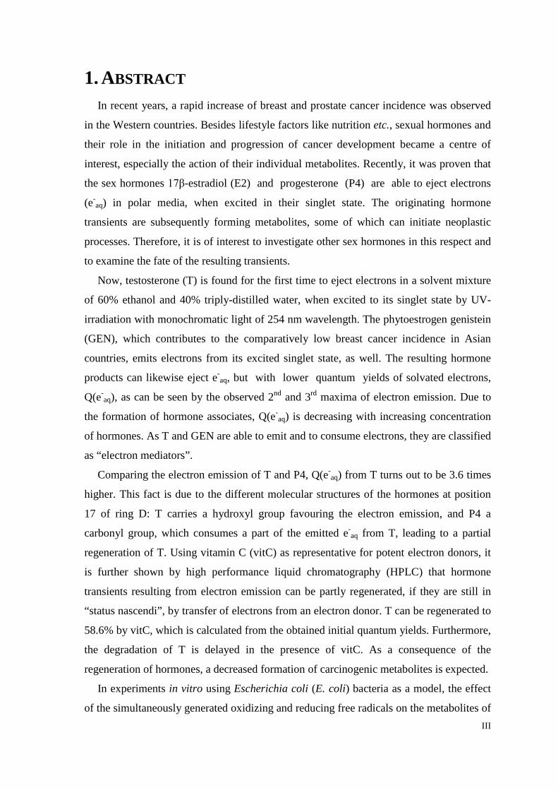
1. ABSTRACT In recent years, a rapid increase of breast and prostate cancer incidence was observed
in the Western countries. Besides lifestyle factors like nutrition etc., sexual hormones and
their role in the initiation and progression of cancer development became a centre of
interest, especially the action of their individual metabolites. Recently, it was proven that
the sex hormones 17β-estradiol (E2) and progesterone (P4) are able to eject electrons
(e-aq) in polar media, when excited in their singlet state. The originating hormone
transients are subsequently forming metabolites, some of which can initiate neoplastic
processes. Therefore, it is of interest to investigate other sex hormones in this respect and
to examine the fate of the resulting transients.
Now, testosterone (T) is found for the first time to eject electrons in a solvent mixture
of 60% ethanol and 40% triply-distilled water, when excited to its singlet state by UV-
irradiation with monochromatic light of 254 nm wavelength. The phytoestrogen genistein
(GEN), which contributes to the comparatively low breast cancer incidence in Asian
countries, emits electrons from its excited singlet state, as well. The resulting hormone
products can likewise eject e-aq, but with lower quantum yields of solvated electrons,
Q(e-aq), as can be seen by the observed 2nd and 3rd maxima of electron emission. Due to
the formation of hormone associates, Q(e-aq) is decreasing with increasing concentration
of hormones. As T and GEN are able to emit and to consume electrons, they are classified
as “electron mediators”.
Comparing the electron emission of T and P4, Q(e-aq) from T turns out to be 3.6 times
higher. This fact is due to the different molecular structures of the hormones at position
17 of ring D: T carries a hydroxyl group favouring the electron emission, and P4 a
carbonyl group, which consumes a part of the emitted e-aq from T, leading to a partial
regeneration of T. Using vitamin C (vitC) as representative for potent electron donors, it
is further shown by high performance liquid chromatography (HPLC) that hormone
transients resulting from electron emission can be partly regenerated, if they are still in
“status nascendi”, by transfer of electrons from an electron donor. T can be regenerated to
58.6% by vitC, which is calculated from the obtained initial quantum yields. Furthermore,
the degradation of T is delayed in the presence of vitC. As a consequence of the
regeneration of hormones, a decreased formation of carcinogenic metabolites is expected.
In experiments in vitro using Escherichia coli (E. coli) bacteria as a model, the effect
of the simultaneously generated oxidizing and reducing free radicals on the metabolites of
IV
different hormones is simulated. Saturation of the bacteria containing medium with
various gases before γ-irradiation allows the production of certain concentrations and
kinds of primary free radicals as a result of water radiolysis: in aerated medium 46% OH•
and 54% O2•-, in medium saturated with N2O 90% OH• and 10% H•, and in medium with
argon 44% e-aq, 10% H• and 46% OH•. Hereby, water-soluble E2 (wsE2) with
incorporated 2-hydroxypropyl-β-cyclodextrin (HBC) is found to be a very powerful
scavenger of OH• and O2•- radicals. Under the attack of reducing species (e-
aq, H•) wsE2
exhibits strong anti-proliferative properties. Water-soluble P4 (wsP4) with incorporated
HBC and mixtures of wsE2 and wsP4 exhibit contrary effects compared to wsE2,
indicating that a combination of wsP4 and wsE2 may strongly reduce the number of
carcinogenic metabolites of wsE2.
T, vitC and their mixture are investigated in a medium containing 4x10⁻² mol/L
ethanol, which has a strong scavenging ability for all primary free radicals except e-aq.
However, in aerated media oxidizing species are operating, whereas in media saturated
with argon or N2O reducing species predominate. T intermediates resulting from attack of
oxidizing free radicals show weak cytotoxic properties, but under reducing conditions, T
induces strong proliferation of E. coli bacteria. These effects are both intensified, when T
is combined with vitC.
The present results offer a deeper insight in the biological behaviour of sex hormones
and their metabolites. Possible reaction mechanisms of hormones and free radicals are
presented, but further radiobiological investigations are required, as the reaction
mechanisms are very complicated and overlap with many other biological processes. The
pulse radiolysis method could essentially contribute to the elucidation of these processes.
The combination experiments with vitC might show new approaches for the application
of hormones in medical therapies.
V
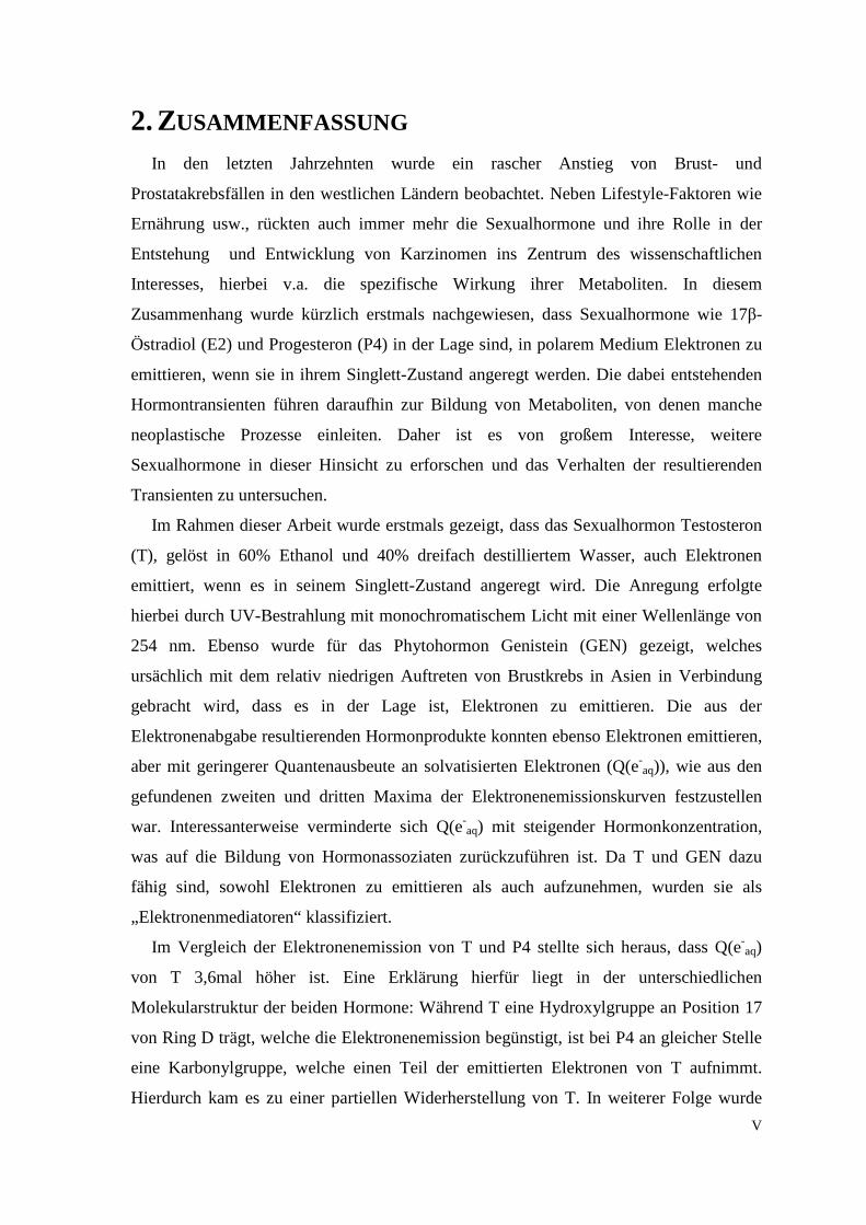
2. ZUSAMMENFASSUNG In den letzten Jahrzehnten wurde ein rascher Anstieg von Brust- und
Prostatakrebsfällen in den westlichen Ländern beobachtet. Neben Lifestyle-Faktoren wie
Ernährung usw., rückten auch immer mehr die Sexualhormone und ihre Rolle in der
Entstehung und Entwicklung von Karzinomen ins Zentrum des wissenschaftlichen
Interesses, hierbei v.a. die spezifische Wirkung ihrer Metaboliten. In diesem
Zusammenhang wurde kürzlich erstmals nachgewiesen, dass Sexualhormone wie 17β-
Östradiol (E2) und Progesteron (P4) in der Lage sind, in polarem Medium Elektronen zu
emittieren, wenn sie in ihrem Singlett-Zustand angeregt werden. Die dabei entstehenden
Hormontransienten führen daraufhin zur Bildung von Metaboliten, von denen manche
neoplastische Prozesse einleiten. Daher ist es von großem Interesse, weitere
Sexualhormone in dieser Hinsicht zu erforschen und das Verhalten der resultierenden
Transienten zu untersuchen.
Im Rahmen dieser Arbeit wurde erstmals gezeigt, dass das Sexualhormon Testosteron
(T), gelöst in 60% Ethanol und 40% dreifach destilliertem Wasser, auch Elektronen
emittiert, wenn es in seinem Singlett-Zustand angeregt wird. Die Anregung erfolgte
hierbei durch UV-Bestrahlung mit monochromatischem Licht mit einer Wellenlänge von
254 nm. Ebenso wurde für das Phytohormon Genistein (GEN) gezeigt, welches
ursächlich mit dem relativ niedrigen Auftreten von Brustkrebs in Asien in Verbindung
gebracht wird, dass es in der Lage ist, Elektronen zu emittieren. Die aus der
Elektronenabgabe resultierenden Hormonprodukte konnten ebenso Elektronen emittieren,
aber mit geringerer Quantenausbeute an solvatisierten Elektronen (Q(e-aq)), wie aus den
gefundenen zweiten und dritten Maxima der Elektronenemissionskurven festzustellen
war. Interessanterweise verminderte sich Q(e-aq) mit steigender Hormonkonzentration,
was auf die Bildung von Hormonassoziaten zurückzuführen ist. Da T und GEN dazu
fähig sind, sowohl Elektronen zu emittieren als auch aufzunehmen, wurden sie als
„Elektronenmediatoren“ klassifiziert.
Im Vergleich der Elektronenemission von T und P4 stellte sich heraus, dass Q(e-aq)
von T 3,6mal höher ist. Eine Erklärung hierfür liegt in der unterschiedlichen
Molekularstruktur der beiden Hormone: Während T eine Hydroxylgruppe an Position 17
von Ring D trägt, welche die Elektronenemission begünstigt, ist bei P4 an gleicher Stelle
eine Karbonylgruppe, welche einen Teil der emittierten Elektronen von T aufnimmt.
Hierdurch kam es zu einer partiellen Widerherstellung von T. In weiterer Folge wurde

VI
anhand von Vitamin C (vitC), welches ein gutes Beispiel für einen starken Elektrondonor
darstellt, mithilfe der HPLC (Hochleistungsflüssigkeitschromatographie) gezeigt, dass die
aus der Elektronenemission resultierenden Hormontransienten zumindest teilweise
regeneriert werden können durch Elektronentransfer, sofern sie sich noch in ihrem „Status
nascendi“ befinden. Hierbei konnte aus den erhaltenen anfänglichen Quantenausbeuten
berechnet werden, dass unter den gegebenen experimentellen Bedingungen T zu 58,6%
durch vitC regeneriert werden kann. Desweiteren wurde festgestellt, dass sich der Abbau
von T in Gegenwart von vitC erheblich verzögert. Durch die Regeneration von Hormonen
wird erwartet, dass sich nur vermindert krebserregende Metaboliten bilden.
Die Auswirkung von oxidierenden und reduzierenden freien Radikalen auf
Hormonmetaboliten wurde mithilfe von In vitro Experimenten mit Escherichia coli
Bakterien simuliert. Durch die Sättigung der Bakterien enthaltenden Lösungen mit
verschiedenen Gasen vor der γ-Bestrahlung konnten definierte Konzentrationen und
Arten von freien Radikalen als Produkte der Wasserradiolyse hergestellt werden: In
lufthältigem Medium waren dies 46% OH• und 54% O2•-, in N2O-gesättigtem Medium
90% OH• und 10% H• und luftfreiem Medium (gesättigt mit Argon) 44% e-aq, 10% H• und
46% OH•. Im Rahmen der Experimente wurde festgestellt, dass wasserlösliches E2
(wsE2), welches als Komplex mit 2-Hydroxypropyl-β-cyclodextrin (HBC) vorliegt, ein
sehr wirksamer Fänger von OH• und O2•- ist. Unter dem Einfluss von reduzierenden
Radikalen (e-aq, H•) zeigte sich eine starke, antiproliferative Wirkung von wsE2 auf die
Bakterien. Konträre Ergebnisse wurden für wasserlösliches P4 alleine (wsP4, im
Komplex mit HBC) und im Gemisch mit wsE2 gefunden, was darauf hindeutet, dass eine
Kombination der beiden Hormone zu einer erheblichen Reduzierung der karzinogenen
Metaboliten von wsE2 führen könnte.
T und vitC wurden auch im Rahmen von In vitro Experimenten mit Escherichia coli
Bakterien untersucht. Allen Medien wurde hierbei 4x10⁻² mol/L Ethanol zugesetzt, da T
nicht in reinem Wasser löslich ist. Da Ethanol ein guter Fänger von allen primären freien
Radikalen außer e-aq ist, waren in lufthältigem Medium nur oxidierende Radikale
vorhanden und in Medien gesättigt mit N2O oder Argon überwiegend reduzierende
Spezies. Die aus dem Angriff von oxidierenden Radikalen entstehenden
Zwischenprodukte von T zeigten eine schwach zytotoxische Wirkung auf die Bakterien.
Unter dem Einfluss von reduzierenden Radikalen induzierte T hingegen starke
Proliferation. VitC intensivierte den jeweiligen Effekt von T.

VII
Die erhaltenen Ergebnisse ermöglichen einen tieferen Einblick in das biologische
Verhalten von Sexualhormonen und ihren Metaboliten. Mögliche Reaktionsmechanismen
von Hormonen mit freien Radikalen werden präsentiert, aber es sind weitere
strahlenbiologische Untersuchungen vonnöten, da die Reaktionsmechanismen sehr
kompliziert sind und mit vielen anderen biologischen Prozessen überlappen. Die
Pulsradiolyse-Methode könnte bedeutend zur Aufklärung dieser Prozesse beitragen. Die
Experimente mit Hormon-vitC-Kombinationen könnten neue Ansätze zur Anwendung
von Hormonen in der Medizin aufzeigen.

1
3. INTRODUCTION
3.1. Hormones The human organism consists of about 1014 cells, which can differ in size,
compartmentalisation etc., and may be very distant from each other. However, for a
coordinated interaction of different cell types of an organism, cell communication is
essential. Different types of cell signalling by chemical messengers can be distinguished
according to the distance between the communicating cells. If cells generate the signal
themselves they are responding to, it is referred to as intern and extern autocrine
signalling, respectively. In the latter case, the signal is secreted before it is transduced to
the cytoplasm again, whereas in the first case signalling occurs only within the cytoplasm.
If the signalling cell is different from the target cell, juxtracrine, paracrine and endocrine
signalling can be distinguished. While juxtracrine signalling requires direct cell contact
by connexons, the messengers need to diffuse over short distances to adjacent cells after
secretion in paracrine signalling. Cells, that are very distant, cannot communicate via
diffusion-mediated messengers, as diffusion is a very slow process. Therefore, a
substance carrying the chemical signal, such as blood, is required. This type of
communication is called endocrine signalling.
Hormones represent a class of chemical messengers, which are produced by
specialized ductless glands or tissues in very low levels. In general, hormones act as
endocrine agents, but cells may also release paracrine or autocrine hormones. Upon
binding to specific receptors, which are expressed by the target cell, various effects may
be initiated within the target cell through signalling cascades. However, hormones can be
distinguished in steroid hormones, peptide/protein hormones and amino acid derivative
hormones.
3.1.1. Sex steroids
Sex hormones, such as estrogens, progestogens and androgens, belong to the class of
steroid hormones. They are essential for the development of sexual characteristics and
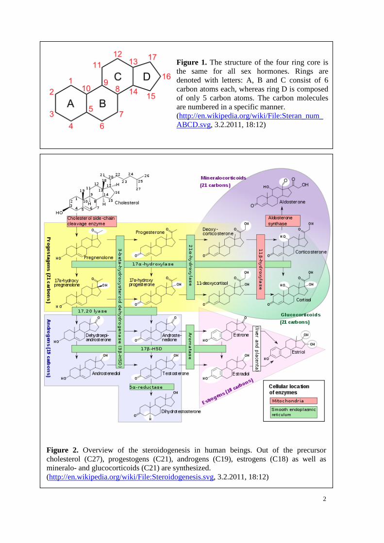
behaviour. All sex hormones are derived from cholesterol and share the same core, which
consists of four rings (Figure 1). While rings A, B and C are cyclohexanic, ring D
consists of solely five carbon atoms. The various sex hormones differ in the oxidative
status of the rings and in their attached, functional groups.
2
Figure 2. Overview of the steroidogenesis in human beings. Out of the precursor cholesterol (C27), progestogens (C21), androgens (C19), estrogens (C18) as well as mineralo- and glucocorticoids (C21) are synthesized. (http://en.wikipedia.org/wiki/File:Steroidogenesis.svg, 3.2.2011, 18:12)
Figure 1. The structure of the four ring core is the same for all sex hormones. Rings are denoted with letters: A, B and C consist of 6 carbon atoms each, whereas ring D is composed of only 5 carbon atoms. The carbon molecules are numbered in a specific manner. (http://en.wikipedia.org/wiki/File:Steran_num_ABCD.svg, 3.2.2011, 18:12)
3
The pathways of the generation of sex hormones from cholesterol are described by the
steroidogenesis (Figure 2). In a first step, cholesterol is converted to pregnenolone by
oxidation processes, which are catalyzed by the Cholesterol side-chain cleavage enzyme
(cyt P450scc). Further oxidation and a tautomerization process by 3β-HSD (3β-
hydroxysteroid dehydrogenase) result in the production of progesterone (P4), the major
progestogen. Out of pregnenolone and P4, 17α-hydroxypregnenolone and 17α-
hydroxyprogesterone (17OHP) can be formed with the help of the 17α-hydroxylase as
catalyzing agent. P4 and 17OHP may also be converted to diverse mineralcorticoids by
hydroxylation steps.
Androgens are generated from 17α-hydroxypregnenolone and 17OHP by the action of
17,20 lyase, which triggers the splitting off the side chain. In a first step,
dehydroepiandrosterone (DHEA) and androstenedione (AE) are produced, which may be
further processed to androstenediol and testosterone (T), respectively, a process requiring
17β-HSD. AE and T may also originate from the conversion of DHEA and
androstenediol, respectively, through oxidation by 3β-HSD. 5α-dihydrotestosterone
(DHT) is produced through reduction of T by the 5α-reductase.
The enzyme aromatase catalyzes the conversion of AE and T to the estrogens estrone
(E1) and estradiol (E2). Estriol (E3), the third naturally occurring estrogen is a metabolite
of E1 and E2 and for the most part made from sulfonated 16-dydroxydehydro-
epiandrosterone (16OH DHEA), which is a derivative of DHEA.
3.1.1.1. Estrogens
The term “estrogens” comprises E1, E2 and their metabolites, which all occur in the
human organism, as well as synthetic and other natural substances with estrogenic
properties. E2, which is the most biologically potent estrogen (1), is produced by all
mammals, females and males, in the gonads, but also in fat cells or the adrenal cortex. Its
action in the development of secondary sex characteristics in women (breast
development, etc.), female reproduction cycle and during pregnancy (placenta
development, etc.) classifies it as the main female sex hormone. Changes in the body
shape affecting bones, joints, fat deposition and structure and skin composition are
attributed to E2, as well (2). These effects are primarily initiated at the time of puberty,
most enhanced during the reproductive years, and become less pronounced after the
menopause, due to an decreasing support with E2. However, E2 is an important factor in
4
spermatogenesis, too, as it is implicated with the inhibition of male germ cells (3). As
sperm counts decrease over the past decades, an association with increasing E2 exposure
in the environment is postulated (4).
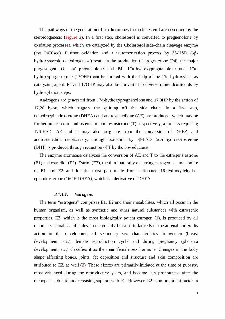
Within the body, estrogens are metabolised to products, which may either have
estrogenic or antiestrogenic activities. In the metabolism of E2, cytochrome P450 (CYP-
450) plays a decisive role, as it triggers the hydroxylation processes at position 2 and 4 of
ring A, resulting in the formation of the catecholestrogens (CEs) 2-hydroxyestrone (2-
OHE1) and 4-hydroxyestrone (4-OHE1), respectively, as well as that of ring D at position
16 (5). Hydroxylation of ring D leads to the generation of a number of metabolites, such
as 16α-hydroxyestrone and E3 (Figure 3). 2-OHE1 can be converted into a semichinone,
thus, the metabolism of E2 may give rise to free radicals (6), besides the radical
scavenging property of E2 due to the phenolic ring A.
In the blood plasma, E2 is bound in large part to SHBG (sex hormone binding
globulin) or serum albumin. Only the unbound, free portion may enter a cell and mediate
the activation of the target cell receptors ER (estrogen receptor) α and β (7, 8). ERs occur
in the cell nucleus or are located to the cytoplasm/cell membrane. Binding of ligands,
such as steroid hormones, to nuclear receptors results into ER homo- and
heterodimerization, when both ERs are co-expressed. The dimers affect gene transcription
by binding to promoters of target genes. The sites of binding to the DNA are called
estrogen responsive elements (9). The nuclear estrogen-ER complex may also interact
with other transcription factors, such as AP-1 (activation protein 1), and influence their
activity (10). Activation of ERs at the plasma membrane mainly results in G-protein
(guanine nucleotide-binding protein) mediated signalling (11).
3.1.1.2. Progestogens
Progestogens exhibit pro-gestational functions, as the name implies. P4, the major
progestogen, is an important factor in the development of the female mammary gland and
is produced during the female menstrual cycle with a peak after ovulation. It causes the
growth of the uterus and many other processes, which are necessary for gestation. Also
non-reproductive tissues are influenced by P4, such as the cardiovascular or the nervous
system (12). In the metabolism of P4 in various tissues, different parts of the molecule are
modified (13).
5
Figure 3. The metabolism of E2 leads to the formation of various A- and D-ring metabolites, which can undergo further degradation (32).
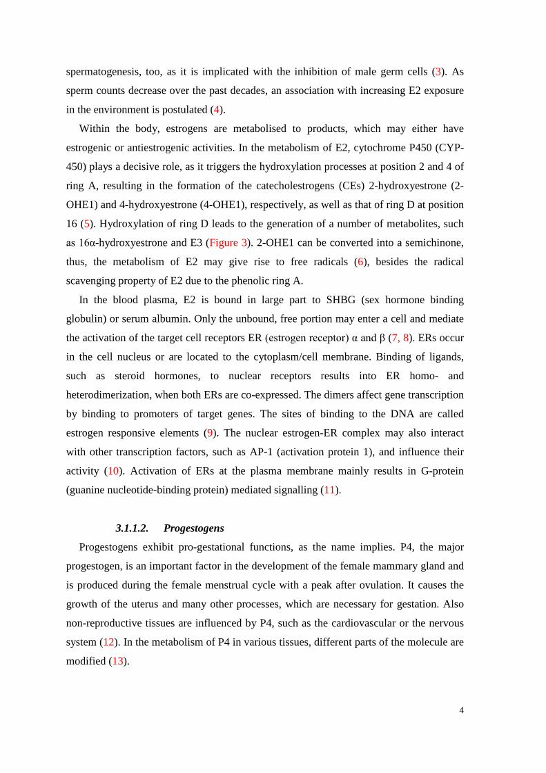
Figure 4. The metabolism of P4 in the breast tissue. 3a-HSO = 3a-hydroxysteroid oxireductase, 20a-HSO = 20a-hydroxysteroid oxireductase (14).
6
In the breast tissue, P4 is metabolized into two classes of metabolites: 5-pregnane-
3,20-dione (5αP) on the one hand, and the 4-pregnen group with 4-pregnen-3α-ol-20-one
(3αHP) and 4-pregnen-20α-ol-3-one (20αHP) on the other hand (Figure 4 (14)).
The nuclear receptors of P4 (PR-A and B), which are activated upon ligand-binding,
mediate the effect of P4 (15). In the course of this, PR-dimers bind to specific target
sequences in gene promoters, which are inducible by P4. Through activation of second
messengers and the corresponding signalling cascades, PRs exhibit also non-genotropic
effects (16). The effect of P4 in different tissues depends on the specific isoform of PR
(PR-A or PR-B), the following nuclear positioning and to a big extent on coregulators
(17). Like ER, PR is a member of the nuclear receptor superfamily.
3.1.1.3. Androgens
While the activity of estrogens and progestogens is mainly associated with the
development and maintenance of female sex characteristics, androgens are decisive for
male features, such as the development of the reproductive tissues (testes, prostate, etc.).
T is of great importance in females, though: A larger part of androgens than of estrogens
is quantitatively produced throughout a woman’s lifetime (18). In humans, synthesis of T
from AE, which is produced in the adrenal glands in large parts, occurs in the testes
(Leydig cells) and ovaries, respectively. T may then be metabolised to DHT, the most
potent androgen, in peripheral tissues by the 5α-reductase, or to estrogens. Hence,
through this pathway of metabolism, T may initiate estrogenic responses. However,
recently it was established, that androgens are also able to specifically bind ERs (19). In
both sexes, T was found to act as suppressor of breast growth (20).
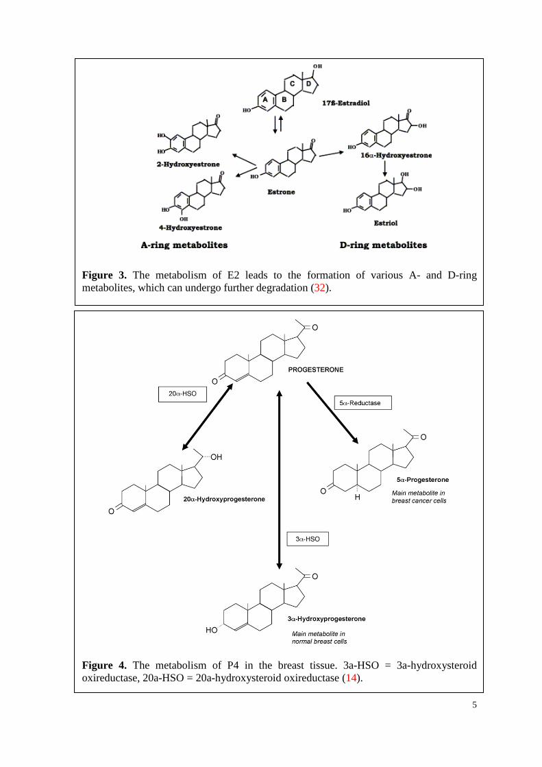
Figure 5. Chemical structure of the isoflavonoid genistein compared with 17β-estradiol (after (96)).
7
As already mentioned for E2, SHBG and serum albumin are the carriers of T and DHT
in the blood stream. Only the unbound fraction of androgens is biologically active. The
effect of T and DHT is mediated by the intracellular, ligand-activated androgen receptor
(AR), which belongs to the nuclear receptor superfamily (15) and acts as transcription
factor on androgen response elements. It is very closely related to PR, and thus,
protestogens are able to bind ARs (21). As mentioned above for estrogens and
progestogens, a rapid, non-genomic effect of androgens is also proposed (22).
3.1.2. Phytohormone Genistein
Phytohormones, such as isoflavones, gained much interest in the last decades.
Basically, phytohormones exhibit important functions in plant development and growth,
but upon ingestion by humans with everyday diet, they may be beneficial for health.
Isoflavonoids, which are contained in soy, representing a significant portion of the Asian
diet, were shown to influence the risk of breast or prostate cancers in Asian countries
significantly (23-25). The main isoflavonoid in soy is genistein (4,5,7-trihydroxy-
isoflavone, GEN), having a similar molecular structure to E2 (Figure 5) and weak
estrogenic properties (101). Thus, it is often referred to as phytoestrogen. GEN is able to
bind to both ERs: in comparison with E2, the affinity to ERα is 4% and to ERβ 87% (26).
By blocking the ERs for other, more potent estrogens, GEN is able to influence the
estrogen metabolism (27). In experiments with MCF-7 cells, GEN was shown to be an
antitumor agent, enhancing the efficiency of cytostatica, such as mitomycin C (28).
However, GEN is also known to be an antioxidant, that may protect cells against
oxidative stress due to its antimutagenic activity (29,30,102).
The reaction rate constants of GEN with various free radicals, which are rather high,
were already determined by pulse radiolysis studies, whereby k(e−aq+GEN) = 6.2x10⁹
L.mol-1.s-1 at pH ∼7 (115) and k(OH+GEN) = 2.3x10¹⁰ L.mol-1.s-1 at pH=8.3 (111).
3.1.3. Sex hormones and cancer
It is well known, that lifestyle-factors like nutrition, smoking, alcohol intake, etc. have
a great impact on the development of various carcinomas. As the incidence for breast and
prostate cancer is steadily rising in the Western countries, steroid hormones and their role
in the initiation and progression of cancer became a centre of interest. For a long time, the
hormone metabolites were thought to be inactive, but now their importance in this respect
8
is widely accepted within the scientific community and they are more and more subject of
studies investigating their impact on various types of cancer cell lines.
For E2 and some of its metabolites, a carcinogenic impact was already demonstrated
(31-33,103). E2 and many of its A-ring metabolites like 2-OHE1 and 4-OHE1 show a
biphasic pattern concerning cell proliferation: in low concentrations (1x10⁻⁸ – 1x10⁻⁶
mol/L) they exhibit a stimulating and in high concentrations (1x10⁻⁵ mol/L and less) an
inhibiting effect on cell proliferation. Metabolites of ring D, however, do not show similar
results (34). It is postulated that metabolites of E2 might even be more relevant in the
induction of breast cancer than E2 itself (104), which implies that the carcinogenity of
estrogens may depend firstly on their metabolism and secondly on the resulting
composition of metabolites.
Furthermore, estrogens are able to induce genetic instabilities, which is also implicated
with the development of cancer (109). Elevated levels of oxidized bases have been found
in estrogen induced cancer, and are even thought to precede cancer development. Thus,
they may be good biomarkers for cancer risk (110).
Up to now, there is no consensus about the action of P4 on cancer cells. The two kinds
of metabolites of P4 in breast tissue (5αP on one hand, and 3αHP and 20αHP on the other
hand) exhibit contrary effects: Whilst 5αP promotes mitogenesis and metastasis, 3αHP
and 20αHP show none of these attributes (13). Also the 5αP- and E2-modulated increase
of ER numbers can be reduced by 3αHP and 20αHP, which underlines the anticancer
impact of the 4-pregnen group of the endogenous produced P4 metabolites. Therefore, P4
metabolites seem to act as modulators on ER levels in various ER-positive MCF-7 breast
cancer cell lines (35,36).
The role of testosterone in cancer initiation and development is not elucidated yet
(37,38). Often, an association between cancer and low or elevated levels of androgens is
suggested (39,40). Experimental studies showed both proliferative (41) and anti-
proliferative (42) action of testosterone on various carcinoma cell lines. Castagnetta et al.
studied the metabolism of T in various prostate cancer cell lines (LNCaP, DU145 and
PC3) and found divergent patterns in this respect: While PC3-cells degradated T very
quickly to high levels of AE, the other two cell lines didn’t convert T to a large part (43).
The steroid receptor status of the cell line highly determinates the catalytic preference of
the steroid metabolism, which can be either oxidative or reductive (43,44). The same was
established for estrogens (45,46).
9
3.1.3.1. Sex hormones act as electron mediators
Recently it was found by Getoff et al. (47) that E2 and P4 can eject solvated electrons
(e-aq), when they become electronically excited in their singlet state (cf. chapter 3.2.2.
“Solvated electrons”). In the meantime, the same behaviour was shown for various other
hormones, such as 4-OHE1 (48), 17OHP (49) and E1 (50). However, E2 is able to eject
much more electrons than P4, the rate of yield is two orders of magnitude higher. A
possible explanation is the difference of the molecular structures of the two hormones.
Taking previously published papers into consideration (51,52), the π-electron structure of
ring A and the OH-group at position 3 of E2 are mainly responsible for the observed
rather high quantum yields of solvated electrons, Q(e-aq). The resulting phenoxyl-type
hormone radical exists in several mesomeric structures, each of which can give rise to
metabolites with pro- or anticancer effects. P4, however, produces a radical cation as a
consequence of electron emission (47). This radical cation is supposed to be a strong
oxidizing species; hence, it can react with various compounds in the cell. During its
regeneration with water, OH• radicals are produced (eq. 1).
P4•+ + H2O P4 + H+ + OH• (47) (1)
By means of electron transfer from an appropriate electron donor (e.g. vitC), P4•+ can
be either regenerated to P4, or it can lead to the formation of metabolites with different
biological properties (53).
In electron emission studies, the polarity of the media was proven to be of great
importance. As steroid hormones are insoluble in water, solvents are often mixtures of
ethanol and water with varying ratios. Thereby, it was found that with increasing water
content, the quantum yields of solvated electrons rose, as well. Furthermore, it was stated
that with increasing hormone concentration (1x10⁻⁵ mol/L and more), Q(e-aq) rapidly
decreased. Hence, a formation of unstable complexes (associates) was suggested (47).
Simultaneously to the emission of electrons, the hormones were also shown to
scavenge e-aq. Therefore, these hormones are classified as “electron mediators” (47). For
E2, the reaction rate constant with solvated electrons in aqueous solutions is known: k(e-aq
+E2) = 2.7x10¹⁰ L.mol-1.s-1 (54).
10
3.2. Free radicals A free radical is defined as any species, molecule or atom, which is capable of
independent existence and contains at least one unpaired electron, which makes it highly
reactive (55). The unpaired electron occupies an orbital by itself. Free radicals can be
negatively or positively charged or electrically neutral.
The simplest free radical is atomic hydrogen (H•), as it contains only one electron,
which, therefore, has to be unpaired.
One possible way of classifying free radicals in aqueous solutions is to divide them
into oxidizing (OH•, O2•-, etc.) and reducing (e-
aq, H•, R•, etc.) free radicals, corresponding
to their redox potential. Otherwise, they can be differentiated into various types according
to the atom they are derived from, e.g. oxygen-centred free radicals (belonging to the
reactive oxygen species, ROS), carbon- or hydrogen-centred free radicals.
3.2.1. Reactive oxygen species
The role of oxygen is very adverse in the human organism: Being an essential
molecule for the aerobic metabolism, it can induce cell toxicity and damage at the same
time. O2 contains two unpaired electrons of same spin state in its outer shell, i.e., it can be
denoted as “bi-radical”. While triplet excited state exceptionally represents the ground
state of molecular oxygen (3O2), which makes it rather reactive, singlet oxygen (1O2)
represents the electronically excited state with two anti-parallel electrons in the outer
orbit. Singlet oxygen is a meta-stable, but non-radical oxygen species. Its high reactivity
compared to that of O2 is due to the removal of the spin restriction.
During the reduction of O2 to H2O, various types of oxygen-centred free radicals are
produced, e.g. the superoxide anion (O2•-) by one-electron reduction of O2, which makes
it less reactive than O2, or the hydroxyl radical (OH•), which is the most reactive species
and very hazardous to biological tissues.
O2 + H• HO2• (2)
O2 + eaq− O2
•− (3)
HO2• ⇌ H+ + O2
•− (pK = 4.8) (66) (4)
HO2• + HO2
• H2O2 + O2 (5)
HO2• + OH• H2O + O2 (6)
11
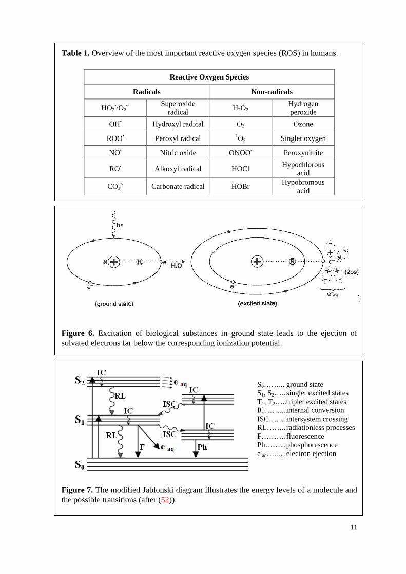
Table 1. Overview of the most important reactive oxygen species (ROS) in humans.
Reactive Oxygen Species
Radicals Non-radicals
HO2•/O2
•- Superoxide radical H2O2
Hydrogen peroxide
OH• Hydroxyl radical O3 Ozone
ROO• Peroxyl radical 1O2 Singlet oxygen
NO• Nitric oxide ONOO- Peroxynitrite
RO• Alkoxyl radical HOCl Hypochlorous acid
CO3•- Carbonate radical HOBr Hypobromous
acid
Figure 6. Excitation of biological substances in ground state leads to the ejection of solvated electrons far below the corresponding ionization potential.
Figure 7. The modified Jablonski diagram illustrates the energy levels of a molecule and the possible transitions (after (52)).
S0……... ground state S1, S2….. singlet excited states T1, T2….. triplet excited states IC……... internal conversion ISC……. intersystem crossing RL…….. radiationless processes F………. fluorescence Ph……... phosphorescence e-
aq…..… electron ejection
12
Table 1 comprises the most important ROS, including both oxygen free radicals and
some non-radicals (56). Cells have developed a number of defense methods against ROS
and their damaging effects, e.g. enzymes, anti-oxidants or repair mechanisms, as survival
is of prime importance. However, ROS also exhibit beneficial actions within the cell.
Therefore, a basic level of ROS is present in the cytoplasm all the time.
3.2.2. Solvated electrons
In 1960, the conversion of γ-irradiated, aqueous CO2 into simple organic compounds
proved the existence of the solvated electrons (e-aq) for the first time (57). Similar results
were obtained by UV-irradiation of aqueous solution containing Fe2+ ions, acting as
electron donors, and CO2 as electron acceptor (pH ~3.7) (57,58). In 1962, the absorption
spectra of solvated electrons in water and in various polar liquids were measured by pulse
radiolysis (59).
The emission of solvated electrons (e-aq) is a consequence of energy input to a
biological molecule (Figure 6). By giving energy to the atom, an electron of the outer
orbital is elevated from ground state into the first singlet excited state. Thus, the distance
of the e- to the nucleus is enlarged and the binding energy (F) is reduced with the square
of distance. Consequently, the dipoles of the surrounding water molecules become
orientated to the polarized atom in less than 10-12 s. The e- is scavenged by the orientated
dipoles of the surrounding water molecules and separates from the atom, becoming a
solvated electron (e-aq) with a salvation shell. It should be noted, that the electron
emission from excited molecules in their singlet state occurs far below the corresponding
ionization potential of a given substrate, because of the very strong dipole action of the
water molecules (60).
The quantum yield of e-aq, Q(e-
aq), emitted from a specific substance depends on
several factors: molecular structure, substituents like -OH, -OH-, -OPO3H2, -OPO3H-, -
COO-,-NH2, -NHCH3, etc., pH of the media and temperature of the solution (52). The
ejection of electrons from molecules in singlet state is competing with other
photophysical processes, as electronically excited molecules tend to return in their
original ground states. The strong relationship between Q(e-aq) and the quantum yield of
fluorescence, QF, can be taken as example in this respect (52). This is illustrated by the
modified Jablonski diagram (Figure 7).
13
3.2.2.1. Simulation of emission of e-aq by UV-irradiation of hormones
The formation of e-aq (solvated electrons) is simulated by irradiation of various organic
compounds, such as hormones, with monochromatic UV-light in aqueous, air-free
solution at pH ~7.4. The basic processes induced by UV-light are shown in eqs. 7 and 8.
AB + hν AB* A• + B• (formation of radicals) (7)
(Excitation) photophysical processes (8) (fluorescence, phosphorescence, etc. )
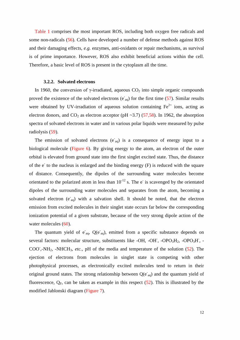
As can be seen in Figure 8, the higher energy portion of the UV-spectrum (overcoming
the ionization potential) is considered to be ionizing, whereas the lower energy portion is
not. Since the experiments in this study are conducted with a UV-lamp emitting light of
λ=254 nm (E=4.85 eV/hν), hormones are just excited in their singlet state and not ionized.
As scavenger for the emitted solvated electrons chloroethanol can be used (eqs. 9,10).
ClC2H4OH + e-aq → Cl− + •C2H4OH (k = 4x10⁸ L.mol-1.s-1, 92) (9)
Q(Cl−) = Q(e-aq) (10)
The resulting yield of Cl− is determined by mercury(II)thiocyanate method (61):
2Cl- + Hg(SCN)2 HgCl2 + 2SCN- (11)
4Cl- + Hg(SCN)2 HgCl42- + 2SCN- (12)
SCN- + Fe3+ Fe(SCN)2+ (13)
3.2.3. Formation of free radicals by ionizing radiation
Basically, radicals are formed by loosing or gaining a single electron from a non
radical, resulting into radical cation and anion, respectively (eqs. 14,15).
X – e- X•+ (single electron oxidation) (14)
Y + e- Y•- (single electron reduction) (15)
Radicals can also be formed by homolytic fission (62), a process requiring significant
amounts of energy (e.g. heat, UV-light, ionizing radiation). Thereby, one electron of the
14
optical
thermal
non-thermal
bonds breaking
10-15 10-10 10-5 1 105 1010 energy (eV)
1010 105 1 10-5 10-10 10-15 λ (cm)
NON-IONIZING IONIZING
Figure 8. Types of radiation in the electromagnetic spectrum. λ = wave length; ELF = extremely low frequency; IR = infrared; VIS = visible light; UV = ultraviolet.
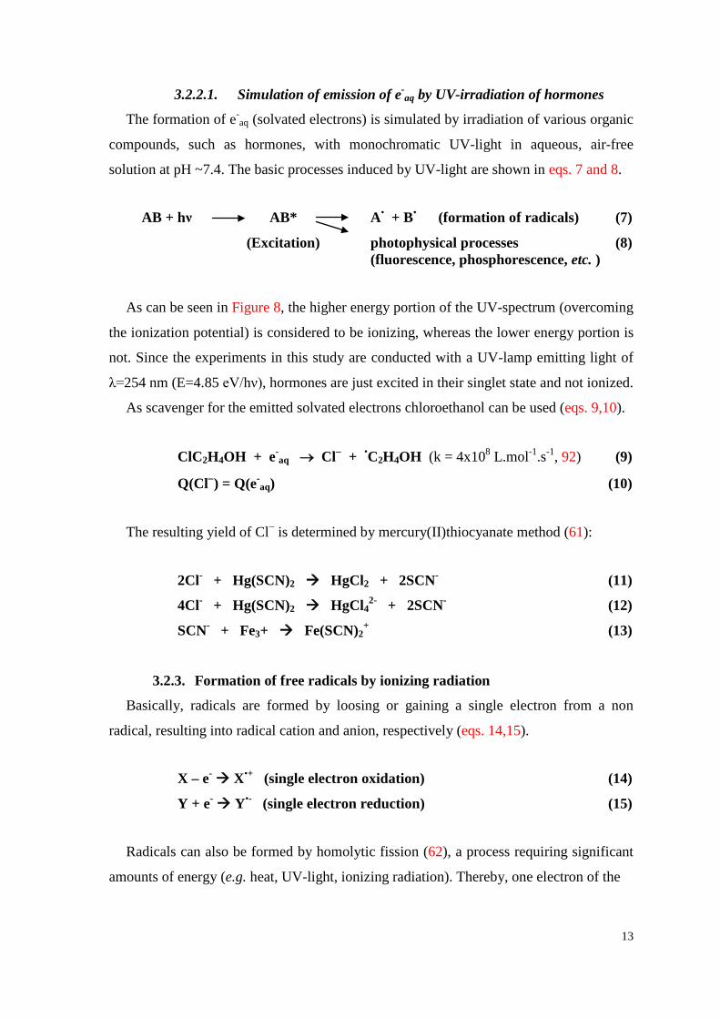
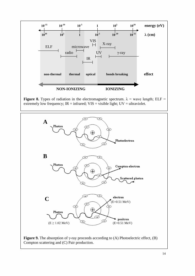
Figure 9. The absorption of γ-ray proceeds according to (A) Photoelectric effect, (B) Compton scattering and (C) Pair production.
ELF radio
microwave
IR
VIS
UV
X-ray
γ-ray
effect
A
B
C (E=0.51 MeV)
(E=0.51 MeV) (E ≥ 1.02 MeV)
15
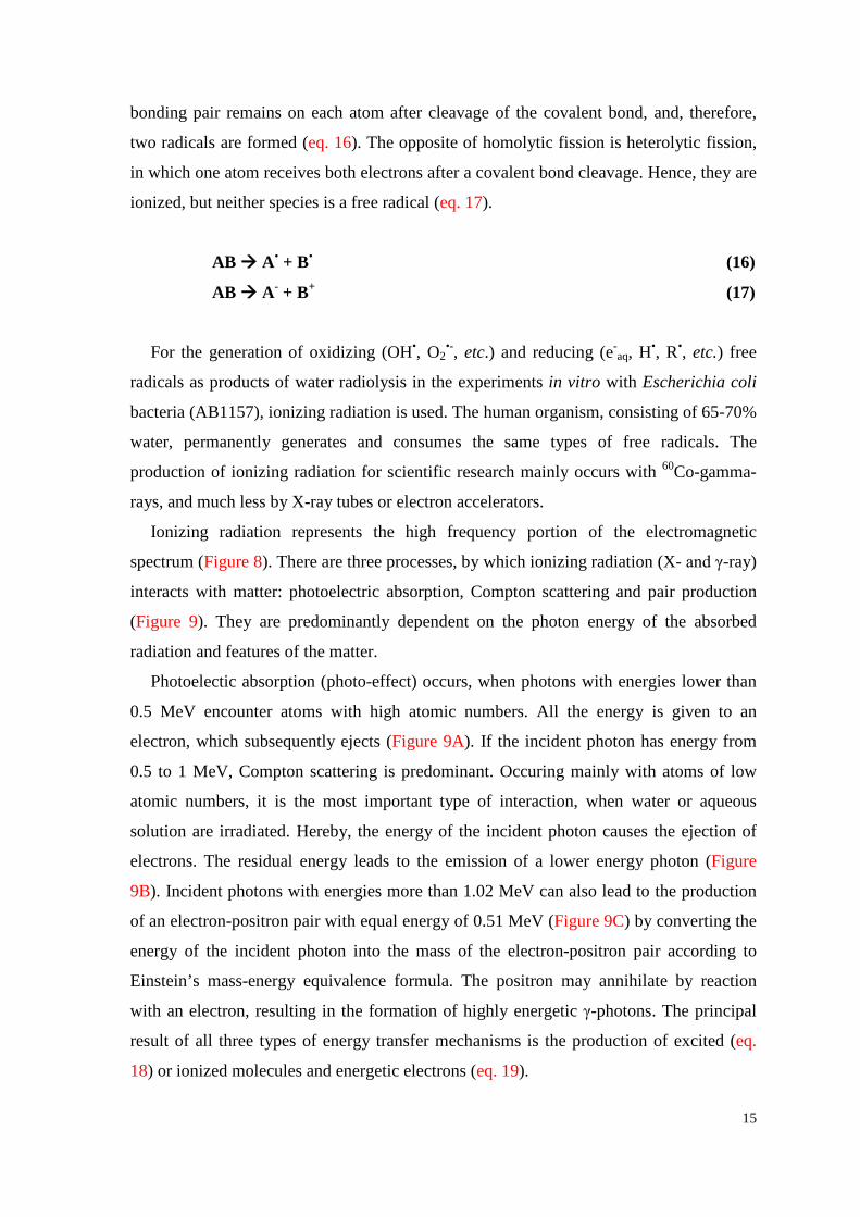
bonding pair remains on each atom after cleavage of the covalent bond, and, therefore,
two radicals are formed (eq. 16). The opposite of homolytic fission is heterolytic fission,
in which one atom receives both electrons after a covalent bond cleavage. Hence, they are
ionized, but neither species is a free radical (eq. 17).
AB A• + B• (16)
AB A- + B+ (17)
For the generation of oxidizing (OH•, O2•-, etc.) and reducing (e-
aq, H•, R•, etc.) free
radicals as products of water radiolysis in the experiments in vitro with Escherichia coli
bacteria (AB1157), ionizing radiation is used. The human organism, consisting of 65-70%
water, permanently generates and consumes the same types of free radicals. The
production of ionizing radiation for scientific research mainly occurs with 60Co-gamma-
rays, and much less by X-ray tubes or electron accelerators.
Ionizing radiation represents the high frequency portion of the electromagnetic
spectrum (Figure 8). There are three processes, by which ionizing radiation (X- and γ-ray)
interacts with matter: photoelectric absorption, Compton scattering and pair production
(Figure 9). They are predominantly dependent on the photon energy of the absorbed
radiation and features of the matter.
Photoelectic absorption (photo-effect) occurs, when photons with energies lower than
0.5 MeV encounter atoms with high atomic numbers. All the energy is given to an
electron, which subsequently ejects (Figure 9A). If the incident photon has energy from
0.5 to 1 MeV, Compton scattering is predominant. Occuring mainly with atoms of low
atomic numbers, it is the most important type of interaction, when water or aqueous
solution are irradiated. Hereby, the energy of the incident photon causes the ejection of
electrons. The residual energy leads to the emission of a lower energy photon (Figure
9B). Incident photons with energies more than 1.02 MeV can also lead to the production
of an electron-positron pair with equal energy of 0.51 MeV (Figure 9C) by converting the
energy of the incident photon into the mass of the electron-positron pair according to
Einstein’s mass-energy equivalence formula. The positron may annihilate by reaction
with an electron, resulting in the formation of highly energetic γ-photons. The principal
result of all three types of energy transfer mechanisms is the production of excited (eq.
18) or ionized molecules and energetic electrons (eq. 19).
16
AB AB* A• + B• (formation of radicals) (18a)
(Excitation) photophysical processes (18b) (fluorescence, phosphorescence etc. )
AB+ + e- (ionization) (19)
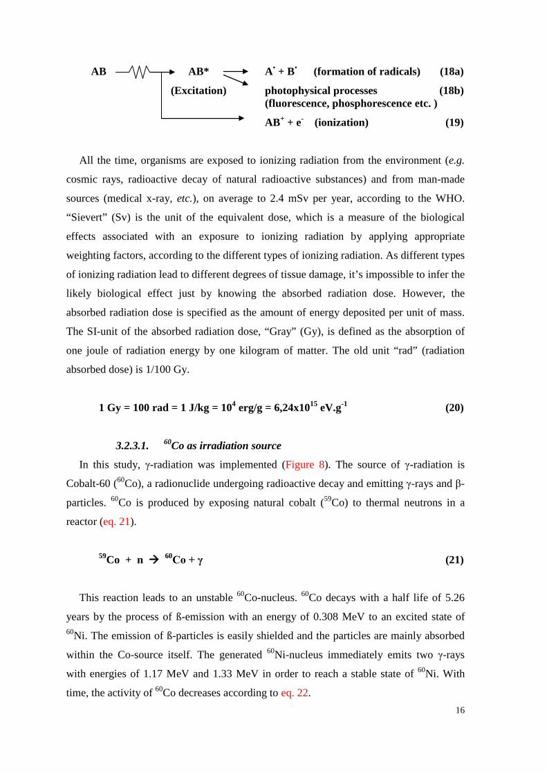
All the time, organisms are exposed to ionizing radiation from the environment (e.g.
cosmic rays, radioactive decay of natural radioactive substances) and from man-made
sources (medical x-ray, etc.), on average to 2.4 mSv per year, according to the WHO.
“Sievert” (Sv) is the unit of the equivalent dose, which is a measure of the biological
effects associated with an exposure to ionizing radiation by applying appropriate
weighting factors, according to the different types of ionizing radiation. As different types
of ionizing radiation lead to different degrees of tissue damage, it’s impossible to infer the
likely biological effect just by knowing the absorbed radiation dose. However, the
absorbed radiation dose is specified as the amount of energy deposited per unit of mass.
The SI-unit of the absorbed radiation dose, “Gray” (Gy), is defined as the absorption of
one joule of radiation energy by one kilogram of matter. The old unit “rad” (radiation
absorbed dose) is 1/100 Gy.
1 Gy = 100 rad = 1 J/kg = 104 erg/g = 6,24x10¹⁵ eV.g-1 (20)
3.2.3.1. 60Co as irradiation source
In this study, γ-radiation was implemented (Figure 8). The source of γ-radiation is
Cobalt-60 (60Co), a radionuclide undergoing radioactive decay and emitting γ-rays and β-
particles. 60Co is produced by exposing natural cobalt (59Co) to thermal neutrons in a
reactor (eq. 21).
59Co + n 60Co + γ (21)
This reaction leads to an unstable 60Co-nucleus. 60Co decays with a half life of 5.26
years by the process of ß-emission with an energy of 0.308 MeV to an excited state of 60Ni. The emission of ß-particles is easily shielded and the particles are mainly absorbed
within the Co-source itself. The generated 60Ni-nucleus immediately emits two γ-rays
with energies of 1.17 MeV and 1.33 MeV in order to reach a stable state of 60Ni. With
time, the activity of 60Co decreases according to eq. 22.
17
A = A0 * e-kt (22) A……... activity
A0……. activity at time 0
k……... radioactive decay constant
t……… time
Several methods exist in order to determine the activity of a γ-source (ionization
chamber, calorimeter, etc.). During this study, the dose-rate of the γ-source was
determined and permanently controlled by means of a Fricke-Dosimeter (63), which was
subsequently modified by saturation with oxygen. Hereby, Fricke solution is exposed to
ionizing radiation. The resulting primary products of water radiolysis oxidize ferrous ions
(Fe2+) to ferric ions (Fe3+) in the presence of air. The increase of Fe3+ can be measured
spectrophotometrically at λ=305 nm (eqs. 23-25). It should be noted, that temperature
strongly affects the molar extinction coefficient (ε305) of ferric ions, as ε305 is proportional
to the temperature of the dosimeter solution (105).
Fe2+ + HO2• + H+ Fe3+ + H2O2 (23)
Fe2+ + H2O2 Fe3+ + OH- + OH• (24)
Fe2+ + OH• Fe3+ + OH- (25)
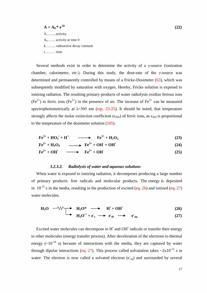
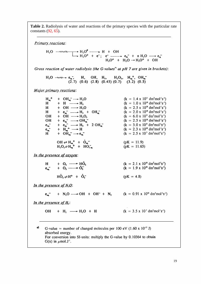
3.2.3.2. Radiolysis of water and aqueous solutions
When water is exposed to ionizing radiation, it decomposes producing a large number
of primary products: free radicals and molecular products. The energy is deposited
in 10-15 s in the media, resulting in the production of excited (eq. 26) and ionized (eq. 27)
water molecules.
H2O H2O* H• + OH• (26)
H2O•+ + e-s e-
th e-
aq (27)
Excited water molecules can decompose to H• and OH• radicals or transfer their energy
to other molecules (energy transfer process). After deceleration of the electrons to thermal
energy (~10-14 s) because of interactions with the media, they are captured by water
through dipolar interactions (eq. 27). This process called solvatation takes ~2x10-12 s in
water: The electron is now called a solvated electron (e-aq) and surrounded by several
18
water molecules. The number of water molecules is dependent on the temperature.
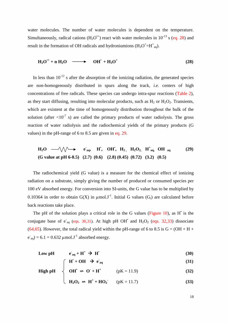
Simultaneously, radical cations (H2O•+) react with water molecules in 10-13 s (eq. 28) and
result in the formation of OH radicals and hydroniumions (H3O+=H+aq).
H2O•+ + n H2O OH• + H3O+ (28)
In less than 10-12 s after the absorption of the ionizing radiation, the generated species
are non-homogenously distributed in spurs along the track, i.e. centers of high
concentrations of free radicals. These species can undergo intra-spur reactions (Table 2),
as they start diffusing, resulting into molecular products, such as H2 or H2O2. Transients,
which are existent at the time of homogenously distribution throughout the bulk of the
solution (after <10-7 s) are called the primary products of water radiolysis. The gross
reaction of water radiolysis and the radiochemical yields of the primary products (G
values) in the pH-range of 6 to 8.5 are given in eq. 29.
H2O e-aq, H•, OH•, H2 , H2O2, H+
aq, OH aq (29)
(G value at pH 6-8.5) (2.7) (0.6) (2.8) (0.45) (0.72) (3.2) (0.5)
The radiochemical yield (G value) is a measure for the chemical effect of ionizing
radiation on a substrate, simply giving the number of produced or consumed species per
100 eV absorbed energy. For conversion into SI-units, the G value has to be multiplied by
0.10364 in order to obtain G(X) in µmol.J-1. Initial G values (Gi) are calculated before
back reactions take place.
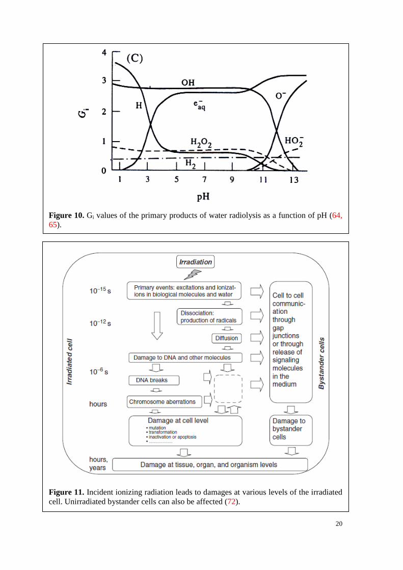
The pH of the solution plays a critical role in the G values (Figure 10), as H• is the
conjugate base of e-aq (eqs. 30,31). At high pH OH• and H2O2 (eqs. 32,33) dissociate
(64,65). However, the total radical yield within the pH-range of 6 to 8.5 is G = (OH + H +
e-aq) = 6.1 = 0.632 µmol.J-1 absorbed energy.
Low pH e-aq + H+ H• (30)
H• + OH e-aq (31)
High pH OH• ⇌ O- + H+ (pK = 11.9) (32)
H2O2 ⇌ H+ + HO2- (pK = 11.7) (33)
19
Table 2. Radiolysis of water and reactions of the primary species with the particular rate constants (92, 65).
20
Figure 10. Gi values of the primary products of water radiolysis as a function of pH (64, 65).
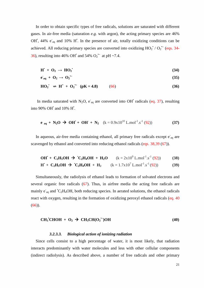
Figure 11. Incident ionizing radiation leads to damages at various levels of the irradiated cell. Unirradiated bystander cells can also be affected (72).
21
In order to obtain specific types of free radicals, solutions are saturated with different
gases. In air-free media (saturation e.g. with argon), the acting primary species are 46%
OH•, 44% e-aq and 10% H•. In the presence of air, totally oxidizing conditions can be
achieved. All reducing primary species are converted into oxidizing HO2• / O2
•− (eqs. 34-
36), resulting into 46% OH• and 54% O2•− at pH ~7.4.
H• + O2 → HO2• (34)
e-aq + O2 → O2
•− (35)
HO2• ⇌ H+ + O2
•− (pK = 4.8) (66) (36)
In media saturated with N2O, e-aq are converted into OH• radicals (eq. 37), resulting
into 90% OH• and 10% H•.
e aq + N2O OH• + OH- + N2 (k = 0.9x10¹⁰ L.mol-1.s-1 (92)) (37)
In aqueous, air-free media containing ethanol, all primary free radicals except e-aq are
scavenged by ethanol and converted into reducing ethanol radicals (eqs. 38,39 (67)).
OH• + C2H5OH •C2H4OH + H2O (k = 2x10⁹ L.mol-1.s-1 (92)) (38)
H• + C2H5OH •C2H4OH + H2 (k = 1.7x10⁷ L.mol-1.s-1 (92)) (39)
Simultaneously, the radiolysis of ethanol leads to formation of solvated electrons and
several organic free radicals (67). Thus, in airfree media the acting free radicals are
mainly e-aq and •C2H4OH, both reducing species. In aerated solutions, the ethanol radicals
react with oxygen, resulting in the formation of oxidizing peroxyl ethanol radicals (eq. 40
(66)).
CH3•CHOH + O2 CH3CH(O2
•−)OH (40)
3.2.3.3. 69BBiological action of ionizing radiation
Since cells consist to a high percentage of water, it is most likely, that radiation
interacts predominantly with water molecules and less with other cellular components
(indirect radiolysis). As described above, a number of free radicals and other primary
22
species result from the radiolysis of water, which start diffusing throughout the solution.
In doing so, it is possible, that besides reacting among themselves, they react with other
substances present in the solution (e.g. the components of a cell). That indirect action of
ionizing radiation is the predominant process when radiating with gamma rays, which are
defined as low LET radiation. LET (linear energy transfer) is a measure for the energy
transferred to a specific material penetrated by an ionizing particle of specified energy per
unit distance traversed. Gamma-rays are highly penetrating and lose their energy less
effectively than e.g. α- and β-rays.
If ionizing radiation interacts with substances of a cell others than water, this process is
referred to as direct effect of ionizing radiation. This process is predominant with high
LET radiation. The excited or ionized atoms of biological compounds may undergo intra-
or intermolecular energy transfer processes or/and intramolecular electron transfer
processes (106), which means that the absorbed energy is able to migrate within the
molecule or even between two molecules. The site of damage can therefore be different
from the site of the interaction with ionizing radiation.
Cell damages, which are induced by ionizing radiation, may lead to mutations or loss
of functions of DNA, proteins or other important cell compounds and to cell death. In E.
coli, the greater part of lethal damage is due to indirect biological action of ionizing
radiation (68,69). Since there is a correlation between radiation sensitivity and amount of
DNA in a cell (70), it is expected, that eukaryotic cells are much more sensitive towards
ionizing radiation than E. coli bacteria, which were implemented in the radiation studies.
Furthermore, the radiosensitivity of cells is highly dependent on the specific phase of cell
cycle (71).
As DNA is always hydrated by water molecules due to its negative charge, it is one of
the main targets for cell damage by ionizing radiation (72). Regarding the rate constants
for the primary products of water radiolysis and DNA (for OH•: k=0.3x10⁹ dm3.mol-1.s-1;
for H•: k=0.08x10⁹ dm3.mol-1.s-1; for e aq: k=0.14x10⁹ dm3.mol-1.s-1) and the
corresponding G values, it can be assumed, that the OH• radicals contribute to a large part
to the damage (70). Radiation-induced lesions in DNA include single strand breaks
(SSB), double strand breaks (DSB), base alterations or loss (apyrimidinic / apurinic (AP)
site), tandem or clustered lesions as well as cross-links (DNA/DNA or DNA/protein
cross-links). The same lesions are results of radiotherapy (73). Hereby, DNA repair is
essential for the organisms, as non-repaired DNA damage usually causes cell death due to
23
the inhibition of necessary biological processes, such as replication or transcription. A
combination of the different lesions in close vicinity on the DNA strand (multiply
damaged site (MDS)) leads to much more harm to cells than single damage events, as the
repair machinery is much more challenged (74). Since DNA repair is not error-free,
mutations can occur, which are not lethal but may be harmful and a possible initiator of
carcinogenesis.
In Figure 11 the biological effects of ionizing radiation are depicted at different spatial
and time levels (72). Interestingly, cells may suffer from radiation damage, which have
never been hit by ionizing radiation, but were in close vicinity to irradiated cells (75, 76).
These bystander effects are due to cell-cell communication between irradiated and
unirradiated cells.
3.2.4. Free radicals in the human organism – only a burden?
For a long time, free radicals were solely associated with damage within the scientific
community. Nowadays, there is a large body of evidence, that oxidizing (OH•, O2•-, etc.)
and reducing (e-aq, H•, R•, etc.) free radicals play a determining role in a huge number of
biological processes in the human organism. Moreover, both types, oxidizing as well as
reducing free radicals, fulfill equally important tasks in living systems.
In the human organism, the generation of free radicals can be induced by exogenous
inducers, such as UV-light, tobacco smoking etc. However, a variety of endogenous
sources is producing free radicals as well, such as the generation of ROS in the
mitochondrion. Thereby, it is essential, that the production of the radicals is well
regulated, as an increased production of ROS exhibits hazardous effects on cells, resulting
in DNA-damage, carcinogenesis and initiation of a number of diseases (diabetes mellitus,
atherosclerosis, neurodegenerative diseases, etc.) by influencing intercellular signaling
cascades or direct oxidation of cellular key-components (79).
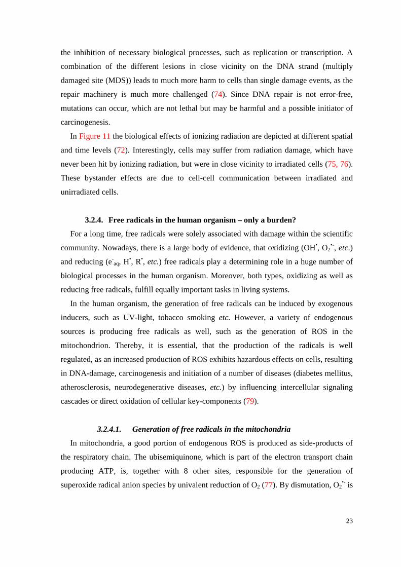
3.2.4.1. Generation of free radicals in the mitochondria
In mitochondria, a good portion of endogenous ROS is produced as side-products of
the respiratory chain. The ubisemiquinone, which is part of the electron transport chain
producing ATP, is, together with 8 other sites, responsible for the generation of
superoxide radical anion species by univalent reduction of O2 (77). By dismutation, O2•- is
24
converted into H2O2. Free Fe(II) and other metals in the cytoplasm promote the Fenton
and Haber-Weiss reaction, leading to the formation of OH• out of O2•− and H2O2.
If detoxification of the ROS by antioxidants or enzymes is not possible due to an
imbalance between ROS production and antioxidant defenses, the mitochondrion suffers
from oxidative stress. As ROS are highly reactive species, they immediately attack nearby
compounds (nucleic acids, membrane lipids, etc.), trying to gain an electron. The
damaged compounds may not be longer able to carry out their function, resulting into
mitochondric dysfunction by mutation or even apoptosis / necrosis by changing the
permeability of the membranes (Figure 12). However, the presence of ROS is also
beneficial, as they play a significant role in signal transduction processes (78,79).
Figure 12. The mitochondrial ROS production is the main source for reactive species in cells. Cyt c: cytochrome c; MOMP: mitochondrial outer membrane permeabilization; mtDNA: mitochondrial DNA; PTP: permeability transition pore; ROS: reactive oxygen species (97).
25
3.2.4.2. Free radicals as regulatory mediators in cell signaling
Oxidative stress has long been thought to be unregulated and an unwanted side-effect
of the aerobic metabolism, as it is involved in various pathological processes. Nowadays,
it becomes more and more evident, that free radicals are of crucial importance in a
number of signalling processes of the cell, mediating diverse biological activities, such as
inflammation or vasodilatation (78). Modulations of cell transduction, which are initiated
by free radicals through specific modifications of cell signalling proteins, are referred to
as „redox cell signalling” (79). Groundbreakingly was the prove, that NFκB, an important
transcription factor for genes of the early defence system, is activated by oxidizing
reagents, such as H2O2, and even ionizing radiation (80,81). The activation of NFκB is a
result of an increase of the degradation of its inhibitor IκB by ROS. Generally, it can be
stated, that oxidative attack on signalling molecules either results into a loss or gain of
function or into a function switch. Meanwhile, a great deal of signalling molecules
regulated by free radicals has been identified and described. Besides the already
mentioned NFκB, the transcription factor AP-1, p38 MAPK and JNK, just to name a few,
seem to be tightly regulated by free radicals (79).
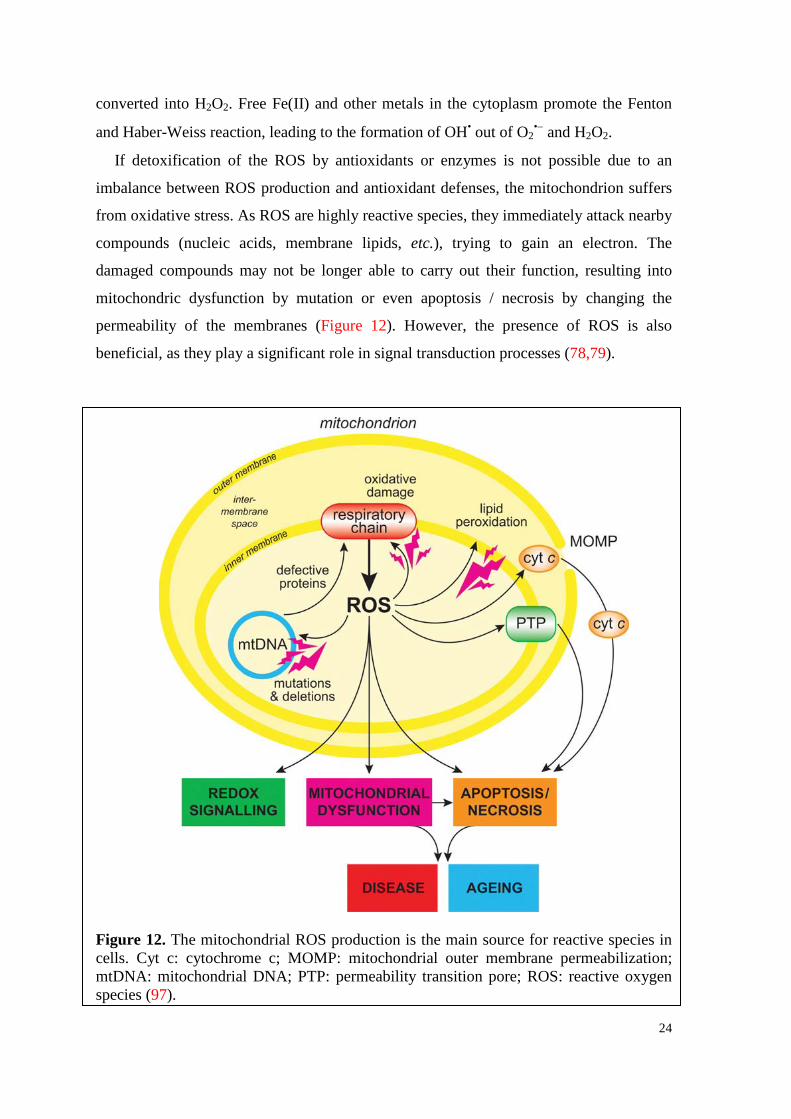
3.2.4.3. Intracellular killing of bacteria by phagocytes
Phagocytosis is a process, in which phagocytes (macrophages and neutrophils) engulf
foreign particles or bacteria and enclose it into a phagosome. After formation of the
phagocytic vacuole by fusion of the phagosome with a lysosome, ROS are generated in
order to kill the internalized bacterium (82). Hereby, the phagocytic NADPH-oxidase,
which assembles in the membrane of the lysosome, catalyzes the synthesis of the
superoxide radical (O2•-) by transferring two electrons from NADPH across the
membrane to O2 (eq. 41).
NADPH − 2e- + 2O2 NADP+ + H+ + 2O2•- (41)
As oxygen uptake is remarkably increased by this reaction, this process is called
“oxidative burst” or “respiratory burst”. This process in not associated with cellular
respiration, though. However, the superoxide radical can be converted by the superoxide
dismutase (SOD) into H2O2 and singlet oxygen. Subsequently, O2•- and H2O2 may form
OH• and again singlet oxygen. These ROS altogether induce lethal damages to the
internalized bacterium, and are, therefore, crucial for the immune response.
26
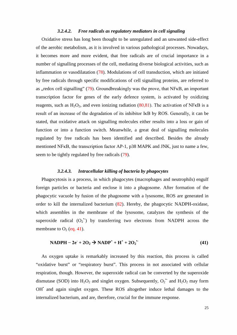
Figure 13. Biologically active states of vitamin C (88).
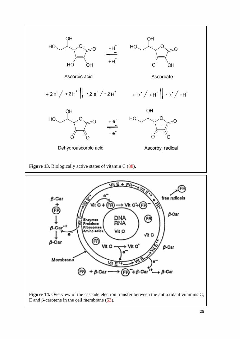
Figure 14. Overview of the cascade electron transfer between the antioxidant vitamins C, E and β-carotene in the cell membrane (53).
27
3.3. Antioxidants Aerobe organisms cope with the poisonous action of oxygen by a complex system of
antioxidant defense mechanisms. Basically, an antioxidant is any substance that reduces
or retards oxidation of an oxidizable substrate (56). There are several processes, by which
antioxidants can protect the cell from damage, whereby the first line of defense is the
prevention of the formation of ROS and other free radicals. This can be done by shielding
important key-substance with the help of chaperones, offering alternative targets for the
attack of free radicals (56) or by decreasing the availability of pro-oxidants, e.g. Fe(II),
which triggers the Fenton and Haber-Weiss reaction (83). If free radicals are already
produced, it is necessary to inhibit chain reactions. Diverse enzymes, such as SOD or
catalase, or proteins, e.g. gluthathione, vitamin C (vitC) or vitamin E (vitE), react with
free radicals in various manners. A large part of the antioxidants becomes oxidized
themselves upon reaction with free radicals by acting as electron donors.
3.3.1. Vitamin C
One of the most potent antioxidants is L-ascorbic acid (AA, vitC, Figure 13), which is
a water-soluble reducing agent and a very potent electron donor. Its name derives from
scorbut (scurvy), a disease resulting from nutritional deficiency of AA, which leads to
defects in collagen synthesis (84). AA is synthesized as product of the hexuronic pathway
in the liver or kidney by the gunololactone oxidase in many organisms. As humans are
deficient of this enzyme, they need to obtain AA by nutrition. Therefore, AA is classified
as vitamin in humans, which is ubiquitously distributed in the cells upon absorption in the
intestine. Depending on the target cells, AA is either actively transported by sodium-
ascorbate co-transporters (SVCTs), or it has to be oxidized to dehydroascorbic acid
(DHA) for transportation by hexose transporters (GLUTs) (85). In the latter case, DHA is
reduced to AA in the cytoplasm again.
The antioxidant properties of AA can be demonstrated by the cascade electron transfer
(Figure 14), by which lipoproteins of membranes are protected from free radical attack
(53). It is known that cell membranes consist to approximately 5% of vitamins C, E and
β-carotene. However, attacking free radicals are reduced in a first defense line by β-
carotene, which is located at the outer membrane layer (eq. 42). The ejected electron from
β-car neutralizes the oxidizing species and becomes oxidized itself to a radical cation (β-
car•+), which in turn is regenerated by vitE (eq. 43).
28
OH/ROO• + β-car OH-/ROO- + β-car•+ (42)
β-car•+ + vitE β-car + vitE•+ (43)
The radical cation (E•+) is subsequently regenerated to vitE by ascorbate (A-) (eq. 44),
which presents a very low reduction potential and is oxidized to an ascorbyl radical (A•).
The relatively stable ascorbyl radicals can dismutate to AA and DHA (eq. 45),
terminating the chain reaction.
vitE•+ + A- vitE + A• (44)
2 A• + H+ AA + DHA (86) (45)
Interestingly, vitC can also act as pro-oxidant by reducing redox-active metals, such as
Fe3+ or Cu2+, triggering the Fenton reaction, which results in the formation of OH• or lipid
peroxidation (87). The inhibitory effect of vitC on cancer cells seems to be partly due to
this process producing reactive species, as cancer cells tend to overexpress GLUTs and,
therefore, accumulate more vitC in the cytoplasm than normal cells (88). Furthermore,
cancer cell lines exhibit a higher basal, endogenous level of reactive species. Hence, a
substance like vitC, which is promoting the production of reactive species, will challenge
tumor cells much more than normal cells (89).
In tumors, hypoxia occurs, a phenomenon due to the rapid growth, leading to regions
within the tumor, which are deprived from oxygen. Tumor cells usually adapt to hypoxia
by activation of HIF1 (hypoxia-inducible factor 1), but AA inhibits this process and, thus,
acts as anticancer agent (90).
Under ionizing irradiation, AA shows a protective effect on E. coli bacteria, especially
in aerated solutions (91). The reaction rate constants for A- with some primary species of
water radiolysis were measured by pulse radiolysis at pH ~7: k(OH•+A-) = 1.1x10¹⁰
dm3.mol-1.s-1, k(H•+A-) = 3x10⁸ dm3.mol-1.s-1 and k(e-aq+A-) = 3.5x10⁸ dm3.mol-1.s-1 (92).
DHA alone and in combination with vitE or/and β-car essentially enhances the antitumor
effect of the cytostaticum mytomycin C (MMC) (107).
29
4. STUDY OBJECTIVES In order to get a deeper insight in the rather complicated biological processes in the
organism, which proceed simultaneously and mostly interact with each other in a
harmonious way, the objectives of the present thesis embrace several superimposed
problems.
Since the breast and prostate cancer risk is steadily increasing in the Western
countries, research focuses more and more on sex hormones and their metabolites, as they
exhibit an impact on the carcinogenesis of most breast- and prostate cancer types (31-46).
Based on previous knowledge, that biological substances having some functional
substituents like -OH, -OH-, -OPO3H2, -OPO3H-, -COO-,-NH2, -NHCH3 etc., are able to
eject electrons when excited in their singlet state in aqueous solutions (52), it is expected
that hormones can principally also emit electrons in polar solvent. The originating
hormone transients are subsequently forming metabolites, some of which can initiate
neoplastic processes. This supposition was proven firstly for E2 and P4 as representatives
of hormones in a mixture of water and ethanol (47). Based on these experimental data and
knowledge, it was of biological interest, to examine this subject matter on other
hormones, such as the main male sex hormone T or the phytohormone GEN. T and E2
differ in their molecular structures only at ring A, where T carries a carbonyl group and
E2 a hydroxyl group, whereas T and P4 differ at ring D, where T carries a hydroxyl group
and P4 a carbonyl group. Therefore, comparisons between these three hormones could
contribute to a better understanding of the impact of the individual functional groups in
this respect. The molecular structure of GEN, which is associated with the comparatively
low breast cancer incidence in Asian countries and is known to exhibit weak estrogenic
properties (101), is similar to E2, and, thus, also of interest.
It is further postulated, that a reduction of the number of carcinogenic metabolites
resulting from hormones after electron ejection can be achieved by the regeneration of
hormones with the help of a potent electron donor, due to an electron transfer from the
donor to the acceptor. Therefore, using vitC as representative for a potent electron donor
and T as acceptor, HPLC-analyses were performed, in order to study the degradation of
the substrates under UV-irradiation and a possible regeneration.
In addition to this, it was of great importance to test the obtained results by
experiments in vitro. In the organism, which consists of 65-70% water, oxidizing (OH•,
O2•-, etc.) as well as reducing (e-
aq, R•, etc.) free radicals are permanently generated,
30
playing a determining role in a huge number of biological processes. A tight regulation is
essential, as a surplus of free radicals may lead to hazardous effects and a great number of
possible damages. However, in order to study the effects of oxidizing and reducing
species on various sex hormones, experiments in vitro with Escherichia coli bacteria were
performed. As the same types of free radicals are generated by ionizing radiation as
products of water radiolysis, it is possible to gain desired kind and concentration of free
radicals under appropriate experimental conditions. Since the ability of E2 and P4 to emit
electrons when excited in their singlet state was already proven, they were investigated
individually and in mixture in this respect first. In the course of these studies, both
hormones were embedded in a water-soluble complex with HBC. Thereafter, also
experiments in vitro with T and vitC were carried out in media containing 4x10⁻² mol/L
ethanol.
Finally, special attempts were made to suggest probable reaction mechanisms
explaining the observed chemical and biological effects.
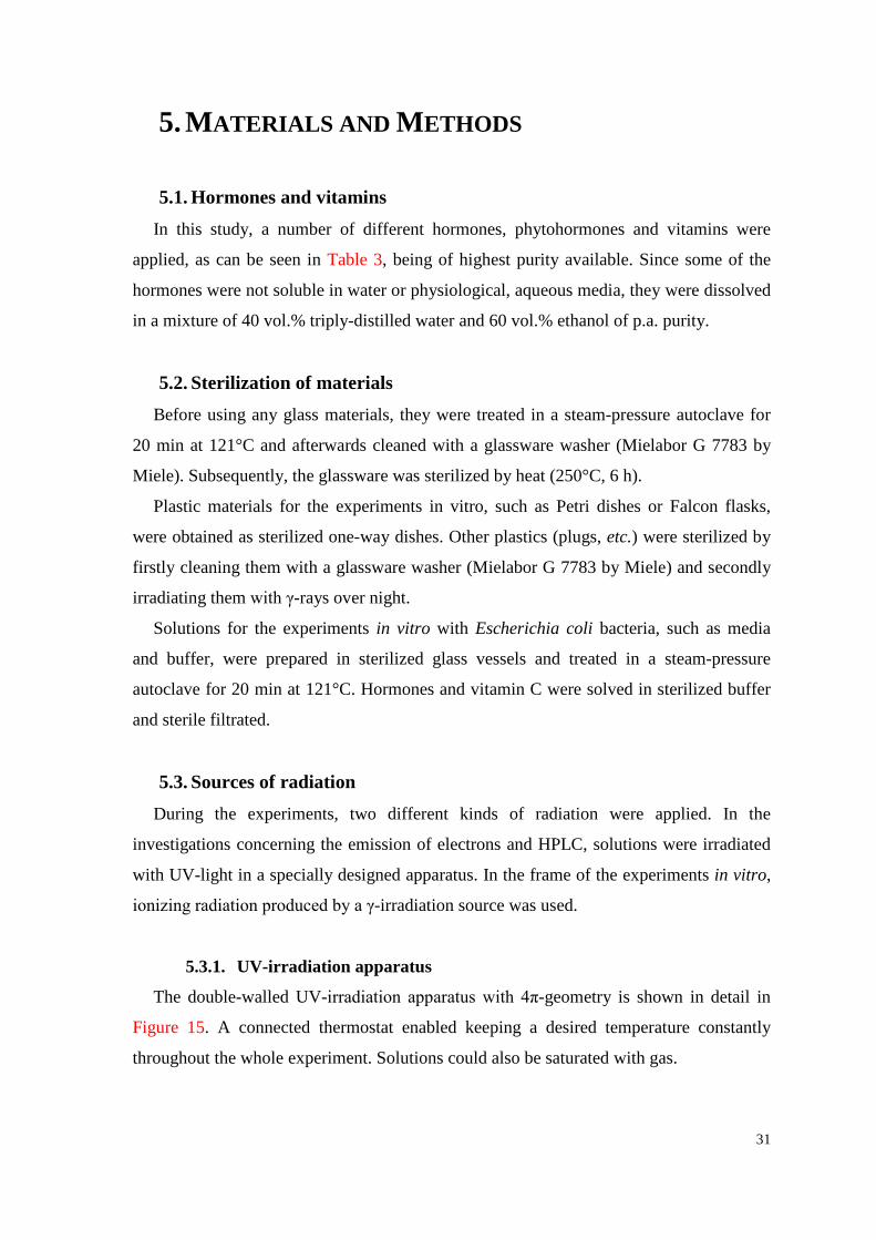
31
5. MATERIALS AND METHODS
5.1. Hormones and vitamins In this study, a number of different hormones, phytohormones and vitamins were
applied, as can be seen in Table 3, being of highest purity available. Since some of the
hormones were not soluble in water or physiological, aqueous media, they were dissolved
in a mixture of 40 vol.% triply-distilled water and 60 vol.% ethanol of p.a. purity.
5.2. Sterilization of materials Before using any glass materials, they were treated in a steam-pressure autoclave for
20 min at 121°C and afterwards cleaned with a glassware washer (Mielabor G 7783 by
Miele). Subsequently, the glassware was sterilized by heat (250°C, 6 h).
Plastic materials for the experiments in vitro, such as Petri dishes or Falcon flasks,
were obtained as sterilized one-way dishes. Other plastics (plugs, etc.) were sterilized by
firstly cleaning them with a glassware washer (Mielabor G 7783 by Miele) and secondly
irradiating them with γ-rays over night.
Solutions for the experiments in vitro with Escherichia coli bacteria, such as media
and buffer, were prepared in sterilized glass vessels and treated in a steam-pressure
autoclave for 20 min at 121°C. Hormones and vitamin C were solved in sterilized buffer
and sterile filtrated.
5.3. Sources of radiation During the experiments, two different kinds of radiation were applied. In the
investigations concerning the emission of electrons and HPLC, solutions were irradiated
with UV-light in a specially designed apparatus. In the frame of the experiments in vitro,
ionizing radiation produced by a γ-irradiation source was used.
5.3.1. UV-irradiation apparatus
The double-walled UV-irradiation apparatus with 4π-geometry is shown in detail in
Figure 15. A connected thermostat enabled keeping a desired temperature constantly
throughout the whole experiment. Solutions could also be saturated with gas.
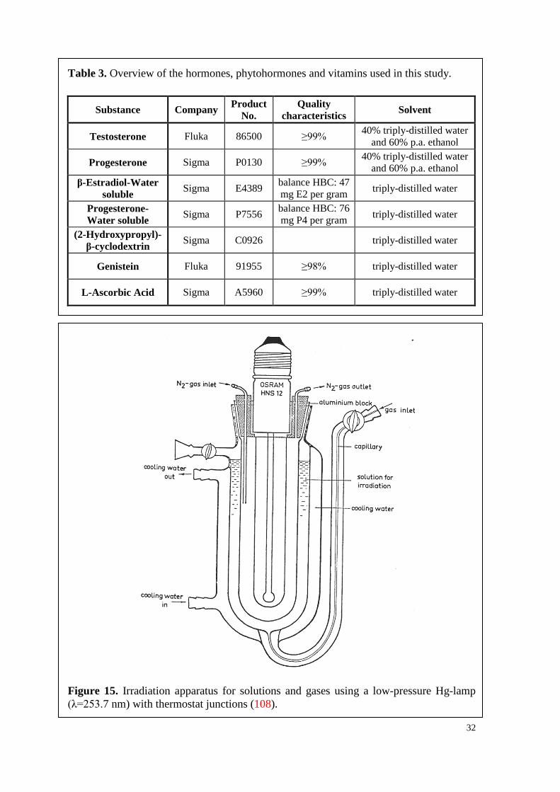
32
Table 3. Overview of the hormones, phytohormones and vitamins used in this study.
Substance Company Product No.
Quality characteristics Solvent
Testosterone Fluka 86500 ≥99% 40% triply-distilled water and 60% p.a. ethanol
Progesterone Sigma P0130 ≥99% 40% triply-distilled water and 60% p.a. ethanol
β-Estradiol-Water soluble Sigma E4389 balance HBC: 47
mg E2 per gram triply-distilled water
Progesterone- Water soluble Sigma P7556 balance HBC: 76
mg P4 per gram triply-distilled water
(2-Hydroxypropyl)- β-cyclodextrin Sigma C0926 triply-distilled water
Genistein Fluka 91955 ≥98% triply-distilled water
L-Ascorbic Acid Sigma A5960 ≥99% triply-distilled water
Figure 15. Irradiation apparatus for solutions and gases using a low-pressure Hg-lamp (λ=253.7 nm) with thermostat junctions (108).
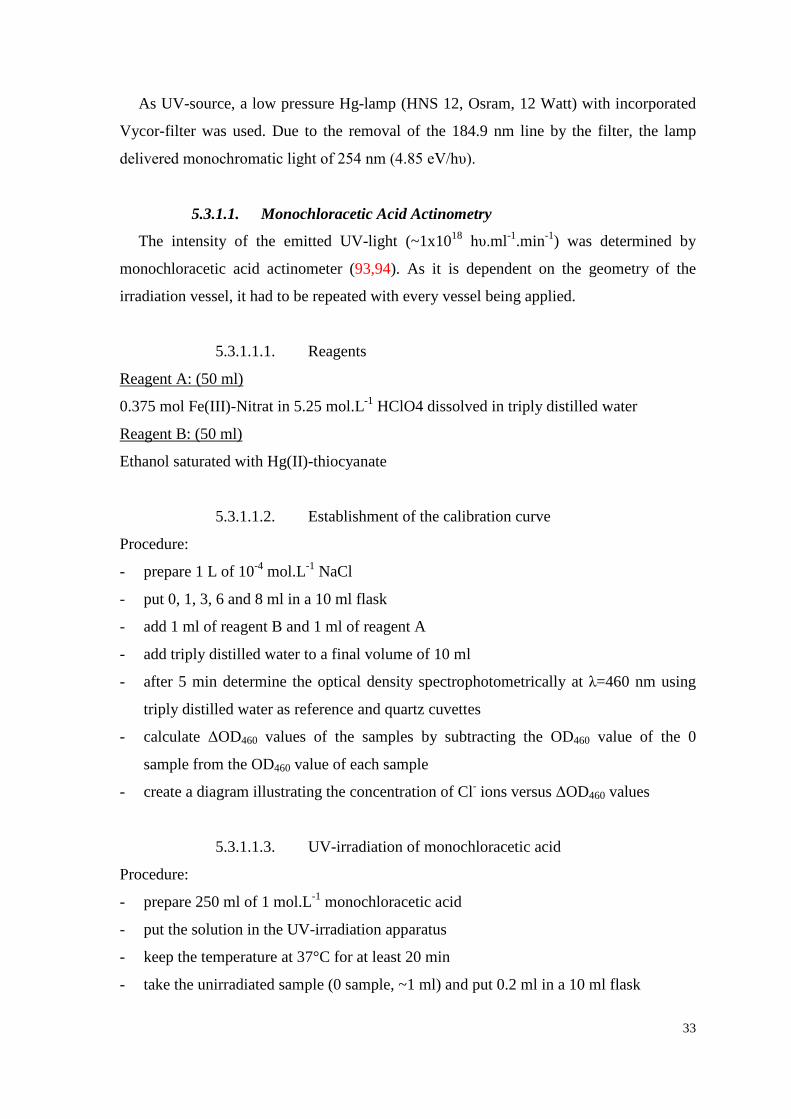
33
As UV-source, a low pressure Hg-lamp (HNS 12, Osram, 12 Watt) with incorporated
Vycor-filter was used. Due to the removal of the 184.9 nm line by the filter, the lamp
delivered monochromatic light of 254 nm (4.85 eV/hυ).
5.3.1.1. Monochloracetic Acid Actinometry
The intensity of the emitted UV-light (~1x10¹⁸ hυ.ml-1.min-1) was determined by
monochloracetic acid actinometer (93,94). As it is dependent on the geometry of the
irradiation vessel, it had to be repeated with every vessel being applied.
5.3.1.1.1. Reagents
0.375 mol Fe(III)-Nitrat in 5.25 mol.L-1 HClO4 dissolved in triply distilled water
Reagent A: (50 ml)
Ethanol saturated with Hg(II)-thiocyanate
Reagent B: (50 ml)
5.3.1.1.2. Establishment of the calibration curve
Procedure:
- prepare 1 L of 10-4 mol.L-1 NaCl
- put 0, 1, 3, 6 and 8 ml in a 10 ml flask
- add 1 ml of reagent B and 1 ml of reagent A
- add triply distilled water to a final volume of 10 ml
- after 5 min determine the optical density spectrophotometrically at λ=460 nm using
triply distilled water as reference and quartz cuvettes
- calculate ΔOD460 values of the samples by subtracting the OD460 value of the 0
sample from the OD460 value of each sample
- create a diagram illustrating the concentration of Cl- ions versus ΔOD460 values
5.3.1.1.3. UV-irradiation of monochloracetic acid
Procedure:
- prepare 250 ml of 1 mol.L-1 monochloracetic acid
- put the solution in the UV-irradiation apparatus
- keep the temperature at 37°C for at least 20 min
- take the unirradiated sample (0 sample, ~1 ml) and put 0.2 ml in a 10 ml flask

34
- add 1 ml of reagent B, 1 ml of reagent A and triply-dist. water to a final volume of 10
ml
- after 5 min determine the optical density spectrophotometrically at λ=460 nm using
triply distilled water as reference and quartz cuvettes
- start UV-irradiation and take samples after 1,2,4 and 6 min and proceed likewise
- calculate ΔOD460 values of the samples by subtracting the OD460 value of the 0
sample from the OD460 value of each sample
- draw a diagram illustrating the duration of UV-irradiation versus ΔOD460 values
- calculate the slope of the curve
- calculate I0 in hυ.ml-1.min-1 according to eq. 46:
NL I0 = k * (46) Q(Cl-) * 1000
k ...... slope of the curve NL ...... Loschmidt number (6.023x10²³ molecules.mol-1) Q(Cl-) ...... quantum yield of Cl-, dependant on the temperature
- multiply the I0 value by 50 because of the different dilutions in the calibration curve
and the irradiation curve
5.3.2. 60Co gamma source
As γ-irradiation source for the generation of free radicals, a 60Co-gamma-irradiator
(Gammacell 220, AECL, Nordion Ltd., Canada), providing a dose rate of approximately
30 Gy.min-1 was used. The equipment is depicted in Figure 16.
5.3.2.1. Fricke dosimetry
The activity of the “Gammacell 220” had to be measured, since 60Co has a half life of
5.26 years. During this study, either Fricke dosimetry was performed, or a computer
program based on Fricke dosimetry was used.
Procedure:
- irradiate Fricke solution (10-2 mol/L FeSO4*7 H2O + 10-2 mol/L NaCl + 0.8 N H2SO4)
with 60Co-gamma-rays for 0, 40, 80, 120, 160 and 240 s
- irradiate two samples for the 0 sec sample, i.e. put the solution into the sample
chamber and lower, but then retract immediately
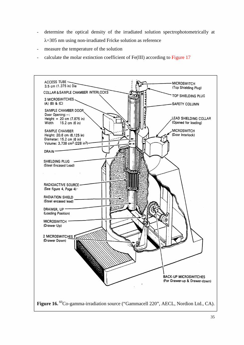
35
- determine the optical density of the irradiated solution spectrophotometrically at
λ=305 nm using non-irradiated Fricke solution as reference
- measure the temperature of the solution
- calculate the molar extinction coefficient of Fe(III) according to Figure 17
Figure 16. 60Co-gamma-irradiation source (“Gammacell 220”, AECL, Nordion Ltd., CA).
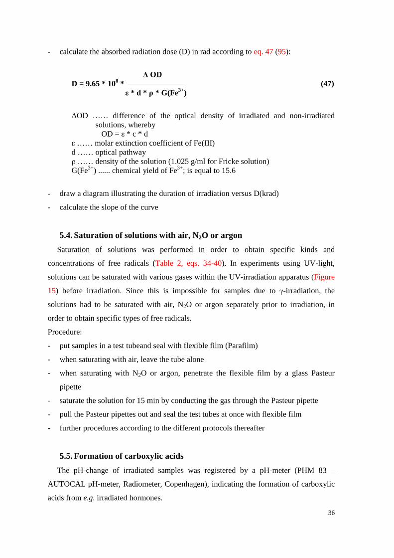
36
- calculate the absorbed radiation dose (D) in rad according to eq. 47 (95):
Δ OD
D = 9.65 * 108 * (47) ε * d * ρ * G(Fe3+)
ΔOD …… difference of the optical density of irradiated and non-irradiated solutions, whereby
OD = ε * c * d ε …… molar extinction coefficient of Fe(III) d …… optical pathway ρ …… density of the solution (1.025 g/ml for Fricke solution) G(Fe3+) ...... chemical yield of Fe3+; is equal to 15.6
- draw a diagram illustrating the duration of irradiation versus D(krad)
- calculate the slope of the curve
5.4. Saturation of solutions with air, N2O or argon Saturation of solutions was performed in order to obtain specific kinds and
concentrations of free radicals (Table 2, eqs. 34-40). In experiments using UV-light,
solutions can be saturated with various gases within the UV-irradiation apparatus (Figure
15) before irradiation. Since this is impossible for samples due to γ-irradiation, the
solutions had to be saturated with air, N2O or argon separately prior to irradiation, in
order to obtain specific types of free radicals.
Procedure:
- put samples in a test tubeand seal with flexible film (Parafilm)
- when saturating with air, leave the tube alone
- when saturating with N2O or argon, penetrate the flexible film by a glass Pasteur
pipette
- saturate the solution for 15 min by conducting the gas through the Pasteur pipette
- pull the Pasteur pipettes out and seal the test tubes at once with flexible film
- further procedures according to the different protocols thereafter
5.5. Formation of carboxylic acids The pH-change of irradiated samples was registered by a pH-meter (PHM 83 –
AUTOCAL pH-meter, Radiometer, Copenhagen), indicating the formation of carboxylic
acids from e.g. irradiated hormones.
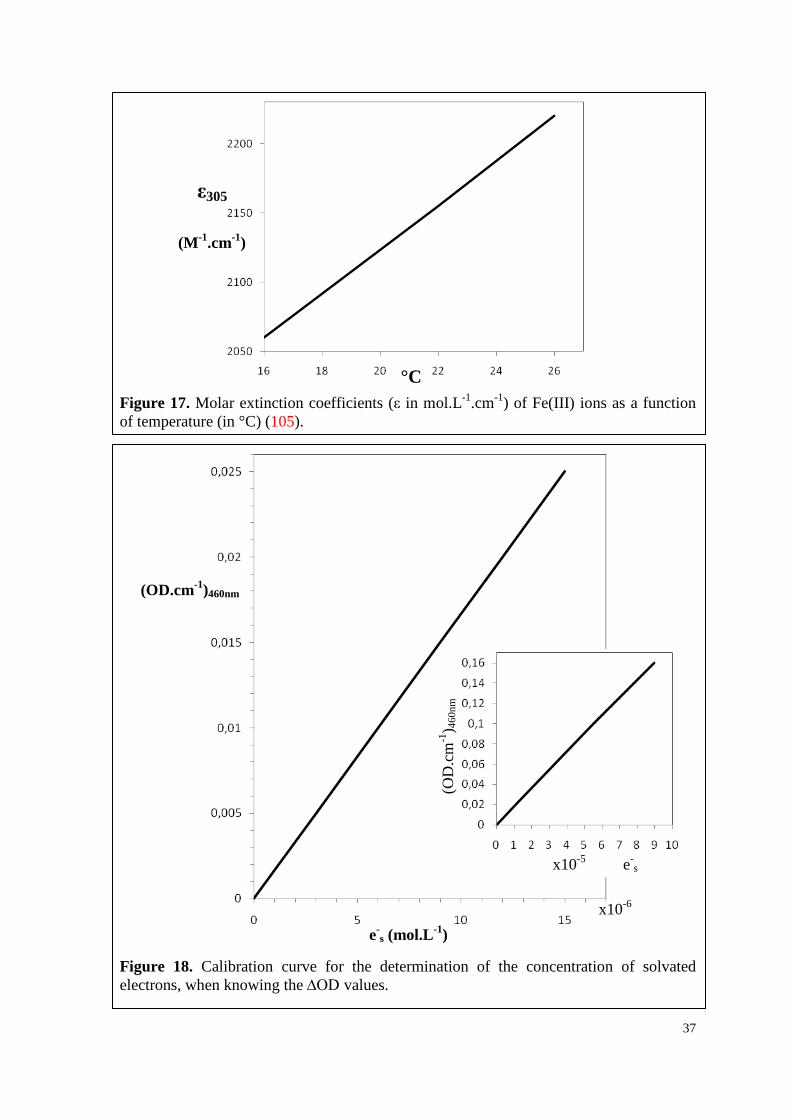
37
Figure 17. Molar extinction coefficients (ε in mol.L-1.cm-1) of Fe(III) ions as a function of temperature (in °C) (105).
Figure 18. Calibration curve for the determination of the concentration of solvated electrons, when knowing the ∆OD values.
x10-6
ε305
(M-1.cm-1)
°C
x10-5 e-s
(OD.cm-1)460nm
e-s (mol.L-1)
(OD
.cm
-1) 4
60nm
38
Procedure:
- adjust samples to pH ~7.4 by adding NaOH or HClO4
- saturate samples with argon, N2O or air
- irradiate with UV- or γ-rays with various doses
- measure the pH exactly 5 min after irradiation
- calculate ΔpH by subtracting the pH value of the non-irradiated sample from the pH
values of the irradiated samples
5.6. Spectrophotometrical measurement of electron ejection The emission of electrons from hormones was simulated by UV-excitation of
hormones in aqueous, air-free solutions (pH~ 7.4, 37°C; cf. chapter 3.2.2.1. “Simulation
of emission of e-aq by UV-irradiation of hormones”). For the registration of absorption
spectra and OD values, a LAMBDA 650 Spectrophotometer (Perkin-Elmer) was used,
belonging to double beam UV/Vis spectrophotometers with microcomputer electronics.
5.6.1. Reagents
0.375 mol Fe(III)-Nitrat in 5.25 mol.L-1 HClO4 dissolved in triply distilled water
Reagent A: (50 ml)
Ethanol saturated with Hg(II)-thiocyanatef
Reagent B: (50 ml)
Applied as delivered
Chloroethanol:
5.6.2. Electron emission
Procedure:
- prepare substrate solution in desired concentration and solvent in a 200 ml flask
(saturate the solvent with argon for 20 minutes in advance!)
- measure pH (~7.4) and correct if necessary by adding NaOH or HClO4
- make an absorption spectrum of the substrate solution from 200-500 nm using the
solvent as reference and quartz cuvettes
- check OD value at 254 nm
- put the substrate solution used for the spectrum and the pH measurement back into
200 ml flask
39
- add 1x10⁻² mol/L ClEtOH to the substrate solution (134 µl)
- put the solution in the UV-irradiation apparatus, saturate with argon for ~30’ and
adjust the temperature to 37°C
- take sample “0” from the radiation-apparatus (~4 ml) before starting UV-irradiation
- put 1 ml of the sample in a 10 ml flask
- add 1ml of reagent B and 1ml of reagent A and fill up with water
- wait for 5 min before measuring OD at 460 nm
- measure pH of the sample with the remaining 3 ml
- start UV-radiation
- take other samples as required and do as described above
- calculate ∆OD by substracting OD of sample “0” from each sample
- determine the concentration of e-aq with the help of the calibration curve (Figure 18)
- calculate the number of absorbed quanta, whereby OD = log (I0/I) and IA = I0 – I
5.7. HPLC analyses The photoinduced degradation of hormones individually and in mixture as well as their
regeneration by electron transfer in a mixture with vitamin C was registered by HPLC-
method. The implemented apparatus was model Hewlett-Packard / Agilent 1100 HPLC
series with a series 1050 diode array detector (DAD), using the Zorbax Eclipse XDB-C18
column (150x4.6mm I.D., 5 µm particle size, Agilent) at a temperature of 30°C.
Testosterone, progesterone and their mixture were separated by eluent A (Table 4), a
linear gradient between mobile phases A (2.5x10⁻⁴ mol/l ammonium acetate in water) and
B (acetonitrile). Detection of the injected samples of 15 µl was performed at 244 nm.
Total run time was 20 min with a flow rate of 0.3 ml.min-1.
Table 4. Eluents used in HPLC-analyses, both driving a linear gradient between 2 mobile phase: 2.5x10⁻⁴ mol/L ammonium acetate in water (AmmAc) and acetonitrile (ACN).
Eluent A Eluent B min ACN 0.25 mM AmmAc min ACN 0.25 mM AmmAc
0 50 50 0 80 20 0.2 60 40 0.6 65 35 8 90 10 24 10 90 9 90 10 27 10 90
9.7 50 50 30 80 20
40
Testosterone, vitamin C as well as their mixture were separated by eluent B (Table 4),
a linear gradient between mobile phases A (2.5x10⁻⁴ mol/l ammonium acetate in water)
and B (acetonitrile). Detection of the injected samples of 25 µl was performed at 244 nm
for T and 250 nm for VitC. The total run time was 35 min, followed by 5 min of post run
with a flow rate of 0.25 ml.min-1.
Procedure:
- prepare substrate solution in desired concentration and solvent in a 200 ml flask
(saturate the solvent with argon in advance!)
- measure pH (~7.4) and correct if necessary by adding NaOH or HClO4
- make an absorption spectrum of the substrate solution from 200-500 nm using the
solvent as reference and quartz cuvettes
- check OD value at 254 nm
- put the substrate solution used for the spectrum and the pH measurement back into
200 ml flask
- put the solution in the UV-irradiation apparatus
- saturate with argon for ~30’ and adjust the temperature to 37°C
- take sample “0” from the radiation-apparatus (~4 ml) before starting UV-irradiation
- put 1 ml of the sample in a vial suitable for the HPLC-apparatus
- measure pH of the sample with the remaining 3 ml
- start UV-radiation
- take other samples as required and do as described above
- calculate ∆OD by substracting OD of sample “0” from each sample
- calculate the number of absorbed quanta, whereby OD = log (I0/I) and IA = I0 – I
- set up the HPLC (computer, pumps, wavelength detector, etc.)
- put the samples in the auto-sampler and start the HPLC
- print HPLC chromatograms afterwards and compare the obtained areas under curve
for the specific peaks
5.8. Experiments in vitro with Escherichia coli bacteria The gram-negative, facultative anaerobe bacterial strain Escherichia coli is very
common in the lower intestine of warm-blooded organisms. Due to its comparatively
simple handling and genetics, E. coli is one of the most favorite model organisms in
41
biochemical research. The strain used in this study (Escherichia coli AB1157; DSM
9036, DSMZ, Braunschweig, Germany) has the following chromosomal markers:
F- thr-1 ara-14 leuB6 Δ(gpt-proA)62 lacY1 tsx-33 qsr- supE44 galK2
lambda- rac- hisG4(Oc) rfbD1 mgl-51 rpsL31 kdgK51 xyl-5 mtl-1
argE3(Oc) thi-1
5.8.1. Media and buffer
10 g/L tryptone, 5 g/L yeast, 10 g/L NaCl, 15 g/L Agar
Agar-medium:
10 g/L tryptone, 5 g/L yeast, 10 g/L NaCl
LB-medium:
8 g/L NaCl, 0.2g/L KCl, 0.25 g/L KH2PO4, 1,46 g/L Na2HPO4.2H20
Buffer-solution (pH 7.4):
5.8.1.1. Preparation of Agar Plates
Procedure:
- prepare agar medium (4 L for ~200 plates)
- sterilize the agar medium in the steam-pressure autoclave for 20 min at 121°C
- let it cool down to approximately 65°C
- pour medium in Petri dishes (20 ml per PD, 9 cm diameter)
- treat the surface with flame in order to kill spores
- store the agar plates in the fridge
5.8.2. Rehydration
The bacterial strain Escherichia coli AB1157 was purchased from DSMZ (Deutsche
Sammlung von Mikroorganismen und Zellkulturen GmbH, Braunschweig, Germany) and
delivered as dried culture. The rehydration was done following the company’s
instructions.
Procedure:
- open the ampoule in a sterile hood
- add 1 ml LB-medium and allow the pellet to rehydrate for 30 min
- mix
42
- transfer into a test tube and add 10 ml LB-medium
- streak a drop onto 4 agar plates
- incubate at 37°C o/n
- remove some colonies by using an inoculation loop for further culturing and streak
onto new agar plates
- repeat several times, before using the bacteria for experiments
5.8.3. Storage
5.8.3.1. Long-term storage
For long time storage (up to 5 years), glycerol stock cultures were prepared by adding
0.85 ml of the bacteria in logarithmic phase in LB-medium to 0.15 ml of the
cryoprotectant glycerol. Subsequently, the well-mixed stock cultures were freezed at -
20°C.
5.8.3.2. Short-term storage
For short term storage (1 month), bacteria were cultivated on an agar plate. Thereby,
some colonies were removed from an old agar plate by using an inoculation loop and
quadrant streaked on a new agar plate. After an o/n incubated at 37°C, the bacteria were
stored at 4°C in a refrigerator.
5.8.4. Radiation-biological analyses
5.8.4.1. Batch culture
The density of the bacteria as well as their phase of growth strongly influences the
results of experiments. Therefore, it was absolutely necessary to work with a defined
concentration of bacteria being in a specific phase of growth throughout all experiments.
For this purpose, batch cultures were grown.
Procedure:
- remove some colonies of the agar cultures with a one-way inoculation loop
- inoculate into 6 ml LB-medium and incubate o/n shaking (140 rpm) in a water-bath at
37°C
- in the next morning add 3 ml to 100 ml fresh LB-medium
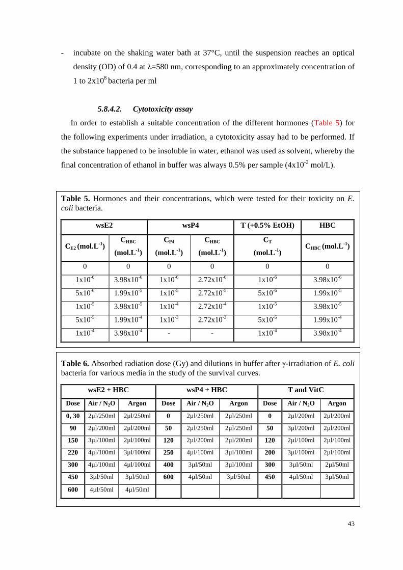
43
- incubate on the shaking water bath at 37°C, until the suspension reaches an optical
density (OD) of 0.4 at λ=580 nm, corresponding to an approximately concentration of
1 to 2x10⁸ bacteria per ml
5.8.4.2. Cytotoxicity assay
In order to establish a suitable concentration of the different hormones (Table 5) for
the following experiments under irradiation, a cytotoxicity assay had to be performed. If
the substance happened to be insoluble in water, ethanol was used as solvent, whereby the
final concentration of ethanol in buffer was always 0.5% per sample (4x10⁻² mol/L).
Table 5. Hormones and their concentrations, which were tested for their toxicity on E. coli bacteria.
wsE2 wsP4 T (+0.5% EtOH) HBC
CE2 (mol.L-1) CHBC
(mol.L-1)
CP4
(mol.L-1)
CHBC
(mol.L-1)
CT
(mol.L-1) CHBC (mol.L-1)
0 0 0 0 0 0
1x10⁻⁶ 3.98x10⁻⁶ 1x10⁻⁶ 2.72x10⁻⁶ 1x10⁻⁶ 3.98x10⁻⁶
5x10⁻⁶ 1.99x10⁻⁵ 1x10⁻⁵ 2.72x10⁻⁵ 5x10⁻⁶ 1.99x10⁻⁵
1x10⁻⁵ 3.98x10⁻⁵ 1x10⁻⁴ 2.72x10⁻⁴ 1x10⁻⁵ 3.98x10⁻⁵
5x10⁻⁵ 1.99x10⁻⁴ 1x10⁻³ 2.72x10⁻³ 5x10⁻⁵ 1.99x10⁻⁴
1x10⁻⁴ 3.98x10⁻⁴ - - 1x10⁻⁴ 3.98x10⁻⁴
Table 6. Absorbed radiation dose (Gy) and dilutions in buffer after γ-irradiation of E. coli bacteria for various media in the study of the survival curves.
wsE2 + HBC wsP4 + HBC T and VitC
Dose Air / N2O Argon Dose Air / N2O Argon Dose Air / N2O Argon
0, 30 2µl/250ml 2µl/250ml 0 2µl/250ml 2µl/250ml 0 2µl/200ml 2µl/200ml
90 2µl/200ml 2µl/200ml 50 2µl/250ml 2µl/250ml 50 3µl/200ml 2µl/200ml
150 3µl/100ml 2µl/100ml 120 2µl/200ml 2µl/200ml 120 2µl/100ml 2µl/100ml
220 4µl/100ml 3µl/100ml 250 4µl/100ml 3µl/100ml 200 3µl/100ml 2µl/100ml
300 4µl/100ml 4µl/100ml 400 3µl/50ml 3µl/100ml 300 3µl/50ml 2µl/50ml
450 3µl/50ml 3µl/50ml 600 4µl/50ml 3µl/50ml 450 4µl/50ml 3µl/50ml
600 4µl/50ml 4µl/50ml
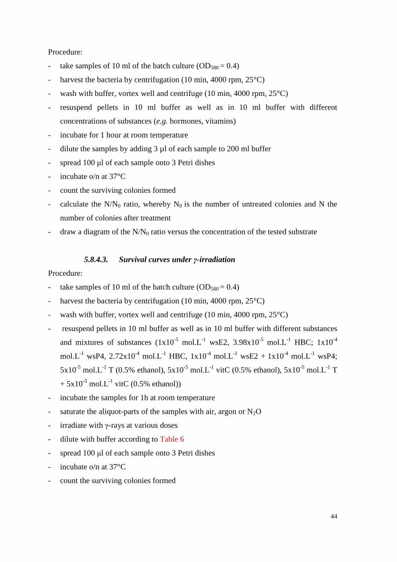
44
Procedure:
- take samples of 10 ml of the batch culture (OD580 = 0.4)
- harvest the bacteria by centrifugation (10 min, 4000 rpm, 25°C)
- wash with buffer, vortex well and centrifuge (10 min, 4000 rpm, 25°C)
- resuspend pellets in 10 ml buffer as well as in 10 ml buffer with different
concentrations of substances (e.g. hormones, vitamins)
- incubate for 1 hour at room temperature
- dilute the samples by adding 3 µl of each sample to 200 ml buffer
- spread 100 μl of each sample onto 3 Petri dishes
- incubate o/n at 37°C
- count the surviving colonies formed
- calculate the N/N0 ratio, whereby N0 is the number of untreated colonies and N the
number of colonies after treatment
- draw a diagram of the N/N0 ratio versus the concentration of the tested substrate
5.8.4.3. Survival curves under γ-irradiation
Procedure:
- take samples of 10 ml of the batch culture (OD580 = 0.4)
- harvest the bacteria by centrifugation (10 min, 4000 rpm, 25°C)
- wash with buffer, vortex well and centrifuge (10 min, 4000 rpm, 25°C)
- resuspend pellets in 10 ml buffer as well as in 10 ml buffer with different substances
and mixtures of substances (1x10⁻⁵ mol.L-1 wsE2, 3.98x10⁻⁵ mol.L-1 HBC; 1x10⁻⁴
mol.L-1 wsP4, 2.72x10⁻⁴ mol.L-1 HBC, 1x10⁻⁴ mol.L-1 wsE2 + 1x10⁻⁴ mol.L-1 wsP4;
5x10⁻⁵ mol.L-1 T (0.5% ethanol), 5x10⁻⁵ mol.L-1 vitC (0.5% ethanol), 5x10⁻⁵ mol.L-1 T
+ 5x10⁻⁵ mol.L-1 vitC (0.5% ethanol))
- incubate the samples for 1h at room temperature
- saturate the aliquot-parts of the samples with air, argon or N2O
- irradiate with γ-rays at various doses
- dilute with buffer according to Table 6
- spread 100 μl of each sample onto 3 Petri dishes
- incubate o/n at 37°C
- count the surviving colonies formed
45
- calculate the N/N0 ratio, whereby N0 is the number of untreated colonies and N the
number of colonies after treatment
- draw a diagram of the N/N0 ratio versus the absorbed radiation dose (Gy)
- calculate ΔD37 values from each survival curve by subtracting the D37 buffer value
from each individual D37 value
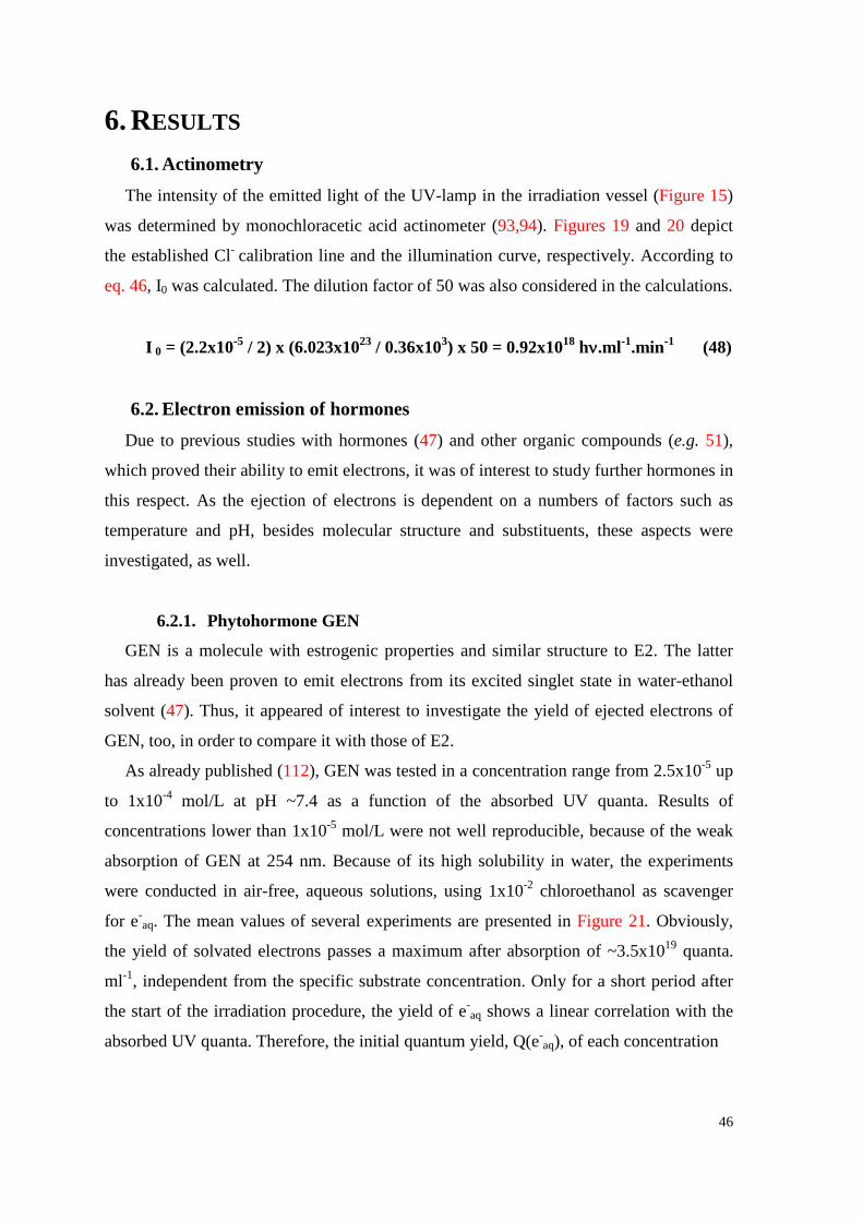
46
6. RESULTS 6.1. Actinometry
The intensity of the emitted light of the UV-lamp in the irradiation vessel (Figure 15)
was determined by monochloracetic acid actinometer (93,94). Figures 19 and 20 depict
the established Cl- calibration line and the illumination curve, respectively. According to
eq. 46, I0 was calculated. The dilution factor of 50 was also considered in the calculations.
I 0 = (2.2x10⁻⁵ / 2) x (6.023x1023 / 0.36x103) x 50 = 0.92x10¹⁸ hν.ml-1.min-1 (48)
6.2. Electron emission of hormones Due to previous studies with hormones (47) and other organic compounds (e.g. 51),
which proved their ability to emit electrons, it was of interest to study further hormones in
this respect. As the ejection of electrons is dependent on a numbers of factors such as
temperature and pH, besides molecular structure and substituents, these aspects were
investigated, as well.
6.2.1. Phytohormone GEN
GEN is a molecule with estrogenic properties and similar structure to E2. The latter
has already been proven to emit electrons from its excited singlet state in water-ethanol
solvent (47). Thus, it appeared of interest to investigate the yield of ejected electrons of
GEN, too, in order to compare it with those of E2.
As already published (112), GEN was tested in a concentration range from 2.5x10⁻⁵ up
to 1x10⁻⁴ mol/L at pH ~7.4 as a function of the absorbed UV quanta. Results of
concentrations lower than 1x10⁻⁵ mol/L were not well reproducible, because of the weak
absorption of GEN at 254 nm. Because of its high solubility in water, the experiments
were conducted in air-free, aqueous solutions, using 1x10⁻² chloroethanol as scavenger
for e-aq. The mean values of several experiments are presented in Figure 21. Obviously,
the yield of solvated electrons passes a maximum after absorption of ~3.5x10¹⁹ quanta.
ml-1, independent from the specific substrate concentration. Only for a short period after
the start of the irradiation procedure, the yield of e-aq shows a linear correlation with the
absorbed UV quanta. Therefore, the initial quantum yield, Q(e-aq), of each concentration
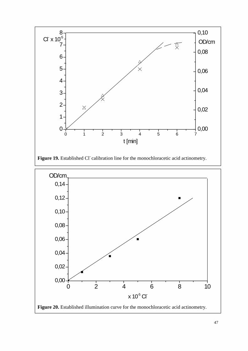
47
Figure 19. Established Cl- calibration line for the monochloracetic acid actinometry.
Figure 20. Established illumination curve for the monochloracetic acid actinometry.
0 1 2 3 4 5 6 70
1
2
3
4
5
6
7
8
0,00
0,02
0,04
0,06
0,08
0,10
OD-Werte
Konzentration
OD/cmCl- x 10-5
t [min]
0 2 4 6 8 100,00
0,02
0,04
0,06
0,08
0,10
0,12
0,14OD/cm
x 10-5 Cl-
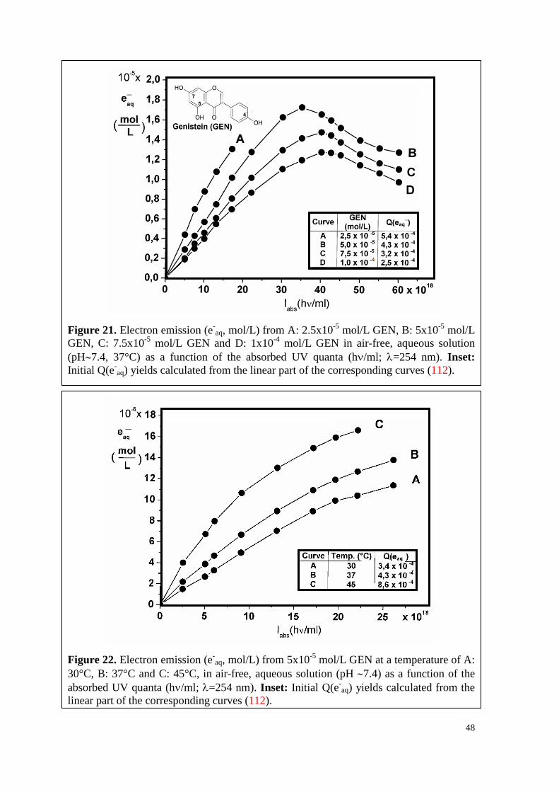
48
Figure 21. Electron emission (e-aq, mol/L) from A: 2.5x10⁻⁵ mol/L GEN, B: 5x10⁻⁵ mol/L
GEN, C: 7.5x10⁻⁵ mol/L GEN and D: 1x10⁻⁴ mol/L GEN in air-free, aqueous solution (pH∼7.4, 37°C) as a function of the absorbed UV quanta (hν/ml; λ=254 nm). Inset: Initial Q(e-
aq) yields calculated from the linear part of the corresponding curves (112).
Figure 22. Electron emission (e-aq, mol/L) from 5x10⁻⁵ mol/L GEN at a temperature of A:
30°C, B: 37°C and C: 45°C, in air-free, aqueous solution (pH ∼7.4) as a function of the absorbed UV quanta (hν/ml; λ=254 nm). Inset: Initial Q(e-
aq) yields calculated from the linear part of the corresponding curves (112).
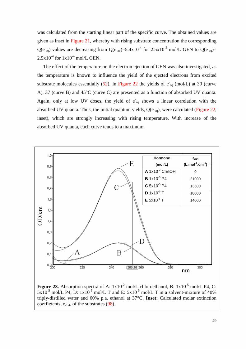
49
was calculated from the starting linear part of the specific curve. The obtained values are
given as inset in Figure 21, whereby with rising substrate concentration the corresponding
Q(e-aq) values are decreasing from Q(e-
aq)=5.4x10⁻⁴ for 2.5x10⁻⁵ mol/L GEN to Q(e-aq)=
2.5x10⁻⁴ for 1x10⁻⁴ mol/L GEN.
The effect of the temperature on the electron ejection of GEN was also investigated, as
the temperature is known to influence the yield of the ejected electrons from excited
substrate molecules essentially (52). In Figure 22 the yields of e-aq (mol/L) at 30 (curve
A), 37 (curve B) and 45°C (curve C) are presented as a function of absorbed UV quanta.
Again, only at low UV doses, the yield of e-aq shows a linear correlation with the
absorbed UV quanta. Thus, the initial quantum yields, Q(e-aq), were calculated (Figure 22,
inset), which are strongly increasing with rising temperature. With increase of the
absorbed UV quanta, each curve tends to a maximum.
Figure 23. Absorption spectra of A: 1x10⁻² mol/L chloroethanol, B: 1x10⁻⁵ mol/L P4, C: 5x10⁻⁵ mol/L P4, D: 1x10⁻⁵ mol/L T and E: 5x10⁻⁵ mol/L T in a solvent-mixture of 40% triply-distilled water and 60% p.a. ethanol at 37°C. Inset: Calculated molar extinction coefficients, ε254, of the substrates (98).
Hormone (mol/L)
ε254 (L.mol-1.cm-1)
A 1x10⁻² ClEtOH 0
B 1x10⁻⁵ P4 21000
C 5x10⁻⁵ P4 13500
D 1x10⁻⁵ T 18000
E 5x10⁻⁵ T 14000
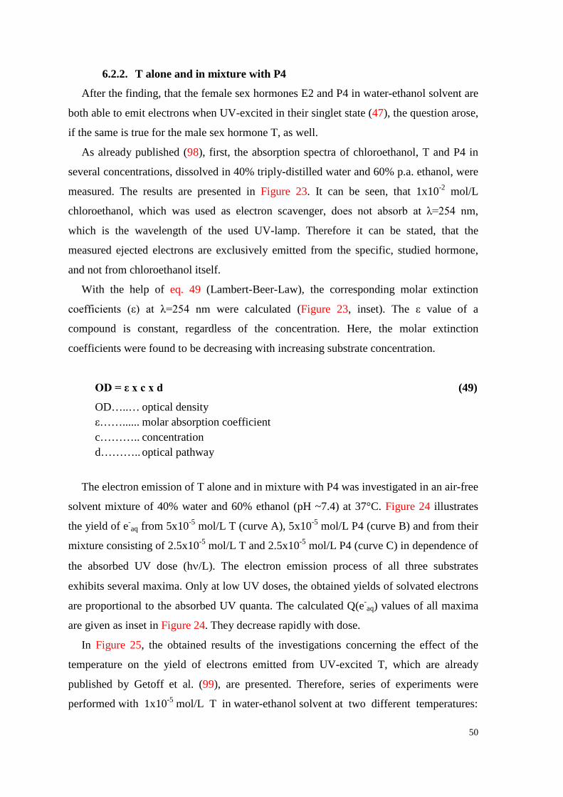
50
6.2.2. T alone and in mixture with P4
After the finding, that the female sex hormones E2 and P4 in water-ethanol solvent are
both able to emit electrons when UV-excited in their singlet state (47), the question arose,
if the same is true for the male sex hormone T, as well.
As already published (98), first, the absorption spectra of chloroethanol, T and P4 in
several concentrations, dissolved in 40% triply-distilled water and 60% p.a. ethanol, were
measured. The results are presented in Figure 23. It can be seen, that 1x10⁻² mol/L
chloroethanol, which was used as electron scavenger, does not absorb at λ=254 nm,
which is the wavelength of the used UV-lamp. Therefore it can be stated, that the
measured ejected electrons are exclusively emitted from the specific, studied hormone,
and not from chloroethanol itself.
With the help of eq. 49 (Lambert-Beer-Law), the corresponding molar extinction
coefficients (ε) at λ=254 nm were calculated (Figure 23, inset). The ε value of a
compound is constant, regardless of the concentration. Here, the molar extinction
coefficients were found to be decreasing with increasing substrate concentration.
OD = ε x c x d (49)
OD…..… optical density ε……...... molar absorption coefficient c……….. concentration d……….. optical pathway
The electron emission of T alone and in mixture with P4 was investigated in an air-free
solvent mixture of 40% water and 60% ethanol (pH ~7.4) at 37°C. Figure 24 illustrates
the yield of e-aq from 5x10⁻⁵ mol/L T (curve A), 5x10⁻⁵ mol/L P4 (curve B) and from their
mixture consisting of 2.5x10⁻⁵ mol/L T and 2.5x10⁻⁵ mol/L P4 (curve C) in dependence of
the absorbed UV dose (hν/L). The electron emission process of all three substrates
exhibits several maxima. Only at low UV doses, the obtained yields of solvated electrons
are proportional to the absorbed UV quanta. The calculated Q(e-aq) values of all maxima
are given as inset in Figure 24. They decrease rapidly with dose.
In Figure 25, the obtained results of the investigations concerning the effect of the
temperature on the yield of electrons emitted from UV-excited T, which are already
published by Getoff et al. (99), are presented. Therefore, series of experiments were
performed with 1x10⁻⁵ mol/L T in water-ethanol solvent at two different temperatures:
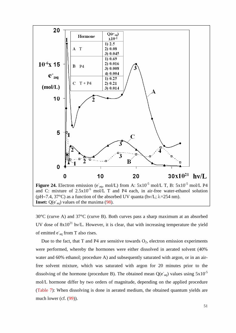
51
Figure 24. Electron emission (e-
aq, mol/L) from A: 5x10⁻⁵ mol/L T, B: 5x10⁻⁵ mol/L P4 and C: mixture of 2.5x10⁻⁵ mol/L T and P4 each, in air-free water-ethanol solution (pH∼7.4, 37°C) as a function of the absorbed UV quanta (hν/L; λ=254 nm). Inset: Q(e-
aq) values of the maxima (98).
30°C (curve A) and 37°C (curve B). Both curves pass a sharp maximum at an absorbed
UV dose of 8x10²¹ hν/L. However, it is clear, that with increasing temperature the yield
of emitted e-aq from T also rises.
Due to the fact, that T and P4 are sensitive towards O2, electron emission experiments
were performed, whereby the hormones were either dissolved in aerated solvent (40%
water and 60% ethanol; procedure A) and subsequently saturated with argon, or in an air-
free solvent mixture, which was saturated with argon for 20 minutes prior to the
dissolving of the hormone (procedure B). The obtained mean Q(e-aq) values using 5x10⁻⁵
mol/L hormone differ by two orders of magnitude, depending on the applied procedure
(Table 7): When dissolving is done in aerated medium, the obtained quantum yields are
much lower (cf. (99)).
T
P4
T + P4
hν/L
(mol/L)
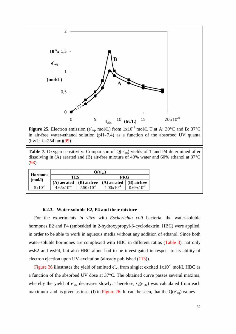
52
Figure 25. Electron emission (e-aq, mol/L) from 1x10⁻⁵ mol/L T at A: 30°C and B: 37°C
in air-free water-ethanol solution (pH∼7.4) as a function of the absorbed UV quanta (hν/L; λ=254 nm)(99).
Table 7. Oxygen sensitivity: Comparison of Q(e-aq) yields of T and P4 determined after
dissolving in (A) aerated and (B) air-free mixture of 40% water and 60% ethanol at 37°C (98).
Hormone (mol/l)
Q(e-aq)
TES PRG (A) aerated (B) airfree (A) aerated (B) airfree
5x10⁻⁵ 4.65x10⁻⁴ 2.50x10⁻² 4.00x10⁻⁴ 0.69x10⁻²
6.2.3. Water-soluble E2, P4 and their mixture
For the experiments in vitro with Escherichia coli bacteria, the water-soluble
hormones E2 and P4 (embedded in 2-hydroxypropyl-β-cyclodextrin, HBC) were applied,
in order to be able to work in aqueous media without any addition of ethanol. Since both
water-soluble hormones are complexed with HBC in different ratios (Table 3), not only
wsE2 and wsP4, but also HBC alone had to be investigated in respect to its ability of
electron ejection upon UV-excitation (already published (113)).
Figure 26 illustrates the yield of emitted e-aq from singlet excited 1x10⁻⁴ mol/L HBC as
a function of the absorbed UV dose at 37°C. The obtained curve passes several maxima,
whereby the yield of e-aq decreases slowly. Therefore, Q(e-
aq) was calculated from each
maximum and is given as inset (I) in Figure 26. It can be seen, that the Q(e-aq) values
10-5x
e-aq
(mol/L)
x1021 Iabs (hν/L)
B
A
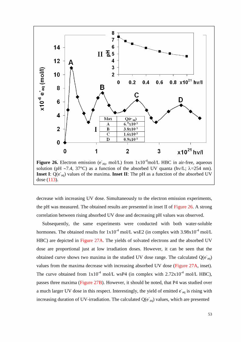
53
Figure 26. Electron emission (e-
aq, mol/L) from 1x10⁻⁴mol/L HBC in air-free, aqueous solution (pH ∼7.4, 37°C) as a function of the absorbed UV quanta (hν/L; λ=254 nm). Inset I: Q(e-
aq) values of the maxima. Inset II: The pH as a function of the absorbed UV dose (113). decrease with increasing UV dose. Simultaneously to the electron emission experiments,
the pH was measured. The obtained results are presented in inset II of Figure 26. A strong
correlation between rising absorbed UV dose and decreasing pH values was observed.
Subsequently, the same experiments were conducted with both water-soluble
hormones. The obtained results for 1x10⁻⁴ mol/L wsE2 (in complex with 3.98x10⁻⁴ mol/L
HBC) are depicted in Figure 27A. The yields of solvated electrons and the absorbed UV
dose are proportional just at low irradiation doses. However, it can be seen that the
obtained curve shows two maxima in the studied UV dose range. The calculated Q(e-aq)
values from the maxima decrease with increasing absorbed UV dose (Figure 27A, inset).
The curve obtained from 1x10⁻⁴ mol/L wsP4 (in complex with 2.72x10⁻⁴ mol/L HBC),
passes three maxima (Figure 27B). However, it should be noted, that P4 was studied over
a much larger UV dose in this respect. Interestingly, the yield of emitted e-aq is rising with
increasing duration of UV-irradiation. The calculated Q(e-aq) values, which are presented
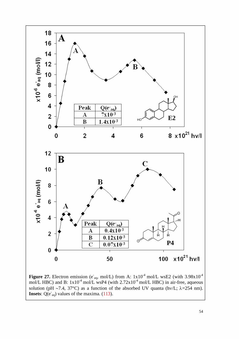
54
Figure 27. Electron emission (e-
aq, mol/L) from A: 1x10⁻⁴ mol/L wsE2 (with 3.98x10⁻⁴
mol/L HBC) and B: 1x10⁻⁴ mol/L wsP4 (with 2.72x10⁻⁴ mol/L HBC) in air-free, aqueous solution (pH ∼7.4, 37°C) as a function of the absorbed UV quanta (hν/L; λ=254 nm). Insets: Q(e-
aq) values of the maxima. (113).
E2
P4
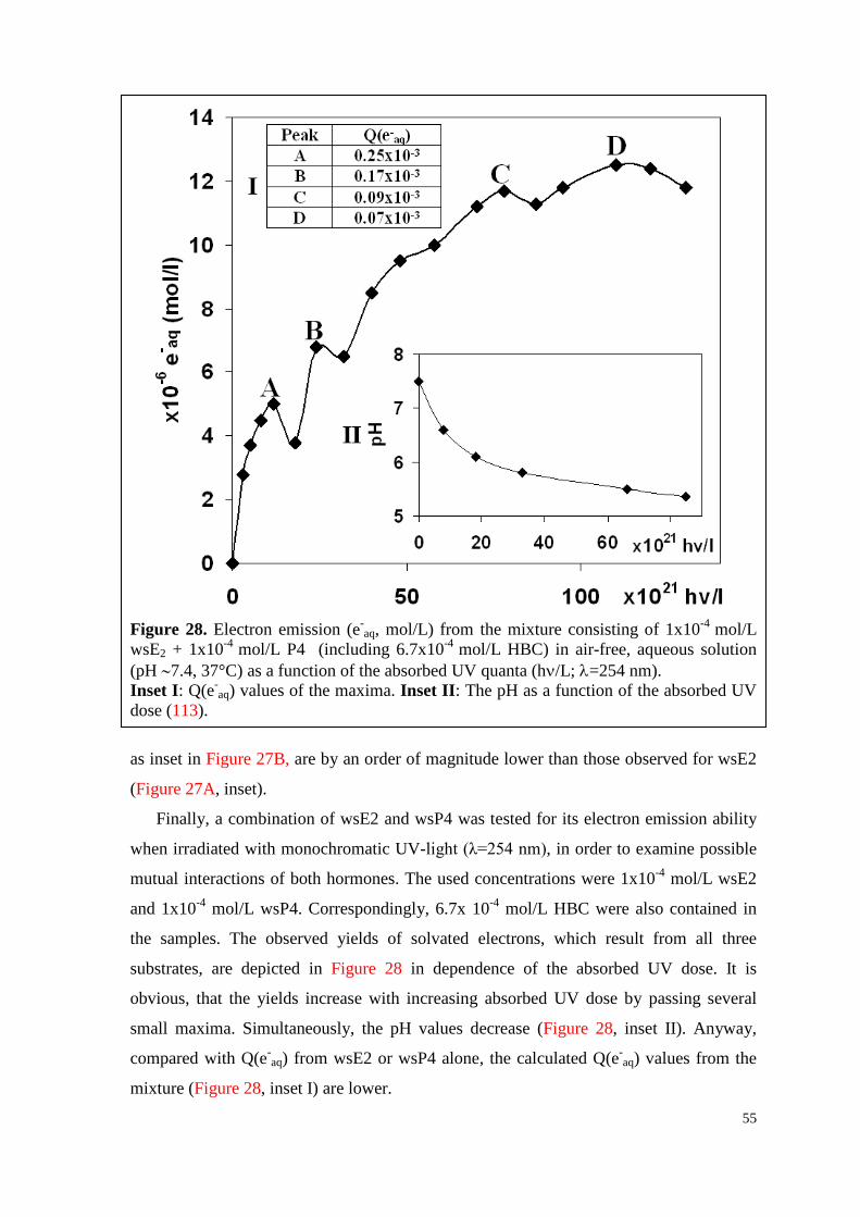
55
Figure 28. Electron emission (e-
aq, mol/L) from the mixture consisting of 1x10⁻⁴ mol/L wsE2 + 1x10⁻⁴ mol/L P4 (including 6.7x10⁻⁴ mol/L HBC) in air-free, aqueous solution (pH ∼7.4, 37°C) as a function of the absorbed UV quanta (hν/L; λ=254 nm). Inset I: Q(e-
aq) values of the maxima. Inset II: The pH as a function of the absorbed UV dose (113).
as inset in Figure 27B, are by an order of magnitude lower than those observed for wsE2
(Figure 27A, inset).
Finally, a combination of wsE2 and wsP4 was tested for its electron emission ability
when irradiated with monochromatic UV-light (λ=254 nm), in order to examine possible
mutual interactions of both hormones. The used concentrations were 1x10⁻⁴ mol/L wsE2
and 1x10⁻⁴ mol/L wsP4. Correspondingly, 6.7x 10⁻⁴ mol/L HBC were also contained in
the samples. The observed yields of solvated electrons, which result from all three
substrates, are depicted in Figure 28 in dependence of the absorbed UV dose. It is
obvious, that the yields increase with increasing absorbed UV dose by passing several
small maxima. Simultaneously, the pH values decrease (Figure 28, inset II). Anyway,
compared with Q(e-aq) from wsE2 or wsP4 alone, the calculated Q(e-
aq) values from the
mixture (Figure 28, inset I) are lower.
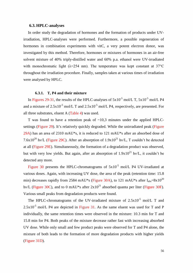
56
6.3. HPLC-analyses In order study the degradation of hormones and the formation of products under UV-
irradiation, HPLC-analyses were performed. Furthermore, a possible regeneration of
hormones in combination experiments with vitC, a very potent electron donor, was
investigated by this method. Therefore, hormones or mixtures of hormones in an air-free
solvent mixture of 40% triply-distilled water and 60% p.a. ethanol were UV-irradiated
with monochromatic light (λ=254 nm). The temperature was kept constant at 37°C
throughout the irradiation procedure. Finally, samples taken at various times of irradiation
were analysed by HPLC.
6.3.1. T, P4 and their mixture
In Figures 29-31, the results of the HPLC-analyses of 5x10⁻⁵ mol/L T, 5x10⁻⁵ mol/L P4
and a mixture of 2.5x10⁻⁵ mol/L T and 2.5x10⁻⁵ mol/L P4, respectively, are presented. For
all three substrates, eluent A (Table 4) was used.
T was found to have a retention peak of ~10,3 minutes under the applied HPLC-
settings (Figure 29). It’s relatively quickly degraded. While the unirradiated peak (Figure
29A) has an area of 2310 mAU*s, it is reduced to 121 mAU*s after an absorbed dose of
7.6x10²⁰ hν/L (Figure 29C). After an absorption of 1.9x10²¹ hν/L, T couldn’t be detected
at all (Figure 29E). Simultaneously, the formation of a degradation product was observed,
but with very low yields. But again, after an absorption of 1.9x10²¹ hν/L, it couldn’t be
detected any more.
Figure 30 presents the HPLC-chromatograms of 5x10⁻⁵ mol/L P4 UV-irradiated at
various doses. Again, with increasing UV dose, the area of the peak (retention time: 15.8
min) decreases rapidly from 2584 mAU*s (Figure 30A), to 121 mAU*s after Iabs=8x10²⁰
hν/L (Figure 30C), and to 0 mAU*s after 2x10²¹ absorbed quanta per liter (Figure 30F).
Various small peaks from degradation products were found.
The HPLC-chromatograms of the UV-irradiated mixture of 2.5x10⁻⁵ mol/L T and
2.5x10⁻⁵ mol/L P4 are depicted in Figure 31. As the same eluent was used for T and P
individually, the same retention times were observed in the mixture: 10.3 min for T and
15.8 min for P4. Both peaks of the mixture decrease rather fast with increasing absorbed
UV dose. While only small and few product peaks were observed for T and P4 alone, the
mixture of both leads to the formation of more degradation products with higher yields
(Figure 31D).
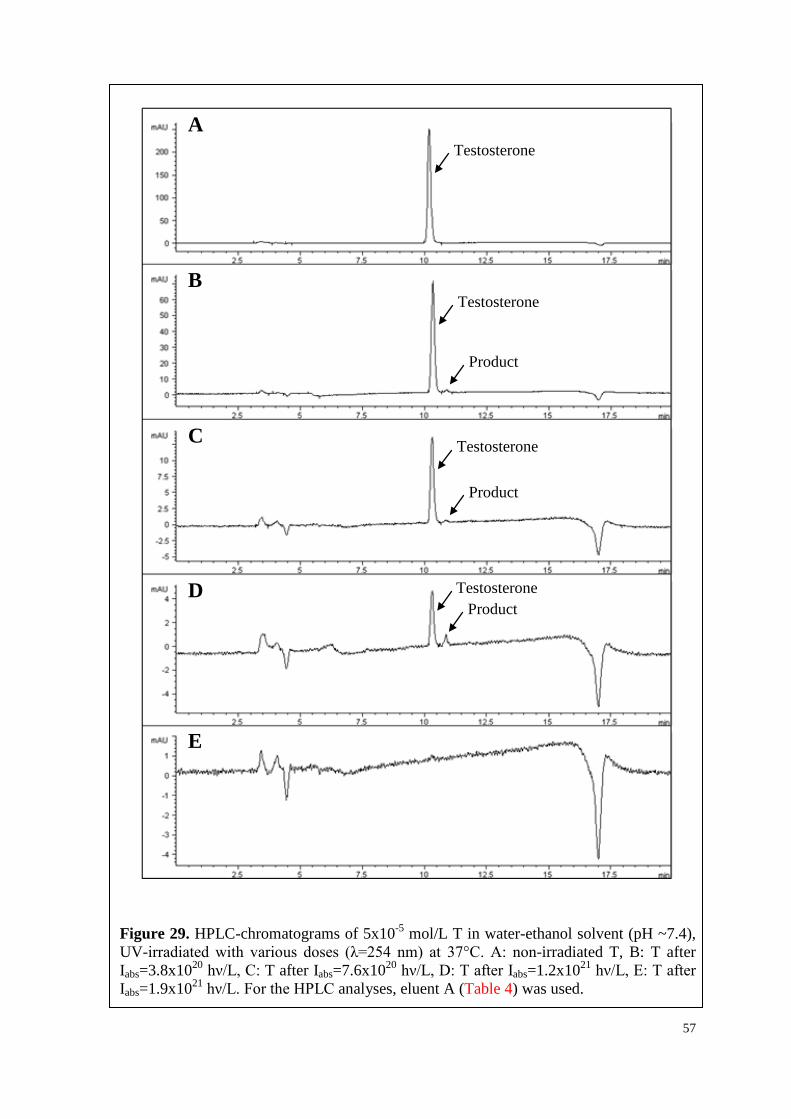
57
Figure 29. HPLC-chromatograms of 5x10⁻⁵ mol/L T in water-ethanol solvent (pH ~7.4), UV-irradiated with various doses (λ=254 nm) at 37°C. A: non-irradiated T, B: T after Iabs=3.8x10²⁰ hν/L, C: T after Iabs=7.6x10²⁰ hν/L, D: T after Iabs=1.2x10²¹ hν/L, E: T after Iabs=1.9x10²¹ hν/L. For the HPLC analyses, eluent A (Table 4) was used.
Testosterone
Testosterone
Testosterone
Testosterone Product
A
B
C
D
E
Product
Product
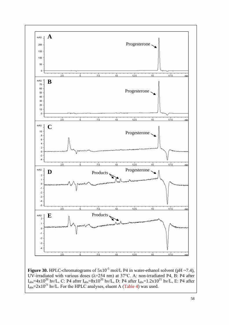
58
Figure 30. HPLC-chromatograms of 5x10⁻⁵ mol/L P4 in water-ethanol solvent (pH ~7.4), UV-irradiated with various doses (λ=254 nm) at 37°C. A: non-irradiated P4, B: P4 after Iabs=4x10²⁰ hν/L, C: P4 after Iabs=8x10²⁰ hν/L, D: P4 after Iabs=1.2x10²¹ hν/L, E: P4 after Iabs=2x10²¹ hν/L. For the HPLC analyses, eluent A (Table 4) was used.
A
B
C
D
E
Progesterone
Progesterone
Progesterone
Progesterone Products
Products
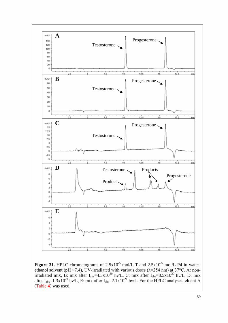
59
Figure 31. HPLC-chromatograms of 2.5x10⁻⁵ mol/L T and 2.5x10⁻⁵ mol/L P4 in water-ethanol solvent (pH ~7.4), UV-irradiated with various doses (λ=254 nm) at 37°C. A: non-irradiated mix, B: mix after Iabs=4.3x10²⁰ hν/L, C: mix after Iabs=8.5x10²⁰ hν/L, D: mix after Iabs=1.3x10²¹ hν/L, E: mix after Iabs=2.1x10²¹ hν/L. For the HPLC analyses, eluent A (Table 4) was used.
A
B
C
D
E
Progesterone
Progesterone
Progesterone
Progesterone Testosterone
Product
Products
Testosterone
Testosterone
Testosterone
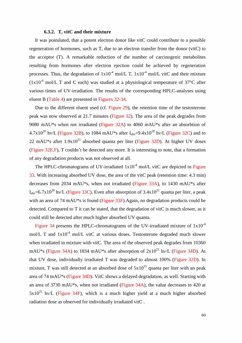
60
6.3.2. T, vitC and their mixture
It was postulated, that a potent electron donor like vitC could contribute to a possible
regeneration of hormones, such as T, due to an electron transfer from the donor (vitC) to
the acceptor (T). A remarkable reduction of the number of carcinogenic metabolites
resulting from hormones after electron ejection could be achieved by regeneration
processes. Thus, the degradation of 1x10⁻⁴ mol/L T, 1x10⁻⁴ mol/L vitC and their mixture
(1x10⁻⁴ mol/L T and C each) was studied at a physiological temperature of 37°C after
various times of UV-irradiation. The results of the corresponding HPLC-analyses using
eluent B (Table 4) are presented in Figures 32-34.
Due to the different eluent used (cf. Figure 29), the retention time of the testosterone
peak was now observed at 21.7 minutes (Figure 32). The area of the peak degrades from
9080 mAU*s when not irradiated (Figure 32A) to 4060 mAU*s after an absorbtion of
4.7x10²⁰ hν/L (Figure 32B), to 1084 mAU*s after Iabs=9.4x10²⁰ hν/L (Figure 32C) and to
22 mAU*s after 1.9x10²¹ absorbed quanta per liter (Figure 32D). At higher UV doses
(Figure 32E,F), T couldn’t be detected any more. It is interesting to note, that a formation
of any degradation products was not observed at all.
The HPLC-chromatograms of UV-irradiated 1x10⁻⁴ mol/L vitC are depicted in Figure
33. With increasing absorbed UV dose, the area of the vitC peak (retention time: 4.3 min)
decreases from 2034 mAU*s, when not irradiated (Figure 33A), to 1430 mAU*s after
Iabs=6.7x10²⁰ hν/L (Figure 33C). Even after absorption of 3.4x10²¹ quanta per liter, a peak
with an area of 74 mAU*s is found (Figure 33F).Again, no degradation products could be
detected. Compared to T it can be stated, that the degradation of vitC is much slower, as it
could still be detected after much higher absorbed UV quanta.
Figure 34 presents the HPLC-chromatograms of the UV-irradiated mixture of 1x10⁻⁴
mol/L T and 1x10⁻⁴ mol/L vitC at various doses. Testosterone degraded much slower
when irradiated in mixture with vitC. The area of the observed peak degrades from 10360
mAU*s (Figure 34A) to 1834 mAU*s after absorption of 2x10²¹ hν/L (Figure 34D). At
that UV dose, individually irradiated T was degraded to almost 100% (Figure 32D). In
mixture, T was still detected at an absorbed dose of 5x10²¹ quanta per liter with an peak
area of 74 mAU*s (Figure 34D). VitC shows a delayed degradation, as well. Starting with
an area of 3730 mAU*s, when not irradiated (Figure 34A), the value decreases to 420 at
5x10²¹ hν/L (Figure 34F), which is a much higher yield at a much higher absorbed
radiation dose as observed for individually irradiated vitC .
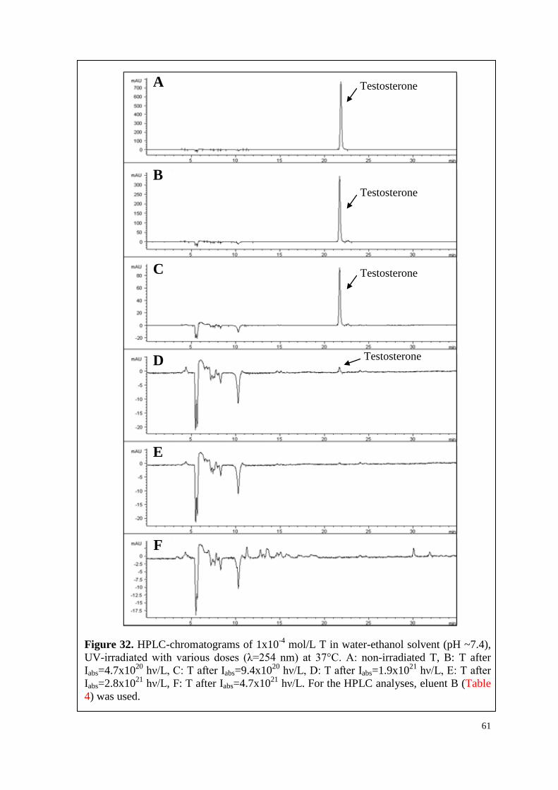
61
Figure 32. HPLC-chromatograms of 1x10⁻⁴ mol/L T in water-ethanol solvent (pH ~7.4), UV-irradiated with various doses (λ=254 nm) at 37°C. A: non-irradiated T, B: T after Iabs=4.7x10²⁰ hν/L, C: T after Iabs=9.4x10²⁰ hν/L, D: T after Iabs=1.9x10²¹ hν/L, E: T after Iabs=2.8x10²¹ hν/L, F: T after Iabs=4.7x10²¹ hν/L. For the HPLC analyses, eluent B (Table 4) was used.
A
B
C
D
E
F
Testosterone
Testosterone
Testosterone
Testosterone
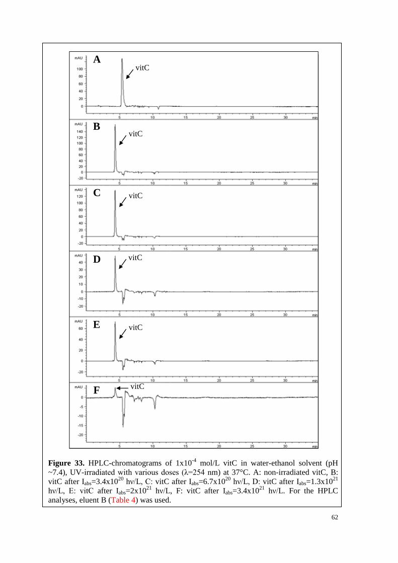
62
Figure 33. HPLC-chromatograms of 1x10⁻⁴ mol/L vitC in water-ethanol solvent (pH ~7.4), UV-irradiated with various doses (λ=254 nm) at 37°C. A: non-irradiated vitC, B: vitC after Iabs=3.4x10²⁰ hν/L, C: vitC after Iabs=6.7x10²⁰ hν/L, D: vitC after Iabs=1.3x10²¹ hν/L, E: vitC after Iabs=2x10²¹ hν/L, F: vitC after Iabs=3.4x10²¹ hν/L. For the HPLC analyses, eluent B (Table 4) was used.
A
B
C
D
E
F
vitC
vitC
vitC
vitC
vitC
vitC
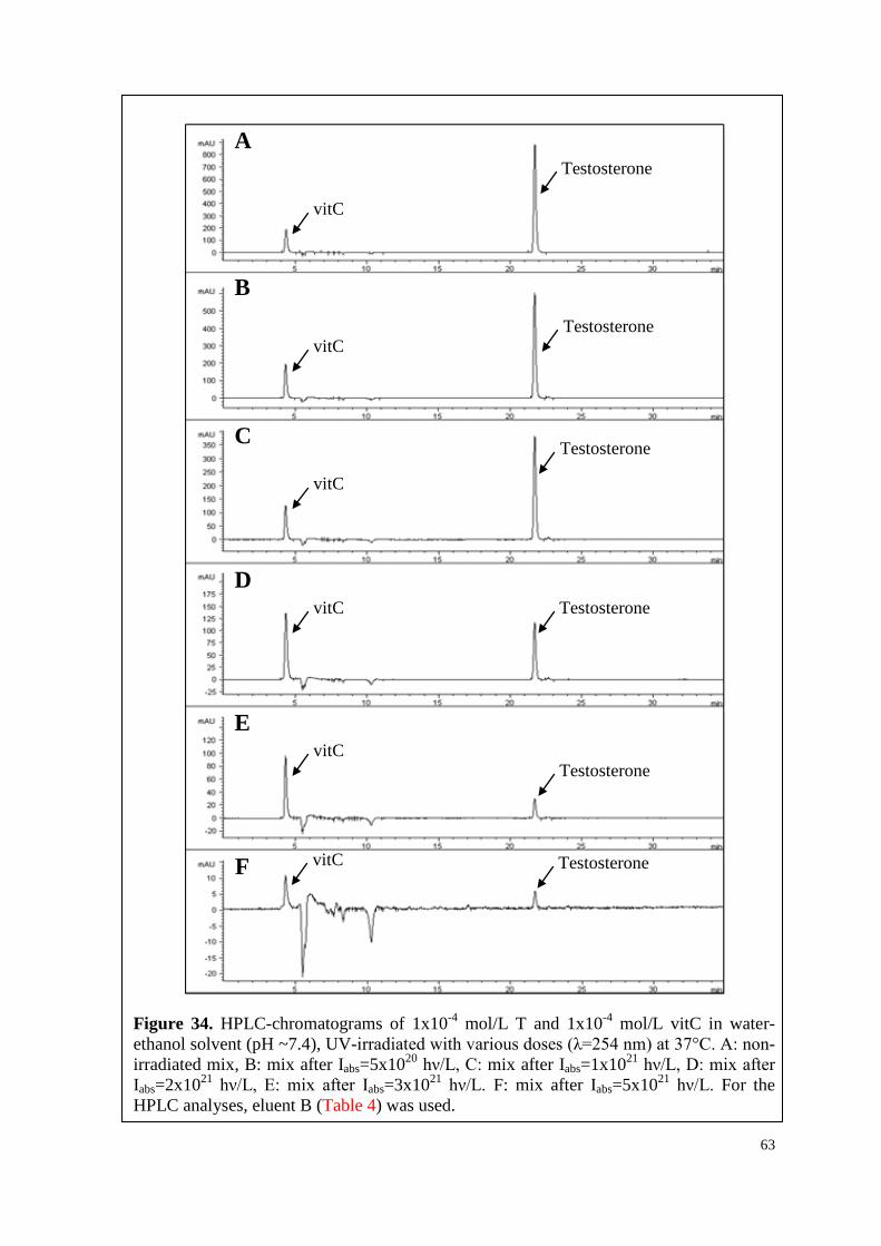
63
Figure 34. HPLC-chromatograms of 1x10⁻⁴ mol/L T and 1x10⁻⁴ mol/L vitC in water-ethanol solvent (pH ~7.4), UV-irradiated with various doses (λ=254 nm) at 37°C. A: non-irradiated mix, B: mix after Iabs=5x10²⁰ hν/L, C: mix after Iabs=1x10²¹ hν/L, D: mix after Iabs=2x10²¹ hν/L, E: mix after Iabs=3x10²¹ hν/L. F: mix after Iabs=5x10²¹ hν/L. For the HPLC analyses, eluent B (Table 4) was used.
A
B
C
D
E
F
vitC
vitC
vitC
vitC
vitC
vitC
Testosterone
Testosterone
Testosterone
Testosterone
Testosterone
Testosterone
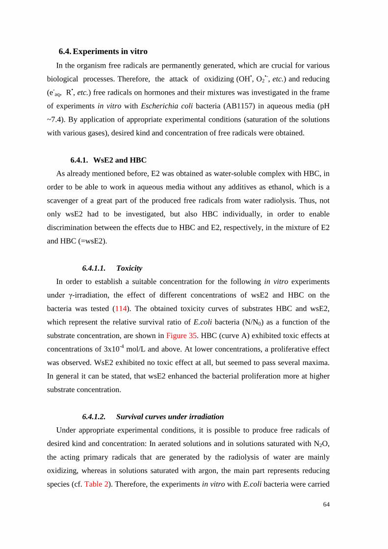
64
6.4. Experiments in vitro In the organism free radicals are permanently generated, which are crucial for various
biological processes. Therefore, the attack of oxidizing (OH•, O2•-, etc.) and reducing
(e-aq, R•, etc.) free radicals on hormones and their mixtures was investigated in the frame
of experiments in vitro with Escherichia coli bacteria (AB1157) in aqueous media (pH
~7.4). By application of appropriate experimental conditions (saturation of the solutions
with various gases), desired kind and concentration of free radicals were obtained.
6.4.1. WsE2 and HBC
As already mentioned before, E2 was obtained as water-soluble complex with HBC, in
order to be able to work in aqueous media without any additives as ethanol, which is a
scavenger of a great part of the produced free radicals from water radiolysis. Thus, not
only wsE2 had to be investigated, but also HBC individually, in order to enable
discrimination between the effects due to HBC and E2, respectively, in the mixture of E2
and HBC (=wsE2).
6.4.1.1. Toxicity
In order to establish a suitable concentration for the following in vitro experiments
under γ-irradiation, the effect of different concentrations of wsE2 and HBC on the
bacteria was tested (114). The obtained toxicity curves of substrates HBC and wsE2,
which represent the relative survival ratio of E.coli bacteria (N/N0) as a function of the
substrate concentration, are shown in Figure 35. HBC (curve A) exhibited toxic effects at
concentrations of 3x10⁻⁴ mol/L and above. At lower concentrations, a proliferative effect
was observed. WsE2 exhibited no toxic effect at all, but seemed to pass several maxima.
In general it can be stated, that wsE2 enhanced the bacterial proliferation more at higher
substrate concentration.
6.4.1.2. Survival curves under irradiation
Under appropriate experimental conditions, it is possible to produce free radicals of
desired kind and concentration: In aerated solutions and in solutions saturated with N2O,
the acting primary radicals that are generated by the radiolysis of water are mainly
oxidizing, whereas in solutions saturated with argon, the main part represents reducing
species (cf. Table 2). Therefore, the experiments in vitro with E.coli bacteria were carried
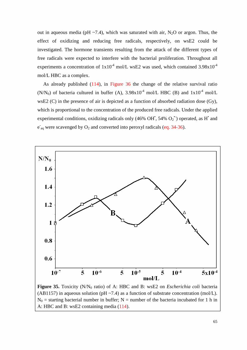
65
out in aqueous media (pH ~7.4), which was saturated with air, N2O or argon. Thus, the
effect of oxidizing and reducing free radicals, respectively, on wsE2 could be
investigated. The hormone transients resulting from the attack of the different types of
free radicals were expected to interfere with the bacterial proliferation. Throughout all
experiments a concentration of 1x10⁻⁴ mol/L wsE2 was used, which contained 3.98x10⁻⁴
mol/L HBC as a complex.
As already published (114), in Figure 36 the change of the relative survival ratio
(N/N0) of bacteria cultured in buffer (A), 3.98x10⁻⁴ mol/L HBC (B) and 1x10⁻⁴ mol/L
wsE2 (C) in the presence of air is depicted as a function of absorbed radiation dose (Gy),
which is proportional to the concentration of the produced free radicals. Under the applied
experimental conditions, oxidizing radicals only (46% OH•, 54% O2•-) operated, as H• and
e-aq were scavenged by O2 and converted into peroxyl radicals (eq. 34-36).
Figure 35. Toxicity (N/N0 ratio) of A: HBC and B: wsE2 on Escherichia coli bacteria (AB1157) in aqueous solution (pH ~7.4) as a function of substrate concentration (mol/L). N0 = starting bacterial number in buffer; N = number of the bacteria incubated for 1 h in A: HBC and B: wsE2 containing media (114).
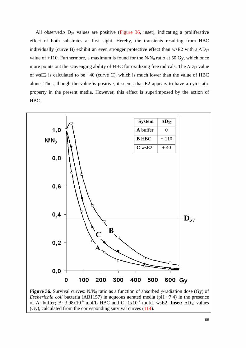
66
All observed ∆ D37 values are positive (Figure 36, inset), indicating a proliferative
effect of both substrates at first sight. Hereby, the transients resulting from HBC
individually (curve B) exhibit an even stronger protective effect than wsE2 with a ∆D37
value of +110. Furthermore, a maximum is found for the N/N0 ratio at 50 Gy, which once
more points out the scavenging ability of HBC for oxidizing free radicals. The ∆D37 value
of wsE2 is calculated to be +40 (curve C), which is much lower than the value of HBC
alone. Thus, though the value is positive, it seems that E2 appears to have a cytostatic
property in the present media. However, this effect is superimposed by the action of
HBC.
Figure 36. Survival curves: N/N0 ratio as a function of absorbed γ-radiation dose (Gy) of Escherichia coli bacteria (AB1157) in aqueous aerated media (pH ~7.4) in the presence of A: buffer; B: 3.98x10⁻⁴ mol/L HBC and C: 1x10⁻⁴ mol/L wsE2. Inset: ∆D37 values (Gy), calculated from the corresponding survival curves (114).
System ΔD37
A buffer 0
B HBC + 110
C wsE2 + 40
67
Figure 37. Survival curves: N/N0 ratio as a function of absorbed γ-radiation dose (Gy) of Escherichia coli bacteria (AB1157) in aqueous media saturated with N2O (pH ~7.4) in the presence of A: buffer; B: 3.98x10⁻⁴ mol/L HBC and C: 1x10⁻⁴ mol/L wsE2. Inset: ∆D37 values (Gy), calculated from the corresponding survival curves (114).
Likewise, studies have been performed in the presence of N2O in a broad range of
radiation dose up to 600 Gy. Under these experimental conditions, all e-aq were converted
into OH• radicals, leading to a mainly oxidizing environment (90% OH• and 10% H•). The
obtained survival curves (N/N0 ratio) represent mean values of several series of
experiments and are presented in Figure 37 (114).
As the reaction rate constant (k) of OH•, the main acting species, and wsE2 is very
high (k ~109-1010 L.mol-1.s-1), wsE2 was expected to act as a very efficient OH•
scavenger. This assumption was approved by the corresponding ΔD37 value of +250
(Figure 37, inset). From the survival curve of E. coli bacteria under irradiation incubated
System ΔD37
A buffer 0
B HBC + 140
C wsE2 + 250
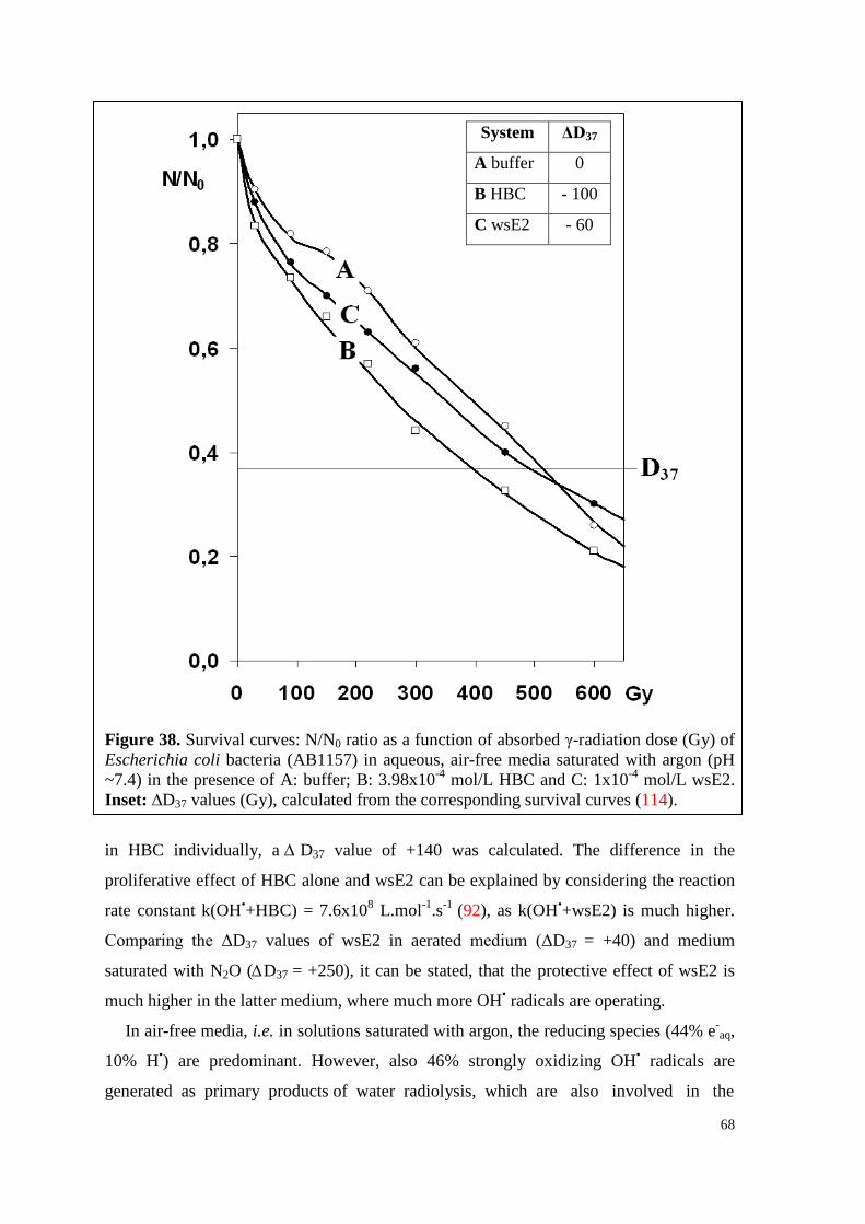
68
Figure 38. Survival curves: N/N0 ratio as a function of absorbed γ-radiation dose (Gy) of Escherichia coli bacteria (AB1157) in aqueous, air-free media saturated with argon (pH ~7.4) in the presence of A: buffer; B: 3.98x10⁻⁴ mol/L HBC and C: 1x10⁻⁴ mol/L wsE2. Inset: ∆D37 values (Gy), calculated from the corresponding survival curves (114).
in HBC individually, a ∆ D37 value of +140 was calculated. The difference in the
proliferative effect of HBC alone and wsE2 can be explained by considering the reaction
rate constant k(OH•+HBC) = 7.6x10⁸ L.mol-1.s-1 (92), as k(OH•+wsE2) is much higher.
Comparing the ΔD37 values of wsE2 in aerated medium (ΔD37 = +40) and medium
saturated with N2O (∆D37 = +250), it can be stated, that the protective effect of wsE2 is
much higher in the latter medium, where much more OH• radicals are operating.
In air-free media, i.e. in solutions saturated with argon, the reducing species (44% e-aq,
10% H•) are predominant. However, also 46% strongly oxidizing OH• radicals are
generated as primary products of water radiolysis, which are also involved in the
System ΔD37
A buffer 0
B HBC - 100
C wsE2 - 60
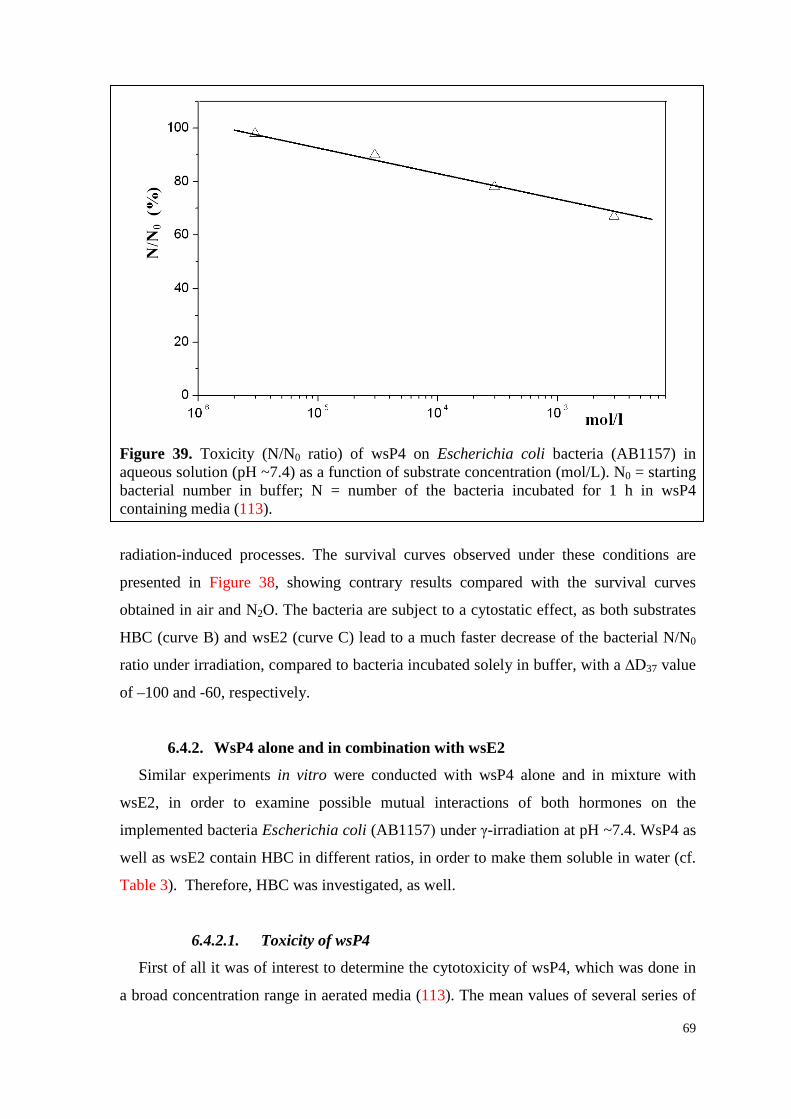
69
Figure 39. Toxicity (N/N0 ratio) of wsP4 on Escherichia coli bacteria (AB1157) in aqueous solution (pH ~7.4) as a function of substrate concentration (mol/L). N0 = starting bacterial number in buffer; N = number of the bacteria incubated for 1 h in wsP4 containing media (113).
radiation-induced processes. The survival curves observed under these conditions are
presented in Figure 38, showing contrary results compared with the survival curves
obtained in air and N2O. The bacteria are subject to a cytostatic effect, as both substrates
HBC (curve B) and wsE2 (curve C) lead to a much faster decrease of the bacterial N/N0
ratio under irradiation, compared to bacteria incubated solely in buffer, with a ∆D37 value
of –100 and -60, respectively.
6.4.2. WsP4 alone and in combination with wsE2
Similar experiments in vitro were conducted with wsP4 alone and in mixture with
wsE2, in order to examine possible mutual interactions of both hormones on the
implemented bacteria Escherichia coli (AB1157) under γ-irradiation at pH ~7.4. WsP4 as
well as wsE2 contain HBC in different ratios, in order to make them soluble in water (cf.
Table 3). Therefore, HBC was investigated, as well.
6.4.2.1. Toxicity of wsP4
First of all it was of interest to determine the cytotoxicity of wsP4, which was done in
a broad concentration range in aerated media (113). The mean values of several series of
70
experiments are presented as N/N0 ratio in dependence of the substrate concentration
(Figure 39). It can be easily seen, that the toxicity of wsP4 to E. coli bacteria (AB1157)
increases linearly with increasing substrate concentration.
6.4.2.2. Survival curves under irradiation
The concentration of 1x10⁻⁴ mol/L wsP4 (including 2.72x10⁻⁴ mol/L HBC) used for all
experiments in vitro under irradiation was chosen according to Figure 39. Hereby, 20% of
the bacteria were deactivated. In the frame of the study, survival curves with 2.72x10⁻⁴
mol/L HBC alone and a mixture of 1x10⁻⁴ mol/L wsP4 and 1x10⁻⁴ mol/L wsE2 including
6.7x10⁻⁴ mol/L HBC were also established. The obtained results in media saturated with
air, N2O and argon, respectively, are presented in Figures 40-42 as a function of the
absorbed radiation dose. ∆D37 values, representing the radiation dose, at which N/N0 is
0.37, were calculated and given as inset in the corresponding Figures.
Figure 40 presents the survival curves of the bacteria in aerated medium (pH ~7.4),
where oxidizing radicals (46% OH•, 54% O2•−) are produced as primary species of water
radiolysis (cf. 113). 2.72x10⁻⁴ mol/L HBC (curve B) exhibits a proliferative effect on the
bacteria, as it was observed before (cf. Figure 36). This fact is also illustrated by the
corresponding positive ∆D37 value (∆D37 = +30), indicating once again the scavenging
ability of HBC for oxidizing free radicals. In contrast, both 1x10⁻⁴ mol/L wsP4 and the
mixture of 1x10⁻⁴ mol/L wsP4 and 1x10⁻⁴ mol/L wsE2 were found to exhibit a cytostatic
action, whereby this effect was slightly more pronounced in the mixture of the two
hormones. However, the ∆D37 values of -35 for wsP4 and -50 for the mixture are
influenced by the protective action of HBC. Therefore, P4 and the mixture of P4 and E2
are expected to sensitize the bacteria much more towards γ-irradiation than their water-
soluble counterparts. Furthermore, it is interesting to note, that though wsE2 showed a
proliferative effect on E. coli bacteria before (cf. Figure 36), it now seems to even
intensity the cytostatic property of wsP4.
The change of the bacterial survival ratio under irradiation in solutions saturated with
N2O was also studied for aqueous solutions containing the above mentioned substrates in
a radiation dose range up to 600 Gy (113). Under these experimental conditions, e-aq are
converted into OH• radicals, but as the reaction rate constants of e-aq with both hormones
are very high, the reaction of e-aq with hormones directly has to be considered, as well.
Figure 41 presents the obtained results, which are completely different from the ones
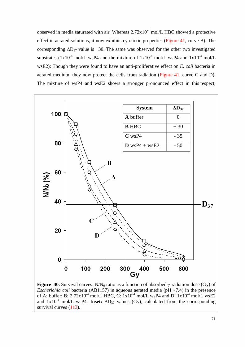
71
observed in media saturated with air. Whereas 2.72x10⁻⁴ mol/L HBC showed a protective
effect in aerated solutions, it now exhibits cytotoxic properties (Figure 41, curve B). The
corresponding ∆D37 value is +30. The same was observed for the other two investigated
substrates (1x10⁻⁴ mol/L wsP4 and the mixture of 1x10⁻⁴ mol/L wsP4 and 1x10⁻⁴ mol/L
wsE2): Though they were found to have an anti-proliferative effect on E. coli bacteria in
aerated medium, they now protect the cells from radiation (Figure 41, curve C and D).
The mixture of wsP4 and wsE2 shows a stronger pronounced effect in this respect,
Figure 40. Survival curves: N/N0 ratio as a function of absorbed γ-radiation dose (Gy) of Escherichia coli bacteria (AB1157) in aqueous aerated media (pH ~7.4) in the presence of A: buffer; B: 2.72x10⁻⁴ mol/L HBC, C: 1x10⁻⁴ mol/L wsP4 and D: 1x10⁻⁴ mol/L wsE2 and 1x10⁻⁴ mol/L wsP4. Inset: ∆D37 values (Gy), calculated from the corresponding survival curves (113).
System ΔD37
A buffer 0
B HBC + 30
C wsP4 - 35
D wsP4 + wsE2 - 50
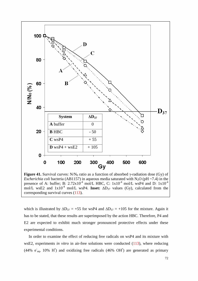
72
Figure 41. Survival curves: N/N0 ratio as a function of absorbed γ-radiation dose (Gy) of Escherichia coli bacteria (AB1157) in aqueous media saturated with N2O (pH ~7.4) in the presence of A: buffer; B: 2.72x10⁻⁴ mol/L HBC, C: 1x10⁻⁴ mol/L wsP4 and D: 1x10⁻⁴ mol/L wsE2 and 1x10⁻⁴ mol/L wsP4. Inset: ∆D37 values (Gy), calculated from the corresponding survival curves (113).
which is illustrated by ∆D37 = +55 for wsP4 and ∆D37 = +105 for the mixture. Again it
has to be stated, that these results are superimposed by the action HBC. Therefore, P4 and
E2 are expected to exhibit much stronger pronounced protective effects under these
experimental conditions.
In order to examine the effect of reducing free radicals on wsP4 and its mixture with
wsE2, experiments in vitro in air-free solutions were conducted (113), where reducing
(44% e-aq, 10% H•) and oxidizing free radicals (46% OH•) are generated as primary
System ΔD37
A buffer 0
B HBC - 50
C wsP4 + 55
D wsP4 + wsE2 + 105
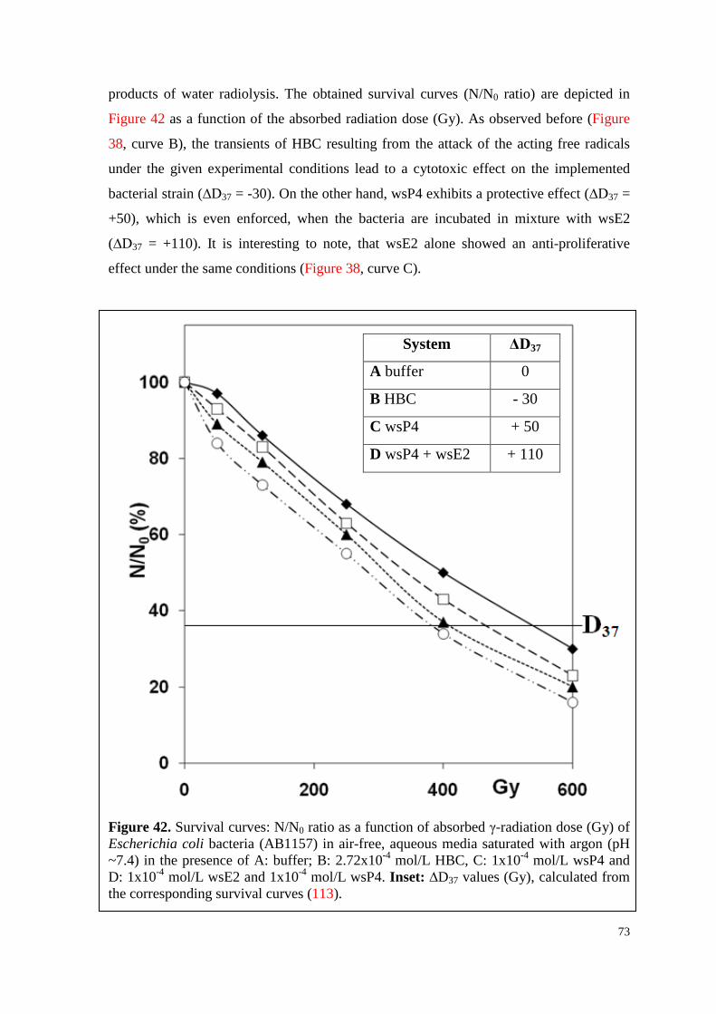
73
products of water radiolysis. The obtained survival curves (N/N0 ratio) are depicted in
Figure 42 as a function of the absorbed radiation dose (Gy). As observed before (Figure
38, curve B), the transients of HBC resulting from the attack of the acting free radicals
under the given experimental conditions lead to a cytotoxic effect on the implemented
bacterial strain (∆D37 = -30). On the other hand, wsP4 exhibits a protective effect (∆D37 =
+50), which is even enforced, when the bacteria are incubated in mixture with wsE2
(∆D37 = +110). It is interesting to note, that wsE2 alone showed an anti-proliferative
effect under the same conditions (Figure 38, curve C).
Figure 42. Survival curves: N/N0 ratio as a function of absorbed γ-radiation dose (Gy) of Escherichia coli bacteria (AB1157) in air-free, aqueous media saturated with argon (pH ~7.4) in the presence of A: buffer; B: 2.72x10⁻⁴ mol/L HBC, C: 1x10⁻⁴ mol/L wsP4 and D: 1x10⁻⁴ mol/L wsE2 and 1x10⁻⁴ mol/L wsP4. Inset: ∆D37 values (Gy), calculated from the corresponding survival curves (113).
System ΔD37
A buffer 0
B HBC - 30
C wsP4 + 50
D wsP4 + wsE2 + 110
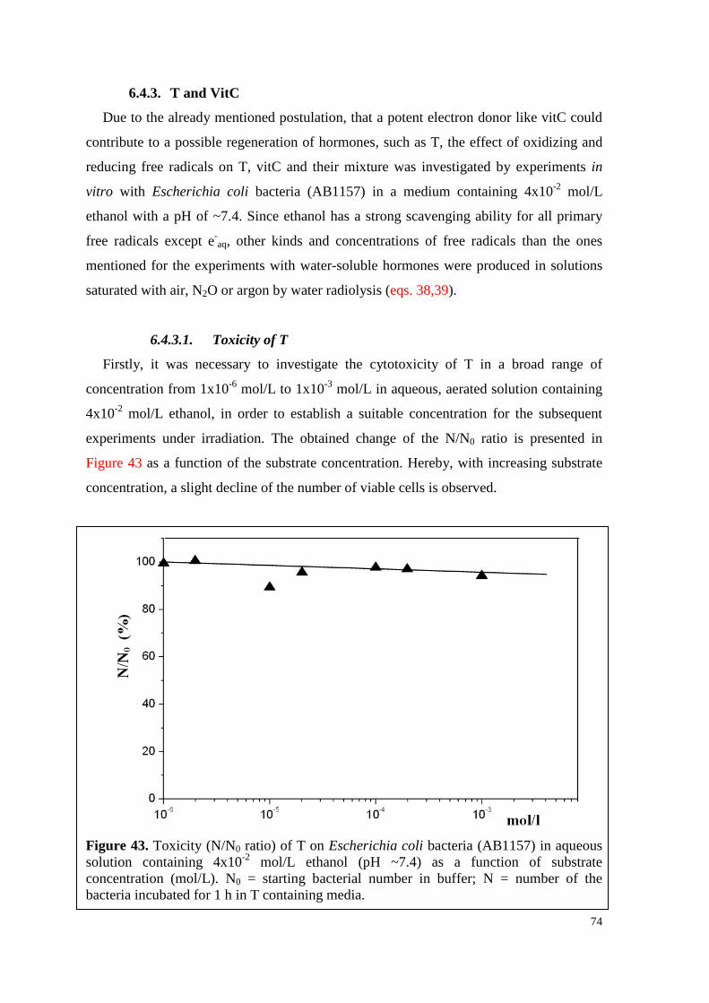
74
6.4.3. T and VitC
Due to the already mentioned postulation, that a potent electron donor like vitC could
contribute to a possible regeneration of hormones, such as T, the effect of oxidizing and
reducing free radicals on T, vitC and their mixture was investigated by experiments in
vitro with Escherichia coli bacteria (AB1157) in a medium containing 4x10⁻² mol/L
ethanol with a pH of ~7.4. Since ethanol has a strong scavenging ability for all primary
free radicals except e-aq, other kinds and concentrations of free radicals than the ones
mentioned for the experiments with water-soluble hormones were produced in solutions
saturated with air, N2O or argon by water radiolysis (eqs. 38,39).
6.4.3.1. Toxicity of T
Firstly, it was necessary to investigate the cytotoxicity of T in a broad range of
concentration from 1x10⁻⁶ mol/L to 1x10⁻³ mol/L in aqueous, aerated solution containing
4x10⁻² mol/L ethanol, in order to establish a suitable concentration for the subsequent
experiments under irradiation. The obtained change of the N/N0 ratio is presented in
Figure 43 as a function of the substrate concentration. Hereby, with increasing substrate
concentration, a slight decline of the number of viable cells is observed.
Figure 43. Toxicity (N/N0 ratio) of T on Escherichia coli bacteria (AB1157) in aqueous solution containing 4x10⁻² mol/L ethanol (pH ~7.4) as a function of substrate concentration (mol/L). N0 = starting bacterial number in buffer; N = number of the bacteria incubated for 1 h in T containing media.
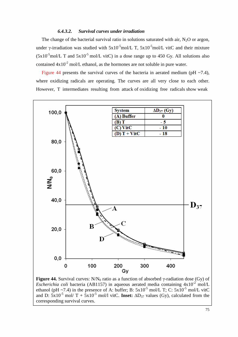
75
6.4.3.2. Survival curves under irradiation
The change of the bacterial survival ratio in solutions saturated with air, N2O or argon,
under γ-irradiation was studied with 5x10⁻⁵mol/L T, 5x10⁻⁵mol/L vitC and their mixture
(5x10⁻⁵mol/L T and 5x10⁻⁵ mol/L vitC) in a dose range up to 450 Gy. All solutions also
contained 4x10⁻² mol/L ethanol, as the hormones are not soluble in pure water.
Figure 44 presents the survival curves of the bacteria in aerated medium (pH ~7.4),
where oxidizing radicals are operating. The curves are all very close to each other.
However, T intermediates resulting from attack of oxidizing free radicals show weak
Figure 44. Survival curves: N/N0 ratio as a function of absorbed γ-radiation dose (Gy) of Escherichia coli bacteria (AB1157) in aqueous aerated media containing 4x10⁻² mol/L ethanol (pH ~7.4) in the presence of A: buffer; B: 5x10⁻⁵ mol/L T; C: 5x10⁻⁵ mol/L vitC and D: 5x10⁻⁵ mol/ T + 5x10⁻⁵ mol/l vitC. Inset: ∆D37 values (Gy), calculated from the corresponding survival curves.
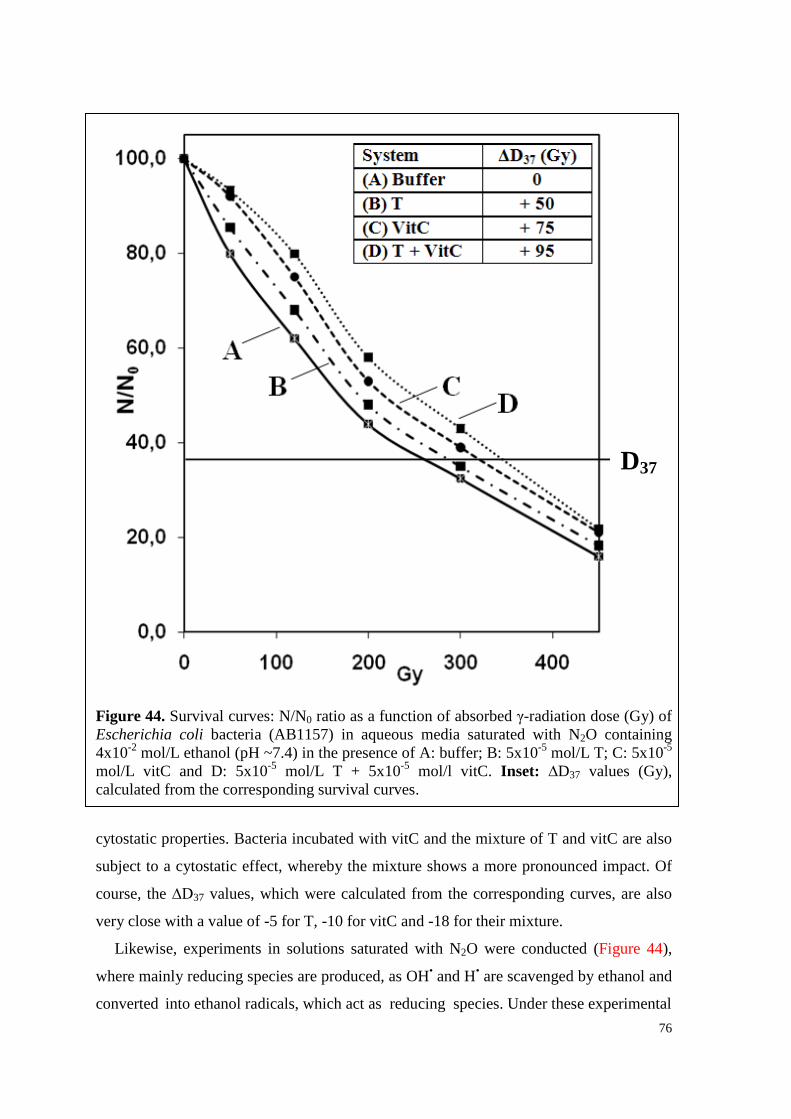
76
Figure 44. Survival curves: N/N0 ratio as a function of absorbed γ-radiation dose (Gy) of Escherichia coli bacteria (AB1157) in aqueous media saturated with N2O containing 4x10⁻² mol/L ethanol (pH ~7.4) in the presence of A: buffer; B: 5x10⁻⁵ mol/L T; C: 5x10⁻⁵ mol/L vitC and D: 5x10⁻⁵ mol/L T + 5x10⁻⁵ mol/l vitC. Inset: ∆D37 values (Gy), calculated from the corresponding survival curves. cytostatic properties. Bacteria incubated with vitC and the mixture of T and vitC are also
subject to a cytostatic effect, whereby the mixture shows a more pronounced impact. Of
course, the ∆D37 values, which were calculated from the corresponding curves, are also
very close with a value of -5 for T, -10 for vitC and -18 for their mixture.
Likewise, experiments in solutions saturated with N2O were conducted (Figure 44),
where mainly reducing species are produced, as OH• and H• are scavenged by ethanol and
converted into ethanol radicals, which act as reducing species. Under these experimental
D37
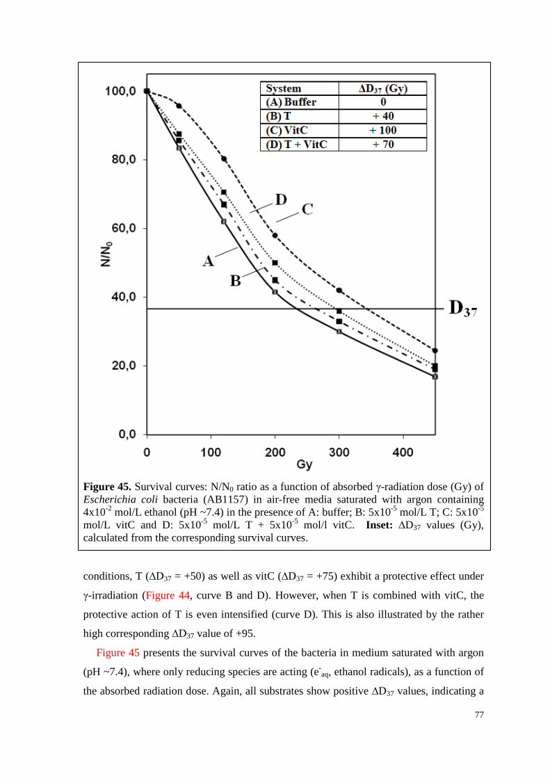
77
Figure 45. Survival curves: N/N0 ratio as a function of absorbed γ-radiation dose (Gy) of Escherichia coli bacteria (AB1157) in air-free media saturated with argon containing 4x10⁻² mol/L ethanol (pH ~7.4) in the presence of A: buffer; B: 5x10⁻⁵ mol/L T; C: 5x10⁻⁵ mol/L vitC and D: 5x10⁻⁵ mol/L T + 5x10⁻⁵ mol/l vitC. Inset: ∆D37 values (Gy), calculated from the corresponding survival curves. conditions, T (∆D37 = +50) as well as vitC (∆D37 = +75) exhibit a protective effect under
γ-irradiation (Figure 44, curve B and D). However, when T is combined with vitC, the
protective action of T is even intensified (curve D). This is also illustrated by the rather
high corresponding ∆D37 value of +95.
Figure 45 presents the survival curves of the bacteria in medium saturated with argon
(pH ~7.4), where only reducing species are acting (e-aq, ethanol radicals), as a function of
the absorbed radiation dose. Again, all substrates show positive ∆D37 values, indicating a
78
proliferative action of the intermediates resulting from the attack of the free radicals on
the bacteria. The corresponding ∆D37 values are +40 for T, +100 for vitC and +70 for
their mixture. It is interesting to note, that the mixture doesn’t exhibit the most
pronounced effect under these experimental conditions, as it was before in the presence of
air (Figure 43) and N2O (Figure 44).
Comparing the data obtained in various media (Figures 43-45), it can be concluded,
that all three substrates exhibit anti-proliferative effects on E. coli bacteria under the
attack of oxidizing radicals, but proliferative effect when they are subject to reducing
species.
79
7. DISCUSSION 7.1. Formation of hormone associates
For all investigated hormones (T, P4, wsE2, wsP4, GEN) it was observed that the
extinction coefficient values at λ=254 nm (ε254 values) were decreasing with increasing
substrate concentration (98,99,112,113), though the ε value of a compound is usually
constant, regardless of the concentration, as already mentioned (Lambert-Beer’s Law).
The obtained values for 1x10⁻⁵ mol/L and 5x10⁻⁵ mol/L T and 1x10⁻⁵ mol/L and 5x10⁻⁵
mol/L P4 are presented in inset of Figure 23. This observation indicates the formation of
hormone associates (hormone clusters), which are preferentially built with higher
substrate concentrations. In the meantime, the same effects were found for other
hormones, such as 4-OHE1 (48), 17OHP (49) and E1 (50).
7.2. Q(e-aq) values decrease with increasing hormone concentration
In the experiments studying the emission of electrons from various hormones as a
result of excitation to their singlet states by UV-irradiation, it was found that with
increasing substrate concentration (mol/L) the Q(e-aq) values are decreasing
(98,99,112,113). Therefore, the obtained Q(e-aq) values, taking GEN as representative
example for hormones, were plotted as a function of the substrate concentration, whereby
a strict linear dependence was found (Figure 46). Furthermore, the number of electron
emitting GEN molecules (in %) was calculated and is given as inset in Figure 46. It
decreases from 36% for 2.5x10⁻⁵ mol/L GEN to 4% for 1x10⁻⁴ mol/L GEN.
There are several explanations for the observed effect of decreasing Q(e-aq) values with
increasing substrate concentration (98,99,112,113). First, the above mentioned hormone
associates have to be considered, as the hormone molecules in the ground state, which are
within a hormone cluster, are able to consume electrons emitted from other hormone
molecules. As a consequence, it is postulated, that with increasing hormone
concentration, a higher number of hormone associates is formed, resulting in an increased
“self-consumption” of e-aq. Thus, Q(e-
aq) values diminish. It should be also noted, that the
dissimination of the absorbed UV quanta is probably not homogenously distributed
within the hormone associates, causing energy quenching. Further, competitive reaction
mechanisms of chloroethanol and the hormones for e-aq (k(e-
aq+ClC2H4OH) = 4x10⁸
L.mol-1.s-1 (92), k(e-aq+hormone) ~4x10⁹ up to 2x10¹⁰ L.mol-1.s-1), contribute to the
observed effect.
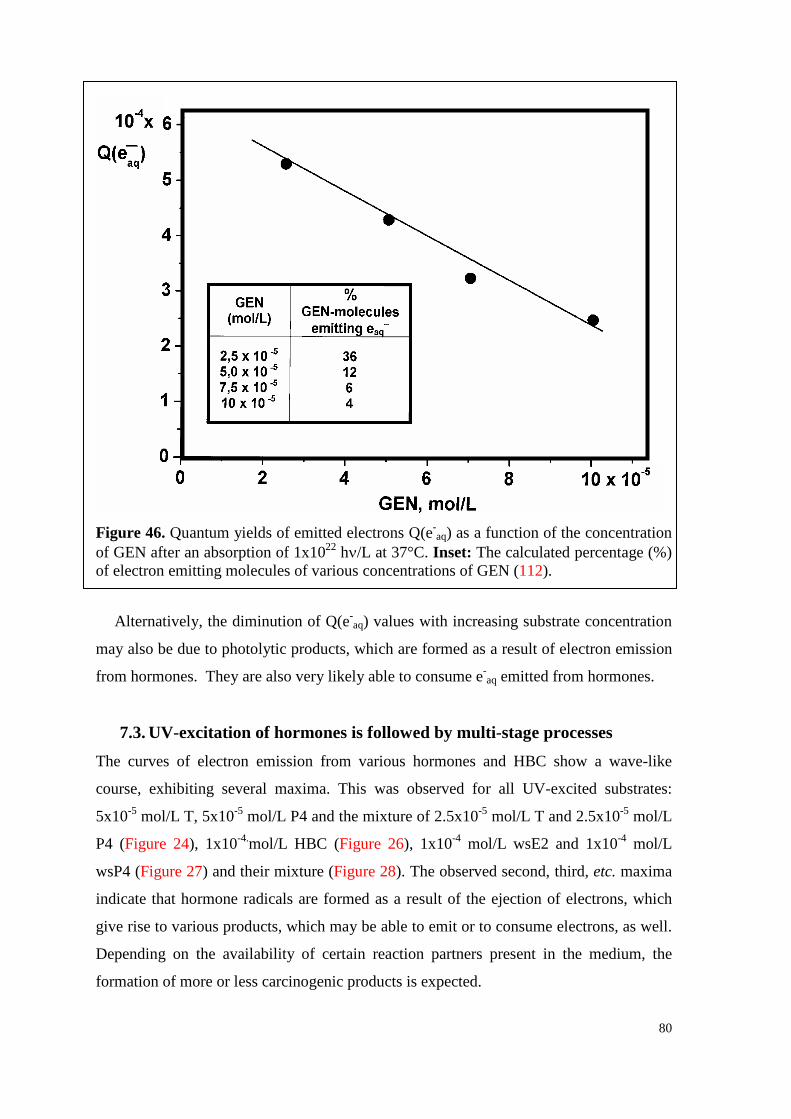
80
Figure 46. Quantum yields of emitted electrons Q(e-
aq) as a function of the concentration of GEN after an absorption of 1x10²² hν/L at 37°C. Inset: The calculated percentage (%) of electron emitting molecules of various concentrations of GEN (112).
Alternatively, the diminution of Q(e-aq) values with increasing substrate concentration
may also be due to photolytic products, which are formed as a result of electron emission
from hormones. They are also very likely able to consume e-aq emitted from hormones.
7.3. UV-excitation of hormones is followed by multi-stage processes The curves of electron emission from various hormones and HBC show a wave-like
course, exhibiting several maxima. This was observed for all UV-excited substrates:
5x10⁻⁵ mol/L T, 5x10⁻⁵ mol/L P4 and the mixture of 2.5x10⁻⁵ mol/L T and 2.5x10⁻⁵ mol/L
P4 (Figure 24), 1x10-4.mol/L HBC (Figure 26), 1x10⁻⁴ mol/L wsE2 and 1x10⁻⁴ mol/L
wsP4 (Figure 27) and their mixture (Figure 28). The observed second, third, etc. maxima
indicate that hormone radicals are formed as a result of the ejection of electrons, which
give rise to various products, which may be able to emit or to consume electrons, as well.
Depending on the availability of certain reaction partners present in the medium, the
formation of more or less carcinogenic products is expected.
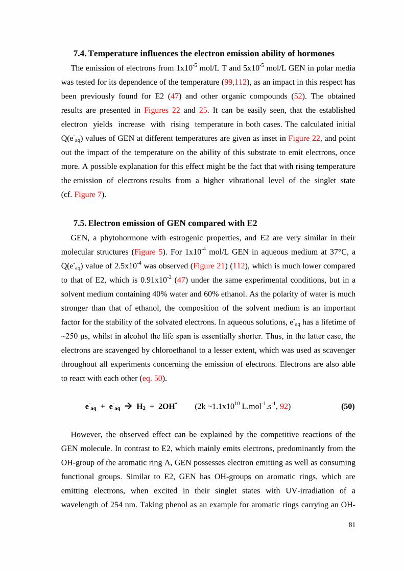
81
7.4. Temperature influences the electron emission ability of hormones The emission of electrons from 1x10⁻⁵ mol/L T and 5x10⁻⁵ mol/L GEN in polar media
was tested for its dependence of the temperature (99,112), as an impact in this respect has
been previously found for E2 (47) and other organic compounds (52). The obtained
results are presented in Figures 22 and 25. It can be easily seen, that the established
electron yields increase with rising temperature in both cases. The calculated initial
Q(e-aq) values of GEN at different temperatures are given as inset in Figure 22, and point
out the impact of the temperature on the ability of this substrate to emit electrons, once
more. A possible explanation for this effect might be the fact that with rising temperature
the emission of electrons results from a higher vibrational level of the singlet state
(cf. Figure 7).
7.5. Electron emission of GEN compared with E2 GEN, a phytohormone with estrogenic properties, and E2 are very similar in their
molecular structures (Figure 5). For 1x10⁻⁴ mol/L GEN in aqueous medium at 37°C, a
Q(e-aq) value of 2.5x10⁻⁴ was observed (Figure 21) (112), which is much lower compared
to that of E2, which is 0.91x10⁻² (47) under the same experimental conditions, but in a
solvent medium containing 40% water and 60% ethanol. As the polarity of water is much
stronger than that of ethanol, the composition of the solvent medium is an important
factor for the stability of the solvated electrons. In aqueous solutions, e-aq has a lifetime of
~250 μs, whilst in alcohol the life span is essentially shorter. Thus, in the latter case, the
electrons are scavenged by chloroethanol to a lesser extent, which was used as scavenger
throughout all experiments concerning the emission of electrons. Electrons are also able
to react with each other (eq. 50).
e-aq + e-
aq H2 + 2OH• (2k ~1.1x10¹⁰ L.mol-1.s-1, 92) (50)
However, the observed effect can be explained by the competitive reactions of the
GEN molecule. In contrast to E2, which mainly emits electrons, predominantly from the
OH-group of the aromatic ring A, GEN possesses electron emitting as well as consuming
functional groups. Similar to E2, GEN has OH-groups on aromatic rings, which are
emitting electrons, when excited in their singlet states with UV-irradiation of a
wavelength of 254 nm. Taking phenol as an example for aromatic rings carrying an OH-
82
group, the corresponding Q(e-aq) value is ~3x10⁻² (92). At the same time, the carbonyl
group (C=O) of GEN as well as the oxygen in the middle ring consume electrons with
reaction rate constants of k(CO+e-aq) = 1.7x10⁹ L.mol-1.s-1 and k(O2+e-
aq) = 1.9x10¹⁰
L.mol-1.s-1, respectively (92,100). As these reaction rate constants are rather high, the
emitted electrons are consumed by GEN itself to a large extent. For comparison, the
reaction rate constant of the electron scavenger chloroethanol is smaller by one to two
orders of magnitude (k ~4x10⁸ L.mol-1.s-1, 92).
7.6. Testosterone 7.6.1. Effect of P4 on the electron emission and degradation of T
In studies concerning the electron emission from UV-excited 5x10⁻⁵ mol/L T, 5x10⁻⁵
mol/L P4 and the mixture of 2.5x10⁻⁵ mol/L T and 2.5x10⁻⁵ mol/L P4 in a solvent
consisting of 40% water and 60% ethanol, the involvement of multi-stage processes can
be easily seen, as all obtained curves exhibit several maxima (Figure 24). Simultaneously,
HPLC-studies were conducted, the results of which are presented in Figures 29-31.
Hereby, highest product yields and most degradation products were found in the UV-
irradiated mixture of T and P4 (Figure 31), indicating that the two hormones possibly
form unstable complexes.
Out of the data obtained by HPLC-analyses, the degradation (%) of the hormones and
their mixture was calculated (98). The results are depicted in Figure 47 as a function of
the absorbed UV dose (hν/L). The initial degradation yields, Q(-hormone)i, were
calculated from the tangent-line of the corresponding curve (insets, Figure 41). At an
absorbed UV dose of 1x10²¹ hν/L, the hormones are already decomposed in large part.
Considering that the first peak of electron emission from the hormones and their mixture
(Figure 24) occurs at the same absorbed UV dose, it can be stated that only the first peak
belongs exclusively to the electron emission from the hormones themselves. Therefore,
observed second, third etc. peaks are only partly a result of the electron ejection of the
individual hormones, as electrons emitted by metabolites play a decisive role in this
respect.
Comparing the electron emission of T and P4 (Figure 24, inset), Q(e-aq) from T turns
out to be 3.6 times higher (Q(e-aq) = 2.5x10⁻² for peak 1 of T and Q(e-
aq) = 0.69x10⁻² for
peak 1 of P4). For the hormone mixture (curve C), the value of peak (1) is even ten times
lower. This fact can be explained by the different molecular structures of the hormones
83
Figure 47. Degradation (%) of A: 5x10⁻⁵ mol/L T, B: 5x10⁻⁵ mol/L P4 and C: mixture of 2.5x10⁻⁵ mol/L T and 2.5x10⁻⁵ mol/L P4 in air-free solvent mixture of 40% water and 60% ethanol (pH ∼7.4, 37°C) as a function of the absorbed UV quanta (hν/L; λ=254 nm).Inset I: Initial hormone degradation yields, Q(-hormone)i, of the substrates. Inset II: Individual degradation (%) of T and P4 in the above mentioned mixture; the calculated Q(-hormone)i values are given in the table (98).
(98). T carries a hydroxyl group favouring the electron emission at position 17 of ring D,
whilst P4 has a carbonyl group at the same position, which consumes a part of the emitted
e-aq. Additionally, both hormones have a double bond at positions 4–5 of ring A, which
represents another source for electron emission.
Eq. 51a illustrates the ejection of electrons from position 17 of ring D of T, resulting in
the formation of a T radical (T•). As by-product, H+ is formed, which is responsible for
the observed decrease of the pH with increasing radiation dose. Furthermore, it is shown,
that the resulting hormone radicals can react with various species (R•x) present in the
T P4
T P4 mix
hν/L
(%)
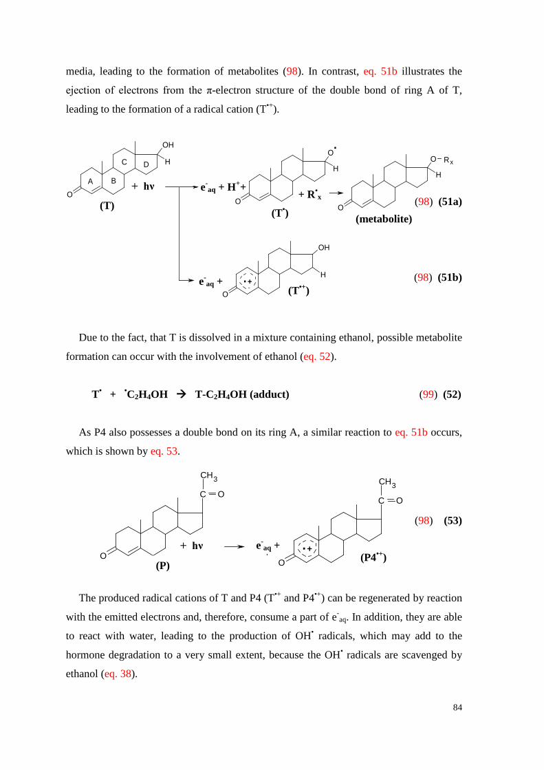
84
OH
O
H
OH
O
H
O
O
H
O
O
HA B
C D
hv
.+
+
e aq
.
(Radical cation)
1(R )
.+e aq + H+
Rx
Rx
(Metabolite)
+ .(TES )+
(TES)
+
media, leading to the formation of metabolites (98). In contrast, eq. 51b illustrates the
ejection of electrons from the π-electron structure of the double bond of ring A of T,
leading to the formation of a radical cation (T•+).
(98) (51a)
(98) (51b)
Due to the fact, that T is dissolved in a mixture containing ethanol, possible metabolite
formation can occur with the involvement of ethanol (eq. 52).
T• + •C2H4OH T-C2H4OH (adduct) (99) (52)
As P4 also possesses a double bond on its ring A, a similar reaction to eq. 51b occurs,
which is shown by eq. 53.
(98) (53)
The produced radical cations of T and P4 (T•+ and P4•+) can be regenerated by reaction
with the emitted electrons and, therefore, consume a part of e-aq. In addition, they are able
to react with water, leading to the production of OH• radicals, which may add to the
hormone degradation to a very small extent, because the OH• radicals are scavenged by
ethanol (eq. 38).
OO
C O
CH3
hv+ e aq + .+
C O
CH3
(PRG ).+
(Radical cation)(PRG)
(T)
+ hν
(T•)
+ R•x
(metabolite)
(T•+) e-
aq +
e-aq + H++
(P)
+ hν e-aq +
(P4•+)
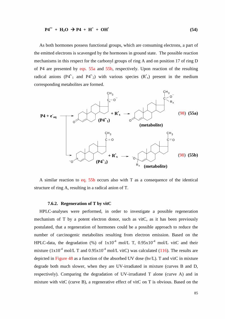
85
O
O O
OO
C O
3CH
e aq+
C O
3CH
.
(PRG ).
(Radical anion)
Rx+.
C O3CH
Rx
(Metabolite)
C O
3CH
.(PRG )
.(Radical anion)
Rx+.
C O
3CH
Rx (Metabolite)
(PRG)
1
2
P4 + e-
aq
P4•+ + H2O P4 + H+ + OH• (54)
As both hormones possess functional groups, which are consuming electrons, a part of
the emitted electrons is scavenged by the hormones in ground state. The possible reaction
mechanisms in this respect for the carbonyl groups of ring A and on position 17 of ring D
of P4 are presented by eqs. 55a and 55b, respectively. Upon reaction of the resulting
radical anions (P4•-1 and P4•-
2) with various species (R•x) present in the medium
corresponding metabolites are formed.
(98) (55a)
(98) (55b)
A similar reaction to eq. 55b occurs also with T as a consequence of the identical
structure of ring A, resulting in a radical anion of T.
7.6.2. Regeneration of T by vitC
HPLC-analyses were performed, in order to investigate a possible regeneration
mechanism of T by a potent electron donor, such as vitC, as it has been previously
postulated, that a regeneration of hormones could be a possible approach to reduce the
number of carcinogenic metabolites resulting from electron emission. Based on the
HPLC-data, the degradation (%) of 1x10⁻⁴ mol/L T, 0.95x10⁻⁴ mol/L vitC and their
mixture (1x10⁻⁴ mol/L T and 0.95x10⁻⁴ mol/L vitC) was calculated (116). The results are
depicted in Figure 48 as a function of the absorbed UV dose (hν/L). T and vitC in mixture
degrade both much slower, when they are UV-irradiated in mixture (curves B and D,
respectively). Comparing the degradation of UV-irradiated T alone (curve A) and in
mixture with vitC (curve B), a regenerative effect of vitC on T is obvious. Based on the
+ R•x
+ R•x
(metabolite)
(metabolite)
(P4•-1)
(P4•-2)
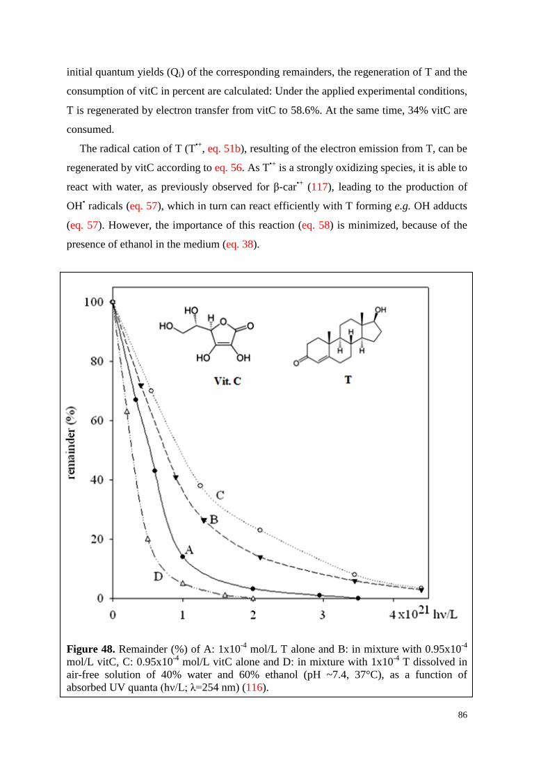
86
initial quantum yields (Qi) of the corresponding remainders, the regeneration of T and the
consumption of vitC in percent are calculated: Under the applied experimental conditions,
T is regenerated by electron transfer from vitC to 58.6%. At the same time, 34% vitC are
consumed.
The radical cation of T (T•+, eq. 51b), resulting of the electron emission from T, can be
regenerated by vitC according to eq. 56. As T•+ is a strongly oxidizing species, it is able to
react with water, as previously observed for β-car•+ (117), leading to the production of
OH• radicals (eq. 57), which in turn can react efficiently with T forming e.g. OH adducts
(eq. 57). However, the importance of this reaction (eq. 58) is minimized, because of the
presence of ethanol in the medium (eq. 38).
Figure 48. Remainder (%) of A: 1x10⁻⁴ mol/L T alone and B: in mixture with 0.95x10⁻⁴ mol/L vitC, C: 0.95x10⁻⁴ mol/L vitC alone and D: in mixture with 1x10⁻⁴ T dissolved in air-free solution of 40% water and 60% ethanol (pH ~7.4, 37°C), as a function of absorbed UV quanta (hν/L; λ=254 nm) (116).
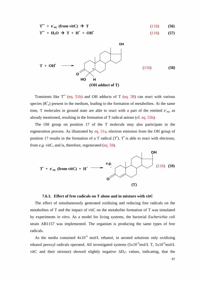
87
PRG + OH
O
C O
CH3
.
HO H( )R2
(PRG OH-adduct)
OH
(T OH-adduct)
T
T•+ + e-aq (from vitC) T (116) (56)
T•+ + H2O T + H+ + OH• (116) (57)
(116) (58)
Transients like T•+ (eq. 51b) and OH adducts of T (eq. 58) can react with various
species (R•x) present in the medium, leading to the formation of metabolites. At the same
time, T molecules in ground state are able to react with a part of the emitted e-aq, as
already mentioned, resulting in the formation of T radical anions (cf. eq. 55b).
The OH group on position 17 of the T molecule may also participate in the
regeneration process. As illustrated by eq. 51a, electron emission from the OH group of
position 17 results in the formation of a T radical (T•). T• is able to react with electrons,
from e.g. vitC, and is, therefore, regenerated (eq. 59).
(116) (59)
7.6.3. Effect of free radicals on T alone and in mixture with vitC
The effect of simultaneously generated oxidizing and reducing free radicals on the
metabolites of T and the impact of vitC on the metabolite formation of T was simulated
by experiments in vitro. As a model for living systems, the bacterial Escherichia coli
strain AB1157 was implemented. The organism is producing the same types of free
radicals.
As the media contained 4x10⁻² mol/L ethanol, in aerated solutions only oxidizing
ethanol peroxyl radicals operated. All investigated systems (5x10⁻⁵mol/L T, 5x10⁻⁵mol/L
vitC and their mixture) showed slightly negative ∆D37 values, indicating, that the
OH
R 5 + eaq (from Vit.C) Haq++
O
(TES regenerated)
e.g.
T + OH•
T• + e-aq (from vitC) + H+
(T)
(OH adduct of T)
e.g.
88
metabolites resulting from the attack of ethanol peroxyl radicals exhibit an anti-
proliferative effect on the bacteria. In media saturated with N2O (ethanol radicals) and
argon (ethanol radicals, e-aq), contrary results were obtained. The ∆D37 values of all
susbtrates are positive, thus it can be stated, that T, vitC and their mixture show a
protective effect under reducing experimental conditions.
Based on the molecular structure of T it is expected, that the ethanol peroxyl radicals
(•O2C2H4OH) predominantly attack the double bond of ring A of T, forming
corresponding peroxyl adducts. Furthermore, to a smaller extent they are able to remove
hydrogen from T. The resulting transients react with various species (R•x) present in the
medium, leading to the formation of metabolites with various biological properties. The
reducing e-aq primarily react with the carbonyl group of ring A. Consequently, T radical
anions (cf. eq. 55b) are produced, which give rise to various metabolites upon reaction
with available species (R•x) in the medium. The OH group on position 17 of ring D can
also be involved in reactions with various types of species. However, in general, the
corresponding reaction rate constants are rather low.
Summing up it can be stated, that oxidizing as well as reducing free radicals are both
able to react very efficiently with T, resulting in the formation of a number of metabolites
with varying biological properties. Hereby, vitC enhances the particular effect of T.
7.7. Water soluble hormones 7.7.1. Electron emission of water soluble hormones
E2 and P4 were obtained as water-soluble complex with HBC, as previously
mentioned. Consequently, the obtained results from studies concerning the electron
emission of wsE2 and wsP4 are an outcome of several, simultaneously proceeding
reactions (113): firstly from ejection of e-aq of the specific hormone and of HBC upon
UV-excitation, and secondly by the partly consumption of e-aq by the substrates, as the
reaction rate constants of both hormones with electrons are rather high: k(E2+e-aq) =
2.7x10¹⁰ L.mol-1.s-1 (54), k(P4+e-aq) ~4x10⁹ L.mol-1.s-1 (99). The reaction rate constant of
HBC and electrons is much lower: k(HBC+e-aq) = 8x10⁷ L.mol-1.s-1 (92). Thirdly, the
photolytic products can also participate in the mentioned processes, and therefore
influence the yield of solvated electrons.
For the observed curve of electron emission of 1x10⁻⁴ mol/L HBC, which exhibits
several maxima, indicating the involvement of multi-stage processes, it can be stated, that
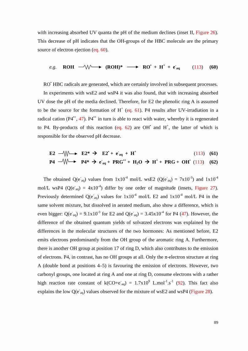
89
with increasing absorbed UV quanta the pH of the medium declines (inset II, Figure 26).
This decrease of pH indicates that the OH-groups of the HBC molecule are the primary
source of electron ejection (eq. 60).
e.g. ROH (ROH)* RO• + H+ + e-aq (113) (60)
RO• HBC radicals are generated, which are certainly involved in subsequent processes.
In experiments with wsE2 and wsP4 it was also found, that with increasing absorbed
UV dose the pH of the media declined. Therefore, for E2 the phenolic ring A is assumed
to be the source for the formation of H+ (eq. 61). P4 results after UV-irradiation in a
radical cation (P4•+, 47). P4•+ in turn is able to react with water, whereby it is regenerated
to P4. By-products of this reaction (eq. 62) are OH• and H+, the latter of which is
responsible for the observed pH decrease.
E2 E2* E2• + e-aq + H+ (113) (61)
P4 P4* e-aq + PRG•+ + H2O H+ + PRG + OH• (113) (62)
The obtained Q(e-aq) values from 1x10⁻⁴ mol/L wsE2 (Q(e-
aq) = 7x10⁻³) and 1x10⁻⁴
mol/L wsP4 (Q(e-aq) = 4x10⁻⁴) differ by one order of magnitude (insets, Figure 27).
Previously determined Q(e-aq) values for 1x10⁻⁴ mol/L E2 and 1x10⁻⁴ mol/L P4 in the
same solvent mixture, but dissolved in aerated medium, also show a difference, which is
even bigger: Q(e-aq) = 9.1x10⁻³ for E2 and Q(e-
aq) = 3.45x10⁻⁴ for P4 (47). However, the
difference of the obtained quantum yields of solvatzed electrons was explained by the
differences in the molecular structures of the two hormones: As mentioned before, E2
emits electrons predominantly from the OH group of the aromatic ring A. Furthermore,
there is another OH group at position 17 of ring D, which also contributes to the emission
of electrons. P4, in contrast, has no OH groups at all. Only the π-electron structure at ring
A (double bond at positions 4–5) is favouring the emission of electrons. However, two
carbonyl groups, one located at ring A and one at ring D, consume electrons with a rather
high reaction rate constant of k(CO+e-aq) = 1.7x10⁹ L.mol-1.s-1 (92). This fact also
explains the low Q(e-aq) values observed for the mixture of wsE2 and wsP4 (Figure 28).
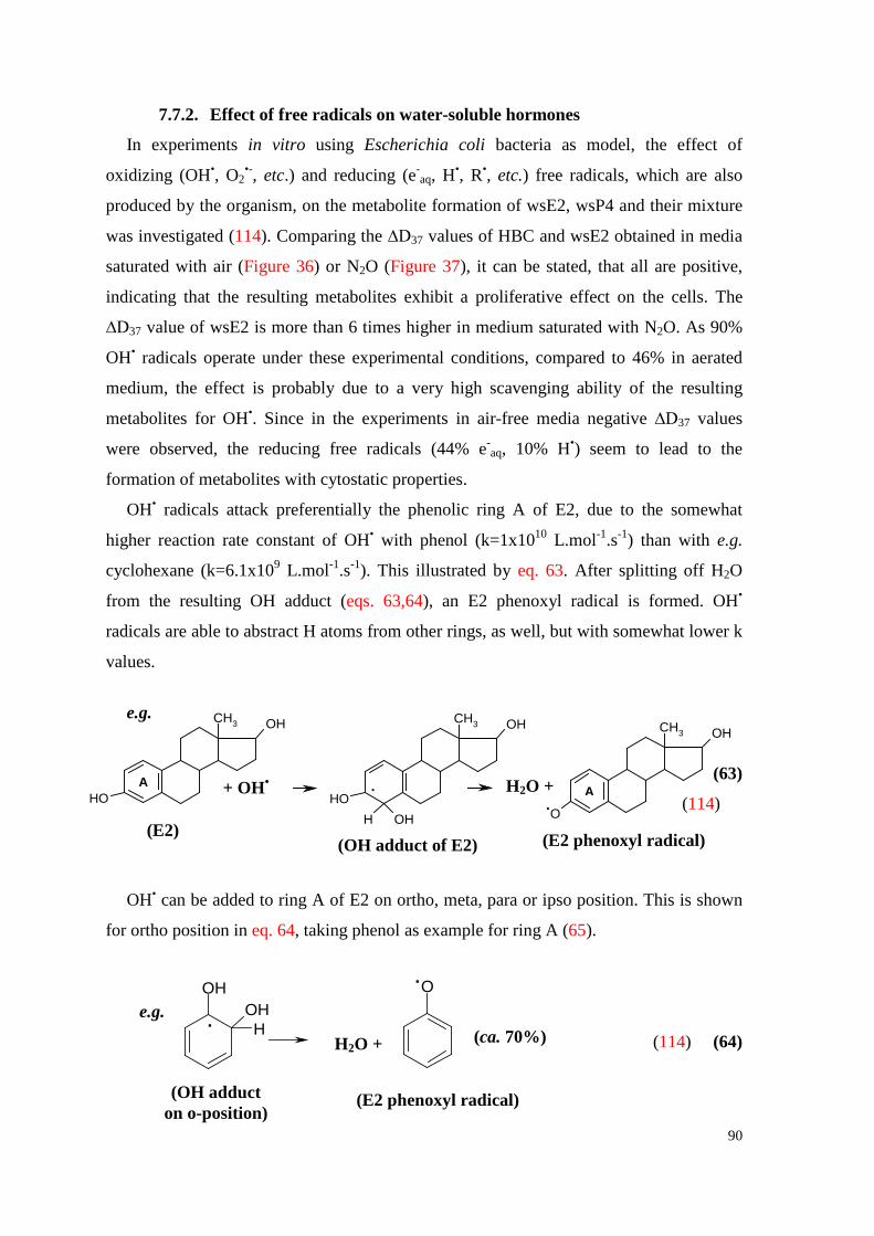
90
OHOHH
O
.
.
H O +2 (ca.70%)
(OH-adduct on o-,m-,p- and ipso positions)
(Phenoxyl radical)
(2)e.g.
OH
CH3 OH
OH
CH3 OH
H OH
CH3 OH
O
H OA + OH A..
2 + (1)
(17ßE2)(OH-adduct) R1( , phenoxyl radical type)
(E2)
+ OH•
7.7.2. Effect of free radicals on water-soluble hormones
In experiments in vitro using Escherichia coli bacteria as model, the effect of
oxidizing (OH•, O2•-, etc.) and reducing (e-
aq, H•, R•, etc.) free radicals, which are also
produced by the organism, on the metabolite formation of wsE2, wsP4 and their mixture
was investigated (114). Comparing the ∆D37 values of HBC and wsE2 obtained in media
saturated with air (Figure 36) or N2O (Figure 37), it can be stated, that all are positive,
indicating that the resulting metabolites exhibit a proliferative effect on the cells. The
∆D37 value of wsE2 is more than 6 times higher in medium saturated with N2O. As 90%
OH• radicals operate under these experimental conditions, compared to 46% in aerated
medium, the effect is probably due to a very high scavenging ability of the resulting
metabolites for OH•. Since in the experiments in air-free media negative ∆D37 values
were observed, the reducing free radicals (44% e-aq, 10% H•) seem to lead to the
formation of metabolites with cytostatic properties.
OH• radicals attack preferentially the phenolic ring A of E2, due to the somewhat
higher reaction rate constant of OH• with phenol (k=1x10¹⁰ L.mol-1.s-1) than with e.g.
cyclohexane (k=6.1x10⁹ L.mol-1.s-1). This illustrated by eq. 63. After splitting off H2O
from the resulting OH adduct (eqs. 63,64), an E2 phenoxyl radical is formed. OH•
radicals are able to abstract H atoms from other rings, as well, but with somewhat lower k
values.
e.g.
(63)
(114)
OH• can be added to ring A of E2 on ortho, meta, para or ipso position. This is shown
for ortho position in eq. 64, taking phenol as example for ring A (65).
(114) (64)
(OH adduct of E2) (E2 phenoxyl radical)
H2O +
H2O +
(OH adduct on o-position)
(E2 phenoxyl radical)
e.g. (ca. 70%)
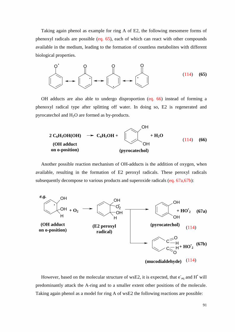
91
O O O O
..
.
.
(3)
OH
OH
2 C H OH(OH) 6 5 C H OH + 6 5 + H O2 (4)
OH
OHH
OHOOH
H
OH
OH
.e.g.
+ O2
.2 + HO2
.
CC
O
O
HH
+ HO2.
(5a)
(5b)
(Pyrocatechol)(OH-adduct on o-position)
(Mucodialdehyde)
Taking again phenol as example for ring A of E2, the following mesomere forms of
phenoxyl radicals are possible (eq. 65), each of which can react with other compounds
available in the medium, leading to the formation of countless metabolites with different
biological properties.
(114) (65)
OH adducts are also able to undergo disproportion (eq. 66) instead of forming a
phenoxyl radical type after splitting off water. In doing so, E2 is regenerated and
pyrocatechol and H2O are formed as by-products.
(114) (66)
Another possible reaction mechanism of OH-adducts is the addition of oxygen, when
available, resulting in the formation of E2 peroxyl radicals. These peroxyl radicals
subsequently decompose to various products and superoxide radicals (eq. 67a,67b):
(67a)
(114)
(67b)
(114)
However, based on the molecular structure of wsE2, it is expected, that e-aq and H• will
predominantly attack the A-ring and to a smaller extent other positions of the molecule.
Taking again phenol as a model for ring A of wsE2 the following reactions are possible:
(pyrocatechol) (OH adduct
on o-position)
(OH adduct on o-position)
e.g.
(pyrocatechol)
(mucodialdehyde)
+ H2O 2 C6H5OH(OH) C6H5OH +
+ O2 + HO•2
+ HO•2
(E2 peroxyl radical)
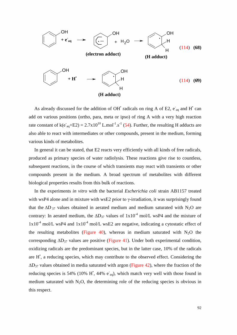
92
OHOH OH
H
H
OH OH
H
H
+ e .aq + H O2.
(electron adduct)( , H-adduct)R2
(6)
+ H.
( )R2
(7)
(114) (68)
(114) (69)
As already discussed for the addition of OH• radicals on ring A of E2, e-aq and H• can
add on various positions (ortho, para, meta or ipso) of ring A with a very high reaction
rate constant of k(e-aq+E2) = 2.7x10¹⁰ L.mol-1.s-1 (54). Further, the resulting H adducts are
also able to react with intermediates or other compounds, present in the medium, forming
various kinds of metabolites.
In general it can be stated, that E2 reacts very efficiently with all kinds of free radicals,
produced as primary species of water radiolysis. These reactions give rise to countless,
subsequent reactions, in the course of which transients may react with transients or other
compounds present in the medium. A broad spectrum of metabolites with different
biological properties results from this bulk of reactions.
In the experiments in vitro with the bacterial Escherichia coli strain AB1157 treated
with wsP4 alone and in mixture with wsE2 prior to γ-irradiation, it was surprisingly found
that the ∆D 37 values obtained in aerated medium and medium saturated with N2O are
contrary: In aerated medium, the ∆D37 values of 1x10⁻⁴ mol/L wsP4 and the mixture of
1x10⁻⁴ mol/L wsP4 and 1x10⁻⁴ mol/L wsE2 are negative, indicating a cytostatic effect of
the resulting metabolites (Figure 40), whereas in medium saturated with N2O the
corresponding ∆D37 values are positive (Figure 41). Under both experimental condition,
oxidizing radicals are the predominant species, but in the latter case, 10% of the radicals
are H•, a reducing species, which may contribute to the observed effect. Considering the
∆D37 values obtained in media saturated with argon (Figure 42), where the fraction of the
reducing species is 54% (10% H•, 44% e-aq), which match very well with those found in
medium saturated with N2O, the determining role of the reducing species is obvious in
this respect.
+ e-aq
+ H•
(electron adduct) (H adduct)
(H adduct)
93
Based on the molecular structure of P4 it is expected, that the oxidizing radicals add
predominantly to the π-electron structure of ring A. In contrast, the reducing species
primarily react with the carbonyl groups of ring A and D. The resulting transients of all
these processes may regenerate, react with each other or with available species (R•x) in
the medium. Hence, as already explained for T and E2, the formation of various
metabolites is conceivable, each of which having unique biological properties.
Comparing the ∆D37 values of wsE2 and wsP4, it can be stated, that the two hormones
show an opposite behaviour in aerated as well as in air-free media, pointing out the
contrary biological action of wsE2 and wsP4. In the mixture, wsE2 enhances the
particular effect of wsP4. Therefore, mixtures of these hormones could be implemented
for reduction of the number of carcinogenic metabolites. However, it should be noted,
that both hormones are influenced by HBC.

94
8. CONCLUSION In the present study it was shown, that the sex hormones T (98,99), wsE2, wsP4 (113)
and phytoestrogen GEN (112) are able to emit electrons upon excitation to their singlet
state in polar media. This was simulated by excitation with monochromatic UV-light
(λ=254 nm; 4.85 eV/hν). Hereby, it was shown, that the yield of the resulting solvated
electrons (e-aq) depends mainly on the specific molecular structure of the substrate,
concentration and temperature. In addition, the absence or presence of oxygen during the
dissolving process of hormones was found to be of importance, as much higher quantum
yields, Q(e-aq), calculated from the tangent of the corresponding curves, were obtained,
when the hormones were dissolved in air-free media. This might be explained by a
possible oxidation of the hormones when dissolved in the presence of oxygen, as oxygen
occurs in triplet state in the atmosphere and is, therefore, rather reactive. The impact of
the temperature on Q(e-aq) values, which increase with temperature, might be due to the
fact that with rising temperature the emission of e-aq results from a higher vibrational
level of the singlet state.
Interestingly, the molar extinction coefficients at λ=254 nm (ε254 values), calculated
from absorption spectra with the help of the Lambert-Beer-Law, decreased with
increasing substrate concentration (98,99,112,113). This observation indicates the
formation of hormone associates (hormone clusters), which are preferentially built with
higher substrate concentrations. Furthermore, it was found that with increasing substrate
concentration (mol/L) the Q(e-aq) values decreased accordingly. This can be due to the
formation of associates, as the hormone molecules in ground state within a hormone
cluster may consume ejected electrons, resulting in an increased “self-consumption” of
e-aq. Moreover, the dissimination of the absorbed UV quanta might not occur
homogenously within the hormone associates, causing energy quenching. Photolytic
products, which are formed as a result of electron emission from hormones, can also
consume emitted electrons. The latter is illustrated by the wave-like course of the curves
of electron emission, exhibiting several maxima, which usually decrease with ongoing
UV-irradiation. By HPLC analyses it was shown that the hormones were decomposed in
large part after absorption of relatively low UV doses. Comparing these results with the
obtained curves of electron emission, it can be stated that only the first peak belongs
exclusively to the electron emission from the particular hormone itself. Observed further
95
peaks are strongly influenced by the electron emission ability of generated photolytic
products.
As already mentioned, the molecular structure of a certain compound is the crucial
factor for its ability to emit electrons. As the Q(e-aq) value observed for GEN is lower than
that of E2, which has been previously established (47), it can be stated, that the carbonyl
group (C=O) of GEN as well as the oxygen in the middle ring are decisive in this respect.
Both consume electrons with rather high reaction rate constants of k(CO+e-aq) = 1.7x10⁹
L.mol-1.s-1 and k(O2+e-aq) = 1.9x10¹⁰ L.mol-1.s-1, respectively (92,100). E2 in contrast
mainly emits electrons, predominantly from the OH group of the aromatic ring A.
Comparing T, P4 and E2, E2 turns out to have the highest Q(e-aq) value, followed by T
and finally P4. This fact is due to the different molecular structures of the hormones at
ring A and D: Ring A of E2 is phenolic, as already mentioned, favouring the electron
emission, whereas ring A of T and P4 only has a double bond on position 4-5, which is a
source of electron emission, indeed, but not as efficient. Furthermore, a carbonyl group is
situated on ring A of T and P4, which consumes a part of the emitted electrons,
explaining the higher observed Q(e-aq) value for E2. Ring D of both E2 and T carries a
hydroxyl group on position 17, which again favours the emission of electrons. P4, in
contrast, has a further carbonyl group on this position. Therefore, P4 emits the lowest
yields of solvated electrons of these hormones.
The established ability of sex hormones to both emit and consume electrons classifies
them as “electron mediators”. This behaviour makes a communication by e-aq between
different biological systems in the organism conceivable (118).
Using vitC as representative for potent electron donors, it was further shown by HPLC
that transients resulting from electron emission of T can be partly regenerated to their
original structure by electron transfer processes, if they are still in “status nascendi”. Due
to the regeneration of T less metabolites are formed. Consequently, also the number of
metabolites initiating neoplastic processes is assumed to be reduced.
As the organism permanently generates oxidizing (OH, O2• , etc.) and reducing (e-
aq,
R•, etc.) free radicals, which are crucial for various biological processes, it was of interest
to investigate the reactions of these radicals with various hormones in the frame of
experiments in vitro with Escherichia coli bacteria (AB1157) in aqueous media (pH
~7.4). By application of appropriate experimental conditions (saturation of the solutions
with various gases), desired kind and concentration of primary free radicals were obtained
96
as a result of the radiolysis of water. WsE2 (embedded in HBC) was found to be a very
efficient radical scavenger of oxidizing radicals, which attack the hormone mainly on its
phenolic ring A. As a consequence, several mesomere forms of transients are possible,
which lead to the formation of metabolites with predominant proliferative effects. The
attack of reducing species (e-aq, H•) occurs to a large part at ring A, as well. However, the
resulting metabolites show strong anti-proliferative properties. WsP4 with incorporated
HBC, in contrast, shows an opposite behaviour. The reducing species, which play a very
important role for its biological effect, are expected to primarily react with the carbonyl
groups of ring A and D, resulting in the formation of radical anions. Mixtures of wsE2
and wsP4 intensify the effects observed for wsP4, indicating that a combination of wsP4
and wsE2 may strongly reduce the number of carcinogenic metabolites of wsE2.
T and a mixture of T and vitC in a media containing 4x10⁻² mol/L ethanol were also
investigated in this respect. T intermediates resulting from attack of oxidizing free
radicals on the double bond of ring A show weak cytotoxic properties. Under reducing
conditions, T induces strong proliferation of E.coli bacteria. These effects are both
enhanced, when T is combined with vitC.
Concluding it might be said, that all investigated sex hormones readily react with
various oxidizing and reducing free radicals, according to their specific molecular
structures with various functional groups and double bonds. The resulting transients,
which often have several mesomere structures, are very eager to react with other
substances. The biological action of the investigated hormones and their mixtures,
therefore, depends on the available compounds in the media. These can critically
influence the behaviour of the resulting metabolites towards cells.
The present research work represents an attempt to get a deep insight in the
enormously complicated, but at the same time synchronized and overlapping biological
processes, which involve hormones and their metabolites. Though some promising results
were obtained, much more work is needed in order to draw these secrets from nature!
97
9. REFERENCES 1. Bennink HJTC. Are all estrogens the same? Maturitas 47:269-275, 2004. 2. Tsai MJ, O'Malley BW. Molecular mechanisms of action of steroid/thyroid receptor
superfamily members. Annu Rev Biochem. 63:451-86, 1994. 3. Pentikäinen V, Erkkilä K, Suomalainen L, Parvinen M, Dunkel L. Estradiol acts as
a germ cell survival factor in the human testis in vitro. Clin Endocrinol Metabolism 85:2057-2067, 2000.
4. Sharpe RM, Skakkebaek NE. Are oestrogens involved in falling sperm counts and
disorders of the male reproductive tract? Lancet 341:1392-1395, 1993. 5. Martucci CP, Fishman J. P450 enzymes of estrogen metabolism. Pharmacol Ther
57:237-257, 1993. 6. Huber JC. Endokrine Gynäkologie – Einführung in die frauenspezifische Medizin.
Wien-München-Bern: Wilhelm Maudrich; 1998. 7. Green S, Walter P, Kumar V, Krust A, Bornert JM, Argos P et al. Human oestrogen
receptor cDNA: sequence, expression and homology to v-erb-A. Nature 320:134-139, 1986.
8. Kuiper GG, Enmark E, Pelto-Huikko M, Nilsson S, Gustafsson JA. Cloning of a
novel receptor expressed in rat prostate and ovary. Proc Natl Acad Sci USA 93:5925-5930, 1996.
9. Paech K, Webb P, Kuiper GG, Nilsson S, Gustafsson J, Kushner PJ et al.
Differential ligand activation of estrogen receptors ERalpha and ERbeta at AP1 sites. Science 277:1508-1510, 1997.
10. Kushner PJ, Agard D, Feng WJ, Lopez G, Schiau A, Uht R, Webb P, Greene G.
Oestrogen receptor function at classical and alternative response elements. Novartis Found Symp 230:20–26, 2000.
11. Levin ER. Integration of the Extranuclear and Nuclear Actions of Estrogen. Mol
Endocrinol 19:1951-1959, 2005. 12. Graham JD, Clarke CL. Physiological action of progesterone in target tissues.
Endocr Rev 18:502-519, 1997.
98
13. Wiebe JP. Progesterone metabolites in breast cancer. Endocrine-related cancer 13:717-738, 2006.
14. Pasqualini JR. Breast cancer and steroid metabolizing enzymes: The role of
progestogens. Maturitas 65:17–21, 2009. 15. Evans RM. The steroid and thyroid hormone receptor superfamily. Science
240:889-95, 1988. 16. Leonhardt SA, Boonyaratanakornkit V, Edwards DP. Progesterone receptor
transcription and non-transcription signaling mechanisms. Steroids 68:761–770, 2003.
17. Scarpin KM, Graham JD, Mote PA, Clarke CL. Progesterone action in human
tissues: regulation by progesterone receptor (PR) isoform expression, nuclear positioning and coregulator expression. Nucl Recept Signal 7: e009, 2009.
18. Dimitrakakis C, Zhou J, Bondy CA. Androgens and mammary growth and
neoplasia. Fertil Steril 77:26-33, 2002. 19. Zava DT, McGuire WL. Androgen action through estrogen receptor in a human
breast cancer cell line. Endocrinol 103:624-631, 1978. 20. Dimitrakakis C, Bondy C. Androgens and the breast. Breast Cancer Res 11:212-
221, 2009. 21. Raudrant D, Rabe T. Progestogens with antiandrogenic properties. Drugs 63:463-
492, 2003. 22. Michels G, Hoppe UC. Rapid actions of androgens. Front Neuroendocrinol 29:182-
198, 2008. 23. Adlercreutz H, Markkanen H, Watanabe S. Plasma concentrations of phyto-
oestrogens in Japanese men. Lancet 342:1209-1210, 1993. 24. Mills PK, Beeson WL, Phillips RL, Fraser GE. Cohort study of diet, lifestyle, and
prostate cancer in Adventist men. Cancer 64:598-604, 1989. 25. Lampe JW, Nishino Y, Ray RM, Wu C, Li W, Lin MG et al. Plasma isoflavones
and fibrocystic breast conditions and breast cancer among women in shanghai, china. Cancer Epidemiol Biomarkers Prev 16:2579–2586, 2007.
99
26. Kuiper GG, Lemmen JG, Carlsson B, Corton JC, Safe SH, van der Saag PT et al. Interaction of estrogenic chemicals and phytoestrogens with estrogen receptor beta. Endocrinol 139:4252–4263, 1998.
27. Banerjee S, Li Y, Wang Z, Sarkar FH. Multi-targeted therapy of cancer by
genistein. Cancer Lett 269:226-242, 2008. 28. Hartmann J, Getoff N. Effect of free radicals on the biological action of genistein in
vitro and synergism with mitomycin C. Anticancer Res 29:3179-3184, 2009. 29. Ruiz-Larrea MB, Mohan AR, Paganga G, Miller NJ, Bolwell GP, Rice-Evans CA.
Antioxidant activity of phytoestrogenic isoflavones. Free Radic Res 26:63-70, 1997.
30. Patel RP, Boersma BJ, Crawford JH, Hogg N, Kirk M, Kalyanaraman B, Parks DA,
Barnes S, Darley-Usmar V. Antioxidant mechanisms of isoflavones in lipid systems: paradoxical effects of peroxyl radical scavenging. Free Radic Biol Med 31:1570-1581, 2001.
31. Cupta M, McDougal A, Safe S. Estrogenic and antiestrogenic activities of 16- and
2-hydroxy metabolites of 17β-estradiol in MCF-7 and T47D-human breast cancer cells. J Steroid Biochem Molec Biol 67:413-419, 1999.
32. Mueck AO, Seeger H, Lippert TH. Estradiol metabolism and malignant disease.
Maturitas 43:1-10, 2002. 33. Seeger H, Deuringer FU, Wallwiener D, Mueck AO.Breast cancer risk during HRT:
influence of estradiol metabolites on breast cancer and endothelial cell proliferation. Maturitas 49:235-240, 2004.
34. Lippert C, Seeger H, Mueck AO. The effect of endogenous estradiol metabolites on
the proliferation of human breast cancer cells. Life Science 72:877-883, 2003. 35. Pawlak KJ, Zang G, Wiebe JP. Membrane 5a-pregnane-2,20-dione(5αP) receptors
in MCF-7 and MCF-10A breast cancer cells are up-regulated by estradiol and 5αP and down-regulated by the progsterone metabolites 3α-dihydroprogesterone and 20α-dihydroprogsterone with associated changes in cell proliferation and cell detachment. J Steriod Biochem Mol Biol 97:278-288, 2005.
36. Pawlak KL, Wiebe JP. Regulation of estrogen receptor (ER) levels in MCF-7 cells
by progesterone metabolites. J Steroid Biochem and Mol Biol 107:172-179, 2007.
100
37. Imamoto T, Suzuki H, Yano M, Kawamura K, Kamiya N, Araki K, Komiya A, Nihei N, Naya Y, Ichikawa T. The role of testosterone in the pathogenesis of prostate cancer. Int J Urol 15:472-480, 2008.
38. Tamimi RM, Hankinson SE, Chen WY, Rosner B, Colditz GA. Combined estrogen
and testosterone use and risk of breast cancer in postmenopausal women. Arch Intern Med 166:1483-1489, 2006.
39. Hoffman MA, DeWolf WC, Morgentaler A. Is low serum free testosterone a
marker for high grade prostate cancer? J Urol 163:824-827, 2000. 40. Cauley JA, Lucas FL, Kuller LH, Stone K, Browner W, Cummings SR. Elevated
serum estradiol and testosterone concentrations are associated with a high risk for breast cancer. Ann Intern Med 130:270-277, 1999.
41. Hackenberg R, Hofmann J, Holzel F, Schulz KD. Stimulatory effects of androgen
and antiandrogen on the in vitro proliferation of human mammary carcinoma cells. J Cancer Res Clin Oncol 114(6):593-601, 1988.
42. Ortmann J, Prifti S, Bohlmann MK, Rehberger-Schneider S, Strowitzki T, Rabe T.
Testosterone and 5 alpha dihydrotestosterone inhibit in vitro growth of human breast cancer cell lines. Gynecol Endocrinol 16:113-20, 2002.
43. Castagnetta L, Granata OM, Polito L, Blasi L, Cannella S, Carruba G. Different
conversion metabolic rates of testosterone are associated to hormone-sensitive status and -response of human prostate cancer cells. J Steroid Biochem Mol Biol 49:351-357, 1994.
44. Castagnetta L, Granata OM, Cocciadiferro L, Saetta A, Polito L, Bronte G, Rizzo S,
Campisi I, Agostara B, Carruba G. Sex steroids, carcinogenesis, and cancer progression. Ann N Y Acad SCI 1028:233-46, 2004.
45. Castagnetta LA, Granata OM, Farrugio F et al. Oxidative and reductive pathways of
estrogens in hormone responsive and non-responsive human breast cancer cells in vitro. J. Steroid Biochem Mol Biol 53:367-374, 1995.
46. Castagnetta LA, Montesanti AM, Granata OM et al. 17β-Hydroxysteroid
dehydrogenase activity in endometrial cancer cells: different metabolic pathways of estradiol in hormone-responsive and non-responsive intact cells. J. Steroid Biochem Mol Biol 55:573-579, 1995.
47. Getoff N, Hartmann J, Quint RM. Photo-induced electron emission from 17β-
estradiol and progesterone and possible biological consequences. J Photobiol B 92:28-41, 2008.
101
48. Getoff N, Gerschpacher M, Hartmann J, Huber JC, Schittl H, Quint RM. The 4-hydroxyestrone: electron emission, formation of secondary metabolites and mechanisms of carcinogenesis. J Photochem Photobiol B 98:20-24, 2010.
49. Getoff N, Danielova I, Hartmann J, Schittl H, Gerschpacher M, Ying S, Quint RM,
Huber JC. Metabolite formation of 17α- hydroxyprogesterone as a consequence of eaq
−-emission and progesterone effect regarding cancer. J In Vivo 24:727-734, 2010. 50. Gerschpacher M, Getoff N, Hartmann J, Schittl H, Danielova I, Ying S, Huber JC,
Quint RM. Electron emission and product analysis of estrone. progesterone interactions studied by experiments in vitro. J Gyn Endo, in print.
51. Getoff N, Solar S, McCormick CB. Photoejection of electrons from flavines in
polar media. Science 201:616-618, 1978. 52. Getoff N. A review of the relationship between Q(e aq) and Q(F) of excited
compounds in aqueous solution. Radiat Phys Chem 34:711-719, 1989. 53. Getoff N. Anti-aging and aging factors in life. The role of free radicals. Radiat Phys
Chem 76:1577-1586, 2007. 54. Tsoni K, Mantaka-Marketoou AE. γ-Radiolysis of some estrogen hormones in
alkaline solution. Chem Chron 14:237-242, 1985. 55. Cheeseman KH, Slater TF. An introduction to free radical biochemistry. Br Med
Bull 49:481-493, 1993. 56. Halliwell B, Gutteridge JMC. Free Radicals in Biology and Medicine. New York:
Oxford University Press, 2007. 57. Getoff N, Scholes G, Weiss J. Reduction of carbon dioxide in aqueous solution
under the influence of radiation. Tetrahedron Lett 18:17-23, 1960. 58. Getoff N. Reduction of carbon dioxide in aqueous solution under influence of UV-
light. (in German) Z Naturforsch 176:87-90, 1962. 59. Hart EJ, Boag JW. Absorption spectrum of hydrated electrons in water and in
aqueous solutions. J Am Chem Soc 84:4090-4095, 1962. 60. Getoff N. Vitamin Free Radicals and Their Anticancer Action. J in vivo 23:599-
612, 2009.
102
61. Florence TM, Farra YJ. Spectrophotometric determination of chloride at the parts-per billion level by mercury (II) thiocyanate method. Anal Chem Acta 54:373-377, 1971.
62. Von Sonntag C. Free-radical-induced DNA damage and its repair. Springer Verlag,
Berlin Heidelberg, 2006. 63. Fricke H, Morse S. Amer J .Roentg Radium Therapy 18:426 f., 1927. 64. Spinks JWT, Woods RJ. An introduction to radiation chemistry. John Wiley &
Sons, Inc., New York, 1976. 65. Getoff N. Radiation induced degradation of water pollutants – state of the art. Rad
Phys Chem 47:581-593, 1996. 66. Getoff N, Prucha M. Radiation induced carbonylation of aqueous methanol. Z Phys
Chem 51:27-34, 1983.
67. Spinks JWT, Woods RJ. An introduction to radiation chemistry. John Wiley & Sons, Inc., New York, 1990.
68. Sanner T, Pih A. Significance and mechanism of the indirect effect in bacterial cells. The relative protective effect of added compounds in Escherichia coli B, irradiated in liquid and in frozen suspension. Rad Research 37:216-227, 1969.
69. Fielden EM, Sapora O, Loverock PS. The application of rapid lysis techniques in radiobiology: III. The effect of radiosensitizers on the production of DNA damage and the time course of its repair. Rad Research 75:54-65, 1978.
70. Farhataziz. Radiation Chemistry. VCH Publishers, Int., 1987.
71. Kiefer J. Biological radiation effects. Springer Verlag, Heidelberg, 1990. 72. Ottolenghi A, Ballarini F. Ionizing radiation effects in biological materials. In:
Encyclopedia of Condensed Matter Physics. ed. by Franco Bassani, Gerald L. Liedl, and Peter Wyder, Elsevier, 28-34, 2005.
73. Thoms J, Bristow RG. DNA repair targeting and radiotherapy: A focus on the therapeutic ratio. Semin Radiat Oncol 20:217-222, 2010.
103
74. David-Cordonnier MH, Boiteux S, O’Neill P. Efficiency of excision of 8-oxo-guanine within DNA clustered damage by XRS5 nuclear extracts and purified human OGG1 protein. Biochem 40:11811-11818, 2001.
75. Little JB. Radiation carcinogenesis. Carcinogenesis 21:397-404, 2000.
76. Michael BD, Belyakov OV, Folkard M, Malcolmson AM, Ozols A, Prise KM. Implications of bystander effects for radiobiology. Radiat Res 2: 397-402, 2000.
77. Andreyev AY, Kushnareva YE Starkov AA. Mitochondrial metabolism of reactive oxygen species. (in Russian) Biochemistry 70:200-214, 2005.
78. Suzuki YJ, Forman HJ, Sevanian A. Oxidants as stimulators of signal transduction. Free Radic Biol Med. 22:269-285, 1997.
79. Dröge W. Free radicals in the physiological control of cell function. Physiol Rev 82:47-95, 2002.
80. Schreck R, Rieber P, Baeuerle PA. Reactive oxygen intermediates as apparently widely used messengers in the activation of the NF-kappa B transcription factor and HIV-1. EMBO J 10:2247-2258, 1991.
81. Schreck R, Albermann K, Baeuerle PA. Nuclear factor kappa B: an oxidative stress-responsive transcription factor of eukaryotic cells (a review). Free Radic Res Commun 17:221-237, 1992.
82. Rosen GM, Pou S, Ramos CL, Cohen MS, Britigan BE. Free radicals and
phagocytic cells. FASEB J 9:200-209, 1995.
83. Dunford H B. Free radicals in iron-containing systems. Free Rad Biol Med 3:405-421, 1987.
84. Svirbely JL, Szent-Györgyi A. The chemical nature of vitamin C. Biochem J (1932) 26:865–870, 1932.
85. Savini I, Rossi A, Pierro C, Avigliano L, Catani MV. SVCT1 and SVCT2: key proteins for vitamin C uptake. Amino Acids 34:347-355, 2008.
86. Carr A, Frei B. Does vitamin C act as a pro-oxidant under physiological conditions? FASEB J 13:1007-1024, 1999.
104
87. Buettner GR, Jurkiewicz BA. Catalytic metals, ascorbate and free radicals: Combinations to avoid. Radiat Res 145:532-541, 1996.
88. Verrax J, Calderon PB. The controversial place of vitamin C in cancer treatment. Biochem Pharmacol 76:1644-1652, 2008.
89. Laurent A, Nicco C, Chéreau C, Goulvestre C, Alexandre J, Alves A. Controlling tumor growth by modulating endogenous production of reactive oxygen species. Cancer Res 65:948-956, 2005.
90. Pagé E, Chan DA, Giaccia AJ, Levine M, Richard DE. 2008. Hypoxia-inducible factor-1 stabilization in nonhypoxic conditions: Role of oxidation and intracellular ascorbate depletion. Mol Biol Cell 19:86-94, 2008.
91. Platzer I, Getoff N. Vitamin C acts as radiation-protecting agent. Radiat Phys Chem 51:73-76, 1998.
92. Buxton GV, Greenstock CL, Helman WP, Ross AB. Critical review of rate constants for reactions of hydrated electrons, hydrogen atoms and hydroxyl radicals (•OH/•O-) in aqueous solution. J Phys Chem 17:518-886, 1988.
93. Neumann-Spallart M, Getoff N. Photolysis of monochloroacetic acid at 253.7 nm in aqueous solution (in German) Monatshefte für Chemie 106:1359-1367, 1975.
94. Neumann-Spallart M, Getoff N. Photolysis and radiolysis of monochloroacetic acid in aqueous solution. Radiation Physics and Chemistry 13:101-105, 1979.
95. IAEA, International Atomic Energy Agency. Manual of food irradiation dosimetry. TRS 178, IAEA, Vienna, 1977.
96. Wood CE, Appt SE, Clarkson TB, Franke AA, et al. Effects of high-dose soy isoflavones and equol on reproductive tissues in female cynomolgus monkeys. Biol Reprod 75:477-486, 2006.
97. Murphy MP. How mitochondria produce reactive oxygen species. Biochem J 417:1-13, 2009.
98. Getoff N, Schittl H, Gerschpacher M, Hartmann J, Danielova I, Ying S, Quint RM. Effect of progesterone on electron emission and degradation of testosterone. J Gyn Endo Early Online, 1-7, 2011.
105
99. Getoff N, Schittl H, Hartmann J, Quint RM. Electron emission from photo-excited testosterone in water-ethanol solution. J Photochem Photobiol 94:179-182, 2009.
100. Getoff N. CO2 and CO utilization: radiation induced carboxylation of aqueous chloroacetic acid to malonic acid. Radiat Phys Chem 67:617-621, 2003.
101. Polkowski K, Mazurek AP. Biological properties of genistein. A review of in vitro and in vivo data. Acta Pol Pharm 57:135-155, 2000.
102. Miyazawa M, Sakano K, Nakamura S, Kosaka H. Antimutagenic activity of isoflavones from soybean seeds. J Agric Food Chem 47:1346-1349, 1999.
103. Mueck AO, Seeger H. Breast cancer: Are oestrogen metabolites carcinogenic? Maturitas 57:42-46, 2007.
104. Zhu BT, Conney AH. Functional role of estrogen metabolism in target cells: review and perspective. Carcinogenesis 19:1-27, 1998.
105. Getoff N. Fortschritte der Strahlenchemie wäßriger Lösungen. (in German) Ost Chem Ztg 63:41-48, 1962.
106. Getoff N. Transient absorption spectra and kinetics of folic acid (vitamin B11) and some kinetic data of folinic acid and methotrexate. Onc Res 15:295-300, 2005.
107. Kammerer C. Getoff N. Synergistic effect of dehydroascorbic acid and mixtures with vitamin E and β-carotene on mitomycin C efficiency under irradiation in vitro. In Vivo 18:795-798, 2004.
108. Getoff N, Schenck GO. Primary products of liquid water photolysis at 1236 A and 1849 A. Photochem.Photobiol. 8:167-178, 1968.
109. Lengauer C, Kinzler KW, Vogelstein B. Genetic instabilities in human cancers. Nature 4:592-604, 1998.
110. Frenkel K, Karkoszka J, Glassman T, Dubin N, Toniolo P, Taioli E, et al. Serum autoantibodies recognizing 5-hydroxymethyl-2′-deoxyuridine, an oxidized DNA base, as biomarkers of cancer risk in women. Cancer Epidemiol Biomarkers Prev 7:49-57, 1998.
106
111. Zielonka J, Gebicki J, Grinkiewicz G. Radical scavenging properties of genistein. Free Radic Biol Med 35:958-965, 2003.
112. Getoff N, Schittl H, Quint RM. Electron emission of phytohormone genistein. Pathway for communication. J Current Bioactive Comp 5:215-218, 2010.
113. Getoff N, Schittl H, Hartmann J, Gerschpacher M, Ying S, Danielova I, Huber JC. Mutual interaction of 17β-estradiol and progesterone: Electron emission. Free radical effect studied by experiments in vitro. J In Vivo 24:535-542, 2010.
114. Getoff N, Schittl H, Gerschpacher M, Hartmann J, Huber JC, Quint RM. 17β-Estradiol acting as an electron mediator: Experiments in vitro. In Vivo 24:173-178, 2010.
115. Cai Z, Li X, Katzumura I. Interaction of hydrated electrons with dietary flavonoids and phenolic acids: rate constants and transient spectra studied by pulse radiolysis. Free Radicals Biol Med 27:822-829, 1999.
116. Getoff N, Hartmann J, Schittl H, Gerschpacher M. Photo-induced regeneration of hormones by electron transfer process. Potential biological and medical consequences. Radiat Phys Chem, accepted (March 2011).
117. Getoff N. Pulse radiolysis of β-carotene in oxygenated DMSO solution. Formation of β-carotene radical cation. Radiat Res 154:692-696, 2000.
118. Getoff N. UV-irradiation-induced electron emission by hormones. Hypothesis for specific communication mechanisms. Radiat Phys Chem 78:945-950, 2009.
107
10. CURRICULUM VITAE PERSONAL INFORMATION
Name Mag.rer.nat. Heike Schittl
Date and Place of Birth February 16th, 1983, Graz, Austria
Citizenship Austria
E-mail [email protected]
EDUCATION
Oct 2007 - present University of Vienna
Doctorate of Life Sciences
Oct 2001 - present University of Vienna
Diploma programme: genetics / microbiology
Oct 2001 – Oct 2007 University of Vienna
Diploma programme: anthropology
Graduation with distinction
Jan 2005 – Jun 2005 Erasmus Exchange Semester
University of Oulu, Oulu, Finland
Sep 1993 – Jun 2001 Gymnasium in Fürstenfeld, Austria
Graduation with distinction
PUBLICATIONS
H. Schittl and N. Getoff, Radiation-induced antitumor properties of vitamin B5
(pantothenic acid) and its effect on Mitomycin C – activity. Experiments in vitro. Onc
Res 16:389-394, 2007.
H. Schittl
N. Getoff,
, R.M. Quint and N. Getoff, Products of aqueous vitamin B5 (pantothenic
acid) formed by free radicals reactions. Radiat Phys Chem 76:1594-1599; 2007.
H. Schittl
N. Getoff,
, J. Hartmann and R.M. Quint, Electron emission from photo-
excited testosterone in water-ethanol solution. J Photochem Photobiol 94:179-182, 2009.
H. Schittl and R.M. Quint, Electron Emission of Phytohormone Genistein.
Pathway for Communication. J Current Bioactive Compounds 5: 215-218, 2009.
108
N. Getoff, M. Gerschpacher, J. Hartmann, J.C. Huber, H. Schittl
N. Getoff,
and R.M. Quint, The
4-hydroxyestrone: Electron Emission, Formation of Secondary Metabolites and
Mechanisms of Carcinogenesis. J Photochem Photobiol B 98:20-24, 2010.
H. Schittl
N. Getoff,
, M. Gerschpacher, J. Hartmann, J.C. Huber, and R.M. Quint, 17β-
Estradiol acting as an electron mediator: Experiments in vitro. In Vivo 24:173-178, 2010.
H. Schittl
N. Getoff, I. Danielova, J. Hartmann,
, J. Hartmann, M. Gerschpacher and J.C. Huber, Mutual
interaction of 17β-Estradiol and progesterone: electron emission. Free radical effect
studied by experiments in vitro. J In Vivo 24:535-541, 2010.
H. Schittl
M. Gerschpacher, N. Getoff, J. Hartmann,
, M. Gerschpacher, Sh. Ying, R.M.
Quint and J.C. Huber, Metabolite Formation of 17α- Hydroxyprogesterone as a
consequence of eaq−-emission and progesterone effect regarding cancer. J In Vivo,
24:727-734. 2010.
H. Schittl
N. Getoff,
, I. Danielova, Sh. Ying, JC.
Huber and R.M. Quint, Electron Emission and Product Analysis of Estrone. Progesterone
interactions studied by experiments in vitro. J Gyn Endo, in print.
H. Schittl
N. Getoff, J. Hartmann,
, M. Gerschpacher, J. Hartmann, I. Danielova, Sh. Ying and R.M.
Quint, Effect of Progesterone on Electron Emission and Degradation of Testosterone. J
Gyn Endo, in print.
H. Schittl
N. Getoff, Sh. Ying,
, M. Gerschpacher and R.M. Quint: Photo-induced
Regeneration of Hormones by Electron Transfer Processes. Potential Biological and
Medical Consequences. Rad Phys Chem, accepted.
H. Schittl, J. Hartmann, M. Gerschpacher, I. Danielova, J.C.
Huber, and R.M. Quint, The dopamine. Electron emission, mechanisms and biological
consequences. Z Naturforsch C, submitted.