Effects of mutation of Asn694 in Aspergillus niger α ... · 2 19 Abstract 20 Aspergillus niger...
25
Instructions for use Title Effects of mutation of Asn694 in Aspergillus niger α-glucosidase on hydrolysis and transglucosylation Author(s) Ma, Min; Okuyama, Masayuki; Sato, Megumi; Tagami, Takayoshi; Klahan, Patcharapa; Kumagai, Yuya; Mori, Haruhide; Kimura, Atsuo Citation Applied microbiology and biotechnology, 101(16), 6399-6408 https://doi.org/10.1007/s00253-017-8402-6 Issue Date 2017-08 Doc URL http://hdl.handle.net/2115/71157 Rights The final publication is available at link.springer.com via http://dx.doi.org/10.1007/s00253-017-8402-6 Type article (author version) Additional Information There are other files related to this item in HUSCAP. Check the above URL. File Information text.pdf Hokkaido University Collection of Scholarly and Academic Papers : HUSCAP
Transcript of Effects of mutation of Asn694 in Aspergillus niger α ... · 2 19 Abstract 20 Aspergillus niger...
Instructions for use
Title Effects of mutation of Asn694 in Aspergillus niger α-glucosidase on hydrolysis and transglucosylation
Author(s) Ma, Min; Okuyama, Masayuki; Sato, Megumi; Tagami, Takayoshi; Klahan, Patcharapa; Kumagai, Yuya; Mori,Haruhide; Kimura, Atsuo
Citation Applied microbiology and biotechnology, 101(16), 6399-6408https://doi.org/10.1007/s00253-017-8402-6
Issue Date 2017-08
Doc URL http://hdl.handle.net/2115/71157
Rights The final publication is available at link.springer.com via http://dx.doi.org/10.1007/s00253-017-8402-6
Type article (author version)
Additional Information There are other files related to this item in HUSCAP. Check the above URL.
File Information text.pdf
Hokkaido University Collection of Scholarly and Academic Papers : HUSCAP
1
Title 1
Effects of mutation of Asn694 in Aspergillus niger α-glucosidase on hydrolysis and 2
transglucosylation 3
4
Authors 5
Min Ma, Masayuki Okuyama*, Megumi Sato, Takayoshi Tagami, Patcharapa Klahan, Yuya Kumagai, 6
Haruhide Mori, and Atsuo Kimura* 7
8
Affiliation 9
Research Faculty of Agriculture, Hokkaido University, Sapporo 060-8589, Japan 10
11
* Corresponding authors: 12
Masayuki Okuyama; Email, [email protected]; Tel, +81-11-706-2816; Fax, +81-11-13
706-2808 14
Atsuo Kimura; Email, [email protected]; Tel and Fax, +81-11-706-2808 15
16
17
18
2
Abstract 19
Aspergillus niger α-glucosidase (ANG), a member of glycoside hydrolase family 31, catalyzes 20
hydrolysis of α-glucosidic linkages at the non-reducing end. In the presence of high concentrations 21
of maltose, the enzyme also catalyzes the formation of α-(1→6)-glucosyl products by 22
transglucosylation and it is used for production of the industrially useful panose and 23
isomaltooligosaccharides. The initial transglucosylation by wild-type ANG in the presence of 100 24
mM maltose [Glc(α1-4)Glc] yields both α-(1→6)- and α-(1→4)-glucosidic linkages, the latter 25
constituting ~25% of the total transfer reaction product. The maltotriose [Glc(α1-4)Glc(α1-4)Glc], 26
α-(1→4)-glucosyl product disappears quickly, whereas the α-(1→6)-glucosyl products panose 27
[Glc(α1-6)Glc(α1-4)Glc], isomaltose [Glc(α1-6)Glc], and isomaltotriose [Glc(α1-6)Glc(α1-6)Glc] 28
accumulate. To modify the transglucosylation properties of ANG, residue Asn694, which was 29
predicted to be involved in formation of the plus subsites of ANG, was replaced with Ala, Leu, Phe, 30
and Trp. Except for N694A, the mutations enhanced the initial velocity of the α-(1→4)-transfer 31
reaction to produce maltotriose, which was then degraded at a rate similar to that by wild-type ANG. 32
With increasing reaction time, N694F and N694W mutations led to the accumulation of larger 33
amounts of isomaltose and isomaltotriose than achieved with the wild-type enzyme. In the final stage 34
of the reaction, the major product was panose (N694A and N694L) or isomaltose (N694F and 35
N694W). 36
37
Keywords 38
α-glucosidase, transglucosylation, isomaltooligosaccharides, Aspergillus niger, structural element, 39
site-directed mutagenesis 40
41
3
Introduction 42
Prebiotics are oligosaccharides used selectively by beneficial bacteria, including the genera 43
Bifidobacterium and Lactobacillus, and regular dietary intake of prebiotics has been shown to 44
improve human health. The conversion of these compounds to short-chain fatty acids such as acetate, 45
propionate, and butyrate improves food intake, reduces inflammatory activity, and promotes insulin 46
signaling (Hur and Lee 2015). The main groups of prebiotics are fructooligosaccharides and 47
galactooligosaccharides. Other beneficial prebiotic carbohydrates include isomaltooligosaccharides 48
(IMOs), xylooligosaccharides, soybean oligosaccharides, lactosucrose, and mannooligosaccharides 49
(Kothari et al. 2014). 50
Among the prebiotic effects of IMOs, panose (PN; α-D-glucopyranosyl-(1→6)-α-D-51
glucopyranosyl-(1→4)-D-glucopyranose), and other oligosaccharides containing α-(1→6)-glucosidic 52
linkage(s) is an increase in the number of bifidobacteria in the intestinal flora (Kohmoto et al. 1988, 53
1991), especially in response to the disaccharide and trisaccharide fractions of IMO (Kaneko et al. 54
1994). PN also promotes significant growth of bifidobacteria in both test cultures and simulations of 55
colonic fermentation (Mäkeläinen et al. 2009; Kohmoto et al. 1992). The industrial production of α-56
(1→6)-glucosidic bond-containing saccharides was achieved by the α-glucosidase-catalyzed 57
transglucosylation of maltose (G2) (Takaku 1988), in a reaction using the α-glucosidase from 58
Aspergillus niger (ANG), which belongs to glycoside hydrolase family 31 (GH31). The ability of 59
ANG to catalyze the formation α-(1→6)-glucosidic linkages has led to substantial interest in this 60
enzyme. 61
α-Glucosidase hydrolyzes the α-glucosidic linkage at the nonreducing end of the substrate. In the 62
presence of high substrate concentrations, the enzyme also catalyzes transglucosylation, which 63
transfers a glucosyl moiety from the donor substrate to the OH-group of the acceptor substrate. The 64
double-displacement mechanism employed by the enzyme progresses through a glycosyl-enzyme 65
intermediate, which involves two functional groups: a catalytic nucleophile and a general acid/base 66
catalyst. The latter transfers a proton to the glucosidic linkage, followed by departure of a leaving 67
group from the substrate. Simultaneously, the nucleophile attacks the anomeric carbon to form the 68
4
intermediate. Hydrolysis results from decomposition (deglycosylation) of the intermediate in a 69
reaction involving a water molecule, with activation assisted by the general base catalyst. 70
Transglucosylation progresses through the same deglycosylation step but with the OH-group from 71
the sugar (alcohol) molecule replacing the water molecule. At high concentrations of G2, α-72
glucosidase initially synthesizes PN (Fig. 1A) and maltotriose (G3) (Fig. 1B) with α-(1→6) and α-73
(1→4) transfers, respectively. With increasing amounts of glucose derived from concomitant 74
hydrolysis and released during the glycosylation step, the enzyme generates isomaltose (IG2) (Fig. 75
1C), and IG2 can act as a further acceptor species for isomaltotriose (IG3) production (Fig. 1D). 76
Transfer to the 2-OH or 3-OH moiety [with the respective formation of kojibiose (KJ) or nigerose 77
(NR)] occurs occasionally. The mechanism of this reaction suggests that the structural element 78
needed for transglucosylation is present at subsite +1 and/or +2 (Fig. 1). Since no three-dimensional 79
structure of ANG is available, we predicted Asn694 to be a candidate residue at the plus subsites 80
based on the structures of homologous GH31 proteins. 81
In this study, we constructed four mutant enzymes, N694A/L/F/W, the hydrolytic activity of 82
which suggested the contribution of Asn694 to the plus subsites. We analyzed the transglucosylation 83
products generated from high concentrations of G2 in its reaction with wild-type ANG and the four 84
Asn694 variants. The results showed that in the initial stage of the reaction, the wild-type enzyme 85
catalyzed the formation of both α-(1→4)- and α-(1→6)-glucosidic linkages. N694L/F/W altered the 86
specificity of the transglucosylation, implicating Asn694 as a structural element that regulates the 87
transglucosylation products of ANG. 88
89
Materials and methods 90
Production of Asn694-mutated ANGs 91
Site-directed mutagenesis of Asn694 was performed according to the manual of the PrimeSTAR 92
mutagenesis basal kit (Takara Bio, Shiga, Japan) using a template [an expression vector (Tagami et 93
al. 2013a) harboring mature ANG cDNA] and primers (Table 1). The nucleotide sequences of the 94
resultant products were analyzed using an ABI PRISM 310 genetic analyzer (Thermo-Applied 95
5
Biosystems, Carlsbad, CA, USA). The production and purification of mutant ANGs were performed 96
as previously described (Tagami et al. 2013a). The purity of the enzymes was determined by SDS-97
PAGE. The concentration of the isolated protein was estimated by amino acid analysis of the protein 98
hydrolysate (6 M HCl, 110°C, 24 h) using a JLC-500/V system (Nihon Denshi, Tokyo, Japan) 99
followed by analysis using a ninhydrin-detection system. 100
Enzyme activity was measured at 37°C for 9 min using a standard reaction mixture consisting of 101
0.2% (w/v) G2 (Nihon Shokuhin Kako, Tokyo, Japan), 40 mM sodium acetate buffer (pH 4.0), 0.02% 102
Triton X-100, and the appropriate concentration of enzyme. The reaction was terminated by the 103
addition of 100 μL 2 M Tris-HCl (pH 7.0). Liberated glucose was quantified using the glucose C-II 104
test (Wako Pure Chemical Industry, Osaka, Japan). One unit of activity was defined as the amount of 105
enzyme hydrolyzing 1 μmol of G2 per min in the above conditions. 106
107
Influence of pH and temperature on activity 108
The optimum pH for activity of the Asn694 mutants was evaluated in the standard reaction conditions, 109
except that Britton-Robinson buffer (40 mM acetic acid, 40 mM phosphoric acid and 40 mM boric 110
acid; pH adjusted with NaOH; pH 2.3–8.3) was substituted for acetate buffer. The enzymes were used 111
at the following concentrations: 0.80 nM for N694A, 0.51 nM for N694L, 0.64 nM for N694F, and 112
0.40 nM for N694W. 113
To investigate pH stability, N694A (79 nM), N694L (51 nM), N694F (64 nM), or N694W (40 114
nM) was incubated at 4°C for 24 h in 10-fold-diluted Britton-Robinson buffer (pH 3.0–11.5) 115
containing 0.1% Triton X-100. The activities of N694L and N694W at pH 2.3–3.2 were evaluated 116
using McIlvaine buffer (0.2 M NaHPO 4 , with the pH adjusted by the addition of 0.1 M citric acid). 117
Residual activity was measured in standard assay conditions. The stable region was defined as the 118
pH range exhibiting residual activity >90% of the original activity. 119
The thermostability of N694A (4.0 nM), N694L (2.6 nM), N694F (3.2 nM), and N694W (2.0 nM) 120
was determined by respectively incubating the enzyme in 20 mM sodium acetate buffer (pH 4.0) 121
containing 0.1% Triton X-100 at 37–70°C for 15 min, after which the residual activity was measured 122
6
in standard assay conditions. The stable region was determined as described above. 123
124
Characterization of hydrolysis and transglucosylation 125
The substrates used to measure the hydrolysis rate were G2, G3, maltotetraose (G4), maltopentaose 126
(G5), maltohexaose (G6), maltoheptaose (G7), KJ, NR (Wako Pure Chemical Industry), IG2, and PN 127
(Tokyo Chemical Industry, Tokyo, Japan). The kinetic parameters k cat and K m were calculated in 128
KaleidaGraph 3.6J (Synergy Software, Reading, PA, USA) from a weighted fit of the Michaelis-129
Menten equation to the initial velocities at eight substrate concentrations, ranging from one-third- to 130
three-fold the K m value. The experiments were repeated three times and mean and standard deviation 131
values were calculated. k cat /K m values were calculated from the mean values of k cat and K m. The 132
concentrations of enzyme were: wild-type ANG (1.3–2.9 nM), N694A (0.35–4.0 nM), N694L (0.73–133
5.1 nM), N694F (0.43–3.2 nM), and N694W (0.27–8.1 nM). 134
The initial transglucosylation velocity (v tg ) of wild-type or mutated ANG was analyzed using G2 135
as the substrate. A reaction mixture consisting of 100 mM G2, enzyme (wild-type, 4.2 nM; N694A, 136
3.2 nM; N694L, 1.7 nM; N694F, 2.6 nM; N694W, 2.0 nM), and 40 mM sodium acetate buffer (pH 137
4.0) was incubated at 37°C. After 2, 4, 8, 12, and 15 min, the reaction was terminated by heating at 138
100°C for 3 min. The concentrations of glucose, G2, G3, PN, and centose [CT; 2,4-di-O-(α-glucosyl)-139
glucose] were measured by high-performance anion-exchange chromatography with pulsed 140
amperometric detection (HPAEC-PAD; Dionex ICS-3000 system; Dionex/Thermo Fisher Scientific, 141
Idstein, Germany) using a CarboPac PA1 column (4 × 250 mm; Dionex/Thermo Fisher Scientific). 142
Carbohydrates were separated by isocratic elution with 400 mM NaOH at a flow rate of 0.8 mL min−1. 143
Standard CT was prepared as described by Song et al. (2013). The v tg was defined as the sum of the 144
initial generation rates of PN, G3, and CT (v PN , v G3 , and v CT , respectively) on the supposition that 145
only G2 was used as an acceptor molecule in the initial reaction. The hydrolysis velocity (v h ) was 146
calculated from both v tg and the glucose-production rate (v glc ): v h = (v glc − v tg ) ∕ 2. The percentage 147
of transglucosylation in the total reaction (R tg ) was estimated as: R tg = v tg ∕(v h + v tg ) × 100 = 2 × v tg 148
∕ (v glc + v tg ) × 100. The proportion of α-(1→4) (R (1,4) ) or α-(1→6) (R (1,6) ) transfer reactions vs. the 149
7
total transglucosylation was calculated from the equations: R (1,4) = v G3 /(v PN + v G3 + v CT ) × 100 and 150
R (1,6) = v PN /(v PN + v G3 + v CT ) × 100. 151
The time courses of the formation of the transglucosylation products were analyzed using reaction 152
mixtures consisting of 100 mM G2 and 1.0 U/mL of each enzyme in 40 mM sodium acetate buffer 153
(pH 4.0). The reactions were incubated at 37°C and samples were taken at 0, 0.5, 1.0, 1.5, 2.0, 2.5, 154
and 3.0 h. A 3-min incubation at 100°C terminated the reactions. The concentration of each product 155
was determined by HPAEC-PAD analysis, as described above. 156
157
Results 158
Selection of target residue for mutagenesis 159
As Fig. 1 shows, the transglucosylation of ANG occurs when G2 molecule binds to the +1/+2 subsites 160
as an acceptor substrate. Modification of these subsites would thus affect the transglucosylation 161
features of the enzyme. As no three-dimensional structure of ANG is available, we predicted the 162
subsite structure from ligand-complexed homologous proteins of GH31. Sugar beet α-glucosidase 163
(SBG) (Tagami et al. 2013b), the N-terminal subunit of maltase-glucoamylase (NtMGAM) (Sim et 164
al. 2008), and its C-terminal subunit (CtMGAM) (Ren et al. 2011) were the highest ranked homologs 165
for ANG among the GH31 enzymes of known structure based on HHpred (Söding et al. 2005). Using 166
the Bioinformatics Toolkit server ( http://toolkit.tuebingen.mpg.de/ ), we built a model structure of 167
ANG using the structure of SBG, which displayed the best probability, E-value, and score with ANG 168
in HHpred. The model structure showed that the +1/+2 subsites of ANG were formed by Asp225, 169
Thr228, Trp343, Trp453, Phe497, Arg644, Phe693, and Asn694 (Fig. 2A). All residues except Thr228, 170
Trp343 and Asn694 were conserved among GH31 α-glucosidases, and thus these residues are 171
probably required for the generic properties of GH31 α-glucosidases. We previously demonstrated 172
that replacement of Thr228 with Phe resulted in creation of a +3 subsite in ANG (Tagami et al. 2013a). 173
An aromatic residue equivalent to Trp343 was proved to be involved in selectivity of α-(1→4) and 174
α-(1→6) glucosidic linkages in both hydrolysis and transglucosylation by homologous enzymes (Tan 175
et al. 2010; Ren et al. 2011; Song et al. 2013). However, mutational analysis of a residue equivalent 176
8
to Asn694 has not been performed and its role in the catalytic reaction remains obscure. Asn694 is 177
on β→α loop 7 of a catalytic TIM barrel domain and is positioned near the +2 subsite in the model 178
structure. The relevant residue varies among GH31 α-glucosidases (Fig. 2B). In CtMGAM, Phe1560, 179
corresponding to Asn694, is involved in the formation of +1/+2/+3 subsites (Fig. 2A). 180
Schwanniomyces occidentalis α-glucosidase (SOG), which is similar to ANG in both hydrolysis and 181
transglucosylation specificities (Song et al. 2013), also has Asn674 at the corresponding position. We 182
therefore hypothesized that Asn694 is involved in the specificities of the hydrolytic reaction and 183
transglucosidation, and its mutation might affect their specificities. Hydrophobic residues of various 184
size, Ala, Leu, Phe, and Trp, were selected as alternate residues for Asn, to increase the hydrophobic 185
interactions between the enzyme and substrate, and to change the specificities. 186
187
Production of Asn694-mutated ANGs 188
The mutant ANGs (N694A/L/F/W) were produced in Pichia pastoris and purified by Ni-affinity 189
chromatography (Tagami et al. 2013a). On SDS-PAGE, each isolated mutant enzyme displayed the 190
same broad, single band as obtained with the wild-type enzyme (Fig. S1). The specific activities of 191
the mutants were: 152 U/mg (N694A), 120 U/mg (N694L), 185 U/mg (N694F), and 155 U/mg 192
(N694W). The stable pH ranges and the optimum pH for G2 hydrolysis were almost the same in the 193
mutants and the wild-type, except for the optimum pHs of N694L and N694F (4.8 and 4.7, 194
respectively), which were slightly higher than that of the wild-type (pH 4.0) (Tagami et al. 2013a) 195
(Fig. S2). The thermal stability was slightly reduced in N694L, for which the residual activity after 196
treatment at 60°C for 15 min was 70%, but the other forms of the enzyme exhibited >80% residual 197
activity after treatment in the same conditions. 198
199
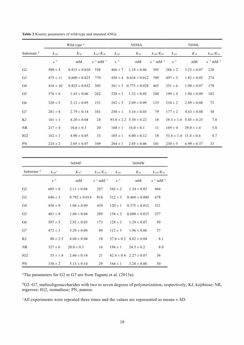
Kinetic analysis of hydrolysis activities of Asn694 mutants 200
Table 2 summarizes the kinetic parameters of the Asn694-mutated ANGs for a series of 201
maltooligosaccharides, α-glucobioses, and PN. The k cat values of N694A were 65–95% of those of 202
the wild-type enzyme, among which the values for NR and IG2 were explicitly decreased. The K m 203
9
values of N694A were similar to those of the wild-type enzyme, but increased slightly for α-204
glucobioses. Of the four mutated ANGs, N694L had the lowest k cat /K m values. Its k cat values for 205
trisaccharides (G3 and PN) were nearly the same as those of wild-type ANG, while for other 206
substrates the k cat values of the mutant were 33–84% of the wild-type values. The K m values of 207
N694L were 1.3–3.5-fold higher than those of the wild-type enzyme. The higher K m values for G3 208
and PN decreased the k cat /K m values by 35% and 30% compared with the wild-type, respectively. 209
The k cat /K m values for other substrates of N694L were also decreased to 14−48% of the wild-type 210
level. In contrast, the k cat values of N694F were 1.1–1.7 times higher than those of the wild-type 211
enzyme for substrates other than KJ, IG2, and PN. This mutation was associated with a 1.1–2.5-fold 212
increase in the K m values for all substrates except IG2. The k cat /K m values of N694F were in the 213
range of 75%–110% of that of the wild-type enzyme, with a significant decrease observed for G2 214
(51%), IG2 (64%), and PN (27%). The k cat values of N694W were 29–90% of those of the wild-type, 215
but for G2 the value was higher. The K m values of this mutant generally decreased relative to the 216
wild-type, with the exceptions of increases for the substrates G2, KJ, NR, and PN. 217
218
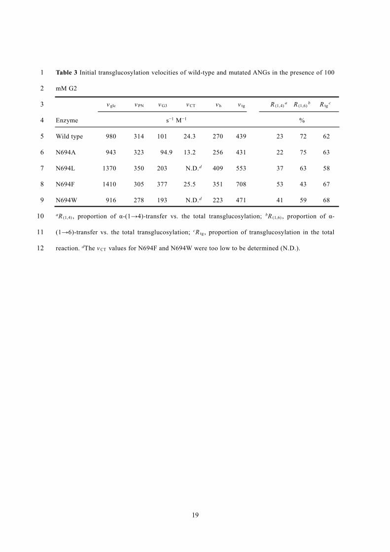
Transglucosylation by wild-type and mutant ANGs 219
The transglucosylation properties of wild-type ANG were investigated because little is known about 220
its initial behavior at high substrate concentrations. The reaction products synthesized from 100 mM 221
G2 in 15 min were glucose, PN, G3, and CT, with v glc , v PN , v G3 , and v CT values of 980, 314, 101, 222
and 24.3 s−1 M−1, respectively (Fig. S3, Table 3). R tg was 62%, and R (1,4) and R (1,6) were 23% and 223
72%, respectively. Thus, wild-type ANG possessed notable α-(1→4)-linkage formation ability. In 224
further reaction with G2, the main transglucosylation products were PN and G3 during the first hour, 225
after which G3 disappeared and IG2 accumulated (Fig. 3A). The concentration of PN increased until 226
2 h, followed by a gradual decrease. At the end of the reaction (3 h), IG2 and PN were the major 227
transglucosylation products, with traces of IG3, KJ, and CT (Fig. 3A3). 228
Transglucosylation by the Asn694 variants was also examined (Table 3). The initial 229
transglucosylation velocities of N694A and N694F were the same as those of wild-type ANG except 230
10
for v G3 of N694F. Neither N694L nor N694W could detectably synthesize CT (detection limit 8×10⁻³ 231
mM). All mutant ANGs except N694A had an increased v G3 compared with wild-type ANG. The 2.0-, 232
3.7-, and 1.9-fold greater velocities for N694L, N694F, and N694W, respectively, resulted in 37, 53, 233
and 41% higher R (1,4) values, respectively. N694A exhibited similar R (1,4) and R (1,6) to the wild-type 234
enzyme. R tg was nearly the same for N694L and the wild-type (58%), but for the mutant, the R (1,6) 235
value decreased to 63%. Transglucosylation by N694F was considerably altered: R tg increased 236
slightly compared with wild-type ANG, but R (1,6) decreased to 43%. The R tg of N694W was also 237
slightly increased, while R (1,6) was 59%. 238
Figure 3B–E depict the time courses of the transglucosylation products formed by the mutant 239
enzymes during the 3-h reaction; in all cases, >90% of the G2 had been consumed at 2 h (Fig. 3B2–240
E2). The reaction profiles of N694A and N694L were similar to that of wild-type ANG (Fig. 3B and 241
C), with early production of PN and G3 and an increase in IG2, as well as a gradual decrease in PN 242
towards the end. One difference was the production of IG2, the amounts of which at 3 h were slightly 243
lower in the N694A and N694L reactions than in the wild-type reaction. Another difference was an 244
earlier decrease in G3 than in the wild-type reaction profile (beginning at 0.5 h for the mutants but 245
at 1 h for the wild-type enzyme) (Fig 3A3–E3). This earlier decrease of G3 was also observed in 246
N694F and N694W reactions. N694F and N694W exhibited reaction profiles distinct from that of the 247
wild-type (Fig. 3D1 and E1). At the beginning of the reaction (30 min), these two mutants produced 248
larger amounts of IG2 and G3, followed by rapid (N694F) or abundant (N694W) accumulation of 249
IG2. While PN formation at 30 min was almost identical for these two mutants and the wild-type 250
enzyme, after reaching its maximum level, the PN concentration either remained within a constant 251
range (N694F) or declined very modestly (N694W). IG3 synthesis by N694F and N694W was also 252
higher than that by the wild-type enzyme. Figure 4 shows the composition of the transglucosylation 253
products at 3 h, the end of the recorded reaction. IG2 and PN were the predominant final products of 254
wild-type ANG in almost identical amounts, whereas PN predominated over IG2 in the reactions 255
catalyzed by N694A and N694L, with the latter enzyme having the highest percentage of PN 256
accumulation among the mutant ANGs. N694F and N694W yielded larger percentages of IG2 and 257
11
IG3 than were obtained with the other enzymes, with N694W resulting in the greatest accumulation 258
of IG2. 259
260
Discussion 261
To investigate the role of Asn694 in hydrolytic activity and the transglucosylation reaction of ANG, 262
we constructed N694A/L/F/W variants. We first investigated their hydrolytic activity. The results of 263
the kinetic analysis of N694A suggested the involvement of Asn694 in the +1 subsite. The k cat /K m 264
value of N694A for G2 was 70% of that for the wild-type enzyme, while there was only a slight 265
decrease in the corresponding values for the other maltooligosaccharides. The mutation of Asn694 to 266
Ala, of which the side chain was non-bulky and chemically inert, might have lowered the affinity at 267
the +1 subsite and thus affected the k cat /K m value for G2. Meanwhile, the affinity at the +2 subsite 268
can offset the lowering of +1 subsite affinity, and thus the effect on the k cat /K m values for the other 269
maltooligosaccharides was small. However, the position of Asn694 seems to be close to +2 subsite 270
in the model structure. Indeed, N694F displayed a slightly higher k cat /K m for G3 compared with wild-271
type ANG, indicating that substitution of Asn with Phe moderately enhanced the affinity at the +2 272
subsite. CtMGAM has Phe1560 at the relevant position, and its aromatic side chain is encircled by 273
three sugar rings of acarbose (Fig. 2A), allowing it to closely associate with the formation of the +1 274
and +2 subsites (Ren et al. 2011). It is possible that maltooligosaccharide substrates bind to N694F 275
in a similar manner to CtMGAM and thus the mutation in ANG causes slight enhancement of the 276
affinity at the +2 subsite. A lower k cat /K m for G2 and its recovery for G3 were observed in N694A as 277
well as N694F and N694W. This kinetic behavior might be explained by the size of the side chains. 278
The change in size of the residue 694 side chain on mutation could affect the orientation of Phe693, 279
which is conserved in GH31 α-glucosidases and closer to the +1 subsite than Asn694 (Fig. 2A). This 280
change in orientation might cause impairment of the +1 subsite and thus the decrease in k cat /K m for 281
G2. In N694L, this kinetic behavior was not observed, which could be rationalized as being due to 282
the similar size of the side chains of Leu and Asn. Among the mutant enzymes, the lowest k cat /K m 283
values for all substrates tested were those of N694L. The N694L mutation could negatively affect the 284
12
function of the general acid/base catalyst. In the modeled structure, Asn694 is located relatively close 285
to Asp660, which serves as the general acid/base catalyst (Fig. 2A). Indeed, a shift in the optimum 286
pH of the hydrolytic activity was observed in N694L. It is possible that increasing the hydrophobicity 287
by this mutation disturbs the ionic state of the general acid/base catalyst. Meanwhile, the optimum 288
pH for N694F also shifted, but its kinetic parameters exhibited no significant decrease compared with 289
the wild-type. The mechanism underlying the decrease in the activity of N694L is unclear. 290
Taken together, Asn694 is likely involved in substrate binding at subsite +2. Our previous report 291
mentioned the involvement of Thr228 on the N-loop of ANG in the formation of the plus subsites 292
(Tagami et al.2013a). Thr228 and Asn694 are located on the N-loop and the β→α loop 7, respectively, 293
but these might be spatially close together based on the model structure (Fig. 2A). It is likely that the 294
plus subsites of ANG are formed by the N-loop and the β→α loop 7. 295
Next, transglucosylation by wild-type ANG and its Asn694 variants were investigated. Formation 296
of the α-(1→6)-glucosidic linkage during wild-type ANG-catalyzed transglucosylation is well-297
established. The NMR study by Shimba et al. (2009) indicated that prolonged reaction of the enzyme 298
resulted in the efficient production of an α-(1→6)-glucosidic linkage. However, analysis of the initial 299
transglucosylation revealed that the wild-type enzyme catalyzed not only α-(1→6) but also α-(1→4) 300
transfer, with the latter accounting for 25% of the total transglucosylation (Table 3). G3, produced 301
together with PN, decreased quickly, as expected from the very high k cat /K m value for this substrate 302
(Table 2). In the double displacement catalytic mechanism, the k cat /K m for hydrolysis is an apparent 303
second-order rate constant that refers to the properties and the reactions of a free enzyme and free 304
substrate (Fersht 1988). Therefore, the G3 produced may be preferentially bound to the enzyme and 305
may either be hydrolyzed or used as the donor substrate for transglucosylation. As the concentration 306
of glucose increased as a product of hydrolysis and transglucosylation (glucose from the reducing-307
end of donor G2), free glucose became an acceptor substrate for IG2 formation (Fig. 3A1 and A2). 308
The k cat /K m value for PN hydrolysis was higher than that for IG2, such that PN was gradually 309
degraded and IG2 accumulated. IG2 then became the acceptor in the transglucosylation reaction, 310
which yielded IG3 in the late stages of the reaction (Fig. 3A3). 311
13
The v G3 values of the mutants N694L, N694F, and N694W were significantly higher than that of 312
wild-type ANG (Table 3), but their v PN values were comparable. This phenomenon was especially 313
pronounced in N694F, in which v G3 exceeded v PN and R (1,4) was 53%. The higher relative k cat /K m 314
values for G3 and PN [(k cat /K m ) G3 ∕(k cat /K m) PN ] seem to be related to the higher R (1,4) . 315
(k cat /K m ) G3 ∕(k cat /K m) PN for N694F was 28.1, greater than that for the wild-type (7.1). However, 316
higher (k cat /K m) G3 also means G3 is more likely to act as a substrate for hydrolysis or as the donor 317
for the next transglucosylation. As a result, the concentration of G3 decreased steeply and most of 318
the G3 formed was then consumed in the late stages of the reaction (Fig. 3C–E). 319
Larger amounts IG2 and IG3 were accumulated by N694F and N694W than by the wild-type, a 320
finding attributable to increased nonproductive binding of IG2. Kinetic analysis of the mutants 321
showed that the mutations decreased both k cat and K m , but had little effect on k cat /K m for IG2 (Table 322
2). This behavior is typical for the nonproductive binding of substrate. The increased nonproductive 323
binding of IG2, which probably occupies the +1 and +2 subsites, would facilitate the accumulation 324
of IG2, by decreasing the rate of its hydrolysis and thus enhancing its function as an acceptor 325
molecule, resulting in the generation of IG3 (Fig. 1C). 326
This study revealed that ANG inherently transfers α-(1→4)-glucosidic linkages, accounting for 327
~25% of the total transfer reactions catalyzed by the enzyme. The mutation of Asn694 to Phe or Trp 328
enhanced the α-(1→4)-glucosyl transfer activity and thus the efficient production of G3 from G2 329
during the initial reaction; thereafter, there was no accumulation of G3 because of its very high 330
k cat /K m value. Two mutants, N694F and N694W, accumulated significantly more of the industry-331
useful IG2 and IG3 in the prolonged reaction, probably owing to the increase in nonproductive 332
binding resulting from the mutations. Thus, mutation is an extremely valuable approach to achieve 333
glycoside-hydrolase-associated oligosaccharide synthesis. Although regulation of the glycoside-334
hydrolase-catalyzed synthesis of glycosidic linkages by mutation is challenging, using the mutants 335
obtained in this study we were able to improve the transglucosylation specificity of ANG. One of the 336
significant results of this study is that mutant enzymes exhibiting commercially interesting 337
transglucosylation properties can be generated without reducing the activity, as often occurs 338
14
following the engineering of residues within/near the active site. 339
340
Acknowledgments 341
We thank the staff of the Instrumental Analysis Division of the Creative Research Institution at 342
Hokkaido University for amino acid analysis. This study was supported in part by the JSPS 343
KAKENHI Grant Number JP26292049 (Atsuo Kimura), and by the JSPS and NRF under the Japan-344
Korea Basic Science Cooperation Program (Atsuo Kimura). 345
346
Conflict of interest 347
The authors declare that they have no competing interests. 348
349
Compliance with Ethical Standards 350
This article does not describe any studies on human participants or animals performed by any of the 351
authors. 352
353
References 354
Fersht A (1988) Structure and mechanism in protein science: a guide to enzyme catalysis and protein 355
folding. W H Freeman and Company, New York 356
Hur KY, Lee M-S (2015) Gut microbiota and metabolic disorders. Diabetes Metab J 39:198–203. 357
doi:10.4093/dmj.2015.39.3.198 358
Kaneko T, Kohmoto T, Kikuchi H, Shiota M, Iino H, Mitsuoka T (1994) Effects of 359
isomaltooligosaccharides with different degrees of polymerization on human fecal 360
bifidobactcria. Biosci Biotechnol Biochem 58:2288–2290. doi:10.1271/bbb.58.2288 361
Kohmoto T, Fukui F, Takaku H, Machida Y, Arai M, Mitsuoka T (1988) Effect of isomalto-362
oligosaccharides on human fecal flora. Bifidobact Microflora 7:61–69. 363
doi:10.12938/bifidus1982.7.2_61 364
Kohmoto T, Fukui F, Takaku H, Mitsuoka T (1991) Dose-response test of isomaltooligosaccharides 365
15
for increasing fecal bifidobacteria. Agric Biol Chem 55:2157–2159. 366
doi:10.1080/00021369.1991.10870921 367
Kohmoto T, Tsuji K, Kaneko T, Shiota M, Fukui F, Takaku H, Nakagawa Y, Ichikawa T, Kobayash 368
S (1992) Metabolism of 13C-isomaltooligosaccharides in healthy men. Biosci Biotechnol 369
Biochem 56:937–940. doi:10.1271/bbb.56.937 370
Kothari D, Patel S (2014) Therapeutic spectrum of nondigestible oligosaccharides: overview of 371
current state and prospect. J Food Sci 79:R1491–R1498. doi:10.1111/1750-3841.12536 372
Mäkeläinen H, Hasselwander O, Rautonen N, Ouwehand AC (2009) Panose, a new prebiotic 373
candidate. Lett Appl Microbiol 49:666–672. doi:10.1111/j.1472-765X.2009.02698.x 374
Ren L, Qin X, Cao X, Wang L, Bai F, Bai G, Shen Y (2011) Structural insight into substrate 375
specificity of human intestinal maltase-glucoamylase. Protein Cell 2:827–836. 376
doi:10.1007/s13238-011-1105-3 377
Shimba N, Shinagawa M, Hoshino W, Yamaguchi H, Yamada N, Suzuki E (2009) Monitoring the 378
hydrolysis and transglycosylation activity of α-glucosidase from Aspergillus niger by nuclear 379
magnetic resonance spectroscopy and mass spectrometry. Anal Biochem 393:23–28. 380
doi:10.1016/j.ab.2009.06.002 381
Sim L, Quezada-Calvillo R, Sterchi EE, Nichols BL, Rose DR (2008) Human intestinal maltase–382
glucoamylase: crystal structure of the N-terminal catalytic subunit and basis of inhibition and 383
substrate specificity. J Mol Biol 375:782–792. doi:10.1016/j.jmb.2007.10.069 384
Söding J, Biegert A, Lupas AN (2005) The HHpred interactive server for protein homology detection 385
and structure prediction. Nucleic Acids Res 33:W244–W248. doi:10.1093/nar/gki408 386
Song KM, Okuyama M, Nishimura M, Tagami T, Mori H, Kimura A (2013) Aromatic residue on β387
→α loop 1 in the catalytic domain is important to the transglycosylation specificity of 388
glycoside hydrolase family 31 α-glucosidase. Biosci Biotechnol Biochem 77:1759–1765. 389
doi:10.1271/bbb.130325 390
Takaku H (1988) Anomalously linked oligosaccharides mixture. In: The amylase research society of 391
Japan (ed) Handbook of amylases and related enzymes. Pergamon press, Oxford, pp 215-217 392
16
Tagami T, Okuyama M, Nakai H, Kim YM, Mori H, Taguchi K, Svensson B, Kimura A (2013a) Key 393
aromatic residues at subsites + 2 and + 3 of glycoside hydrolase family 31 α-glucosidase 394
contribute to recognition of long-chain substrates. Biochim Biophys Acta - Proteins 395
Proteomics 1834:329–335. doi:10.1016/j.bbapap.2012.08.007 396
Tagami T, Yamashita K, Okuyama M, Mori H, Yao M, Kimura A (2013b) Molecular basis for the 397
recognition of long-chain substrates by plant α-glucosidases. J Biol Chem 288: 19296–19303. 398
doi:10.1074/jbc.M113.465211 399
Tagami T, Yamashita K, Okuyama M, Mori H, Yao M, Kimura A (2015) Structural advantage of 400
sugar beet α-glucosidase to stabilize the Michaelis complex with long-chain substrate. J Biol 401
Chem 290:1796–1803. doi:10.1074/jbc.M114.606939 402
Tan K, Tesar C, Wilton R, Keigher L, Babnigg G, Joachimiak A (2010) Novel α-glucosidase from 403
human gut microbiome: substrate specificities and their switch. FASEB J 24:3939–3949. doi: 404
10.1096/fj.10-156257 405
406
17
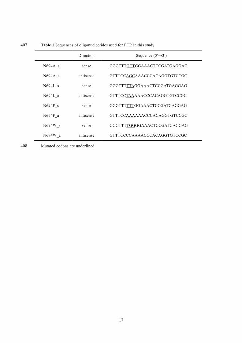
Table 1 Sequences of oligonucleotides used for PCR in this study 407
Direction Sequence (5′→3′)
N694A_s sense GGGTTTGCTGGAAACTCCGATGAGGAG
N694A_a antisense GTTTCCAGCAAACCCACAGGTGTCCGC
N694L_s sense GGGTTTTTAGGAAACTCCGATGAGGAG
N694L_a antisense GTTTCCTAAAAACCCACAGGTGTCCGC
N694F_s sense GGGTTTTTTGGAAACTCCGATGAGGAG
N694F_a antisense GTTTCCAAAAAACCCACAGGTGTCCGC
N694W_s sense GGGTTTTGGGGAAACTCCGATGAGGAG
N694W_a antisense GTTTCCCCAAAACCCACAGGTGTCCGC
Mutated codons are underlined.408
18
Table 2 Kinetic parameters of wild-type and mutated ANGs
Wild type a N694A N694L
Substrate b k cat K m k cat /K m k cat K m k cat /K m k cat K m k cat /K m
s−1 mM s−1 mM−1 s−1 mM s−1 mM−1 s−1 mM s−1 mM−1
G2 509 ± 5 0.913 ± 0.010 558 466 ± 7 1.18 ± 0.06 395 388 ± 2 3.23 ± 0.07 120
G3 475 ± 11 0.609 ± 0.025 779 450 ± 4 0.634 ± 0.012 709 497 ± 5 1.82 ± 0.05 274
G4 416 ± 10 0.823 ± 0.032 505 361 ± 3 0.775 ± 0.028 465 351 ± 6 1.98 ± 0.07 178
G5 376 ± 6 1.43 ± 0.06 262 328 ± 3 1.32 ± 0.05 248 199 ± 4 1.94 ± 0.09 102
G6 320 ± 5 2.12 ± 0.05 151 282 ± 5 2.09 ± 0.09 135 210 ± 2 2.89 ± 0.08 73
G7 281 ± 8 2.79 ± 0.14 101 250 ± 1 3.16 ± 0.03 79 177 ± 2 4.63 ± 0.08 38
KJ 101 ± 1 4.20 ± 0.04 24 93.0 ± 2.2 5.30 ± 0.22 18 38.3 ± 1.0 5.45 ± 0.25 7.0
NR 217 ± 4 10.6 ± 0.3 20 168 ± 1 16.0 ± 0.1 11 169 ± 4 29.0 ± 1.6 5.8
IG2 162 ± 1 4.90 ± 0.05 33 105 ± 1 6.00 ± 0.12 18 53.8 ± 1.4 11.4 ± 0.6 4.7
PN 224 ± 2 2.05 ± 0.07 109 204 ± 1 2.05 ± 0.06 101 230 ± 5 6.99 ± 0.37 33
N694F N694W
Substrate b k cat c K m c k cat /K m k cat K m k cat /K m
s−1 mM s−1 mM−1 s−1 mM s−1 mM−1
G2 605 ± 8 2.11 ± 0.04 287 542 ± 2 1.34 ± 0.03 404
G3 646 ± 3 0.792 ± 0.014 816 312 ± 3 0.460 ± 0.004 678
G4 456 ± 9 1.04 ± 0.09 439 120 ± 1 0.375 ± 0.012 321
G5 461 ± 8 1.60 ± 0.04 289 154 ± 3 0.600 ± 0.015 257
G6 507 ± 5 2.92 ± 0.03 173 128 ± 3 1.29 ± 0.07 99
G7 472 ± 3 5.29 ± 0.04 89 112 ± 3 1.96 ± 0.06 57
KJ 80 ± 2.5 4.60 ± 0.04 18 37.6 ± 0.2 4.62 ± 0.04 8.1
NR 327 ± 6 20.0 ± 0.3 16 196 ± 1 24.5 ± 0.2 8.0
IG2 55 ± 1.4 2.60 ± 0.14 21 82.4 ± 0.4 2.27 ± 0.07 36
PN 150 ± 2 5.13 ± 0.14 29 164 ± 1 3.28 ± 0.06 50
aThe parameters for G2 to G7 are from Tagami et al. (2013a).
bG2–G7, maltooligosaccharides with two to seven degrees of polymerization, respectively; KJ, kojibiose; NR, nigerose; IG2, isomaltose; PN, panose.
cAll experiments were repeated three times and the values are represented as means ± SD.
19
Table 3 Initial transglucosylation velocities of wild-type and mutated ANGs in the presence of 100 1
mM G2 2
v glc v PN v G3 v CT v h v tg R (1,4)a R (1,6)
b R tgc 3
Enzyme s−1 M−1 % 4
Wild type 980 314 101 24.3 270 439 23 72 62 5
N694A 943 323 94.9 13.2 256 431 22 75 63 6
N694L 1370 350 203 N.D.d 409 553 37 63 58 7
N694F 1410 305 377 25.5 351 708 53 43 67 8
N694W 916 278 193 N.D.d 223 471 41 59 68 9
aR (1,4) , proportion of α-(1→4)-transfer vs. the total transglucosylation; bR (1,6) , proportion of α-10
(1→6)-transfer vs. the total transglucosylation; cR tg , proportion of transglucosylation in the total 11
reaction. dThe v CT values for N694F and N694W were too low to be determined (N.D.). 12
20
Figure legends 13
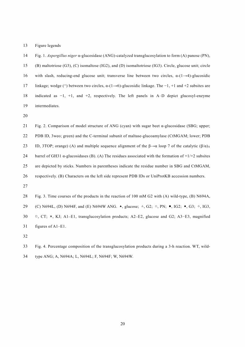
Fig. 1. Aspergillus niger α-glucosidase (ANG)-catalyzed transglucosylation to form (A) panose (PN), 14
(B) maltotriose (G3), (C) isomaltose (IG2), and (D) isomaltotriose (IG3). Circle, glucose unit; circle 15
with slash, reducing-end glucose unit; transverse line between two circles, α-(1→4)-glucosidic 16
linkage; wedge (^) between two circles, α-(1→6)-glucosidic linkage. The −1, +1 and +2 subsites are 17
indicated as −1, +1, and +2, respectively. The left panels in A–D depict glucosyl-enzyme 18
intermediates. 19
20
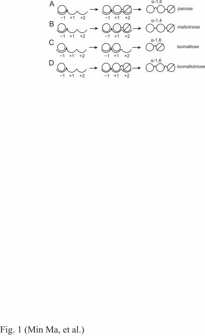
Fig. 2. Comparison of model structure of ANG (cyan) with sugar beet α-glucosidase (SBG; upper; 21
PDB ID, 3weo; green) and the C-terminal subunit of maltase-glucoamylase (CtMGAM; lower; PDB 22
ID, 3TOP; orange) (A) and multiple sequence alignment of the β→α loop 7 of the catalytic (β/α) 8 23
barrel of GH31 α-glucosidases (B). (A) The residues associated with the formation of +1/+2 subsites 24
are depicted by sticks. Numbers in parentheses indicate the residue number in SBG and CtMGAM, 25
respectively. (B) Characters on the left side represent PDB IDs or UniProtKB accession numbers. 26
27
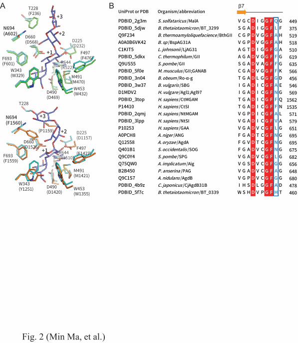
Fig. 3. Time courses of the products in the reaction of 100 mM G2 with (A) wild-type, (B) N694A, 28
(C) N694L, (D) N694F, and (E) N694W ANG. ▲, glucose; △, G2; ○, PN; ●, IG2; ◆, G3; ◇, IG3, 29
□, CT; ×, KJ; A1–E1, transglucosylation products; A2–E2, glucose and G2; A3−E3, magnified 30
figures of A1–E1. 31
32
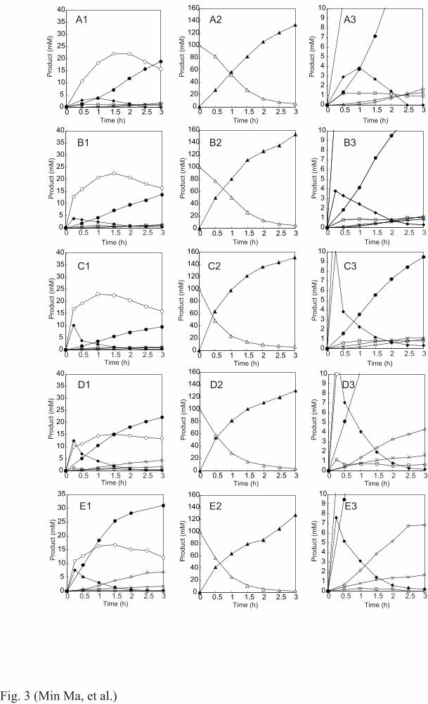
Fig. 4. Percentage composition of the transglucosylation products during a 3-h reaction. WT, wild-33
type ANG; A, N694A; L, N694L; F, N694F; W, N694W. 34
−1 +1 +2
−1 +1 +2
−1 +1 +2
−1 +1 +2
−1 +1 +2
−1 +1 +2
α-1,6
α-1,6
α-1,6
panose
isomaltose
isomaltotriose
A
C
D
Fig. 1 (Min Ma, et al.)
−1 +1 +2 −1 +1 +2
α-1,4maltotrioseB
β7BA
Fig. 2 (Min Ma, et al.)
05
10152025303540
0 0.5 1 1.5 2 2.5 30
20406080
100120140160
0 0.5 1 1.5 2 2.5 3
020406080
100120140160
0 0.5 1 1.5 2 2.5 3
020406080
100120140160
0 0.5 1 1.5 2 2.5 3
020406080
100120140160
0 0.5 1 1.5 2 2.5 3
020406080
100120140160
0 0.5 1 1.5 2 2.5 3
05
10152025303540
0 0.5 1 1.5 2 2.5 3
05
10152025303540
0 0.5 1 1.5 2 2.5 3
05
10152025303540
0 0.5 1 1.5 2 2.5 3
0
5
10
15
20
25
30
35
0 0.5 1 1.5 2 2.5 3
Prod
uct (
mM
)
Time (h)
Prod
uct (
mM
)
Time (h)
Prod
uct (
mM
)
Time (h)
Prod
uct (
mM
)
Time (h)
Prod
uct (
mM
)
Time (h)
Prod
uct (
mM
)
Time (h)
Prod
uct (
mM
)
Time (h)
Prod
uct (
mM
)
Time (h)
Prod
uct (
mM
)
Time (h)
Prod
uct (
mM
)
Time (h)
Prod
uct (
mM
)
Time (h)
Prod
uct (
mM
)
Time (h)
Prod
uct (
mM
)
Time (h)
Prod
uct (
mM
)
Time (h)
Prod
uct (
mM
)
Time (h)
A1
B1
C1
A2 A3
B3
C3
D3
E3
B2
D1
E1
D2
E2
C2
Fig. 3 (Min Ma, et al.)
0123456789
10
0 0.5 1 1.5 2 2.5 3
0123456789
10
0 0.5 1 1.5 2 2.5 3
0123456789
10
0 0.5 1 1.5 2 2.5 3
0123456789
10
0 0.5 1 1.5 2 2.5 3
0123456789
10
0 0.5 1 1.5 2 2.5 3
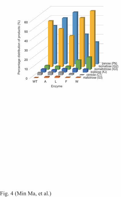
Fig. 4 (Min Ma, et al.)
Enzyme
Perc
enta
ge d
istri
butio
n of
pro
duct
s (%
)
0
10
20
30
40
50
60
WT A L F W
panose (PN)isomaltose (IG2)
isomaltotriose (IG3)kojibiose (KJ)
centose (CT)maltotriose (G3)