Effect of anatomical location (mandible vs maxilla) of ...
10
261 RESEARCH Effect of anatomical location (mandible vs maxilla) of dental implants on the BMP-2, BMP-7, sRANKL and OPG levels in peri- implant crevicular fluid during osseointegration. A pilot study Mehmet Sağlam(0000-0002-2703-2462) , Doğan Dolanmaz(0000-0002-7390-1147) β , Emrah Koçak(0000-0002-4188-1348) γ , Burcu Gürsoytrak(0000-0002-9893-0649) λ , Özgür İnan(0000-0001-5990-6454) μ , Niyazi Dündar(0000-0002-6558-3775) φ , Sema S. Hakkı(0000-0001-8665-6235) ω Selcuk Dent J, 2019; 6: 261-270 (Doi: 10.15311/selcukdentj.450292) Department of Periodontology, Faculty of Dentistry, İzmir Katip Çelebi University, İzmir, Turkey β Department of Oral and Maxillofacial Surgery, Faculty of Dentistry, Bezmiȃlem, İstanbul, Turkey γ İzzet Baysal Mouth and Dental Health Center, Bolu, Turkey λ Department of Oral and Maxillofacial Surgery, Faculty of Dentistry, Adnan Menderes University, Aydın, Turkey μ Department of Prosthodontics, Faculty of Dentistry, Selçuk University, Konya, Turkey φ Research Center of Dental Faculty, Selcuk University, Konya, Turkey ω Department of Periodontology, Faculty of Dentistry, Selcuk University, Konya, Turkey ÖZ Dental implantların anatomik lokasyonlarının (mandibula ile maxilla) osseointegrasyon süresince peri-implant oluğu sıvısındaki BMP-2, BMP-7, sRANKL ve OPG seviyeleri üzerine etkisi. Pilot bir çalışma Amaç: Bu çalışmanın amacı, hem maksilla hemde mandibulaya yerleştirilen implantların periimplant oluğu sıvısında osseointegrasyon süresi boyunca kemik morfogenetik protein-2 (BMP-2), BMP-7, çözünür reseptör aktivatör nükleer faktör kappa B ligandı (sRANKL) ve osteoprotegrin (OPG) seviyelerinin araştırılmasıdır. Gereç ve Yöntemler: Bu çalışmada 33 hasta (17 bayan ve 16 erkek; ortalama yaş 47.03±11.23) yer almıştır. Hem maxillaya (Grup 1/n=18) hemde mandibulaya (Grup 2/n=15) olmak üzere toplam 33 implant yerleştirilmiştir. Peri-implant oluğu sıvısı (PIS) örnekleri, modifiye plak indeksi (MPI), gingival indeks (GI) ve sondlama cep derinliği (SCD) ölçümleri cerrahiden sonra 1. ve 3. ayda alındı. BMP-2/-7, sRANKL ve OPG PIS seviyeleri ELIZA ile incelendi. Bulgular: İyileşme süresince herhangi bir komplikasyon gözlenmedi.Gruplar arasında sRANKL, OPG, BMP-2 ve BMP-7 PIS seviyeleri ve incelenen klinik parametreler açısından herhangi bir zaman periyodunda anlamlı fark gözlenmedi (p>0.05). PIS hacmi 1. ayda grup 2 de grup 1’e göre fazla iken, 3. ayda PIS hacmi grup 1 de grup 2’ye göre fazlaydı (p<0.05). PIS hacmi ile sRANKL arasında pozitif (p<0.05) ve BMP-2 ile BMP-7 arasında güçlü pozitif korelasyon (p<0.01) mevcuttu. Sonuç: Bu pilot çalışmanın sonuçları, dental implantların anatomik lokasyonları açısından BMP-2, BMP-7, sRANKL, ve OPG PIS seviyelerinde anlamlı bir fark gösterememiştir. Kemik ile ilişkili biyobelirteçler ve dental implantların anatomik lokasyonu arasındaki ilişkinin değerlendirilmesi için iyi dizayn edilmiş çalışmalar yapılmalıdır. ANAHTAR KELİMELERK Kemik morfogenetik proteinleri, dental implant, osteoprotegrin, reseptör aktivatör nükleer kappa B ligand Başvuru Tarihi: 11 Nisan 2018 Yayına Kabul Tarihi: 16 Aralık 2018 ABSTRACT Effect of anatomical location (mandible vs maxilla) of dental implants on the BMP-2, BMP-7, sRANKL and OPG levels in peri-implant crevicular fluid during osseointegration. A pilot study Background: The aim of this study was to investigate levels of bone morphogenetic protein-2 (BMP-2), BMP-7, soluble receptor activator of nuclear factor-kB ligand (sRANKL) and osteoprotegerin (OPG) in the peri-implant crevicular fluid (PICF) of implants placed in both maxilla and mandible during the osseointegration period. Materials and Methods: Thirty-three patients (17 females and 16 males; mean age 47.03±11.23 years) were included in this study. A total of 33 implants were placed in both of maxilla (Group 1/n=18) and mandible (group 2/n=15). Peri-implant crevicular fluid (PICF) samples, modified plaque index (MPI), gingival index (GI) and probing depth (PD) measurements were obtained at 1 and 3 months after surgery. PICF levels of BMP-2/-7, sRANKL and OPG were analyzed by ELISA. Results: No complications were observed during the healing period. No significant differences were observed in the PICF levels of sRANKL, OPG, BMP-2 and BMP-7 and evaluated clinical parameters between groups at any time point (p>0.05). While PICF volume of group 2 was greater than group 1 at first month, PICF volume of group 1 was greater than group 2 at 3 months (p<0.05). There was a positive correlation between sRANKL levels and PICF volume (p<0.05) and a strong correlation between BMP-2 and BMP-7 (p<0.01). Conclusion: The results of this pilot study didn’t show any significant difference in PICF levels of BMP-2, BMP-7, sRANKL, and OPG in terms of anatomic location of dental implants. Further well- designed studies should be carried out to evaluate the relationship between bone related biomarkers and anatomic location of dental implants. KEYWORDS Bone morphogenetic proteins, dental implant, osteoprotegerin, receptor activator of nuclear factor-kappa B
Transcript of Effect of anatomical location (mandible vs maxilla) of ...
261
RESEARCH
Effect of anatomical location (mandible vs maxilla) of dental
implants on the BMP-2, BMP-7, sRANKL and OPG levels in peri-
implant crevicular fluid during osseointegration. A pilot study
Mehmet Sağlam(0000-0002-2703-2462), Doğan Dolanmaz(0000-0002-7390-1147)
β, Emrah Koçak(0000-0002-4188-1348)
γ,
Burcu Gürsoytrak(0000-0002-9893-0649)λ, Özgür İnan(0000-0001-5990-6454)
μ, Niyazi Dündar(0000-0002-6558-3775)
φ,
Sema S. Hakkı(0000-0001-8665-6235)ω
Selcuk Dent J, 2019; 6: 261-270 (Doi: 10.15311/selcukdentj.450292)
Department of Periodontology, Faculty of Dentistry, İzmir Katip Çelebi University, İzmir, Turkey β Department of Oral and Maxillofacial Surgery, Faculty of Dentistry, Bezmiȃlem, İstanbul, Turkey γ İzzet Baysal Mouth and Dental Health Center, Bolu, Turkey λ Department of Oral and Maxillofacial Surgery, Faculty of Dentistry, Adnan Menderes University, Aydın, Turkey μ Department of Prosthodontics, Faculty of Dentistry, Selçuk University, Konya, Turkey φ Research Center of Dental Faculty, Selcuk University, Konya, Turkey ω Department of Periodontology, Faculty of Dentistry, Selcuk University, Konya, Turkey
ÖZ
Dental implantların anatomik lokasyonlarının (mandibula ile
maxilla) osseointegrasyon süresince peri-implant oluğu
sıvısındaki BMP-2, BMP-7, sRANKL ve OPG seviyeleri
üzerine etkisi. Pilot bir çalışma
Amaç: Bu çalışmanın amacı, hem maksilla hemde mandibulaya
yerleştirilen implantların periimplant oluğu sıvısında
osseointegrasyon süresi boyunca kemik morfogenetik protein-2
(BMP-2), BMP-7, çözünür reseptör aktivatör nükleer faktör
kappa B ligandı (sRANKL) ve osteoprotegrin (OPG)
seviyelerinin araştırılmasıdır.
Gereç ve Yöntemler: Bu çalışmada 33 hasta (17 bayan ve 16
erkek; ortalama yaş 47.03±11.23) yer almıştır. Hem maxillaya
(Grup 1/n=18) hemde mandibulaya (Grup 2/n=15) olmak üzere
toplam 33 implant yerleştirilmiştir. Peri-implant oluğu sıvısı (PIS)
örnekleri, modifiye plak indeksi (MPI), gingival indeks (GI) ve
sondlama cep derinliği (SCD) ölçümleri cerrahiden sonra 1. ve
3. ayda alındı. BMP-2/-7, sRANKL ve OPG PIS seviyeleri ELIZA
ile incelendi.
Bulgular: İyileşme süresince herhangi bir komplikasyon
gözlenmedi.Gruplar arasında sRANKL, OPG, BMP-2 ve BMP-7
PIS seviyeleri ve incelenen klinik parametreler açısından
herhangi bir zaman periyodunda anlamlı fark gözlenmedi
(p>0.05). PIS hacmi 1. ayda grup 2 de grup 1’e göre fazla iken,
3. ayda PIS hacmi grup 1 de grup 2’ye göre fazlaydı (p<0.05).
PIS hacmi ile sRANKL arasında pozitif (p<0.05) ve BMP-2 ile
BMP-7 arasında güçlü pozitif korelasyon (p<0.01) mevcuttu.
Sonuç: Bu pilot çalışmanın sonuçları, dental implantların
anatomik lokasyonları açısından BMP-2, BMP-7, sRANKL, ve
OPG PIS seviyelerinde anlamlı bir fark gösterememiştir. Kemik
ile ilişkili biyobelirteçler ve dental implantların anatomik
lokasyonu arasındaki ilişkinin değerlendirilmesi için iyi dizayn
edilmiş çalışmalar yapılmalıdır.
ANAHTAR KELİMELERK
Kemik morfogenetik proteinleri, dental implant,
osteoprotegrin, reseptör aktivatör nükleer kappa B ligand
Başvuru Tarihi: 11 Nisan 2018
Yayına Kabul Tarihi: 16 Aralık 2018
Yayına Kbul ABSTRACT
Effect of anatomical location (mandible vs maxilla) of dental
implants on the BMP-2, BMP-7, sRANKL and OPG levels in
peri-implant crevicular fluid during osseointegration. A pilot
study
Background: The aim of this study was to investigate levels of bone
morphogenetic protein-2 (BMP-2), BMP-7, soluble receptor activator
of nuclear factor-kB ligand (sRANKL) and osteoprotegerin (OPG) in
the peri-implant crevicular fluid (PICF) of implants placed in both
maxilla and mandible during the osseointegration period.
Materials and Methods: Thirty-three patients (17 females and 16
males; mean age 47.03±11.23 years) were included in this study. A
total of 33 implants were placed in both of maxilla (Group 1/n=18)
and mandible (group 2/n=15). Peri-implant crevicular fluid (PICF)
samples, modified plaque index (MPI), gingival index (GI) and
probing depth (PD) measurements were obtained at 1 and 3
months after surgery. PICF levels of BMP-2/-7, sRANKL and OPG
were analyzed by ELISA.
Results: No complications were observed during the healing
period. No significant differences were observed in the PICF levels
of sRANKL, OPG, BMP-2 and BMP-7 and evaluated clinical
parameters between groups at any time point (p>0.05). While PICF
volume of group 2 was greater than group 1 at first month, PICF
volume of group 1 was greater than group 2 at 3 months (p<0.05).
There was a positive correlation between sRANKL levels and PICF
volume (p<0.05) and a strong correlation between BMP-2 and
BMP-7 (p<0.01).
Conclusion: The results of this pilot study didn’t show any
significant difference in PICF levels of BMP-2, BMP-7, sRANKL, and
OPG in terms of anatomic location of dental implants. Further well-
designed studies should be carried out to evaluate the relationship
between bone related biomarkers and anatomic location of dental
implants.
KEYWORDS
Bone morphogenetic proteins, dental implant, osteoprotegerin,
receptor activator of nuclear factor-kappa B
Effect of anatomical location (mandible vs maxilla) of dental implants on the BMP-2, BMP-7, sRANKL and OPG levels in peri-implant crevicular fluid during osseointegration. A pilot study Cilt 6 • Sayı 3
262
Osseointegration term has been used to describe
a direct structural and functional relationship
between living bone tissue and a load-carrying
implant surface.1 A series of some cellular and
extracellular biological events occur between
bone tissue and implant surface during bone
healing around implants.2 At the bone-implant
interface, the expression of several growth and
differentiation factors by the activated blood cells,
mediates this cascade of biological events.3
Platelet derived growth factor, insulin-like growth
factors, transforming growth factors and fibroblast
growth factor enhance bone healing by inducing
migration, proliferation, and differentiation into
bone cells of mesenchymal-derived cells.4 Other
important bone biological factors that play
essential roles in osteogenesis are bone
morphogenetic proteins (BMPs). BMPs that
belong to the transforming growth factor-β
superfamily are secreted signaling proteins which
serve as regulating matrix synthesis, cell
proliferation and tissue differentiation.5,6
BMPs
stimulate bone tissue formation by differentiating
mesenchymal stem cells to osteoblastic and
chondroblastic cells.7 BMP-2 and BMP-7 are the
most effective types that induce complete
morphogenesis of bone tissue.8 Like other bone
morphogenetic proteins, BMP-2 has an essential
function in the development of cartilage and bone
tissue.9 BMP-7 is also known as osteogenic
protein-1and plays a key role in the transformation
of MSCs into bone and cartilage.9
RANKL and OPG are critical factors in the control
of osseous healing, which are both produced by
osteoblasts. RANKL is a cell membrane-bound
protein responsible for stimulation of osteoclast
differentiation and bone resorption.10
OPG
(osteoclastogenesis inhibitory factor) counteracts
the biological activities of RANKL by preventing its
interaction with its receptor (RANK).11,12
It has
been indicated that a balanced RANKL/OPG
expression is important in physiological bone
remodeling.13
The availability of an adequate quantity and
quality of bone at the implant site are critical local
factors for osseointegration. The quality of bone
support differs due to the anatomic location, thus
implant outcomes are sometimes categorized
according to anatomic location. The types of
bones related tothe quality of cortical bone and
trabecular bone density were classified by
Lekholm and Zarb. According to their
classification, the bones types are classified as
D1, D2, D3 and D4 bone.14
The D1 bone consists
of dense cortical and trabecular bone.15
The
percentages of light microscopic bone-implant
contact (BIC) are highest in D1 bone and more
than 80%. Fewer blood vessels are present in D1
bone than in the other three types. The structure
of this bone type is almost all cortical and the
regeneration capacity is impaired because of the
poor blood circulation. D1 bone existsmore often
percentages of light microscopic bone-implant contact
(BIC) are highest in D1 bone and more than 80%. Fewer
blood vessels are present in D1 bone than in the other
three types. The structure of this bone type is almost all
cortical and the regeneration capacity is impaired because
of the poor blood circulation. D1 bone existsmore often in
anterior regions of mandibles. The D2 is composed of a
thick crestal layer of dense-to-porous cortical bone and
coarse trabecular bone under the cortical bone.16
In D2
bone there is high amount of blood in contact with the
implant surface and primary stability is good.15
The D2
bone trabeculae are 40% to 60% stronger than D3
trabeculae. This bone is found generally in the anterior
region of mandible, followed by the posterior region of
mandible. D2 bone type ensures desirable implant healing,
and osseointegration is very foreseeable. D3 is composed
combination of thinner porous cortical bone on the crest
and fine trabecular bone on the inside. The trabecula in D3
bone is approximately 50% more fragile than in D2 bone.
D3 bone is found more frequently in the anterior regions of
maxilla and posterior regions of both arch. The BIC is also
less favorable in D3 bone compared to D2 bone. D4 bone
has very few density and small amount or no cortical
crestal bone. This type of bone is observed most often in
the posterior maxilla. Bone trabeculae are dispersed and,
as a result, achievement of primary stability of any implant
typepresents a surgical challenge.16
Biological factors play an important role in
osseointegration, and a few data is present about the
biological factors that play essential roles in bone tissue
healing and remodeling around dental implants. The
purposes of this study were to measure BMP-2, BMP-7,
sRANKL, and OPG levels in periimplant crevicular fluid
(PICF) around non-submerged implants, which were
placed at different locations of maxilla and mandible at 1
and 3 months after surgery; and to correlate these values
with clinical parameters.
MATERIALS AND METHODS
Thirty-three patients (17 females and 16 males; mean age
47.03±11.23 years) who were attending the Oral and
Maxillofacial Surgery Department at Selcuk University
Faculty of Dentistry (during the 2011-2013 academic years)
and scheduled for implant surgery were included in this
study. The study protocol was approved by the Ethics
Commission of Selcuk University Faculty of Medicine for
human subjects. None of the patients had a history of
systemic disease and had received antibiotics within the
prior 6 months. All patients were nonsmokers. Informed
consent was obtained from each patient before clinical
examination and PICF sampling. All patients received oral
hygiene instructions and supragingival scaling. Full-mouth
subgingival scaling and root planing under local anesthesia
was performed in a single appointment for chronic
periodontitis patients. There was no patient who has
≥4mm periodontal pocket depth after periodontal therapy.
A total of 33 implants from single implant system
(Nucleoss, Izmir, Turkey) were placed using standard
surgical procedures.
Selcuk Dent J. 2019 Sağlam M, Dolanmaz D, Koçak E, Poyraz B, İnan Ö, Dündar N, Hakkı S
263
and 0.2% chlorhexidine mouthwash (Corsodyl,
GlaxoSmithKline Consumer Healthcare, UK) twice a day for 10
days. The silk sutures were removed 10 days after surgery.
The implants had no probe for resonance frequency analysis.
The primary stability was evaluated clinically and by using
periapical radiography.
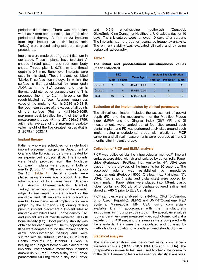
Table 1.
The initial and post-treatment microhardness values
(mean±standard
SEX
Mean Age
Implant Site Distribution
Male Female Anterior Premolar Molar
Group 1 9 9 47.44±11.95 5 11 2
Group 2 7 8 46.53±10.70 0 5 10
Total 16 17 47.03±11.23 5 16 12
Evaluation of the implant status by clinical parameters
The clinical examination included the assessment of pocket
depth (PD) and the measurement of the Modified Plaque
Index (MPI)18
and the Gingival Index (GI)19
MPI and GI
measurements were carried out at four sites around each
dental implant and PD was performed at six sites around each
implant using a periodontal probe with plastic tip. PICF
sampling and clinical measurements were recorded at 1 and 3
months after implant therapy.
Collection of PICF and ELISA analysis
PICF was collected via the intracrevicular method.20
Implant
surfaces were dried with air and isolated by cotton rolls. Paper
strips (Periopaper, ProFlow, Inc., Amityville, NY, USA) were
placed into the crevices of the implants for 30 seconds. The
adsorbed volume was established by impedance
measurements (Periotron 8000, Oraflow, Inc., Plainview, NY,
USA). Two strips (mesial and distal sites) were pooled for
each implant. Paper strips were placed into 1.5-mL plastic
tubes containing 500 µL of phosphate-buffered saline and
stored at −80°C prior to ELISA analysis.
PICF samples were analyzed for sRANKL, OPG (BioVendor,
Brno, Czech Republic), BMP-2 and BMP-7(Quantikine, R&D
Systems, Minneapolis, MN, USA) using commercially
available kits in accordance with the manufacturer’s
instructions as in our previous study.21
The absorbance values
(optical densities) were measured spectrophotometrically at a
wavelength of 450 nm, and the samples were compared with
the standards. Data were then calculated and obtained by
methods of interpolation of a predetermined standard curve.
Statistical analysis
The statistical analysis was performed using commercially
available software (SPSS v.20.0, IBM, Chicago, IL,USA). The
Shapiro-Wilks normality test was used to check the normality
of the data. Parametric tests were used for statistical analyses.
Independent sample T-test was used in comparison between
groups for each time point. Paired t-test was used in
comparison between the two time points for the same group.
Associations among mean levels of the biomarkers and
clinical parameters were also examined using the Spearman
rank correlation test.
periodontitis patients. There was no patient
who has ≥4mm periodontal pocket depth after
periodontal therapy. A total of 33 implants
from single implant system (Nucleoss, Izmir,
Turkey) were placed using standard surgical
procedures.
Implants were made out of grade 4 titanium in
our study. These implants have two-start V-
shaped thread pattern and root form body
shape. Thread pitch is 0.75 mm and thread
depth is 0.3 mm. Bone level implants were
used in this study. These implants exhibited
‘Maxicell’ surface technology, in which the
surface is first sandblasted by large grain
Al2O3, as in the SLA surface, and then is
thermal acid etched for surface cleaning. This
produces fine 1- to 2-µm micropits on the
rough-blasted surface. Average roughness
value of the implants (Ra) is 3,2361±0,2315,
the root mean square of the values of all points
of the surface (Rq) is 4,1316±0,3085,
maximum peak-to-valley height of the entire
measurement trace (Rt) is 27,1536±3,1756,
arithmetic average of the maximum peak-to-
valley height of the five greatest values (Rz) is
21,9079±1,6022.17
Implant therapy
Patients who were scheduled for single tooth
implant placement surgery in Department of
Oral and Maxillofacial Surgery were treated by
an experienced surgeon (DD). The implants
were kindly provided from the Nucleoss
Company. Implants were placed in both of
maxilla (Group 1/n=18) and mandible (group
2/n=15) (Table 1). Dental implants were
placed using a one-stage protocol. After the
administration of local anesthesia (Ultracain
DS, Aventis Pharmacheuticals, Istanbul,
Turkey), an incision was made on the alveolar
ridge. Fifteen implants were placed in the
mandible and 18 implants were placed in
maxilla. Bone densities at implant sites were
judged by the surgeon (DD) during drilling
prior to implant placement.14
Implant sites at
mandible exhibited Class II bone density (D2)
and implant sites at maxilla exhibited Class III
bone density (D3). Good primary stability was
obtained for each implant. The mucoperiosteal
flaps were adapted around the implant neck to
allow non-submerged healing and were
sutured with silk sutures (Sterisilk, SSM Sterile
Health Products Inc, Istanbul, Turkey). A
healing cap (gingival former) was placed for all
implants. Postoperative medication included
amoxicillin 500 mg 3 times a day for 10 days,
paracetamol 500 mg twice a day for 5 days,
and 0.2% chlorhexidine mouthwash (Corsodyl,
GlaxoSmithKline Consumer Healthcare, UK)
twice a day for 10 days. The silk sutures were
removed 10 days after surgery. The implants
had no probe for resonance frequency
analysis. The primary stability was evaluated
Effect of anatomical location (mandible vs maxilla) of dental implants on the BMP-2, BMP-7, sRANKL and OPG levels in peri-implant crevicular fluid during osseointegration. A pilot study Cilt 6 • Sayı 3
264
Independent sample T-test was used in
comparison between groups for each time point.
Paired t-test was used in comparison between the
two time points for the same group. Associations
among mean levels of the biomarkers and clinical
parameters were also examined using the
Spearman rank correlation test.
RESULTS
A total of 33 implants were placed using one-stage
protocol. The patient demographic data and
implant site distribution were presented in Table 1.
The implants showed no clinical signs of peri-
implant infection or noticeable mobility during the
healing period.
Clinical assessments
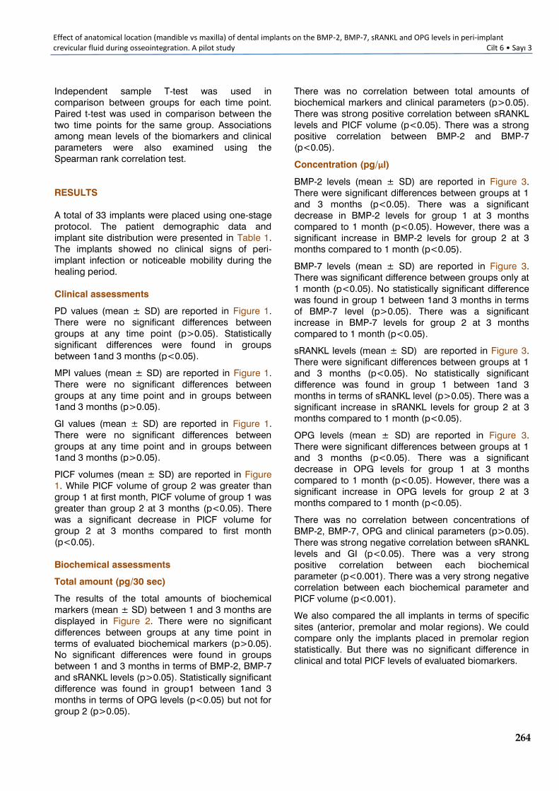
PD values (mean ± SD) are reported in Figure 1.
There were no significant differences between
groups at any time point (p>0.05). Statistically
significant differences were found in groups
between 1and 3 months (p<0.05).
MPI values (mean ± SD) are reported in Figure 1.
There were no significant differences between
groups at any time point and in groups between
1and 3 months (p>0.05).
GI values (mean ± SD) are reported in Figure 1.
There were no significant differences between
groups at any time point and in groups between
1and 3 months (p>0.05).
PICF volumes (mean ± SD) are reported in Figure
1. While PICF volume of group 2 was greater than
group 1 at first month, PICF volume of group 1 was
greater than group 2 at 3 months (p<0.05). There
was a significant decrease in PICF volume for
group 2 at 3 months compared to first month
(p<0.05).
Biochemical assessments
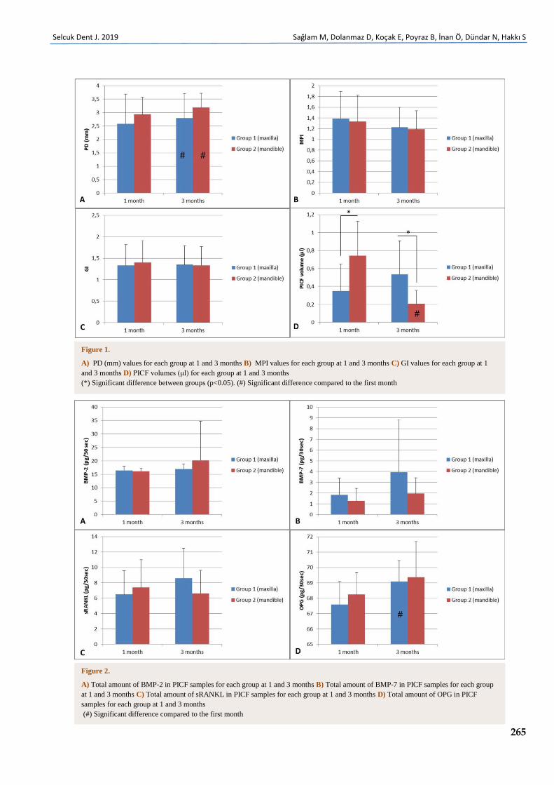
Total amount (pg/30 sec)
The results of the total amounts of biochemical
markers (mean ± SD) between 1 and 3 months are
displayed in Figure 2. There were no significant
differences between groups at any time point in
terms of evaluated biochemical markers (p>0.05).
No significant differences were found in groups
between 1 and 3 months in terms of BMP-2, BMP-7
and sRANKL levels (p>0.05). Statistically significant
difference was found in group1 between 1and 3
months in terms of OPG levels (p<0.05) but not for
group 2 (p>0.05).
There was no correlation between total amounts of
biochemical markers and clinical parameters
(p>0.05). There was strong positive correlation
between sRANKL levels and PICF volume (p<0.05).
There was no correlation between total amounts of
biochemical markers and clinical parameters (p>0.05).
There was strong positive correlation between sRANKL
levels and PICF volume (p<0.05). There was a strong
positive correlation between BMP-2 and BMP-7
(p<0.05).
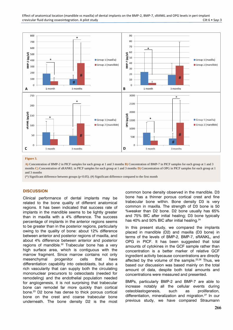
Concentration (pg/μl)
BMP-2 levels (mean ± SD) are reported in Figure 3.
There were significant differences between groups at 1
and 3 months (p<0.05). There was a significant
decrease in BMP-2 levels for group 1 at 3 months
compared to 1 month (p<0.05). However, there was a
significant increase in BMP-2 levels for group 2 at 3
months compared to 1 month (p<0.05).
BMP-7 levels (mean ± SD) are reported in Figure 3.
There was significant difference between groups only at
1 month (p<0.05). No statistically significant difference
was found in group 1 between 1and 3 months in terms
of BMP-7 level (p>0.05). There was a significant
increase in BMP-7 levels for group 2 at 3 months
compared to 1 month (p<0.05).
sRANKL levels (mean ± SD) are reported in Figure 3.
There were significant differences between groups at 1
and 3 months (p<0.05). No statistically significant
difference was found in group 1 between 1and 3
months in terms of sRANKL level (p>0.05). There was a
significant increase in sRANKL levels for group 2 at 3
months compared to 1 month (p<0.05).
OPG levels (mean ± SD) are reported in Figure 3.
There were significant differences between groups at 1
and 3 months (p<0.05). There was a significant
decrease in OPG levels for group 1 at 3 months
compared to 1 month (p<0.05). However, there was a
significant increase in OPG levels for group 2 at 3
months compared to 1 month (p<0.05).
There was no correlation between concentrations of
BMP-2, BMP-7, OPG and clinical parameters (p>0.05).
There was strong negative correlation between sRANKL
levels and GI (p<0.05). There was a very strong
positive correlation between each biochemical
parameter (p<0.001). There was a very strong negative
correlation between each biochemical parameter and
PICF volume (p<0.001).
We also compared the all implants in terms of specific
sites (anterior, premolar and molar regions). We could
compare only the implants placed in premolar region
statistically. But there was no significant difference in
clinical and total PICF levels of evaluated biomarkers.
Selcuk Dent J. 2019 Sağlam M, Dolanmaz D, Koçak E, Poyraz B, İnan Ö, Dündar N, Hakkı S
265
Figure 1.
A) PD (mm) values for each group at 1 and 3 months B) MPI values for each group at 1 and 3 months C) GI values for each group at 1
and 3 months D) PICF volumes (μl) for each group at 1 and 3 months
(*) Significant difference between groups (p<0.05). (#) Significant difference compared to the first month
Figure 2.
A) Total amount of BMP-2 in PICF samples for each group at 1 and 3 months B) Total amount of BMP-7 in PICF samples for each group
at 1 and 3 months C) Total amount of sRANKL in PICF samples for each group at 1 and 3 months D) Total amount of OPG in PICF
samples for each group at 1 and 3 months
(#) Significant difference compared to the first month
Effect of anatomical location (mandible vs maxilla) of dental implants on the BMP-2, BMP-7, sRANKL and OPG levels in peri-implant crevicular fluid during osseointegration. A pilot study Cilt 6 • Sayı 3
266
Figure 3.
A) Concentration of BMP-2 in PICF samples for each group at 1 and 3 months B) Concentration of BMP-7 in PICF samples for each group at 1 and 3
months C) Concentration of sRANKL in PICF samples for each group at 1 and 3 months D) Concentration of OPG in PICF samples for each group at 1
and 3 months
(*) Significant difference between groups (p<0.05). (#) Significant difference compared to the first month
DISCUSSION
Clinical performance of dental implants may be
related to the bone quality of different anatomical
regions. It has been indicated that success rate of
implants in the mandible seems to be lightly greater
than in maxilla with a 4% difference. The success
percentage of implants in the anterior regions seems
to be greater than in the posterior regions, particularly
owing to the quality of bone: about 12% difference
between anterior and posterior regions of maxilla, and
about 4% difference between anterior and posterior
regions of mandible.22
Trabecular bone has a very
high surface area, which is contiguous with the
marrow fragment. Since marrow contains not only
mesenchymal progenitor cells that have
differentiation capability into osteoblasts, but also a
rich vascularity that can supply both the circulating
mononuclear precursors to osteoclasts (needed for
remodeling) and the endothelial population needed
for angiogenesis, it is not surprising that trabecular
bone can remodel far more quickly than cortical
bone.23
D2 bone has dense to thick porous cortical
bone on the crest and coarse trabecular bone
underneath. The bone density D2 is the most
common bone density observed in the mandible. D3
bone has a thinner porous cortical crest and fine
trabecular bone within. Bone density D3 is very
common in maxilla. The strength of D3 bone is 50
%weaker than D2 bone. D2 bone usually has 65%
and 75% BIC after initial healing; D3 bone typically
common bone density observed in the mandible. D3
bone has a thinner porous cortical crest and fine
trabecular bone within. Bone density D3 is very
common in maxilla. The strength of D3 bone is 50
%weaker than D2 bone. D2 bone usually has 65%
and 75% BIC after initial healing; D3 bone typically
has 40% and 50% BIC after initial healing.24
In this present study, we compared the implants
placed in mandible (D2) and maxilla (D3 bone) in
terms of the levels of BMP-2, BMP-7, sRANKL, and
OPG in PICF. It has been suggested that total
amounts of cytokines in the GCF sample rather than
concentration is a better marker of relative GCF
ingredient activity because concentrations are directly
affected by the volume of the sample.25,26
Thus, we
based our discussion was based mainly on the total
amount of data, despite both total amounts and
concentrations were measured and presented.
BMPs, particularly BMP-2 and BMP-7 are able to
increase notably all the cellular events during
osteoblastogenesis, such as proliferation,
differentiation, mineralization and migration.27
In our
previous study, we have compared Straumann
SLActive and SLA surface implants
Selcuk Dent J. 2019 Sağlam M, Dolanmaz D, Koçak E, Poyraz B, İnan Ö, Dündar N, Hakkı S
267
SLActive and SLA surface implants with Nucleoss
implants used in this study in terms of PICF levels of
BMP-2, BMP-7, sRANKL and OPG. All implants were
placed in mandible only and we observed that PICF
levels of BMP-2 and BMP-7 were similar between all
implant systems at 1 and 3 months.21
In this present
study, there was also no significant difference in PICF
levels of BMP-2 and BMP-7 between groups at all-time
points. Vlacic-Zischike et al. indicated that a number
of genes related tothe TGFβ‑BMP signaling cascade
(BMP2, BMP6, CREBBP, SP1, RBL2, ACVR1, TBS3,
and ZFYVE16) were significantly differentially
upregulated with culture on the SLActive surface and
BMP2 expression has the largest fold change
increase, which was consequently affirmed at the
protein level by ELISA.28
Mamalis et al. reported that
after 7 days of culture, the gene expression of BMP-7
by hPDL cells was significantly upregulated in
response to the SLActive surface compared to the
SLA surface29
Eriksson et al. reported that more BMP-
2-positive cells were observed on hydrophilic titanium
discs than on hydrophobic ones after 1 week.30
It was
demonstrated that surface roughness induced BMP-2
mRNA expression, especially at the early time point of
24 h.31
According to these studies, we think that
differences in BMP-2 and BMP-7 levels may be due to
implant surface topography, especially in early time
periods. In present study, only one type implants
(SLA) were used and it is not an unexpected event
that we couldn’t find any difference in terms of BMP-2
and BMP-7 levels in PICF. We observed a strong
positive correlation between BMP-2 and BMP-7 levels
in PICF.
RANKL/RANK/OPG system is essential for bone
homeostasis. The binding of RANKL to its receptor
RANK on the surface of pre-osteoclasts induces their
differentiation into mature osteoclasts, thus leading to
bone resorption.10 It is expressed by activated T and
B cells, osteoblasts, periodontal, gingival fibroblasts
and epithelial cells.32-34
Osteoprotegerin (OPG) is a
soluble tumor necrosis factor receptor-like molecule
that serves as a decoy receptor and blocks the
binding of RANKL to RANK and thus inhibits
osteoclastogenesis. OPG is expressed by osteoblasts,
periodontal ligament cells, gingival fibroblasts and
epithelial cells.32,35
In our previous study, we also
observed that PICF levels of sRANKL and OPG were
similar between all implant systems at 1 and 3
months.21
In this present study, there was no
significant difference in PICF levels of sRANKL and
OPG between groups at all-time points. In two studies
researched sRANKL levels in PICF, no significant
correlation was found between the PICF levels of
sRANKL and the clinical parameters (PD, MPI and GI)
measured around the dental implants.36,37
We couldn’t
find any significant correlation between the PICF levels
of sRANKL and the clinical parameters as in these
studies. Monov et al 36 detected sRANKL in 35% of
their samples, whereas Arikan et al 37 detected
sRANKL in 12% of the samples in their studies. We
detected sRANKL in all of the samples as in the study
of Sarlati et al. 38 Our findings can be explained by
the periodontal health status of the dental implants.
Such a difference may account for different levels of
studies. Monov et al36
detected sRANKL in 35% of
their samples, whereas Arikan et al37
detected sRANKL
in 12% of the samples in their studies. We detected
sRANKL in all of the samples as in the study of Sarlati
et al.38
Our findings can be explained by the
periodontal health status of the dental implants. Such
a difference may account for different levels of sub-
clinical inflammation among healthy subjects or
differences in the sensitivity of various ELISA kits
employed in each study.39
There was strong positive
correlation between sRANKL levels and PICF volume
in this present study. The detected sRANKL levels
may be associated with sub-clinical inflammation or
bone remodeling in the dental implants in our study.
Güncü et al. reported that although the PICF RANKL
level in gingivitis/inflamed group was higher than the
level of healthy/non-inflamed group, the difference
between groups did not reach the statistically
significant level.40
Sarlati et al. also demonstrated that
there were no statistically significant differences in
sRANKL concentration between healthy group, peri-
implant mucositis and periimplantitis.38
In our study,
there was no clinical inflammation signs (edema,
bleeding, change in color or pus formation) in the
placed implants, thus we think that finding a difference
in RANKL levels of healthy implants is difficult
considering the findings of studies mentioned above.
It was indicated that levels of RANKL, OPG, M- CSF
and other mediators involved in osteoclast formation
are also regulated in response to different metal
particles in vitro and these differences may reflect the
osteoclastogenic potential of different chemical
composition of biomaterials.38
Rausch-fan et al. reported that OPG production of
primary human alveolar osteoblasts and human
osteoblast-like MG63 cells were higher in SLActive
surfaces compared to SLA surfaces in in vitro
conditions.41
In another study, it was demonstrated
that the gene expression of OPG by hPDL cells, which
have osteoblast-like properties, was significantly
upregulated in response to the SLActive surface
compared to SLA surface.29
According to the findings
of these studies, we think that differences in PICF
OPG levels may be due to implant surface topography
as PICF BMP-2 and BMP-7 levels in our present study.
Güncü et al. demonstrated that PICF OPG levels were
significantly greater in gingivitis/inflamed group
compared to healthy/non-inflamed group by using
ELISA.40
However, Hall et al. demonstrated that OPG
levels in PICF were similar for the subjects in the
healthy and peri-implantitis group by using
quantitative polymerase chain reaction.42
Differences
in these studies may be due to analyze method, PICF
sampling procedure, study population. We didn’t
observe any differences in OPG level between groups.
This condition may be related to clinically healthy
condition of all implants.
Arikan et al. showed that the total amount of OPG was
positively correlated with gingival index, BOP, and
PICF volume. 37 They suggested that locally
produced OPG correlated with the local signs of
inflammation in periodontal and/or peri-implant
tissues. However, we observed that no significant
Effect of anatomical location (mandible vs maxilla) of dental implants on the BMP-2, BMP-7, sRANKL and OPG levels in peri-implant crevicular fluid during osseointegration. A pilot study Cilt 6 • Sayı 3
268
Arikan et al. showed that the total amount of OPG was
positively correlated with gingival index, BOP, and PICF
volume.37
They suggested that locally produced OPG
correlated with the local signs of inflammation in
periodontal and/or peri-implant tissues. However, we
observed that no significant correlation between OPG,
PICF volume and other evaluated clinical parameters in
this present study. The population of a study by Arikan
et al. had an unbalanced distribution of samples into
three periodontal health categories.37
They investigated
79 healthy implants, four implants with peri-implant
mucositis and three implants with peri-implantitis. In
our study, all implants were healthy. The differences in
correlations between their study and ours may be due
to this situation.
There were no significant differences in clinical
parameters between groups at 1 and 3 months. PD
values significantly increased at 3 months compared to
first month in both groups. Some authors concluded
that increased pocket depth could be correlated with a
higher degree of inflammation of the peri-implant
mucosa.43-45
In our study, there was a strong positive
correlation between PD and GI values but there wasn’t
any increase in GI values at 3 months compared to first
month in both groups. All implants were already
clinically healthy. Bengazi et al reported that a slight
decrease in mean probing depth (0.2 mm) in
Brånemark oral implants with fixed prosthesis at follow
up period and apical migration of the soft tissue margin
mainly occurred during the first 6 months of
observation period. They suggested that the recession
of the periimplant soft tissue margin mainly may be the
result of a remodelling of the soft tissue.46
Other
investigators reported that a significant increase in peri-
implant probing values.47,48
But follow up periods were
1 and 3 years in these studies. It was also concluded
that peri-implant PD measurements are more sensitive
to force variation than the corresponding
measurements around teeth.49
This situation also may
be the reason of increased PD values in this study
although clinical parameters were measured by the
same examiner.
PICF volume values were significantly different
between groups at all-time points. There was no
correlation between PICF volume and clinical
parameters. While PICF volume change was not
significant for group 1, PICF volume decreased at 3
months compared to first month for group 2. Although
all implants were clinically healthy, the changes in PICF
volume may be related to sub-clinical inflammation
around dental implants.
In our study, first and 3 months after surgery were
chosen for the PICF sampling times. We waited for
complete epithelial healing to prevent the possible
effect of inflammatory events on biomarkers in PICF. A
fully epithelialized gingival crevice with a well-defined
epithelial attachment can be occurred one month after
flap surgery. Furthermore, woven bone is the first bone
tissue that is formed in osseointegration and its
formation clearly dominates the healing area within the
first 4 to 6 weeks after surgery.50 Thus, the first month
effect of inflammatory events on biomarkers in PICF.
A fully epithelialized gingival crevice with a well-
defined epithelial attachment can be occurred one
month after flap surgery. Furthermore, woven bone
is the first bone tissue that is formed in
osseointegration and its formation clearly dominates
the healing area within the first 4 to 6 weeks after
surgery.50
Thus, the first month after surgery was
decided to be the first time point for PICF sampling.
A healing time of 3 to 6 months was recommended
for the conventional protocol of implant loading.51
In
our study, implant loading was performed at 3
months after surgery and this time period was
decided to be the second PICF sampling time.
Prosthetic appointments were arranged for the
patients after completing PICF samplings.
CONCLUSION
In our pilot study, the significant effect of anatomic
location on the levels of BMP-2, BMP-7, sRANKL,
and OPG in PICF was not observed. Both volume
and density of available bone are important factors
for osseointegration of dental implants. Bone
volume and density varies from site to site and from
patient to patient. In our study, implants in mandible
were placed in D2 bone and implants in maxilla
were placed in D3. There were no implants in D1
and D4 bones. The lack of these groups was a
limitation in our study. This was a pilot study and it
was not possible to calculate a power analysis to
determine the number of implants in each group. In
view of our findings, further well-designed studies
with sample size needed for ≥ 80% statistical power
could be conducted, and different time points
(might be earlier) and other biochemical markers
might be chosen for PICF sampling to evaluate the
relationship between anatomic location of implants
and bone remodeling parameters.
Acknowledgments
Authors thank to Nucleoss Company, Izmir, Turkey
for kindly providing implants used in this study. The
authors declare no potential conflicts of interest with
respect to the authorship and/or publication of this
article. The authors are grateful to Bulent Ozkan
(Izmir Katip Celebi Univesity, Department of
Biostatistics) for his help in statistical analyses.
Selcuk Dent J. 2019 Sağlam M, Dolanmaz D, Koçak E, Poyraz B, İnan Ö, Dündar N, Hakkı S
269
REFERENCES
1. Mavrogenis AF, Dimitriou R, Parvizi J, Babis GC.
Biology of implant osseointegration. J Musculoskelet
Neuronal Interact 2009; 9: 61-71.
2. Fini M, Giavaresi G, Torricelli P, et al. Osteoporosis
and biomaterial osteointegration. Biomed
Pharmacother 2004; 58: 487-93.
3. Davies JE. Mechanisms of endosseous integration.
Int J Prosthodont 1998; 11: 391-401.
4. Kalfas IH. Principles of bone healing. Neurosurg
Focus 2001; 10:E1.
5. Kugimiya F, Kawaguchi H, Kamekura S, et al.
Involvement of endogenous bone morphogenetic
protein (BMP) 2 and BMP6 in bone formation. J Biol
Chem 2005; 280: 35704-12.
6. Garrison KR, Shemilt I, Donell S, et al. Bone
morphogenetic protein (BMP) for fracture healing in
adults. Cochrane Database Syst Rev 2010:
CD006950.
7. Yilgor P, Tuzlakoglu K, Reis RL, Hasirci N, Hasirci V.
Incorporation of a sequential BMP-2/BMP-7 delivery
system into chitosan-based scaffolds for bone tissue
engineering. Biomaterials 2009; 30: 3551-9.
8. Bessa PC, Casal M, Reis RL. Bone morphogenetic
proteins in tissue engineering: the road from
laboratory to clinic, part II (BMP delivery). J Tissue
Eng Regen Med 2008; 2: 81-96.
9. Chen D, Zhao M, Mundy GR. Bone morphogenetic
proteins. Growth Factors 2004; 22: 233-41.
10. Belibasakis GN, Bostanci N. The RANKL-OPG system
in clinical periodontology. J Clin Periodontol 2012;
39: 239-48.
11. Yasuda H, Shima N, Nakagawa N, et al. Osteoclast
differentiation factor is a ligand for
osteoprotegerin/osteoclastogenesis-inhibitory factor
and is identical to TRANCE/RANKL. Proc Natl Acad
Sci U S A 1998; 95: 3597-602.
12. Suda T, Takahashi N, Udagawa N, Jimi E, Gillespie
MT, Martin TJ. Modulation of osteoclast differentiation
and function by the new members of the tumor
necrosis factor receptor and ligand families. Endocr
Rev 1999; 20: 345-57.
13. Hughes FJ, Turner W, Belibasakis G, Martuscelli G.
Effects of growth factors and cytokines on osteoblast
differentiation. Periodontol 2000 2006; 41: 48-72.
14. Lekholm O, Zarb, G.A. Patient selection and
preparation. In: In:Branemark PI et al Editors: Tissue
integrated prostheses - Osseointegration in clinical
dentistry. Chicago: Quintessence, 1985: 199-209.
15. Elias CN. Factors Affecting the Success of Dental
Implants. In: Turkyilmaz I, ed. Implant Dentistry - A
Rapidly Evolving Practice: InTech, 2011: 342.
16. Gulsahi A. Bone Quality Assessment for Dental
Implants. In: Turkyilmaz I, ed. Implant Dentistry - The
Most Promising Discipline of Dentistry: InTech, 2011:
444-5.
17. 17. Dolanmaz D, Saglam M, Inan O, et al. Monitoring
bone morphogenetic protein-2 and -7, soluble
receptor activator of nuclear factor-kappaB ligand
and osteoprotegerin levels in the peri-implant sulcular
fluid during the osseointegration of hydrophilic-
17. Dolanmaz D, Saglam M, Inan O, et al. Monitoring
bone morphogenetic protein-2 and -7, soluble
receptor activator of nuclear factor-kappaB
ligand and osteoprotegerin levels in the peri-
implant sulcular fluid during the osseointegration
of hydrophilic-modified sandblasted acid-etched
and sandblasted acid-etched surface dental
implants. J Periodontal Res 2015; 50: 62-73.
18. Mombelli A, van Oosten MA, Schurch E, Jr.,
Land NP. The microbiota associated with
successful or failing osseointegrated titanium
implants. Oral Microbiol Immunol 1987;2: 145-
51.
19. Loe H. The Gingival Index, the Plaque Index and
the Retention Index Systems. J Periodontol 1967;
38: Suppl: 610-6.
20. Griffiths GS. Formation, collection and
significance of gingival crevice fluid. Periodontol
2000 2003; 31: 32-42.
21. Dolanmaz D, Saglam M, Inan O, Dundar N,
Alniacık G, Gursoy Trak B, Kocak E, Hakki SS.
Monitoring bone morphogenetic protein-2 and -
7, soluble receptor activator of nuclear factor-kB
ligand and osteoprotegerin levels in the peri-
implant sulcular fluid during the osseointegration
of hydrophilic-modified sandblasted acid-etched
and sandblasted acid-etched surface dental
implants. J Periodontal Res 2014; DOI:
10.1111/jre.12182.
22. Tolstunov L. Implant zones of the jaws: implant
location and related success rate. J Oral
Implantol 2007; 33: 211-20.
23. Davies JE. Understanding peri-implant
endosseous healing. J Dent Educ 2003; 67: 932-
49.
24. Misch C. Bone density: A Key Determinant for
Treatment Planning. In: Misch C, Abbas, H.A.,
ed. Contemporary Implant Dentistry. vol. Third
edition. Canada Elsevier:135-40.
25. Lamster IB, Oshrain RL, Gordon JM. Enzyme
activity in human gingival crevicular fluid:
considerations in data reporting based on
analysis of individual crevicular sites. J Clin
Periodontol 1986; 13: 799-804.
26. Nakashima K, Roehrich N, Cimasoni G.
Osteocalcin, prostaglandin E2 and alkaline
phosphatase in gingival crevicular fluid: their
relations to periodontal status. J Clin Periodontol
1994; 21: 327-33.
27. Bi W, Gu Z, Zheng Y, Zhang X, Guo J, Wu G.
Heterodimeric BMP-2/7 antagonizes the
inhibition of all-trans retinoic acid and promotes
the osteoblastogenesis. PLoS One 2013; 8:
e78198.
28. 28. Vlacic-Zischke J, Hamlet SM, Friis T, Tonetti
MS, Ivanovski S. The influence of surface
microroughness and hydrophilicity of titanium on
the up-regulation of TGFbeta/BMP signalling in
osteoblasts. Biomaterials 2011;32:665-671.
Effect of anatomical location (mandible vs maxilla) of dental implants on the BMP-2, BMP-7, sRANKL and OPG levels in peri-implant crevicular fluid during osseointegration. A pilot study Cilt 6 • Sayı 3
270
42. Vlacic-Zischke J, Hamlet SM, Friis T, Tonetti MS,
Ivanovski S. The influence of surface microroughness
and hydrophilicity of titanium on the up-regulation of
TGFbeta/BMP signalling in osteoblasts. Biomaterials
2011; 32: 665-71.
43. Mamalis AA, Markopoulou C, Vrotsos I, Koutsilirieris
M. Chemical modification of an implant surface
increases osteogenesis and simultaneously reduces
osteoclastogenesis: an in vitro study. Clin Oral
Implants Res 2011; 22: 619-26.
44. Eriksson C, Nygren H, Ohlson K. Implantation of
hydrophilic and hydrophobic titanium discs in rat
tibia: cellular reactions on the surfaces during the first
3 weeks in bone. Biomaterials 2004; 25: 4759-66.
45. Takebe J, Ito S, Champagne CM, Cooper LF,
Ishibashi K. Anodic oxidation and hydrothermal
treatment of commercially pure titanium surfaces
increases expression of bone morphogenetic protein-
2 in the adherent macrophage cell line J774A.1. J
Biomed Mater Res A 2007; 80: 711-8.
46. Boyce BF, Xing L. Biology of RANK, RANKL, and
osteoprotegerin. Arthritis Res Ther 2007; 9 Suppl 1:
S1.
47. Nagasawa T, Kiji M, Yashiro R, et al. Roles of receptor
activator of nuclear factor-kappaB ligand (RANKL)
and osteoprotegerin in periodontal health and
disease. Periodontol 2000 2007; 43: 65-84.
48. Boyce BF, Xing L. Functions of RANKL/RANK/OPG in
bone modeling and remodeling. Arch Biochem
Biophys 2008; 473: 139-46.
49. Bartold PM, Cantley MD, Haynes DR. Mechanisms
and control of pathologic bone loss in periodontitis.
Periodontol 2000 2010; 53: 55-69.
50. Monov G, Strbac GD, Baron M, Kandler B, Watzek G,
Gruber R. Soluble RANKL in crevicular fluid of dental
implants: a pilot study. Clin Implant Dent Relat Res
2006; 8: 135-41.
51. Arikan F, Buduneli N, Kutukculer N. Osteoprotegerin
levels in peri-implant crevicular fluid. Clin Oral
Implants Res 2008; 19: 283-88.
52. Sarlati F, Sattari M, Gazar AG, Rafsenjani AN.
Receptor activator of nuclear factor kappa B ligand
(RANKL) levels in peri-implant crevicular fluid. Iran J
Immunol 2010; 7: 226-33.
53. Bostanci N, Ilgenli T, Emingil G, et al. Gingival
crevicular fluid levels of RANKL and OPG in
periodontal diseases: implications of their relative
ratio. J Clin Periodontol 2007; 34: 370-6.
54. Guncu GN, Akman AC, Gunday S, Yamalik N, Berker
E. Effect of inflammation on cytokine levels and bone
remodelling markers in peri-implant sulcus fluid: a
preliminary report. Cytokine 2012; 59: 313-6.
55. Rausch-fan X, Qu Z, Wieland M, Matejka M, Schedle
A. Differentiation and cytokine synthesis of human
alveolar osteoblasts compared to osteoblast-like cells
(MG63) in response to titanium surfaces. Dent Mater
2008; 24: 102-10.
56. 42. Hall J, Britse AO, Jemt T, Friberg B. A
controlled clinical exploratory study on genetic
markers for peri-implantitis. Eur J Oral Implantol
2011;4:371-382.
57. 43. Pontoriero R, Tonelli MP, Carnevale G,
Mombelli A, Nyman SR, Lang NP. Experimentally
42. Hall J, Britse AO, Jemt T, Friberg B. A
controlled clinical exploratory study on
genetic markers for peri-implantitis. Eur J Oral
Implantol 2011;4: 371-82.
43. Pontoriero R, Tonelli MP, Carnevale G,
Mombelli A, Nyman SR, Lang NP.
Experimentally induced peri-implant
mucositis. A clinical study in humans. Clin
Oral Implants Res 1994; 5: 254-9.
44. Lekholm U, Ericsson I, Adell R, Slots J. The
condition of the soft tissues at tooth and
fixture abutments supporting fixed bridges. A
microbiological and histological study. J Clin
Periodontol 1986; 13: 558-62.
45. Quirynen M, van Steenberghe D, Jacobs R,
Schotte A, Darius P. The reliability of pocket
probing around screw-type implants. Clin Oral
Implants Res 1991; 2: 186-92.
46. Bengazi F, Wennstrom JL, Lekholm U.
Recession of the soft tissue margin at oral
implants. A 2-year longitudinal prospective
study. Clin Oral Implants Res 1996; 7: 303-10.
47. Weber HP, Crohin CC, Fiorellini JP. A 5-year
prospective clinical and radiographic study of
non-submerged dental implants. Clin Oral
Implants Res 2000; 11: 144-53.
48. Behneke A, Behneke N, d'Hoedt B, Wagner
W. Hard and soft tissue reactions to ITI screw
implants: 3-year longitudinal results of a
prospective study. Int J Oral Maxillofac
Implants 1997; 12: 749-57.
49. Salvi GE, Lang NP. Diagnostic parameters for
monitoring peri-implant conditions. Int J Oral
Maxillofac Implants 2004; 19 Suppl: 116-27.
50. Schenk RK, Buser D. Osseointegration: a
reality. Periodontol 2000 1998; 17: 22-35.
51. Ioannidou E, Doufexi A. Does loading time
affect implant survival? A meta-analysis of
1,266 implants. J Periodontol 2005; 76: 1252-
8.
Corresponding Author:
Assist. Prof Mehmet SAĞLAM, PhD, DDS
Faculty of Dentistry, Department of Periodontolog
İzmir Katip Celebi University, İzmir, Turkey
Fax : +90 (232) 325 25 35
Phone : +90 (530) 324 99 48
e-Mail : [email protected]