Distrutti Molecular Pain 6 ... · Distrutti et al. Molecular Pain 2010, 6:36 Page 2 of 16 model...
16
MOLECULAR PAIN Distrutti et al. Molecular Pain 2010, 6:36 http://www.molecularpain.com/content/6/1/36 Open Access RESEARCH © 2010 Distrutti et al; licensee BioMed Central Ltd. This is an Open Access article distributed under the terms of the Creative Commons Attribution License (http://creativecommons.org/licenses/by/2.0), which permits unrestricted use, distribution, and reproduction in any medium, provided the original work is properly cited. Research Hydrogen sulphide induces μ opioid receptor-dependent analgesia in a rodent model of visceral pain Eleonora Distrutti* 1 , Sabrina Cipriani 2 , Barbara Renga 2 , Andrea Mencarelli 2 , Marco Migliorati 2 , Stefano Cianetti 3 and Stefano Fiorucci 2 Abstract Background: Hydrogen sulphide (H 2 S) is a gaseous neuro-mediator that exerts analgesic effects in rodent models of visceral pain by activating K ATP channels. A body of evidence support the notion that K ATP channels interact with endogenous opioids. Whether H 2 S-induced analgesia involves opioid receptors is unknown. Methods: The perception of painful sensation induced by colorectal distension (CRD) in conscious rats was measured by assessing the abdominal withdrawal reflex. The contribution of opioid receptors to H 2 S-induced analgesia was investigated by administering rats with selective μ, κ and δ opioid receptor antagonists and antisenses. To investigate whether H 2 S causes μ opioid receptor (MOR) transactivation, the neuronal like cells SKNMCs were challenged with H 2 S in the presence of MOR agonist (DAMGO) or antagonist (CTAP). MOR activation and phosphorylation, its association to β arrestin and internalization were measured. Results: H 2 S exerted a potent analgesic effects on CRD-induced pain. H 2 S-induced analgesia required the activation of the opioid system. By pharmacological and molecular analyses, a robust inhibition of H 2 S-induced analgesia was observed in response to central administration of CTAP and MOR antisense, while κ and δ receptors were less involved. H 2 S caused MOR transactivation and internalization in SKNMCs by a mechanism that required AKT phosphorylation. MOR transactivation was inhibited by LY294002, a PI3K inhibitor, and glibenclamide, a K ATP channels blocker. Conclusions: This study provides pharmacological and molecular evidence that antinociception exerted by H 2 S in a rodent model of visceral pain is modulated by the transactivation of MOR. This observation provides support for development of new pharmacological approaches to visceral pain. Introduction Visceral pain is the most common sign of acute and chronic gastrointestinal, pelvic and genitourinary dis- eases. As one of the most common causes of persistent disability, visceral pain represents a frequent reason for patients to seek medical treatment. Despite multiple therapeutic approaches, the treatment of visceral pain remains a significant challenge. A complex network of signaling molecules mediates perception of visceral pain [1]. Hydrogen sulphide (H 2 S) is a gaseous neuromodulator generated from L-cysteine by the activity of two pyrodoxal-5'-phosphate-dependent enzymes, the cystathionine γ-lyase (CSE) and the cysta- thionine β-synthase (CBS) [2-5], that exerts regulatory activities in the gastrointestinal tract [1,4]. In the central nervous system H 2 S mediates the induction of hippocam- pal long-term potentiation [6-8] and the release of the corticotropin releasing hormone from the hypothalamus [9], enhances NMDA receptor-mediated responses [8] and protects against peroxynitrite-induced neuronal tox- icity [10]. ATP-sensitive potassium (K ATP ) channels have been identified as important mediators of several effects exerted by H 2 S [2,3,10]. Thus, glibenclamide, a K ATP channels blocker, attenuates analgesic effect of H 2 S in a * Correspondence: [email protected] 1 S.C. di Gastroenterologia, Azienda Ospedaliera di Perugia, Perugia, Italia Full list of author information is available at the end of the article
Transcript of Distrutti Molecular Pain 6 ... · Distrutti et al. Molecular Pain 2010, 6:36 Page 2 of 16 model...
MOLECULAR PAINDistrutti et al. Molecular Pain 2010, 6:36http://www.molecularpain.com/content/6/1/36
Open AccessR E S E A R C H
ResearchHydrogen sulphide induces μ opioid receptor-dependent analgesia in a rodent model of visceral painEleonora Distrutti*1, Sabrina Cipriani2, Barbara Renga2, Andrea Mencarelli2, Marco Migliorati2, Stefano Cianetti3 and Stefano Fiorucci2
AbstractBackground: Hydrogen sulphide (H2S) is a gaseous neuro-mediator that exerts analgesic effects in rodent models of visceral pain by activating KATP channels. A body of evidence support the notion that KATP channels interact with endogenous opioids. Whether H2S-induced analgesia involves opioid receptors is unknown.
Methods: The perception of painful sensation induced by colorectal distension (CRD) in conscious rats was measured by assessing the abdominal withdrawal reflex. The contribution of opioid receptors to H2S-induced analgesia was investigated by administering rats with selective μ, κ and δ opioid receptor antagonists and antisenses. To investigate whether H2S causes μ opioid receptor (MOR) transactivation, the neuronal like cells SKNMCs were challenged with H2S in the presence of MOR agonist (DAMGO) or antagonist (CTAP). MOR activation and phosphorylation, its association to β arrestin and internalization were measured.
Results: H2S exerted a potent analgesic effects on CRD-induced pain. H2S-induced analgesia required the activation of the opioid system. By pharmacological and molecular analyses, a robust inhibition of H2S-induced analgesia was observed in response to central administration of CTAP and MOR antisense, while κ and δ receptors were less involved. H2S caused MOR transactivation and internalization in SKNMCs by a mechanism that required AKT phosphorylation. MOR transactivation was inhibited by LY294002, a PI3K inhibitor, and glibenclamide, a KATP channels blocker.
Conclusions: This study provides pharmacological and molecular evidence that antinociception exerted by H2S in a rodent model of visceral pain is modulated by the transactivation of MOR. This observation provides support for development of new pharmacological approaches to visceral pain.
IntroductionVisceral pain is the most common sign of acute andchronic gastrointestinal, pelvic and genitourinary dis-eases. As one of the most common causes of persistentdisability, visceral pain represents a frequent reason forpatients to seek medical treatment. Despite multipletherapeutic approaches, the treatment of visceral painremains a significant challenge.
A complex network of signaling molecules mediatesperception of visceral pain [1]. Hydrogen sulphide (H2S)is a gaseous neuromodulator generated from L-cysteine
by the activity of two pyrodoxal-5'-phosphate-dependentenzymes, the cystathionine γ-lyase (CSE) and the cysta-thionine β-synthase (CBS) [2-5], that exerts regulatoryactivities in the gastrointestinal tract [1,4]. In the centralnervous system H2S mediates the induction of hippocam-pal long-term potentiation [6-8] and the release of thecorticotropin releasing hormone from the hypothalamus[9], enhances NMDA receptor-mediated responses [8]and protects against peroxynitrite-induced neuronal tox-icity [10]. ATP-sensitive potassium (KATP) channels havebeen identified as important mediators of several effectsexerted by H2S [2,3,10]. Thus, glibenclamide, a KATPchannels blocker, attenuates analgesic effect of H2S in a
* Correspondence: [email protected] S.C. di Gastroenterologia, Azienda Ospedaliera di Perugia, Perugia, ItaliaFull list of author information is available at the end of the article
© 2010 Distrutti et al; licensee BioMed Central Ltd. This is an Open Access article distributed under the terms of the Creative CommonsAttribution License (http://creativecommons.org/licenses/by/2.0), which permits unrestricted use, distribution, and reproduction inany medium, provided the original work is properly cited.
Distrutti et al. Molecular Pain 2010, 6:36http://www.molecularpain.com/content/6/1/36
Page 2 of 16
model of visceral pain induced by colorectal distension(CRD) in healthy and post-colitis, allodynic rats [11,12].
Opioid receptors are G protein-coupled receptors(GPCRs) and the main receptors involved in the modula-tion of pain in mammals [13,14]. The principal opioidreceptor subtypes, μ (MOR), δ (DOR) and κ (KOR), areall expressed in the spinal cord and in the brain contribut-ing to the modulation of nociceptive transmission. Inaddition, the μ and κ opioid receptors are also expressedin the enteric nervous system. MOR is the preferredreceptor for potent analgesics with high potential forabuse, such as morphine [14]. Endogenous opioids,including enkephalins, endorphins and opiates like etor-phine, induce rapid μ receptor endocytosis in neuronsand transfected cells [15,16], a process called internaliza-tion that is widely used as a marker of MOR activation[17,18].
Opioid receptors and KATP channels converge in regu-lating release of neurotransmitters, smooth muscle con-tractions and neuronal excitability with both signalingpathways being effective in attenuating perception of vis-ceral painful sensations in animal models and patients[19,20]. Whether H2S signaling integrates with the opioidsystem, however, is still unknown.
In the present study we provide evidence that antinoci-ception exerted by H2S in a rodent model of visceral painis selectively modulated by the intervention of μ opioidreceptors. By in vitro studies we demonstrated that a pre-viously unrecognized neuronal circuit with H2S-activatedKATP channels transactivating the μ opioid receptor sup-ports the analgesic activities of H2S. These results iden-tify new pharmacological targets in the treatment ofchronic visceral pain.
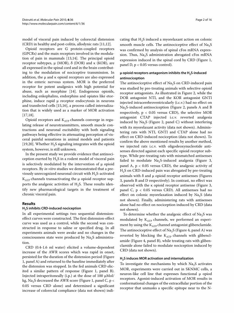
ResultsH2S inhibits CRD-induced nociceptionIn all experimental settings two sequential distension-effect curves were constructed. The first distension-effectcurve was used as a control, while the second was con-structed in response to saline or specified drug. In allexperiments animals were awake and no changes in theconsciousness state were produced by Na2S administra-tion.
CRD (0.4-1.6 ml water) elicited a volume-dependentincrease of the AWR scores which was rapid in onset,persisted for the duration of the distension period (Figure1, panel A) and returned to the baseline immediately afterthe distension was stopped. In the fed animals CRD elic-ited a similar pattern of response (Figure 1, panel B).Injected intraperitoneally (i.p.) at the dose of 100 μMol/kg, Na2S decreased the AWR score (Figure 1, panel C, p <0.05 versus CRD alone) and determined a significantincrease of colorectal compliance (data not shown) indi-
cating that H2S induced a myorelaxant action on colonicsmooth muscle cells. The antinociceptive effect of Na2Swas confirmed by analysis of spinal cFos mRNA expres-sion. Thus, Na2S administration abrogated cFos mRNAexpression induced in the spinal cord by CRD (Figure 1,panel D, p < 0.05 versus control).
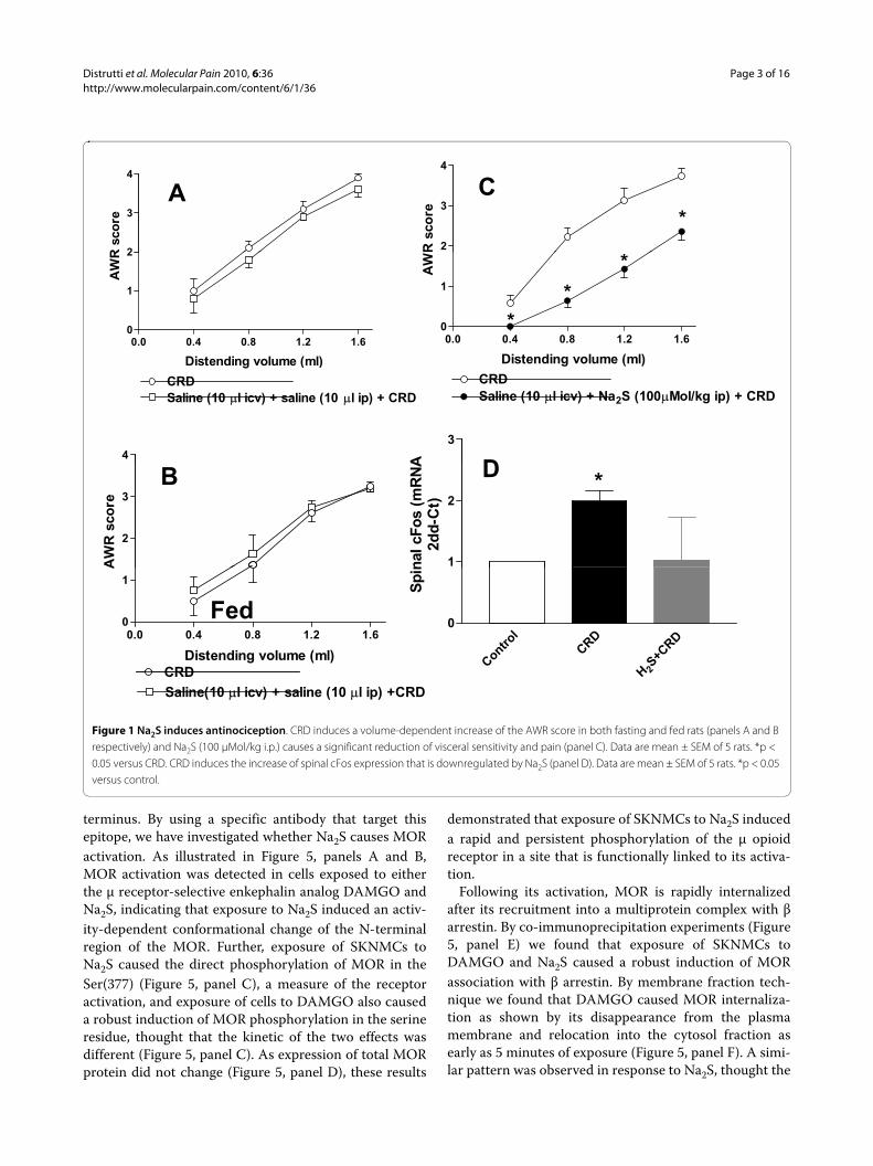
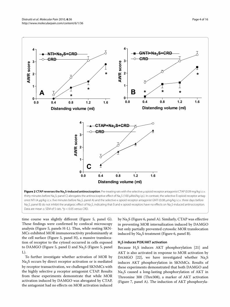
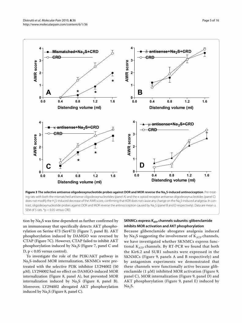
μ opioid receptors antagonism inhibits the H2S-induced antinociceptionThe antinociceptive effect of Na2S on CRD-induced painwas studied by pre-treating animals with selective opioidreceptor antagonists. As illustrated in Figure 2, while theDOR antagonist NTI, and the KOR antagonist GNTIinjected intracerebroventricularly (i.c.v.) had no effect onNa2S-induced antinociception (Figure 2, panels A and Brespectively, p < 0.05 versus CRD), the selective MORantagonist CTAP injected i.c.v. reverted analgesiainduced by Na2S (Figure 2, panel C) without interferingwith its myorelaxant activity (data not shown). Adminis-tering rats with NTI, GNTI and CTAP alone had noeffect on CRD-induced nociception (data not shown). Toconfirm the above mentioned results by another method,we injected rats i.c.v. with oligodeoxynucleotide anti-senses directed against each specific opioid receptor sub-type. While pre-treating rats with mismatched antisensesfailed to modulate Na2S-induced analgesia (Figure 3,panel A, p < 0.05 versus CRD), the analgesic activity ofH2S on CRD-induced pain was abrogated by pre-treatinganimals with δ and μ opioid receptor antisenses (Figures3, panels B and D respectively). In contrast, no effect wasobserved with the κ opioid receptor antisense (Figure 3,panel C, p < 0.05 versus CRD). All antisenses had noeffect on colonic myorelaxation induced by Na2S (datanot shown). Finally, administering rats with antisensesalone had no effect on nociception induced by CRD (datanot shown).
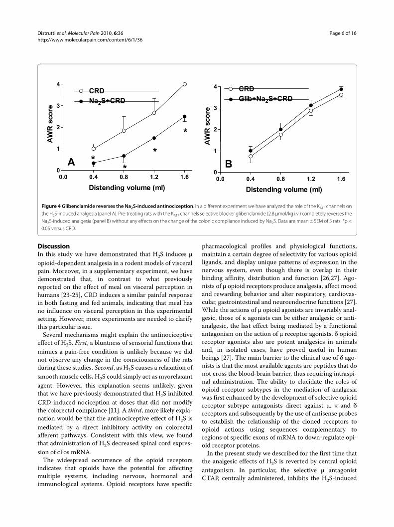
To determine whether the analgesic effect of Na2S wasmodulated by KATP channels, we performed an experi-ment by using the KATP channel antagonist glibenclamide.The antinociceptive effect of Na2S (Figure 4, panel A) wasreverted by blocking the KATP channels with glibencl-amide (Figure 4, panel B), while treating rats with gliben-clamide alone failed to modulate nociception induced byCRD (data not shown).
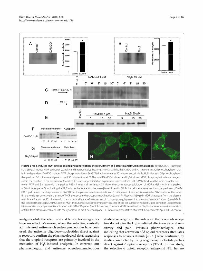
H2S induces MOR activation and internalizationTo investigate the mechanisms by which Na2S activatesMOR, experiments were carried out in SKNMC cells, aneuron-like cell line that expresses functional μ opiodreceptors. Agonist-induced activation of MOR results inconformational changes of the extracellular portion of thereceptor that unmasks a specific epitope near to the N-
Distrutti et al. Molecular Pain 2010, 6:36http://www.molecularpain.com/content/6/1/36
Page 3 of 16
terminus. By using a specific antibody that target thisepitope, we have investigated whether Na2S causes MORactivation. As illustrated in Figure 5, panels A and B,MOR activation was detected in cells exposed to eitherthe μ receptor-selective enkephalin analog DAMGO andNa2S, indicating that exposure to Na2S induced an activ-ity-dependent conformational change of the N-terminalregion of the MOR. Further, exposure of SKNMCs toNa2S caused the direct phosphorylation of MOR in theSer(377) (Figure 5, panel C), a measure of the receptoractivation, and exposure of cells to DAMGO also causeda robust induction of MOR phosphorylation in the serineresidue, thought that the kinetic of the two effects wasdifferent (Figure 5, panel C). As expression of total MORprotein did not change (Figure 5, panel D), these results
demonstrated that exposure of SKNMCs to Na2S induceda rapid and persistent phosphorylation of the μ opioidreceptor in a site that is functionally linked to its activa-tion.
Following its activation, MOR is rapidly internalizedafter its recruitment into a multiprotein complex with βarrestin. By co-immunoprecipitation experiments (Figure5, panel E) we found that exposure of SKNMCs toDAMGO and Na2S caused a robust induction of MORassociation with β arrestin. By membrane fraction tech-nique we found that DAMGO caused MOR internaliza-tion as shown by its disappearance from the plasmamembrane and relocation into the cytosol fraction asearly as 5 minutes of exposure (Figure 5, panel F). A simi-lar pattern was observed in response to Na2S, thought the
Figure 1 Na2S induces antinociception. CRD induces a volume-dependent increase of the AWR score in both fasting and fed rats (panels A and B respectively) and Na2S (100 μMol/kg i.p.) causes a significant reduction of visceral sensitivity and pain (panel C). Data are mean ± SEM of 5 rats. *p < 0.05 versus CRD. CRD induces the increase of spinal cFos expression that is downregulated by Na2S (panel D). Data are mean ± SEM of 5 rats. *p < 0.05 versus control.
A C
2
3
4
R s
co
re
2
3
4
*
R s
co
re
0.0 0.4 0.8 1.2 1.6
0
1
2
Di t di l ( l)
AW
R
0.0 0.4 0.8 1.2 1.60
1
*
*
*
Distending volume (ml)
AW
CRD
Saline (10 �l icv) + saline (10 �l ip) + CRD
Distending volume (ml)
CRD
Saline (10 �l icv) + Na2S (100�Mol/kg ip) + CRD
Distending volume (ml)
3
A
4
DB
1
2
*
nal
cF
os (
mR
NA
2d
d-C
t)
2
3
AW
R s
co
re
D
Contr
ol
CRD
H 2S+C
RD
0
Sp
in
0.0 0.4 0.8 1.2 1.60
1
CRD
Distending volume (ml)
A
Fed
H 2CRD
Saline(10 �l icv) + saline (10 �l ip) +CRD
Distrutti et al. Molecular Pain 2010, 6:36http://www.molecularpain.com/content/6/1/36
Page 4 of 16
time course was slightly different (Figure 5, panel G).These findings were confirmed by confocal microscopyanalysis (Figure 5, panels H-L). Thus, while resting SKN-MCs exhibited MOR immunoreactivity predominantly atthe cell surface (Figure 5, panel H), a massive transloca-tion of receptor to the cytosol occurred in cells exposedto DAMGO (Figure 5, panel I) and Na2S (Figure 5, panelL).
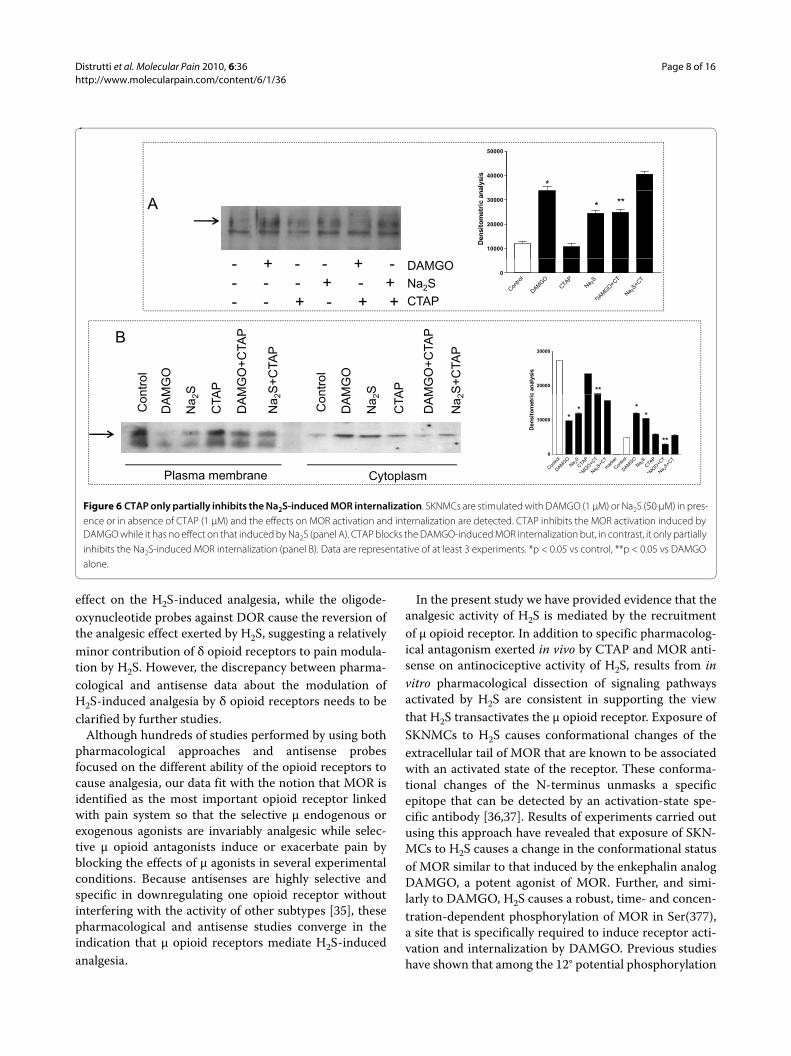
To further investigate whether activation of MOR byNa2S occurs by direct receptor activation or is mediatedby receptor transactivation, we challenged SKNMCs withthe highly selective μ receptor antagonist CTAP. Resultsfrom these experiments demonstrate that while MORactivation induced by DAMGO was abrogated by CTAP,the antagonist had no effects on MOR activation induced
by Na2S (Figure 6, panel A). Similarly, CTAP was effectivein preventing MOR internalization induced by DAMGObut only partially prevented cytosolic MOR translocationinduced by Na2S treatment (Figure 6, panel B).
H2S induces PI3K/AKT activationBecause H2S induces AKT phosphorylation [21] andAKT is also activated in response to MOR activation byDAMGO [22], we have investigated whether Na2Sinduces AKT phosphorylation in SKNMCs. Results ofthese experiments demonstrated that both DAMGO andNa2S caused a long-lasting phosphorylation of AKT inThreonine 308 (Thre308), a marker of AKT activation(Figure 7, panel A). The induction of AKT phosphoryla-
Figure 2 CTAP reverses the Na2S-induced antinociception. Pre-treating rats with the selective μ opioid receptor antagonist CTAP (0.09 mg/kg i.c.v. thirty minutes before Na2S; panel C) abrogates the antinociceptive effect of Na2S (100 μMol/kg i.p.). In contrast, the selective δ opioid receptor antag-onist NTI (4 μg/kg i.c.v. five minutes before Na2S, panel A) and the selective κ opioid receptor antagonist GNTI (0.08 μmg/kg i.c.v. three days before Na2S, panel B) do not inhibit the analgesic effect of Na2S, indicating that δ and κ opioid receptors have no effects on Na2S-induced antinociception. Data are mean ± SEM of 5 rats. *p < 0.05 versus CRD.
3
4
CRD
NTI+Na2S+CRD
re
3
4
CRD
GNTI+Na2S+CRD
re
1
2
*
*
AW
R s
co
r
1
2
* * *
*
AW
R s
co
r
A B
0.0 0.4 0.8 1.2 1.6
0 *
Distending volume (ml)
0.0 0.4 0.8 1.2 1.60
Distending volume (ml)
4
A B
2
3
4
CRD
CTAP+Na2S+CRD
WR
sco
re
0.0 0.4 0.8 1.2 1.60
1
Di t di l ( l)
AW
C
Distending volume (ml)
Distrutti et al. Molecular Pain 2010, 6:36http://www.molecularpain.com/content/6/1/36
Page 5 of 16
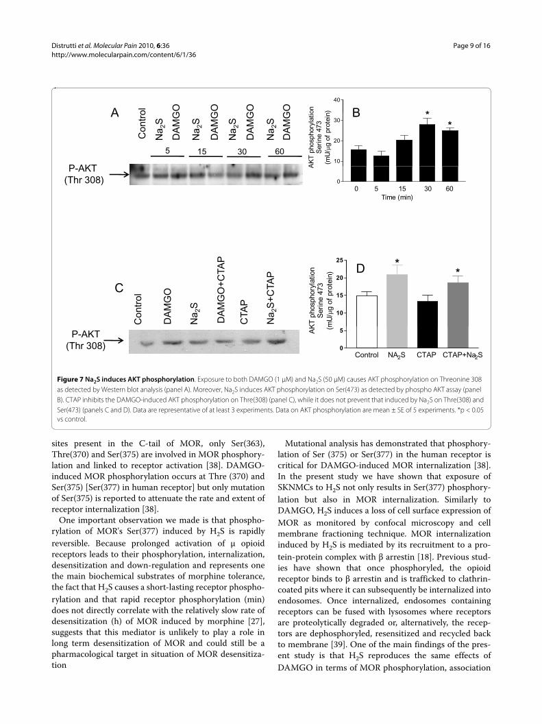
tion by Na2S was time dependent as further confirmed byan immunoassay that specifically detects AKT phospho-rylation on Serine 473 (Ser473) (Figure 7, panel B). AKTphosphorylation induced by DAMGO was reversed byCTAP (Figure 7C). However, CTAP failed to inhibit AKTphosphorylation induced by Na2S (Figure 7, panel C andD, p < 0.05 versus control).
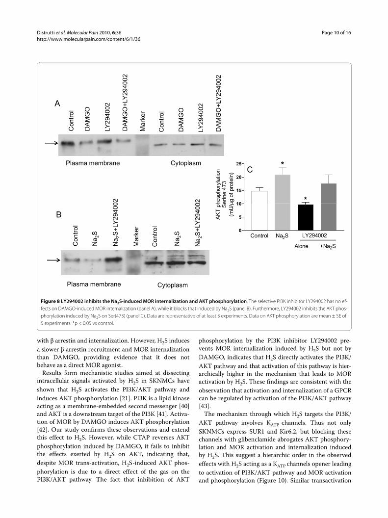
To investigate the role of the PI3K/AKT pathway inNa2S-induced MOR internalization, SKNMCs were pre-treated with the selective PI3K inhibitor LY294002 (50μM). LY294002 had no effect on DAMGO-induced MORinternalization (Figure 8, panel A), but prevented MORinternalization induced by Na2S (Figure 8, panel B).Moreover, LY294002 abrogated AKT phosphorylationinduced by Na2S (Figure 8, panel C).
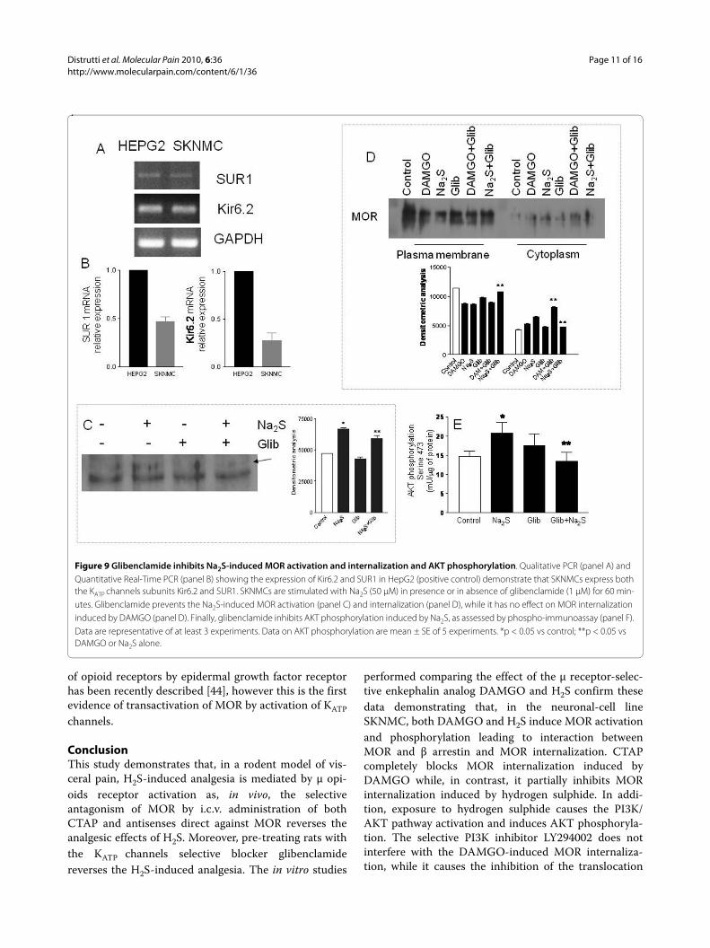
SKNMCs express KATP channels subunits: glibenclamide inhibits MOR activation and AKT phosphorylationBecause glibenclamide abrogates analgesia inducedby Na2S suggesting the involvement of KATP channels,we have investigated whether SKNMCs express func-tional KATP channels. By RT-PCR we found that boththe Kir6.2 and SUR1 subunits were expressed in theSKNMCs (Figure 9, panels A and B respectively) andby antagonism experiments we demonstrated thatthese channels were functionally active because glib-enclamide (1 μM) inhibited MOR activation (Figure 9,panel C), MOR internalization (Figure 9, panel D) andAKT phosphorylation (Figure 9, panel E) induced byNa2S.
Figure 3 The selective antisense oligodeoxynucleotide probes against DOR and MOR reverse the Na2S-induced antinociception. Pre-treat-ing rats with both the mismatched antisense oligodeoxynucleotides (panel A) and the κ opioid receptor antisense oligodeoxynucleotides (panel C) does not modify the H2S-induced decrease of the AWR score, confirming that KOR does not cause any change on the Na2S-induced analgesia. In con-trast, oligodeoxynucleotide probes against DOR and MOR reverse the antinociception caused by Na2S (panel B and D respectively). Data are mean ± SEM of 5 rats. *p < 0.05 versus CRD.
3
4
CRD
Mismatched+Na2S+CRD
sco
re
3
4
CRD
� antisense+Na2S+CRD
sco
re
0 0 0 4 0 8 1 2 1 6
0
1
2
* *
*AW
R s
0 0 0 0 8 1 2 1 60
1
2
AW
R s
A B0.0 0.4 0.8 1.2 1.6
Distending volume (ml)
0.0 0.4 0.8 1.2 1.6
Distending volume (ml)
4
� antisense+Na2S+CRD
4
CRD
� antisense+Na2S+CRD
1
2
3 CRD
*
*AW
R s
co
re
1
2
3
CRDA
WR
sco
re
0.0 0.4 0.8 1.2 1.60
1 *
Distending volume (ml)
0.0 0.4 0.8 1.2 1.60
1
Distending volume (ml)
C D
Distrutti et al. Molecular Pain 2010, 6:36http://www.molecularpain.com/content/6/1/36
Page 6 of 16
DiscussionIn this study we have demonstrated that H2S induces μopioid-dependent analgesia in a rodent models of visceralpain. Moreover, in a supplementary experiment, we havedemonstrated that, in contrast to what previouslyreported on the effect of meal on visceral perception inhumans [23-25], CRD induces a similar painful responsein both fasting and fed animals, indicating that meal hasno influence on visceral perception in this experimentalsetting. However, more experiments are needed to clarifythis particular issue.
Several mechanisms might explain the antinociceptiveeffect of H2S. First, a bluntness of sensorial functions thatmimics a pain-free condition is unlikely because we didnot observe any change in the consciousness of the ratsduring these studies. Second, as H2S causes a relaxation ofsmooth muscle cells, H2S could simply act as myorelaxantagent. However, this explanation seems unlikely, giventhat we have previously demonstrated that H2S inhibitedCRD-induced nociception at doses that did not modifythe colorectal compliance [11]. A third, more likely expla-nation would be that the antinociceptive effect of H2S ismediated by a direct inhibitory activity on colorectalafferent pathways. Consistent with this view, we foundthat administration of H2S decreased spinal cord expres-sion of cFos mRNA.
The widespread occurrence of the opioid receptorsindicates that opioids have the potential for affectingmultiple systems, including nervous, hormonal andimmunological systems. Opioid receptors have specific
pharmacological profiles and physiological functions,maintain a certain degree of selectivity for various opioidligands, and display unique patterns of expression in thenervous system, even though there is overlap in theirbinding affinity, distribution and function [26,27]. Ago-nists of μ opioid receptors produce analgesia, affect moodand rewarding behavior and alter respiratory, cardiovas-cular, gastrointestinal and neuroendocrine functions [27].While the actions of μ opioid agonists are invariably anal-gesic, those of κ agonists can be either analgesic or anti-analgesic, the last effect being mediated by a functionalantagonism on the action of μ receptor agonists. δ opioidreceptor agonists also are potent analgesics in animalsand, in isolated cases, have proved useful in humanbeings [27]. The main barrier to the clinical use of δ ago-nists is that the most available agents are peptides that donot cross the blood-brain barrier, thus requiring intraspi-nal administration. The ability to elucidate the roles ofopioid receptor subtypes in the mediation of analgesiawas first enhanced by the development of selective opioidreceptor subtype antagonists direct against μ, κ and δreceptors and subsequently by the use of antisense probesto establish the relationship of the cloned receptors toopioid actions using sequences complementary toregions of specific exons of mRNA to down-regulate opi-oid receptor proteins.
In the present study we described for the first time thatthe analgesic effects of H2S is reverted by central opioidantagonism. In particular, the selective μ antagonistCTAP, centrally administered, inhibits the H2S-induced
Figure 4 Glibenclamide reverses the Na2S-induced antinociception. In a different experiment we have analyzed the role of the KATP channels on the H2S-induced analgesia (panel A). Pre-treating rats with the KATP channels selective blocker glibenclamide (2.8 μmol/kg i.v.) completely reverses the Na2S-induced analgesia (panel B) without any effects on the change of the colonic compliance induced by Na2S. Data are mean ± SEM of 5 rats. *p < 0.05 versus CRD.
4
CRD4 CRD
Glib+N S+CRD
2
3Na2S+CRD
AW
R s
co
re
* 2
3
Glib+Na2S+CRD
AW
R s
co
re
B0.0 0.4 0.8 1.2 1.6
0
1
Distending ol me (ml)
A
*
**
0.0 0.4 0.8 1.2 1.6
0
1
A
A
Distending volume (ml) Distending volume (ml)
Distrutti et al. Molecular Pain 2010, 6:36http://www.molecularpain.com/content/6/1/36
Page 7 of 16
analgesia while the selective κ and δ receptor antagonistshave no effect. Moreover, when the selective, centrallyadministered antisense olygodeoxynucleotides have beenused, the antisense oligodeoxynucleotides direct againstμ receptors confirm the pharmacological data, suggestingthat the μ opioid receptors are primarily involved in themediation of H2S-induced analgesia. In contrast, ourpharmacological and antisense oligodeoxynucleotides
studies converge onto the indication that κ opioids recep-tors do not alter the H2S-mediated effects on visceral sen-sitivity and pain. Previous pharmacological dataindicating that activation of δ opioid receptors attenuatesresponses to noxious stimuli [28-31] were confirmed bystudies conducted by using olygodeoxynucleotide probesdirect against δ opioids receptors [32-34]. In our study,the selective δ opioid receptor antagonist NTI has no
Figure 5 Na2S induces MOR activation and phosphorylation, the recruitment of β arrestin and MOR internalization. Both DAMGO (1 μM) and Na2S (50 μM) induce MOR activation (panel A and B respectively). Treating SKNMCs with both DAMGO and Na2S results in MOR phosphorylation that is time-dependent. DAMGO induces MOR phosphorylation at Ser(377) that is maximal at 30 minutes and, similarly, H2S induces MOR phosphorylation that peaks at 3-6 minutes and persists until 30 minutes (panel C). The total DAMGO-induced and H2S-induced MOR phosphorylation is unchanged within the duration of the experiment (panel D). Co-immunoprecipitation experiments demonstrate that DAMGO induces the rapid complex be-tween MOR and β arrestin with the peak at 5-15 minutes and, similarly, H2S induces the co-immunoprecipitation of MOR and β arrestin that peaked at 30 minutes (panel E), indicating that H2S induces the interaction between β arrestin and MOR. At the cell membrane fractioning experiments, DAM-GO (1 μM) causes the disappearance of MOR from the plasma membrane fraction at 5 minutes and this effect is maximal at 60 minutes. At the same time there is a progressive increment of MOR presence in the cytoplasmatic fraction (panel F). After Na2S (50 μM), MOR disappears from the plasma membrane fraction at 30 minutes with the maximal effect at 60 minutes and, in contemporary, it passes into the cytoplasmatic fraction (panel G). At the confocal microscopy SKNMCs exhibit MOR immunoreactivity predominantly localized at the cell surface in nonstimulated condition (panel H) and it translocates to cytoplasm after activation with DAMGO (panel I), which is known to induce MOR internalization. Na2S induces a massive translocation of MOR from plasma membrane into the cytoplasm in most neurons (panel L). Data are representative of at least 3 experiments. *p < 0.05 vs control.
A
Con
trol
DA
MG
O
Con
trol
Na 2
S
B50000
75000
*
etr
ic a
naly
sis
40000
50000
60000
*
ric a
naly
sis
Control Na2S0
25000
Den
sit
om
e
0
10000
20000
30000
Den
sit
om
etr
Con
trol
3� 6� 9� 30�15� 3� 6� 9� 30�15�
DAMGO 1 μM Na2S 50 μM
pMOR Ser(377)C
Control Na2SControl DAMGO
0
pMOR Ser(377)
MOR
C
D
Con
trol
5 15 30 5 15 30DAMGO 1 μM Na2S 50 μM
E
5� 15� 30� 60�- 5� 15� 30� 60�-
DAMGO 1 �M
Plasma membrane CytoplasmF
5� 15� 30� 60�-5� 15� 30� 60�-Na2S 50 �M
Plasma membrane Cytoplasm
H I L
G
Distrutti et al. Molecular Pain 2010, 6:36http://www.molecularpain.com/content/6/1/36
Page 8 of 16
effect on the H2S-induced analgesia, while the oligode-oxynucleotide probes against DOR cause the reversion ofthe analgesic effect exerted by H2S, suggesting a relativelyminor contribution of δ opioid receptors to pain modula-tion by H2S. However, the discrepancy between pharma-cological and antisense data about the modulation ofH2S-induced analgesia by δ opioid receptors needs to beclarified by further studies.
Although hundreds of studies performed by using bothpharmacological approaches and antisense probesfocused on the different ability of the opioid receptors tocause analgesia, our data fit with the notion that MOR isidentified as the most important opioid receptor linkedwith pain system so that the selective μ endogenous orexogenous agonists are invariably analgesic while selec-tive μ opioid antagonists induce or exacerbate pain byblocking the effects of μ agonists in several experimentalconditions. Because antisenses are highly selective andspecific in downregulating one opioid receptor withoutinterfering with the activity of other subtypes [35], thesepharmacological and antisense studies converge in theindication that μ opioid receptors mediate H2S-inducedanalgesia.
In the present study we have provided evidence that theanalgesic activity of H2S is mediated by the recruitmentof μ opioid receptor. In addition to specific pharmacolog-ical antagonism exerted in vivo by CTAP and MOR anti-sense on antinociceptive activity of H2S, results from invitro pharmacological dissection of signaling pathwaysactivated by H2S are consistent in supporting the viewthat H2S transactivates the μ opioid receptor. Exposure ofSKNMCs to H2S causes conformational changes of theextracellular tail of MOR that are known to be associatedwith an activated state of the receptor. These conforma-tional changes of the N-terminus unmasks a specificepitope that can be detected by an activation-state spe-cific antibody [36,37]. Results of experiments carried outusing this approach have revealed that exposure of SKN-MCs to H2S causes a change in the conformational statusof MOR similar to that induced by the enkephalin analogDAMGO, a potent agonist of MOR. Further, and simi-larly to DAMGO, H2S causes a robust, time- and concen-tration-dependent phosphorylation of MOR in Ser(377),a site that is specifically required to induce receptor acti-vation and internalization by DAMGO. Previous studieshave shown that among the 12° potential phosphorylation
Figure 6 CTAP only partially inhibits the Na2S-induced MOR internalization. SKNMCs are stimulated with DAMGO (1 μM) or Na2S (50 μM) in pres-ence or in absence of CTAP (1 μM) and the effects on MOR activation and internalization are detected. CTAP inhibits the MOR activation induced by DAMGO while it has no effect on that induced by Na2S (panel A). CTAP blocks the DAMGO-induced MOR internalization but, in contrast, it only partially inhibits the Na2S-induced MOR internalization (panel B). Data are representative of at least 3 experiments. *p < 0.05 vs control, **p < 0.05 vs DAMGO alone.
40000
50000
*
aly
sis
A
CTAP10000
20000
30000
* **
Den
sit
om
etr
ic a
na
DAMGONa2SCTAP
- + - - + -- - - + - +- - + - + +
Contro
l
DAMGO
CTAPNa 2
S
DAMGO+CT
Na 2S+C
T0
ntro
l
MG
O
S AP
2S+C
TAP
MG
O
2S AP
2S+C
TAP
MG
O+C
TAP
MG
O+C
TAP
ntro
lB
20000
30000
c a
naly
sis
**
Con
DA
M
Na 2
CTA
Na 2
DA
M
Na 2
CTA
Na 2
DA
M
DA
M
Con
ntrol
MGO
a 2S
TAP+C
T+CT
rker
ntrol
MGO
a 2S
TAP+CT
+CT
0
10000 ** *
*
**
Den
sit
om
etr
ic
Plasma membrane CytoplasmCon
tr
DAMG
Na 2CTA
DAMGO+C
Na 2S+C
mark
Contr
DAMG
Na 2CTA
DAMGO+C
Na 2S+C
Distrutti et al. Molecular Pain 2010, 6:36http://www.molecularpain.com/content/6/1/36
Page 9 of 16
sites present in the C-tail of MOR, only Ser(363),Thre(370) and Ser(375) are involved in MOR phosphory-lation and linked to receptor activation [38]. DAMGO-induced MOR phosphorylation occurs at Thre (370) andSer(375) [Ser(377) in human receptor] but only mutationof Ser(375) is reported to attenuate the rate and extent ofreceptor internalization [38].
One important observation we made is that phospho-rylation of MOR's Ser(377) induced by H2S is rapidlyreversible. Because prolonged activation of μ opioidreceptors leads to their phosphorylation, internalization,desensitization and down-regulation and represents onethe main biochemical substrates of morphine tolerance,the fact that H2S causes a short-lasting receptor phospho-rylation and that rapid receptor phosphorylation (min)does not directly correlate with the relatively slow rate ofdesensitization (h) of MOR induced by morphine [27],suggests that this mediator is unlikely to play a role inlong term desensitization of MOR and could still be apharmacological target in situation of MOR desensitiza-tion
Mutational analysis has demonstrated that phosphory-lation of Ser (375) or Ser(377) in the human receptor iscritical for DAMGO-induced MOR internalization [38].In the present study we have shown that exposure ofSKNMCs to H2S not only results in Ser(377) phosphory-lation but also in MOR internalization. Similarly toDAMGO, H2S induces a loss of cell surface expression ofMOR as monitored by confocal microscopy and cellmembrane fractioning technique. MOR internalizationinduced by H2S is mediated by its recruitment to a pro-tein-protein complex with β arrestin [18]. Previous stud-ies have shown that once phosphoryled, the opioidreceptor binds to β arrestin and is trafficked to clathrin-coated pits where it can subsequently be internalized intoendosomes. Once internalized, endosomes containingreceptors can be fused with lysosomes where receptorsare proteolytically degraded or, alternatively, the recep-tors are dephosphoryled, resensitized and recycled backto membrane [39]. One of the main findings of the pres-ent study is that H2S reproduces the same effects ofDAMGO in terms of MOR phosphorylation, association
Figure 7 Na2S induces AKT phosphorylation. Exposure to both DAMGO (1 μM) and Na2S (50 μM) causes AKT phosphorylation on Threonine 308 as detected by Western blot analysis (panel A). Moreover, Na2S induces AKT phosphorylation on Ser(473) as detected by phospho AKT assay (panel B). CTAP inhibits the DAMGO-induced AKT phosphorylation on Thre(308) (panel C), while it does not prevent that induced by Na2S on Thre(308) and Ser(473) (panels C and D). Data are representative of at least 3 experiments. Data on AKT phosphorylation are mean ± SE of 5 experiments. *p < 0.05 vs control.
A40
*on ) Bol GO
GO
GO
GOA
10
20
30*
*
AK
T p
hosp
hory
latio
Ser
ine
473
(mU
/�g
of p
rote
in B
5 15 30 60C
ontr
o
Na 2
S
DA
MG
Na 2
S
DA
MG
Na 2
S
DA
MG
Na 2
S
DA
MG
P AKT0
0 5 15 30 60Time (min)
AP-AKT(Thr 308)
TAP
P D25 *
*on )
DA
MG
O+C
T
Con
trol
DA
MG
O
Na 2
S
CTA
P
Na 2
S+C
TAP
C
10
15
20
KT
pho
spho
ryla
tioS
erin
e 47
3m
U/�
g of
pro
tein
)P-AKT
(Thr 308)Control NA2S CTAP CTAP+Na2S
0
5AK (m
Distrutti et al. Molecular Pain 2010, 6:36http://www.molecularpain.com/content/6/1/36
Page 10 of 16
with β arrestin and internalization. However, H2S inducesa slower β arrestin recruitment and MOR internalizationthan DAMGO, providing evidence that it does notbehave as a direct MOR agonist.
Results form mechanistic studies aimed at dissectingintracellular signals activated by H2S in SKNMCs haveshown that H2S activates the PI3K/AKT pathway andinduces AKT phosphorylation [21]. PI3K is a lipid kinaseacting as a membrane-embedded second messenger [40]and AKT is a downstream target of the PI3K [41]. Activa-tion of MOR by DAMGO induces AKT phosphorylation[42]. Our study confirms these observations and extendthis effect to H2S. However, while CTAP reverses AKTphosphorylation induced by DAMGO, it fails to inhibitthe effects exerted by H2S on AKT, indicating that,despite MOR trans-activation, H2S-induced AKT phos-phorylation is due to a direct effect of the gas on thePI3K/AKT pathway. The fact that inhibition of AKT
phosphorylation by the PI3K inhibitor LY294002 pre-vents MOR internalization induced by H2S but not byDAMGO, indicates that H2S directly activates the PI3K/AKT pathway and that activation of this pathway is hier-archically higher in the mechanism that leads to MORactivation by H2S. These findings are consistent with theobservation that activation and internalization of a GPCRcan be regulated by activation of the PI3K/AKT pathway[43].
The mechanism through which H2S targets the PI3K/AKT pathway involves KATP channels. Thus not onlySKNMCs express SUR1 and Kir6.2, but blocking thesechannels with glibenclamide abrogates AKT phosphory-lation and MOR activation and internalization inducedby H2S. This suggest a hierarchic order in the observedeffects with H2S acting as a KATP channels opener leadingto activation of PI3K/AKT pathway and MOR activationand phosphorylation (Figure 10). Similar transactivation
Figure 8 LY294002 inhibits the Na2S-induced MOR internalization and AKT phosphorylation. The selective PI3K inhibitor LY294002 has no ef-fects on DAMGO-induced MOR internalization (panel A), while it blocks that induced by Na2S (panel B). Furthermore, LY294002 inhibits the AKT phos-phorylation induced by Na2S on Ser(473) (panel C). Data are representative of at least 3 experiments. Data on AKT phosphorylation are mean ± SE of 5 experiments. *p < 0.05 vs control.
Y29
4002
Y29
4002
Con
trol
DA
MG
O
LY29
4002
DA
MG
O+L
Y
Con
trol
DA
MG
O
LY29
4002
DA
MG
O+L
Y
Mar
ker
A
25 *CPlasma membrane Cytoplasm
10
15
20
*hosp
hory
latio
ner
ine
473
� g o
f pr
otei
n)
C
02 02
Control Na2S0
5
10
LY294002
AK
T p
hS
e(m
U/�
B
ontr
ol
a 2S
a 2S
+LY
2940
0
ontr
ol
a 2S
a 2S
+LY
2940
0
arke
r
Alone +Na2SCo
Na
Na
Co
Na
Na
Ma
Plasma membrane Cytoplasm
Distrutti et al. Molecular Pain 2010, 6:36http://www.molecularpain.com/content/6/1/36
Page 11 of 16
of opioid receptors by epidermal growth factor receptorhas been recently described [44], however this is the firstevidence of transactivation of MOR by activation of KATPchannels.
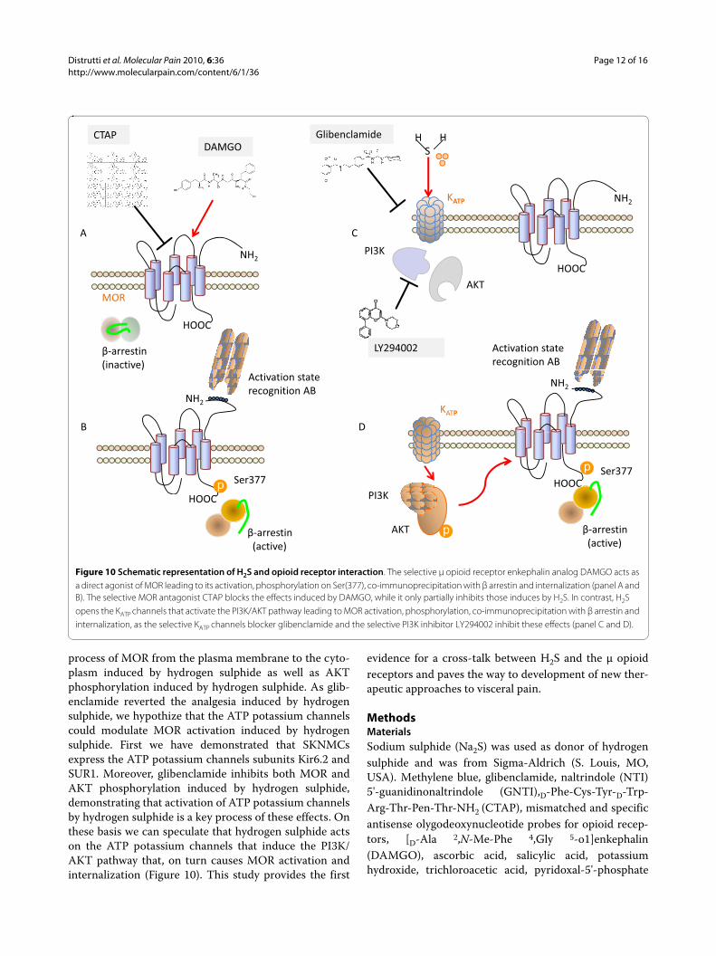
ConclusionThis study demonstrates that, in a rodent model of vis-ceral pain, H2S-induced analgesia is mediated by μ opi-oids receptor activation as, in vivo, the selectiveantagonism of MOR by i.c.v. administration of bothCTAP and antisenses direct against MOR reverses theanalgesic effects of H2S. Moreover, pre-treating rats withthe KATP channels selective blocker glibenclamidereverses the H2S-induced analgesia. The in vitro studies
performed comparing the effect of the μ receptor-selec-tive enkephalin analog DAMGO and H2S confirm thesedata demonstrating that, in the neuronal-cell lineSKNMC, both DAMGO and H2S induce MOR activationand phosphorylation leading to interaction betweenMOR and β arrestin and MOR internalization. CTAPcompletely blocks MOR internalization induced byDAMGO while, in contrast, it partially inhibits MORinternalization induced by hydrogen sulphide. In addi-tion, exposure to hydrogen sulphide causes the PI3K/AKT pathway activation and induces AKT phosphoryla-tion. The selective PI3K inhibitor LY294002 does notinterfere with the DAMGO-induced MOR internaliza-tion, while it causes the inhibition of the translocation
Figure 9 Glibenclamide inhibits Na2S-induced MOR activation and internalization and AKT phosphorylation. Qualitative PCR (panel A) and Quantitative Real-Time PCR (panel B) showing the expression of Kir6.2 and SUR1 in HepG2 (positive control) demonstrate that SKNMCs express both the KATP channels subunits Kir6.2 and SUR1. SKNMCs are stimulated with Na2S (50 μM) in presence or in absence of glibenclamide (1 μM) for 60 min-utes. Glibenclamide prevents the Na2S-induced MOR activation (panel C) and internalization (panel D), while it has no effect on MOR internalization induced by DAMGO (panel D). Finally, glibenclamide inhibits AKT phosphorylation induced by Na2S, as assessed by phospho-immunoassay (panel F). Data are representative of at least 3 experiments. Data on AKT phosphorylation are mean ± SE of 5 experiments. *p < 0.05 vs control; **p < 0.05 vs DAMGO or Na2S alone.
Distrutti et al. Molecular Pain 2010, 6:36http://www.molecularpain.com/content/6/1/36
Page 12 of 16
process of MOR from the plasma membrane to the cyto-plasm induced by hydrogen sulphide as well as AKTphosphorylation induced by hydrogen sulphide. As glib-enclamide reverted the analgesia induced by hydrogensulphide, we hypothize that the ATP potassium channelscould modulate MOR activation induced by hydrogensulphide. First we have demonstrated that SKNMCsexpress the ATP potassium channels subunits Kir6.2 andSUR1. Moreover, glibenclamide inhibits both MOR andAKT phosphorylation induced by hydrogen sulphide,demonstrating that activation of ATP potassium channelsby hydrogen sulphide is a key process of these effects. Onthese basis we can speculate that hydrogen sulphide actson the ATP potassium channels that induce the PI3K/AKT pathway that, on turn causes MOR activation andinternalization (Figure 10). This study provides the first
evidence for a cross-talk between H2S and the μ opioidreceptors and paves the way to development of new ther-apeutic approaches to visceral pain.
MethodsMaterialsSodium sulphide (Na2S) was used as donor of hydrogensulphide and was from Sigma-Aldrich (S. Louis, MO,USA). Methylene blue, glibenclamide, naltrindole (NTI)5'-guanidinonaltrindole (GNTI),D-Phe-Cys-Tyr-D-Trp-Arg-Thr-Pen-Thr-NH2 (CTAP), mismatched and specificantisense olygodeoxynucleotide probes for opioid recep-tors, [D-Ala 2,N-Me-Phe 4,Gly 5-o1]enkephalin(DAMGO), ascorbic acid, salicylic acid, potassiumhydroxide, trichloroacetic acid, pyridoxal-5'-phosphate
Figure 10 Schematic representation of H2S and opioid receptor interaction. The selective μ opioid receptor enkephalin analog DAMGO acts as a direct agonist of MOR leading to its activation, phosphorylation on Ser(377), co-immunoprecipitation with β arrestin and internalization (panel A and B). The selective MOR antagonist CTAP blocks the effects induced by DAMGO, while it only partially inhibits those induces by H2S. In contrast, H2S opens the KATP channels that activate the PI3K/AKT pathway leading to MOR activation, phosphorylation, co-immunoprecipitation with β arrestin and internalization, as the selective KATP channels blocker glibenclamide and the selective PI3K inhibitor LY294002 inhibit these effects (panel C and D).
H�������HS
GlibenclamideDAMGO
CTAP
NH2KATP
A C
HOOC
PI3K
AKT
NH2
MOR
LY294002 Activation�staterecognition�AB
HOOC
��arrestin(inactive)
NH2
( )
NH2
Activation�staterecognition�AB
B D
KATP
HOOCPI3K
Ser377Ser377
PI3K
AKT ��arrestin(active)
HOOC
��arrestin(active)
Distrutti et al. Molecular Pain 2010, 6:36http://www.molecularpain.com/content/6/1/36
Page 13 of 16
and calmodulin were from Sigma-Aldrich (S. Louis, MO,USA). Tissue Protein Extraction Reagent (T-PER) wasobtained by Pierce Biotechnology (Rockford, IL, USA).
In vivo experimentsAnimalsMale, Wistar rats (200-250 g, Charles River, Monza, Italy)were housed in plastic cages and maintained under con-trolled conditions with 12-hour light/dark cycles (lightson at 07.00). Tap water and standard laboratory chowwere freely available (Additional file 1). It has been dem-onstrated that the nutrients induce an enhancement ofthe colorectal sensitivity in both healthy subjects [23] andIBS patients [24,25]. To avoid the influence of the meal oncolorectal perception and pain, food was withdrawn 12hours before surgical procedures and CRD recordings inall in vivo experiments [11,12]. However, to verifywhether meal could influence the perception of CRD-induced visceral pain, we performed a supplementaryexperiment on fed rats (n = 5). Experimental procedureswere approved by our institutional animal research com-mittees and were in accordance with nationally approvedguidelines for the treatment of laboratory animals.Surgical procedureRat were anesthetized by an i.p. injection of 70 mg/kgpenthotal and were then mounted in a stereotaxic instru-ment. To perform the i.c.v. injection, a guide cannula(Alzet Brain Infusion Kit II, 3-5 mm) was inserted stereo-taxically into the right lateral cerebral ventricle. The ste-reotaxic coordinates were 1,6 mm right laterally and 0,8mm dorsoventrally from the bregma and 3,5 mm belowthe dura. Drugs dissolved in 10 μl saline were injectedinto the cerebral ventricle by insertion of an injectioncannula (28 gauge stainless steel tube) connected to acatheter tube into the guide cannula which was con-nected to a syringe. In each injection 10 μl of vehicle ordrugs were delivered manually into the ventricle over 3min. At the end of each experiment, methylene blue solu-tion was injected through the injection cannula to verifyits correct placement in the right lateral ventricle. Ratsexhibiting motor deficits after the surgical procedurewere not used in the subsequent experiments.CRD and behavioral testingAll experiments began 1 week after the surgical proce-dure. Distending procedure were performed as previouslydescribed (Additional file 2). The behavioral response toCRD was assessed by measuring the abdominal with-drawal reflex (AWR) as previously described [45,46](Additional file 2).Effects of H2S on colonic nociceptionThe control group (n = 5) consisted of fasting rats thatunderwent surgical procedures but not CRD, while theCRD group consisted of fasting rats that underwent sur-gical procedures and two sets of CRD, the first acting as
control. To investigate whether H2S administration mod-ulates sensitivity and pain induced by CRD, rats weretreated i.p. with vehicle (CRD group) or Na2S, an H2Sdonor, at the dose of 100 μMol/kg five minutes beforeCRD.Effects of the opioid and KATP channels inhibitorsThe role of the δ, κ and μ opioid receptors in the H2S-induced antinociception was investigated by pre-treatingrats with selective opioid receptor antagonists adminis-tered at final volume of 10 μl i.c.v.: NTI, a δ opioid recep-tor antagonist (4 μg/kg), was injected 5 minutes beforeNa2S [47]; GNTI, a κ opioid receptor antagonist (0.08mg/kg), was administered three days before Na2S [48];CTAP, a μ opioid receptor antagonist (0.09 mg/kg), wasadministered 30 minutes before Na2S [49]. Controlexperiments were performed by injecting rats with NTI,GNTI and CTAP alone (n = 5 rats/group).
For antisense experiments rats were pretreated withantisense oligodeoxynucleotides direct against specific
Table 1: Primer used for antisense experiments
Probe sequence Probe sequence
DOR-1 opioid receptor clone
Exon 1 AS TGT CCG TCT CCA CCG TGC
Exon 2 AS ATC AAG TAC TTG GCG CTC TG
Exon 3 AS AAC ACG CAG ATC TTG GTC AC
KOR-1 opioid receptor clone
Exon 1 AS GCT GCT GAT CCT CTG AGC CCA
Exon 2 AS CCA AAG CAT CTG CCA AAG CCA
Exon 3 AS GGC GCA GGA TCA TCA GGG TGT
MOR-1 opioid receptor clone
Exon 1 AS CGC CCC AGC CTC TTC CTC T
Exon 2 AS TTG GTG GCA GTC TTC ATT TTG G
Exon 3 AS TGA GCA GGT TCT CCC AGT ACC A
Exon 4 AS GGG CAA TGG AGC AGT TTC TG
Mismatch CGC CCC GAC CTC TTC CCT T
Distrutti et al. Molecular Pain 2010, 6:36http://www.molecularpain.com/content/6/1/36
Page 14 of 16
exons of DOR, KOR and MOR. A mismatched antisensewas used as control (Table 1). All antisense olygodeoxy-nucleotides were administered i.c.v. in dose of 10 μg in 10μl volume saline [50,52,53]. Treatment with antisenseswas performed on day 1, 3 and 5 and the behavioral testwas performed at day 6 [51] (Additional file 3).
The involvement of KATP channels in the analgesiceffects of H2S was assessed by pre-treating rats with glib-enclamide, a selective KATP channel blocker, at a dose of2.8 μmol/kg injected intravenously (i.v.) for 20 minutesbefore Na2S administration [11,12] (Additional file 4).
At the end of the CRD procedures, rats were sacrificedand spinal cords (L1-L5) collected for RT-PCR analysis ofcFOS [54] (additional file 5) using the following sense andantisense primers: gtctggttccttctatgcag and taggtagtg-cagctgggagt.
In vitro experimentsThe immortalized human neuronal SKNMCs were usedfor in vitro studies. Cells were grown in Minimum Essen-tial Medium with Earl's salts supplemented with 10%FBS, L-glutamine, penicillin and streptomycin, and regu-larly passaged to maintain exponential growth.
For in vitro studies DAMGO was used at the dose of 1μM and Na2S at the dose of 50 μM. To determine whetherH2S induces MOR activation, SKNMCs were stimulatedwith DAMGO or Na2S and MOR activation detected byWestern blot analysis using a specific antibody raisedagainst a specific epitope in the N-terminus of the recep-tor that becomes exposed in response to conformationalchanges induced by receptor activation [55]. This activa-tion-state specific antibody exhibits enhanced recogni-tion of activated receptor [36,37]. In addition, activationof MOR by H2S was detected by Western blot analysis ofreceptor phosphorylation on Serine (Ser) (377). Finally,because MOR activation results in receptor recruitmentto β arrestin, co-immunoprecipitation experiments wereperformed to investigate whether H2S induces the forma-tion of a protein-protein complex between MOR and βarrestin (Additional file 6).Effect of H2S on MOR internalizationTo investigate whether exposure of SKNMCs to H2Sinduces MOR internalization, cells were treated with theμ receptor-selective enkephalin analog DAMGO [15,16]and Na2S alone or in combination with the MOR antago-nist CTAP. Internalization of the receptor was assessed byWestern blot analysis by measuring its translocation fromthe cell membrane fraction to the cytosol and by confocalmicroscopy (Nikon) using a specific anti-MOR immuno-fluorescent antibody (Additional file 7).Effect of H2S on AKT phosphorylationSKNMCs were exposed to DAMGO and Na2S up to 60minutes and Western blot analysis performed on whole
cell lysates using a specific antibody that detected thephosphorylated form of AKT on Thre(308). AKT phos-phorylation was also detected by measuring the AKTphosphorylated form on Ser(473) (phospho-AKT ELISAKIT, Biosource).
Activation of the PI3K/AKT pathway was tested byexposing SKNMCs to the selective PI3K inhibitorLY294002 (50 μM) in the presence of DAMGO or Na2S(Additional file 8).Effect of KATP channels blockadeExpression of KATP channels in SKNMCs was evaluatedby assessing the expression of Kir6.2 and SUR1 sub-units(Additional file 9). Qualitative and quantitative PCR wereperformed by using the following sense and antisenseprimers: hGAPDH: gaaggtgaaggtcggagt and catgggtg-gaatcatattggaa; hSUR.1: gtccagatcatgggaggcta and cagaa-gacagcccctgagac; hKir6.2: gtcaccagcatccactcctt andggggacttcaaatgttgcat. The effects of glibenclamide (1 μM)on AKT phosphorylation and MOR activation and inter-nalization were determined (Additional file 9).Densitometric analysisAll the densitometric analysis have been performed byusing the Image J software.Statistical analysisBehavioral data are presented as mean ± SE, with samplesizes of at least 5 rats per group. Statistical comparisonsof unpaired data were performed by the Mann-Whitneytest, while statistical comparisons of paired data wereperformed by the Wilcoxon signed rank test. Densito-metric data have been analyzed with Turkey's multiplecomparison test. Data on AKT phosphorylation are pre-sented as mean ± SE, with sample sizes of at least 5 exper-iments per group. An associated probability (p value) ofless that 5% was considered significant.
Additional material
Additional file 1 Animals. This file describes the animals used.Additional file 2 CRD and behavioral testing. This file describes the behavioral testing used in the in vivo studies.Additional file 3 Effects of the opioid receptors antagonism. This file describes the methods used for blocking the opioid receptors.Additional file 4 Effects of KATP channels. This file describes the method used for blocking the KATP channels.
Additional file 5 Spinal cFOS expression. This file describes the method used for determining spinal cFos expression.Additional file 6 Effect of H2S on MOR. This file describes the methods used to determine MOR activationAdditional file 7 Effect of H2S on MOR internalization. This file describes the methods used to detect MOR internalization.Additional file 8 Effect of H2S on AKT phosphorylation. This file describes the methods used to determine AKT phosphorylation.Additional file 9 Effects of glibenclamide. This file describes the meth-ods used to determine the effects of KATP channels blockade.
Distrutti et al. Molecular Pain 2010, 6:36http://www.molecularpain.com/content/6/1/36
Page 15 of 16
Competing interestsThe authors declare that they have no competing interests.
Authors' contributionsED and SF conceived the study and wrote the manuscript. S Cianetti wrote themanuscript. S Cipriani and AM carried out the in vivo studies and helped todraft the manuscript (Methods section). BR and MM carried out the in vitrostudies and helped to draft the manuscript (Methods section). All authors readand approved the final manuscript.
Author Details1S.C. di Gastroenterologia, Azienda Ospedaliera di Perugia, Perugia, Italia, 2Dipartimento di Medicina Clinica e Sperimentale, Università degli Studi di Perugia, Perugia, Italia and 3Dipartimento di Scienze Chirurgiche, Radiologiche e Odontostomatologiche, Università degli Studi di Perugia, Perugia, Italia
References1. Fiorucci S, Distrutti E, Cirino G, Wallace JL: The emerging roles of
hydrogen sulfide in the gastrointestinal tract and liver. Gastroenterology 2006, 131:259-271.
2. Wang R: Two's company three's a crowd: can H2S be the third endogenous gaseous transmitter? FASEB J 2002, 16:1792-1798.
3. Boehning D, Snyder SH: Novel neural modulators. Annu Rev Neurosci 2003, 26:105-131.
4. Schemann M, Grundy D: >Role of hydrogen sulfide in visceral nociception. Gut 2009, 58:744-747.
5. Zhao W, Zhang J, Lu Y, Wang R: The vasorelaxant effect of H2S as a novel endogenous gaseous KATP channel opener. EMBO J 2001, 20:6008-6016.
6. Abe K, Kimura H: The possible role of hydrogen sulfide as an endogenous neuromodulator. J Neurosci 1996, 16:1066-1071.
7. Eto K, Ogasawara M, Umemura K, Nagai Y, Kimura H: Hydrogen sulfide is produced in response to neuronal excitation. J Neurosci 2002, 22:3386-3391.
8. Kimura H: Hydrogen sulfide induces cyclic AMP and modulates the NMDA receptors. Biochem Biophys Res Commun 2000, 267:129-133.
9. Dello Russo C, Tringali G, Ragazzoni E, Maggiano N, Menini E, Vairano M, Preziosi P, Navarra P: Evidence that hydrogen sulphide can modulate hypothalamo-pituitary-adrenal axis function:in vitro and in vivo studies in the rat. J Neuroendocrinol 2000, 12:225-233.
10. Whiteman M, Armstrong JS, Chu SH, Jia-Ling S, Wong BS, Cheung NS, Halliwell B, Moore PK: The novel neuromodulator hydrogen sulfide: an endogenous peroxynitrite 'scavenger'? J Neurochem 2004, 90:765-768.
11. Distrutti E, Sediari L, Mencarelli A, Renga B, Orlandi S, Antonelli E, Roviezzo F, Morelli A, Cirino G, Wallace JL, Fiorucci S: Evidence that hydrogen sulfide exerts antinociceptive effects in the gastrointestinal tract by activating KATP channels. J Pharmacol Exp Ther 2006, 316:325-335.
12. Distrutti E, Sediari L, Mencarelli A, Renga B, Orlandi S, Russo G, Caliendo G, Santagada V, Cirino G, Wallace JL, Fiorucci S: 5-Amino-2-hydroxybenzoic acid 4-(5-thioxo-5H-[1,2]dithiol-3yl)-phenyl ester (ATB-429), a hydrogen sulfide-releasing derivative of mesalamine exerts antinociceptive effects in a model of postinflammatory hypersensitivity. J Pharmacol Exp Ther 2006, 319:447-458.
13. Uhl GR, Childers S, Pasternak G: An opiate-receptor gene family reunion. Trend Neurosci 1994, 17:89-93.
14. Reisine T, Bell GI: Molecular biology of opioid receptors. Trends Neurosci 1993, 16:506-510.
15. Sternini C, Brecha NC, Minnis J, D'Agostino G, Balestra B, Fiori E, Tonini M: Role of agonist-dependent receptor internalization in the regulation of μ opioid receptors. Neuroscience 2000, 98:233-241.
16. Sternini C, Spann M, Anton B, Keith DE Jr, Bunnett NW, von Zastrow M, Evans C, Brecha NC: Agonist-selective endocytosis of μ-opioid receptor by neurons in vivo. Proc Natl Acad Sci 1996, 93:9241-9246.
17. Mantyh PW, DeMaster E, Malhotra A, Ghilardi JR, Rogers SD, Mantyh CR, Liu H, Basbaum AI, Vigna SR, Maggio JE: Receptor endocytosis and dendrite reshaping in spinal neurons after somatosensory stimulation. Science 1995, 268:1629-1632.
18. Ferguson SS, Zhang J, Barak LS, Caron MG: Role of beta-arrestins in the intracellular trafficking of G-protein-coupled receptors. Adv Pharmacol 1998, 42:420-424.
19. Rodrigues ARA, Duarte IDG: The peripheral antinociceptive effect induced by morphine is associated with ATP-sensitive K + channels. Br J Pharmacol 2000, 129:110-114.
20. Ocaña M, Cendán CM, Cobos EJ, Entrena JM, Baeyens JM: Potassium channels and pain: present realities and future opportunities. Eur J Pharmacol 2004, 500:203-219.
21. Yong QC, Lee SW, Foo CS, Neo KL, Chen X, Bian JS: Endogenous hydrogen sulphide mediates the cardioprotection induced by ischemic postconditioning. Am J Physiol Hearth Circ Physiol 2008, 295:H1330-H1340.
22. Polakiewicz RD, Schieferl SM, Gingras AC, Sonenberg N, Comb MJ: μ-Opioid receptor activates signalling pathways implicated in cell survival and translational control. J Biol Chem 1998, 273:23534-23541.
23. Musial F, Crowell MD, Kalveram KT, Enck P: Nutrient ingestion increases rectal sensitivity in humans. Physiol Behav 1994, 55:953-956.
24. Distrutti E, Hauer SK, Fiorucci S, Pensi MO, Morelli A: Intraduodenal lipids increase perception of rectal distension in IBS patients. Gastroenterology 2000, 118(4, suppl 2):A138.
25. Simrén M, Agerforz P, Björnsson ES, Abrahamsson H: Nutrient-dependent enhancement of rectal sensitivity in irritable bowel syndrome (IBS). Neurogastroenterol Motil 2007, 19:20-29.
26. Raynor K, Kong H, Chen Y, Yasuda K, Yu L, Bell GI, Reisine T: Pharmacological characterization of the clones kappa delta and mu opioid receptor. Mol Pharmacol 1994, 45:330-334.
27. Gutstein HB, Akil H: Opioid analgesics. In Goodman and Gilman's The pharmacological basis of therapeutics. Edited by: Hardman JGL, Limbird LE. New York: McGraw-Hill; 2001:569-619.
28. Danzebrink RM, Green SA, Gebhart GF: Spinal mu and delta but not kappa opioid-receptor agonists attenuate responses to noxious colorectal distension in the rat. Pain 1995, 63:39-47.
29. Marker CL, Lujan R, Loh HH, Wickman K: Spinal G-protein-gated potassium channels contribute in a dose-dependent manner to the analgesic effect of μ- and δ- but not κ-opioids. J Neurosci 2005, 25:3551-3559.
30. Pacheco DF, Reis GM, Francischi JN, Castro MS, Perez AC, Duarte ID: Delta-opioid receptor agonist SNC80 elicits peripheral antinociception via delta (1) and delta (2) receptors and activation of the l-arginine/nitric oxide/cyclic GMP pathway. Life Sci 2005, 78:54-60.
31. Gendron L, Pintar JE, Chavkin C: Essential role of mu opioids receptor in the regulation of delta opioids receptor-mediated antihyperalgesia. Neurosci 2007, 150:807-817.
32. Bilsky EJ, Wang T, Lai J, Porreca F: Selective blockade of peripheral delta opioids agonist induced antinociception by intrathecal administration of delta receptor antisense oligodeoxynucleotide. Neurosci Lett 1996, 220:155-158.
33. Fraser GL, Pradhan AA, Clarke PB, Wahlestedt C: Supraspinal antinociceptive response to [D-Pen(2,5)]-enkephalin (DPDPE) is pharmacologically distinct from that to other delta-agonists in the rat. J Pharmacol Exp Ther 2000, 295:1135-1141.
34. Lohmann AB, Welch SP: Antisense to opioids receptors attenuate ATP-gated K+ channel opener-induced antinociception. Eur J Pharmacol 1999, 384:147-152.
35. Pasternak GW, Standifer KM: Mapping of opioid receptors using antisense oligodeoxynucleotides: correlating their molecular biology and pharmacology. Trend Pharmacol Sci 1995, 16:344-350.
36. Gupta A, Décaillot FM, Gomes I, Tkalych O, Heimann AS, Ferro ES, Devi LA: Conformation state-sensitive antibodies to G-protein-coupled receptors. J Biol Chem 2007, 282:5116-5124.
37. Gupta A, Rozenfeld R, Gomes I, Raehal KM, Décaillot FM, Bohn LM, Devi LA: Post-activation-mediated changes in opioid receptors detected by N-terminal antibodies. J Biol Chem 2008, 283:10735-10744.
38. El Kouhen R, Burd AL, Erickson-Herbrandson LJ, Chang CY, Law PY, Loh HH: Phosphorylation of Ser 363, Thr 370 and Ser 375 residues within the carboxyl tail differentially regulates μ-opioid receptor internalization. J Biol Chem 2001, 276:12774-12780.
39. Ferguson SS: Evolving concepts in G protein-coupled receptor endocytosis: the role in receptor desensitization and signaling. Pharmacol Rev 2001, 53:1-24.
40. Whitman M, Downes CP, Keeler M, Keller T, Cantley L: Type I phosphatidylinositol kinase makes a novel inositol phospholipids phosphatidylinositol-3-phosphate. Nature 1988, 332:644-646.
Received: 13 October 2009 Accepted: 11 June 2010 Published: 11 June 2010This article is available from: http://www.molecularpain.com/content/6/1/36© 2010 Distrutti et al; licensee BioMed Central Ltd. This is an Open Access article distributed under the terms of the Creative Commons Attribution License (http://creativecommons.org/licenses/by/2.0), which permits unrestricted use, distribution, and reproduction in any medium, provided the original work is properly cited.Molecular Pain 2010, 6:36
http://www.ncbi.nlm.nih.gov/entrez/query.fcgi?cmd=Retrieve&db=PubMed&dopt=Abstract&list_uids=8558235
http://www.ncbi.nlm.nih.gov/entrez/query.fcgi?cmd=Retrieve&db=PubMed&dopt=Abstract&list_uids=7515530
http://www.ncbi.nlm.nih.gov/entrez/query.fcgi?cmd=Retrieve&db=PubMed&dopt=Abstract&list_uids=7509520
http://www.ncbi.nlm.nih.gov/entrez/query.fcgi?cmd=Retrieve&db=PubMed&dopt=Abstract&list_uids=8799185
http://www.ncbi.nlm.nih.gov/entrez/query.fcgi?cmd=Retrieve&db=PubMed&dopt=Abstract&list_uids=7539937
http://www.ncbi.nlm.nih.gov/entrez/query.fcgi?cmd=Retrieve&db=PubMed&dopt=Abstract&list_uids=9327929
http://www.ncbi.nlm.nih.gov/entrez/query.fcgi?cmd=Retrieve&db=PubMed&dopt=Abstract&list_uids=9722592
http://www.ncbi.nlm.nih.gov/entrez/query.fcgi?cmd=Retrieve&db=PubMed&dopt=Abstract&list_uids=8022917
http://www.ncbi.nlm.nih.gov/entrez/query.fcgi?cmd=Retrieve&db=PubMed&dopt=Abstract&list_uids=8114680
http://www.ncbi.nlm.nih.gov/entrez/query.fcgi?cmd=Retrieve&db=PubMed&dopt=Abstract&list_uids=8577489
Distrutti et al. Molecular Pain 2010, 6:36http://www.molecularpain.com/content/6/1/36
Page 16 of 16
41. Chan TO, Rittenhouse SE, Tsichlis PN: AKT/PKB and other D3 phosphoinositide-regulated kinases: activation by phosphoinositide-dependent phosphorylation. Annu Rev Biochem 1999, 68:965-914.
42. Iglesias M, Segura MF, Comella JX, Olmos G: Mu-opioid receptor activation prevents apoptosis following serum withdrawal in differentiated SH-SY5Y cells and cortical neurons via phosphatidylinositol 3-kinase. Neuropharmacology 2003, 44:482-492.
43. Gavi S, Shumay E, Wang H-y, Malbon CC: G-protein-coupled receptors and tyrosin kinases: crossroads in cell signalling and regulation. Trends Endocrinol Met 2006, 17:46-52.
44. Chen Y, Long H, Wu Z, Jiang X, Ma L: EGF transregulates opioid receptors through EGFR-mediated GRK2 phosphorylation and activation. Mol Biol Cell 2008, 19:2973-2983.
45. Al-Chaer ED, Kawasaki M, Pasricha PJ: A new model of chronic visceral hypersensitivity in adult rats induced by colon irritation during postnatal development. Gastroenterology 2000, 119:1276-1285.
46. Ness TJ, Gebhart GF: Visceral pain: a review of experimental studies. Pain 1990, 41:167-234.
47. Calcagnetti DJ, Holtzman SG: Delta Opioid antagonist naltrindole, selectively blocks analgesia induced by DPDPE but not DAMGO or morphine. Pharmacol Biochem Behav 1991, 38:185-190.
48. Jewett DC, Grace MK, Jones RM, Billington CJ, Portoghese PS, Levine AS: The kappa-opioid antagonist GNTI reduces U50,488-, DAMGO-, and deprivation-induced feeding but not butorphanol- and neuropeptide Y-induced feeding in rats. Brain Res 2001, 909:75-80.
49. Sterious SN, Walker EA: Potency differences for D-Phe-Cys-Tyr-D-Trp-Arg-Thr-Pen-Thr-NH2 as an antagonist of peptide and alkaloid micro-agonists in an antinociception assay. J Pharmacol Exp Ther 2003, 304:301-309.
50. Rossi GC, Pan Y-X, Brown GP, Pasternak GW: Antisense mapping the MOR-1 opioid receptor: evidence of alternative splicing and a novel morphine-6β-glucuronide receptor. FEBS Letters 1995, 369:192-196.
51. Rossi GC, Leventhal L, Pan YX, Cole J, Su W, Bodnar RJ, Pasternak GW: Antisense mapping of MOR-1 in rats: distinguishing between morphine and morphine-6β-glucoronide antinociception. J Pharmacol Exp Ther 1997, 281:109-114.
52. Silva RM, Grossman HC, Hadjimarkou MM, Rossi GC, Pasternak GW, Bodnar RJ: Dynorphin A1-17--induced feeding: pharmacological characterization using selective opioid antagonists and antisense probes in rats. J Pharmacol Exp Ther 2002, 301:513-518.
53. Israel Y, Kandov Y, Khaimova E, Kest A, Lewis SR, Pasternak GW, Pan YX, Rossi GC, Bodnar RJ: NPY-induced feeding: pharmacological characterization using selective opioid antagonists and antisense probes in rats. Peptides 2005, 26:1167-1175.
54. Bonaz B, Rivière PJ, Sinniger V, Pascaud X, Junien JL, Fournet J, Feuerstein C: Fedotozine, a kappa-opioid agonist prevents spinal and supra-spinal Fos expression induced by a noxious visceral stimulus in the rat. Neurogastroenterol Motil 2000, 2:135-147.
55. Gomes I, Gupta A, Singh SP, Sharma SK: Monoclonal antibody to the delta opioid receptor acts as an agonist in dual regulation of adenylate cyclase in NG 108-15 cells. FEBS Lett 1999, 456:126-130.
doi: 10.1186/1744-8069-6-36Cite this article as: Distrutti et al., Hydrogen sulphide induces ? opioid receptor-dependent analgesia in a rodent model of visceral pain Molecular Pain 2010, 6:36
http://www.ncbi.nlm.nih.gov/entrez/query.fcgi?cmd=Retrieve&db=PubMed&dopt=Abstract&list_uids=2195438
http://www.ncbi.nlm.nih.gov/entrez/query.fcgi?cmd=Retrieve&db=PubMed&dopt=Abstract&list_uids=2017444
http://www.ncbi.nlm.nih.gov/entrez/query.fcgi?cmd=Retrieve&db=PubMed&dopt=Abstract&list_uids=7649256

![the Australian Pain Society JULY 2013 NEwSlEttEr · Pain Symptom Manage. 2013 Feb 1. pii: S0885-3924(12)00835-4. doi: 10.1016/j.jpainsymman.2012.10.231. [Epub ahead of print] The](https://static.fdocument.org/doc/165x107/5ecf892bef43e453bf24d5dc/the-australian-pain-society-july-2013-newsletter-pain-symptom-manage-2013-feb-1.jpg)