Deactivation study of amanitin toxicity in poisonous ...
7
Open Access Research Article Journal of Forensic Research J o u r n a l o f F o r e n s i c R e s e a r c h ISSN: 2157-7145 Narongchai and Narongchai, J Forensic Res 2017, 8:6 DOI: 10.4172/2157-7145.1000396 Volume 8 • Issue 6 • 1000396 J Forensic Res, an open access journal ISSN: 2157-7145 *Corresponding author: Paitoon Narongchai, Department of Forensic Medicine, Faculty of Medicine, ChiangMai University, Chiang Mai, 50200, Thailand, E-mail: [email protected] Received October 12, 2016; Accepted October 31, 2017; Published November 06, 2017 Citation: Narongchai P, Narongchai S (2017) Deactivation Study of α-Amanitin Toxicity in Poisonous Amanita spp. Mushrooms by the Common Substances In Vitro. J Forensic Res 8: 396. doi: 10.4172/2157-7145.1000396 Copyright: © 2017 Narongchai P, et al. This is an open-access article distributed under the terms of the Creative Commons Attribution License, which permits unrestricted use, distribution, and reproduction in any medium, provided the original author and source are credited. Keywords: α-Amanitin; Poisonous Amanita; Mushrooms; Acetic acid; Sodium carbonate; Potassium permanganate; Calcium hydroxide Introduction e poisonous mushrooms which are very dangerous toxicity are Amanita mushroom species: A. phalloides, (death cap) A. virosa, (destroying angel), A. verna, A. bisporigera, Galerina marginata and Conocybe filaris (Figures 1A and 1B). Alpha-amanitin is the most potent of the toxins occurring in poisonous mushrooms in the genus Amanita [1,2]. e lethal oral dose (LD50) of α-amanitin in mice is 0.1-0.3 mg/kg. One cap of A. phalloides, A. virosa or A. verna contains ten amatoxins. Ingestion of A. phalloides results in the highest mortality rate of all poisonous mushrooms, ranging from 50 to 90%. Unfortunately, distinguishing poisonous and non-poisonous mushrooms by morphology alone is challenging due to their similar gross appearance. eoretically, there are ten important amatoxins: [3] -amanitin, β-amanitin, γ-amanitin, ε-amanitin, ð-amanitin, amanullin, amanullinic acid, amaninamide, amanin and proamanullin [4] ese differ from the other amatoxins in that they are heat resisitant, alcohol and lipid soluable [5] and indigestible in gastric and small intestinal enzymes. In addition, they can be absorbed rapidly by both gastric and duodenal tissues. One hundred grams of of A. phalloides contain eight milligrams of α-amanitin [1] e RNA polymerase II and III inhibitor α-amanitin [2] affects many organs, especially liver cells, resulting in necrotic hepatic cytolysis [3]. e mortality rate of α-amanitin poisoning is very high because of the difficulty diagnosis, the delayed toxicity (average 24 hrs.), the severity of the toxicity and the administration of an ineffective antidote. e cycle peptide chain, α-amanitin, has eight amino acids: asparagine, hydroxyl proline, dihydroxy isoleucine, hydroxyl-mercapto-tryptophan, glycine, isoleucine, glycine and alanine. Hydroxyl proline and dihydroxy isoleucine activate RNA polymerase II and III by binding to the enzyme molecule, enhancing the activating effect by means of glycine and isoleucine binding [6-8]. e immediate cause of death from mushroom poisoning is kidney, liver and respiratory failure which occur within one week of ingestion. Deactivation Study of α-Amanitin Toxicity in Poisonous Amanita spp. Mushrooms by the Common Substances In Vitro Paitoon Narongchai* and Siripun Narongchai Department of Forensic Medicine, Faculty of Medicine, ChiangMai University, Chiang Mai, Thailand Abstract The purpose of this research was to find out the substance which deactivate α-Amanitin Toxicity The materials and methods used in the study include analysis with high performance liquid chromatography (HPLC) to: 1.Demonstrate the standard α-amanitin at concentrations of 25, 50 and 100 µg/ml 2.Determine the deactivation of α-amanitin with 1) 18% acetic acid 2), calcium hydroxide 40 mg/ml, 3) potassium permanganate 20 mg/ml, 4) sodium bicarbonate 20 mg/ml 3.Report the statistical analysis as the mean ± standard deviation (SD) and paired t-test. The result revealed that potassium permanganate could eliminate 100 percent of the α-amanitin at 25, 50 and 100 µg/ml. Calcium hydroxide, sodium bicarbonate and acetic acid had lower elimination rates at those concentrations: 68.43 ± 2.58 (-71.4, -67.2, -66.7%), 21.48 ± 10.23 (-29.4, -25.2, -9.9%) and 3.21 ± 0.02% (-3.2, -3.2, +1.1%), respectively. The conclusion of this study was suggested that potassium permanganate could be applied as an absorbent substance during gastric lavage in patients with mushroom poisoning. It also might be effective as a cleansing wash for uncooked mushrooms. Investigation of potassium permanganate’s ability to absorb α-amanitin in animal models and humans should be considered. . Patients with α-amanitin poisoning can develop severe toxic hepatitis, centrilobular necrosis, liver steatosis, and acute tubulointerstitial necrosis leading to hepatorenal syndrome all of which have a high mortality rate. e most effective treatment is emptying the stomach promptly by performing gastric lavage with 1:2,000 tannic acid or 1:10,000 potassium permanganase plus triggering emesis. A warm saline solution of potassium permaganase can be used as an emergency treatment to reduce toxins in the intestines and stomach. us, KMnO 4 , NaHCO 3 , Ca(OH) 2 and acetic acid should be considered as a cleanser for uncooked mushrooms to deactivate α-amanitin toxicity and potentially reduce the mortality rate from liver failure, acute renal failure, respiratory failure and gastro-intestinal haemorrhage[9]. Materials and Methods In this study, experiments were performed on α-amanitin at concentrations of 25, 50 and 100 µg/ml eached mixed with 1) 18% acetic acid 2), calcium hydroxide 40 mg/ml, 3) potassium permanganate 20 mg/ml, 4) sodium bicarbonate 20 mg/ml (results analyzed with high performance liquid chromatography (HPLC) with Luna C18 (150 × 4.6 mm I.D., 5 micron) from Phenomenex ® , USA), or 5) mobile phase (a mixture of 0.02 M aqueous ammonium acetate and acetonitrile (88/12, v/v) pH 5.0 at an absorbance of 280 nm). Glacial acetic acid was used to
Transcript of Deactivation study of amanitin toxicity in poisonous ...

Open AccessResearch Article
Journal of Forensic Research
Jour
nal o
f Forensic Research
ISSN: 2157-7145
Narongchai and Narongchai, J Forensic Res 2017, 8:6DOI: 10.4172/2157-7145.1000396
Volume 8 • Issue 6 • 1000396J Forensic Res, an open access journalISSN: 2157-7145
*Corresponding author: Paitoon Narongchai, Department of Forensic Medicine, Faculty of Medicine, ChiangMai University, Chiang Mai, 50200, Thailand, E-mail: [email protected]
Received October 12, 2016; Accepted October 31, 2017; Published November 06, 2017
Citation: Narongchai P, Narongchai S (2017) Deactivation Study of α-Amanitin Toxicity in Poisonous Amanita spp. Mushrooms by the Common Substances In Vitro. J Forensic Res 8: 396. doi: 10.4172/2157-7145.1000396
Copyright: © 2017 Narongchai P, et al. This is an open-access article distributed under the terms of the Creative Commons Attribution License, which permits unrestricted use, distribution, and reproduction in any medium, provided the original author and source are credited.
Keywords: α-Amanitin; Poisonous Amanita; Mushrooms; Acetic acid; Sodium carbonate; Potassium permanganate; Calcium hydroxide
IntroductionThe poisonous mushrooms which are very dangerous toxicity
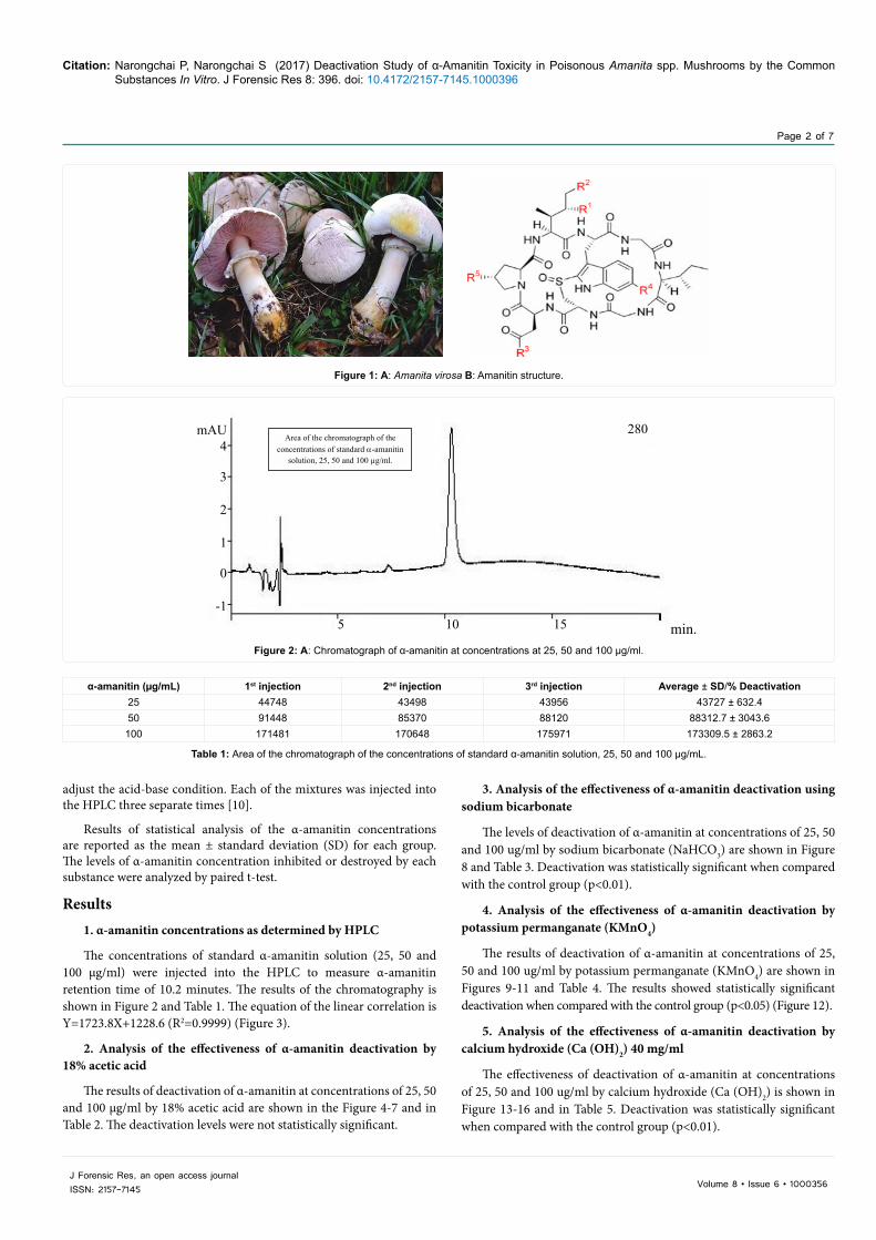
are Amanita mushroom species: A. phalloides, (death cap) A. virosa, (destroying angel), A. verna, A. bisporigera, Galerina marginata and Conocybe filaris (Figures 1A and 1B). Alpha-amanitin is the most potent of the toxins occurring in poisonous mushrooms in the genus Amanita [1,2]. The lethal oral dose (LD50) of α-amanitin in mice is 0.1-0.3 mg/kg. One cap of A. phalloides, A. virosa or A. verna contains ten amatoxins. Ingestion of A. phalloides results in the highest mortality rate of all poisonous mushrooms, ranging from 50 to 90%. Unfortunately, distinguishing poisonous and non-poisonous mushrooms by morphology alone is challenging due to their similar gross appearance. Theoretically, there are ten important amatoxins: [3] -amanitin, β-amanitin, γ-amanitin, ε-amanitin, ð-amanitin, amanullin, amanullinic acid, amaninamide, amanin and proamanullin [4] These differ from the other amatoxins in that they are heat resisitant, alcohol and lipid soluable [5] and indigestible in gastric and small intestinal enzymes. In addition, they can be absorbed rapidly by both gastric and duodenal tissues. One hundred grams of of A. phalloides contain eight milligrams of α-amanitin [1] The RNA polymerase II and III inhibitor α-amanitin [2] affects many organs, especially liver cells, resulting in necrotic hepatic cytolysis [3]. The mortality rate of α-amanitin poisoning is very high because of the difficulty diagnosis, the delayed toxicity (average 24 hrs.), the severity of the toxicity and the administration of an ineffective antidote. The cycle peptide chain, α-amanitin, has eight amino acids: asparagine, hydroxyl proline, dihydroxy isoleucine, hydroxyl-mercapto-tryptophan, glycine, isoleucine, glycine and alanine. Hydroxyl proline and dihydroxy isoleucine activate RNA polymerase II and III by binding to the enzyme molecule, enhancing the activating effect by means of glycine and isoleucine binding [6-8]. The immediate cause of death from mushroom poisoning is kidney, liver and respiratory failure which occur within one week of ingestion.
Deactivation Study of α-Amanitin Toxicity in Poisonous Amanita spp. Mushrooms by the Common Substances In VitroPaitoon Narongchai* and Siripun Narongchai Department of Forensic Medicine, Faculty of Medicine, ChiangMai University, Chiang Mai, Thailand
AbstractThe purpose of this research was to find out the substance which deactivate α-Amanitin Toxicity
The materials and methods used in the study include analysis with high performance liquid chromatography (HPLC) to:
1.Demonstrate the standard α-amanitin at concentrations of 25, 50 and 100 µg/ml
2.Determine the deactivation of α-amanitin with 1) 18% acetic acid 2), calcium hydroxide 40 mg/ml, 3) potassium permanganate 20 mg/ml, 4) sodium bicarbonate 20 mg/ml
3.Report the statistical analysis as the mean ± standard deviation (SD) and paired t-test.
The result revealed that potassium permanganate could eliminate 100 percent of the α-amanitin at 25, 50 and 100 µg/ml. Calcium hydroxide, sodium bicarbonate and acetic acid had lower elimination rates at those concentrations: 68.43 ± 2.58 (-71.4, -67.2, -66.7%), 21.48 ± 10.23 (-29.4, -25.2, -9.9%) and 3.21 ± 0.02% (-3.2, -3.2, +1.1%), respectively. The conclusion of this study was suggested that potassium permanganate could be applied as an absorbent substance during gastric lavage in patients with mushroom poisoning. It also might be effective as a cleansing wash for uncooked mushrooms. Investigation of potassium permanganate’s ability to absorb α-amanitin in animal models and humans should be considered. .
Patients with α-amanitin poisoning can develop severe toxic hepatitis, centrilobular necrosis, liver steatosis, and acute tubulointerstitial necrosis leading to hepatorenal syndrome all of which have a high mortality rate. The most effective treatment is emptying the stomach promptly by performing gastric lavage with 1:2,000 tannic acid or 1:10,000 potassium permanganase plus triggering emesis. A warm saline solution of potassium permaganase can be used as an emergency treatment to reduce toxins in the intestines and stomach. Thus, KMnO4, NaHCO3, Ca(OH)2 and acetic acid should be considered as a cleanser for uncooked mushrooms to deactivate α-amanitin toxicity and potentially reduce the mortality rate from liver failure, acute renal failure, respiratory failure and gastro-intestinal haemorrhage[9].
Materials and MethodsIn this study, experiments were performed on α-amanitin at
concentrations of 25, 50 and 100 µg/ml eached mixed with 1) 18% acetic acid 2), calcium hydroxide 40 mg/ml, 3) potassium permanganate 20 mg/ml, 4) sodium bicarbonate 20 mg/ml (results analyzed with high performance liquid chromatography (HPLC) with Luna C18 (150 × 4.6 mm I.D., 5 micron) from Phenomenex®, USA), or 5) mobile phase (a mixture of 0.02 M aqueous ammonium acetate and acetonitrile (88/12, v/v) pH 5.0 at an absorbance of 280 nm). Glacial acetic acid was used to
Citation: Narongchai P, Narongchai S (2017) Deactivation Study of α-Amanitin Toxicity in Poisonous Amanita spp. Mushrooms by the Common Substances In Vitro. J Forensic Res 8: 396. doi: 10.4172/2157-7145.1000396
Page 2 of 7
J Forensic Res, an open access journalISSN: 2157-7145 Volume 8 • Issue 6 • 1000356
3. Analysis of the effectiveness of α-amanitin deactivation using sodium bicarbonate
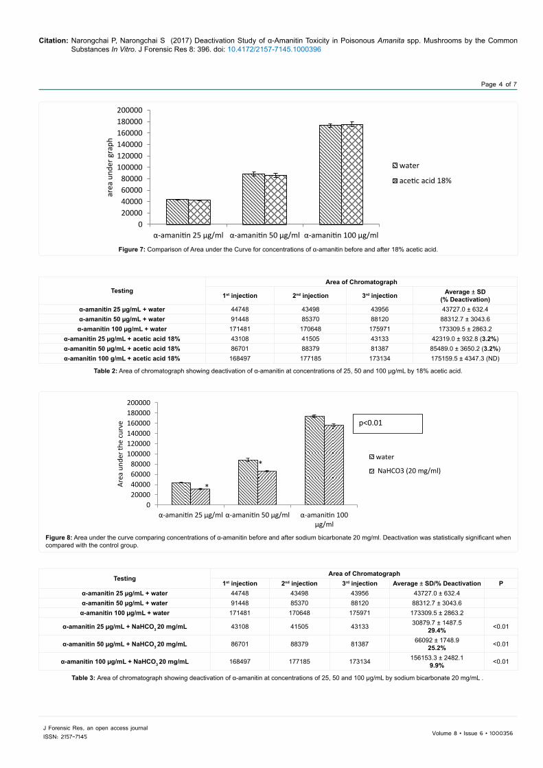
The levels of deactivation of α-amanitin at concentrations of 25, 50 and 100 ug/ml by sodium bicarbonate (NaHCO3) are shown in Figure 8 and Table 3. Deactivation was statistically significant when compared with the control group (p<0.01).
4. Analysis of the effectiveness of α-amanitin deactivation by potassium permanganate (KMnO4)
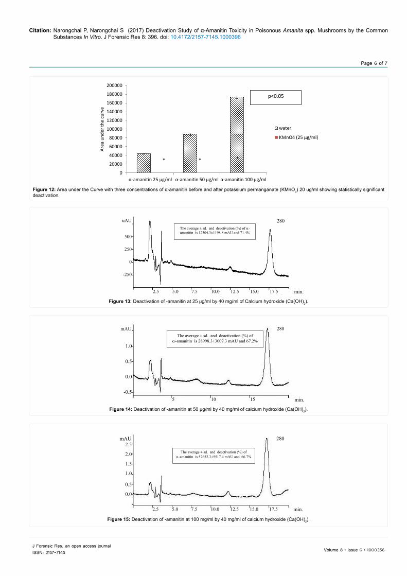
The results of deactivation of α-amanitin at concentrations of 25, 50 and 100 ug/ml by potassium permanganate (KMnO4) are shown in Figures 9-11 and Table 4. The results showed statistically significant deactivation when compared with the control group (p<0.05) (Figure 12).
5. Analysis of the effectiveness of α-amanitin deactivation by calcium hydroxide (Ca (OH)2) 40 mg/ml
The effectiveness of deactivation of α-amanitin at concentrations of 25, 50 and 100 ug/ml by calcium hydroxide (Ca (OH)2) is shown in Figure 13-16 and in Table 5. Deactivation was statistically significant when compared with the control group (p<0.01).
adjust the acid-base condition. Each of the mixtures was injected into the HPLC three separate times [10].
Results of statistical analysis of the α-amanitin concentrations are reported as the mean ± standard deviation (SD) for each group. The levels of α-amanitin concentration inhibited or destroyed by each substance were analyzed by paired t-test.
Results1. α-amanitin concentrations as determined by HPLC
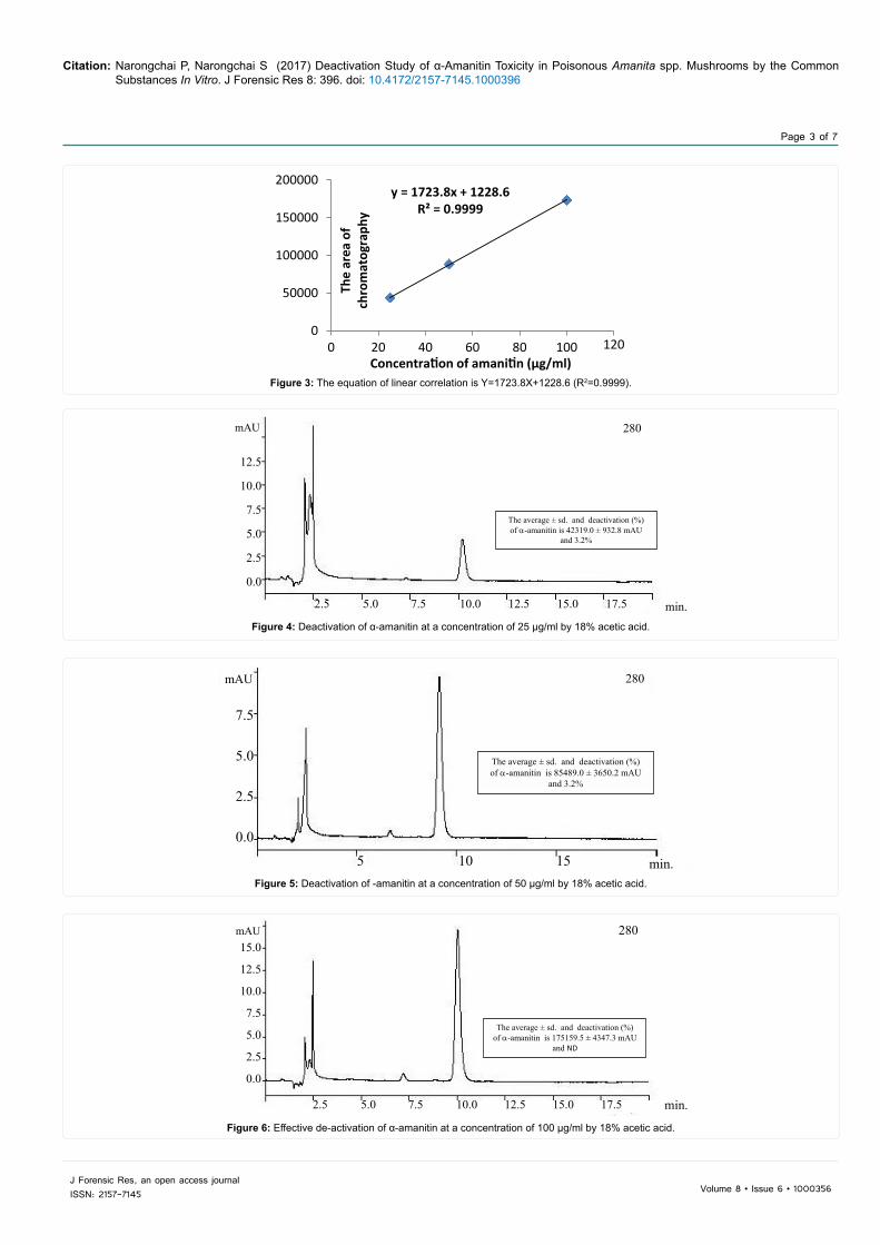
The concentrations of standard α-amanitin solution (25, 50 and 100 µg/ml) were injected into the HPLC to measure α-amanitin retention time of 10.2 minutes. The results of the chromatography is shown in Figure 2 and Table 1. The equation of the linear correlation is Y=1723.8X+1228.6 (R2=0.9999) (Figure 3).
2. Analysis of the effectiveness of α-amanitin deactivation by 18% acetic acid
The results of deactivation of α-amanitin at concentrations of 25, 50 and 100 µg/ml by 18% acetic acid are shown in the Figure 4-7 and in Table 2. The deactivation levels were not statistically significant.
Figure 1: A: Amanita virosa B: Amanitin structure.
Figure 2: A: Chromatograph of α-amanitin at concentrations at 25, 50 and 100 µg/ml.
min.
mAU4
3
2
1
0
-15 10 15
280
potassium permanganate 20 mg/ml, 4) sodium bicarbonate 20 mg/ml (results analyzed with high performance liquid chromatography (HPLC) with Luna C18 (150×4.6 mm I.D., 5 micron) from Phenomenex®, USA), or 5) mobile phase (a mixture of 0.02 M aqueous ammonium acetate and acetonitrile (88/12, v/v) pH 5.0 at an absorbance of 280 nm). Glacial acetic acid was used to adjust the acid-base condition. Each of the mixtures was injected into the HPLC three separate times.(10)
Results of statistical analysis of the -amanitin concentrations are reported as the mean ± standard deviation (SD) for each group. The levels of -amanitin concentration inhibited or destroyed by each substance were analyzed by paired t-test. Results: 1. -amanitin concentrations as determined by HPLC
The concentrations of standard -amanitin solution (25, 50 and 100 µg/ml) were injected into the HPLC to measure -amanitin retention time of 10.2 minutes. The results of the chromatography is shown in Figure 1.1 and Table 1.1. The equation of the linear correlation is Y = 1723.8X + 1228.6 (R2 = 0.9999) (Figure 1.2)
Figure 1.1 Chromatograph of -amanitin at concentrations at 25, 50 and 100 µg/ml
min.
Table 1.1 Area of the chromatograph of the concentrations of standard -amanitin solution, 25, 50 and 100 µg/ml.
-amanitin (µg/ml)
1st injection
2nd injection
3rd injection
Average ± SD
25 44748 43498 43956 43727± 632.4
50 91448 85370 88120 88312.7± 3043.6
100 171481 170648 175971 173309.5± 2863.2
Area of the chromatograph of the concentrations of standard -amanitin
solution, 25, 50 and 100 µg/ml.
α-amanitin (µg/mL) 1st injection 2nd injection 3rd injection Average ± SD/% Deactivation25 44748 43498 43956 43727 ± 632.450 91448 85370 88120 88312.7 ± 3043.6
100 171481 170648 175971 173309.5 ± 2863.2
Table 1: Area of the chromatograph of the concentrations of standard α-amanitin solution, 25, 50 and 100 µg/mL.
Citation: Narongchai P, Narongchai S (2017) Deactivation Study of α-Amanitin Toxicity in Poisonous Amanita spp. Mushrooms by the Common Substances In Vitro. J Forensic Res 8: 396. doi: 10.4172/2157-7145.1000396
Page 3 of 7
J Forensic Res, an open access journalISSN: 2157-7145 Volume 8 • Issue 6 • 1000356
Figure 3: The equation of linear correlation is Y=1723.8X+1228.6 (R2=0.9999).
y = 1723.8x + 1228.6R² = 0.9999
0
50000
100000
150000
200000
0 20 40 60 80 100 120Th
e ar
ea o
f ch
rom
atog
raph
y
Concentra on of amani n (µg/ml)
Figure 4: Deactivation of α-amanitin at a concentration of 25 µg/ml by 18% acetic acid.
mAU
12.5
10.0
7.5
5.0
2.5
0.0
2.5 5.0 7.5 10.0 12.5 15.0 17.5
280
min.
The average ± sd. and deactivation (%) of α-amanitin is 42319.0 ± 932.8 mAU
and 3.2%
Figure 5: Deactivation of -amanitin at a concentration of 50 µg/ml by 18% acetic acid.
min.
mAU
5 10 15
280
7.5
5.0
2.5
0.0
The average ± sd. and deactivation (%) of α-amanitin is 85489.0 ± 3650.2 mAU
and 3.2%
Figure 6: Effective de-activation of α-amanitin at a concentration of 100 µg/ml by 18% acetic acid.
280mAU15.0
12.5
10.0
7.5
5.0
2.5
0.0
2.5 5.0 7.5 10.0 12.5 15.0 17.5 min.
The average ± sd. and deactivation (%) of α-amanitin is 175159.5 ± 4347.3 mAU
and ND
Citation: Narongchai P, Narongchai S (2017) Deactivation Study of α-Amanitin Toxicity in Poisonous Amanita spp. Mushrooms by the Common Substances In Vitro. J Forensic Res 8: 396. doi: 10.4172/2157-7145.1000396
Page 4 of 7
J Forensic Res, an open access journalISSN: 2157-7145 Volume 8 • Issue 6 • 1000356
Figure 7: Comparison of Area under the Curve for concentrations of α-amanitin before and after 18% acetic acid.
020000400006000080000
100000120000140000160000180000200000
α-amanitin 25 µg/ml α-amanitin 50 µg/ml α-amanitin 100 µg/ml
area
und
er g
raph
water
acetic acid 18%
Figure 8: Area under the curve comparing concentrations of α-amanitin before and after sodium bicarbonate 20 mg/ml. Deactivation was statistically significant when compared with the control group.
020000400006000080000
100000120000140000160000180000200000
α-amanitin 25 µg/ml α-amanitin 50 µg/ml α-amanitin 100 µg/ml
Area
und
er th
e cu
rve
water
NaHCO3 (20 mg/ml)
*
*
*
p<0.01
TestingArea of Chromatograph
1st injection 2nd injection 3rd injection Average ± SD(% Deactivation)
α-amanitin 25 µg/mL + water 44748 43498 43956 43727.0 ± 632.4α-amanitin 50 µg/mL + water 91448 85370 88120 88312.7 ± 3043.6α-amanitin 100 µg/mL + water 171481 170648 175971 173309.5 ± 2863.2
α-amanitin 25 µg/mL + acetic acid 18% 43108 41505 43133 42319.0 ± 932.8 (3.2%)α-amanitin 50 µg/mL + acetic acid 18% 86701 88379 81387 85489.0 ± 3650.2 (3.2%)α-amanitin 100 g/mL + acetic acid 18% 168497 177185 173134 175159.5 ± 4347.3 (ND)
Table 2: Area of chromatograph showing deactivation of α-amanitin at concentrations of 25, 50 and 100 µg/mL by 18% acetic acid.
TestingArea of Chromatograph
1st injection 2nd injection 3rd injection Average ± SD/% Deactivation Pα-amanitin 25 µg/mL + water 44748 43498 43956 43727.0 ± 632.4α-amanitin 50 µg/mL + water 91448 85370 88120 88312.7 ± 3043.6
α-amanitin 100 µg/mL + water 171481 170648 175971 173309.5 ± 2863.2
α-amanitin 25 µg/mL + NaHCO3 20 mg/mL 43108 41505 43133 30879.7 ± 1487.529.4% <0.01
α-amanitin 50 µg/mL + NaHCO3 20 mg/mL 86701 88379 81387 66092 ± 1748.925.2% <0.01
α-amanitin 100 µg/mL + NaHCO3 20 mg/mL 168497 177185 173134 156153.3 ± 2482.19.9% <0.01
Table 3: Area of chromatograph showing deactivation of α-amanitin at concentrations of 25, 50 and 100 µg/mL by sodium bicarbonate 20 mg/mL .

Citation: Narongchai P, Narongchai S (2017) Deactivation Study of α-Amanitin Toxicity in Poisonous Amanita spp. Mushrooms by the Common Substances In Vitro. J Forensic Res 8: 396. doi: 10.4172/2157-7145.1000396
Page 5 of 7
J Forensic Res, an open access journalISSN: 2157-7145 Volume 8 • Issue 6 • 1000356
mAU
500
400
300
200
100
05 10 15
280
min.
The average ± sd. and deactivation (%) of α-amanitin is 0.0 mAU and 100%
Figure 9: Deactivation of -amanitin at a concentration of 25 µg/ml by 20 ug/ml of potassium permanganate (KMnO4).
5 10 15
280
min.
AU
0.75
0.50
0.25
0.00
The average ± sd. and deactivation (%) of α-amanitin is 0.0 mAU and 100%
Figure 10: Deactivation of -amanitin at 50 µg/ml by 20 ug/ml potassium permanganate (KMnO4).
Figure 11: Deactivation of -amanitin at a concentration of 100 µg/ml by 20 ug/ml of potassium permanganate (KMnO4) (p<0.05).
280
2.5 5.0 7.5 10.0 12.5 15.0 17.5 min.
AU
0.75
0.50
0.25
0.00
The average ± sd. and deactivation (%) of α-amanitin is 0.0 mAU and 100%
TestingArea of Chromatograph
1st injection 1st injection 1st injection Average ± SD/% Deactivation pα-amanitin 25 µg/mL + water 44748 43498 43956 43727.0 ± 632.4α-amanitin 50 µg/mL + water 91448 85370 88120 88312.7 ± 3043.6
α-amanitin 100 µg/mL + water 171481 170648 175971 173309.5 ± 2863.2α-amanitin 25 µg/mL + KMnO4 (20 µg/mL) 43108 41505 43133 0/-100% <0.05α-amanitin 50 µg/mL + KMnO4 (20 µg/mL) 86701 88379 81387 0/-100% <0.05α-amanitin 100 µg/mL + KMnO4 (20 µg/mL) 168497 177185 173134 0/-100% <0.05
Table 4: Area of chromatograph showing deactivation of α-amanitin at concentrations of 25,50 and 100 µg/mL by 20 ug/mL of Potassium permanganate (KMnO4).
Discussion and ConclusionsFour common substances, Potassium permanganate (KMnO4),
calcium hydroxide (Ca (OH)2), sodium carbonate (NaHCO3) and acetic acid are regularly used for vegetable detoxification. As mushrooms are
one of the most popular ingredients in Thai food, especially in rural areas, routine detoxification of potential toxins in uncooked mushrooms is one solution that should be considered to avoid mushroom poisoning, particularly α-amanitin toxicity.
Citation: Narongchai P, Narongchai S (2017) Deactivation Study of α-Amanitin Toxicity in Poisonous Amanita spp. Mushrooms by the Common Substances In Vitro. J Forensic Res 8: 396. doi: 10.4172/2157-7145.1000396
Page 6 of 7
J Forensic Res, an open access journalISSN: 2157-7145 Volume 8 • Issue 6 • 1000356
Figure 12: Area under the Curve with three concentrations of α-amanitin before and after potassium permanganate (KMnO4) 20 ug/ml showing statistically significant deactivation.
0
20000
40000
60000
80000
100000
120000
140000
160000
180000
200000
α-amanitin 25 µg/ml α-amanitin 50 µg/ml α-amanitin 100 µg/ml
Area
und
er th
e cu
rve
water
KMnO4 (25 µg/ml)
* * *
p<0.05
Figure 13: Deactivation of -amanitin at 25 µg/ml by 40 mg/ml of Calcium hydroxide (Ca(OH)2).
280
2.5 5.0 7.5 10.0 12.5 15.0 17.5 min.
uAU
500
250
0
-250
The average ± sd. and deactivation (%) of α-amanitin is 12504.3±1198.8 mAU and 71.4%
Figure 14: Deactivation of -amanitin at 50 µg/ml by 40 mg/ml of calcium hydroxide (Ca(OH)2).
min.
mAU
5 10 15
280
1.0
0.5
0.0
-0.5
The average ± sd. and deactivation (%) of α-amanitin is 28998.3±3007.3 mAU and 67.2%
Figure 15: Deactivation of -amanitin at 100 mg/ml by 40 mg/ml of calcium hydroxide (Ca(OH)2).
mAU2.5
2.0
1.5
1.0
0.5
0.0
280
2.5 5.0 7.5 10.0 12.5 15.0 17.5 min.
The average ± sd. and deactivation (%) of α-amanitin is 57652.3±5517.4 mAU and 66.7%

Citation: Narongchai P, Narongchai S (2017) Deactivation Study of α-Amanitin Toxicity in Poisonous Amanita spp. Mushrooms by the Common Substances In Vitro. J Forensic Res 8: 396. doi: 10.4172/2157-7145.1000396
Page 7 of 7
J Forensic Res, an open access journalISSN: 2157-7145 Volume 8 • Issue 6 • 1000356
Figure 16: Area under the curve comparing three concentrations of α-amanitin before and after calcium hydroxide 40 mg/ml. Deactivation was statistically significant compared with the control group.
020000400006000080000
100000120000140000160000180000200000
α-amanitin 25 µg/ml α-amanitin 50 µg/ml α-amanitin 100 µg/ml
Area
und
er th
e cu
rve
water
Ca(OH)2 (40 mg/ml)
*
*
*
p<0.01
Potassium permanganate (KMnO4) is an antiseptic, alkali, and highly soluble detoxification substance. It contains potassium (K+) and permanganate ions which are strong oxidizing and organic substance destroying agents. Manganese oxide (MnO2) destroys the cell wall and cell membrain of bacteria, protozoa, parasites and algae. This study revealed that potassium permanganate (KMnO4) 20 mg/ml completely destroyed α-amanitin at concentrations of 25, 50 and 100 µg/ml. We found that potassium permanganate (KMnO4) is the strongest of the oxidizing agents tested and that it has the ability to destroy or deactivate peptides of α-amanitin molecules in vitro [11,12]. Potassium permanganate (KMnO4) could potentially be used for gastric lavage in patients with mushroom poisoning. Additional experiments in animal models should be conducted to evaluate the effectiveness of these four substances.
Calcium hydroxide (Ca (OH)2) and sodium carbonate (NaHCO3) have an alkaline property which can significantly deactivate alkaline hydrolysis [13,14] caused by α-amanitin. Acetic acid has an antiseptic acid property but it cannot destroy the cell membranes or cell walls of any vegetable including α-amanitin because it has a low pKa value. α-amanitin in poisonous mushrooms cannot be destroyed by acids or by gastric or duodenal enzymes [15].
Potassium permanganate (KMnO4) is the most effective deactivation substance for α-amanitin. It completely inhibits α-amanitin activation. Both calcium hydroxide (Ca (OH)2) and sodium carbonate (NaHCO3) (to a lesser degree) inhibit α-amanitin, but they do not do so completely. Acetic acid only very weakly inhibits α-amanitin. Based on these findings, washing uncooked poisonous Amanita spp. mushrooms with KMnO4 to detoxify or deactivate α-amanitin before ingestion would definitely be beneficial to health, especially in rural areas where many people gather their own mushrooms from forested areas rather than purchasing them at a market.
Ethical Approval
This research did not involve any human participants or animals.
References1. Defendenti C, Bonacina E, Mauroni M, Gelosa L (1998) Validation of a high
performance liquid chromatographic method for alpha amanitin determination in urine. Forensic Sci Inter. 92: 59-68.
2. Meinecke B, Meinecke-Tillmann S (1993) Effects of alpha-amanitin on nuclear maturation of porcine oocytes in vitro. J Reprod Fertil 98: 195-201.
3. Michelot D, Labia R (1988) Alpha-amanitin: A possible suicide substance-like toxin involving the sulphoxide moiety of the bridged cyclopeptide. Drug Metabol Drug Interact 6: 265-74.
4. Baumann K, Muenter K, Faulstich H (1993) Identification of structural features involved in binding of α-amanitin to a monoclonal antibody. Biochemistry 32: 4043-50.
5. Christine KS, Hans P (2003) Cytotoxic fungi –an overview. Toxicon 42: 339-49.
6. Wieland T (1983) The toxic peptides from Amanita mushrooms. Int J Pept Prot Res 22: 257–76.
7. Baumann K, Zanotti G, Faulstich H (1994) A beta-turn in alpha-amanitin is the most important structural feature for binding to RNA polymerase II and three monoclonalantibodies. Prot Sci 3: 750-6.
8. Pomilio AB, Battista ME, Vitale AA (2001) Semiempirical AM1 and ab initio parameters of the lethal cyclopeptides alpha-amanitin and its related thioether, S-sulphoxide, sulphone, and O-methyl derivative. J Mol struct: Theochem. 536: 243-62.
9. Mas A (2005) Mushrooms. J Hepatol 42: 166-9.
10. Jehl F, Gallion C, Birckel P, Jaeger A, Flesch F, et al. (1985) Determination of α-amanitin and α -amanitin in human biological fluids by High-performance liquid chromatography. Anal Biochem 149: 35-42.
11. Hopwood D (1969) Fixation of proteins by osmium tetroxide potassium dichromate and potassium permanganate. Histochem Cell Biol. 18: 250-60.
12. Lewis GP, Rowe PB (1979) Oxidative and reductive cleavage of folates-A critical appraisal. Anal Biochem 93: 91-7.
13. Zheleva A, Tolekova A, Zhelev M, Uzunova V, Platikanova M et al. (2007) Free radical reactions might contribute to severe alpha amanitin hepatotoxicity- A hypothesis. Med Hypothesis. 69: 361-7.
14. Leist M, Gantner F, Naumann H, Bluethmann H, Vogt K, et al. (1997) Tumor necrosis factor-induced apoptosis during the poisoning of mice with hepatotoxin. Gastroenterology 112: 923-34.
15. Vetter J (1998) Toxin of Amanita phalloides. Toxicon 36: 13-24.
TestingArea of Chromatograph
1st injection 2nd injection 3rd injection Average ± SD/% Deactivation Pα –amanitin 25 µg/mL + water 44748 43498 43956 43727.0 ± 632.4α –amanitin 50 µg/mL + water 91448 85370 88120 88312.7 ± 3043.6
α –amanitin 100 µg/mL + water 171481 170648 175971 173309.5 ± 2863.2α –amanitin 25 µg/mL + Ca(OH)2 (40 mg/mL) 43108 41505 43133 12504.3 ± 1198.8/-71.4% <0.01α –amanitin 50 µg/mL + Ca(OH)2 (40 mg/mL) 86701 88379 81387 28998.3 ± 3007.3/-67.2% <0.01
α –amanitin 100 µg/mL + Ca(OH)2 (40 mg/mL) 168497 177185 173134 57652.3 ± 5517.4/-66.7% <0.01
Table 5: Area of chromatograph showing deactivation of α-amanitin at 25,50 and 100µg/mL by 40 mg/mL of Calcium hydroxide.
![B-TOUCH intelligence passenger airbag deactivation warning lamp intelligence pre heating display reconfigurable intelligence radio command display ... passat [1997] (3b) edi (ΗΛΕΚΤΡΟΝΙΚΗ](https://static.fdocument.org/doc/165x107/6123f6172259f476611dad53/b-intelligence-passenger-airbag-deactivation-warning-lamp-intelligence-pre-heating.jpg)
![Computational Modeling of Glucose Toxicity in Pancreatic Β-cells [Update]](https://static.fdocument.org/doc/165x107/577cb4f61a28aba7118cd93d/computational-modeling-of-glucose-toxicity-in-pancreatic-cells-update.jpg)