Current Protocols in Nucleic Acid Chemistry || Synthesis of 2���- O ...
19
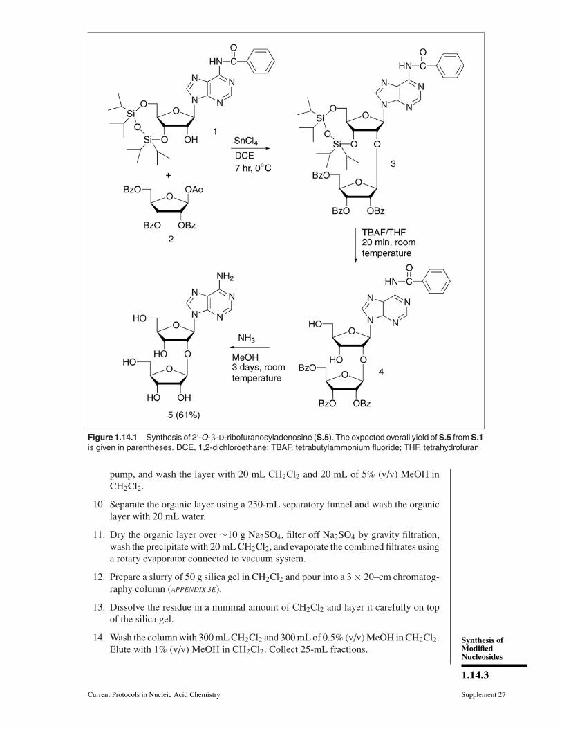
UNIT 1.14 Synthesis of 2 -O-β-d- Ribofuranosylnucleosides This unit describes a three-step procedure for the preparation of 2 -O-β-D-ribofurano- sylnucleosides (Mikhailov et al., 1997a; Markiewicz et al., 1998). First, the complete syn- thesis of 2 -O-β-D-ribofuranosyladenosine is described (see Basic Protocol). As shown in Figure 1.14.1, the procedure involves (1) condensation of a small excess of 1-O-acetyl- 2,3,5-tri-O-benzoyl-β-D-ribofuranose activated with tin tetrachloride with an N-protected 3 ,5 -O-tetraisopropyldisiloxane-1,3-diyl-ribonucleoside in 1,2-dichloroethane, (2) re- moval of silyl protecting group with tetrabutylammonium fluoride, and (3) deacylation with ammonia in methanol. Using these procedures, 2 -O-β-D-ribofuranosyladenosine is prepared in a 61% overall yield. The 2 -O-ribosylation proceeds stereospecifically with the formation of a β-glycosidic bond. The presence of a participating 2-O-benzoyl group leads exclusively to 1,2-trans-ribofuranoside. This reaction is carried out under mild conditions (0 ◦ C, 1,2-dichloroethane, 2 hr for pyrimidine nucleosides, 7 to 16 hr for purine derivatives) and yields of the target compounds are 72% to 80% (Mikhailov et al., 1997a). At room temperature, the reaction occurs faster (30 min), but the yields in the coupling steps are lower (40% to 77%; Markiewicz et al., 1998). These variations are presented as slightly modified procedures for the preparation of four other 2 -O-β-D-ribofuranosylnucleosides (see Alternate Protocols 1 through 4; Fig. 1.14.3). CAUTION: Carry out all operations involving organic solvents and reagents in a well- ventilated fume hood, and wear gloves and protective glasses. BASIC PROTOCOL PREPARATION OF 2 -O- β-D-RIBOFURANOSYLADENOSINE The first step of preparation of 2 -O-β-D-ribofuranosyladenosine involves the conden- sation of N 6 -benzoyl-3 ,5 -(1,1,3,3-O-tetraisopropyldisiloxane-1,3-diyl)adenosine (S.1; Markiewicz and Wiewiorowski, 1986; UNIT 2.4) with 1-O-acetyl-2,3,5-tri-O-benzoyl-β-D- ribofuranose (S.2) in the presence of tin tetrachloride in 1,2-dichloroethane at 0 ◦ C (Fig. 1.14.1). To obtain the product S.3 in high yield and to simplify its isolation, it is recom- mended to run the reaction under nitrogen and to perform preactivation of the sugar with tin tetrachloride; usually the reaction is complete in 7 to 8 hr. During workup of the reac- tion mixture with saturated sodium bicarbonate solution, a suspension is formed, which should be filtered through a 2- to 3-cm layer of Hyflo Super Cel before the separation of organic and aqueous layers. The second step involves removal of the silyl protec- tion from the 3 - and 5 -hydroxyl groups with tetrabutylammonium fluoride (TBAF) in tetrahydrofuran (THF) for 15 to 20 min at room temperature followed by silica gel column chromatography. In the last step, all benzoyl groups from S.4 are removed with 5 M ammonia (half-saturated at 0 ◦ C) in methanol for 2 to 3 days at room temperature followed by recrystallization from methanol to give 2 -O-β-D-ribofuranosyladenosine (S.5) in an overall yield of 61%. Materials 1-O-Acetyl-2,3,5-tri-O-benzoyl-β-D-ribofuranose (S.2) N 6 -Benzoyl-3 ,5 -(1,1,3,3-O-tetraisopropyldisiloxane-1,3-diyl)adenosine (S.1; UNIT 2.4; Fig. 2.4.5) Phosphorus pentoxide (P 2 O 5 ) Balloon of nitrogen or argon 1,2-Dichloroethane, anhydrous Tin tetrachloride (SnCl 4 ) Methanol (MeOH), analytical grade Contributed by Sergey N. Mikhailov, Ekaterina V. Efimtseva, Andrei A. Rodionov, Georgii V. Bobkov, Irina V. Kulikova, and Piet Herdewijn Current Protocols in Nucleic Acid Chemistry (2006) 1.14.1-1.14.19 Copyright C 2006 by John Wiley & Sons, Inc. Synthesis of Modified Nucleosides 1.14.1 Supplement 27
Transcript of Current Protocols in Nucleic Acid Chemistry || Synthesis of 2���- O ...

UNIT 1.14Synthesis of 2′-O-β-d-Ribofuranosylnucleosides
This unit describes a three-step procedure for the preparation of 2′-O-β-D-ribofurano-sylnucleosides (Mikhailov et al., 1997a; Markiewicz et al., 1998). First, the complete syn-thesis of 2′-O-β-D-ribofuranosyladenosine is described (see Basic Protocol). As shownin Figure 1.14.1, the procedure involves (1) condensation of a small excess of 1-O-acetyl-2,3,5-tri-O-benzoyl-β-D-ribofuranose activated with tin tetrachloride with an N-protected3′,5′-O-tetraisopropyldisiloxane-1,3-diyl-ribonucleoside in 1,2-dichloroethane, (2) re-moval of silyl protecting group with tetrabutylammonium fluoride, and (3) deacylationwith ammonia in methanol. Using these procedures, 2′-O-β-D-ribofuranosyladenosineis prepared in a 61% overall yield. The 2′-O-ribosylation proceeds stereospecificallywith the formation of a β-glycosidic bond. The presence of a participating 2-O-benzoylgroup leads exclusively to 1,2-trans-ribofuranoside. This reaction is carried out undermild conditions (0◦C, 1,2-dichloroethane, 2 hr for pyrimidine nucleosides, 7 to 16 hrfor purine derivatives) and yields of the target compounds are 72% to 80% (Mikhailovet al., 1997a). At room temperature, the reaction occurs faster (30 min), but theyields in the coupling steps are lower (40% to 77%; Markiewicz et al., 1998). Thesevariations are presented as slightly modified procedures for the preparation of four other2′-O-β-D-ribofuranosylnucleosides (see Alternate Protocols 1 through 4; Fig. 1.14.3).
CAUTION: Carry out all operations involving organic solvents and reagents in a well-ventilated fume hood, and wear gloves and protective glasses.
BASICPROTOCOL
PREPARATION OF 2′-O-ββ-D-RIBOFURANOSYLADENOSINE
The first step of preparation of 2′-O-β-D-ribofuranosyladenosine involves the conden-sation of N6-benzoyl-3′,5′-(1,1,3,3-O-tetraisopropyldisiloxane-1,3-diyl)adenosine (S.1;Markiewicz and Wiewiorowski, 1986; UNIT 2.4) with 1-O-acetyl-2,3,5-tri-O-benzoyl-β-D-ribofuranose (S.2) in the presence of tin tetrachloride in 1,2-dichloroethane at 0◦C (Fig.1.14.1). To obtain the product S.3 in high yield and to simplify its isolation, it is recom-mended to run the reaction under nitrogen and to perform preactivation of the sugar withtin tetrachloride; usually the reaction is complete in 7 to 8 hr. During workup of the reac-tion mixture with saturated sodium bicarbonate solution, a suspension is formed, whichshould be filtered through a 2- to 3-cm layer of Hyflo Super Cel before the separationof organic and aqueous layers. The second step involves removal of the silyl protec-tion from the 3′- and 5′-hydroxyl groups with tetrabutylammonium fluoride (TBAF)in tetrahydrofuran (THF) for 15 to 20 min at room temperature followed by silica gelcolumn chromatography. In the last step, all benzoyl groups from S.4 are removed with5 M ammonia (half-saturated at 0◦C) in methanol for 2 to 3 days at room temperaturefollowed by recrystallization from methanol to give 2′-O-β-D-ribofuranosyladenosine(S.5) in an overall yield of 61%.
Materials
1-O-Acetyl-2,3,5-tri-O-benzoyl-β-D-ribofuranose (S.2)N6-Benzoyl-3′,5′-(1,1,3,3-O-tetraisopropyldisiloxane-1,3-diyl)adenosine
(S.1; UNIT 2.4; Fig. 2.4.5)Phosphorus pentoxide (P2O5)Balloon of nitrogen or argon1,2-Dichloroethane, anhydrousTin tetrachloride (SnCl4)Methanol (MeOH), analytical grade
Contributed by Sergey N. Mikhailov, Ekaterina V. Efimtseva, Andrei A. Rodionov, Georgii V.Bobkov, Irina V. Kulikova, and Piet HerdewijnCurrent Protocols in Nucleic Acid Chemistry (2006) 1.14.1-1.14.19Copyright C© 2006 by John Wiley & Sons, Inc.
Synthesis ofModifiedNucleosides
1.14.1
Supplement 27

Synthesis of2′-O-ββ-d-
Ribofuranosyl-nucleosides
1.14.2
Supplement 27 Current Protocols in Nucleic Acid Chemistry
Methylene chloride (CH2Cl2), reagent gradeSaturated sodium bicarbonate solution (sat. NaHCO3)Hyflo Super Cel (Fluka)Sodium sulfate, anhydrous (Na2SO4)Silica gel (e.g., Kieselgel 60, 0.06 to 0.20 mm; Merck)Tetrabutylammonium fluoride trihydrate (TBAF)Tetrahydrofuran (THF), reagent gradeChloroform (CHCl3), reagent grade5 M ammonia in methanol (half-saturated at 0◦C)Diethyl ether, reagent grade
50- and 250-mL round-bottom flasksVacuum desiccatorVacuum oil pumpTLC plate: silica-coated aluminum plate with fluorescent indicator (Merck silica
gel 60 F254)254-nm UV lampLong disposable capillaries100-mL funnels with sintered glass disc filters (porosity 3)100- and 250-mL separatory funnelRotary evaporator equipped with a water aspirator3 × 20–cm and 3 × 15–cm sintered glass chromatography columns, porosity 3Stainless steel spatula
Additional reagents and equipment for TLC (APPENDIX 3D) and columnchromatography (APPENDIX 3E)
Prepare S.31. Weigh 1.26 g (2.5 mmol) of 1-O-acetyl-2,3,5-tri-O-benzoyl-β-D-ribofuranose (S.2)
into a 250-mL round-bottom flask containing a stir bar and weigh 1.23 g (2 mmol)of nucleoside S.1 into a separate flask.
2. Put both flasks in a vacuum desiccator with phosphorus pentoxide and evacuateusing a vacuum oil pump for 10 to 15 min. Close the stopcock of the desiccator andleave overnight at room temperature.
3. Connect the desiccator with a balloon of nitrogen or argon and open the stopcockof the desiccator. Open the desiccator and quickly close the flasks.
4. Dissolve 1-O-acetyl-2,3,5-tri-O-benzoyl-β-D-ribofuranose (S.2) in 25 mL of 1,2-dichloroethane, close with stopcock and balloon of nitrogen or argon, and put theflask in the ice-water bath (Fig. 1.14.2A).
5. Add 0.35 mL (3 mmol) of tin tetrachloride in one portion under stirring and keepthe reaction mixture for 10 min at 0◦C.
6. Add 1.23 g (2 mmol) of nucleoside S.1 in one portion and stir the reaction mixturefor 7 hr at 0◦C.
7. Monitor reaction by TLC (APPENDIX 3D) using 2% (v/v) MeOH in CH2Cl2. To protectthe reaction mixture from atmospheric moisture, remove the sample using a longcapillary as shown in Figure 1.14.2B.
The starting compound S.1 (Rf = 0.21) usually disappears after 6 to 7 hr at 0◦C. Theproduct S.3 (Rf = 0.31) moves faster in the same solvent.
8. Add 10 mL saturated sodium bicarbonate solution and stir the suspension for 20min at 0◦C.
9. Lay a 2- to 3-cm layer of Hyflo Super Cel on a 100-mL funnel with sintered disc(porosity 3) and wash it with 20 mL CH2Cl2. Filter the suspension using a vacuum
Synthesis ofModifiedNucleosides
1.14.3
Current Protocols in Nucleic Acid Chemistry Supplement 27
Figure 1.14.1 Synthesis of 2′-O-β-D-ribofuranosyladenosine (S.5). The expected overall yield of S.5 from S.1is given in parentheses. DCE, 1,2-dichloroethane; TBAF, tetrabutylammonium fluoride; THF, tetrahydrofuran.
pump, and wash the layer with 20 mL CH2Cl2 and 20 mL of 5% (v/v) MeOH inCH2Cl2.
10. Separate the organic layer using a 250-mL separatory funnel and wash the organiclayer with 20 mL water.
11. Dry the organic layer over ∼10 g Na2SO4, filter off Na2SO4 by gravity filtration,wash the precipitate with 20 mL CH2Cl2, and evaporate the combined filtrates usinga rotary evaporator connected to vacuum system.
12. Prepare a slurry of 50 g silica gel in CH2Cl2 and pour into a 3 × 20–cm chromatog-raphy column (APPENDIX 3E).
13. Dissolve the residue in a minimal amount of CH2Cl2 and layer it carefully on topof the silica gel.
14. Wash the column with 300 mL CH2Cl2 and 300 mL of 0.5% (v/v) MeOH in CH2Cl2.Elute with 1% (v/v) MeOH in CH2Cl2. Collect 25-mL fractions.

Synthesis of2′-O-ββ-d-
Ribofuranosyl-nucleosides
1.14.4
Supplement 27 Current Protocols in Nucleic Acid Chemistry
Figure 1.14.2 Protection from atmospheric moisture for synthesis of 2′-O-β-D-ribofur-anosylnucleosides. (A) A 250-mL round-bottom flask with adaptor and stopcock connected witha balloon of nitrogen or argon in an ice-water bath. (B) Checking the reaction mixture with a longcapillary (diameter ∼1 mm). The diameter of the hole in the stopcock is ∼3 mm.
15. Evaluate fractions by TLC using 2% (v/v) MeOH in CH2Cl2 and combine thefractions that contain only S.3. Evaporate the volatile materials from the combinedfractions using a rotary evaporator connected to vacuum system, and dry the residualfoam for 2 to 3 hr using a vacuum oil pump.
16. Grind the foam with a stainless steel spatula and dry the resulting powder 12 to 16hr using a vacuum oil pump.
17. Characterize the product by TLC, 1H NMR, and 13C NMR.
The compound is stable for at least 12 months at ambient temperature.
N6-Benzoyl-9-[3,5-(1,1,3,3-tetraisopropyldisiloxane-1,3-diyl)-2-O-(2,3,5-tri-O-benzoyl-β-D-ribofuranosyl)-β-D-ribofuranosyl]adenine (S.3). Yield of white amorphous solid1.57 g (74%). TLC: Rf 0.31 (98:2 v/v CH2Cl2/MeOH).1H NMR (400 MHz, CDCl3):9.02 brs (1H, NH), 8.72 s (1H, H8), 8.15 s (1H, H2), 8.03-7.87 m (8H, Bz), 7.61-7.28m (12H, Bz), 6.09 d (1H, J1′,2′ = 1.0 Hz, H1′, Ado), 5.96 dd (1H, J3′,2′ = 4.8 Hz, J3′,4′
= 6.2 Hz, H3′, Rib), 5.88 d (1H, H2′, Rib), 5.82 s (1H, H1′, Rib), 4.96 dd (1H, J3′,2′ =4.7 Hz, J3′,4′ = 9.0 Hz, H3′, Ado), 4.90 dd (1H, H2′, Ado), 4.81-4.65 m (3H, H4′, Ado;H4′,5′a, Rib), 4.18 d (1H, J5′a,5′b = −13.4 Hz, H5′a, Ado), 4.13 dd (1H, J5′b,4′ = 1.0Hz, J5′b,5′a = −9.5 Hz, H5′b, Rib), 4.03 dd (1H, J5′b,4′ = 1.5 Hz, H5′b, Ado), 1.08-1.03m (28H, iPr). 13C NMR (CDCl3): 166.01, 165.37, 164.97 and 164.43 (C=O), 152.70(C2), 150.76 (C6), 149.35 (C4), 141.80 (C8), 133.41, 133.16, 132.69, 129.71, 129.64,129.10, 128.83, 128.34 and 127.78 (Bz), 123.40 (C5), 105.65 (C1′, Rib), 88.81 (C1′,Ado), 81.34 (C4′, Ado), 79.55 (C4′, Rib), 78.47 (C2′, Ado), 75.51 (C2′, Rib), 72.53 (C3′,Rib), 69.84 (C3′, Ado), 65.27 (C5′, Rib), 59.70 (C5′, Ado), 17.26, 17.04, 16.87, 16.77,13.31, 12.89, 12.74 and 12.59 (iPr).
Synthesis ofModifiedNucleosides
1.14.5
Current Protocols in Nucleic Acid Chemistry Supplement 27
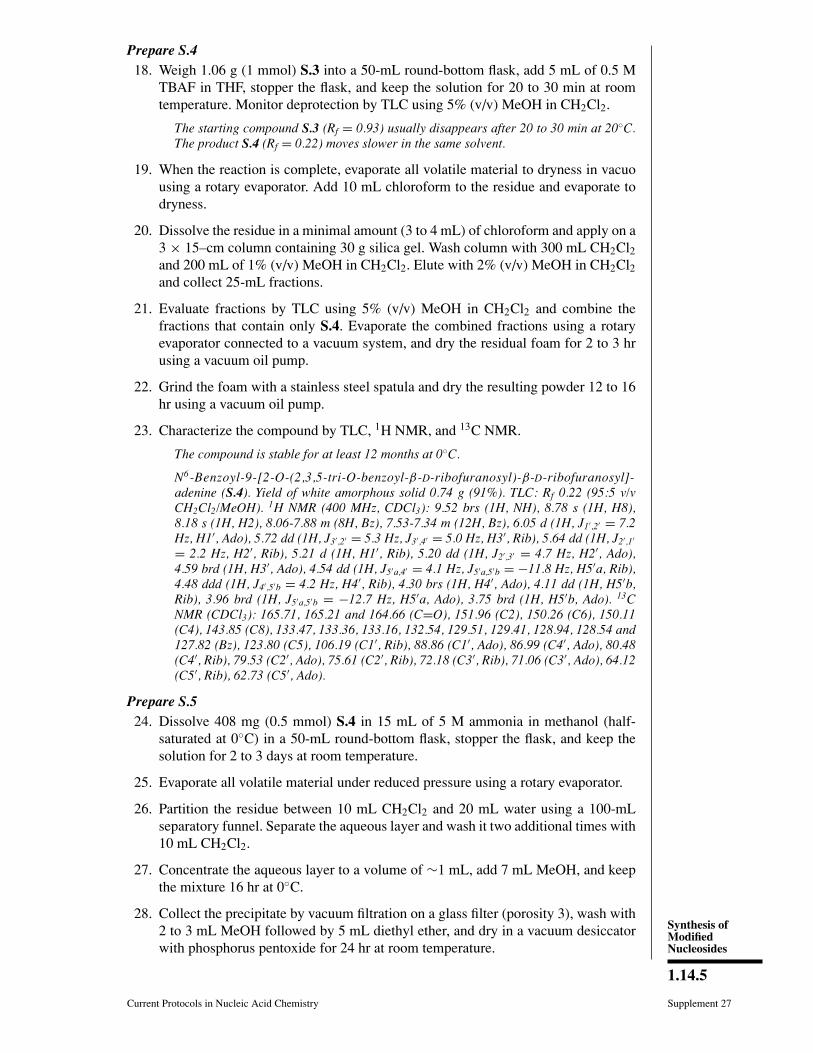
Prepare S.418. Weigh 1.06 g (1 mmol) S.3 into a 50-mL round-bottom flask, add 5 mL of 0.5 M
TBAF in THF, stopper the flask, and keep the solution for 20 to 30 min at roomtemperature. Monitor deprotection by TLC using 5% (v/v) MeOH in CH2Cl2.
The starting compound S.3 (Rf = 0.93) usually disappears after 20 to 30 min at 20◦C.The product S.4 (Rf = 0.22) moves slower in the same solvent.
19. When the reaction is complete, evaporate all volatile material to dryness in vacuousing a rotary evaporator. Add 10 mL chloroform to the residue and evaporate todryness.
20. Dissolve the residue in a minimal amount (3 to 4 mL) of chloroform and apply on a3 × 15–cm column containing 30 g silica gel. Wash column with 300 mL CH2Cl2and 200 mL of 1% (v/v) MeOH in CH2Cl2. Elute with 2% (v/v) MeOH in CH2Cl2and collect 25-mL fractions.
21. Evaluate fractions by TLC using 5% (v/v) MeOH in CH2Cl2 and combine thefractions that contain only S.4. Evaporate the combined fractions using a rotaryevaporator connected to a vacuum system, and dry the residual foam for 2 to 3 hrusing a vacuum oil pump.
22. Grind the foam with a stainless steel spatula and dry the resulting powder 12 to 16hr using a vacuum oil pump.
23. Characterize the compound by TLC, 1H NMR, and 13C NMR.
The compound is stable for at least 12 months at 0◦C.
N6-Benzoyl-9-[2-O-(2,3,5-tri-O-benzoyl-β -D-ribofuranosyl)-β-D-ribofuranosyl]-adenine (S.4). Yield of white amorphous solid 0.74 g (91%). TLC: Rf 0.22 (95:5 v/vCH2Cl2/MeOH). 1H NMR (400 MHz, CDCl3): 9.52 brs (1H, NH), 8.78 s (1H, H8),8.18 s (1H, H2), 8.06-7.88 m (8H, Bz), 7.53-7.34 m (12H, Bz), 6.05 d (1H, J1′,2′ = 7.2Hz, H1′, Ado), 5.72 dd (1H, J3′,2′ = 5.3 Hz, J3′,4′ = 5.0 Hz, H3′, Rib), 5.64 dd (1H, J2′,1′
= 2.2 Hz, H2′, Rib), 5.21 d (1H, H1′, Rib), 5.20 dd (1H, J2′,3′ = 4.7 Hz, H2′, Ado),4.59 brd (1H, H3′, Ado), 4.54 dd (1H, J5′a,4′ = 4.1 Hz, J5′a,5′b = −11.8 Hz, H5′a, Rib),4.48 ddd (1H, J4′,5′b = 4.2 Hz, H4′, Rib), 4.30 brs (1H, H4′, Ado), 4.11 dd (1H, H5′b,Rib), 3.96 brd (1H, J5′a,5′b = −12.7 Hz, H5′a, Ado), 3.75 brd (1H, H5′b, Ado). 13CNMR (CDCl3): 165.71, 165.21 and 164.66 (C=O), 151.96 (C2), 150.26 (C6), 150.11(C4), 143.85 (C8), 133.47, 133.36, 133.16, 132.54, 129.51, 129.41, 128.94, 128.54 and127.82 (Bz), 123.80 (C5), 106.19 (C1′, Rib), 88.86 (C1′, Ado), 86.99 (C4′, Ado), 80.48(C4′, Rib), 79.53 (C2′, Ado), 75.61 (C2′, Rib), 72.18 (C3′, Rib), 71.06 (C3′, Ado), 64.12(C5′, Rib), 62.73 (C5′, Ado).
Prepare S.524. Dissolve 408 mg (0.5 mmol) S.4 in 15 mL of 5 M ammonia in methanol (half-
saturated at 0◦C) in a 50-mL round-bottom flask, stopper the flask, and keep thesolution for 2 to 3 days at room temperature.
25. Evaporate all volatile material under reduced pressure using a rotary evaporator.
26. Partition the residue between 10 mL CH2Cl2 and 20 mL water using a 100-mLseparatory funnel. Separate the aqueous layer and wash it two additional times with10 mL CH2Cl2.
27. Concentrate the aqueous layer to a volume of ∼1 mL, add 7 mL MeOH, and keepthe mixture 16 hr at 0◦C.
28. Collect the precipitate by vacuum filtration on a glass filter (porosity 3), wash with2 to 3 mL MeOH followed by 5 mL diethyl ether, and dry in a vacuum desiccatorwith phosphorus pentoxide for 24 hr at room temperature.
Synthesis of2′-O-ββ-d-
Ribofuranosyl-nucleosides
1.14.6
Supplement 27 Current Protocols in Nucleic Acid Chemistry
29. Characterize the compound by TLC, UV spectroscopy, 1H NMR, and 13C NMR.
The compound is stable for at least 12 months at 0◦C.
9-(2-O-β-D-Ribofuranosyl-β-D-ribofuranosyl)adenine (S.5): Yield of white crystals 190mg (91%). According to elemental analysis, S.5 is obtained as a monohydrate. m.p.212◦-214◦C (softening at 162◦-164◦C). TLC: Rf 0.12 (8:2 v/v CH2Cl2/MeOH). [α]20
D
−97◦ (c 0.76, DMSO). UV (pH 7-13): λmax 261 nm (ε 14200); (pH 1): λmax261 nm (ε13700). LSIMS (C15H21N5O8 + H): calcd. 400.1468, found 400.1465. 1H NMR (400MHz, D2O): 8.32 s (1H, H8, Ade), 8.19 s (1H, H2, Ade), 6.12 d (1H, J1′,2′ = 6.4 Hz,H1′, Ado), 5.07 s (1H, H1′, Rib), 4.80 dd (1H, J2′,3′ = 5.0 Hz, H2′, Ado), 4.55 dd (1H,J3′,4′ = 3.3 Hz, H3′, Ado), 4.29 ddd (1H, J4′,5′a = 2.6 Hz, J4′,5′b = 3.6 Hz, H4′ Ado),4.13 d (1H, J2′,3′ = 4.5 Hz, H2′, Rib), 3.99 dd (1H, J3′,4′ = 7.4 Hz, H3′, Rib), 3.92 dd(1H, J5′a,5′b = −13.0 Hz, H5′a, Ado), 3.83 dd (1H, H5′b, Ado), 3.82 ddd (1H, J4′,5′a =3.7 Hz, J4′,5′b = 6.8 Hz, H4′ Rib), 3.32 dd (1H, J5′a,5′b = −12.0 Hz, H5′a, Rib), 2.75 dd(1H, H5′b, Rib). 13C NMR (D2O): 156.14 (C6), 153.06 (C2), 149.74 (C4), 141.08 (C8),119.42 (C5), 106.42 (C1′, Rib), 87.44 (C1′, Ado), 86.84 (C4′, Ado), 83.11 (C4′, Rib),78.64 (C2′, Ado), 74.75 (C2′, Rib), 71.31 (C3′, Rib), 69.43 (C3′, Ado), 63.16 (C5′, Rib),61.88 (C5′, Ado).
ALTERNATEPROTOCOL 1
PREPARATION OF 2′-O-ββ-D-RIBOFURANOSYLURIDINE
3′,5′-(1,1,3,3-O-Tetraisopropyldisiloxane-1,3-diyl)uridine (S.6a; Markiewicz andWiewiorowski, 1986; UNIT 2.10; Fig. 2.10.2) is converted to 2′-O-β-D-ribofuranosyluridine(S.9a) with an overall yield of 56% (Mikhailov et al., 1997a) using the steps outlinedin the Basic Protocol (Fig. 1.14.3). The reaction uses the same molar equivalent ofstarting nucleoside (i.e., 2 mmol S.6a) and the same amounts of other reagents as inthe Basic Protocol. The condensation reaction of pyrimidine nucleoside S.6a with 1-O-acetyl-2,3,5-tri-O-benzoyl-β-D-ribofuranose (S.2) is carried out in 1,2-dichloroethanefor 2 hr at 0◦C. 2′-O-β-D-Ribofuranosyluridine (S.9a) is isolated by crystallization froma minimal amount of water.
1-[3,5-(1,1,3,3-Tetraisopropyldisiloxane-1,3-diyl)-2-O-(2,3,5-tri-O-benzoyl-β-D-ribofuranosyl)-β-D-ribofuranosyl]uracil (S.7a). Yield of white amorphous solid 76%.TLC: Rf 0.30 (98:2 v/v CH2Cl2/MeOH). 1H NMR (400 MHz, CDCl3): 8.35 brs (1H,NH), 8.05-7.85 m (6H, Bz), 7.80 d (1H, J6,5 = 8.0 Hz, H6), 7.56-7.26 m (9H, Bz), 5.88dd (1H, J3′,2′ = 5.0 Hz, J3′,4′ = 6.5 Hz, H3′, Rib), 5.82 d (1H, H2′, Rib), 5.81 s (1H,H1′, Urd), 5.80 s (1H, H1′, Rib), 5.65 d (1H, H5), 4.80-4.75 m (3H, H3′,4′, Urd; H4′,Rib), 4.41 d (1H, J2′,3′ = 4.8 Hz, H2′, Urd), 4.30 dd (1H, J5′a,4′ = 4.0 Hz, J5′a,5′b =−9.6 Hz, H5′a, Rib), 4.23 d (1H, J5′a,5′b = −13.4 Hz, H5′a, Urd), 4.08 dd (1H, J5′b,4′
= 1.0 Hz, H5′b, Rib), 3.95 dd (1H, J5′,4′ = 1.5 Hz, H5′b, Urd), 1.09-0.96 m (28H, iPr).13C NMR (400 MHz, CDCl3): 166.01, 165.22 and 164.95 (C=O), 163.51 (C4), 149.68(C2), 139.41 (C6), 133.31, 133.21, 133.00, 132.83, 129.64, 129.10, 128.87, 128.32 and128.21 (Bz), 105.44 (C1′, Rib), 101.45 (C5), 89.25 (C1′, Urd), 81.52 (C4′, Urd), 79.33(C4′, Rib), 78.47 (C2′, Urd), 75.47 (C2′, Rib), 72.96 (C3′, Rib), 68.71 (C3′, Urd), 65.60(C5′, Rib), 59.22 (C5′, Urd), 17.22, 17.12, 17.01, 16.77, 16.67, 13.26, 12.91, 12.75 and12.42 (iPr).
1-[2-O-(2,3,5-Tri-O-benzoyl-β-D-ribofuranosyl)-β-D-ribofuranosyl]uracil (S.8a). Yieldof white amorphous solid 92%. TLC: Rf 0.20 (95:5 v/v CH2Cl2/MeOH). 1H NMR (400MHz, CDCl3): 8.47 brs (1H, NH), 8.08-7.91 m (6H, Bz), 7.54-7.36 m (10H, H6, Bz),5.82 dd (1H, J3′,2′ = 5.3 Hz, J3′,4′ = 5.9 Hz, H3′, Rib), 5.74 dd (1H, J2′,1′ = 1.9 Hz, H2′,Rib), 5.70 d (1H, J1′,2′ = 4.7 Hz, H1′, Urd), 5.65 d (1H, J5,6 = 8.0 Hz, H5), 5.48 d (1H,H1′, Rib), 4.78-4.69 m (3H, H2′, Urd; H4′,5′a, Rib), 4.49-4.42 m (2H, H3′, Urd; H5′b,Rib), 3.93-3.87 m (2H, H4′,5′a, Urd), 3.75 dd (1H, J5′b,4′ = 2.3 Hz, J5′b,5′a = −12.4Hz, H5′b, Urd). 13C NMR (400 MHz, CDCl3): 166.12 and 165.46 (C=O), 163.78 (C4),150.41 (C2), 142.54 (C6), 133.51, 133.47, 133.28, 129.69, 129.34, 128.68 and 128.42(Bz), 106.82 (C1′, Rib), 102.08 (C5), 91.10 (C1′, Urd), 84.51 (C4′, Urd), 80.65 (C4′,Rib), 79.75 (C2′, Urd), 75.77 (C2′, Rib), 72.40 (C3′, Rib), 69.08 (C3′, Urd), 64.65 (C5′,Rib), 61.14 (C5′, Urd).

Synthesis ofModifiedNucleosides
1.14.7
Current Protocols in Nucleic Acid Chemistry Supplement 27
Figure 1.14.3 Synthesis of 2′-O-β-D-ribofuranosyluridine (S.9a), 2′-O-β-D-ribofuranosylthy-midine (S.9b), 2′-O-β-D-ribofuranosylcytidine (S.9c), and 2′-O-β-D-ribofuranosylguanosine(S.9d). The expected overall yields are given in parentheses. DCE, 1,2-dichloroethane; TBAF,tetrabutylammonium fluoride; THF, tetrahydrofuran.
Synthesis of2′-O-ββ-d-
Ribofuranosyl-nucleosides
1.14.8
Supplement 27 Current Protocols in Nucleic Acid Chemistry
1-(2-O-β-D-Ribofuranosyl-β-D-ribofuranosyl)uracil (S.9a): Yield of white crystals 80%.m.p. 224◦-225◦C (water). TLC: Rf 0.15 (8:2 v/v CH2Cl2/MeOH). [α]20
D −36◦ (c 0.68,DMSO). UV (pH 1-7): λmax 262 nm (ε 9400); (pH 13): λmax 262 nm (ε 6900). LSIMS(C14H20N2O10+ H): calcd. 377.1196, found 377.2801. 1H NMR (400 MHz, D2O): 7.87d (1H, J5,6 = 8.1 Hz, H6, Ura), 6.04 d (1H, J1′,2′ = 5.0 Hz, H1′, Urd), 5,92 d (1H, H5,Ura), 5.15 s (1H, H1′, Rib), 4.44 dd (1H, J2′,3′ = 5.4 Hz, H2′, Urd), 4.36 dd (1H, J3′,4′
= 5.1 Hz, H3′, Urd), 4.19 dd (1H, J3′,2′ = 4.8 Hz, J3′,4′ = 6.9 Hz, H3′, Rib), 4.16 d (1H,H2′, Rib), 4.11 ddd (1H, J4′,5′a = 2.9 Hz, J4′,5′b = 4.5 Hz, H4′, Urd), 3.99 ddd (1H, J4′,5′a= 3.4 Hz, J4′,5′b = 6.6 Hz, H4′, Rib), 3.88 dd (1H, J5′a,5′b = −12.7 Hz, H5′a, Urd), 3.79dd (1H, H5′b, Urd), 3.75 dd (1H, J5′a,5′b = −12.1 Hz, H5′a, Rib), 3.48 dd (1H, H5′b,Rib). 13C NMR (D2O): 166.66 (C4), 152.17 (C2), 142.55 (C6), 107.28 (C1′, Rib), 103.06(C5), 88.47 (C1′, Urd), 85.11 (C4′, Urd), 83.35 (C4′, Rib), 78.93 (C2′, Urd), 74.85 (C2′,Rib), 71.14 (C3′, Rib), 68.87 (C3′, Urd), 63.34 (C5′, Rib), 61.17 (C5′, Urd).
ALTERNATEPROTOCOL 2
PREPARATION OF 2′-O-ββ-D-RIBOFURANOSYLTHYMIDINE
3′,5′-(1,1,3,3-O-Tetraisopropyldisiloxane-1,3-diyl)thymidine (S.6b; Markiewicz andWiewiorowski, 1986; see Support Protocol) is converted to 2′-O-β-D-ribofur-anosylthymidine (S.9b) with an overall yield of 48% (Mikhailov et al., 1997a) usingthe steps outlined in the Basic Protocol (Fig. 1.14.3). The reaction uses the same molarequivalent of starting nucleoside (i.e., 2 mmol S.6b) and the same amounts of otherreagents as in the Basic Protocol. The condensation reaction of pyrimidine nucleosideS.6b with 1-O-acetyl-2,3,5-tri-O-benzoyl-β-D-ribofuranose (S.2) is carried out in 1,2-dichloroethane for 2 hr at 0◦C. 2′-O-β-D-Ribofuranosylthymidine (S.9b) is isolated bycrystallization from a minimal amount of 9:1 (v/v) ethanol/water.
1-[3,5-(1,1,3,3-Tetraisopropyldisiloxane-1,3-diyl)-2-O-(2,3,5-tri-O-benzoyl-β-D-ribofuranosyl)-β-D-ribofuranosyl]thymine (S.7b). Yield of white amorphous solid 74%.TLC: Rf 0.30 (98:2 v/v CH2Cl2/MeOH). 1H NMR (400 MHz, CDCl3): 8.21 brs (1H,NH), 8.00-7.83 m (6H, Bz), 7.55-7.25 m (10H, Bz, H6), 5.85 dd (1H, J3′,2′ = 5.0 Hz,J3′,4′ = 6.5 Hz, H3′, Rib), 5.78 d (1H, H2′, Rib), 5.76 s (1H, H1′, Thd), 5.75 s (1H,H1′, Rib), 4.77-4.69 m (3H, H3′,4′, Thd; H4′, Rib), 4.39 d (1H, J2′,3′ = 4.3 Hz, H2′,Thd), 4.30 dd (1H, J5′a,4′ = 4.4 Hz, J5′a,5′b = −9.5 Hz, H5′a, Rib), 4.18 d (1H, J5′a,5′b =−13.4 Hz, H5′a, Thd), 4.04 dd (1H, J5′b,4′ = 2.0 Hz, H5′b, Rib), 3.92 dd (1H, J5′b,4′ =2.6 Hz, H5′b, Thd), 1.86 d (3H, J5,6 = 1.2 Hz, Me5), 1.07-0.92 m (28H, iPr). 13C NMR(CDCl3): 166.11 and 165.32 (C=O), 165.01 (C4), 149.55 (C2), 135.27 (C6), 133.39,133.30, 132.96, 129.72, 129.18, 128.94, 128.41, 128.29 and 128.25 (Bz), 110.09 (C5),105.51 (C1′, Rib), 89.64 (C1′, Thd), 81.48 (C4′, Thd), 79.38 (C4′, Rib), 78.52 (C2′,Thd), 75.53 (C2′, Rib), 73.05 (C3′, Rib), 69.02 (C3′, Thd), 65.77 (C5′, Rib), 59.25 (C5′,Thd), 17.41, 17.33, 17.22, 17.13, 17.01, 16.88, 16.77, 13.42, 12.86 and 12.68 (iPr),12.57 (Me5).
1-[2-O-(2,3,5-Tri-O-benzoyl-β-D-ribofuranosyl)-β-D-ribofuranosyl]thymine (S.8b).Yield of white amorphous solid 87%. TLC: Rf 0.20 (95:5 v/v CH2Cl2/MeOH). 1H NMR(400 MHz, CDCl3-CD3OD): 7.98-7.81 m (6H, Bz), 7.42 q (1H, J6,5 = 1.2 Hz, H6), 7.49-7.25 m (9H, Bz), 5.76 d (1H, J1′,2′ = 4.2 Hz, H1′, Thd), 5.74 dd (1H, J3′,2′ = 5.1 Hz, J3′,4′
= 6.2 Hz, H3′, Rib), 5.69 dd (1H, J2′,1′ = 1.2 Hz, H2′, Rib), 5.44 d (1H, H1′, Rib), 4.53ddd (1H, J4′,5′a = 4.8 Hz, J4′,5′b = 5.9 Hz, H4′, Rib), 4.41 dd (1H, J5′a,5′b = −11.8 Hz,H5′a, Rib), 4.30 dd (1H, H5′b, Rib), 4.28 dd (1H, J2′,3′ = 5.3 Hz, H2′, Thd), 4.13 dd (1H,J3′,4′ = 5.6 Hz, H3′, Thd), 3.77 ddd (1H, J4′,5′a = 2.3 Hz, J4′,5′b = 2.5 Hz, H4′, Thd), 3.66dd (1H, J5′a,5′b = −12.4 Hz, H5′a, Thd), 3.52 dd (1H, H5′b, Thd), 1.76 d (3H, Me5). 13CNMR (CDCl3-CD3OD): 166.15 and 165.48 (C=O), 164.33 (C4), 150.55 (C2), 137.74(C6), 133.62, 133.47, 133.21, 129.62, 129.54, 129.18, 128.55, 128.48, 128.38, 128.31and 128.28 (Bz), 110.48 (C5), 106.31 (C1′, Rib), 89.92 (C1′, Thd), 84.36 (C4′, Thd),79.31 (C4′, Rib), 77.32 (C2′, Thd), 75.57 (C2′, Rib), 72.36 (C3′, Rib), 68.89 (C3′, Thd),64.69 (C5′, Rib), 60.79 (C5′, Thd), 11.96 (Me5).
Synthesis ofModifiedNucleosides
1.14.9
Current Protocols in Nucleic Acid Chemistry Supplement 27
1-(2-O-β-D-Ribofuranosyl-β-D-ribofuranosyl)thymine (S.9b). Yield of white crystals75%. m.p. 236◦-237◦C (aq. EtOH). TLC: Rf 0.16 (8:2 v/v CH2Cl2/MeOH). [α]20
D
−52◦ (c 0.82, water). UV (pH 1-7): λmax269 nm (ε9400); (pH 13): λmax 262 nm(ε7100). LSIMS (C15H22N2O10+ H): calcd. 391.1352, found 391.1362. 1H NMR (400MHz, D2O): 7.66 q (1H, J6,CH3 = 1.2 Hz, H6, Thy), 6.03 d (1H, J1′,2′ = 5.5 Hz,H1′, Thd), 5.12 s (1H, H1′, Rib), 4.44 dd (1H, J2′,3′ = 5.5 Hz, H2′, Thd), 4.38dd (1H, J3′,4′ = 4.7 Hz, H3′, Thd), 4.14 dd (1H, J2′,3′ = 4.7 Hz, J3′,4′ = 6.7 Hz,H3′, Rib), 4.13 dd (1H, H2′, Rib), 4.11 ddd (1H, J4′,5′a = 3.1 Hz, J4′,5′b = 4.5 Hz, H4′,Urd), 3.99 ddd (1H, J4′,5′a = 3.6 Hz, J4′,5′b = 6.7 Hz, H4′, Rib), 3.88 dd (1H, J5′a,5′b =−12.7 Hz, H5′a, Thd), 3.81 dd (1H, H5′b, Urd), 3.71 dd (1H, J5′a,5′b = −12.1 Hz, H5′a,Rib), 3.44 dd (1H, H5′b, Rib), 1.91 d (1H, Me5). 13C NMR (D2O): 167.28 (C4), 152.71(C2), 138.49 (C6), 112.82 (C5), 107.71 (C1′, Rib), 88.50 (C1′, Thd), 85.60 (C4′, Thd),83.88 (C4′, Rib), 79.17 (C2′, Thd), 75.32 (C2′, Rib), 71.74 (C3′, Rib), 69.32 (C3′, Thd),64.03 (C5′, Rib), 61.71 (C5′, Thd), 12.47 (Me5).
ALTERNATEPROTOCOL 3
PREPARATION OF 2′-O-ββ-D-RIBOFURANOSYLCYTIDINE
N4-Benzoyl-3′,5′-(1,1,3,3-O-tetraisopropyldisiloxane-1,3-diyl)cytidine (S.6c; Markiewiczand Wiewiorowski, 1986; UNIT 2.4; Fig. 2.4.4) is converted to 2′-O-β-D-ribofur-anosylcytidine (S.9c) with an overall yield of 72% (Mikhailov et al., 1997a) usingthe steps outlined in the Basic Protocol (Fig. 1.14.3). The reaction uses the same molarequivalent of starting nucleoside (i.e., 2 mmol S.6c) and the same amounts of otherreagents as in the Basic Protocol. The condensation reaction of pyrimidine nucleosideS.6c with 1-O-acetyl-2,3,5-tri-O-benzoyl-β-D-ribofuranose (S.2) is carried out in 1,2-dichloroethane for 2 hr at 0◦C. After step 26, the aqueous layer containing product S.9cis evaporated to dryness in vacuo, the residue is dissolved in 5 mL methanol, and isevaporated to dryness again. The residual foam is dried for 2 to 3 hr using a vacuum oilpump, and is finally dried in a vacuum desiccator with phosphorus pentoxide.
N4-Benzoyl-1-[3,5-(1,1,3,3-tetraisopropyldisiloxane-1,3-diyl)-2-O-(2,3,5-tri-O-benzoyl-β-D-ribofuranosyl)-β-D-ribofuranosyl]cytosine (S.7c). Yield of white amorphous solid80%. TLC: Rf 0.32 (98:2 v/v CH2Cl2/MeOH). 1H NMR (400 MHz, CDCl3): 8.77 brs(1H, NH), 8.30 d (1H, J6,5 = 7.6 Hz, H6), 8.02-7.82 m (8H, Bz), 7.55-7.24 m (13H,H5, Bz), 5.96 s (1H, H1′, Cyd), 5.92 dd (1H, J3′,2′ = 5.0 Hz, J3′,4′ = 6.3 Hz, H3′, Rib),5.89 s (1H, H1′, Rib), 5.81 d (1H, H2′, Rib), 4.83-4.78 m (3H, H3′,4′, Cyd; H4′, Rib),4.49 d (1H, J2′,3′ = 4.1 Hz, H2′, Cyd), 4.28 dd (1H, J5′a,4′ = 3.8 Hz, J5′a,5′b = −9.4Hz, H5′a, Rib), 4.24 d (1H, J5′a,5′b = −13.4 Hz, H5′a, Cyd), 4.17 dd (1H, J5′b,4′ = 1.0Hz, H5′b, Rib), 3.98 dd (1H, J5′,4′ = 1.5 Hz, H5′b, Cyd), 1.10-0.96 m (28H, iPr). 13CNMR (CDCl3): 166.09, 165.28 and 165.05 (C=O) 162.40 (C4), 155.02 (C2), 144.36(C6), 133.31, 133.18, 132.80, 129.74, 129.23, 128.98, 128.37, 128.21 and 127.53 (Bz),105.48 (C1′, Rib), 96.02 (C5), 90.14 (C1′, Cyd), 81.81 (C4′, Cyd), 79.07 (C4′, Rib),78.51 (C2′, Cyd), 75.63 (C2′, Rib), 73.10 (C3′, Rib), 68.71 (C3′, Cyd), 65.51 and 59.32(C5′), 17.41, 17.28, 17.06, 16.91, 16.77, 13.31, 13.02, 12.85 and 12.50 (iPr).
N4-Benzoyl-1-[2-O-(2,3,5-tri-O-benzoyl-β-D-ribofuranosyl)-β-D-ribofuranosyl]-cytosine (S.8c). Yield of white amorphous solid 95%. TLC: Rf 0.25 (95:5 v/vCH2Cl2/MeOH). 1H NMR (400 MHz, CDCl3): 9.18 brs (1H, NH), 8.05-7.89 m (9H,H6, Bz), 7.58-7.33 m (13H, H5, Bz), 5.84 dd (1H, J3′,2′ = 5.2 Hz, J3′,4′ = 5.3 Hz, H3′,Rib), 5.77 d (1H, J1′,2′ = 3.8 Hz, H1′, Cyd), 5.74 dd (1H, J2′,1′ = 1.6 Hz, H2′, Rib), 5.60d (1H, H1′, Rib), 4.92 dd (1H, J2′,3′ = 5.0 Hz, H2′, Cyd), 4.76 dd (1H, J5′a,4′ = 4.0 Hz,J5′a,5′b = −11.9 Hz, H5′a, Rib), 4.69 ddd (1H, J4′,3′ = 5.3 Hz, J4′,5′a = 4.3 Hz, H4′, Rib),4.49-4.45 m (2H, H3′, Cyd; H5′b, Rib), 4.03 brd (1H, J4′,3′ = 5.3 Hz, H4′, Cyd), 3.94brd (1H, J5′a,5′b = −12.8 Hz, H5′a, Cyd), 3.77 brd (1H, H5′b, Cyd). 13C NMR (CDCl3):166.66, 166.05 and 165.37 (C=O), 162.77 (C4), 155.31 (C2), 147.17 (C6), 133.46,133.35, 133.14, 132.95, 129.67, 129.42, 128.76, 128.35 and 127.71 (Bz), 106.66 (C1′,Rib), 96.87 (C5), 92.38 (C1′, Cyd), 84.87 (C4′, Cyd), 80.67 (C4′, Rib), 79.51 (C2′, Cyd),75.87 (C2′, Rib), 72.57 (C3′, Rib), 68.34 (C3′, Cyd), 64.82 (C5′, Rib), 60.56 (C5′, Cyd).
Synthesis of2′-O-ββ-d-
Ribofuranosyl-nucleosides
1.14.10
Supplement 27 Current Protocols in Nucleic Acid Chemistry
1-(2-O-β-D-Ribofuranosyl-β-D-ribofuranosyl)cytosine (S.9c): Yield of white amorphoussolid 95%. TLC: Rf 0.04 (8:2 v/v CH2Cl2/MeOH). [α]20
D −29◦ (c 0.53, water). UV (pH7-13): λmax271 nm (ε8200); (pH 1): λmax 279 nm (ε 12200). LSIMS (C14H21N3O9 + H):calcd. 376.1355, found 376.1338. 1H NMR (400 MHz, D2O): 7.81 d (1H, J5,6 = 7.6 Hz,H6, Cyt), 6.06 d (1H, H5, Cyt), 6.04 d (1H, J1′,2′ = 5.1 Hz, H1′, Cyd), 5.12 s (1H, H1′,Rib), 4.37 dd (1H, J2′,3′ = 5.5 Hz, H2′, Cyd), 4.33 dd (1H, J3′,4′ = 4.7 Hz, H3′, Cyd), 4.17dd (1H, J3′,2′ = 4.6, J3′,4′ = 6.8, H3′, Rib), 4.16 d (1H, H2′, Rib), 4.11 ddd (1H, J4′,5′a =3.0 Hz, J4′,5′b = 4.4 Hz, H4′, Cyd), 3.97 ddd (1H, J4′,5′a = 3.4 Hz, J4′,5′b = 6.7 Hz, H4′,Rib), 3.88 (1H, J5′a,5′b = −12.7 Hz, H5′a, Cyd), 3.79 dd (1H, H5′b, Cyd), 3.71 dd (1H,J5′a,5′b = −12.2 Hz, H5′a, Rib), 3.37 dd (1H, H5′b, Rib). 13C NMR (D2O): 166.57 (C4),158.03 (C2), 142.28 (C6), 106.96 (C1′, Rib), 97.17 (C5), 88.70 (C1′, Cyd), 85.08 (C4′,Cyd), 83.30 (C4′, Rib), 78.92 (C2′, Cyd), 74.84 (C2′, Rib), 71.23 (C3′, Rib), 68.90 (C3′,Cyd), 63.51 (C5′, Rib), 61.35 (C5′, Cyd).
ALTERNATEPROTOCOL 4
PREPARATION OF 2′-O-ββ-D-RIBOFURANOSYLGUANOSINE
N2-Isobutyryl-3′,5′-(1,1,3,3-O-tetraisopropyldisiloxane-1,3-diyl)guanosine (S.6d;Markiewicz and Wiewiorowski, 1986; UNIT 2.4; Fig. 2.4.6) is converted to 2′-O-β-D-ribofuranosylguanosine (S.9d) with an overall yield of 46% (Mikhailov et al., 1997a)using the steps outlined in the Basic Protocol (Fig. 1.14.3) with some modificationsdescribed here. The reaction uses the same molar equivalent of starting nucleoside (i.e.,2 mmol S.6d) and the same amounts of other reagents as in the Basic Protocol. The con-densation reaction of guanosine nucleoside S.6d with 1-O-acetyl-2,3,5-tri-O-benzoyl-β-D-ribofuranose (S.2) is carried out in 1,2-dichloroethane for 16 hr at 0◦C. The guanosinederivatives form stable complexes with tin tetrachloride. After the condensation reaction(step 6), the reaction mixture is taken up with a long capillary (Fig. 1.14.2B) and is addedto a microcentrifuge tube containing 0.2 mL saturated sodium bicarbonate solution and0.2 mL ethyl acetate. The tube is shaken and kept for 5 to 10 min at 20◦C to destroy thetin tetrachloride complexes. A sample from the upper organic layer is then checked byTLC using 2% (v/v) MeOH in CH2Cl2 (step 7). The starting compound S.6d (Rf = 0.14)usually disappears after 16 hr at 0◦C, and the product S.7d (Rf = 0.25) moves faster inthe same solvent. After TLC, the purification of S.7d resumes with step 8. After step 26,the aqueous layer containing 2′-O-β-D-ribofuranosylguanosine (S.9d) is evaporated todryness in vacuo. The residue is dissolved in 10 mL ethanol and the mixture is kept for16 hr at 0◦C. The resulting precipitate is collected by vacuum filtration on a glass filter(porosity 3), washed with 5 ml diethyl ether, and dried for 24 hr in a vacuum desiccatorwith phosphorus pentoxide.
N2- Isobutyryl-9-[3,5-(1,1,3,3-tetraisopropyldisiloxane-1,3-diyl)-2-O-(2,3,5-tri-O-benzoyl-β-D-ribofuranosyl)-β-D-ribofuranosyl]guanine (S.7d). Yield of whiteamorphous solid 72%. TLC: Rf 0.25 (98:2 v/v CH2Cl2/MeOH). 1H NMR (400 MHz,CDCl3): 12.19 brs (1H, NH), 9.81 brs (1H, NH), 8.08 s (1H, H8), 8.00-7.82 m (6H, Bz),7.55-7.26 m (9H, Bz), 6.10 dd (1H, J3′,2′ = 5.1 Hz, J3′,4′ = 5.8 Hz, H3′, Rib), 5.86 dd(1H, J2′,1′ = 0.9 Hz, H2′, Rib), 5.79 d (1H, H1′, Rib), 5.72 s (1H, H1′, Guo), 4.87-4.53m (5H, H2′,3′,4′, Guo; H4′,5′a, Rib), 4.23 d (1H, J5′a,5′b = −13.5 Hz, H5′a, Guo), 4.15dd (1H, J5′b,4′ = 2.2 Hz, J5′b,5′a = −9.4 Hz, H5′b, Rib), 3.96 dd (1H, J5′,4′ = 2.5 Hz,H5′b, Guo), 2.86 sep (1H, J = 6.8 Hz, CH, iBu), 1.32 d (3H, Me, iBu), 1.22 d (3H, Me,iBu), 1.07-0.92 m (28H, iPr). 13C NMR (CDCl3): 179.42, 167.87, 165.34 and 165.00(C=O), 155.42 (C6), 148.27 (C2), 147.09 (C4), 135.91 (C8), 134.01, 133.57, 129.71,128.95, 128.83, 128.66 and 128.45 (Bz), 121.60 (C5), 105.46 (C1′, Rib), 87.32 (C1′,Guo), 81.17 (C4′, Guo), 79.38 (C4′, Rib), 78.75 (C2′, Guo), 75.81 (C2′, Rib), 73.29(C3′, Rib), 69.31 (C3′, Guo), 65.65 (C5′, Rib), 59.41 (C5′, Guo), 36.10 (CH, iBu), 19.21(Me, iBu), 18.92 (Me, iBu), 17.48, 17.31, 17.13, 16.96, 16.80, 16.71, 13.32, 13.06,12.82 and 12.53 (iPr).
Synthesis ofModifiedNucleosides
1.14.11
Current Protocols in Nucleic Acid Chemistry Supplement 27
N2-Isobutyryl-9-[2-O-(2,3,5-tri-O-benzoyl-β-D-ribofuranosyl)-β-D-ribofuranosyl]-guanine (S.8d). Yield of white amorphous solid 84%. TLC: Rf 0.15 (95:5 v/vCH2Cl2/MeOH). 1H NMR (400 MHz, CDCl3): 12.27 brs (1H, NH), 9.67 brs (1H, NH),8.62 s (1H, H8), 7.99-7.88 m (6H, Bz), 7.55-7.32 m (9H, Bz), 6.07 dd (1H, J3′,2′ =5.0 Hz, J3′,4′ = 5.9 Hz, H3′, Rib), 5.88 d (1H, J1′,2′ = 1.9 Hz, H1′, Guo), 5.85 d (1H,H2′, Rib), 5.80 s (1H, H1′, Rib), 4.76 dd (1H, J5′a,4′ = 5.9 Hz, J5′a,5′b = −11.3 Hz,H5′a, Rib), 4.73-4.66 m (2H, H3′, Guo; H4′, Rib), 4.53 dd (1H, J5′b,4′ = 3.7 Hz, H5′b,Rib), 4.40 dd (1H, J2′,3′ = 4.4 Hz, H2′, Guo), 4.17 brd (1H, J4′,3′ = 8.2 Hz, H4′, Guo),4.06brs (2H, H5′a,5′b, Guo), 2.82 sep (1H, J = 6.8 Hz, CH, iBu), 1.31 d (3H, Me, iBu),1.24 d (3H, Me, iBu). 13C NMR (CDCl3): 179.73, 167.77 and 165.27 (C=O), 155.95(C6), 148.03 (C2), 147.48 (C4), 138.98 (C8), 133.93, 133.48, 129.87, 129.73, 129.69,128.99, 128.83, 128.64, 128.45 and 128.39 (Bz), 120.50 (C5), 105.48 (C1′, Rib), 87.97(C1′, Guo), 83.67 (C4′, Guo), 79.25 (C4′, Rib), 77.32 (C2′, Guo), 75.82 (C2′, Rib),73.07 (C3′, Rib), 68.79 (C3′, Guo), 65.57 (C5′, Rib), 59.83 (C5′, Guo), 36.05 (CH, iBu),19.13 (Me, iBu), 18.85 (Me, iBu).
9-(2-O-β-D-Ribofuranosyl-β-D-ribofuranosyl)guanine (S.9d): Yield of white crystals76%. m.p. 215◦-216◦C (EtOH). TLC: Rf 0.09 (8:2 v/v CH2Cl2/MeOH). [α]20
D −75◦(c 0.92, water). UV (pH 1): λmax 257 nm (ε 10800); (pH 7): λmax 254 nm (ε 12000);(pH 13): λmax 263 nm (ε 10000). LSIMS (C15H21N5O9 + H): calcd. 416.1417, found416.1413. 1H NMR (400 MHz, D2O): 8.03 s (1H, H6, Guo), 6.01 d (1H, J1′,2′ = 6.3 Hz,H1′, Guo), 5,12 d (1H, J1′,2′ = 0.8 Hz, H1′, Rib), 4.81 dd (1H, J2′,3′ = 5.3 Hz, H2′, Guo),4.56 dd (1H, J3′,4′ = 3.3 Hz, H3′, Guo), 4.26 ddd (1H, J4′,5′a = 3.1 Hz, J4′,5′b = 4.1 Hz,H4′, Guo), 4.17 dd (1H, J2′,3′ = 4.6 Hz, H2′, Rib), 4.08 dd (1H, J3′,4′ = 7.3 Hz, H3′,Rib), 3.92 ddd (1H, J4′,5′a = 3.8 Hz, J4′,5′b = 6.8 Hz, H4′, Rib), 3.91 dd (1H, J5′a,5′b =−12.8 Hz, H5′a, Guo), 3.85 dd (1H, H5′b, Guo), 3.47 dd (1H, J5′a,5′b = −11.9 Hz, H5′a,Rib), 3.01 dd (1H, H5′b, Rib). 13C NMR (D2O): 159.89 (C6), 154.86 (C2), 150.85 (C4),139.28 (C8), 117.67 (C5), 107.18 (C1′, Rib), 87.52 (C1′, Guo), 87.03 (C4′, Guo), 83.80(C4′, Rib), 78.98 (C2′, Guo), 75.37 (C2′, Rib), 72.06 (C3′, Rib), 69.96 (C3′, Guo), 64.02(C5′, Rib), 62.41 (C5′, Guo).
SUPPORTPROTOCOL
PREPARATION OF THE 3′,5′-PROTECTED RIBOTHYMIDINE
1-[3,5-(1,1,3,3-Tetraisopropyldisiloxane-1,3-diyl)-β-D-ribofuranosyl]thymine (S.6b) isprepared starting from thymine and 1-O-acetyl-2,3,5-tri-O-benzoyl-β-D-ribofuranose(Fig. 1.14.4). A procedure proposed by Vorbruggen and Ruh-Pohlenz (2000) has com-pletely replaced the previously employed methods for the synthesis of nucleosides andtheir analogs. The method involves glycosylation of trimethylsilyl derivatives of hetero-cyclic bases with peracylated sugars in aprotic solvents in the presence of Lewis acids suchas SnCl4 or trimethylsilyl trifluoromethanesulfonate (TMSOTf; UNIT 1.13). The use of thisapproach has significantly simplified the reaction procedure and increased the yields ofthe target products. The presence of a 2-O-acyl group in a carbohydrate residue is believedto be crucial for the stereochemistry of this reaction, since it stabilizes C1 carbonium iongenerated via an intermediate 1,2-acyloxonim ion and yields 1,2-trans derivatives.
In the first step of this procedure, crystalline 1-(2,3,5-tri-O-benzoyl-β-D-ribofuranosyl)-thymine (S.11) is prepared from thymine derivative S.10 and 1-O-acetyl-2,3,5-tri-O-benzoyl-β-D-ribofuranose (S.2) in a yield of 82%. In the second step, the cleavage ofthe benzoyl protecting groups is achieved using sodium methoxide in methanol un-der mild conditions, and 1-(β-D-ribofuranosyl)thymine (S.12) is purified by crystal-lization from ethanol. In the third step, nucleoside S.12 is simultaneously protectedon its 3′- and 5′-hydroxy groups by the bifunctional protecting reagent 1,3-dichloro-1,1,3,3-tetraisopropyldisiloxane developed by Markiewicz and Wiewiorowski (1986).The derivative S.6b is formed in high yield (86% from S.12) due to the higher reactivityof the primary 5′-hydroxy group and the subsequently favorable cyclization to the 3′-hydroxy group. Chromatography is required only in the last step of the sequence. Usingthis three-step procedure, S.6b is prepared in an overall yield of 55%.

Synthesis of2′-O-ββ-d-
Ribofuranosyl-nucleosides
1.14.12
Supplement 27 Current Protocols in Nucleic Acid Chemistry
Figure 1.14.4 Synthesis of 1-[3,5-(1,1,3,3-tetraisopropyldisiloxane-1,3-diyl)-β-D-ribofuranosyl]-thymine (S.6b) The expected overall yield is given in parentheses. DCE, 1,2-dichloroethane; TMSOTf, trimethylsilyl trifluoromethanesulfonate; TPDSCl2, 1,3-dichloro-1,1,3,3-tetraisopropyldisiloxane.
Additional Materials (also see Basic Protocol)
ThymineAmmonium sulfate [(NH4)2SO4]1,1,1,3,3,3-Hexamethyldisilazane, reagent gradeCalcium chloride (CaCl2), anhydrousTrimethylsilyl trifluoromethanesulfonate (TMSOTf)0.2 N sodium methoxide (NaOMe), freshly prepared from sodium and dry
methanolDowex 50 × 4 (100 to 200 mesh) in H+ formPyridine, anhydrousMarkiewicz reagent: 1,3-dichloro-1,1,3,3-tetraisopropyldisiloxane, 96% pure
(Wacker)Toluene, reagent grade
100-, 250-, and 500-mL round-bottom flasksReflux condensersCaCl2 protection tubesOil bath with temperature controlAdapters with stopcocks and vacuum pump (Fig. 1.14.5)250- and 500-mL separatory funnelsGlass filters (porosity 3)3 × 20–cm chromatography columns
Synthesis ofModifiedNucleosides
1.14.13
Current Protocols in Nucleic Acid Chemistry Supplement 27
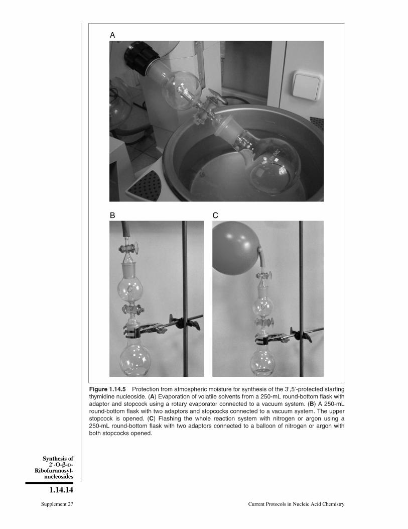
Prepare S.111. Weigh 3.78 g (30 mmol) of thymine and 20 mg of (NH4)2SO4 into a 250-mL
round-bottom flask, add 50 mL of 1,1,1,3,3,3-hexamethyldisilazane (b.p. 125◦C),and attach a condenser equipped with CaCl2 protection tube.
2. Reflux the mixture in an oil bath set at 130◦C until complete dissolution of thymine(10 to 12 hr).
3. Cool the flask, remove the condenser, and attach an adaptor with a stopcock (Fig.1.14.5A). Concentrate the solution to a viscous oil using a rotary evaporator con-nected to a vacuum system (bath temperature ∼30◦ to 35◦C).
The resulting bis-O-trimethylsilylthymine S.10 is very hygroscopic.
4. Close the stopcock of adaptor and attach a second adaptor with stopcock to theflask. Connect the system to a vacuum pump (Fig. 1.14.5B), evacuate the system,and close the upper stopcock. Connect the system with a balloon of nitrogen orargon and open the upper stopcock to fill the adaptor with nitrogen. Repeat thisprocedure (evacuation and flash with nitrogen) and then open both stopcocks to fillthe entire system with nitrogen (Fig. 1.14.5C).
5. Dissolve the residue in 30 mL of anhydrous 1,2-dichloroethane and concentrate thesolution to a viscous oil using a rotary evaporator with a bath temperature ∼30◦ to35◦C.
6. Repeat step 4.
7. Dissolve the residue in 120 mL of anhydrous 1,2-dichloroethane. Add 12.6 g(25 mmol) of 1-O-acetyl-2,3,5-tri-O-benzoyl-β-D-ribofuranose (S.2) in one portionand 5.79 mL (32 mmol) of trimethylsilyl trifluoromethanesulfonate (TMSOTf) withgentle hand stirring.
8. Attach a condenser equipped with CaCl2 protection tube and heat the clear solutionfor 1 hr in a 100◦C oil bath. Monitor reaction by TLC in 98:2 (v/v) CH2Cl2/MeOH.
The starting S.2 (Rf = 0.91) usually disappears after 1 hr at 100◦C. The product S.11(Rf = 0.45) moves slower in the same solvent.
9. Cool the flask to room temperature. Remove the condenser, add 50 mL of saturatedsodium bicarbonate solution, and stir the suspension for 20 min at 20◦C.
10. Put a 2- to 3-cm layer of Hyflo Super Cel on a 100-mL funnel with a sintered disc(porosity 3) and wash with 20 mL CH2Cl2. Filter the suspension using vacuumpump and wash the layer with 50 mL CH2Cl2.
11. Separate the organic layer using a 250-mL separatory funnel and wash the organiclayer with 50 mL of water.
12. Dry the organic layer over ∼20 g Na2SO4, filter off Na2SO4 by gravity filtration,and wash the precipitate with 40 mL CH2Cl2.
13. Concentrate the combined filtrates to a solid using a rotary evaporator connected tovacuum system.
14. Add 100 mL of ethanol, attach a condenser, and reflux (79◦C) until the solid iscompletely dissolved. Cool the flask to room temperature and keep the mixture for16 hr at 0◦C.
15. Collect the precipitate by vacuum filtration on a glass filter (porosity 3), washthe precipitate with 10 mL ethanol, and dry S.11 over phosphorus pentoxide in avacuum desiccator overnight.
Synthesis of2′-O-ββ-d-
Ribofuranosyl-nucleosides
1.14.14
Supplement 27 Current Protocols in Nucleic Acid Chemistry
Figure 1.14.5 Protection from atmospheric moisture for synthesis of the 3′,5′-protected startingthymidine nucleoside. (A) Evaporation of volatile solvents from a 250-mL round-bottom flask withadaptor and stopcock using a rotary evaporator connected to a vacuum system. (B) A 250-mLround-bottom flask with two adaptors and stopcocks connected to a vacuum system. The upperstopcock is opened. (C) Flashing the whole reaction system with nitrogen or argon using a250-mL round-bottom flask with two adaptors connected to a balloon of nitrogen or argon withboth stopcocks opened.

Synthesis ofModifiedNucleosides
1.14.15
Current Protocols in Nucleic Acid Chemistry Supplement 27
16. Characterize the compound by TLC and 1H NMR.
The compound is stable for at least 12 months at 20◦C.
1-(2,3,5-Tri-O-benzoyl-β-D-ribofuranosyl)thymine (S.11): Yield of white crystals 11.7 g(82%). m.p. 158◦-159◦C (EtOH). TLC: Rf 0.45 (98:2 v/v CH2Cl2/MeOH). 1H NMR (400MHz, CDCl3): 8.32 brs (1H, NH), 8.15-7.32 m (15H, Ph), 7.13 q (1H, J6,Me = 1.2, H6),6.39 d (1H, J1′,2′ = 6.4, H1′), 5.87 dd (1H, J3′,2′ = 6.0, J3′,4′ = 3.7, H3′), 5.74 dd (1H,H2′), 4.83 dd (1H, J5′a,4′ = 2.6, J5′a,5′b = −12.2, H5′a), 4.63 ddd (1H, J4′,5′b = 3.4, H4′),4.61 dd (1H, H5′b), 1.55 d (3H, Me).
Prepare S.1217. Weigh 11.4 g (20 mmol) S.11 into a 500-mL round-bottom flask. Add 200 mL of 0.2 N
NaOMe, stopper the flask, and stir the reaction for 30 min at room temperature.
18. Monitor reaction by TLC in 98:2 (v/v) CH2Cl2/MeOH.
The starting S.11 (Rf = 0.45) usually disappears after 30 min at 20◦C. The product S.12(Rf = 0.01) moves slower in the same solvent.
19. Remove MeOH under reduced pressure using a rotary evaporator in vacuo anddissolve the residue in 100 mL water and 100 mL chloroform.
20. Separate the aqueous layer using a 500-mL separatory funnel and wash with 50 mLchloroform.
21. Apply aqueous solution to a 3 × 20–cm chromatography column containing 50 mLof Dowex 50 × 4 (100 to 200 mesh) in H+ form packed in water.
22. Elute with water and collect 50-mL UV-containing fractions until elution is com-plete (elution volume will vary from 200 to 400 mL). Monitor fractions by TLCusing 9:1 (v/v) CH2Cl2/MeOH (Rf = 0.11.).
23. Combine the fractions that contain S.12 in a 500-mL round-bottom flask andevaporate to dryness using a rotary evaporator connected to a vacuum system.
24. Add 30 mL EtOH, stopper the flask, and keep the mixture for 16 hr at 0◦C.
25. Collect the precipitate by vacuum filtration on a glass filter (porosity 3), wash theprecipitate with 10 mL EtOH followed by 10 mL diethyl ether, and dry S.12 overphosphorus pentoxide in a vacuum desiccator overnight.
26. Characterize the compound by TLC, UV, and 1H NMR.
The compound is stable for at least 12 months at 20◦C.
1-(β-D-Ribofuranosyl)thymine (S.12): Yield of white crystals 4.0 g (78%). m.p. 180◦-181◦C (EtOH). TLC: Rf 0.11 (9:1 v/v CH2Cl2/MeOH). UV (pH 1-7): λmax 269 nm(ε9200); (pH 13): λmax 264 nm (ε7300). 1H NMR (400 MHz, D2O): 7.62 s (1H, H6),5.85 d (1H, J1′,2′ = 4.9 Hz, H1′), 4.29 dd (1H, J2′,3′ = 5.2 Hz, H2′), 4.19 dd (1H, J3′,4′ =5.4 Hz, H3′), 4.08 ddd (1H, J4′,5′a = 3.0 Hz, J4′,5′b = 4.3 Hz, H4′), 3.87 dd (1H, J5′a,5′b= −12.8 Hz, H5′a), 3.78 dd (1H, H5′b), 1.85 s (3H, Me).
Prepare S.6b27. Weigh 2.6 g (10 mmol) of dry S.12 in a 100-mL round-bottom flask and add 20 mL
anhydrous pyridine. Evaporate using a rotary evaporator equipped with a wateraspirator and then apply a dry nitrogen atmosphere (steps 3 to 4, Fig. 1.14.5).
28. Dissolve the residue in 40 mL pyridine with magnetic stirring. Rapidly add 3.14 mL(10 mmol) 1,3-dichloro-1,1,3,3-tetraisopropyldisiloxane (Markiewicz reagent) inone portion, stopper the flask, and keep 4 hr at 20◦C.
Synthesis of2′-O-ββ-d-
Ribofuranosyl-nucleosides
1.14.16
Supplement 27 Current Protocols in Nucleic Acid Chemistry
29. Monitor reaction by TLC using 98:2 (v/v) CH2Cl2/MeOH.
The starting S.12 (Rf = 0.01) usually disappears after 4 to 6 hr at 20◦C. The productS.6b (Rf = 0.23) moves faster in the same solvent.
30. Remove nearly all the pyridine using a rotary evaporator connected to a vacuumsystem and dissolve the residue in 50 mL water and 100 mL CH2Cl2.
31. Separate the organic layer using a 250-mL separatory funnel and wash with 20 mLsaturated sodium bicarbonate solution and then with 20 mL water.
32. Dry the organic layer over ∼10 g Na2SO4, filter off Na2SO4 by gravity filtration,and wash the precipitate with 20 mL CH2Cl2.
33. Evaporate the combined filtrates using a rotary evaporator connected to a vacuumsystem. Remove traces of pyridine by co-evaporating two times with 20 mL toluene.
34. Prepare a slurry of 50 g of silica gel in CH2Cl2 and pour it into a 3 × 20–cmchromatography column.
35. Dissolve the obtained residue in a minimal amount CH2Cl2 and layer it carefullyon top of the silica gel.
36. Wash column with 300 mL CH2Cl2 and 300 mL of 0.5% (v/v) MeOH in CH2Cl2.Elute with 1% (v/v) MeOH in CH2Cl2. Collect 25-mL fractions.
37. Evaluate fractions by TLC and combine the fractions that contain only S.6b. Evap-orate the volatile materials from the combined fractions using a rotary evaporatorconnected to a vacuum system, and dry the residual foam for 2 to 3 hr using avacuum oil pump.
38. Grind the foam with a stainless steel spatula and dry the resulting powder 12 to 16hr using a vacuum oil pump.
39. Characterize the product by TLC and 1H NMR.
The compound is stable for at least 12 months at ambient temperature.
1-[3,5-(1,1,3,3-Tetraisopropyldisiloxane-1,3-diyl)-β-D-ribofuranosyl]thymine (S.6b):Yield of white amorphous solid 4.30 g (86%). TLC: Rf 0.23 (98:2 v/v CH2Cl2/MeOH).1H NMR (400 MHz, CDCl3): 9.18 brs (1H, NH), 7.44 s (1H, H6), 5.70 s (1H, H1′), 4.38dd (1H, J3′,2′ = 5.1, Hz J3′,4′ = 8.6 Hz, H3′), 4.18 dd (1H, J5′a,4′ = 2.5 Hz, J5′a,5′b =−13.2 Hz, H5′a), 4.17 d (1H, H2′), 3.95 ddd (1H, J4′,5′b = 2.9 Hz, H4′), 4.01 dd (1H,H5′b), 1.89 s (3H, Me), 1.09-1.02 m (28H, iPr). 13C NMR (CDCl3): 163.93 (C4), 150.21(C2), 135.99 (C6), 110.74 (C5), 91.42 (C1′), 82.14 (C4′), 75.15 (C2′), 69.57 (C3′), 60.81(C5′), 17.57, 17.50, 17.41, 17.38, 17.22, 17.14, 17.10, 17.02, 13.59, 13.15, 12.91 and12.76 (iPr), 12.63 (Me).
COMMENTARY
Background InformationDisaccharide nucleosides belong to an im-
portant group of natural compounds that formpoly(ADP-ribose) and are found in tRNA,antibiotics, and other physiologically activecompounds. To date, ∼100 disaccharide nu-cleosides and related derivatives have beenisolated from a variety of natural sources.These compounds contain an extra carbohy-drate residue linked to one of the nucleosidehydroxyl groups via an O-glycosidic bond.The disaccharide residue and heterocyclic basemake their properties similar to those of carbo-
hydrates and nucleosides. These compoundsmanifest a broad spectrum of biological activ-ities, including antibacterial, fungicidal, herbi-cidal, antitumoral, and antiviral (Lerner, 1991;Efimtseva and Mikhailov, 2004; Nauwelaertset al., 2004).
An effective and simple synthesis of 2′-O-β-D-ribofuranosylnucleosides, minor tRNAcomponents, has been recently elaborated(Mikhailov et al., 1997a; Markiewicz et al.,1998). The method consists of the condensa-tion of a small excess of 1-O-acetyl-2,3,5-tri-O-benzoyl-β-D-ribofuranose activated with
Synthesis ofModifiedNucleosides
1.14.17
Current Protocols in Nucleic Acid Chemistry Supplement 27
tin tetrachloride with N-protected 3′,5′-O-tetraisopropyldisiloxane-1,3-diyl-ribonucleo-sides in 1,2-dichloroethane. O-Glycosylationproceeds stereospecifically with the formationof a β-glycosidic bond. Yields of disaccharidenucleosides reach 70% to 80% if the reactionis performed at 0◦C (Mikhailov et al., 1997a).O-Glycosylation proceeds stereospecificallywith formation of a β-glycosidic bond.
After complete deprotection, 2′-O-β-D-ribofuranosylnucleoside disaccharidenucleosides are obtained in good overallyields (Mikhailov et al., 1997a; Markiewiczet al., 1998). This method has been usedfor the preparation of pyrimidine 3′-O-β-D-ribofuranosyl-2′-deoxyribonucleosides and3′-O-β-D-ribopyranosyl-2′-deoxyribonucleo-sides (Mikhailov et al., 1996), and for5′-O-β-D-ribofuranosyl-2′-deoxyribonucleo-sides and 5′-O-β-D-ribofuranosylnucleosides(Mikhailov et al., 1997b, 1998). To studythe broad applicability of the method, someother sugars such as fully acylated D- andL-arabinofuranose, D-ribopyranose, andD-erythrofuranose have been used in the O-glycosylation reaction, and the correspondingdisaccharide nucleosides have been incor-porated into oligodeoxyribonucleotides andoligoribonucleotides (Efimtseva et al., 2001).Melting point determinations demonstratedthat modified oligonucleotides containingdisaccharide nucleosides form stable duplexeswith complementary RNA (�Tm = 0◦C;Efimtseva et al., 2001). The duplex RNAmaintains an A-type helical geometry with theextra 2′-O-ribose moiety located in the minorgroove. This implies that the voluminousextra sugar moiety has almost no effect on thestability of the RNA duplex (Luyten et al.,2000).
Oligonucleotides containing 2′-O-ribofur-anosylnucleosides have been used as modi-fied primers in RNA-templated DNA synthe-sis catalyzed by HIV reverse transcriptase. Itwas shown that an additional 2′-ribofuranoseresidue in a specific position (-3-4) of theprimer prevents elongation (Andreeva et al.,2002). An important feature of these oligonu-cleotides is the presence of an additionalcis-diol group, which may be readily oxi-dized with sodium periodate to give oligonu-cleotide derivatives with aldehyde groupsplaced anywhere in the sequence (Efimtsevaand Mikhailov, 2002). Such oligonucleotideswere used for affinity labelling of differentenzymes (Tunitskaya et al., 1999; Gritsenkoet al., 2002). The incorporation of disaccha-ride nucleosides into oligonucleotides opens
new possibilities for the functionalization ofnucleic acids. The extra sugar ring attached to anucleoside can serve several purposes. For thesynthetic work, it is important that acyl block-ing groups used in disaccharide synthesis arecompatible with the chemistry of automatedoligonucleotide synthesis.
Compound CharacterizationThe structure of the compound is supported
by NMR spectroscopy and mass spectrometry.The 1H NMR spectra of the obtained com-pounds are rather complicated due to the pres-ence of two ribofuranose residues. In spite ofthis, most of the chemical shifts and couplingconstants may be calculated directly from theNMR spectra. In some cases, comparison withthe published spectra of disaccharide nucleo-sides and 1H-13C correlation and COSY spec-tra were used for assignment. The chemicalshifts were assigned using double resonancetechniques and COSY experiments.
Several conclusions were drawn from the1H NMR spectral analysis. In nucleosides S.1and S.6 and disaccharide nucleosides S.3 andS.7 with a 1,1,3,3-tetraisopropyldisiloxane-1,3-diyl group, the coupling constants J1′,2′
of nucleoside moieties are <0.5 Hz (Robinset al., 1983). The O-glycosylation reactionproceeded stereospecifically with the forma-tion of 1′′,2′′-trans-substituted disaccharidenucleosides S.3 and S.7. The coupling con-stants (J1′ ′,2′ ′ ) in the additional ribose moietyare <0.5 Hz. As in the case of disaccharidenucleosides (Mikhailov et al., 1997a) and 2′-O-methylnucleosides (Robins et al., 1981), in-corporation of the 2′-O-substitutent results ina low field shift (+2+3 ppm) of C2′ of thenucleoside moiety in 13C NMR spectra.
Critical Parameters andTroubleshooting
The synthesis of 2′-O-β-D-ribofuranosyl-nucleosides is short, straightforward, andfairly efficient. However, careful attention todetails of basic organic synthesis proceduresis required. Preparation of the various com-pounds requires prior experience with routinechemical laboratory techniques such as sol-vent evaporation, extraction, TLC, and columnchromatography. Characterization of the prod-ucts demands knowledge of 1H and 13C NMR,UV, and mass spectroscopy. General labora-tory safety is also of primary concern whenhazardous materials are involved. Strict ad-herence to the outlined methods is thereforehighly recommended.

Synthesis of2′-O-ββ-d-
Ribofuranosyl-nucleosides
1.14.18
Supplement 27 Current Protocols in Nucleic Acid Chemistry
All solvents should be distilled before use.Anhydrous solvents are very important. Theyshould be either freshly distilled and storedover molecular sieves, or be taken from afreshly opened bottle of commercially pre-pared anhydrous solvent. It is important foreach step of the syntheses that the startingcompounds be thoroughly dried either by co-evaporation with anhydrous pyridine or ina desiccator over P2O5. Protection of reac-tion mixtures from atmospheric moisture (asshown in Figs. 1.14.2 and 1.14.5) will enhanceyields, especially in the case of ribosylationand silylation reactions.
For all compounds, a sample of 50 to 100mg should be kept as a reference. In all cases,the reaction progress is followed by TLC. Abaseline is marked and the spots (∼1 opticalunit) of the reaction mixture and starting ma-terial are placed at equal distances. For thepreparation of S.3 and S.7, where the Rf val-ues of the starting compounds and products arevery similar, a mixed sample containing bothstarting compound and reaction mixture maybe placed on the TLC plate. After developingin the appropriate solvent, the end-line of theTLC is marked, and spots are identified undera UV lamp (254 nm).
Before each column purification, TLC isperformed on the reaction mixture to evaluatethe completeness of the reaction and to choosethe optimal elution system for column chro-matography. The silica gel for column chro-matography (0.06 to 0.20 mm) is suspendedin methylene chloride and loaded into the col-umn, which is tightly packed by gentle tap-ping until there is a constant volume. Thecrude product is dissolved in a minimal vol-ume of solvent and is carefully applied usinga glass Pasteur pipet. After sample applica-tion, the flask and column walls are washedwith a minimal volume of solvent. For protec-tion, the silica layer is topped by a 1-cm layerof sand.
Anticipated ResultsThe method presented here generally pro-
duces high yields even for inexperiencedworkers. Good overall yields (46% to 72%)of the final 2′-O-ribofuranosylnucleosides (S.5and S.9) from the Markiewicz-protected nu-cleosides (S.1 and S.6) are expected follow-ing these procedures. 2′-O-Ribosylation pro-ceeds stereospecifically with formation of aβ-glycosidic bond. The first ribosylation reac-tion is carried out under mild conditions (0◦C,1,2-dichloroethane, 2 hr for pyrimidine nucle-osides, 7 to 16 hr for purine derivatives) and
the yields of the target compounds are 72% to80% (Mikhailov et al., 1997a). At room tem-perature, the reaction occurs faster (30 min),but the yields in the coupling steps are lower(40% to 77%; Markiewicz et al., 1998).
Time ConsiderationsThe three-step preparation of 2′-O-
β-D-ribofuranosylnucleosides starting fromMarkiewicz-protected nucleosides can be ac-complished in 1 week. Each column chro-matography step requires 3 to 5 hr. Normallythe first two steps (ribosylation and silylation)may be performed in 3 to 4 working days,including purification and spectroscopic anal-ysis. The final deacylation reaction normallyproceeds in 2 to 3 days at room temperatureand may be run over a weekend. The three-steppreparation of the protected thymine startingnucleoside (S.6b) may be accomplished in 1week.
Literature CitedAndreeva, O.I., Golubeva, A.S., Kochetkov, S.N.,
Van Aerschot, A., Herdewijn, P., Efimtseva,E.V., Ermolinsky, B.S., and Mikhailov, S.N.2002. An additional 2′-ribofuranose residue ata specific position of DNA primer preventsits elongation by HIV-1 reverse trancriptase.Bioorg. Med. Chem. Lett. 12:681-684.
Efimtseva, E.V. and Mikhailov, S.N. 2002. Dis-accharide nucleosides and oligonucleotides ontheir bases. New tools for the study of en-zymes of nucleic acid metabolism. Biochemistry67:1136-1144.
Efimtseva, E.V. and Mikhailov, S.N. 2004. Disac-charide nucleosides. Russ. Chem. Rev. 73:401-414.
Efimtseva, E.V., Bobkov, G.V., Mikhailov, S.N.,Van Aerschot, A., Schepers, G., Busson, R.,Rozenski, J., and Herdewijn, P. 2001. Oligonu-cleotides containing disaccharide nucleosides.Helv. Chim. Acta 84:2387-2397.
Gritsenko, O.M., Koudan, E.V., Mikhailov, S.N.,Ermolinsky, B.S., Van Aerschot, A., Herdewijn,P., and Gromova, E.S. 2002. Affinity modifi-cation of EcoRII DNA methyltransferase bythe dialdehyde-substituted DNA duplexes: Map-ping the enzyme region that interacts with DNA.Nucleosides Nucleotides Nucleic Acids 21:753-764.
Lerner, L.M. 1991. Synthesis and Properties of Var-ious Disaccharide Nucleosides. In Chemistryof Nucleosides and Nucleotides, Vol. 2 (L.B.Townsend, ed.) pp. 27-79. Plenum Press, NewYork.
Luyten, I., Esnouf, R.M., Mikhailov, S.N.,Efimtseva, E.V., Michiels, P., Heus, H.A.,Hilbers, C.W., and Herdewijn, P. 2000. So-lution structure of a RNA decamer duplex,containing 9-[(2-O-(β-D-ribofuranosyl)-(β-D-ribofuranosyl]adenine, a special residue in lower
Synthesis ofModifiedNucleosides
1.14.19
Current Protocols in Nucleic Acid Chemistry Supplement 27
eukaryotic initiator tRNAs. Helv. Chim. Acta83:1278-1289.
Markiewicz, W.T. and Wiewiorowski, M. 1986.1-[3,5-(1,1,3,3-Tetraisopropyldisiloxane-1,3-diyl)ribonucleosides. In Nucleic Acid Chem-istry. Improved and New Synthetic Proceduresand Techniques, Part 3: (L.B. Townsend andR.S. Tipson, eds.) pp. 229-231. John Wiley &Sons, Hoboken.
Markiewicz, W.T., Niewczyk, A., Gdaniec, Z.,Adamiak, D.A., Dauter, Z., Rypniewski, W., andChmielewski, M. 1998. Studies on synthesisand structure of O-β-D-ribofuranosyl(1′ ′→2′)ribonucleosides and oligonucleotides.Nucleosides Nucleotides Nucleic Acids 17:411-424.
Mikhailov, S.N., De Clercq, E., and Herdewijn,P. 1996. Ribosylation of pyrimidine 2′-deoxynucleosides. Nucleosides NucleotidesNucleic Acids 15:1323-1334.
Mikhailov, S.N., Efimtseva, E.V., Gurskaya, G.V.,Zavodnik, V.E., De Bruyn, A., Janssen, G.,Rozenski, J., and Herdewijn, P. 1997a. An ef-ficient synthesis and physico-chemical proper-ties of 2′-O-β-D-ribofuranosyl-nucleosides, mi-nor tRNA components. J. Carbohydr. Chem.16:75-92.
Mikhailov, S.N., Rodionov, A.A., Efimtseva,E.V., Fomitcheva, M.V., Padyukova, N.Sh.,Herdewijn, P., and Oivanen, M. 1997b. Prepa-ration of pyrimidine 5′-O-β-D-ribofuranosyl-nucleosides, and hydrolytic stability of O-D-ribofuranosyl-nucleosides. Carbohydrate Lett.2:321-328.
Mikhailov, S.N., Rodionov, A.A., Efimtseva,E.V., Ermolinsky, B.S., Fomitcheva, M.V.,Padyukova, N.Sh., Rothenbacher, K.,Lescrinier, E., and Herdewijn, P. 1998.Formation of trisaccharide nucleoside duringdisaccharide nucleoside synthesis. Eur. J. Org.Chem. 2193-2199.
Nauwelaerts, K., Efimtseva, E.V., Mikhailov, S.N.,and Herdewijn, P. 2004. Disaccharide nucleo-sides, an important group of natural compoundsIn Frontiers in Nucleosides and Nucleic Acids(R.F. Schinazi and D.C. Liotta, eds.) pp 187-220.IHL Press, Arlington, MA.
Robins, M.J., Hansske, F., and Bermier, S.E. 1981.Nucleic acid related compounds. 36. Synthe-sis of the 2′-O-methyl and 3′-O-methyl ethersguanosine and 2-amino adenosine and correla-tion of O′-methylnucleoside C13 NMR spectralshifts. Can. J. Chem. 59:3360-3364.
Robins, M.J., Wilson, J.S., Sawyer, L., andJames, M.N.G. 1983. Nucleic acid relatedcompounds. 41. Restricted furanose conforma-tions of 3′,5′-(1,1,3,3-tetraisopropyldisilox-1,3-diyl)nucleosides provide a convenient evalua-tion of anomeric configuration. Can. J. Chem.59:1911-1920.
Tunitskaya, V.L., Rusakova, E.E., Memelova,L.V., Kochetkov, S.N., Van Aerschot, A.,Herdewijn, P., Efimtseva, E.V., Ermolinsky,B.S., and Mikhailov, S.N. 1999. The map-ping of T7 RNA polymerase active site withnovel reagents—Oligonucleotides with reactivedialdehyde groups. FEBS Lett. 442:20-24.
Vorbruggen, H. and Ruh-Pohlenz, C. 2000. Synthe-sis of nucleosides. Org. React. 55:1-630.
Contributed by Sergey N. Mikhailov,Ekaterina V. Efimtseva, Andrei A.Rodionov, Georgii V. Bobkov, andIrina V. Kulikova
Russian Academy of SciencesEngelhardt Institute of Molecular BiologyMoscow, Russia
Piet HerdewijnRega Institute for Medical ResearchLeuven, Belgium