![Secure Concurrent Constraint Programming - …catuscia/Talks/060820_ICLP/sccp.pdfRemark 1. (Behavioral Charecterization ... Notice thecorrespondencebetween (νx ... [Mil95] J. Millen.](https://static.fdocument.org/doc/165x107/5ab3d54a7f8b9ac3348ea67e/secure-concurrent-constraint-programming-catusciatalks060820iclpsccppdfremark.jpg)
Concurrent Chemoradiotherapy Shows Long-Term Survival … · Concurrent Chemoradiotherapy Shows...
9
Yonsei Med J http://www.eymj.org Volume 55 Number 6 November 2014 1489 Concurrent Chemoradiotherapy Shows Long-Term Survival after Conversion from Locally Advanced to Resectable Hepatocellular Carcinoma Ik Jae Lee, 1 Jun Won Kim, 1 Kwang Hyub Han, 2 Ja Kyung Kim, 2 Kyung Sik Kim, 3 Jin Sub Choi, 3 Young Nyun Park, 4 and Jinsil Seong 1 Departments of 1 Radiation Oncology, 2 Internal Medicine, 3 Surgery, and 4 Pathology, Yonsei Liver Cancer Special Clinic, Yonsei University College of Medicine, Seoul, Korea. Received: November 18, 2013 Revised: January 15, 2014 Accepted: February 11, 2014 Corresponding author: Dr. Jinsil Seong, Department of Radiation Oncology, Yonsei University College of Medicine, 50-1 Yonsei-ro, Seodaemun-gu, Seoul 120-752, Korea. Tel: 82-2-2228-8095, Fax: 82-2-2227-7823 E-mail: [email protected] This study was selected for poster presentation at the 54th annual meeting of the American Society for Therapeutic Radiation Oncology (ASTRO) in Boston, MA, on October 28–31, 2012. ∙ The authors have no financial conflicts of interest. © Copyright: Yonsei University College of Medicine 2014 This is an Open Access article distributed under the terms of the Creative Commons Attribution Non- Commercial License (http://creativecommons.org/ licenses/by-nc/3.0) which permits unrestricted non- commercial use, distribution, and reproduction in any medium, provided the original work is properly cited. Purpose: For locally unresectable hepatocellular carcinoma (HCC) patients, concur- rent chemoradiotherapy (CCRT) has been applied as a loco-regional treatment. After shrinkage of tumors in selected patients, surgical resection is performed. The aim of this study was to evaluate prognostic factors and long-term survivors in such pa- tients. Materials and Methods: From January 2000 to January 2009, 264 patients with HCC were treated with CCRT (45 Gy with fractional dose of 1.8 Gy), and intra- arterial chemotherapy was administered during radiotherapy. Eighteen of these pa- tients (6.8%) underwent hepatic resection after showing a response to CCRT. Cases were considered resectable when tumor-free margins and sufficient remnant vol- umes were obtained without extrahepatic metastasis. Prior to operation, there were six patients with complete remission, 11 with partial remission, and six with stable disease according to modified Response Evaluation Criteria in Solid Tumors. Re- sults: In pathologic review, four patients (22.2%) showed total necrosis and seven patients (38.9%) showed 70‒99% necrosis. A high level of necrosis (≥80%) was cor- related with low risk for extrahepatic metastasis and long-term survival. In univariate analyses, vessel invasion and capsular infiltration were significantly correlated with disease free survival (DFS) ( p=0.017 and 0.013, respectively), and vessel invasion was significantly correlated with overall survival (OS) (p=0.013). In multivariate analyses, capsule infiltration was a significant factor for DFS ( p=0.016) and vessel invasion was significant for OS ( p=0.015). Conclusion: CCRT showed favorable re- sponses and locally advanced HCC converted into resectable tumor after CCRT in selected patients. Long-term survivors showed the pathological features of near total necrosis, as well as negative capsule and vessel invasion. Key Words: Hepatocellular carcinoma, concurrent chemoradiotherapy, intra-arte- rial chemotherapy, hepatic resection INTRODUCTION Hepatocellular carcinoma (HCC) is a major disease in Asian countries. Global Original Article http://dx.doi.org/10.3349/ymj.2014.55.6.1489 pISSN: 0513-5796, eISSN: 1976-2437 Yonsei Med J 55(6):1489-1497, 2014
Transcript of Concurrent Chemoradiotherapy Shows Long-Term Survival … · Concurrent Chemoradiotherapy Shows...
Yonsei Med J http://www.eymj.org Volume 55 Number 6 November 2014 1489
Concurrent Chemoradiotherapy Shows Long-Term Survival after Conversion from Locally Advanced to Resectable
Hepatocellular Carcinoma
Ik Jae Lee,1 Jun Won Kim,1 Kwang Hyub Han,2 Ja Kyung Kim,2 Kyung Sik Kim,3 Jin Sub Choi,3 Young Nyun Park,4 and Jinsil Seong1
Departments of 1Radiation Oncology, 2Internal Medicine, 3Surgery, and 4Pathology, Yonsei Liver Cancer Special Clinic, Yonsei University College of Medicine, Seoul, Korea.
Received: November 18, 2013Revised: January 15, 2014Accepted: February 11, 2014Corresponding author: Dr. Jinsil Seong, Department of Radiation Oncology, Yonsei University College of Medicine,50-1 Yonsei-ro, Seodaemun-gu, Seoul 120-752, Korea.Tel: 82-2-2228-8095, Fax: 82-2-2227-7823E-mail: [email protected]
This study was selected for poster presentation at the 54th annual meeting of the American Society for Therapeutic Radiation Oncology (ASTRO) in Boston, MA, on October 28–31, 2012.
∙ The authors have no financial conflicts of interest.
© Copyright:Yonsei University College of Medicine 2014
This is an Open Access article distributed under the terms of the Creative Commons Attribution Non-Commercial License (http://creativecommons.org/ licenses/by-nc/3.0) which permits unrestricted non-commercial use, distribution, and reproduction in any medium, provided the original work is properly cited.
Purpose: For locally unresectable hepatocellular carcinoma (HCC) patients, concur-rent chemoradiotherapy (CCRT) has been applied as a loco-regional treatment. After shrinkage of tumors in selected patients, surgical resection is performed. The aim of this study was to evaluate prognostic factors and long-term survivors in such pa-tients. Materials and Methods: From January 2000 to January 2009, 264 patients with HCC were treated with CCRT (45 Gy with fractional dose of 1.8 Gy), and intra-arterial chemotherapy was administered during radiotherapy. Eighteen of these pa-tients (6.8%) underwent hepatic resection after showing a response to CCRT. Cases were considered resectable when tumor-free margins and sufficient remnant vol-umes were obtained without extrahepatic metastasis. Prior to operation, there were six patients with complete remission, 11 with partial remission, and six with stable disease according to modified Response Evaluation Criteria in Solid Tumors. Re-sults: In pathologic review, four patients (22.2%) showed total necrosis and seven patients (38.9%) showed 70‒99% necrosis. A high level of necrosis (≥80%) was cor-related with low risk for extrahepatic metastasis and long-term survival. In univariate analyses, vessel invasion and capsular infiltration were significantly correlated with disease free survival (DFS) (p=0.017 and 0.013, respectively), and vessel invasion was significantly correlated with overall survival (OS) (p=0.013). In multivariate analyses, capsule infiltration was a significant factor for DFS (p=0.016) and vessel invasion was significant for OS (p=0.015). Conclusion: CCRT showed favorable re-sponses and locally advanced HCC converted into resectable tumor after CCRT in selected patients. Long-term survivors showed the pathological features of near total necrosis, as well as negative capsule and vessel invasion.
Key Words: Hepatocellular carcinoma, concurrent chemoradiotherapy, intra-arte-rial chemotherapy, hepatic resection
INTRODUCTION
Hepatocellular carcinoma (HCC) is a major disease in Asian countries. Global
Original Article http://dx.doi.org/10.3349/ymj.2014.55.6.1489pISSN: 0513-5796, eISSN: 1976-2437 Yonsei Med J 55(6):1489-1497, 2014

Ik Jae Lee, et al.
Yonsei Med J http://www.eymj.org Volume 55 Number 6 November 20141490
cluding metastasis, portal vein invasion, and performance 0‒2. In this stage, National Comprehensive Cancer Network guidelines suggest that radiotherapy (RT) can be considered as an alternative to ablation/embolization techniques for un-resectable HCC with category 2B level evidence. Lee and Seong4 reviewed the optimal indications for RT according to the BCLC staging system and the application of various RT modalities and prescription according to the extent of dis-ease. Combining RT with radiosensitizers such as concurrent chemoradiotherapy (CCRT) is an also attractive strategy to increase the therapeutic ratio for these patients.5
Several studies reported downstaging or total necrosis of the tumor after non-surgical modalities, including trans-catheter arterial chemoembolization (TACE), hepatic artery chemo-infusion, combined systemic chemotherapy, exter-nal radiation, and radioimmunotherapy.6-9 Although tumor shrinkage was sufficient to allow salvage liver resection following therapy in only a small proportion of patients, the potential for curative therapy in responders also gives hope to patients with unresectable HCC.
In our institute, patients with locally advanced BCLC stage C were treated with CCRT when surgery was not fea-sible due to portal vein tumor thrombosis or inadequate non-tumor liver volume.10 Among them, some patients un-derwent surgical resection after showing a response to CCRT. In this study, we evaluated prognostic factors and long-term survival in patients who underwent hepatic re-section after CCRT.
MATERIALS AND METHODS
PatientsFrom January 2000 to January 2009, 264 patients with lo-cally advanced HCC were treated with external beam RT and concurrent intra-arterial (iA) chemotherapy (CTx) when surgery was not feasible due to portal vein tumor thrombo-sis or inadequate non-tumor liver volume. In some patients, CCRT showed a favorable response and eighteen of these patients (6.8%) underwent hepatic resection after multidis-ciplinary HCC conference. All patients provided informed written consent for participation in this study, which was approved by our Institutional Review Board. Table 1 shows the clinical characteristics of these patients. The median fol-low-up of the 18 patients was 40 months.
Diagnosis of HCC was based on either pathologic confir-mation or radiologic findings with an elevated serum α-feto-
cancer statistics showed this disease as the third most com-mon cause of cancer-related deaths worldwide.1 Since HCC is unresectable in the majority of patients at the time of initial visiting, patients are frequently referred to non curative treat-ments. Based on the results of randomized clinical trials, sorafenib has become the first-line therapy for advanced Bar-celona Clinic Liver Cancer (BCLC) stage C HCC.2,3 Howev-er, BCLC stage C represents a variety of disease features, in-
Table 1. Patient CharacteristicsCharacteristics Value (range) No. of patients (%)
Age Median: 51.5 (34‒68)
Sex Male 17 (94.4) Female 1 (5.6)Etiology HBV 15 (83.3) Non-B, -C 3 (16.7)Child-Pugh class A 15 (83.3) B 3 (16.7)ICG-R15 (%) Mean: 10.7 (4‒21)Main lesion Rt. lobe 13 (72.2) Lt. lobe 5 (27.8)Clinical stage (Japanese TNM) II 5 (27.8) III 6 (33.3) IV-A 7 (38.9)Tumor size before CCRT (cm) Mean: 10.7 (5‒20)
PVT No 12 (66.7) Yes 6 (33.3)Lymph node involvement No 16 (88.9) Yes 2 (11.1)Previous treatment No 12 (66.7) Yes 6 (33.3)AFP, ng/mL ≥400 9 (50.0) <400 9 (50.0)PIVKA-II, mAU/mL ≥200 13 (72.2) <200 5 (17.8)
HBV, hepatitis B virus; ICG, indocyanine green; CCRT, concurrent chemora-diotherapy; PVT, portal vein thrombosis; AFP, α-fetoprotein; PIVKA, protein induced by vitamin K absence.
Chemoradiation Followed by Resection for HCC
Yonsei Med J http://www.eymj.org Volume 55 Number 6 November 2014 1491
were obtained.12 The resectability of tumors was finally de-termined at multidisciplinary HCC conferences before and after CCRT.
Evaluation and statistical analysisTwo months after the completion of CCRT and at short-inter-val (3‒6 months) follow-up, abdominal/pelvic CT was used to evaluate radiologic tumor response. Modified Response Evaluation Criteria in Solid Tumors criteria were used to evaluate responses to CCRT.13 Serum AFP and protein in-duced by vitamin K absence (PIVKA-II) were measured at diagnosis and 2 months after CCRT for evaluation of chemi-cal response, as well as at 3‒6 months during follow-up, to detect recurrence. For chemical response, complete remis-sion (cCR) was defined as normalization of tumor marker, partial response (cPR) as at least a 50% decrease from the pretreatment level, progressive disease (cPD) as >25% in-crease of tumor marker from the pretreatment level, and stable disease (cSD) as any changes that do not meet other criteria.
A pathologist evaluated the resected tumor for evidence of gross and histologic necrosis after serial sectioning using a standard protocol (10 mm slice thickness). Slides were prepared using routine hematoxylin and eosin staining. The irradiated lesion was examined for the presence of viable neoplastic tissue, capsular invasion, microscopic vascular invasion, tumor size, tumor multiplicity, and resection mar-gins, and a pathologist quantitatively evaluated the percent-age of necrosis.
Patient survival was measured from the beginning of ra-diotherapy. Overall survival (OS) and disease free survival (DFS) were analyzed using proportional hazards (Cox) re-gression. The median survival time was estimated using Kaplan-Meier method.
RESULTS
Radiologic and chemical responses of CCRTRadiologic and chemical responses were determined 2 months after the completion of CCRT and immediately be-fore surgical resection. Table 2 shows the results of the treat-ment. Two months following CCRT, two (11.1%), 13 (72.2%), and three (16.7%) patients achieved radiological CR, PR, and SD, respectively. Immediately before the oper-ation, six (33.3%), 11 (61.1%), and one (5.6%) patients achieved CR, PR, and SD, respectively. Among the nine pa-
protein (AFP). If computed tomography (CT), magnetic resonance imaging (MRI), or arteriography detected a cir-rhotic liver with a hepatic mass larger than 2 cm (which suggests HCC) accompanied by AFP elevated more than 200 ng/mL, a biopsy was not necessary for HCC diagnosis. In order to be selected as a candidate for the CCRT proto-col, patients needed to have an Eastern Cooperative Oncol-ogy Group performance status of 0‒2 and adequate liver function [indocyanine green R15 (ICG R15) <30% and Child-Pugh class A or B]. Patients with tumors of diffuse or multifocal bilobal involvement were excluded to avoid whole-liver irradiation.
Concurrent chemoradiationAll of the 18 patients were treated with three-dimensional conformal radiotherapy. The gross tumor volume (GTV) was defined as the radiographically abnormal areas detect-ed in the CT or MRI images, and a minimum of 5 mm around the GTV was added to determine the clinical target volume (CTV). The range of diaphragmatic movement was measured via fluoroscope in order to incorporate cranio-caudal movement of the liver into defining the internal tar-get volume (ITV). In defining planning target volume (PTV), an additional 5 mm for set-up error was added to ITV. A total of 45 Gy was prescribed to the isocenter in 25 fractions (1.8 Gy/fraction) over 5 weeks using a 6-megavolt (MV) or 10-MV linear accelerator with the intention to de-liver 95% of the prescribed dose encompassing the PTV around the CTV.10
Continuous-infusion hepatic arterial 5-fluorouracil (5-FU) at a dose of 500 mg/day was delivered during the first and fifth weeks of RT. One month after CCRT, hepatic arte-rial 5-FU (at a dose of 500 mg/m2 for 5 hours on Days 1‒3) and cisplatin (at a dose of 60 mg/m2 for 2 hours on Day 2) were administered every 4 weeks for 3 to 12 cycles accord-ing to tumor response.
Indication and criteria for salvage surgery after CCRTTo evaluate operability, all patients underwent preoperative liver function tests, Child-Push score reassessment, and measurement of the ICG R15. The extent of resection was determined according to the ICG R15 and gross findings of the liver during a laparotomy. ICG R15 <10% was regard-ed as having no impaired liver function.11 For our hospital, cases were considered resectable if there was no extrahe-patic metastasis on preoperative imaging studies, and if ad-equate tumor-free margins and sufficient remnant volumes
Ik Jae Lee, et al.
Yonsei Med J http://www.eymj.org Volume 55 Number 6 November 20141492
patients at 2 months from CCRT, and cCR, cPR, or cPD were shown in five (28%), three (17%), and one (5.5%) pa-tients immediately before the operation, respectively. Among the 13 patients with elevated PIVKA-II above 250 ng/mL, cCR, cPR, and cPD were observed in six (33%), five (28%), and two (11%) patients 2 months after CCRT. The cCR, cPR, and cPD were observed in seven (39%), five (28%), and one (5.5%) patients immediately before the operation, respectively.
Pathologic findings after CCRT followed by operationThe median time interval from CCRT to surgical resection was 6.2 months (range 1‒21 months). The numbers of pa-tients who received right lobectomy, left lobectomy, central lobectomy, and bisegmentectomy were 12, four, one, and one, respectively. Table 3 shows reports from the patholog-ic review. Single and multiple tumors were found in 10 (56%) and eight (44%) patients, respectively, and four pa-tients (22%) had tumors larger than 10 cm in diameter. Ves-sel invasion and capsular infiltration each were confirmed in nine patients (50%), and close resection margin (≤1.0 cm) was detected in seven patients (39%). Four patients (22%) had total necrosis, seven patients (39%) had 70‒99% necrosis, and less than 70% necrosis was observed in seven
tients whose AFP level was above 200 ng/mL at diagnosis, cCR and cPR were achieved in two (11%) and seven (39%)
Table 2. Treatment Results of CCRTResponse after CCRT At 2 months (%) Before OP (%)Radiologic CR 2/18 (11.1) 6/18 (33.3) PR 13/18 (72.2) 11/18 (61.1) SD 3/18 (16.7) 1/18 (5.6)AFP iAFP ≥200 ng/mL CR 2/18 (11.1) 5/18 (27.8) PR 7/18 (38.9) 3/18 (16.7) SD 0/18 (-) 0/18 (-) PD 0/18 (-) 1/18 (5.5) iAFP <200 ng/mL 9/18 (50.0) 9/18 (50.0)PIVKA-II iPIVKA-II ≥250 ng/mL CR 6/18 (33.3) 7/18 (38.9) PR 5/18 (27.8) 5/18 (27.8) SD 2/18 (11.1) 0/18 (-) PD 0/18 (-) 1/18 (5.5) iPIVKA-II <250 ng/mL 5/18 (27.8) 5/18 (27.8)
CCRT, concurrent chemoradiation; CR, complete remission; PR, partial response; SD, stable disease; iAFP, α-fetoprotein at diagnosis; iPIVKA, protein induced by vitamin K absence at diagnosis; OP, operation; PD, pro-gressive disease.
Table 3. Pathologic Findings after Operation
Pathologic findings No. of patients (%)Elevated Preop AFP
(≥200 ng/mL) Elevated Preop PIVKA-II
(≥250 ng/mL)p value (chi-square)
No. of tumors Single 10 (55.6)
0.87 1.00 Multiple 8 (44.4)Tumor size <10 cm 14 (77.8)
0.005 0.27 ≥10 cm 4 (22.2)Microscopic vascular invasion No 9 (50.0)
0.13 0.25 Yes 9 (50.0)Capsular invasion No 9 (50.0)
0.13 0.02 Yes 9 (50.0)Margin of resection ≤1.0 cm No 11 (61.1)
0.06 0.76 Yes 7 (38.9)Degree of necrosis <80% 7 (77.8)
0.87 0.009 ≥80% 11 (61.1) <100% 14 (77.8)
0.42 0.27 100% 4 (22.2)
No., number; AFP, α-fetoprotein; PIVKA, protein induced by vitamin K absence.
Chemoradiation Followed by Resection for HCC
Yonsei Med J http://www.eymj.org Volume 55 Number 6 November 2014 1493
the degree of necrosis above 80% (p=0.009).
Patterns of recurrenceThe number of patients with intrahepatic metastasis was six (33.3%) and distant metastasis was eight (44.4%). For dis-tant metastases, lung was the most common site, followed by lymph nodes, pleural/omental seeding, and the brain. In the analysis of patterns of treatment failure (Table 4), high necrosis rate (≥80%) was associated with a low incidence of extrahepatic metastasis (p=0.02).
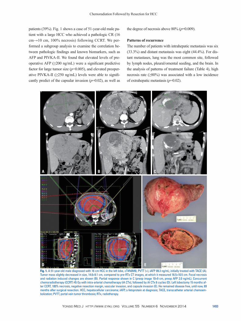
patients (39%). Fig. 1 shows a case of 51-year-old male pa-tient with a large HCC who achieved a pathologic CR (16 cm→10 cm, 100% necrosis) following CCRT. We per-formed a subgroup analysis to examine the correlation be-tween pathologic findings and known biomarkers, such as AFP and PIVKA-II. We found that elevated levels of pre-operative AFP (≥200 ng/mL) were a significant predictive factor for large tumor size (p=0.005), and elevated preoper-ative PIVKA-II (≥250 ng/mL) levels were able to signifi-cantly predict of the capsular invasion (p=0.02), as well as
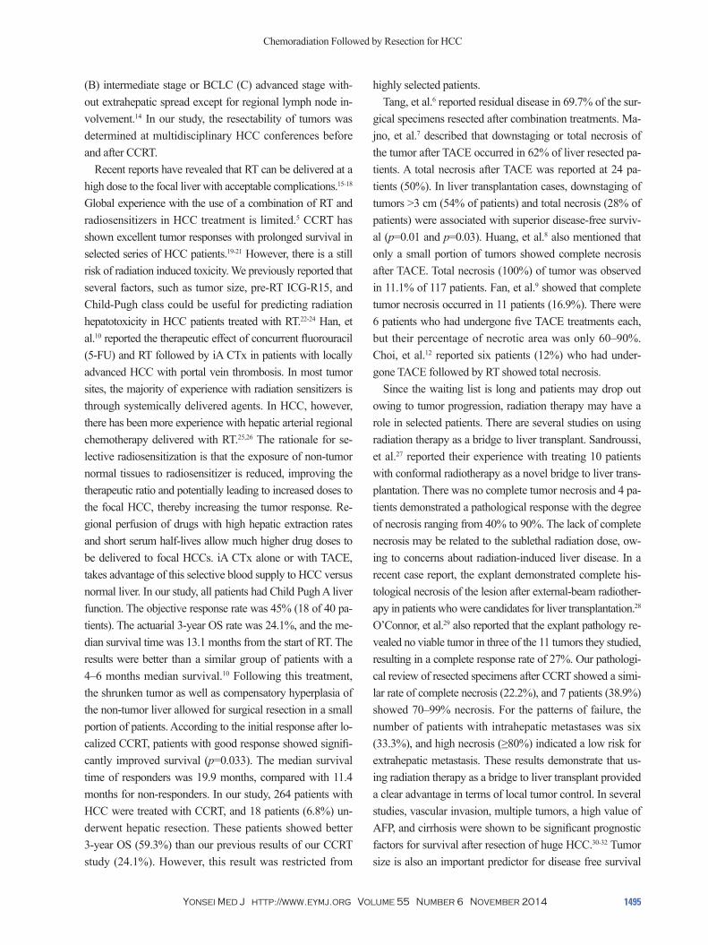
Fig. 1. A 51-year-old male diagnosed with 16 cm HCC in the left lobe, cT4N0M0, PVTT (+), iAFP 89.3 ng/mL; initially treated with TACE (A). Tumor mass slightly decreased in size, 14.6×9.1 cm, compared to pre-RTx CT images, at which it measured 16.5×10.5 cm. Focal necrosis and radiation induced changes are shown (B). Partial response shown in C (preop image 10×9 cm, preop AFP 2.0 ng/mL). Concurrent chemoradiotherapy (CCRT) 45 Gy with intra-arterial chemotherapy (iA CTx), followed by iA CTx 6 cycles (D). Left lobectomy 15 months af-ter CCRT, 100% necrosis, negative resection margin, vascular invasion, and capsule invasion (E). He remained disease free, until now, 69 months after surgical resection. HCC, hepatocellular carcinoma; iAFP, α-fetoprotein at diagnosis; TACE, transcatheter arterial chemoem-bolization; PVTT, portal vein tumor thrombosis; RTx, radiotherapy.
A
D
B C
E
Ik Jae Lee, et al.
Yonsei Med J http://www.eymj.org Volume 55 Number 6 November 20141494
crosis, and two PRs with 90‒100% necrosis were achieved, and neither vascular nor capsular invasion was detected in pathologic findings.
DISCUSSION
Surgical resection of tumors gives the best opportunity of a cure for patients with HCC. However, the resectability of HCC is low at initial diagnosis (10‒30%). Among the un-rectable patients, patients with poor general condition or decompensated liver disease could be candidates for tumor downstaging and salvage liver resection. The decision mak-ing for the unresectability of HCC include a large tumor volume with insufficient normal hepatic mass after liver re-section, extensive and multifocal bilobar tumors, extrahe-patic spread of the disease, and tumors with main portal vein/hepatic vein/inferior vena cava tumor thrombus. For Asian patients, 80‒90% of cases of HCC develop in cir-rhotic livers. Although a normal liver can permit up to 80% of resection of functional liver volume, cirrhotic liver is a major risk factor for the development of postoperative liver insufficiency, failure, and mortality. Even minor resection of a cirrhotic liver can result in post-resection liver failure. Therefore, the resection of liver that should be safely moni-tored according to the degree of cirrhosis, the functional re-serve, and the regenerative response to the surgical inter-vention. A main vessels tumor thrombus usually occurs as a late complication of large or diffuse HCC, and is associated with a poor median survival of 2 months. Such tumors are therefore traditionally considered unsuitable for partial hep-atectomy. However, the decision on whether or not a tumor is resectable is still largely subjective. This is a common criticism of studies on salvage surgery following tumor downstaging. In radiation oncologists’ view, “locally ad-vanced hepatocellular carcinoma” is defined as disease not acceptable to surgical resection or immediate liver trans-plantation. The extent of disease could be defined by BCLC
Survival analysis and prognostic factorsThe 3-year OS and DFS were 59.3% and 33.3%, respective-ly, and the median OS and DFS were 61.8 months and 24.1 months, respectively. In univariate analyses, vessel invasion was significant for decreased OS (p=0.013), while other variables including capsule invasion, positive resection mar-gin, necrosis less than 100%, and tumor size ≥10 cm showed no significant correlation with OS. Findings of vessel inva-sion and capsular infiltration were significant for decreased DFS (p=0.017 and 0.013). In multivariate analyses, vessel invasion was a significant factor for OS (p=0.015), and cap-sule infiltration was significantly correlated with DFS (p= 0.016) (Table 5).
Patients who had no evidence of disease for long-term (>5 years)Table 6 shows clinical and pathologic factors of the three pa-tients who had no evidence of disease for long-term (>5 years) at the time of surgical treatment. The stage of these pa-tients was III or VI-A, and the range of tumor size was 7‒16 cm. Because two of them had portal vein thrombosis and an-other had extensive multi-lobar tumors, although the tumor size was not huge (7 cm), they did not undergo surgery as the first treatment. After CCRT, these patients showed good re-sponse (CR or PR) and the disease was converted to opera-ble stage. Before surgical treatment, preoperative levels of tumor markers (AFP and PIVKA-II) were normalized in all patients. After operation, one radiologic CR with 100% ne-
Table 4. Pathologic Variables and Patterns of Intra/Extrahe-patic Metastases
Pathologic factorsIntrahepatic mets Extrahepatic mets
p value (chi-square)Tumor size ≥10 cm 0.109 0.163Necrosis 100% 0.423 0.043Necrosis ≥80% 0.094 0.020Resection margin (+) 0.737 0.914Vessel invasion (+) 0.317 0.058Capsule invasion (+) 0.317 0.058
Table 5. Univariate and Multivariate Analysis for Overall and Disease Free Survival
VariablesUnivariate analysis Multivariate analysis
OS DFS OS DFSVessel invasion 0.013 0.017 0.015 0.275Capsule invasion 0.163 0.013 0.908 0.016Resection margin 0.299 0.594 0.234 0.510Necrosis 100% 0.215 0.276 0.713 0.583Tumor size ≥10 cm 0.120 0.764 0.292 0.524
OS, overall survival; DFS, disease free survival.
Chemoradiation Followed by Resection for HCC
Yonsei Med J http://www.eymj.org Volume 55 Number 6 November 2014 1495
highly selected patients.Tang, et al.6 reported residual disease in 69.7% of the sur-
gical specimens resected after combination treatments. Ma-jno, et al.7 described that downstaging or total necrosis of the tumor after TACE occurred in 62% of liver resected pa-tients. A total necrosis after TACE was reported at 24 pa-tients (50%). In liver transplantation cases, downstaging of tumors >3 cm (54% of patients) and total necrosis (28% of patients) were associated with superior disease-free surviv-al (p=0.01 and p=0.03). Huang, et al.8 also mentioned that only a small portion of tumors showed complete necrosis after TACE. Total necrosis (100%) of tumor was observed in 11.1% of 117 patients. Fan, et al.9 showed that complete tumor necrosis occurred in 11 patients (16.9%). There were 6 patients who had undergone five TACE treatments each, but their percentage of necrotic area was only 60‒90%. Choi, et al.12 reported six patients (12%) who had under-gone TACE followed by RT showed total necrosis.
Since the waiting list is long and patients may drop out owing to tumor progression, radiation therapy may have a role in selected patients. There are several studies on using radiation therapy as a bridge to liver transplant. Sandroussi, et al.27 reported their experience with treating 10 patients with conformal radiotherapy as a novel bridge to liver trans-plantation. There was no complete tumor necrosis and 4 pa-tients demonstrated a pathological response with the degree of necrosis ranging from 40% to 90%. The lack of complete necrosis may be related to the sublethal radiation dose, ow-ing to concerns about radiation-induced liver disease. In a recent case report, the explant demonstrated complete his-tological necrosis of the lesion after external-beam radiother-apy in patients who were candidates for liver transplantation.28 O’Connor, et al.29 also reported that the explant pathology re-vealed no viable tumor in three of the 11 tumors they studied, resulting in a complete response rate of 27%. Our pathologi-cal review of resected specimens after CCRT showed a simi-lar rate of complete necrosis (22.2%), and 7 patients (38.9%) showed 70‒99% necrosis. For the patterns of failure, the number of patients with intrahepatic metastases was six (33.3%), and high necrosis (≥80%) indicated a low risk for extrahepatic metastasis. These results demonstrate that us-ing radiation therapy as a bridge to liver transplant provided a clear advantage in terms of local tumor control. In several studies, vascular invasion, multiple tumors, a high value of AFP, and cirrhosis were shown to be significant prognostic factors for survival after resection of huge HCC.30-32 Tumor size is also an important predictor for disease free survival
(B) intermediate stage or BCLC (C) advanced stage with-out extrahepatic spread except for regional lymph node in-volvement.14 In our study, the resectability of tumors was determined at multidisciplinary HCC conferences before and after CCRT.
Recent reports have revealed that RT can be delivered at a high dose to the focal liver with acceptable complications.15-18 Global experience with the use of a combination of RT and radiosensitizers in HCC treatment is limited.5 CCRT has shown excellent tumor responses with prolonged survival in selected series of HCC patients.19-21 However, there is a still risk of radiation induced toxicity. We previously reported that several factors, such as tumor size, pre-RT ICG-R15, and Child-Pugh class could be useful for predicting radiation hepatotoxicity in HCC patients treated with RT.22-24 Han, et al.10 reported the therapeutic effect of concurrent fluorouracil (5-FU) and RT followed by iA CTx in patients with locally advanced HCC with portal vein thrombosis. In most tumor sites, the majority of experience with radiation sensitizers is through systemically delivered agents. In HCC, however, there has been more experience with hepatic arterial regional chemotherapy delivered with RT.25,26 The rationale for se-lective radiosensitization is that the exposure of non-tumor normal tissues to radiosensitizer is reduced, improving the therapeutic ratio and potentially leading to increased doses to the focal HCC, thereby increasing the tumor response. Re-gional perfusion of drugs with high hepatic extraction rates and short serum half-lives allow much higher drug doses to be delivered to focal HCCs. iA CTx alone or with TACE, takes advantage of this selective blood supply to HCC versus normal liver. In our study, all patients had Child Pugh A liver function. The objective response rate was 45% (18 of 40 pa-tients). The actuarial 3-year OS rate was 24.1%, and the me-dian survival time was 13.1 months from the start of RT. The results were better than a similar group of patients with a 4‒6 months median survival.10 Following this treatment, the shrunken tumor as well as compensatory hyperplasia of the non-tumor liver allowed for surgical resection in a small portion of patients. According to the initial response after lo-calized CCRT, patients with good response showed signifi-cantly improved survival (p=0.033). The median survival time of responders was 19.9 months, compared with 11.4 months for non-responders. In our study, 264 patients with HCC were treated with CCRT, and 18 patients (6.8%) un-derwent hepatic resection. These patients showed better 3-year OS (59.3%) than our previous results of our CCRT study (24.1%). However, this result was restricted from
Ik Jae Lee, et al.
Yonsei Med J http://www.eymj.org Volume 55 Number 6 November 20141496
CCRT showed favorable response and locally advanced HCC converted into resectable tumor after CCRT. Long-term survivors showed the pathological features of near total necrosis, as well as negative capsule and vessel invasion.
ACKNOWLEDGEMENTS
This study was supported by the faculty research grant from Yonsei University College of Medicine (6-2011-0077) and Basic Science Research Program through the National Research Foundation of Korea (NRF) funded by the Minis-try of Education, Science and Technology (2010-0004085).
REFERENCES
1. Parkin DM, Bray F, Ferlay J, Pisani P. Global cancer statistics, 2002. CA Cancer J Clin 2005;55:74-108.
2. Llovet JM, Ricci S, Mazzaferro V, Hilgard P, Gane E, Blanc JF, et al. Sorafenib in advanced hepatocellular carcinoma. N Engl J Med 2008;359:378-90.
3. Cheng AL, Kang YK, Chen Z, Tsao CJ, Qin S, Kim JS, et al. Effi-cacy and safety of sorafenib in patients in the Asia-Pacific region with advanced hepatocellular carcinoma: a phase III randomised, double-blind, placebo-controlled trial. Lancet Oncol 2009;10:25-34.
4. Lee IJ, Seong J. Radiotherapeutic strategies in the management of hepatocellular carcinoma. Oncology 2011;81 Suppl 1:123-33.
5. Lee IJ, Seong J. Radiosensitizers in hepatocellular carcinoma. Semin Radiat Oncol 2011;21:303-11.
6. Tang ZY, Yu YQ, Zhou XD, Ma ZC, Lu JZ, Liu KD, et al. Cytore-duction and sequential resection: a hope for unresectable primary liver cancer. J Surg Oncol 1991;47:27-31.
7. Majno PE, Adam R, Bismuth H, Castaing D, Ariche A, Krissat J, et al. Influence of preoperative transarterial lipiodol chemoemboli-zation on resection and transplantation for hepatocellular carcino-ma in patients with cirrhosis. Ann Surg 1997;226:688-701.
8. Huang J, He X, Lin X, Zhang C, Li J. Effect of preoperative trans-catheter arterial chemoembolization on tumor cell activity in he-patocellular carcinoma. Chin Med J (Engl) 2000;113:446-8.
9. Fan J, Tang ZY, Yu YQ, Wu ZQ, Ma ZC, Zhou XD, et al. Im-proved survival with resection after transcatheter arterial chemo-embolization (TACE) for unresectable hepatocellular carcinoma.
and overall survival after resection in patients with HCC. When the tumor size is bigger, the incidence of vascular in-vasion and intrahepatic metastasis also increases. Choi, et al.12 evaluated the surgical outcomes of 50 patients with ≥10 cm HCC. The 5 year-overall survival was 40.2%, and a multivariate analysis showed that the single nodular tu-mor type was the only significant prognostic factor for sur-vival. These patients also had a more incidence of high AFP >1000 IU/mL, microscopic vascular invasion, and advanced stage tumors than patients with smaller tumors. Mean tumor size of the present study was 10.7 cm before CCRT, and histologic prognostic factors such as vessel invasion, capsu-lar infiltration, resection margin positivity, and tumor size were also analyzed in this study. Vessel invasion was signifi-cant for OS and DFS, and capsular infiltration was signifi-cant for DFS in univariate analysis. In multivariate analyses, vessel invasion was a significant factor for OS, and capsule infiltration was significantly correlated with DFS. Patients who remained without evidence of disease for a long time also showed favorable pathologic findings (Table 6), and these results were similar to surgical studies.
In our previous results, we suggested CCRT followed by iA CTx to be a useful modality.10,33,34 This study described that CCRT could also achieved downstaging and conversion to operable status in selected patients with unresectable HCC. Our data suggest that there is hope for long-term sur-vival through surgical resection. However, this study has lim-itations of a small sample size (18 patients) and the risk of type II error is high. Notwithstanding, the results are similar to those of other studies on TACE followed by downstaging surgery or other results for surgical outcomes for large HCC.9,12 Our data suggest that long-term survival may be possible in total necrosis, as well as capsular and vessel in-vasion negative patients. Therefore, tailored strategies ac-cording to the pathologic findings after downstaging sur-gery after CCRT may be effective. However, more clinical experience and prospective studies are needed to determine the role of downstaging by CCRT. In selected patients,
Table 6. Clinical Characteristics of Patients Who Remained without Evidence of Disease for >5 Years
S/A Size (cm)
Pre-CRT AFP
Pre-OP AFP
Pre-CRT PIVKA
Pre-OP PIVKA After RT Resp TtOP
(m)Necro (%) RM VI CI DFS
(months)M/34 11 7400 3.49 1286 <10 iA CTx#8 CR 15 100 Close - - 97M/52 7 8400 2.5 30 36 iA CTx#3 PR 4 90 Close - - 69M/51 16 89 2.01 >2000 30 iA CTx#6 PR 15 100 - - - 69
S/A, sex/age; CRT, chemoradiation; OP, operation; AFP, α-fetoprotein; PIVKA, protein induced by vitamin K absence; TtOP, time interval to operation; RM, resection margin; VI, vascular invasion; CI, capsular infiltration; DFS, disease free survival; iA CTx, intra-arterial chemotherapy; CR, complete remission; PR, partial response; RT, radiotherapy.
Chemoradiation Followed by Resection for HCC
Yonsei Med J http://www.eymj.org Volume 55 Number 6 November 2014 1497
Int 2012;32:1165-71.23. Lee IJ, Seong J, Shim SJ, Han KH. Radiotherapeutic parameters
predictive of liver complications induced by liver tumor radiother-apy. Int J Radiat Oncol Biol Phys 2009;73:154-8.
24. Lee IJ, Seong J, Shim SJ, Han KH, Chon CY. [Reappraisal of risk factors predicting liver complications from radiotherapy for hepa-tocellular carcinoma]. Korean J Hepatol 2006;12:420-8.
25. Wellwood JM, Cady B, Oberfield RA. Treatment of primary liver cancer: response to regional chemotherapy. Clin Oncol 1979;5:25-31.
26. Atiq OT, Kemeny N, Niedzwiecki D, Botet J. Treatment of unre-sectable primary liver cancer with intrahepatic fluorodeoxyuridine and mitomycin C through an implantable pump. Cancer 1992; 69:920-4.
27. Sandroussi C, Dawson LA, Lee M, Guindi M, Fischer S, Ghanek-ar A, et al. Radiotherapy as a bridge to liver transplantation for he-patocellular carcinoma. Transpl Int 2010;23:299-306.
28. Wigg A, Hon K, Mosel L, Sladden N, Palumbo K. Down-staging of hepatocellular carcinoma via external-beam radiotherapy with subsequent liver transplantation: a case report. Liver Transpl 2013;19:1119-24.
29. O’Connor JK, Trotter J, Davis GL, Dempster J, Klintmalm GB, Goldstein RM. Long-term outcomes of stereotactic body radiation therapy in the treatment of hepatocellular cancer as a bridge to transplantation. Liver Transpl 2012;18:949-54.
30. Pawlik TM, Poon RT, Abdalla EK, Zorzi D, Ikai I, Curley SA, et al. Critical appraisal of the clinical and pathologic predictors of survival after resection of large hepatocellular carcinoma. Arch Surg 2005;140:450-7.
31. Nagano Y, Tanaka K, Togo S, Matsuo K, Kunisaki C, Sugita M, et al. Efficacy of hepatic resection for hepatocellular carcinomas larger than 10 cm. World J Surg 2005;29:66-71.
32. Pandey D, Lee KH, Wai CT, Wagholikar G, Tan KC. Long term outcome and prognostic factors for large hepatocellular carcinoma (10 cm or more) after surgical resection. Ann Surg Oncol 2007;14:2817-23.
33. Seong J. Challenge and hope in radiotherapy of hepatocellular carcinoma. Yonsei Med J 2009;50:601-12.
34. Choi SB, Kim KS, Park YN, Choi JS, Lee WJ, Seong J, et al. The efficacy of hepatic resection after neoadjuvant transarterial che-moembolization (TACE) and radiation therapy in hepatocellular carcinoma greater than 5 cm in size. J Korean Med Sci 2009; 24:242-7.
Dig Surg 1998;15:674-8.10. Han KH, Seong J, Kim JK, Ahn SH, Lee do Y, Chon CY. Pilot
clinical trial of localized concurrent chemoradiation therapy for locally advanced hepatocellular carcinoma with portal vein throm-bosis. Cancer 2008;113:995-1003.
11. Makuuchi M, Kosuge T, Takayama T, Yamazaki S, Kakazu T, Mi-yagawa S, et al. Surgery for small liver cancers. Semin Surg Oncol 1993;9:298-304.
12. Choi GH, Han DH, Kim DH, Choi SB, Kang CM, Kim KS, et al. Outcome after curative resection for a huge (>or=10 cm) hepato-cellular carcinoma and prognostic significance of gross tumor classification. Am J Surg 2009;198:693-701.
13. Lencioni R, Llovet JM. Modified RECIST (mRECIST) assessment for hepatocellular carcinoma. Semin Liver Dis 2010;30:52-60.
14. Llovet JM, Brú C, Bruix J. Prognosis of hepatocellular carcinoma: the BCLC staging classification. Semin Liver Dis 1999;19:329-38.
15. Lawrence TS, Tesser RJ, ten Haken RK. An application of dose volume histograms to the treatment of intrahepatic malignancies with radiation therapy. Int J Radiat Oncol Biol Phys 1990;19: 1041-7.
16. Emami B, Lyman J, Brown A, Coia L, Goitein M, Munzenrider JE, et al. Tolerance of normal tissue to therapeutic irradiation. Int J Radiat Oncol Biol Phys 1991;21:109-22.
17. Seong J, Keum KC, Han KH, Lee DY, Lee JT, Chon CY, et al. Combined transcatheter arterial chemoembolization and local ra-diotherapy of unresectable hepatocellular carcinoma. Int J Radiat Oncol Biol Phys 1999;43:393-7.
18. Kim YI, Park HC, Lim do H, Park HJ, Kang SW, Park SY, et al. Changes of the liver volume and the Child-Pugh score after high dose hypofractionated radiotherapy in patients with small hepato-cellular carcinoma. Radiat Oncol J 2012;30:189-96.
19. Han KH, Lee JT, Seong J. Treatment of non-resectable hepatocel-lular carcinoma. J Gastroenterol Hepatol 2002;17 Suppl 3:S424-7.
20. Ben-Josef E, Normolle D, Ensminger WD, Walker S, Tatro D, Ten Haken RK, et al. Phase II trial of high-dose conformal radiation therapy with concurrent hepatic artery floxuridine for unresectable intrahepatic malignancies. J Clin Oncol 2005;23:8739-47.
21. Dawson LA, McGinn CJ, Normolle D, Ten Haken RK, Walker S, Ensminger W, et al. Escalated focal liver radiation and concurrent hepatic artery fluorodeoxyuridine for unresectable intrahepatic malignancies. J Clin Oncol 2000;18:2210-8.
22. Yoon HI, Koom WS, Lee IJ, Jeong K, Chung Y, Kim JK, et al. The significance of ICG-R15 in predicting hepatic toxicity in pa-tients receiving radiotherapy for hepatocellular carcinoma. Liver