Cellular Mechanisms Underlying the Anxiolytic Effect of Low Doses of Peripheral...
10
Cellular Mechanisms Underlying the Anxiolytic Effect of Low Doses of Peripheral D 9 -Tetrahydrocannabinol in Rats Tiziana Rubino* ,1,3 , Mariaelvina Sala 2,3 , Daniela Vigano ` 1 , Daniela Braida 2 , Chiara Castiglioni 1 , Valeria Limonta 2 , Cinzia Guidali 1 , Natalia Realini 1 and Daniela Parolaro 1 1 DBSF, Pharmacology Section and Center of Neuroscience, University of Insubria, Busto Arsizio, Varese, Italy; 2 Department of Pharmacology, Chemotherapy and Medical Toxicology, Faculty of Sciences, University of Milan, Milan, Italy We investigated the effect of low doses of intraperitoneal D 9 -tetrahydrocannabinol (THC) on anxiety behavior in rats using the elevated plus maze (EPM). An anxiolytic effect was obtained in a range of doses between 0.075 and 1.5 mg/kg, the 0.75 dose being the most effective. Pretreatment with the CB1 receptor antagonist AM251 fully reversed THC’s effect, suggesting CB1 receptors were involved. In order to elucidate the neuroanatomical substrates underlying the effect of the maximal effective dose of THC, we investigated cFos expression in anxiety-related brain regions (prefrontal cortex, nucleus accumbens, amygdala, and hippocampus) of rats exposed to the EPM. THC significantly lowered the amount of cFos in prefrontal cortex and amygdala without affecting the other cerebral areas. As there is increasing evidence that CREB function regulates anxiety-like behavior in rats, the second biochemical parameter we measured was phosphorylated CREB in the same brain areas. Rats treated with THC showed a significant increase in CREB activation in the prefrontal cortex and hippocampus. In the prefrontal cortex this increased activation was linked to an increase in ERK activation, whereas in the hippocampus there was a drop in the activity of CAMKII, a kinase with inhibitory effect on CREB activation. All these effects were reversed by AM251 pretreatment, suggesting that stimulation of CB1 receptors is fundamental for triggering the biochemical events. Our results suggest that the stimulation of these receptors in the prefrontal cortex, amygdala, and hippocampus with the subsequent activation of different signaling pathways is the first event underlying the effects of cannabinoids on anxious states. Neuropsychopharmacology (2007) 32, 2036–2045; doi:10.1038/sj.npp.1301330; published online 7 February 2007 Keywords: THC; anxiety; cFos; brain regions; CREB; cellular mechanisms INTRODUCTION The role of cannabinoids in the modulation of anxious states is still a matter of controversy. In humans, cannabis use could promote either relaxation and euphoria, thus relieving anxious state, or anxiety and panic attacks, depending on subjects, and, within the same subject, on the emotional state prior the use (Iversen, 2003). Similarly, animal experiments with different cannabinoid agonists revealed both anxiolytic-like and anxiogenic-like responses (for a review, see Viveros et al, 2005) depending on the doses and the familiarity of the environment. Natural and synthetic cannabinoids seem to display a dose-dependent biphasic profile in rats and mice when using standard anxiety models. Low doses of these compounds produced anxiolytic-like responses (Valjent et al, 2002; Berrendero and Maldonado, 2002; Genn et al, 2004; Marco et al, 2004; Rodriguez de Fonseca et al, 1996), whereas higher doses resulted in anxiogenic-like effects in several models of anxiety (Onaivi et al, 1990; Arevalo et al, 2001; Marin et al, 2003; Genn et al, 2004; Marco et al, 2004). Recent data obtained with pharmacological agents that enhance the endocannabinoid tone join this complex picture suggesting that anandamide too has a role in the regulation of anxiety states (Bortolato et al, 2006; Kathuria et al, 2003). Peripheral injection of the endocannabinoid transport inhibitor AM404 or the active inhibitor of fatty acid amide hydrolase URB597 produced anxiolytic-like effects in different rat models of anxiety. These effects were accompanied by an increase in the brain level of anandamide and were prevented by CB1 receptor blockade. Besides the confusing picture from behavioral experi- ments, the mechanisms of the effects of cannabinoids on anxiety-related responses still need further investigation. Although it has been proposed that the corticotropin- releasing hormone (CRH) system as well as CCK and serotonin (see for review Viveros et al, 2005) may participate in cannabinoids’ effects on anxiety behavior, the intracellular pathways activated by the stimulation of CB1 receptors, involved too, are still unknown. Received 10 April 2006; revised 21 November 2006; accepted 11 December 2006 *Correspondence: Dr T Rubino, DBSF, Pharmacology Section and Center of Neuroscience, University of Insubria, via A. da Giussano 10, 21052 Busto Arsizio, Varese, Italy, Tel: + 39 0331 339416, Fax: + 39 0331 339459, E-mail: [email protected] 3 These two authors contributed equally to this work Neuropsychopharmacology (2007) 32, 2036–2045 & 2007 Nature Publishing Group All rights reserved 0893-133X/07 $30.00 www.neuropsychopharmacology.org
Transcript of Cellular Mechanisms Underlying the Anxiolytic Effect of Low Doses of Peripheral...
Cellular Mechanisms Underlying the Anxiolytic Effect of LowDoses of Peripheral D9-Tetrahydrocannabinol in Rats
Tiziana Rubino*,1,3, Mariaelvina Sala2,3, Daniela Vigano1, Daniela Braida2, Chiara Castiglioni1,Valeria Limonta2, Cinzia Guidali1, Natalia Realini1 and Daniela Parolaro1
1DBSF, Pharmacology Section and Center of Neuroscience, University of Insubria, Busto Arsizio, Varese, Italy; 2Department of Pharmacology,
Chemotherapy and Medical Toxicology, Faculty of Sciences, University of Milan, Milan, Italy
We investigated the effect of low doses of intraperitoneal D9-tetrahydrocannabinol (THC) on anxiety behavior in rats using the elevated
plus maze (EPM). An anxiolytic effect was obtained in a range of doses between 0.075 and 1.5 mg/kg, the 0.75 dose being the most
effective. Pretreatment with the CB1 receptor antagonist AM251 fully reversed THC’s effect, suggesting CB1 receptors were involved. In
order to elucidate the neuroanatomical substrates underlying the effect of the maximal effective dose of THC, we investigated cFos
expression in anxiety-related brain regions (prefrontal cortex, nucleus accumbens, amygdala, and hippocampus) of rats exposed to the
EPM. THC significantly lowered the amount of cFos in prefrontal cortex and amygdala without affecting the other cerebral areas. As
there is increasing evidence that CREB function regulates anxiety-like behavior in rats, the second biochemical parameter we measured
was phosphorylated CREB in the same brain areas. Rats treated with THC showed a significant increase in CREB activation in the
prefrontal cortex and hippocampus. In the prefrontal cortex this increased activation was linked to an increase in ERK activation, whereas
in the hippocampus there was a drop in the activity of CAMKII, a kinase with inhibitory effect on CREB activation. All these effects were
reversed by AM251 pretreatment, suggesting that stimulation of CB1 receptors is fundamental for triggering the biochemical events. Our
results suggest that the stimulation of these receptors in the prefrontal cortex, amygdala, and hippocampus with the subsequent
activation of different signaling pathways is the first event underlying the effects of cannabinoids on anxious states.
Neuropsychopharmacology (2007) 32, 2036–2045; doi:10.1038/sj.npp.1301330; published online 7 February 2007
Keywords: THC; anxiety; cFos; brain regions; CREB; cellular mechanisms
��������������������������������������������������
INTRODUCTION
The role of cannabinoids in the modulation of anxiousstates is still a matter of controversy. In humans, cannabisuse could promote either relaxation and euphoria, thusrelieving anxious state, or anxiety and panic attacks,depending on subjects, and, within the same subject, onthe emotional state prior the use (Iversen, 2003). Similarly,animal experiments with different cannabinoid agonistsrevealed both anxiolytic-like and anxiogenic-like responses(for a review, see Viveros et al, 2005) depending on thedoses and the familiarity of the environment. Natural andsynthetic cannabinoids seem to display a dose-dependentbiphasic profile in rats and mice when using standardanxiety models. Low doses of these compounds producedanxiolytic-like responses (Valjent et al, 2002; Berrendero
and Maldonado, 2002; Genn et al, 2004; Marco et al, 2004;Rodriguez de Fonseca et al, 1996), whereas higher dosesresulted in anxiogenic-like effects in several models ofanxiety (Onaivi et al, 1990; Arevalo et al, 2001; Marin et al,2003; Genn et al, 2004; Marco et al, 2004). Recent dataobtained with pharmacological agents that enhance theendocannabinoid tone join this complex picture suggestingthat anandamide too has a role in the regulation of anxietystates (Bortolato et al, 2006; Kathuria et al, 2003). Peripheralinjection of the endocannabinoid transport inhibitorAM404 or the active inhibitor of fatty acid amide hydrolaseURB597 produced anxiolytic-like effects in different ratmodels of anxiety. These effects were accompanied by anincrease in the brain level of anandamide and wereprevented by CB1 receptor blockade.
Besides the confusing picture from behavioral experi-ments, the mechanisms of the effects of cannabinoids onanxiety-related responses still need further investigation.Although it has been proposed that the corticotropin-releasing hormone (CRH) system as well as CCK andserotonin (see for review Viveros et al, 2005) mayparticipate in cannabinoids’ effects on anxiety behavior,the intracellular pathways activated by the stimulation ofCB1 receptors, involved too, are still unknown.
Received 10 April 2006; revised 21 November 2006; accepted 11December 2006
*Correspondence: Dr T Rubino, DBSF, Pharmacology Section andCenter of Neuroscience, University of Insubria, via A. da Giussano 10,21052 Busto Arsizio, Varese, Italy, Tel: + 39 0331 339416, Fax: + 390331 339459, E-mail: [email protected] two authors contributed equally to this work
Neuropsychopharmacology (2007) 32, 2036–2045& 2007 Nature Publishing Group All rights reserved 0893-133X/07 $30.00
www.neuropsychopharmacology.org
The present study investigated the effect of low doses ofTHC on anxiety behavior in rats, measured in the EPM. Toclarify the neuroanatomical substrates of the effect ofintraperitoneally injected THC, we also investigated cFosexpression in anxiety-related brain regions (prefrontalcortex, nucleus accumbens, amygdala, and hippocampus)of rats exposed to the EPM. Assessment of cFos expressionis the most widely used functional anatomical mapping toolto identify cells and extended circuitries that becomeactivated in response to various stimuli including anxio-genic environment (Kovacs, 1998). The second biochemicalparameter we measured was phosphorylated CREB (ieactivated CREB) in nuclear extracts from the prefrontalcortex, nucleus accumbens, amygdala, and hippocampus ofrats treated with THC and exposed to EPM, as CREBfunction probably regulates anxiety-like behavior in rats(Barrot et al, 2002; Pandey, 2003). In the positive brainregions we then investigated the CB1-dependent intracel-lular pathways activated by THC that might account for theCREB modulation. There are numerous protein kinases thatregulate CREB activity differently: protein kinase A (PKA)and ERK-activated ribosomal S6 kinases (RSKs) phosphor-ylate CREB and lead to activation of gene transcription(Carlezon et al, 2005); by contrast, Ca2 + /calmodulin-dependent kinase (CAMK) II reduces CREB-mediated genetranscription (Wu and McMurray, 2001). In vivo stimula-tion of CB1 receptors can affect PKA and ERK activity(Rubino et al, 2000, 2004; Valjent et al, 2001; Hampson et al,1995), but there is no evidence of their ability to alter CAMKII in vivo, although the inhibition of voltage-gated calciumchannels is an established cannabinoid transduction path-way (Howlett and Mukhopadhyay, 2000). We thereforeinvestigated the activity of PKA, ERK, and CAMKII.
METHODS
Subjects
All the experimental procedures followed the guidelines ofthe Italian Council on Animal Care, and were approved bythe Italian Government decree No. 33/2004. All efforts weremade to minimize the number of animals used and theirsuffering.
At the beginning of the experiment, maze-naıve maleSprague–Dawley rats (Charles-River, Calco, Italy), weighing150–175 g, were housed in cages (10 per cage) in aclimatically controlled colony room under a 12-h light–dark cycle (lights on at 0800 hours). Food and water werecontinuously available and each animal was handled dailyduring the 7 days preceding the experiment. The day beforethe experiment animals were individually housed andrandomly assigned to each experimental group (10 ratsper group). Rats were used only once.
Drugs and Treatments
The following drugs were used: THC (kindly supplied byGW Pharma, Salisbury, England), in a range of dosesbetween 0.015 and 3 mg/kg and dissolved in cremophor,ethanol, and saline (1 : 1 : 18). AM251 (3 mg/kg) wasobtained from Sigma-Aldrich MO (St Louis, USA), anddissolved in DMSO 10% and saline 90%. THC was given
acutely i.p. 30 min before, whereas AM251 was given 40 minbefore the test. SL327 (Tocris, Bristol, UK) was dissolved in30% DMSO. The doses of the drugs were calculated as salt.
Surgery
For intracerebral surgery, rats were anesthetized withchloral hydrate (400 mg/kg i.p.) and guide cannulae forprefrontal cortex microinjections were stereotaxically im-planted unilaterally and cemented to the skull (coordinateswith references to bregma according to stereotaxic atlasof Paxinos and Watson: prefrontal cortex AP + 2.7 mm, ML0.8 mm, DV 3 mm). Immediately after surgery, rats weregiven an injection of amoxicilline (20 mg/kg) and thenallowed 7 days to recover from surgery before anybehavioral testing.
Behavioral Studies
Elevated plus maze. The elevated plus maze (EPM)consisted of two opposite open arms (50� 10 cm) and twoenclosed arms (50� 10� 40 cm) extended from a commoncentral platform (10� 10 cm) based on a design validated byLister (1987). The entire apparatus was constructed ofwhite-painted wood, elevated to a height of 50 cm abovefloor level and placed in the center of a small room that waslit with fluorescent lights.
Upon completion of injection made in the colony room,animals were transported to plus-maze laboratory tofacilitate adaptation to novel surroundings for 30 min.Then, rats were placed individually onto the center of theapparatus facing an open arm, and the time spent on andentries onto each arm were detected for 5 min. The mazewas wiped clean with water and dried after each trial. Allexperimental sessions were conducted during the morningtime of the light cycle (1000–1400 hours). An arm entry wasrecorded when all four paws of the rat were in the arm. Thenumber of open- and closed-arm entries and the time spentin each arm were recorded. Data were expressed as thepercentage (open or closed time/total time� 100; open orclosed entries/total entries� 100). The percentage of timespent in the open arms and the percentage of open-armentries were used as measures of anxiety (Hogg, 1996).
Spontaneous motor activity. Spontaneous motor activitywas evaluated as previously described () in an activity cage(43� 43� 32 cm) (Ugo Basile, Varese, Italy), placed in asound-attenuating room. The cage was fitted with twoparallel horizontal and vertical infrared beams located 2 and6 cm from the floor. Cumulative horizontal and verticalmovement counts were recorded 30 min after treatmentwith vehicle or THC for 15 min.
Biochemical Studies
After the EPM session, rats were killed and brains quicklyremoved. In these studies, a group of animals handled as theones exposed to EPM but not tested (naıve) was also presentto check the level of the biochemical parameters in basalconditions. The cerebral areas (prefrontal cortex, nucleusaccumbens, amygdala, and hippocampus) were obtainedwithin a few minutes by regional dissection on ice according
Neurochemical anxiolytic effect correlates of THCT Rubino et al
2037
Neuropsychopharmacology
to Heffner et al (1980), and immediately frozen in liquidnitrogen and stored at –801C until used.
Tissue preparation. Each brain region was homogenized inan appropriate volume of ice-cold Buffer A (10 mM HepespH 7.5, 1.5 mM MgCl2, 10 mM KCl, 2 mM DTT, 1 mM PMSF,1 mM EDTA, 1 mM EGTA, 2 mM sodium orthovanadate,50 mM NaF, 10 mM sodium pyrophosfate, 0.5% Triton,5 mg/ml aprotinin, and 5 mg/ml leupeptin) and centrifugedat 13 000 rpm at 41C for 3 min. The supernatant was usedfor cytosolic preparation and pellet for nuclear extracts.Cytosolic preparation: the supernatant was saved, centri-fuged at 11 200 rpm at 41C for 20 min and the resultingsupernatant was used as soluble fraction. Nuclear extractspreparation: the pellet was resuspended in an appropriatevolume of ice-cold Buffer C (20 mM Hepes pH 7.5, 400 mMNaCl, 1.5 mM MgCl2, 10 mM NaF, 10 mM Na2MoO4, 0.1 mMEDTA, 1 mM sodium orthovanadate, 10 mM PNPP, 10 mMb-glycerophosphate, 20% glycerol, 2 mM DTT, and proteaseinhibitors as above) and homogenized. After 30 minincubation on ice with gently rocking, samples werecentrifuged at 13 000 rpm at 41C for 10 min and the resultingsupernatant was saved and used as nuclear extract. Proteinconcentrations in the respective fractions were determinedaccording to the Micro-BCA assay kit (Pierce, Rockford,IL, USA).
Western blotting. Equivalent amounts of protein from thesoluble fraction for each sample were resolved by 10% SDS-PAGE, blotted electrophoretically to nitrocellulose mem-brane, blocked and then incubated overnight at 41C with arabbit polyclonal antibody that selectively recognizedphospho-ERK 1-2 (Cell Signalling Technology Inc., Beverly,MA, USA; diluted 1 : 1000) or phospho-CAMKII (ChemiconInternational, Temecula, CA, USA; diluted 1 : 750). Boundantibodies were detected with horseradish peroxidase-linked anti-rabbit antibody (Santa Cruz BiotechnologyInc., Santa Cruz, CA, USA; diluted 1 : 3000) and developedusing an enhanced chemiluminescence reagent (AmershamBiosciences, Milan, Italy). To control for protein loading,the membranes were then stripped with T-PBS for 1 h at601C and reprobed with a rabbit polyclonal antibodyraised against total ERK (Santa Cruz Biotechnology Inc.,diluted 1 : 5000) or total CAMKII (Chemicon International,diluted 1 : 1000). The relevant immunoreactive bands werequantified with scanning densitometry using Scion Imagesoftware. To allow comparison between different autoradio-graphic films, the density of the bands was expressed as apercentage of the average of control. The value of activephospho-proteins was normalized to the amount of totalproteins in the same sample and expressed as a percentageof controls.
PKA activity. The amount of PKA activated in vivo wasassayed according to the instructions of the SignaTECTPKA assay system (Promega, Madison, WI, USA). In brief,the isolated brain regions were homogenized in ice-coldbuffer containing 0.25 M sucrose, 50 mM Tris-HCl (pH 7.5),5 mM EGTA, 5 mM EDTA, 1 mM phenylmethylsulfonylfluoride, 0.1 mM dithiothreitol, 0.1% Triton X-100, 10 g/mlleupeptin, and 10 g/ml aprotinin. The homogenized tissues
were briefly centrifuged at 13 000 g at 41C, and the super-natant was used as cellular extract. Lysate (5 ml) was addedto a reaction mixture containing 10 mCi [32P]ATP, 5mMcAMP, 100 mM PKA biotinylated peptide substrate, 40 mMTris-HCl (pH 7.4), 25 mM MgCl2, and 100 mg/ml bovineserum albumin. Each reaction was immediately incubated at301C for 5 min and then terminated by adding 12.5 ml of7.5 M guanidine hydrochloride. An aliquot of the termi-nated reactions was spotted onto a phosphocellulose filter.After the filters were washed with 2 M NaCl in 1% H3PO4,and deionized water, respectively, the amount of 32P-labeledsubstrate in the filter was measured by scintillationcounting.
Transcription factors evaluation. The level of activatedtranscription factors was measured in the nuclear extractsusing transcription factor assay kits (Active Motif,Rixensart, Belgium) based on ELISA method to detect andquantify pCREB and c-Fos activation. Briefly, oligonucleo-tides containing a cAMP-responsive element (CRE) or aTPA-responsive element (TRE) have been immobilized in a96-well plate. CREB or cFos contained in nuclear cellextracts bind specifically to these oligonucleotides. By usingan antibody directed against phosphorylated CREB orc-Fos, the equivalent transcription factors bound to theoligonucleotide are detected. Addition of a secondaryantibody conjugated to horseradish peroxidase providessensitive colorimetric readout that is quantified by spectro-photometry at 450 nm.
Statistical Analysis
Data were expressed as mean7SEM and analyzed with one-way ANOVA or Kruskal–Wallis test followed by post hocTukey’s or Dunnett’s test. A po0.05 was considered to besignificant throughout. All statistical calculations werecarried out using Prism, version 4 software (GraphPad,San Diego, CA, USA).
RESULTS
Behavioral Findings
Figure 1 shows the differences between groups with respectto the percentage of EPM parameters at all THC doses,administered 30 min before the test. One-way ANOVAindicated a significant effect of treatment on thepercentage of open-arm entries (F(6,63) ¼ 14.34, po0.0001),closed-arm entries (F(6,63) ¼ 18.26, po0.0001), open-armtime (F(6,63)¼ 8.01, po0.001), and closed-arm time(F(6,63) ¼ 6.58, p¼ 0.0003). Post hoc analysis found signifi-cant differences between THC-treated groups and thevehicle group for doses between 0.075 and 1.5 mg/kg.The anxiolytic effect of THC was dose dependent inthe range between 0.015 and 0.75 mg/kg (r2
open-arm entries ¼0.95, r2
open-arm time ¼ 0.96, po0.02). The maximal anxiolyticeffect was obtained with the dose of 0.75 mg/kg. The highestdose (3 mg/kg) was ineffective.
The effect of the CB1 receptor antagonist AM251 givenalone or with THC (0.75 mg/kg) is reported in Figure 2.One-way ANOVA showed that treatment significantlyaffected the percentage of open-arm entries (F(3,36) ¼
Neurochemical anxiolytic effect correlates of THCT Rubino et al
2038
Neuropsychopharmacology
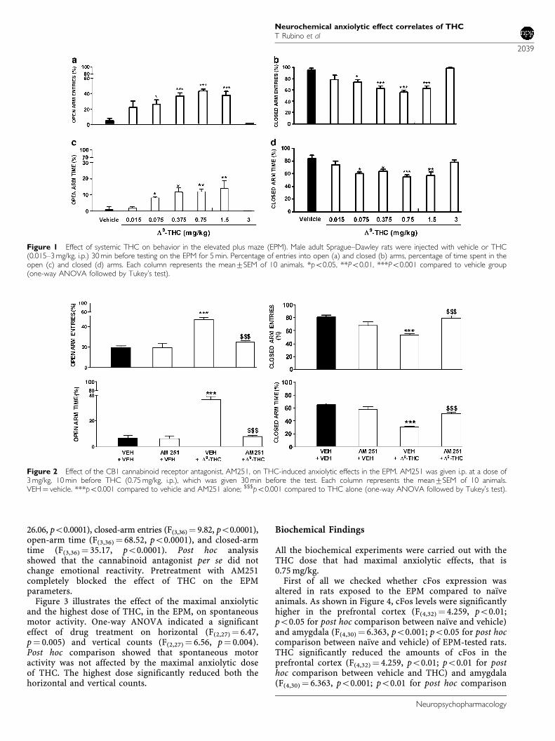
26.06, po0.0001), closed-arm entries (F(3,36)¼ 9.82, po0.0001),open-arm time (F(3,36) ¼ 68.52, po0.0001), and closed-armtime (F(3,36) ¼ 35.17, po0.0001). Post hoc analysisshowed that the cannabinoid antagonist per se did notchange emotional reactivity. Pretreatment with AM251completely blocked the effect of THC on the EPMparameters.
Figure 3 illustrates the effect of the maximal anxiolyticand the highest dose of THC, in the EPM, on spontaneousmotor activity. One-way ANOVA indicated a significanteffect of drug treatment on horizontal (F(2,27)¼ 6.47,p¼ 0.005) and vertical counts (F(2,27)¼ 6.56, p¼ 0.004).Post hoc comparison showed that spontaneous motoractivity was not affected by the maximal anxiolytic doseof THC. The highest dose significantly reduced both thehorizontal and vertical counts.
Biochemical Findings
All the biochemical experiments were carried out with theTHC dose that had maximal anxiolytic effects, that is0.75 mg/kg.
First of all we checked whether cFos expression wasaltered in rats exposed to the EPM compared to naıveanimals. As shown in Figure 4, cFos levels were significantlyhigher in the prefrontal cortex (F(4,32) ¼ 4.259, po0.01;po0.05 for post hoc comparison between naıve and vehicle)and amygdala (F(4,30) ¼ 6.363, po0.001; po0.05 for post hoccomparison between naıve and vehicle) of EPM-tested rats.THC significantly reduced the amounts of cFos in theprefrontal cortex (F(4,32)¼ 4.259, po0.01; po0.01 for posthoc comparison between vehicle and THC) and amygdala(F(4,30) ¼ 6.363, po0.001; po0.01 for post hoc comparison
Figure 1 Effect of systemic THC on behavior in the elevated plus maze (EPM). Male adult Sprague–Dawley rats were injected with vehicle or THC(0.015–3 mg/kg, i.p.) 30 min before testing on the EPM for 5 min. Percentage of entries into open (a) and closed (b) arms, percentage of time spent in theopen (c) and closed (d) arms. Each column represents the mean7SEM of 10 animals. *po0.05, **Po0.01, ***Po0.001 compared to vehicle group(one-way ANOVA followed by Tukey’s test).
Figure 2 Effect of the CB1 cannabinoid receptor antagonist, AM251, on THC-induced anxiolytic effects in the EPM. AM251 was given i.p. at a dose of3 mg/kg, 10 min before THC (0.75 mg/kg, i.p.), which was given 30 min before the test. Each column represents the mean7SEM of 10 animals.VEH¼ vehicle. ***po0.001 compared to vehicle and AM251 alone; $$$po0.001 compared to THC alone (one-way ANOVA followed by Tukey’s test).
Neurochemical anxiolytic effect correlates of THCT Rubino et al
2039
Neuropsychopharmacology
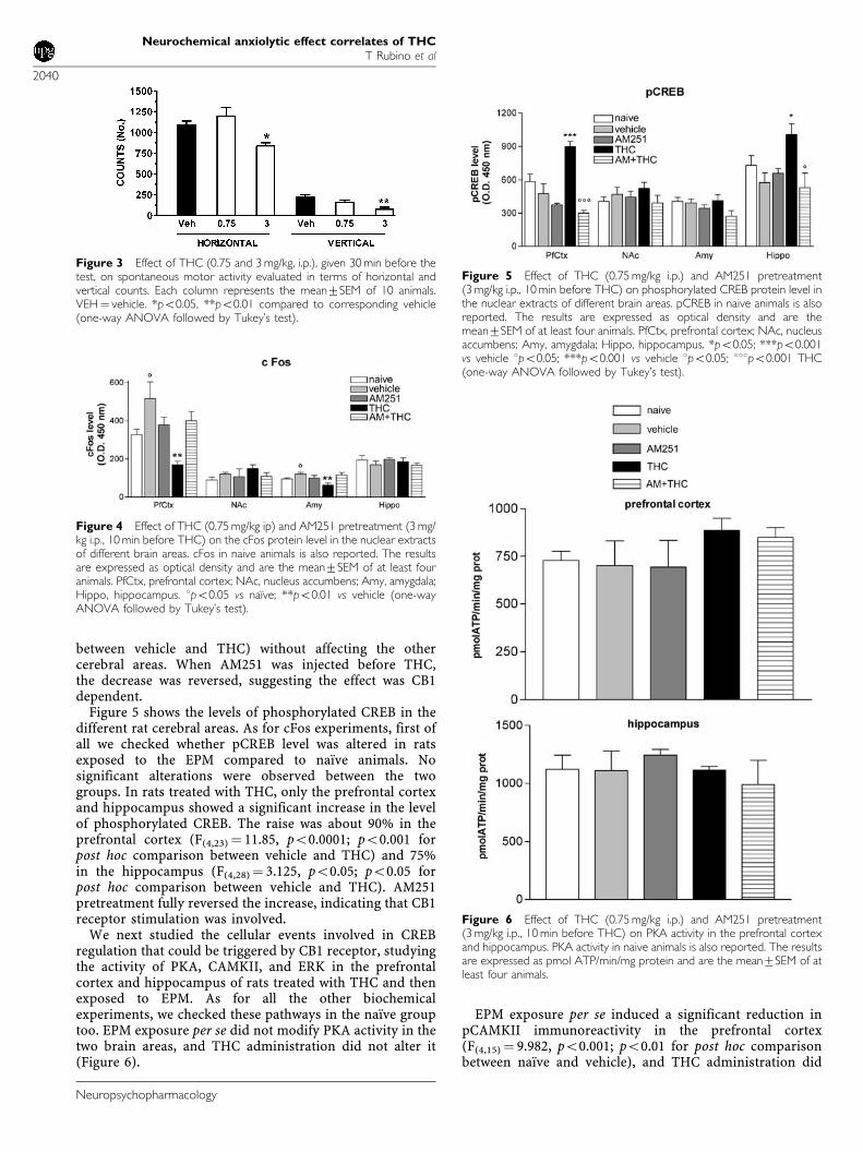
between vehicle and THC) without affecting the othercerebral areas. When AM251 was injected before THC,the decrease was reversed, suggesting the effect was CB1dependent.
Figure 5 shows the levels of phosphorylated CREB in thedifferent rat cerebral areas. As for cFos experiments, first ofall we checked whether pCREB level was altered in ratsexposed to the EPM compared to naıve animals. Nosignificant alterations were observed between the twogroups. In rats treated with THC, only the prefrontal cortexand hippocampus showed a significant increase in the levelof phosphorylated CREB. The raise was about 90% in theprefrontal cortex (F(4,23) ¼ 11.85, po0.0001; po0.001 forpost hoc comparison between vehicle and THC) and 75%in the hippocampus (F(4,28)¼ 3.125, po0.05; po0.05 forpost hoc comparison between vehicle and THC). AM251pretreatment fully reversed the increase, indicating that CB1receptor stimulation was involved.
We next studied the cellular events involved in CREBregulation that could be triggered by CB1 receptor, studyingthe activity of PKA, CAMKII, and ERK in the prefrontalcortex and hippocampus of rats treated with THC and thenexposed to EPM. As for all the other biochemicalexperiments, we checked these pathways in the naıve grouptoo. EPM exposure per se did not modify PKA activity in thetwo brain areas, and THC administration did not alter it(Figure 6).
EPM exposure per se induced a significant reduction inpCAMKII immunoreactivity in the prefrontal cortex(F(4,15) ¼ 9.982, po0.001; po0.01 for post hoc comparisonbetween naıve and vehicle), and THC administration did
Figure 3 Effect of THC (0.75 and 3 mg/kg, i.p.), given 30 min before thetest, on spontaneous motor activity evaluated in terms of horizontal andvertical counts. Each column represents the mean7SEM of 10 animals.VEH¼ vehicle. *po0.05, **po0.01 compared to corresponding vehicle(one-way ANOVA followed by Tukey’s test).
Figure 4 Effect of THC (0.75 mg/kg ip) and AM251 pretreatment (3 mg/kg i.p., 10 min before THC) on the cFos protein level in the nuclear extractsof different brain areas. cFos in naive animals is also reported. The resultsare expressed as optical density and are the mean7SEM of at least fouranimals. PfCtx, prefrontal cortex; NAc, nucleus accumbens; Amy, amygdala;Hippo, hippocampus. 1po0.05 vs naıve; **po0.01 vs vehicle (one-wayANOVA followed by Tukey’s test).
Figure 5 Effect of THC (0.75 mg/kg i.p.) and AM251 pretreatment(3 mg/kg i.p., 10 min before THC) on phosphorylated CREB protein level inthe nuclear extracts of different brain areas. pCREB in naive animals is alsoreported. The results are expressed as optical density and are themean7SEM of at least four animals. PfCtx, prefrontal cortex; NAc, nucleusaccumbens; Amy, amygdala; Hippo, hippocampus. *po0.05; ***po0.001vs vehicle 1po0.05; ***po0.001 vs vehicle 1po0.05; 111po0.001 THC(one-way ANOVA followed by Tukey’s test).
Figure 6 Effect of THC (0.75 mg/kg i.p.) and AM251 pretreatment(3 mg/kg i.p., 10 min before THC) on PKA activity in the prefrontal cortexand hippocampus. PKA activity in naive animals is also reported. The resultsare expressed as pmol ATP/min/mg protein and are the mean7SEM of atleast four animals.
Neurochemical anxiolytic effect correlates of THCT Rubino et al
2040
Neuropsychopharmacology
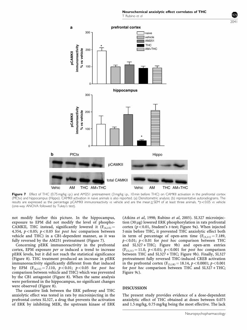
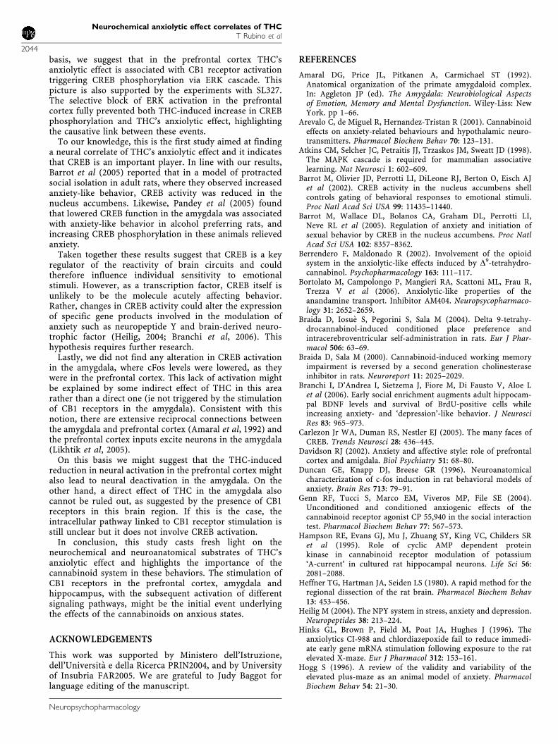
not modify further this picture. In the hippocampus,exposure to EPM did not modify the level of phospho-CAMKII, THC instead, significantly lowered it (F(4,15) ¼4.354, po0.05; po0.05 for post hoc comparison betweenvehicle and THC) in a CB1-dependent manner, as it wasfully reversed by the AM251 pretreatment (Figure 7).
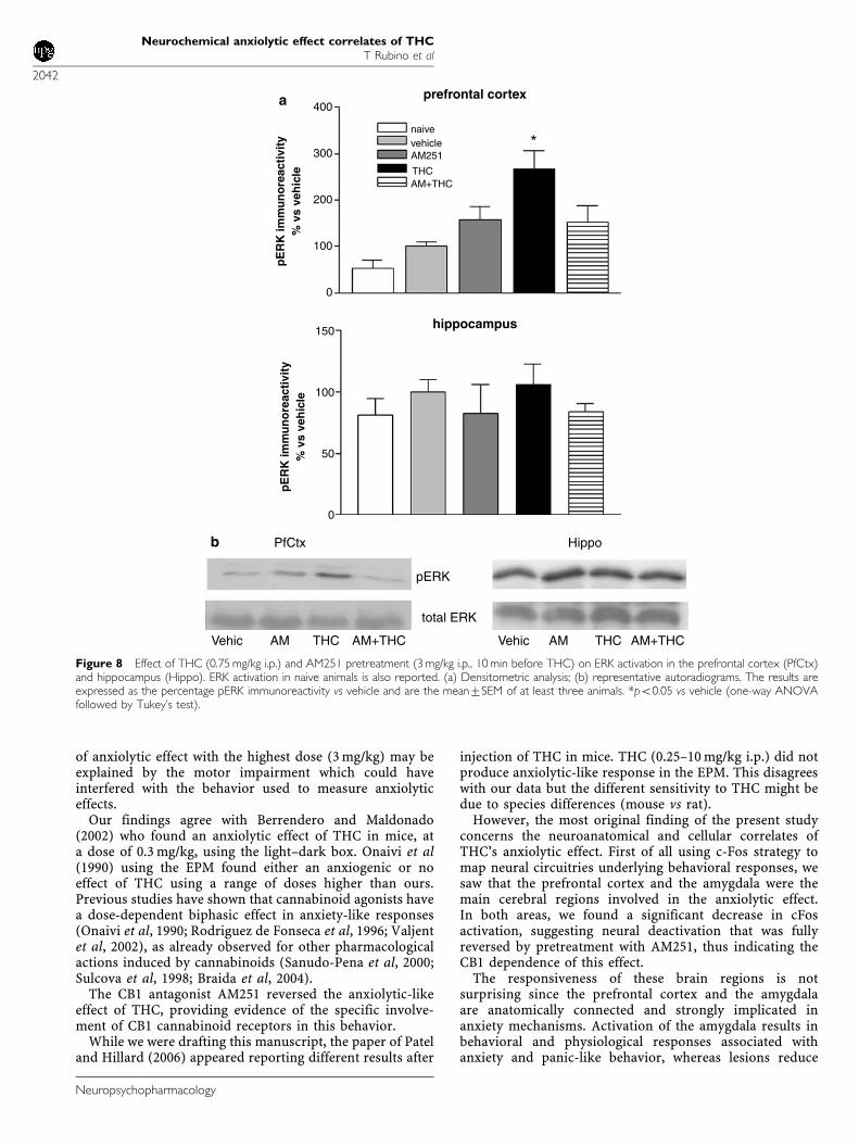
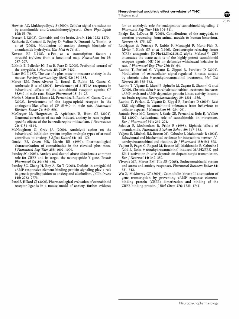
Concerning pERK immunoreactivity in the prefrontalcortex, EPM exposure per se induced a trend to increasepERK levels, but it did not reach the statistical significance(Figure 8). THC treatment produced an increase in pERKimmunoreactivity significantly different from that inducedby EPM (F(4,11) ¼ 7.110, po0.01; po0.05 for post hoccomparison between vehicle and THC) which was preventedby the CB1 antagonist (Figure 8). When the same analysiswere performed in the hippocampus, no significant changeswere observed (Figure 8).
The causative link between the ERK pathway and THCanxiolytic effect was tested in rats by microinjecting in theprefrontal cortex SL327, a drug that prevents the activationof ERK by inhibiting MEK, the upstream kinase of ERK
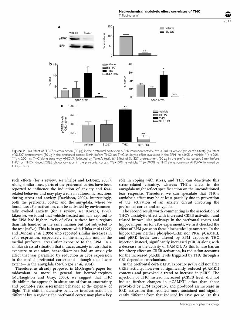
(Atkins et al, 1998; Rubino et al, 2005). SL327 microinjec-tion (30 mg) lowered ERK phosphorylation in rats prefrontalcortex (po0.01, Student’s t-test; Figure 9a). When injected5 min before THC, it prevented THC anxiolytic effect bothin term of percentage of open-arm time (F(3,11) ¼ 7.189,po0.01; po0.01 for post hoc comparison between THCand SL327 + THC; Figure 9b) and open-arm entries(F(3,11) ¼ 11.0, po0.01; po0.001 for post hoc comparisonbetween THC and SL327 + THC; Figure 9b). Finally, SL327pretreatment fully reversed THC-induced CREB activationin the prefrontal cortex (F(3,18)¼ 18.14, po0.0001; po0.001for post hoc comparison between THC and SL327 + THC;Figure 9c).
DISCUSSION
The present study provides evidence of a dose-dependentanxiolytic effect of THC obtained at doses between 0.075and 1.5 mg/kg, 0.75 mg/kg being the most effective. The lack
prefrontal cortexa
b
300 naivevehicleAM251
200
100
0
300
200
100
0
HippoPfCtx
pCAMKII
total CAMKII
AMAM THC AM+THC AM+THCTHCVehicVehic
pC
AM
KII
imm
un
ore
acti
vity
% v
s ve
hic
le
pC
AM
KII
imm
un
ore
acti
vity
% v
s ve
hic
le
hippocampus
*
*
THCAM+THC
Figure 7 Effect of THC (0.75 mg/kg i.p.) and AM251 pretreatment (3 mg/kg i.p., 10 min before THC) on CAMKII activation in the prefrontal cortex(PfCtx) and hippocampus (Hippo). CAMKII activation in naive animals is also reported. (a) Densitometric analysis; (b) representative autoradiograms. Theresults are expressed as the percentage pCAMKII immunoreactivity vs vehicle and are the mean7SEM of at least three animals. *po0.05 vs vehicle(one-way ANOVA followed by Tukey’s test).
Neurochemical anxiolytic effect correlates of THCT Rubino et al
2041
Neuropsychopharmacology
of anxiolytic effect with the highest dose (3 mg/kg) may beexplained by the motor impairment which could haveinterfered with the behavior used to measure anxiolyticeffects.
Our findings agree with Berrendero and Maldonado(2002) who found an anxiolytic effect of THC in mice, ata dose of 0.3 mg/kg, using the light–dark box. Onaivi et al(1990) using the EPM found either an anxiogenic or noeffect of THC using a range of doses higher than ours.Previous studies have shown that cannabinoid agonists havea dose-dependent biphasic effect in anxiety-like responses(Onaivi et al, 1990; Rodriguez de Fonseca et al, 1996; Valjentet al, 2002), as already observed for other pharmacologicalactions induced by cannabinoids (Sanudo-Pena et al, 2000;Sulcova et al, 1998; Braida et al, 2004).
The CB1 antagonist AM251 reversed the anxiolytic-likeeffect of THC, providing evidence of the specific involve-ment of CB1 cannabinoid receptors in this behavior.
While we were drafting this manuscript, the paper of Pateland Hillard (2006) appeared reporting different results after
injection of THC in mice. THC (0.25–10 mg/kg i.p.) did notproduce anxiolytic-like response in the EPM. This disagreeswith our data but the different sensitivity to THC might bedue to species differences (mouse vs rat).
However, the most original finding of the present studyconcerns the neuroanatomical and cellular correlates ofTHC’s anxiolytic effect. First of all using c-Fos strategy tomap neural circuitries underlying behavioral responses, wesaw that the prefrontal cortex and the amygdala were themain cerebral regions involved in the anxiolytic effect.In both areas, we found a significant decrease in cFosactivation, suggesting neural deactivation that was fullyreversed by pretreatment with AM251, thus indicating theCB1 dependence of this effect.
The responsiveness of these brain regions is notsurprising since the prefrontal cortex and the amygdalaare anatomically connected and strongly implicated inanxiety mechanisms. Activation of the amygdala results inbehavioral and physiological responses associated withanxiety and panic-like behavior, whereas lesions reduce
prefrontal cortex
naive
400
300
200
100
100
0
0
150
50
vehicle *AM251
HippoPfCtx
pERK
total ERK
AM THC AM+THCVehic AM THC AM+THCVehic
pE
RK
imm
un
ore
acti
vity
% v
s ve
hic
lep
ER
K im
mu
no
reac
tivi
ty%
vs
veh
icle
hippocampus
THCAM+THC
a
b
Figure 8 Effect of THC (0.75 mg/kg i.p.) and AM251 pretreatment (3 mg/kg i.p., 10 min before THC) on ERK activation in the prefrontal cortex (PfCtx)and hippocampus (Hippo). ERK activation in naive animals is also reported. (a) Densitometric analysis; (b) representative autoradiograms. The results areexpressed as the percentage pERK immunoreactivity vs vehicle and are the mean7SEM of at least three animals. *po0.05 vs vehicle (one-way ANOVAfollowed by Tukey’s test).
Neurochemical anxiolytic effect correlates of THCT Rubino et al
2042
Neuropsychopharmacology
such effects (for a review, see Phelps and LeDoux, 2005).Along similar lines, parts of the prefrontal cortex have beenreported to influence the induction of anxiety and fear-related behavior and may play a role in autonomic reactionsduring stress and anxiety (Davidson, 2002). Interestingly,both the prefrontal cortex and the amygdala, where wefound less cFos activation, can be activated by environmen-tally evoked anxiety (for a review, see Kovacs, 1998).Likewise, we found that vehicle-treated animals exposed tothe EPM had higher levels of cFos in these brain regionsthan rats handled in the same manner but not subjected tothe test (naıve). This is in agreement with Hinks et al (1996)and Duncan et al (1996) who reported similar increases incFos expression, respectively in the amygdala and in themedial prefrontal areas after exposure to the EPM. In asimilar stressful situation that induces anxiety in rats, that isexposure to cat odor, benzodiazepines had an anxiolyticeffect that was paralleled by reduction in cFos expressionin the medial prefrontal cortex andFthough to a lesserextentFin the amygdala (McGregor et al, 2004).
Therefore, as already proposed in McGregor’s paper formidazolam or more in general for benzodiazepines(McNaughton and Gray, 2000), we suggest that THCdisinhibits the approach in situations of fear or uncertaintyand promotes risk assessment behavior at the expense offlight. This shift in defensive behavior involves action ondifferent brain regions: the prefrontal cortex may play a key
role in coping with stress, and THC can deactivate thisstress-related circuitry, whereas THC’s effect in theamygdala might reflect specific action on the unconditionedfear response. Therefore, we can speculate that THC’sanxiolytic effect may be at least partially due to preventionof the activation of an anxiety circuit involving theprefrontal cortex and amygdala.
The second result worth commenting is the association ofTHC’s anxiolytic effect with increased CREB activation andrelated intracellular pathways in the prefrontal cortex andhippocampus. As for cFos experiments, we first checked theeffect of EPM per se on these biochemical parameters. In thehippocampus neither phospho-CREB nor PKA, pCAMKII,and pERK levels were altered by EPM exposure. THCinjection instead, significantly increased pCREB along witha decrease in the activity of CAMKII. As this kinase has aninhibitory effect on CREB activation, its reduction accountsfor the increased pCREB levels triggered by THC through aCB1-dependent mechanism.
In the prefrontal cortex EPM exposure per se did not alterCREB activity, however it significantly reduced pCAMKIIcontents and provoked a trend to increase in pERK. Theinjection of THC instead increased pCREB level, did notinduce further changes in pCAMKII other than thoseprovoked by EPM exposure, and produced an increase inERK activation that resulted more sustained and signifi-cantly different from that induced by EPM per se. On this
vehicle
150
100
50
0
vehicle15 40
30
20
10
0
vehicle
**
**
**
SL327
SL 327SL327
THCSL+THC
vehicle
vehicle
SL327
SL327
THC
THC
SL+THC
SL+THC
pERK
ERK
10
5
% ti
me
pCR
EB
leve
lsO
.D.4
50nm
% e
ntrie
s
% v
s ve
hicl
e
0
1000
750
500
250
0
open arms open arms
a
b
c
Figure 9 (a) Effect of SL327 microinjection (30 mg) in the prefrontal cortex on p-ERK immunoreactivity. **po0.01 vs vehicle (Student’s t-test). (b) Effectof SL327 pretreatment (30 mg in the prefrontal cortex, 5 min before THC) on THC anxiolytic effect evaluated in the EPM. *po0.05 vs vehicle; 11po0.01,111po0.001 vs THC alone (one-way ANOVA followed by Tukey’s test). (c) Effect of SL 327 pretreatment (30 mg in the prefrontal cortex, 5 min beforeTHC) on THC-induced CREB phosphorylation in the prefrontal cortex. **po0.01 vs vehicle; 111po0.001 vs THC alone (one-way ANOVA followed byTukey’s test).
Neurochemical anxiolytic effect correlates of THCT Rubino et al
2043
Neuropsychopharmacology
basis, we suggest that in the prefrontal cortex THC’sanxiolytic effect is associated with CB1 receptor activationtriggering CREB phosphorylation via ERK cascade. Thispicture is also supported by the experiments with SL327.The selective block of ERK activation in the prefrontalcortex fully prevented both THC-induced increase in CREBphosphorylation and THC’s anxiolytic effect, highlightingthe causative link between these events.
To our knowledge, this is the first study aimed at findinga neural correlate of THC’s anxiolytic effect and it indicatesthat CREB is an important player. In line with our results,Barrot et al (2005) reported that in a model of protractedsocial isolation in adult rats, where they observed increasedanxiety-like behavior, CREB activity was reduced in thenucleus accumbens. Likewise, Pandey et al (2005) foundthat lowered CREB function in the amygdala was associatedwith anxiety-like behavior in alcohol preferring rats, andincreasing CREB phosphorylation in these animals relievedanxiety.
Taken together these results suggest that CREB is a keyregulator of the reactivity of brain circuits and couldtherefore influence individual sensitivity to emotionalstimuli. However, as a transcription factor, CREB itself isunlikely to be the molecule acutely affecting behavior.Rather, changes in CREB activity could alter the expressionof specific gene products involved in the modulation ofanxiety such as neuropeptide Y and brain-derived neuro-trophic factor (Heilig, 2004; Branchi et al, 2006). Thishypothesis requires further research.
Lastly, we did not find any alteration in CREB activationin the amygdala, where cFos levels were lowered, as theywere in the prefrontal cortex. This lack of activation mightbe explained by some indirect effect of THC in this arearather than a direct one (ie not triggered by the stimulationof CB1 receptors in the amygdala). Consistent with thisnotion, there are extensive reciprocal connections betweenthe amygdala and prefrontal cortex (Amaral et al, 1992) andthe prefrontal cortex inputs excite neurons in the amygdala(Likhtik et al, 2005).
On this basis we might suggest that the THC-inducedreduction in neural activation in the prefrontal cortex mightalso lead to neural deactivation in the amygdala. On theother hand, a direct effect of THC in the amygdala alsocannot be ruled out, as suggested by the presence of CB1receptors in this brain region. If this is the case, theintracellular pathway linked to CB1 receptor stimulation isstill unclear but it does not involve CREB activation.
In conclusion, this study casts fresh light on theneurochemical and neuroanatomical substrates of THC’sanxiolytic effect and highlights the importance of thecannabinoid system in these behaviors. The stimulation ofCB1 receptors in the prefrontal cortex, amygdala andhippocampus, with the subsequent activation of differentsignaling pathways, might be the initial event underlyingthe effects of the cannabinoids on anxious states.
ACKNOWLEDGEMENTS
This work was supported by Ministero dell’Istruzione,dell’Universita e della Ricerca PRIN2004, and by Universityof Insubria FAR2005. We are grateful to Judy Baggot forlanguage editing of the manuscript.
REFERENCES
Amaral DG, Price JL, Pitkanen A, Carmichael ST (1992).Anatomical organization of the primate amygdaloid complex.In: Aggleton JP (ed). The Amygdala: Neurobiological Aspectsof Emotion, Memory and Mental Dysfunction. Wiley-Liss: NewYork. pp 1–66.
Arevalo C, de Miguel R, Hernandez-Tristan R (2001). Cannabinoideffects on anxiety-related behaviours and hypothalamic neuro-transmitters. Pharmacol Biochem Behav 70: 123–131.
Atkins CM, Selcher JC, Petraitis JJ, Trzaskos JM, Sweatt JD (1998).The MAPK cascade is required for mammalian associativelearning. Nat Neurosci 1: 602–609.
Barrot M, Olivier JD, Perrotti LI, DiLeone RJ, Berton O, Eisch AJet al (2002). CREB activity in the nucleus accumbens shellcontrols gating of behavioral responses to emotional stimuli.Proc Natl Acad Sci USA 99: 11435–11440.
Barrot M, Wallace DL, Bolanos CA, Graham DL, Perrotti LI,Neve RL et al (2005). Regulation of anxiety and initiation ofsexual behavior by CREB in the nucleus accumbens. Proc NatlAcad Sci USA 102: 8357–8362.
Berrendero F, Maldonado R (2002). Involvement of the opioidsystem in the anxiolytic-like effects induced by D9-tetrahydro-cannabinol. Psychopharmacology 163: 111–117.
Bortolato M, Campolongo P, Mangieri RA, Scattoni ML, Frau R,Trezza V et al (2006). Anxiolytic-like properties of theanandamine transport. Inhibitor AM404. Neuropsycopharmaco-logy 31: 2652–2659.
Braida D, Iosue S, Pegorini S, Sala M (2004). Delta 9-tetrahy-drocannabinol-induced conditioned place preference andintracerebroventricular self-administration in rats. Eur J Phar-macol 506: 63–69.
Braida D, Sala M (2000). Cannabinoid-induced working memoryimpairment is reversed by a second generation cholinesteraseinhibitor in rats. Neuroreport 11: 2025–2029.
Branchi I, D’Andrea I, Sietzema J, Fiore M, Di Fausto V, Aloe Let al (2006). Early social enrichment augments adult hippocam-pal BDNF levels and survival of BrdU-positive cells whileincreasing anxiety- and ‘depression’-like behavior. J NeurosciRes 83: 965–973.
Carlezon Jr WA, Duman RS, Nestler EJ (2005). The many faces ofCREB. Trends Neurosci 28: 436–445.
Davidson RJ (2002). Anxiety and affective style: role of prefrontalcortex and amigdala. Biol Psychiatry 51: 68–80.
Duncan GE, Knapp DJ, Breese GR (1996). Neuroanatomicalcharacterization of c-fos induction in rat behavioral models ofanxiety. Brain Res 713: 79–91.
Genn RF, Tucci S, Marco EM, Viveros MP, File SE (2004).Unconditioned and conditioned anxiogenic effects of thecannabinoid receptor agonist CP 55,940 in the social interactiontest. Pharmacol Biochem Behav 77: 567–573.
Hampson RE, Evans GJ, Mu J, Zhuang SY, King VC, Childers SRet al (1995). Role of cyclic AMP dependent proteinkinase in cannabinoid receptor modulation of potassium‘A-current’ in cultured rat hippocampal neurons. Life Sci 56:2081–2088.
Heffner TG, Hartman JA, Seiden LS (1980). A rapid method for theregional dissection of the rat brain. Pharmacol Biochem Behav13: 453–456.
Heilig M (2004). The NPY system in stress, anxiety and depression.Neuropeptides 38: 213–224.
Hinks GL, Brown P, Field M, Poat JA, Hughes J (1996). Theanxiolytics CI-988 and chlordiazepoxide fail to reduce immedi-ate early gene mRNA stimulation following exposure to the ratelevated X-maze. Eur J Pharmacol 312: 153–161.
Hogg S (1996). A review of the validity and variability of theelevated plus-maze as an animal model of anxiety. PharmacolBiochem Behav 54: 21–30.
Neurochemical anxiolytic effect correlates of THCT Rubino et al
2044
Neuropsychopharmacology
Howlett AC, Mukhopadhyay S (2000). Cellular signal transductionby anandamide and 2-arachidonoylglycerol. Chem Phys Lipids108: 53–70.
Iversen L (2003). Cannabis and the brain. Brain 126: 1252–1270.Kathuria S, Gaetani S, Fegley D, Valino F, Duranti A, Tontini A
et al (2003). Modulation of anxiety through blockade ofanandamide hydrolysis. Nat Med 9: 76–81.
Kovacs KJ (1998). c-Fos as a transcription factor: astressful (re)view from a functional map. Neurochem Int 33:287–297.
Likhtik E, Pelletier JG, Paz R, Pare D (2005). Prefrontal control ofthe amygdala. J Neurosci 25: 7429–7437.
Lister RG (1987). The use of a plus-maze to measure anxiety in themouse. Psychopharmacology (Berl) 92: 180–185.
Marco EM, Perez-Alvarez L, Borcel E, Rubio M, Guaza C,Ambrosio E et al (2004). Involvement of 5-HT1A receptors inbehavioural effects of the cannabinoid receptor agonist CP55,940 in male rats. Behav Pharmacol 15: 21–27.
Marin S, Marco E, Biscaia M, Fernandez B, Rubio M, Guaza C et al(2003). Involvement of the kappa-opioid receptor in theanxiogenic-like effect of CP 55 940 in male rats. PharmacolBiochem Behav 74: 649–656.
McGregor IS, Hargreaves G, Apfelbach R, Hunt GE (2004).Neuronal correlates of cat odr-induced anxiety in rats: region-specific effects of the benzodiazepine midazolam. J Neuroscince24: 4134–4144.
McNaughton N, Gray JA (2000). Anxiolytic action on thebehavioural inhibition system implies multiple types of arousalcontribute to anxiety. J Affect Disord 61: 161–176.
Onaivi ES, Green MR, Martin BR (1990). Pharmacologicalcharacterization of cannabinoids in the elevated plus maze.J Pharmacol Exp Ther 253: 1002–1009.
Pandey SC (2003). Anxiety and alcohol abuse disorders: a commonrole for CREB and its target, the neuropeptide Y gene. TrendsPharmacol Sci 24: 456–460.
Pandey SC, Zhang H, Roy A, Xu T (2005). Deficits in amygdaloidcAMP-responsive element-binding protein signaling play a rolein genetic predisposition to anxiety and alcoholism. J Clin Invest115: 2762–2773.
Patel S, Hillard CJ (2006). Pharmacological evaluation of cannabinoidreceptor ligands in a mouse model of anxiety: further evidence
for an anxiolytic role for endogenous cannabinoid signaling. JPharmacol Exp Ther 318: 304–311.
Phelps EA, LeDoux JE (2005). Contributions of the amygdala toemotion processing: from animal models to human behaviour.Neuron 48: 175–187.
Rodriguez de Fonseca F, Rubio P, Menzaghi F, Merlo-Pich E,Rivier J, Koob GF et al (1996). Corticotropin-releasing factor(CRF) antagonist [D-Phe12,Nle21,38,C alpha MeLeu37] CRFattenuates the acute actions of the highly potent cannabinoidreceptor agonist HU-210 on defensive-withdrawal behavior inrats. J Pharmacol Exp Ther 276: 56–64.
Rubino T, Forlani G, Vigano D, Zippel R, Parolaro D (2004).Modulation of extracellular signal-regulated kinases cacadeby chronic delta 9-tetrahydrocannabinol treatment. Mol CellNeurosi 25: 355–362.
Rubino T, Vigano D, Massi P, Spinello M, Zagato E, Gianoni G et al(2000). Chronic delta 9-tetrahydrocannabinol treatment increasescAMP levels and cAMP-dependent protein kinase activity in somerat brain regions. Neuropharmacology 39: 1331–1336.
Rubino T, Forlani G, Vigano D, Zippel R, Parolaro D (2005). Ras/ERK signalling in cannabinoid tolerance: from behaviour tocellular aspects. J Neurochem 93: 984–991.
Sanudo-Pena MC, Romero J, Seale GE, Fernandez-Ruiz JJ, WalkerJM (2000). Activational role of cannabinoids on movement.Eur J Pharmacol 391: 269–274.
Sulcova E, Mechoulam R, Fride E (1998). Biphasic effects ofanandamide. Pharmacol Biochem Behav 59: 347–352.
Valjent E, Mitchell JM, Besson MJ, Caboche J, Maldonado R (2002).Behavioural and biochemical evidence for interactions between D9-tetrahydrocannabinol and nicotine. Br J Pharmacol 135: 564–578.
Valjent E, Pages C, Rogard M, Besson MJ, Maldonado R, Caboche J(2001). Delta 9-tetrahydrocannabinol-induced MAPK/ERK andElk-1 activation in vivo depends on dopaminergic transmission.Eur J Neurosci 14: 342–352.
Viveros MP, Marco EM, File SE (2005). Endocannabinoid systemand stress and anxiety responses. Pharmacol Biochem Behav 81:331–342.
Wu X, McMurray CT (2001). Calmodulin kinase II attenuation ofgene transcription by preventing cAMP response element-binding protein (CREB) dimerization and binding of theCREB-binding protein. J Biol Chem 276: 1735–1741.
Neurochemical anxiolytic effect correlates of THCT Rubino et al
2045
Neuropsychopharmacology