Cell Structure Chapter 3. Looking at Cells 1 st microscope invented in the 1600s Robert Hooke (Cork...
29
Cell Structure Chapter 3
-
Upload
rosanna-osborne -
Category
Documents
-
view
215 -
download
0
Transcript of Cell Structure Chapter 3. Looking at Cells 1 st microscope invented in the 1600s Robert Hooke (Cork...
Cell Structure
Chapter 3
Looking at Cells
1st microscope invented in the 1600s Robert Hooke (Cork Cells) Scientists use the Metric System or
International Systems of Measurements SI units are based on powers of 10
– μ = micro μm = micrometer
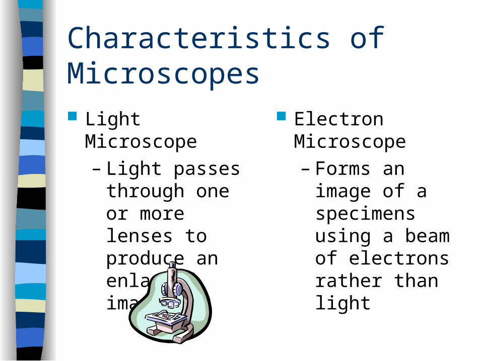
Characteristics of Microscopes
Light Microscope– Light passes
through one or more lenses to produce an enlarged image
Electron Microscope– Forms an image
of a specimens using a beam of electrons rather than light
Characteristics of Microscopes
MAGNIFICATION – is the quality of making an image appear larger than its actual size
RESOLUTION – is the measure of the clarity of an image
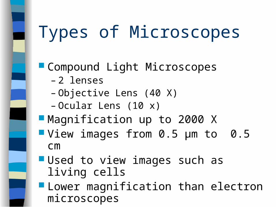
Types of Microscopes
Compound Light Microscopes– 2 lenses– Objective Lens (40 X)– Ocular Lens (10 x)
Magnification up to 2000 X View images from 0.5 μm to 0.5 cm Used to view images such as living cells Lower magnification than electron
microscopes
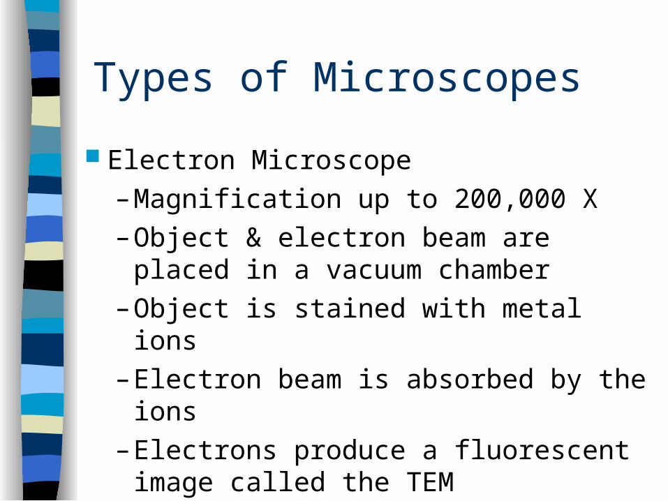
Types of Microscopes
Electron Microscope
– Magnification up to 200,000 X
– Object & electron beam are placed in a vacuum chamber
– Object is stained with metal ions
– Electron beam is absorbed by the ions
– Electrons produce a fluorescent image called the TEM
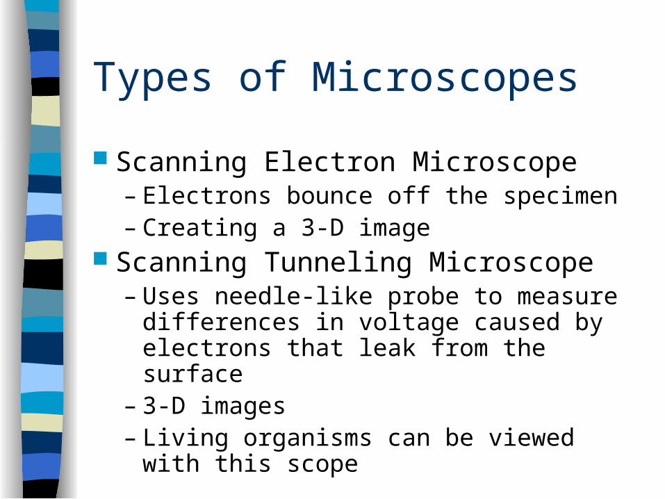
Types of Microscopes
Scanning Electron Microscope– Electrons bounce off the specimen– Creating a 3-D image
Scanning Tunneling Microscope– Uses needle-like probe to measure
differences in voltage caused by electrons that leak from the surface
– 3-D images– Living organisms can be viewed with this
scope
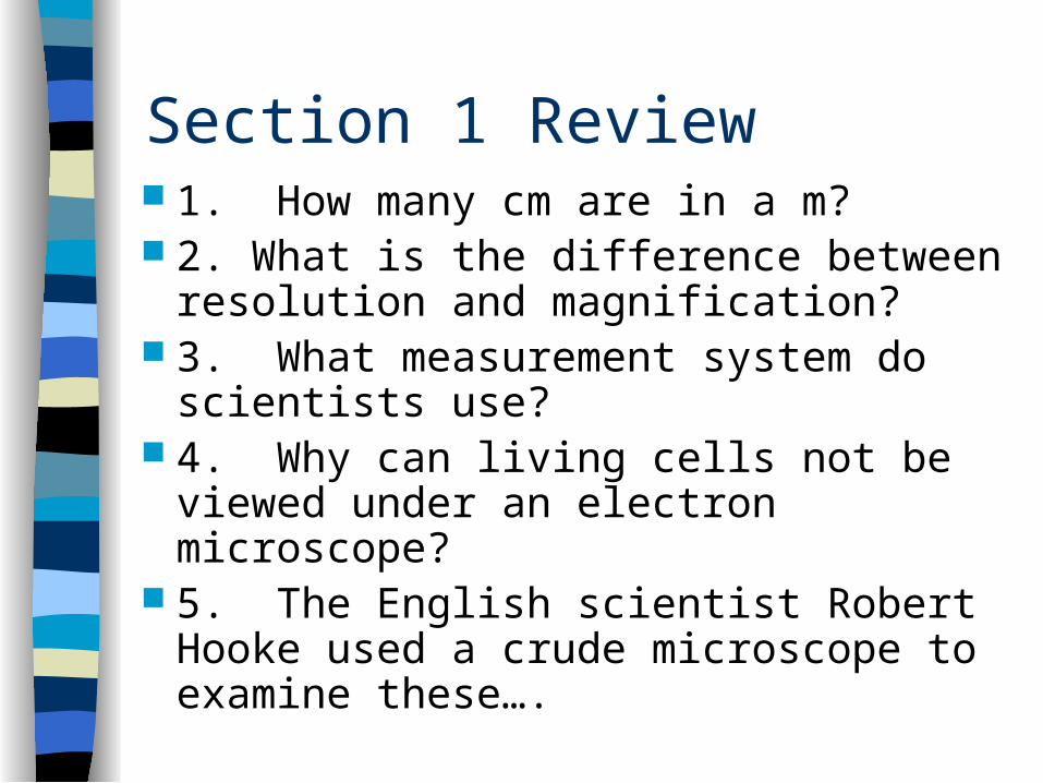
Section 1 Review 1. How many cm are in a m? 2. What is the difference between
resolution and magnification? 3. What measurement system do
scientists use? 4. Why can living cells not be viewed
under an electron microscope? 5. The English scientist Robert Hooke
used a crude microscope to examine these….
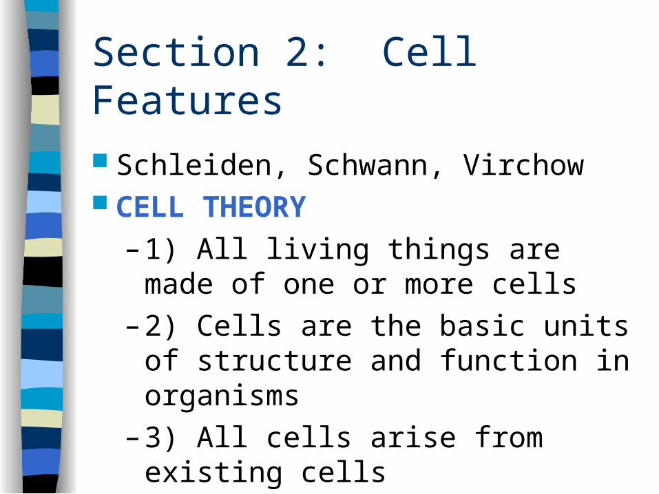
Section 2: Cell Features
Schleiden, Schwann, Virchow CELL THEORY
– 1) All living things are made of one or more cells
– 2) Cells are the basic units of structure and function in organisms
– 3) All cells arise from existing cells
Cell Size
Small cells are more efficient than large cells
All substances that enter or leave a cells have to cross the cell’s surface
Surface area- to- Volume – LOW – substances can not enter or leave
Small cells have a higher surface area- to – volume ratio– Shorter distance to travel
Common Features of Cells
CELL MEMBRANE – encloses the cell– Regulates what enters & leaves the cell
CYTOPLASM – cell’s interior CYTOSKELETON – fibers suspended
in the cytoplasm RIBOSOMES – cellular structures on
which proteins are made DNA – instructions for the cell

Prokaryotes
The smallest and simplest cells Are single-celled organisms Lack a NUCLEUS Examples: Bacteria Reproduce Rapidly Do not need oxygen to survive Some can make their own food
Characteristics of Prokaryotes CYTOPLASM – semi-solid material that
surrounds everything inside the cell– DNA, Ribosomes & enzymes
CELL WALL – surrounds the cell membrane– Function: Provides support &
structure– Found in plants, fungi & some
bacteria– Made of polysaccharides
Characteristics of Prokaryotes
Capsule – surrounds the cell– Cling to lots of things
Flagella – long, threadlike structures that extend from the cell surface– Function: helps the cell move
Eukaryotic Cells Have a Nucleus NUCLEUS – houses the DNA Organelles - internal compartments
– Carry out specific activities in the cell Vesicles – move proteins & other
molecules from cell to cell CILLIA – short hair-like projections
– Functions: Movement & transport of materials across the cell membrane
Eukaryotic Cells
CYTOSOL – fluid material that makes up the cytoplasm
CYTOSKELETON– Made of protein fibers– Function: Holds the cell together– All connected to one another– 3 kinds of fibers – Actin Fibers,
Microtubules & Intermediate Fibers
Cytoskeleton
Actin Fibers – determine the shape Microtubules – act as a highway
system
– Transport info from the nucleus to the rest of the cell
Intermediate Fibers – are an anchor for ribosomes & enzymes
The Cell Membrane
Selective Permeability – Only let certain things enter or exit
Phospholipid = 1 lipid + 2 fatty acids
– Head Polar
• Phosphate Group
• Attracted to water
– 2 Tails NonPolar
• Repel water
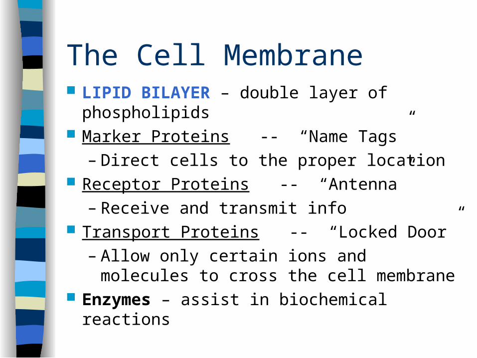
The Cell Membrane LIPID BILAYER – double layer of
phospholipids Marker Proteins -- “Name Tags”
– Direct cells to the proper location Receptor Proteins -- “Antenna”
– Receive and transmit info Transport Proteins -- “Locked Door”
– Allow only certain ions and molecules to cross the cell membrane
Enzymes – assist in biochemical reactions
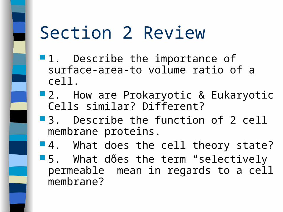
Section 2 Review 1. Describe the importance of surface-
area-to volume ratio of a cell. 2. How are Prokaryotic & Eukaryotic
Cells similar? Different? 3. Describe the function of 2 cell
membrane proteins. 4. What does the cell theory state? 5. What does the term “selectively
permeable” mean in regards to a cell membrane?

Section 3: Cell Organelles Cell functions are controlled by the
NUCLEUS
– Surrounded by the NUCLEAR ENVELOPE• Made up of 2 lipid bilayers• NUCLEAR PORES
–Ribosomal RNA pass through the pores into the cytoplasm
• Ribosomes are partially assembled in the NUCLEOLUS
The Nucleus
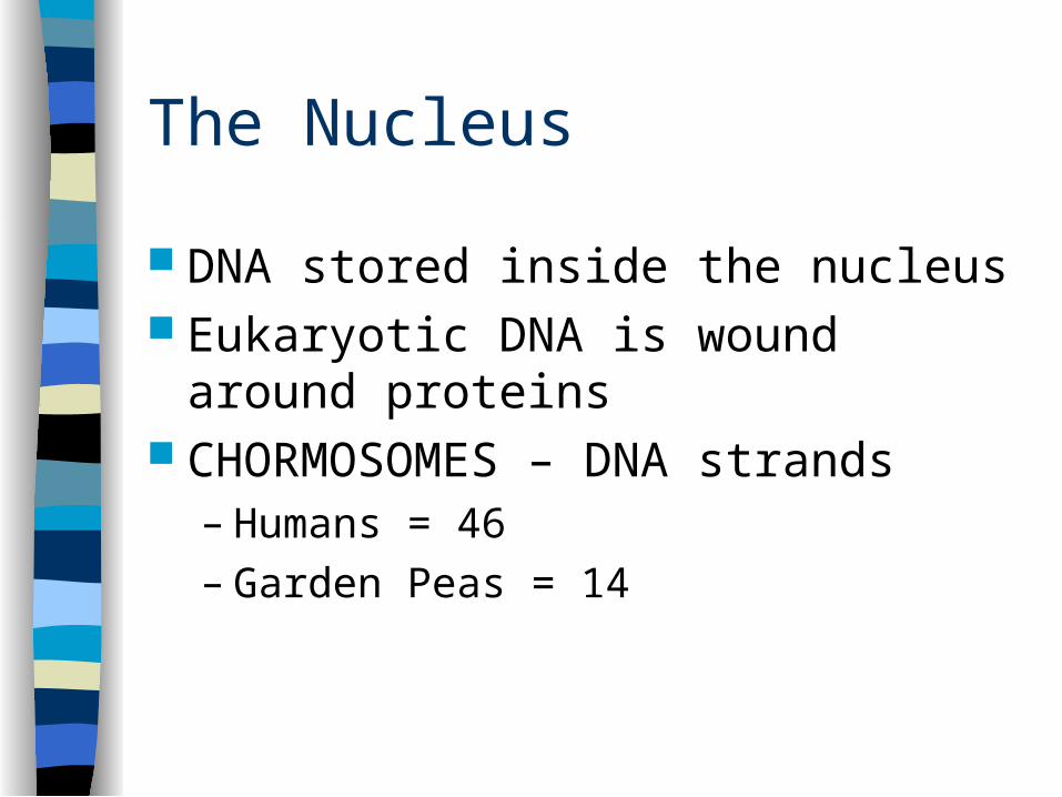
DNA stored inside the nucleus Eukaryotic DNA is wound around
proteins CHORMOSOMES – DNA strands
– Humans = 46– Garden Peas = 14
Ribosomes & the ER
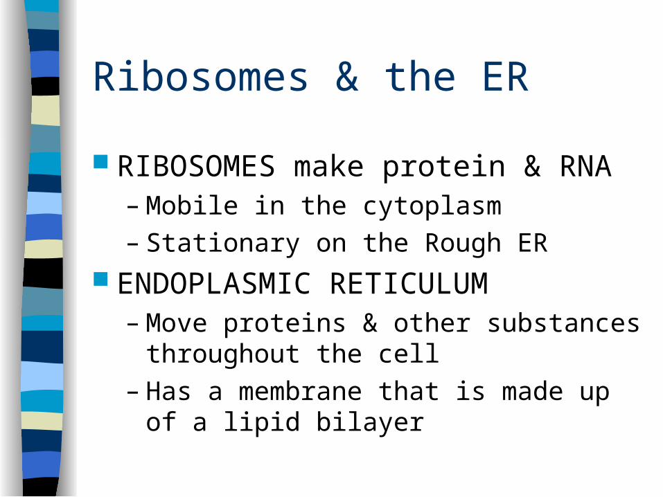
RIBOSOMES make protein & RNA– Mobile in the cytoplasm– Stationary on the Rough ER
ENDOPLASMIC RETICULUM– Move proteins & other substances
throughout the cell– Has a membrane that is made up of a lipid
bilayer
Ribosomes & the ER
VESICLE – is a small membrane bound sac that transports substances inside the cell– Separates proteins made on the Rough ER
from proteins made in the cytoplasm SMOOTH ENDOPLASMIC RETICULUM
– Lacks ribosomes– Function: Makes lipids & breaks down toxic
substances
Packaging & Distribution of Proteins
GOLGI APPARATUS– Is a set of flattened, membrane-bound sacs
that package & distribute – Proteins are modified in the GA & enter new
vesicles– Vesicles take the proteins outside the cell
OR– Vesicles remain in the cell & become
LYSOMES • Contain digestive enzymes
Mitochondria
Function: Harvest energy to make ATP Muscle cells can have 100s – 1000s 2 Membranes (Inner & Outer)
– Here chemical reactions produce ATP Also has DNA
– Circular – similar to prokaryotic DNA
Structures of Plant Cells
3 Organelles NOT found in Animal Cells 1) CELL WALL
– Adds additional support, give shape, protects from damage & connects with adjacent cells
2) Chloroplasts– Use light to make carbohydrates from
carbon dioxide and water– Surrounded by 2 layered membrane &
contains DNA
Structures of Plant Cells
3) Central Vacuole– Takes up most of volume – Contains
• Water• Ions• Nutrients• Waste
– When full it makes the cell rigid– Allows the plant to stand upright
Section 3: Review
1. Describe the role of the nucleus in cell activities.
2. Sequence the course of newly made proteins from the rough ER to the outside of the cell.
3. Name 2 organelles a plant has that an animal cells does not.
4. The mitochondria has the nickname “The Powerhouse”. Why is this a good nickname?