Biochimica et Biophysica Acta - University of East Anglia free-Hank's buffer salt solution (HBSS),...
12
PAK1 modulates a PPARγ/NF-κB cascade in intestinal inflammation Kyle Dammann a , Vineeta Khare a , Michaela Lang a , Thierry Claudel b , Felix Harpain a , Nicolas Granofszky a , Rayko Evstatiev a , Jonathan M. Williams c , D. Mark Pritchard c , Alastair Watson d , Christoph Gasche a, ⁎ a Medical University of Vienna, Department of Internal Medicine III, Division of Gastroenterology and Hepatology, Christian Doppler Laboratory for Molecular Cancer Chemoprevention, Vienna, Austria b Medical University of Vienna, Department of Internal Medicine III, Division of Gastroenterology and Hepatology, Hans Popper Laboratory for Molecular Hepatology, Vienna, Austria c Department of Gastroenterology, University of Liverpool, Liverpool, United Kingdom d Norwich Medical School, University of East Anglia, Norwich Research Park, Norwich, United Kingdom abstract article info Article history: Received 24 March 2015 Received in revised form 13 May 2015 Accepted 28 May 2015 Available online 31 May 2015 Keywords: PAK1 NF-κB PPARγ Inflammation Ulcerative colitis Colitis-associated cancer P21-activated kinases (PAKs) are multifunctional effectors of Rho GTPases with both kinase and scaffolding activ- ity. Here, we investigated the effects of inflammation on PAK1 signaling and its role in colitis-driven carcinogen- esis. PAK1 and p-PAK1 (Thr423) were assessed by immunohistochemistry, immunofluorescence, and Western blot. C57BL6/J wildtype mice were treated with a single intraperitoneal TNFα injection. Small intestinal organoids from these mice and from PAK1-KO mice were cultured with TNFα. NF-κB and PPARγ were analyzed upon PAK1 overexpression and silencing for transcriptional/translational regulation. PAK1 expression and activa- tion was increased on the luminal intestinal epithelial surface in inflammatory bowel disease and colitis- associated cancer. PAK1 was phosphorylated upon treatment with IFNγ, IL-1β, and TNFα. In vivo, mice adminis- tered with TNFα showed increased p-PAK1 in intestinal villi, which was associated with nuclear p65 and NF-κB activation. p65 nuclear translocation downstream of TNFα was strongly inhibited in PAK1-KO small intestinal organoids. PAK1 overexpression induced a PAK1–p65 interaction as visualized by co-immunoprecipitation, nuclear translocation, and increased NF-κB transactivation, all of which were impeded by kinase-dead PAK1. Moreover, PAK1 overexpression downregulated PPARγ and mesalamine recovered PPARγ through PAK1 inhibi- tion. On the other hand PAK1 silencing inhibited NF-κB, which was recovered using BADGE, a PPARγ antagonist. Altogether these data demonstrate that PAK1 overexpression and activation in inflammation and colitis- associated cancer promote NF-κB activity via suppression of PPARγ in intestinal epithelial cells. © 2015 The Authors. Published by Elsevier B.V. This is an open access article under the CC BY-NC-ND license (http://creativecommons.org/licenses/by-nc-nd/4.0/). 1. Introduction Inflammatory bowel diseases (IBD) such as Crohn's disease (CD) and ulcerative colitis (UC) are associated with an increased risk of develop- ing colorectal cancer (CRC). Mesalamine, 5-aminosalicyclic acid (5- ASA), is an anti-inflammatory drug used to treat UC, and epidemiologi- cal evidence suggests that it has chemopreventive effects [1]. We previ- ously identified p-21 activated kinase-1 (PAK1) as a 5-ASA target [2]. PAK1 is a serine/threonine kinase effector of the small Rho GTPases Rac1/Cdc42 [3], which regulates cytoskeletal dynamics and epithelial cell (IEC) migration and homeostasis. Recently, we demonstrated that PAK1 is overexpressed in IBD and CAC and promotes cell survival path- ways [4–6]. However, the exact cause and consequence of PAK1 overex- pression in intestinal inflammation have yet to be defined. Several studies support the notion that canonical NF-κB activation promotes intestinal tumorigenesis through the upregulation of pro- inflammatory cytokines, proliferation, and cell survival [7–10]. NF-κB activation is regulated by the transcription factor RelA (p65). At basal levels, p65 is sequestered in the cytoplasm by its inhibitors IKKα/β and IκB. Upon pathway activation, IκB is degraded, and p65 translocates into the nucleus [11]. NF-κB signaling in immune cells drives the ex- pression of pro-inflammatory cytokines such as TNFα or IL-1β, which subsequently activate NF-κB in IECs thereby promoting cell survival [12]. In support of this, TNFα-administered NF-κB1 -/- mice show in- creased IEC apoptosis [12]. PAK1 was previously reported to stimulate NF-κB [13], albeit its exact mechanism within the canonical pathway is unknown. Here, we have investigated the effect of PAK1 activation in IECs upon inflammation and its relevance for NF-κB signaling. We observed that Biochimica et Biophysica Acta 1853 (2015) 2349–2360 Abbreviations: PAK1, p-21 activated kinase 1; PPARγ, peroxisome proliferator associ- ated receptor gamma; IBD, inflammatory bowel disease; NF-κB, nuclear factor-kappa B; CRC, colorectal cancer; UC, ulcerative colitis; CD, Crohn's disease; CAC, colitis-associated cancer; EV, empty vector; WT, wild type; KD, kinase dead; KO, knock out; SIO, small intes- tinal organoids; SB, small bowel; LB, large bowel; IEC, intestinal epithelial cells; Rosi, Rosiglitazone ⁎ Corresponding author at: Division of Gastroenterology and Hepatology, Medical University of Vienna, Währinger Gürtel 18-20, 1090 Vienna, Austria. E-mail address: [email protected] (C. Gasche). http://dx.doi.org/10.1016/j.bbamcr.2015.05.031 0167-4889/© 2015 The Authors. Published by Elsevier B.V. This is an open access article under the CC BY-NC-ND license (http://creativecommons.org/licenses/by-nc-nd/4.0/). Contents lists available at ScienceDirect Biochimica et Biophysica Acta journal homepage: www.elsevier.com/locate/bbamcr
Transcript of Biochimica et Biophysica Acta - University of East Anglia free-Hank's buffer salt solution (HBSS),...

Biochimica et Biophysica Acta 1853 (2015) 2349–2360
Contents lists available at ScienceDirect
Biochimica et Biophysica Acta
j ourna l homepage: www.e lsev ie r .com/ locate /bbamcr
PAK1 modulates a PPARγ/NF-κB cascade in intestinal inflammation
Kyle Dammann a, Vineeta Khare a, Michaela Lang a, Thierry Claudel b, Felix Harpain a, Nicolas Granofszky a,Rayko Evstatiev a, Jonathan M. Williams c, D. Mark Pritchard c, Alastair Watson d, Christoph Gasche a,⁎a Medical University of Vienna, Department of Internal Medicine III, Division of Gastroenterology and Hepatology, Christian Doppler Laboratory for Molecular Cancer Chemoprevention, Vienna,Austriab Medical University of Vienna, Department of Internal Medicine III, Division of Gastroenterology and Hepatology, Hans Popper Laboratory for Molecular Hepatology, Vienna, Austriac Department of Gastroenterology, University of Liverpool, Liverpool, United Kingdomd Norwich Medical School, University of East Anglia, Norwich Research Park, Norwich, United Kingdom
Abbreviations: PAK1, p-21 activated kinase 1; PPARγ,ated receptor gamma; IBD, inflammatory bowel disease;CRC, colorectal cancer; UC, ulcerative colitis; CD, Crohn'scancer; EV, empty vector;WT, wild type; KD, kinase dead;tinal organoids; SB, small bowel; LB, large bowel; IEC, iRosiglitazone⁎ Corresponding author at: Division of Gastroentero
University of Vienna, Währinger Gürtel 18-20, 1090 ViennE-mail address: [email protected] (
http://dx.doi.org/10.1016/j.bbamcr.2015.05.0310167-4889/© 2015 The Authors. Published by Elsevier B.V
a b s t r a c t
a r t i c l e i n f oArticle history:Received 24 March 2015Received in revised form 13 May 2015Accepted 28 May 2015Available online 31 May 2015
Keywords:PAK1NF-κBPPARγInflammationUlcerative colitisColitis-associated cancer
P21-activated kinases (PAKs) aremultifunctional effectors of Rho GTPaseswith both kinase and scaffolding activ-ity. Here, we investigated the effects of inflammation on PAK1 signaling and its role in colitis-driven carcinogen-esis. PAK1 and p-PAK1 (Thr423) were assessed by immunohistochemistry, immunofluorescence, and Westernblot. C57BL6/J wildtype mice were treated with a single intraperitoneal TNFα injection. Small intestinalorganoids from these mice and from PAK1-KO mice were cultured with TNFα. NF-κB and PPARγwere analyzedupon PAK1overexpression and silencing for transcriptional/translational regulation. PAK1expression and activa-tion was increased on the luminal intestinal epithelial surface in inflammatory bowel disease and colitis-associated cancer. PAK1was phosphorylated upon treatment with IFNγ, IL-1β, and TNFα. In vivo, mice adminis-tered with TNFα showed increased p-PAK1 in intestinal villi, which was associated with nuclear p65 and NF-κBactivation. p65 nuclear translocation downstream of TNFα was strongly inhibited in PAK1-KO small intestinalorganoids. PAK1 overexpression induced a PAK1–p65 interaction as visualized by co-immunoprecipitation,nuclear translocation, and increased NF-κB transactivation, all of which were impeded by kinase-dead PAK1.Moreover, PAK1 overexpression downregulated PPARγ andmesalamine recovered PPARγ through PAK1 inhibi-tion. On the other hand PAK1 silencing inhibited NF-κB, which was recovered using BADGE, a PPARγ antagonist.Altogether these data demonstrate that PAK1 overexpression and activation in inflammation and colitis-associated cancer promote NF-κB activity via suppression of PPARγ in intestinal epithelial cells.
© 2015 The Authors. Published by Elsevier B.V. This is an open access article under the CC BY-NC-ND license(http://creativecommons.org/licenses/by-nc-nd/4.0/).
1. Introduction
Inflammatory bowel diseases (IBD) such as Crohn's disease (CD) andulcerative colitis (UC) are associated with an increased risk of develop-ing colorectal cancer (CRC). Mesalamine, 5-aminosalicyclic acid (5-ASA), is an anti-inflammatory drug used to treat UC, and epidemiologi-cal evidence suggests that it has chemopreventive effects [1]. We previ-ously identified p-21 activated kinase-1 (PAK1) as a 5-ASA target [2].PAK1 is a serine/threonine kinase effector of the small Rho GTPasesRac1/Cdc42 [3], which regulates cytoskeletal dynamics and epithelial
peroxisome proliferator associ-NF-κB, nuclear factor-kappa B;disease; CAC, colitis-associatedKO, knock out; SIO, small intes-ntestinal epithelial cells; Rosi,
logy and Hepatology, Medicala, Austria.C. Gasche).
. This is an open access article under
cell (IEC) migration and homeostasis. Recently, we demonstrated thatPAK1 is overexpressed in IBD and CAC and promotes cell survival path-ways [4–6]. However, the exact cause and consequence of PAK1 overex-pression in intestinal inflammation have yet to be defined.
Several studies support the notion that canonical NF-κB activationpromotes intestinal tumorigenesis through the upregulation of pro-inflammatory cytokines, proliferation, and cell survival [7–10]. NF-κBactivation is regulated by the transcription factor RelA (p65). At basallevels, p65 is sequestered in the cytoplasm by its inhibitors IKKα/βand IκB. Upon pathway activation, IκB is degraded, and p65 translocatesinto the nucleus [11]. NF-κB signaling in immune cells drives the ex-pression of pro-inflammatory cytokines such as TNFα or IL-1β, whichsubsequently activate NF-κB in IECs thereby promoting cell survival[12]. In support of this, TNFα-administered NF-κB1−/− mice show in-creased IEC apoptosis [12]. PAK1 was previously reported to stimulateNF-κB [13], albeit its exact mechanism within the canonical pathwayis unknown.
Here, we have investigated the effect of PAK1 activation in IECs uponinflammation and its relevance for NF-κB signaling. We observed that
the CC BY-NC-ND license (http://creativecommons.org/licenses/by-nc-nd/4.0/).
2350 K. Dammann et al. / Biochimica et Biophysica Acta 1853 (2015) 2349–2360
2351K. Dammann et al. / Biochimica et Biophysica Acta 1853 (2015) 2349–2360
several cytokines including TNFα can activate PAK1and induce its nuclearlocalization. In IECs, such PAK1 activation induces a p65 interaction andassists its nuclear translocation through inhibition of PPARγ.
2. Materials and methods
2.1. Animal experiments
9 week old, C57BL/6 mice were provided by Charles River (Margate,UK) and housed at the University of Liverpool. All procedures wereperformed according to UK Home Office license guidelines. Mice wereeuthanized 1.5 h after receiving an intraperitoneal injection of TNFαat 0.33 mg/kg body weight. The large (LB) and small bowel (SB) weredissected and paraffin embedded as previously described.
2.2. Cell culture, isolation of small intestinal epithelial organoids, andreagents
Primary human colon epithelial cells (HCEC-1CT), a generous giftfrom Dr. Jerry W. Shay, were cultured as described previously [14].HCT116 cells were purchased from ATCC. Small intestinal organoids(SIO) were isolated, from C57BL/6 wild type (WT) and PAK1−/−
(PAK1 KO) mice (purchased from MMRRC UNC, Chapel Hills, NC), andmaintained as previously described [15]. All cells were serum starvedin incomplete medium for 12 h prior to treatment with recombinantprotein. Reagents (Supplementary Table S2) were applied 2–12 hprior to inflammatory cytokines. Cells were harvested at 60–70%confluency.
2.3. Co-culture
Polymorphonuclear cells (PMN) were isolated, activated, andcultured using a previously established co-culture system [16]. Briefly,isolated PMNs were activated using 50 ng/mL phorbol 12-myristate13-acetate (PMA) at 37 °C for 30 min. PMNs were washed twice withCa/Mg free-Hank's buffer salt solution (HBSS), and applied to theupper chamber of a 0.45 μm Transwell insert in a 1:50 epithelial/PMNratio. Following treatment, PMNswere discarded, and colonic epithelialcells were harvested for western blot analysis.
2.4. Immunohistochemical analysis
Immunohistochemistry (IHC) was performed on 3–5 μm tissue sec-tions from human samples, andmouse SB or LB [6]. The human sampleswere obtained from the Clinical Institute of Pathology at the MedicalUniversity of Vienna. The study was approved by the local ethics com-mittee. Samples were selected from endoscopic biopsies or surgicalspecimens of active IBD and CAC patients. Control specimens weretaken fromnormal colon tissue. CDpatients displayed active colonic dis-ease, and CAC included both adenoma and invasive tumors.
2.5. Immunoreactivity score
An immunoreactivity score (IRS) was generated by two blinded in-vestigators. Separate IRSs were generated for the small bowel (SB)and the large bowel (LB). The SB included villus and crypt epithelialstaining, while the LB included surface and crypt epithelial staining.The intensity of staining included a score from 0–3, 0 (no staining), 1(low), 2 (medium), 3 (high). The percentage of positively stained
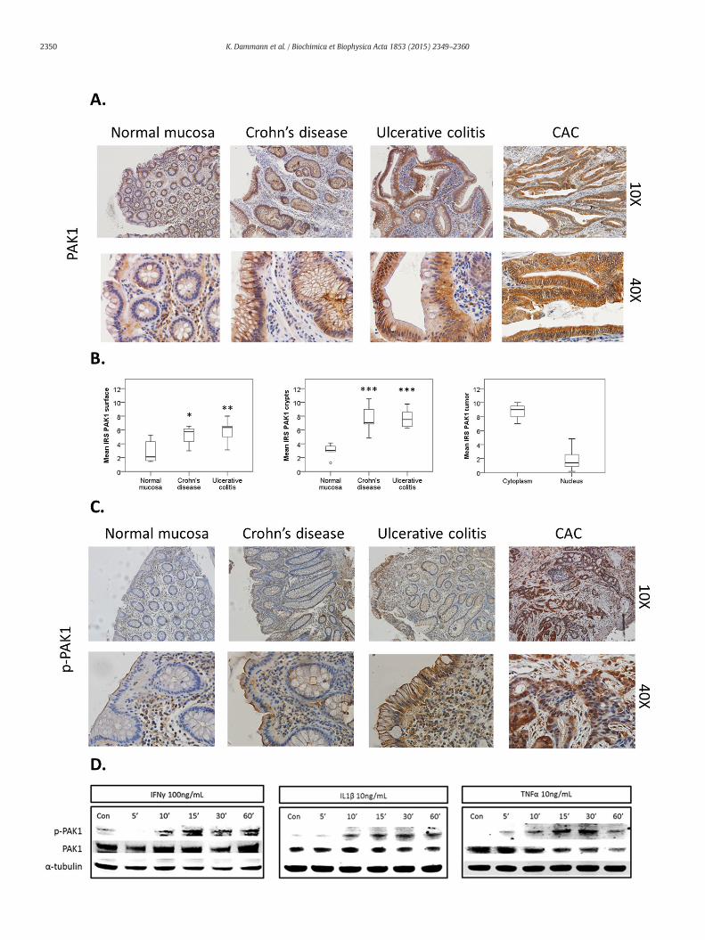
Fig. 1. (A–D). PAK1 is overexpressed and activated in IBD. (A) Human colonic tissue stained for Psurface and crypts in both Crohn's disease (CD), ulcerative colitis (UC), and in tumors of colitis asnormalmucosa (n=6), CD (n=7), and UC (n= 6) at the luminal surface and crypts. PAK1 is otissue stained for p-PAK1. In CD andUC, PAK1 activation in epithelial cells is found at themembris found within the nuclei and cytoplasm of tumor cells. (D) Inflammatory cytokines activate Peither untreated (Con) or treatedwith IFNγ, IL-1β, or TNFα for 5–60min and analyzed for PAK1activation at 30 min. α-Tubulin was utilized as a loading control.
epithelial cells was evaluated on a scale from 0–4, 0 (b1%), 1 (1–10%),2 (10–50%), 3 (51–80%), and 4 (N80%) in 4 separate fields of view. Themean intensity and percentage of positively stained epithelial cellswas multiplied to generate the IRS, and the highest score attainablewas 12. Data shown represent the mean of the 2 scorers' findings.
2.6. Quantitative real-time PCR
RNA was isolated using TRIzol reagent (Life Technologies). cDNAsynthesiswas carriedout using Thermoscript RT-PCRSystem(Invitrogen)according to manufacturer's protocol. Quantitative RT-PCR was carriedout in triplicate using Fast SYBR GreenMasterMix with primers (Supple-mentary Table S3). All data were normalized to the endogenous control36b4. Relative quantification of transcripts were calculated using thecomparative CT method.
2.7. Western blotting (WB) and co-immunoprecipitation (co-IP)
Cells werewashed in PBS and harvested in RIPA buffer for whole celllysates. Nuclear and cytoplasmic extracts were collected as previouslydescribed [17]. SIOwere released frommatrigel in ice cold PBS, and cen-trifuged at 1000 RPM for 5 min. The matrigel was discarded and theorganoids were sonicated for 2–3 s in RIPA buffer. All lysates were har-vested for 30min on ice and centrifuged at 4 °C for 30min. Protein con-centration was determined by Bradford assay and measured on an(Anthos 2010) photometer. For co-immunoprecipitation (co-IP),750 μg of protein were incubated with 2 μg rabbit or mouse IgG or pri-mary antibody shaking at 4 °C overnight followed by incubation withA/G agarose beads for 2 h at room temp. Samples were washed 5×with PBS and added to SDS sample buffer. For WB, 25–50 μg proteinwere added to SDS sample buffer for 10 min at 95 °C. Protein sampleswere separated using SDS-PAGE and transferred on a PVDF membrane.Membranes were blocked in Western blocker solution (Sigma) for 1 h,and incubatedwith primary antibodies (Supplementary Table S1) over-night at 4 °C. Protein bands were visualized using IRDye coupled sec-ondary antibodies and imaged on an Odyssey infrared imaging system(LI-COR). Images were obtained and densitometry was calculatedusing Odyssey application software version 3.0.21.
2.8. Electroporation, PAK1 overexpression, and RNA interference
Transient transfection inHCEC-1CTwas achieved via electroporationusing Amaxa basic nucleofector 2B kit and device for primary mamma-lian epithelial cells (Lonza) [17]. PAK1 was overexpressed with 5 μg ofpCMV6M-PAK1 (WT-PAK1), a generous gift fromDr. Jonathan Chernoff,pCMV6M-PAK1K299R (KD-PAK1) (Addgene) or a PCMV6Mempty vec-tor (EV) (Addgene) for 72 h. PAK1 knockdown using RNA interferencewas achieved using 100 nM siPAK1 (Dharmacon) or 100 nM scrambledsiRNA (Santa Cruz) for 48 h.
2.9. Immunofluorescence
HCEC-1CT and SIOwere fixed in 4% paraformaldehyde at room tem-perature for 30 min and blocked in 0.1% Triton-X-100/2% horse serumfor 1 h. Samples were incubated with primary antibody in 1% BSA/PBSovernight at 4 °C, then washed and incubated with fluorescent second-ary antibody for 1 h at room temperature. Samples were washed, dried,and mounted in medium containing DAPI (Vector Laboratories), andimaged on an Olympus IX81 or BX51 microscope.
AK1. PAK1 expression increases in comparison to normalmucosa at the luminal epithelialsociated cancer (CAC). (B) Box plots comparemean PAK1 immunoreactivity scores (IRS) inverexpressed in CAC (n=8). The circle is the outlier from the boxplot. (C) Human colonicane, specifically at the luminal surface, but not in the crypts. In CAC, PAK1 phosphorylationAK1 in HCEC-1CT. WB of HCEC-1CT RIPA whole cell lysates using 50 μg protein. Cells wereactivationwith a p-PAK1Thr 423 antibody. TNFα induced themost profoundeffect in PAK1
2352 K. Dammann et al. / Biochimica et Biophysica Acta 1853 (2015) 2349–2360
2.10. Luciferase assay
HCEC-1CT cells were transiently transfected with a TkpGL3NF-κB orPPAR luciferase response element, or co-transfected with scrRNA,siPAK1, EV, WT-PAK1, or KD-PAK1 and seeded into 24 well plates.Cells were lysed with a lysis buffer containing 4% Triton X-100, 100 mMGlycyl-glycine, 100mMMgSO4, and 250mMEGTA for 20min on a shakerat room temperature. 20 μL of lysate were added into a 96 well plate andanalyzed on a Plate Chameleon V photometer (Hidex) in which 100 μL ofluciferase activity buffer containing 100 mM Glycyl-glycine, 100 mMMgSO4, 20 mM ATP (Sigma), and 2.5 mM D-luciferin (Sigma) wereinjected per well. All measurements were performed using technicaltriplicates.
2.11. Statistics
Statistical analysis was performed using SPSS (version 21.0). Metricoutcome variables were compared using univariate analysis of variance(ANOVA) and Tukey HSD post-hoc tests. RT-PCR data and immunoreac-tivity scores were analyzed using a 2-tailed t-test. p-Values less than0.05 were considered significant (*p b 0.05, **p b 0.01, ***p b 0.001).All data are expressed as mean ± standard deviation.
3. Results
3.1. Pro-inflammatory cytokines promote PAK1 activation in IEC
PAK1 is associated with gut tumorigenesis [5,18,19] andwe recentlydemonstrated its overexpression in IBD and colitis-associated cancer(CAC) [6]. Here we investigated PAK1 activation and signaling in IBDand CAC. Normal intestinal mucosa, CD, UC, and CAC samples werestained with both a PAK1 and p-PAK1 antibody that detects phosphor-ylation at Thr-423, a residue that is essential for PAK1 activation [20].In comparison to normal mucosa, total and nuclear PAK1 was over-expressed at the luminal epithelial surface and crypts in CD and UC, aswell as in tumors of CAC (Fig. 1A–B). PAK1 activationwas absent withinthe normal mucosa, and cytoplasmic p-PAK1 levels increased at the lu-minal intestinal epithelial surface in both UC and CD, but not within thecrypts. CAC tumor samples displayed increased PAK1 phosphorylationin both the cytoplasm and nucleus (Fig. 1C, Supplementary Fig. S1A).
IFNγ, IL-1β, and TNFα are pro-inflammatory cytokines whichcontribute to gut inflammation in IBD [21]. We investigated their effecton PAK1 activation in HCEC-1CT cells. IFNγ, IL-1β, and TNFα inducedphosphorylation of PAK1 at Thr-423 within 10 to 30 min (Fig. 1D). Im-munofluorescence of p-PAK1 revealed cytoplasmic levels of p-PAK1 in-creased upon TNFα stimulation, and this effect was abolished uponPAK1 kinase inhibition using IPA-3 (Supplementary Fig. S1B). We furtherverified the specificity of our p-PAK1 antibody using IPA-3 upon WT-PAK1 overexpression in the presence of TNFα (Supplementary Fig. S1C).PAK1 overexpression also resulted in PAK1 auto-phosphorylation. IFNγ,IL-1β, and TNFα also induced p-PAK1 in the colorectal cancer cell lineHCT116 as early as 5 min peaking within the 30–60 min range (Supple-mentary Fig. S1D). Accumulation of polymorphonuclear leukocytes(PMNs) within colonic crypts is a key feature of UC [16]. HCEC-1CT cells
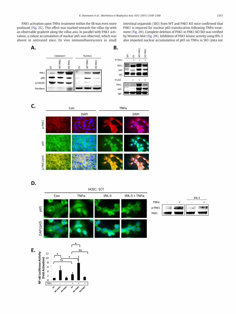
Fig. 2. (A–H). PAK1 overexpression regulates NF-kB activation downstream of TNFα in HCEC-1WT-PAK1 or KD-PAK1 and probed for PAK1 and p65.WT-PAK1 but not KD-PAK1 overexpressioand Fibrillarin (nucleus). (B) Co-IP of PAK1 (top) and p65 (bottom) probed for PAK1 and p65co-staining in untreated (Con) and TNFα treated cells. Upper panel p-PAK1, middle panel p6PAK1 and p65 colocalized upon TNFα. (D) IF of p65 or merged DAPI/p65 in HCEC-1CT. Cells waccumulation of p65 by TNFα is impeded by p-PAK1 inhibition. The WB verified p-PAK1 activWT-PAK1, and KD-PAK1 overexpression in untreated and TNFα treated cells. Data a(F) Immunohistochemistry of large bowel (LB) or small bowel (SB) mouse tissue sections staineparing SB crypt andvilli expression, and LB crypt and surface expression in controlmice (n=3)in TNFα injectedmice. TNFα increases p-PAK1 expression and p65 (black arrow) nuclear translcontrol. Graphs showmean villi p-PAK1 andp65 IRSs (±SD) (n=3)mice per group (t-test, 2 tasmall intestinal organoids (SIO) with or without TNFα. WB of PAK1 protein expression in WT
were co-cultured with freshly isolated PMNs using Transwell inserts.Total PAK1 expression increased more than two-fold at 60 min (Supple-mentary Fig. S1E). Taken together, these data demonstrate that pro-inflammatory cytokines and activated PMNs regulate PAK1 activationand expression in human colonic epithelial cells.
3.2. PAK1 regulates NF-κB activation downstream of TNFα
Our data showed that TNFα, which plays a key role in the pathogen-esis of IBD [21], and activates canonical NF-κB signaling, induced PAK1activation. Next, we investigated the role of PAK1 in NF-κB signaling.Control (EV), WT-PAK1, and KD-PAK1 vectors were overexpressed inHCEC-1CT and Western blot analysis was performed on cytoplasmicand nuclear fractions. WT-PAK1 overexpression induced p65 nucleartranslocation in the absence of TNFα, whereas KD-PAK1 did not(Fig. 2A). Co-immunoprecipitation showed a direct interaction of p65and PAK1 upon WT-PAK1 overexpression which was reduced uponKD-PAK1 indicating the importance of auto-phosphorylation of PAK1for interaction with p65 (Fig. 2B). In the absence of TNFα, immunofluo-rescence staining of p-PAK1 and p65 revealed localization of both pro-teins in the cytoplasm. TNFα induced translocation and co-localizationof p-PAK1 and p65 in the nucleus (Fig. 2C). Immunofluorescence inHCEC-1CT revealed that TNFα activated nuclear translocation of p65was blocked upon PAK1 kinase inhibition using IPA-3 (Fig. 2D). P-PAK1 inhibition by IPA-3 was confirmed by Western blot (Fig. 2D).Western blot analysis supported these observations and demonstratedthat KD-PAK1 abrogated the effect of TNFα on p65nuclear translocation(Supplementary Fig. S2A). Densitometry of nuclear p65 revealed thatWT-PAK1 overexpressing cells had increased nuclear p65 and weremore sensitive to TNFα in comparison to EV or KD-PAK1 (SupplementaryFig. S2A). Further, the effect of PAK1 overexpression and the depen-dence of PAK1 kinase activity on NF-κB transactivation were con-firmed using an NF-κB luciferase reporter assay (Fig. 2E). Thesedata demonstrate that PAK1 overexpression or its activation byTNFα results in a p-PAK1–p65 interaction, nuclear translocation,and transcriptional activation of NF-κB.
As the findings above indicate a concerted action of PAK1 and p65,we further assessed the distribution of such a PAK1–p65 interaction inthemouse intestine. PAK1 and p-PAK1were expressed in the cytoplasmwithin cells of small bowel (SB) villi and of the large bowel (LB) luminalsurface (Fig. 2F). P65 was also cytoplasmic and its localization resem-bled PAK1 and p-PAK1 in the LB (Fig. 2F). Less than 1%of IECs had nucle-ar p65. These data suggest thatwithout inflammation, PAK1 andp65 areco-expressed in differentiated IEC of SB villi and LB luminal surface andcanonical NF-κB signaling is not active.
Considering that PAK1 and p65 expression was associated through-out themouse intestine, we investigated the effect of TNFα on PAK1 ex-pression and activation in vivo. Mice were i.p. injected with TNFα 1.5 hbefore sacrificing and the SB and LB were collected for immunohisto-chemistry. Total PAK1 expressionwas not altered upon TNFα treatment(data not shown). We observed a modest increase in p-PAK1 at the LBepithelial surface (Supplementary Fig. S2B). Nuclear p65, which wasnot present in control mice, also increased upon TNFα treatment atthe luminal surface (Supplementary Fig. S2B).
CT. (A) WB of HCEC-1CT cytoplasmic and nuclear lysates. Cells were transfected with EV,n increased nuclear translocation of p65. Loading controls includedα-tubulin (cytoplasm)demonstrating a PAK1–p65 interaction. (C) Immunofluorescence (IF) of p-PAK1 and p655, and lower panel merged p-PAK1/p65 images with or without merged DAPI channel.ere pre-treated with 10 μM IPA-3 (2 h) followed by 10 ng/mL TNFα for 30 min. Nuclearation by TNFα and its inhibition by IPA-3 in HCEC-1CT. (E) NF-κB luciferase assay of EV,re representative of 3 independent experiments, ANOVA, Tukey HSD; *p b 0.05.d for PAK1, p-PAK1, or p65. Box plots aremean PAK1 immunoreactivity scores (IRS) com-mice (t-test, 2 tailed; ***pb 0.001). (G) IHC of p-PAK1and p65within small intestinal tissueocation within villi, but not in crypts. PAK1 KOmicewere stained for p-PAK1 as a negativeiled; ***pb 0.001) (H) IF of p65 ormergedDAPI/p65 inwild type (WT) and PAK1KOmouseor PAK1 KO SIO.
2353K. Dammann et al. / Biochimica et Biophysica Acta 1853 (2015) 2349–2360
PAK1 activation upon TNFα treatment within the SB was evenmoreprofound (Fig. 2G). This effect was marked towards the villus tip withan observable gradient along the villus axis. In parallel with PAK1 acti-vation, a robust accumulation of nuclear p65 was observed, which wasabsent in untreated mice. Ex vivo immunofluorescence in small
intestinal organoids (SIO) from WT and PAK1 KO mice confirmed thatPAK1 is required for nuclear p65 translocation following TNFα treat-ment (Fig. 2H). Complete deletion of PAK1 in PAK1 KO SIO was verifiedbyWestern blot (Fig. 2H). Inhibition of PAK1 kinase activity using IPA-3also impeded nuclear accumulation of p65 on TNFα in SIO (data not
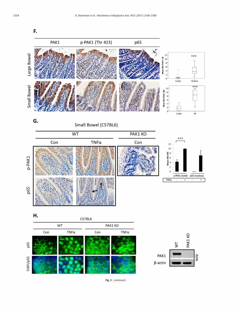
Fig. 2 (continued).
2354 K. Dammann et al. / Biochimica et Biophysica Acta 1853 (2015) 2349–2360

Fig. 3. (A–F). PAK1 is required for complete activation of NF-κB downstream of IκB in HCEC-1CT. (A) EV, WT-PAK1, or KD-PAK1 overexpressing cells were treated with 10 ng/mL TNFα(30 min). RIPA lysates were analyzed by WB for PAK1, p-IKKα/β, IKKα, p-IκB-α, and IκBα expression. WT or KD-PAK1 overexpression did not interfere with the degradation of IκB fol-lowing TNFα treatment. (B)WB of RIPA lysates analyzed for p-p65 and p65. Cellswere treatedwith TNFα or pretreated (12 h)with 10 μM IPA-3 or 20mM5-ASA. Pretreatmentwith IPA-3or 5-ASA blocked the effect of TNFα on p65. (C)WB of whole cell (RIPA) lysates probed for PAK1, p65, and β-actin. PAK1 knock down by both 5-ASA and siPAK1 blocked total p65 proteinlevels. (D) Cells were co-transfected with a NF-κB luciferase reporter and siPAK1 or scrRNA. Pretreatment with 5-ASA and PAK1 knockdown impeded the effect of TNFα on NF-κB tran-scriptional activation. Data are representative of 3 independent experiments. (E) Relative mRNA expression of RelA upon siPAK1 or scrRNA with or without 5-ASA treatment. PAK1 inhi-bition increased transcription of RelA. (F) HCEC-1CT cells were treated with 20mM 5-ASA (24 h) with or without the proteasomal inhibitor MG132 (6 h). RIPA lysates were analyzed forp65 and α-tubulin by WB. 5-ASA inhibits p65 but not upon pretreatment with MG132. All data are representative of 3 independent experiments.
2355K. Dammann et al. / Biochimica et Biophysica Acta 1853 (2015) 2349–2360

2356 K. Dammann et al. / Biochimica et Biophysica Acta 1853 (2015) 2349–2360

Fig. 4. (A–G). PAK1modulates a PPARγ/p65 cascade. (A) HCEC-1CT cells were transfectedwith a NF-kB luciferase reporter and treated with 1 μM Rosi ± 20mM 5-ASA (24 h). Rosiand 5-ASA blocked NF-kB transcriptional activity ANOVA, Tukey HSD; ***p b 0.001.(B) Rosi treated cells were fractionated and analyzed by WB for PAK1 and p65. Rosiblocked PAK1 and p65 expression. (C) IF of p-PAK1 and PPARγ upon TNFα (30 min)with or without 20 mM 5-ASA pretreatment (24 h). TNFα resulted in p-PAK1 activationand PPARγ nuclear export and downregulation which was blocked by 5-ASA. (D) The ef-fect of EV, WT-PAK1, and KD-PAK1 overexpression on PPARγwas analyzed byWB.α-Tu-bulin was used as a loading control. Densitometry of PPARγ levels normalized toα-tubulin, data are mean and SD from 2 independent experiments. (E) RTPCR of PAK1and PPARγ after EV or WT-PAK1 overexpression with or without 5-ASA treatment(24 h). PAK1 overexpression blocked PPARγ at the mRNA level, an effect that was recov-ered by 5-ASA. Data are representative of 3 independent experiments (t-test, 2 tailed;**p b 0.01). (F) PPAR luciferase reporter assay in PAK1 knock down cells using scrRNAor siPAK1. siPAK1 activated PPAR transcription, and this effect was recovered upon0.1 μM BADGE pretreatment (12 h), ANOVA, Tukey HSD; *p b 0.05. (G) NF-kB luciferaseassay in PAK1 knock down cells using scrRNA or siPAK1. Cells were pretreated withBADGE (12 h) with or without 5-ASA (24 h). BADGE recovered the effect of 5-ASA orsiPAK1 on NF-kB. All data are representative of 3 independent experiments ANOVA,Tukey HSD; **p b 0.01, ***p b 0.001.
2357K. Dammann et al. / Biochimica et Biophysica Acta 1853 (2015) 2349–2360
shown). These data support a role for PAK1 in NF-κB signaling down-stream of TNFα in IECs.
3.3. PAK1 is required for complete activation of NF-κB downstream of IκBin IECs
In order to delineate PAK1's role within canonical NF-κB signaling, wefurther investigated the IKKα/β-IκB cascade in HCEC-1CT cells. If PAK1was upstream of IKKα/β or IκB, PAK1 overexpression would modulateIkB degradation. Western blot analysis revealed that neither WT-PAK1nor KD-PAK1 mediated TNFα's effect on IκB degradation (Fig. 3A). PAK1knockdown (siPAK1) reduced p65 nuclear translocation upon TNFαtreatment, but did not affect IκB phosphorylation or degradation furthersuggesting that PAK1 acts downstream of IκB (Supplementary Fig. S3A).
Considering that PAK1 associates with p65 upon TNFα stimulation,we investigated its effect on p65 stability. PAK1 inhibitors IPA-3 as
well as 5-ASA reduced total p65 protein levels with and without TNFα(Fig. 3B), although 5-ASA but not IPA-3 inhibited p65 phosphorylation.Compared to TNF-α treatment, 5-ASA treatment reduced p65 levelsover time upon stimulation with TNF-α and this effect wasmore prom-inent at 24 h. IPA-3 however, inhibited total p65 levels but did not re-duce p65 phosphorylation. This suggested that the effect of PAK1inhibition on p65 is a result of p65 stability, but not phosphorylation.Mechanistically, 5-ASA reduced PAK1 expression to a similar extent assiPAK1 and both resulted in reduction of p65 total protein levels(Fig. 3C) and p65 transcriptional activity (Fig. 3D). 5-ASA did not displayan additive effect to siPAK1 alone suggesting that 5-ASA utilizes PAK1 toreduce p65. TNFα transactivation of NF-κB was blocked upon 5-ASA,siPAK1, and their combination. NF-κB transactivation by TNFα wasalso blocked by IPA-3 (Supplementary Fig. S3B).
We further investigated whether siPAK1 or 5-ASA reduced p65 atthe transcriptional level. Surprisingly, both siPAK1 and 5-ASA increasedRelAmRNA (Fig. 3E). Inhibition of the proteasomewithMG132, howev-er, blocked 5-ASA's effect on p65 suggestive that PAK1's role within thecanonical NF-κB pathway is rather posttranslational (Fig. 3F). Thesedata demonstrate that (1) PAK1 stabilizes p65 protein downstream ofIkB, (2) PAK1 is required for complete activation of NF-κB by TNFα,and (3) 5-ASA inhibits NF-κB transcriptional activity via inhibition oftotal PAK1 in IECs.
3.4. PAK1 modulates a PPARγ/p65 cascade within IECs
Our data indicated that 5-ASA blocks NF-κB in IECs through inhibi-tion of PAK1. 5-ASA is also a known agonist of PPAR gamma (PPARγ)[22] and PPARγ polyubiquitinates and degrades p65 [23]. PPARγ ishighly expressed in the gut, and regulates IEC differentiation andmigra-tion within SB villi [24]. Within the LB, PPARγ activation has anti-inflammatory and chemopreventive effects [22,25].
First, we confirmed that both Rosiglitazone (Rosi; a PPARγ agonist)and 5-ASA – to a lesser degree – increased PPAR transactivation (datanot shown). Both also blocked NF-κB signaling in our system (Fig. 4A).Interestingly, Rosi did not add to 5-ASA's effect onNF-κB inhibition, sug-gesting that both agonists utilize PPARγ activation to inhibit NF-κB(Fig. 4A). These data were confirmed at the protein level in whichPPARγ activation by Rosi reduced p65 levels in both cytoplasm and nu-cleus (Fig. 4B). Rosi also reduced PAK1 protein abundance.
Pro-inflammatory cytokines, such as TNFα, result in PPARγ degrada-tion [26], therefore we investigated the TNFα-PAK1-PPARγ cascade viaimmunofluorescence. Control HCEC-1CT cells had low levels of p-PAK1,and PPARγ was present in the nucleus and cytoplasm. TNFα treatmentresulted in PAK1 phosphorylation concomitantwith an apparent reduc-tion of nuclear and cytoplasmic PPARγ expression (Fig. 4C). 5-ASAblocked activation of PAK1 and retained PPARγ levels in the presenceof TNFα. These data suggest that TNFα results in activation of PAK1and in reduction of PPARγ, both of which can be blocked by 5-ASA.
To better understand the interaction between PAK1 and PPARγ, weoverexpressed PAK1 in HCEC-1CT. Total PPARγ protein levels werereduced upon WT-PAK1 but not upon KD-PAK1 transfection (Fig. 4D).At the transcriptional level, 5-ASA inhibited PAK1 while increasingPPARγ mRNA levels (Fig. 4E). WT-PAK1 overexpression decreasedPPARγmRNA levels and5-ASA recovered PPARγ levels. These data dem-onstrate that PAK1 activation and overexpression downregulate PPARγboth at the protein andmRNA levels, which can be recovered by 5-ASA.
WT-PAK1 overexpression increased the effect of TNFα on NF-κBtransactivation (as shown in Fig. 2E). If this was a result of PPARγ inhibi-tion, PPARγ activation should recover this effect. NF-κB luciferase assayshowed that in control cells (EV), Rosi blocked TNFα transactivation ofNF-κB (Supplementary Fig. S4A). Rosi also blocked the effect of WT-PAK1 on NF-κB transactivation. The synergistic effect of WT-PAK1 andTNFα on NF-κB transactivation was also reduced upon PPARγ activationby Rosi. These data were further verified by Western blot in which Rosiinhibited nuclear PAK1 and p65 upon TNFα in WT-PAK1 overexpressing
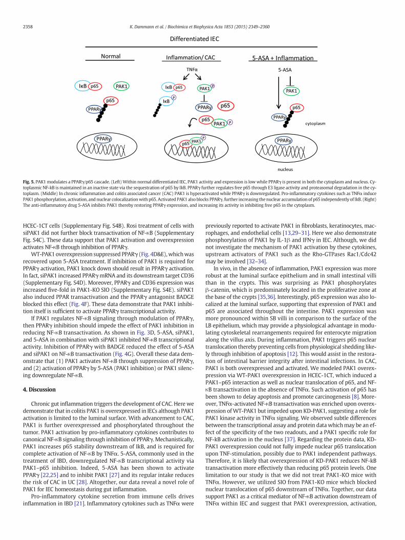
Fig. 5. PAK1 modulates a PPARγ/p65 cascade. (Left)Within normal differentiated IEC, PAK1 activity and expression is low while PPARγ is present in both the cytoplasm and nucleus. Cy-toplasmic NF-kB is maintained in an inactive state via the sequestration of p65 by IkB. PPARγ further regulates free p65 through E3 ligase activity and proteasomal degradation in the cy-toplasm. (Middle) In chronic inflammation and colitis associated cancer (CAC) PAK1 is hyperactivated while PPARγ is downregulated. Pro-inflammatory cytokines such as TNFα inducePAK1phosphorylation, activation, and nuclear colocalizationwith p65. Activated PAK1 also blocks PPARγ, further increasing the nuclear accumulation of p65 independently of IkB. (Right)The anti-inflammatory drug 5-ASA inhibits PAK1 thereby restoring PPARγ expression, and increasing its activity in inhibiting free p65 in the cytoplasm.
2358 K. Dammann et al. / Biochimica et Biophysica Acta 1853 (2015) 2349–2360
HCEC-1CT cells (Supplementary Fig. S4B). Rosi treatment of cells withsiPAK1 did not further block transactivation of NF-κB (SupplementaryFig. S4C). These data support that PAK1 activation and overexpressionactivates NF-κB through inhibition of PPARγ.
WT-PAK1overexpression suppressed PPARγ (Fig. 4D&E),whichwasrecovered upon 5-ASA treatment. If inhibition of PAK1 is required forPPARγ activation, PAK1 knock down should result in PPARγ activation.In fact, siPAK1 increased PPARγmRNA and its downstream target CD36(Supplementary Fig. S4D). Moreover, PPARγ and CD36 expression wasincreased five-fold in PAK1-KO SIO (Supplementary Fig. S4E). siPAK1also induced PPAR transactivation and the PPARγ antagonist BADGEblocked this effect (Fig. 4F). These data demonstrate that PAK1 inhibi-tion itself is sufficient to activate PPARγ transcriptional activity.
If PAK1 regulates NF-κB signaling through modulation of PPARγ,then PPARγ inhibition should impede the effect of PAK1 inhibition inreducing NF-κB transactivation. As shown in Fig. 3D, 5-ASA, siPAK1,and 5-ASA in combination with siPAK1 inhibited NF-κB transcriptionalactivity. Inhibition of PPARγ with BADGE reduced the effect of 5-ASAand siPAK1 on NF-κB transactivation (Fig. 4G). Overall these data dem-onstrate that (1) PAK1 activates NF-κB through suppression of PPARγ,and (2) activation of PPARγ by 5-ASA (PAK1 inhibition) or PAK1 silenc-ing downregulate NF-κB.
4. Discussion
Chronic gut inflammation triggers the development of CAC. Here wedemonstrate that in colitis PAK1 is overexpressed in IECs although PAK1activation is limited to the luminal surface. With advancement to CAC,PAK1 is further overexpressed and phosphorylated throughout thetumor. PAK1 activation by pro-inflammatory cytokines contributes tocanonical NF-κB signaling through inhibition of PPARγ. Mechanistically,PAK1 increases p65 stability downstream of IkB, and is required forcomplete activation of NF-κB by TNFα. 5-ASA, commonly used in thetreatment of IBD, downregulated NF-κB transcriptional activity viaPAK1–p65 inhibition. Indeed, 5-ASA has been shown to activatePPARγ [22,25] and to inhibit PAK1 [27] and its regular intake reducesthe risk of CAC in UC [28]. Altogether, our data reveal a novel role ofPAK1 for IEC homeostasis during gut inflammation.
Pro-inflammatory cytokine secretion from immune cells drivesinflammation in IBD [21]. Inflammatory cytokines such as TNFα were
previously reported to activate PAK1 in fibroblasts, keratinocytes, mac-rophages, and endothelial cells [13,29–31]. Here we also demonstratephosphorylation of PAK1 by IL-1β and IFNγ in IEC. Although, we didnot investigate the mechanism of PAK1 activation by these cytokines,upstream activators of PAK1 such as the Rho-GTPases Rac1/Cdc42may be involved [32–34].
In vivo, in the absence of inflammation, PAK1 expression was morerobust at the luminal surface epithelium and in small intestinal villithan in the crypts. This was surprising as PAK1 phosphorylatesβ-catenin, which is predominately located in the proliferative zone atthe base of the crypts [35,36]. Interestingly, p65 expression was also lo-calized at the luminal surface, supporting that expression of PAK1 andp65 are associated throughout the intestine. PAK1 expression wasmore pronounced within SB villi in comparison to the surface of theLB epithelium, which may provide a physiological advantage in modu-lating cytoskeletal rearrangements required for enterocyte migrationalong the villus axis. During inflammation, PAK1 triggers p65 nucleartranslocation thereby preventing cells from physiological shedding like-ly through inhibition of apoptosis [12]. This would assist in the restora-tion of intestinal barrier integrity after intestinal infections. In CAC,PAK1 is both overexpressed and activated. We modeled PAK1 overex-pression via WT-PAK1 overexpression in HCEC-1CT, which induced aPAK1–p65 interaction as well as nuclear translocation of p65, and NF-κB transactivation in the absence of TNFα. Such activation of p65 hasbeen shown to delay apoptosis and promote carcinogenesis [8]. More-over, TNFα-activated NF-κB transactivation was enriched upon overex-pression ofWT-PAK1 but impeded upon KD-PAK1, suggesting a role forPAK1 kinase activity in TNFα signaling. We observed subtle differencesbetween the transcriptional assay and protein data whichmay be an ef-fect of the specificity of the two readouts, and a PAK1 specific role forNF-kB activation in the nucleus [37]. Regarding the protein data, KD-PAK1 overexpression could not fully impede nuclear p65 translocationupon TNF-stimulation, possibly due to PAK1 independent pathways.Therefore, it is likely that overexpression of KD-PAK1 reduces NF-kBtransactivation more effectively than reducing p65 protein levels. Onelimitation to our study is that we did not treat PAK1-KO mice withTNFα. However, we utilized SIO from PAK1-KO mice which blockednuclear translocation of p65 downstream of TNFα. Together, our datasupport PAK1 as a critical mediator of NF-κB activation downstream ofTNFα within IEC and suggest that PAK1 overexpression, activation,
2359K. Dammann et al. / Biochimica et Biophysica Acta 1853 (2015) 2349–2360
and nuclear localization may contribute to tumor development by sus-taining NF-κB activity.
TGFβ activated kinase (TAK1) is required for canonical NF-κB activa-tion primarily through phosphorylation of IKKα and through inducingIkB degradation [38]. Neither PAK1 overexpression nor knockdowninterfered with the upstream IKKα/IkB cascade, suggesting thatPAK1's effect on p65 is through an alternative pathway separate fromTAK1 and downstream of IkB.
Besides its role in fatty acid oxidation/transport, and increasing insu-lin sensitivity [39], PPARγ was recently reported to be an E3 ubiquitinligase, which induces p65 degradation [23]. 5-ASA acts as a PPARγ ago-nist and also inhibits PAK1 and NF-κB [22,27,40]. Here we demonstratethat 5-ASA induced degradation of p65 protein (Fig. 3C) indicative ofPPARγ's E3 ubiquitin ligase activity. Thus, it was imperative to investi-gate the PAK1/PPARγ axis. In fact, upon pro-inflammatory signals (asobserved in IBD), PAK1 activation suppresses PPARγ and contributesto NF-κB signaling by stabilizing p65. This notion is supported by ourobservation that total p65 levels are reduced by both 5-ASA and PAK1inhibition (IPA-3, siPAK1). TNFα initiates PPARγ recruitment and deg-radation of p65 in mouse embryonic fibroblasts [23]. In our system,TNFα increased PAK1 activation concurrent with PPARγ downregula-tion (Fig. 4C). As PAK1 overexpression inhibits PPARγ at the proteinand transcriptional levels, it is likely that PAK1 itself, or its downstreamtargets (JNK or ERK) could phosphorylate PPARγ leading to its nuclearexport and proteasomal degradation [41]. Moreover, PPARγ is knownto regulate its own expression [42], therefore down regulation ofPPARγ at the protein level would result in reduced transcription,which we also observed in our system (Fig. 4D&E). PAK1 activation oroverexpression could inhibit PPARγ transcription through multiplepathways. In pluripotent mesenchymal stem cells, TAK1 suppressesPPARγ via a NF-κB interacting kinase (NIK) cascade [26]. PAK1 activationof NIK was previously described [43] and may result in PPARγ inhibition.It is plausible that PAK1 activation and subsequent phosphorylation ofSMART [44], Snail [45], or the estrogen receptor [46] may downregulatePPARγ [47–49]. Nevertheless, our data implicate PAK1 in modulating aPPARγ/p65 cascade, which has not previously been reported.
Wedid not study the effect of PAK1 on other PPARs as 5-ASA is a spe-cific agonist for PPARγ [22], and only PPARγ but not PPARα or δ inducesp65 degradation [23]. Interestingly, both 5-ASA and Rosiglitazoneinhibited PAK1 (Figs. 3C and 4B). One explanation may be upon ligandactivation, PPARγmay not onlymark p65 but also a PAK1–p65 complexfor proteasomal degradation. 5-ASA also inhibits PAK1 at the mRNAlevel which is suggestive of an additional mechanism independent ofPPARγ ligand activation.
We propose that PAK1 is another player in PPARγ/NF-κB cascadein intestinal epithelial cells (Fig. 5). In differentiated normal IECs,PPARγ regulates NF-κB through proteasomal degradation of p65.However, in the presence of inflammation or CAC, PAK1 is over-expressed and activated, whereas PPARγ is suppressed [50]. PAK1overexpression or activation blocks PPARγ leading to increasedNF-κB transcriptional activation. In addition we further demonstratethat inhibition of PAK1 by 5-ASA also leads to activation of PPARγ'sE3 ligase activity.
Overall, these findings support a role for PAK1 in epithelial cellhomeostasis in IBD and CAC. Pharmacological inhibition of PAK1 overex-pression may recover PPARγ levels and impair the inflammatory re-sponse such as NF-kB activation in IEC.
Supplementary data to this article can be found online at http://dx.doi.org/10.1016/j.bbamcr.2015.05.031.
Disclosure of potential conflicts of interest
CG has a research collaboration with Shire Pharmaceuticals and re-ceived research support, lecturing or consulting honoraria from Ferringand Dr. Falk Pharma.
Author contributions
KD: conception and design, acquisition of data, analysis and inter-pretation of data, writing of the manuscript, VK: conception and design,acquisition of data, analysis and interpretation of data and revision ofmanuscript, ML: analysis and interpretation of data, TC: interpretationof data, proof-reading of the manuscript, development of methodology,FH: analysis and interpretation of data, NG: development ofmethodology,RE: statistical analysis, JW: acquisition of data, analysis and interpretationof data, MP: interpretation of data, revision of the manuscript, AW: ob-tained funding, revision of manuscript, CG: obtained funding, conceptionanddesign, study supervision, revision of themanuscript. All authors readand approved the final manuscript.
Acknowledgments
The financial support by the Federal Ministry of Economy, Familyand Youth and the National Foundation for Research, Technology andDevelopment is gratefully acknowledged. We acknowledge Dr. AndresI. Roig and Dr. Jerry W. Shay for providing HCEC-1CT cells and Dr.Jonathan Chernoff for the WT-PAK1 overexpression plasmid.
Grant supportThe study was supported by Austrian Science Fund (P24121
to Christoph Gasche). The research leading to these results hasreceived funding from the European Community's Seventh FrameworkProgramme (FP7/2007-2013) under grant agreement no. 305564(SysmedIBD).
References
[1] D.T. Rubin, M.R. Cruz-Correa, C. Gasche, J.R. Jass, G.R. Lichtenstein, E.A. Montgomery,R.H. Riddell, M.D. Rutter, T.A. Ullman, F.S. Velayos, S. Itzkowitz, A.S.A.i.C.C.P.M.Group, Colorectal cancer prevention in inflammatory bowel disease and the roleof 5-aminosalicylic acid: a clinical review and update, Inflamm. Bowel Dis. 14(2008) 265–274.
[2] V. Khare, A. Lyakhovich, K. Dammann, M. Lang, M. Borgmann, B. Tichy, S.Pospisilova, G. Luciani, C. Campregher, R. Evstatiev, M. Pflueger, H. Hundsberger,C. Gasche, Mesalamine modulates intercellular adhesion through inhibition ofp-21 activated kinase-1, Biochem. Pharmacol. 85 (2013) 234–244.
[3] M.C. Parrini, M. Lei, S.C. Harrison, B.J. Mayer, Pak1 kinase homodimers areautoinhibited in trans and dissociated upon activation by Cdc42 and Rac1, Mol.Cell 9 (2002) 73–83.
[4] R. Kumar, A.E. Gururaj, C.J. Barnes, p21-Activated kinases in cancer, Nat. Rev. Cancer6 (2006) 459–471.
[5] K. Dammann, V. Khare, C. Gasche, Tracing PAKs from GI inflammation to cancer, Gut63 (2014) 1173–1184.
[6] V. Khare, K. Dammann, M. Asboth, A. Krnjic, M. Jambrich, C. Gasche, Overexpressionof PAK1 promotes cell survival in inflammatory bowel diseases and colitis-associated cancer, Inflamm. Bowel Dis. 21 (2015) 287–296.
[7] F.R. Greten, L. Eckmann, T.F. Greten, J.M. Park, Z.W. Li, L.J. Egan, M.F. Kagnoff, M.Karin, IKKbeta links inflammation and tumorigenesis in a mouse model of colitis-associated cancer, Cell 118 (2004) 285–296.
[8] M. Karin, NF-kappaB as a critical link between inflammation and cancer, Cold SpringHarb. Perspect. Biol. 1 (2009) a000141.
[9] S. Schwitalla, A.A. Fingerle, P. Cammareri, T. Nebelsiek, S.I. Goktuna, P.K. Ziegler, O.Canli, J. Heijmans, D.J. Huels, G. Moreaux, R.A. Rupec, M. Gerhard, R. Schmid, N.Barker, H. Clevers, R. Lang, J. Neumann, T. Kirchner, M.M. Taketo, G.R. van denBrink, O.J. Sansom, M.C. Arkan, F.R. Greten, Intestinal tumorigenesis initiated by de-differentiation and acquisition of stem-cell-like properties, Cell 152 (2013) 25–38.
[10] K.B. Myant, P. Cammareri, E.J. McGhee, R.A. Ridgway, D.J. Huels, J.B. Cordero, S.Schwitalla, G. Kalna, E.L. Ogg, D. Athineos, P. Timpson, M. Vidal, G.I. Murray, F.R.Greten, K.I. Anderson, O.J. Sansom, ROS production and NF-kappaB activation trig-gered by RAC1 facilitateWNT-driven intestinal stem cell proliferation and colorectalcancer initiation, Cell Stem Cell 12 (2013) 761–773.
[11] B. Hoesel, J.A. Schmid, The complexity of NF-kappaB signaling in inflammation andcancer, Mol. Cancer 12 (2013) 86.
[12] J.M. Williams, C.A. Duckworth, A.J. Watson, M.R. Frey, J.C. Miguel, M.D. Burkitt, R.Sutton, K.R. Hughes, L.J. Hall, J.H. Caamano, B.J. Campbell, D.M. Pritchard, A mousemodel of pathological small intestinal epithelial cell apoptosis and shedding in-duced by systemic administration of lipopolysaccharide, Dis. Model. Mech. 6(2013) 1388–1399.
[13] J.A. Frost, J.L. Swantek, S. Stippec, M.J. Yin, R. Gaynor, M.H. Cobb, Stimulation ofNFkappa B activity by multiple signaling pathways requires PAK1, J. Biol. Chem.275 (2000) 19693–19699.
[14] A.I. Roig, U. Eskiocak, S.K. Hight, S.B. Kim, O. Delgado, R.F. Souza, S.J. Spechler, W.E.Wright, J.W. Shay, Immortalized epithelial cells derived from human colon biopsies
2360 K. Dammann et al. / Biochimica et Biophysica Acta 1853 (2015) 2349–2360
express stem cell markers and differentiate in vitro, Gastroenterology 138 (2010)1012–1021 (e1011-1015).
[15] T. Sato, R.G. Vries, H.J. Snippert, M. van de Wetering, N. Barker, D.E. Stange, J.H. vanEs, A. Abo, P. Kujala, P.J. Peters, H. Clevers, Single Lgr5 stem cells build crypt-villusstructures in vitro without a mesenchymal niche, Nature 459 (2009) 262–265.
[16] C. Campregher, M.G. Luciani, C. Gasche, Activated neutrophils induce an hMSH2-dependent G2/M checkpoint arrest and replication errors at a (CA)13-repeat incolon epithelial cells, Gut 57 (2008) 780–787.
[17] V. Khare, M. Lang, K. Dammann, C. Campregher, A. Lyakhovich, C. Gasche, Modula-tion of N-glycosylation by mesalamine facilitates membranous E-cadherin expres-sion in colon epithelial cells, Biochem. Pharmacol. 87 (2014) 312–320.
[18] J.H. Carter, L.E. Douglass, J.A. Deddens, B.M. Colligan, T.R. Bhatt, J.O. Pemberton, S.Konicek, J. Hom, M. Marshall, J.R. Graff, Pak-1 expression increases with progressionof colorectal carcinomas to metastasis, Clin. Cancer Res. 10 (2004) 3448–3456.
[19] M. Radu, G. Semenova, R. Kosoff, J. Chernoff, PAK signalling during the developmentand progression of cancer, Nat. Rev. Cancer 14 (2014) 13–25.
[20] F.T. Zenke, C.C. King, B.P. Bohl, G.M. Bokoch, Identification of a central phosphoryla-tion site in p21-activated kinase regulating autoinhibition and kinase activity, J. Biol.Chem. 274 (1999) 32565–32573.
[21] M.F. Neurath, Cytokines in inflammatory bowel disease, Nat. Rev. Immunol. 14(2014) 329–342.
[22] C. Rousseaux, B. Lefebvre, L. Dubuquoy, P. Lefebvre, O. Romano, J. Auwerx, D.Metzger, W. Wahli, B. Desvergne, G.C. Naccari, P. Chavatte, A. Farce, P. Bulois, A.Cortot, J.F. Colombel, P. Desreumaux, Intestinal antiinflammatory effect of5-aminosalicylic acid is dependent on peroxisome proliferator-activatedreceptor-gamma, J. Exp. Med. 201 (2005) 1205–1215.
[23] Y. Hou, F. Moreau, K. Chadee, PPARgamma is an E3 ligase that induces the degrada-tion of NFkappaB/p65, Nat. Commun. 3 (2012) 1300.
[24] L. Chen, B.M. Necela, W. Su, M. Yanagisawa, P.Z. Anastasiadis, A.P. Fields, E.A.Thompson, Peroxisome proliferator-activated receptor gamma promotes epithelialto mesenchymal transformation by Rho GTPase-dependent activation of ERK1/2, J.Biol. Chem. 281 (2006) 24575–24587.
[25] C. Rousseaux, N. El-Jamal, M. Fumery, C. Dubuquoy, O. Romano, D. Chatelain, A.Langlois, B. Bertin, D. Buob, J.F. Colombel, A. Cortot, P. Desreumaux, L. Dubuquoy,The 5-aminosalicylic acid antineoplastic effect in the intestine is mediated byPPARgamma, Carcinogenesis 34 (2013) 2580–2586.
[26] M. Suzawa, I. Takada, J. Yanagisawa, F. Ohtake, S. Ogawa, T. Yamauchi, T. Kadowaki,Y. Takeuchi, H. Shibuya, Y. Gotoh, K. Matsumoto, S. Kato, Cytokines suppress adipo-genesis and PPAR-gamma function through the TAK1/TAB1/NIK cascade, Nat. CellBiol. 5 (2003) 224–230.
[27] K.W. Dammann, V. Khare, M. Lang, N. Granofszky, C. Gasche, Tu1674 PAK1 mediatesNF-KB signaling in colitis and colitis-associated cancer, Gastroenterology 144 (2013)S-819.
[28] F.S. Velayos, J.P. Terdiman, J.M. Walsh, Effect of 5-aminosalicylate use on colorectalcancer and dysplasia risk: a systematic review and metaanalysis of observationalstudies, Am. J. Gastroenterol. 100 (2005) 1345–1353.
[29] L. Zhou, C. Yan, R.G. Gieling, Y. Kida, W. Garner, W. Li, Y.P. Han, Tumor necrosisfactor-alpha induced expression of matrix metalloproteinase-9 through p21-activated kinase-1, BMC Immunol. 10 (2009) 15.
[30] D. Fu, Y. Yang, Y. Xiao, H. Lin, Y. Ye, Z. Zhan, L. Liang, X. Yang, L. Sun, H. Xu, Role ofp21-activated kinase 1 in regulating the migration and invasion of fibroblast-likesynoviocytes from rheumatoid arthritis patients, Rheumatology (Oxford) 51(2012) 1170–1180.
[31] R.A. Stockton, E. Schaefer, M.A. Schwartz, p21-Activated kinase regulates endothelialpermeability through modulation of contractility, J. Biol. Chem. 279 (2004)46621–46630.
[32] S. Kant, W. Swat, S. Zhang, Z.Y. Zhang, B.G. Neel, R.A. Flavell, R.J. Davis, TNF-stimulated MAP kinase activation mediated by a Rho family GTPase signaling path-way, Genes Dev. 25 (2011) 2069–2078.
[33] B. Wojciak-Stothard, A. Entwistle, R. Garg, A.J. Ridley, Regulation of TNF-alpha-induced reorganization of the actin cytoskeleton and cell–cell junctions by Rho,Rac, and Cdc42 in human endothelial cells, J. Cell. Physiol. 176 (1998) 150–165.
[34] A. Puls, A.G. Eliopoulos, C.D. Nobes, T. Bridges, L.S. Young, A. Hall, Activation of thesmall GTPase Cdc42 by the inflammatory cytokines TNF(alpha) and IL-1, and bythe Epstein–Barr virus transforming protein LMP1, J. Cell Sci. 112 (Pt 17) (1999)2983–2992.
[35] M.H. Park, D.J. Kim, S.T. You, C.S. Lee, H.K. Kim, S.M. Park, E.Y. Shin, E.G. Kim, Phos-phorylation of beta-catenin at serine 663 regulates its transcriptional activity,Biochem. Biophys. Res. Commun. 419 (3) (2012) 543–549.
[36] G. Zhu, Y. Wang, B. Huang, J. Liang, Y. Ding, A. Xu, W. Wu, A Rac1/PAK1 cascade con-trols beta-catenin activation in colon cancer cells, Oncogene 31 (2012) 1001–1012.
[37] S. Jagadeeshan, Y.R. Krishnamoorthy, M. Singhal, A. Subramanian, J. Mavuluri, A.Lakshmi, A. Roshini, G. Baskar, M. Ravi, L.D. Joseph, K. Sadasivan, A. Krishnan, A.S.Nair, G. Venkatraman, S.K. Rayala, Transcriptional regulation of fibronectin byp21-activated kinase-1 modulates pancreatic tumorigenesis, Oncogene 34 (2015)455–464.
[38] H. Sakurai, Targeting of TAK1 in inflammatory disorders and cancer, TrendsPharmacol. Sci. 33 (2012) 522–530.
[39] S. Hauser, G. Adelmant, P. Sarraf, H.M.Wright, E. Mueller, B.M. Spiegelman, Degrada-tion of the peroxisome proliferator-activated receptor gamma is linked to ligand-dependent activation, J. Biol. Chem. 275 (2000) 18527–18533.
[40] H. Kim, H. Jeon, H. Kong, Y. Yang, B. Choi, Y.M. Kim, L. Neckers, Y. Jung, A molecularmechanism for the anti-inflammatory effect of taurine-conjugated 5-aminosalicylicacid in inflamed colon, Mol. Pharmacol. 69 (2006) 1405–1412.
[41] R. Yin, Y.G. Dong, H.L. Li, PPARgamma phosphorylation mediated by JNK MAPK: apotential role in macrophage-derived foam cell formation, Acta Pharmacol. Sin. 27(2006) 1146–1152.
[42] P. Tontonoz, E. Hu, B.M. Spiegelman, Stimulation of adipogenesis in fibroblasts byPPAR gamma 2, a lipid-activated transcription factor, Cell 79 (1994) 1147–1156.
[43] M. Neumann, A. Foryst-Ludwig, S. Klar, K. Schweitzer, M. Naumann, The PAK1autoregulatory domain is required for interaction with NIK in Helicobacter pylori-induced NF-kappaB activation, Biol. Chem. 387 (2006) 79–86.
[44] R.K. Vadlamudi, B. Manavathi, R.R. Singh, D. Nguyen, F. Li, R. Kumar, An essential roleof Pak1 phosphorylation of SHARP in Notch signaling, Oncogene 24 (2005)4591–4596.
[45] Z. Yang, S. Rayala, D. Nguyen, R.K. Vadlamudi, S. Chen, R. Kumar, Pak1 phosphoryla-tion of snail, a master regulator of epithelial-to-mesenchyme transition, modulatessnail's subcellular localization and functions, Cancer Res. 65 (2005) 3179–3184.
[46] R.A. Wang, A. Mazumdar, R.K. Vadlamudi, R. Kumar, P21-activated kinase-1 phos-phorylates and transactivates estrogen receptor-alpha and promotes hyperplasiain mammary epithelium, EMBO J. 21 (2002) 5437–5447.
[47] C. Yu, K. Markan, K.A. Temple, D. Deplewski, M.J. Brady, R.N. Cohen, The nuclear re-ceptor corepressors NCoR and SMRT decrease peroxisome proliferator-activated re-ceptor gamma transcriptional activity and repress 3T3-L1 adipogenesis, J. Biol.Chem. 280 (2005) 13600–13605.
[48] Y.H. Lee, S.H. Kim, Y.J. Lee, E.S. Kang, B.W. Lee, B.S. Cha, J.W. Kim, D.H. Song, H.C. Lee,Transcription factor Snail is a novel regulator of adipocyte differentiation viainhibiting the expression of peroxisome proliferator-activated receptor gamma,Cell. Mol. Life Sci. 70 (2013) 3959–3971.
[49] X. Wang, M.W. Kilgore, Signal cross-talk between estrogen receptor alpha and betaand the peroxisome proliferator-activated receptor gamma1 in MDA-MB-231 andMCF-7 breast cancer cells, Mol. Cell. Endocrinol. 194 (2002) 123–133.
[50] L. Dubuquoy, E.A. Jansson, S. Deeb, S. Rakotobe, M. Karoui, J.F. Colombel, J. Auwerx, S.Pettersson, P. Desreumaux, Impaired expression of peroxisome proliferator-activated receptor gamma in ulcerative colitis, Gastroenterology 124 (2003)1265–1276.

![Biochimica et Biophysica Acta - COnnecting REpositoriesby volatile anesthetics [20,21]. Reliable structures for individual sub-types of nAChRs, especially their TM domains, are also](https://static.fdocument.org/doc/165x107/60f824a6246e9522bd1db7e7/biochimica-et-biophysica-acta-connecting-repositories-by-volatile-anesthetics.jpg)
![Biochimica et Biophysica Acta - University of Illinois at ... · the harmless dissipation of excess excitation energy, as heat, in the thylakoids (see e.g. [14,15] for review). The](https://static.fdocument.org/doc/165x107/5c684f1e09d3f2f5638b5530/biochimica-et-biophysica-acta-university-of-illinois-at-the-harmless-dissipation.jpg)