![Chemistry of C-C π-bonds Lectures 5-8: Aromatic …€œOrganic Chemistry”, Clayden, Greeves, Wothers and Warren, OUP, 2000. Chapter 22 [2]. “Aromatic Chemistry” by Malcolm](https://static.fdocument.org/doc/165x107/5ad8e0b07f8b9a32618e1e06/chemistry-of-c-c-bonds-lectures-5-8-aromatic-organic-chemistry-clayden.jpg)
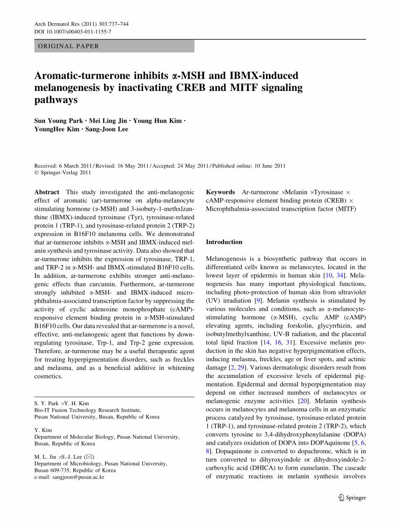
Aromatic-turmerone inhibits α-MSH and IBMX-induced melanogenesis by inactivating CREB and MITF...
8
ORIGINAL PAPER Aromatic-turmerone inhibits a-MSH and IBMX-induced melanogenesis by inactivating CREB and MITF signaling pathways Sun Young Park • Mei Ling Jin • Young Hun Kim • YoungHee Kim • Sang-Joon Lee Received: 6 March 2011 / Revised: 16 May 2011 / Accepted: 24 May 2011 / Published online: 10 June 2011 Ó Springer-Verlag 2011 Abstract This study investigated the anti-melanogenic effect of aromatic (ar)-turmerone on alpha-melanocyte stimulating hormone (a-MSH) and 3-isobuty-1-methxlzan- thine (IBMX)-induced tyrosinase (Tyr), tyrosinase-related protein 1 (TRP-1), and tyrosinase-related protein 2 (TRP-2) expression in B16F10 melanoma cells. We demonstrated that ar-turmerone inhibits a-MSH and IBMX-induced mel- anin synthesis and tyrosinase activity. Data also showed that ar-turmerone inhibits the expression of tyrosinase, TRP-1, and TRP-2 in a-MSH- and IBMX-stimulated B16F10 cells. In addition, ar-turmerone exhibits stronger anti-melano- genic effects than curcumin. Furthermore, ar-turmerone strongly inhibited a-MSH- and IBMX-induced micro- phthalmia-associated transcription factor by suppressing the activity of cyclic adenosine monophosphate (cAMP)- responsive element binding protein in a-MSH-stimulated B16F10 cells. Our data revealed that ar-turmerone is a novel, effective, anti-melanogenic agent that functions by down- regulating tyrosinase, Trp-1, and Trp-2 gene expression. Therefore, ar-turmerone may be a useful therapeutic agent for treating hyperpigmentation disorders, such as freckles and melasma, and as a beneficial additive in whitening cosmetics. Keywords Ar-turmerone Á Melanin Á Tyrosinase Á cAMP-responsive element binding protein (CREB) Á Microphthalmia-associated transcription factor (MITF) Introduction Melanogenesis is a biosynthetic pathway that occurs in differentiated cells known as melanocytes, located in the lowest layer of epidermis in human skin [10, 34]. Mela- nogenesis has many important physiological functions, including photo-protection of human skin from ultraviolet (UV) irradiation [9]. Melanin synthesis is stimulated by various molecules and conditions, such as a-melanocyte- stimulating hormone (a-MSH), cyclic AMP (cAMP) elevating agents, including forskolin, glycyrrhizin, and isobutylmethylxanthine, UV-B radiation, and the placental total lipid fraction [14, 16, 31]. Excessive melanin pro- duction in the skin has negative hyperpigmentation effects, inducing melasma, freckles, age or liver spots, and actinic damage [2, 29]. Various dermatologic disorders result from the accumulation of excessive levels of epidermal pig- mentation. Epidermal and dermal hyperpigmentation may depend on either increased numbers of melanocytes or melanogenic enzyme activities [20]. Melanin synthesis occurs in melanocytes and melanoma cells in an enzymatic process catalyzed by tyrosinase, tyrosinase-related protein 1 (TRP-1), and tyrosinase-related protein 2 (TRP-2), which converts tyrosine to 3,4-dihydroxyphenylalanine (DOPA) and catalyzes oxidation of DOPA into DOPAquinone [5, 6, 8]. Dopaquinone is converted to dopachrome, which is in turn converted to dihyroxyindole or dihydroxyindole-2- carboxylic acid (DHICA) to form eumelanin. The cascade of enzymatic reactions in melanin synthesis involves S. Y. Park Á Y. H. Kim Bio-IT Fusion Technology Research Institute, Pusan National University, Busan, Republic of Korea Y. Kim Department of Molecular Biology, Pusan National University, Busan, Republic of Korea M. L. Jin Á S.-J. Lee (&) Department of Microbiology, Pusan National University, Busan 609-735, Republic of Korea e-mail: [email protected] 123 Arch Dermatol Res (2011) 303:737–744 DOI 10.1007/s00403-011-1155-7
-
Upload
sun-young-park -
Category
Documents
-
view
213 -
download
1
Transcript of Aromatic-turmerone inhibits α-MSH and IBMX-induced melanogenesis by inactivating CREB and MITF...
ORIGINAL PAPER
Aromatic-turmerone inhibits a-MSH and IBMX-inducedmelanogenesis by inactivating CREB and MITF signalingpathways
Sun Young Park • Mei Ling Jin • Young Hun Kim •
YoungHee Kim • Sang-Joon Lee
Received: 6 March 2011 / Revised: 16 May 2011 / Accepted: 24 May 2011 / Published online: 10 June 2011
� Springer-Verlag 2011
Abstract This study investigated the anti-melanogenic
effect of aromatic (ar)-turmerone on alpha-melanocyte
stimulating hormone (a-MSH) and 3-isobuty-1-methxlzan-
thine (IBMX)-induced tyrosinase (Tyr), tyrosinase-related
protein 1 (TRP-1), and tyrosinase-related protein 2 (TRP-2)
expression in B16F10 melanoma cells. We demonstrated
that ar-turmerone inhibits a-MSH and IBMX-induced mel-
anin synthesis and tyrosinase activity. Data also showed that
ar-turmerone inhibits the expression of tyrosinase, TRP-1,
and TRP-2 in a-MSH- and IBMX-stimulated B16F10 cells.
In addition, ar-turmerone exhibits stronger anti-melano-
genic effects than curcumin. Furthermore, ar-turmerone
strongly inhibited a-MSH- and IBMX-induced micro-
phthalmia-associated transcription factor by suppressing the
activity of cyclic adenosine monophosphate (cAMP)-
responsive element binding protein in a-MSH-stimulated
B16F10 cells. Our data revealed that ar-turmerone is a novel,
effective, anti-melanogenic agent that functions by down-
regulating tyrosinase, Trp-1, and Trp-2 gene expression.
Therefore, ar-turmerone may be a useful therapeutic agent
for treating hyperpigmentation disorders, such as freckles
and melasma, and as a beneficial additive in whitening
cosmetics.
Keywords Ar-turmerone � Melanin � Tyrosinase �cAMP-responsive element binding protein (CREB) �Microphthalmia-associated transcription factor (MITF)
Introduction
Melanogenesis is a biosynthetic pathway that occurs in
differentiated cells known as melanocytes, located in the
lowest layer of epidermis in human skin [10, 34]. Mela-
nogenesis has many important physiological functions,
including photo-protection of human skin from ultraviolet
(UV) irradiation [9]. Melanin synthesis is stimulated by
various molecules and conditions, such as a-melanocyte-
stimulating hormone (a-MSH), cyclic AMP (cAMP)
elevating agents, including forskolin, glycyrrhizin, and
isobutylmethylxanthine, UV-B radiation, and the placental
total lipid fraction [14, 16, 31]. Excessive melanin pro-
duction in the skin has negative hyperpigmentation effects,
inducing melasma, freckles, age or liver spots, and actinic
damage [2, 29]. Various dermatologic disorders result from
the accumulation of excessive levels of epidermal pig-
mentation. Epidermal and dermal hyperpigmentation may
depend on either increased numbers of melanocytes or
melanogenic enzyme activities [20]. Melanin synthesis
occurs in melanocytes and melanoma cells in an enzymatic
process catalyzed by tyrosinase, tyrosinase-related protein
1 (TRP-1), and tyrosinase-related protein 2 (TRP-2), which
converts tyrosine to 3,4-dihydroxyphenylalanine (DOPA)
and catalyzes oxidation of DOPA into DOPAquinone [5, 6,
8]. Dopaquinone is converted to dopachrome, which is in
turn converted to dihyroxyindole or dihydroxyindole-2-
carboxylic acid (DHICA) to form eumelanin. The cascade
of enzymatic reactions in melanin synthesis involves
S. Y. Park � Y. H. Kim
Bio-IT Fusion Technology Research Institute,
Pusan National University, Busan, Republic of Korea
Y. Kim
Department of Molecular Biology, Pusan National University,
Busan, Republic of Korea
M. L. Jin � S.-J. Lee (&)
Department of Microbiology, Pusan National University,
Busan 609-735, Republic of Korea
e-mail: [email protected]
123
Arch Dermatol Res (2011) 303:737–744
DOI 10.1007/s00403-011-1155-7
tyrosinase, TRP-1, and dopachrome tautomerase (DCT),
also known as TRP-2 [7, 13, 22].
cAMP-dependent protein kinase A (PKA) signaling
pathways are the predominant cascades participating in
melanin synthesis. Furthermore, stimulators, such as
a-MSH, forskolin and IBMX, control the expression of
tyrosinase, TRP-1 and TRP-2 by modulating the activation
of transcription factors such as MITF and CREB and
through protein kinase A signaling pathways [18]. MITF is
the most important transcriptional regulator of tyrosinase
activity and is involved in melanocyte pigmentation, pro-
liferation, survival, and differentiation [3, 33]. Alpha-
melanocyte stimulating hormone (a-MSH) induces MITF
expression by increasing cAMP levels after binding to the
melanocortin 1 receptor [22]. The MITF gene contains two
promoters, one of which is a cAMP-responsive element
(CRE). Phosphorylation of a CRE binding protein (CREB)
promotes MITF expression [12, 19].
Aromatic (ar)-turmerone is a naturally occurring tur-
meric oil, which was initially isolated from Curcuma
longa, that has been used for centuries in Southeast Asia as
both a remedy and a food. Curcumin, demethoxycurcumin,
bisdemethoxycurcumin, ar-turmerone, a-turmerone, and
b-turmerone are the major bioactive compounds found in
C. longa. In modern pharmacological studies, C. longa
constituents, particularly curcumin, have been shown to
have anti-inflammatory, anti-cancer [11], anti-oxidative,
chemopreventive, immunomodulatory, and potentially
chemotherapeutic properties [1, 15]. Recently, the anti-
melanogenic effects of partially purified C. longa and
curcumin have been reported. However, the effects of other
components of this plant on the melanogenesis signaling
pathway have not been investigated. Ar-turmerone is a
component of C. longa. Therefore, in this study we
examined the effect of ar-turmerone on a-MSH and IBMX-
induced melanogenesis and signaling pathways in B16F10
melanoma cells.
Materials and methods
Materials
Ar-turmerone, a-MSH, IBMX, L-3,4-dihydroxyphenylala-
nine (L-DOPA), curcumin, 3-(4,5-dimetylthiazol-2-yl)-2,5-
diphenyl tetrazolium bromide (MTT), H89 and all other
reagents were purchased from Sigma-Aldrich (St. Louis,
MO, USA). Antibodies recognizing phospho-CREB and
CREB were obtained from Cell Signaling Technology
(Danvers, MA, USA). Tyrosinase, TRP-1, TRP-2, and
MITF antibodies were supplied by Santa Cruz Biotech-
nology (Santa Cruz, CA, USA).
Cell culture
The B16-F10 murine melanoma cell line obtained from the
American Type Culture Collection (ATCC; Rockville,
MD, USA) was grown as monolayers in Dulbecco’s
modified Eagle’s medium (DMEM; GIBCO BRL, Carls-
bad, CA, USA) supplemented with 10% heat-inactivated
fetal bovine serum (FBS; GIBCO BRL). The cells were
incubated at 37�C in a humidified atmosphere containing
5% CO2 and 95% air. To avoid changes in cell character-
istics produced by extended cell culture periods, cells were
used for experiments between passages 15 and 25. Each
cell suspension was split every 2 days to maintain expo-
nential growth.
Cell viability assay
Cells were incubated in wells of a 24-well plate at a density
of 4 9 104 cells/well. MTT solution (50 lg/ml) was added
to each well. The plates were then incubated for an addi-
tional 3 h at 37�C in a 5% CO2 atmosphere, after which the
supernatant was removed. Formazan crystals that had
formed in viable cells were solubilized using dimethyl-
sulfoxide (DMSO). The absorbance of each well was
measured at 570 nm using an enzyme-linked immunosor-
bent assay (ELISA) reader (Wallace, Boston, MA, USA).
Determination of intracellular melanin content
The melanin content was measured using slight modifica-
tion of a previously described method [32]. Briefly, cells
were treated with 0.5 lM a-MSH in the absence or pres-
ence of ar-turmerone for 48 h. Then the media was aspi-
rated and the cells were washed twice with PBS. Cells were
harvested from each well in a phosphate buffered saline
(PBS). Cells were pelleted by centrifugation (2,000 rpm)
and the melanin was dissolved by treatment with 500 ll of
1 N NaOH in 10% DMSO at 80�C for 1 h. Relative mel-
anin content was determined by measuring absorbance at
475 nm in a microplate reader. Melanin production was
calculated by normalizing melanin values with protein
content (absorbance/lg protein). Protein content was
determined using the Bradford dye reagent (Bio-Rad,
Hercules, CA, USA).
B16F10 cell tyrosinase activity assay
Tyrosinase activity was determined by measuring the rate
of dopachrome formation of L-DOPA and was measured by
the method of Akao et al. [24] with a slight modification.
Briefly, cells grown in 6-well dishes were treated with
0.5 lM a-MSH in the absence or presence of ar-turmerone
738 Arch Dermatol Res (2011) 303:737–744
123
for 48 h. Cells were then washed in ice-cold PBS and lysed
in PBS containing 1% (w/v) Triton X-100 and 0.1 mM
phenylmethanesulfonylfluoride (PMSF). The lysates were
then centrifuged at 13,000 rpm for 30 min to obtain the
supernatant as the crude tyrosinase extract for the activity
assay. The tyrosinase substrate, L-DOPA (2 mg/ml), was
prepared in phosphate lysis buffer. Each extract was placed
in a 96-well plate and the enzymatic reaction was initiated
by adding L-DOPA solution. After incubation, dopachrome
formation was assayed by measuring absorbance at 475 nm
using a microplate reader. The value of each measurement
was expressed as a percentage of the control. Tyrosinase
activity was calculated by normalizing tyrosinase activity
values with protein content (absorbance/lg protein). Pro-
tein content was determined using the Bradford dye reagent
(Bio-Rad, Hercules, CA, USA).
Determination of intracellular cAMP level
cAMP concentration was measured using a cyclic AMP
EIA kit (Cayman Chemical Company, Ann Arbor, MI,
USA). Briefly, cells were lysed in 0.1 M HCl to inhibit
phosphodiesterase activity. After neutralization and dilu-
tion, a fixed amount of cAMP conjugate was added to
compete with cAMP in the cell lysate for sites on rabbit
polyclonal antibody immobilized on a 96-well plate. Pro-
tein content in the cell lysate was determined using the
Bradford dye reagent. For cAMP measurements, 50 lg of
protein was used for sample analysis, which was performed
according to the manufacturer’s instructions.
Quantitative real-time polymerase chain reaction (PCR)
Total RNA was isolated from cells using RNA spin mini
RNA isolation kit (GE Healthcare, Uppsala, Sweden)
according to the manufacturer’s instructions. cDNA was
synthesized from 1 lg of total RNA using Maxime RT
PreMix (Intron Biotechnology, Gyeonggi-do, Korea) and
anchored oligo-dT15-primers. Real-time PCR was per-
formed using a Chromo4TM instrument (Bio-Rad) and
SYBR Green Master Mix (Applied Biosystems, Foster
City, CA, USA). Relative amounts of target mRNA were
determined using the comparative threshold (Ct) method by
normalizing target mRNA Ct values to those for glyceral-
dehyde 3-phosphate dehydrogenase (DCt).
Western blot analysis
Cells were harvested in ice-cold lysis buffer consisting of
1% Triton X-100, 1% deoxycholate, and 0.1% sodium
dodecyl sulfate (SDS). The protein content of cell lysates
was determined using the Bradford reagent. Protein amounts
in each sample (50 lg total protein) were resolved by 7.5%
SDS-polyacrylamide gel electrophoresis (SDS-PAGE),
transferred to a polyvinylidene difluoride (PVDF) mem-
brane, and exposed to the appropriate antibodies. Proteins
were visualized using an enhanced chemiluminescence
detection system (Amersham Biosciences, Piscataway, NJ,
USA) and horseradish peroxidase-conjugated anti-rabbit or
anti-mouse secondary antibodies. Images were acquired
using an ImageQuant 350 analyzer (Amersham Biosciences,
Uppsala, Sweden) and densitometry was performed using
the ImageQuant TL software (Amersham Biosciences).
Statistical analysis
Data is expressed as mean [standard error (SE)]. Each
experiment was repeated at least three times. Statistical
analysis was performed using SPSS, version 16.0 software
(SPSS, Inc., Chicago, IL, USA) to determine significant
differences. We used either one- or two-way analysis of
variance (ANOVA) followed by Dunn’s post hoc tests for
analyses. Values of *p \ 0.05 were considered statistically
significant.
Results
Ar-turmerone inhibits a-MSH- and IBMX-induced
melanin synthesis and tyrosinase activity in B16F10
cells
The effect of ar-turmerone on melanogenesis, activated by
a-MSH and IBMX with its intracellular signaling pathway,
was examined. We determined whether ar-turmerone
inhibited cellular melanin synthesis in a-MSH- and IBMX-
stimulated cells. B16F10 mouse melanoma cells were
exposed to 0.5 lM a-MSH and 0.1 mM IBMX in the
presence of ar-turmerone or curcumin, and melanin syn-
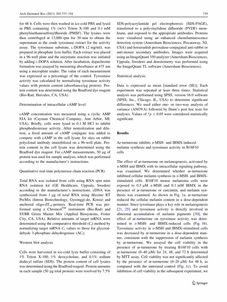
thesis was examined. As shown in Fig. 1a, ar-turmerone
reduced the cellular melanin content in a dose-dependent
manner. Since tyrosinase plays a key role in melanogenesis
[21, 25] and tyrosinase activity is directly involved in
abnormal accumulation of melanin pigments [30], the
effect of ar-turmerone on tyrosinase activity was deter-
mined in a-MSH- and IBMX-induced cells (Fig. 1b).
Tyrosinase activity in a-MSH and IBMX-stimulated cells
was decreased by ar-turmerone in a dose-dependent man-
ner, consistent with the suppression of melanin synthesis
by ar-turmerone. We assayed the cell viability in the
presence of ar-turmerone by treating B16F10 cells with
ar-turmerone (0–40 lM) for 24, 48, and 72 h determined
by MTT assay. Cell viability was not significantly affected
by the presence of ar-turmerone (0–20 lM) for 48 h, as
compared with the untreated control (Fig. 1c). To avoid
inhibition of cell viability in the subsequent experiment, we
Arch Dermatol Res (2011) 303:737–744 739
123
chose to use ar-turmerone concentrations between 5 and
20 lM and an incubation time of 48 h. These results sug-
gest that ar-turmerone reduces melanin synthesis by
inhibiting tyrosinase activity. Curcumin, a well-studied
compound present in C. longa, was used for comparison.
By treating cells with curcumin and ar-turmerone at
equivalent concentrations of 20 lM, the inhibitory effect of
ar-turmerone was shown to be more effective than that of
curcumin. Collectively, these results demonstrate that ar-
turmerone have an inhibitory effect on a-MSH- and IBMX-
induced melanin synthesis and tyrosinase activity.
Inhibition of a-MSH and IBMX-induced expression
of tyrosinase, TRP-1, and TRP-2 by ar-turmerone
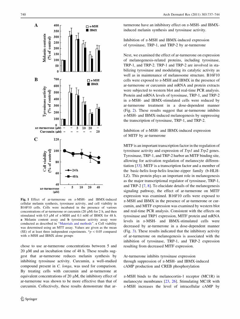
Next, we examined the effect of ar-turmerone on expression
of melanogenesis-related proteins, including tyrosinase,
TRP-1, and TRP-2. TRP-1 and TRP-2 are involved in sta-
bilizing tyrosinase and modulating its catalytic activity as
well as in maintenance of melanosome structure. B16F10
cells were exposed to a-MSH and IBMX in the presence of
ar-turmerone or curcumin and mRNA and protein extracts
were subjected to western blot and real-time PCR analysis.
Protein and mRNA levels of tyrosinase, TRP-1, and TRP-2
in a-MSH- and IBMX-stimulated cells were reduced by
ar-turmerone treatment in a dose-dependent manner
(Fig. 2). These results suggest that ar-turmerone inhibits
a-MSH- and IBMX-induced melanogenesis by suppressing
the transcription of tyrosinase, TRP-1, and TRP-2.
Inhibition of a-MSH- and IBMX-induced expression
of MITF by ar-turmerone
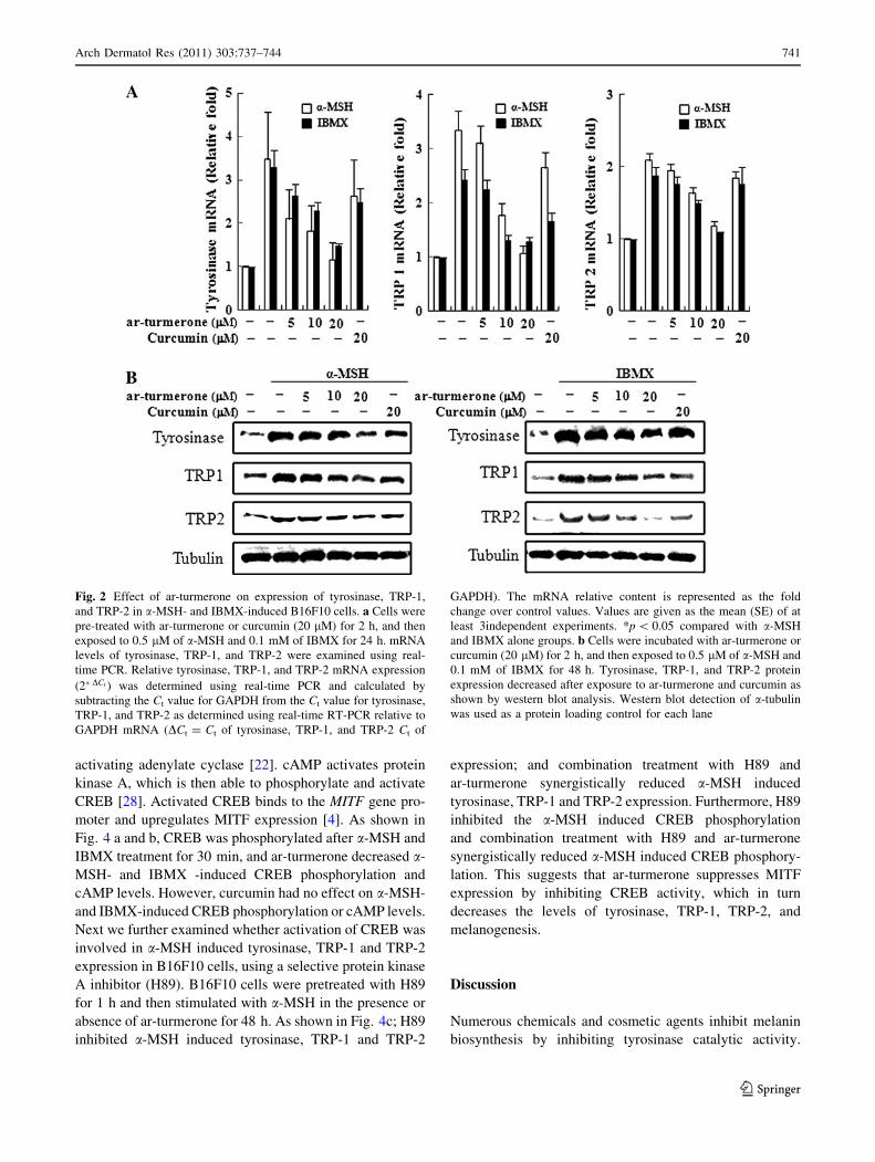
MITF is an important transcription factor in the regulation of
tyrosinase activity and expression of Trp1 and Trp2 genes.
Tyrosinase, TRP-1, and TRP-2 harbor an MITF binding site,
allowing for activation regulation of melanocyte differen-
tiation [33]. MITF is a transcription factor and a member of
the basic-helix-loop-helix-leucine-zipper family (b-HLH-
LZ). This protein plays an important role in melanogenesis
as the major transcriptional regulator of tyrosinase, TRP-1,
and TRP-2 [7, 8]. To elucidate details of the melanogenesis
signaling pathway, the effect of ar-turmerone on MITF
expression was examined. B16F10 cells were exposed to
a-MSH and IBMX in the presence of ar-turmerone or cur-
cumin, and MITF expression was examined by western blot
and real-time PCR analysis. Consistent with the effects on
tyrosinase and TRP1 expression, MITF protein and mRNA
levels in a-MSH- and IBMX-stimulated cells were
decreased by ar-turmerone in a dose-dependent manner
(Fig. 3). These results indicated that the inhibitory activity
of ar-turmerone on melanogenesis is associated with the
inhibition of tyrosinase, TRP-1, and TRP-2 expression
resulting from decreased MITF expression.
Ar-turmerone inhibits tyrosinase expression
through suppression of a-MSH- and IBMX-induced
cAMP production and CREB phosphorylation
a-MSH binds to the melanocortin-1 receptor (MC1R) in
melanocyte membranes [23, 26]. Stimulating MC1R with
a-MSH increases the level of intracellular cAMP by
Fig. 1 Effect of ar-turmerone on a-MSH- and IBMX-induced
cellular melanin synthesis, tyrosinase activity, and cell viability in
B16F10 cells. Cells were incubated in the presence of various
concentrations of ar-turmerone or curcumin (20 lM) for 2 h, and then
stimulated with 0.5 lM of a-MSH and 0.1 mM of IBMX for 48 h.
a Melanin content assay and b tyrosinase activity assay were
conducted as described in ‘‘Materials and methods’’. c Cell viability
was determined using an MTT assay. Values are given as the mean
(SE) of at least three independent experiments. *p \ 0.05 compared
with a-MSH and IBMX alone groups
740 Arch Dermatol Res (2011) 303:737–744
123
activating adenylate cyclase [22]. cAMP activates protein
kinase A, which is then able to phosphorylate and activate
CREB [28]. Activated CREB binds to the MITF gene pro-
moter and upregulates MITF expression [4]. As shown in
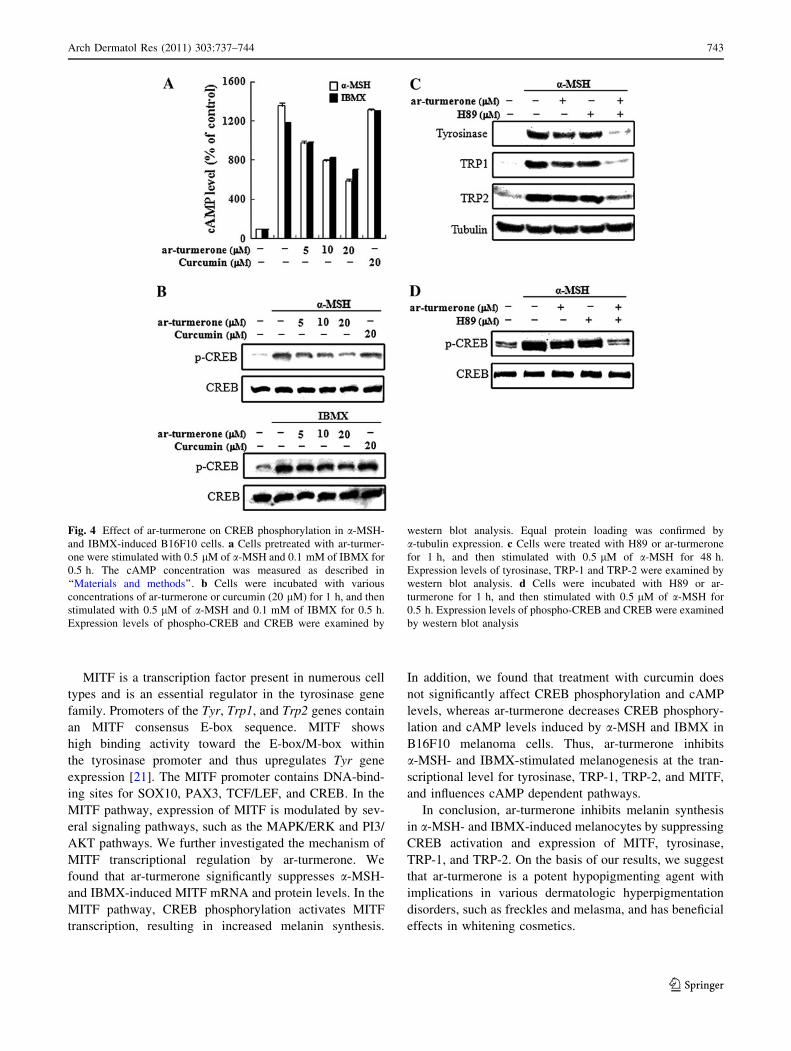
Fig. 4 a and b, CREB was phosphorylated after a-MSH and
IBMX treatment for 30 min, and ar-turmerone decreased a-
MSH- and IBMX -induced CREB phosphorylation and
cAMP levels. However, curcumin had no effect on a-MSH-
and IBMX-induced CREB phosphorylation or cAMP levels.
Next we further examined whether activation of CREB was
involved in a-MSH induced tyrosinase, TRP-1 and TRP-2
expression in B16F10 cells, using a selective protein kinase
A inhibitor (H89). B16F10 cells were pretreated with H89
for 1 h and then stimulated with a-MSH in the presence or
absence of ar-turmerone for 48 h. As shown in Fig. 4c; H89
inhibited a-MSH induced tyrosinase, TRP-1 and TRP-2
expression; and combination treatment with H89 and
ar-turmerone synergistically reduced a-MSH induced
tyrosinase, TRP-1 and TRP-2 expression. Furthermore, H89
inhibited the a-MSH induced CREB phosphorylation
and combination treatment with H89 and ar-turmerone
synergistically reduced a-MSH induced CREB phosphory-
lation. This suggests that ar-turmerone suppresses MITF
expression by inhibiting CREB activity, which in turn
decreases the levels of tyrosinase, TRP-1, TRP-2, and
melanogenesis.
Discussion
Numerous chemicals and cosmetic agents inhibit melanin
biosynthesis by inhibiting tyrosinase catalytic activity.
Fig. 2 Effect of ar-turmerone on expression of tyrosinase, TRP-1,
and TRP-2 in a-MSH- and IBMX-induced B16F10 cells. a Cells were
pre-treated with ar-turmerone or curcumin (20 lM) for 2 h, and then
exposed to 0.5 lM of a-MSH and 0.1 mM of IBMX for 24 h. mRNA
levels of tyrosinase, TRP-1, and TRP-2 were examined using real-
time PCR. Relative tyrosinase, TRP-1, and TRP-2 mRNA expression
(2�DCt ) was determined using real-time PCR and calculated by
subtracting the Ct value for GAPDH from the Ct value for tyrosinase,
TRP-1, and TRP-2 as determined using real-time RT-PCR relative to
GAPDH mRNA (DCt = Ct of tyrosinase, TRP-1, and TRP-2 Ct of
GAPDH). The mRNA relative content is represented as the fold
change over control values. Values are given as the mean (SE) of at
least 3independent experiments. *p \ 0.05 compared with a-MSH
and IBMX alone groups. b Cells were incubated with ar-turmerone or
curcumin (20 lM) for 2 h, and then exposed to 0.5 lM of a-MSH and
0.1 mM of IBMX for 48 h. Tyrosinase, TRP-1, and TRP-2 protein
expression decreased after exposure to ar-turmerone and curcumin as
shown by western blot analysis. Western blot detection of a-tubulin
was used as a protein loading control for each lane
Arch Dermatol Res (2011) 303:737–744 741
123
A number of tyrosinase inhibitors are available from nat-
ural and synthetic sources, but only a few are used as skin-
whitening agents because many inhibitors have drawbacks
such as safety concerns and low activity [27]. To overcome
these limitations, natural compounds derived from plant
extracts were investigated. Ar-turmerone is an abundant
component of turmeric, which has been traditionally used
in cooking, medicines, cosmetic formulations, and fabric
dying for over 2,000 years in Asia. Ar-turmerone has been
shown to be highly biologically active and possesses anti-
oxidant, anti-inflammatory, and anti-tumor properties. The
safety of ar-turmerone is certified by traditional usage as
well as our results (Fig. 1a). Thus, ar-turmerone can be
employed as an agent in functional cosmetics to develop
safe and effective skin-whitening treatments. Consistent
with our results, partially purified C. longa has been
reported to suppress a-MSH-stimulated melanogenesis
[17]. In this study, utilizing B16F10 melanoma cell lines,
the data presented here show that ar-trumerone suppressed
a-MSH, IBMX-induced protein kinase A signaling in
a-MSH, IBMX-induced melanin synthesis via tyrosinase,
TRP-1 and TRP-2 expression. Nevertheless, further studies
are needed to investigate the anti-melanogenic effect of
ar-trumerone in animal models.
a-MSH and IBMX are crucial cAMP-elevating agents;
these compounds exhibit differences in their mechanism of
activity. a-MSH combines with its receptor, melanocortin 1
receptor (MC1R), and activates adenylate cyclase, which
can increase the intracellular cAMP concentration. Com-
pared with a-MSH, IBMX inhibits cAMP phosphodies-
terase, increasing the intracellular cAMP concentration.
Tyrosinase is a key enzyme in melanin synthesis and
production and is primarily regulated by tyrosinase
expression and activation. In this study, we detected an
inhibitory effect of ar-turmerone on melanin synthesis and
tyrosinase activity induced by a-MSH and IBMX. To rule
out the possibility that the effect of ar-turmerone on
a-MSH and IBMX stimulates melanin synthesis as a con-
sequence of its cytotoxic effect, non-lethal concentrations
(B20lM) of ar-turmerone were used. A proper incubation
time was essential in evaluating the effect of ar-turmerone
on a-MSH- and IBMX-stimulated melanin synthesis. We
incubated cells treated with varying concentrations of
a-MSH and IBMX for various durations (data not shown).
As shown in Fig. 1, ar-turmerone inhibits melanin syn-
thesis and tyrosinase activity. To further explore the exact
mechanism of ar-turmerone inhibition on a-MSH- and
IBMX-induced melanogenesis, we performed western
blotting and real-time RT-PCR to determine tyrosinase,
TRP-1, and TRP-2 expression at the protein and mRNA
levels. We found that ar-turmerone reduces mRNA and
protein induction for tyrosinase, TRP-1, and TRP-2 in
response to a-MSH and IBMX. Curcumin, well known for
its antioxidative and anti-melanogenic effects, was used as
a comparison control, and it significantly inhibited melanin
synthesis and tyrosinase activity at a concentration of
20 lM. However, under these conditions, ar-turmerone is
more effective than curcumin.
Fig. 3 Effect of ar-turmerone on MITF expression in a-MSH- and
IBMX-induced B16F10 cells. a Cells were incubated with various
concentrations of ar-turmerone or curcumin (20 lM) for 2 h, and then
stimulated with 0.5 lM of a-MSH and 0.1 mM of IBMX for 24 h.
mRNA levels of MITF were examined using real-time PCR. Relative
MITF mRNA expression (2�DCt ) was determined using real-time PCR
and calculated by subtracting the Ct value for GAPDH from the Ct
value for MITF as determined using real-time RT-PCR relative to
GAPDH mRNA (DCt = Ct of MITF -Ct of GAPDH). The mRNA
relative content is represented as the fold change over control. Values
are given as the mean (SE) of at least three independent experiments.
*p \ 0.05 compared with a-MSH and IBMX alone groups. b Cells
were incubated with ar-turmerone or curcumin (C, 20 lM) for 2 h,
and then exposed to 0.5 lM of a-MSH and 0.1 mM of IBMX for
48 h. Western blot analysis shows that MITF protein expression was
decreased. Western blot detection of a-tubulin was used as a protein
loading control for each lane
742 Arch Dermatol Res (2011) 303:737–744
123
MITF is a transcription factor present in numerous cell
types and is an essential regulator in the tyrosinase gene
family. Promoters of the Tyr, Trp1, and Trp2 genes contain
an MITF consensus E-box sequence. MITF shows
high binding activity toward the E-box/M-box within
the tyrosinase promoter and thus upregulates Tyr gene
expression [21]. The MITF promoter contains DNA-bind-
ing sites for SOX10, PAX3, TCF/LEF, and CREB. In the
MITF pathway, expression of MITF is modulated by sev-
eral signaling pathways, such as the MAPK/ERK and PI3/
AKT pathways. We further investigated the mechanism of
MITF transcriptional regulation by ar-turmerone. We
found that ar-turmerone significantly suppresses a-MSH-
and IBMX-induced MITF mRNA and protein levels. In the
MITF pathway, CREB phosphorylation activates MITF
transcription, resulting in increased melanin synthesis.
In addition, we found that treatment with curcumin does
not significantly affect CREB phosphorylation and cAMP
levels, whereas ar-turmerone decreases CREB phosphory-
lation and cAMP levels induced by a-MSH and IBMX in
B16F10 melanoma cells. Thus, ar-turmerone inhibits
a-MSH- and IBMX-stimulated melanogenesis at the tran-
scriptional level for tyrosinase, TRP-1, TRP-2, and MITF,
and influences cAMP dependent pathways.
In conclusion, ar-turmerone inhibits melanin synthesis
in a-MSH- and IBMX-induced melanocytes by suppressing
CREB activation and expression of MITF, tyrosinase,
TRP-1, and TRP-2. On the basis of our results, we suggest
that ar-turmerone is a potent hypopigmenting agent with
implications in various dermatologic hyperpigmentation
disorders, such as freckles and melasma, and has beneficial
effects in whitening cosmetics.
Fig. 4 Effect of ar-turmerone on CREB phosphorylation in a-MSH-
and IBMX-induced B16F10 cells. a Cells pretreated with ar-turmer-
one were stimulated with 0.5 lM of a-MSH and 0.1 mM of IBMX for
0.5 h. The cAMP concentration was measured as described in
‘‘Materials and methods’’. b Cells were incubated with various
concentrations of ar-turmerone or curcumin (20 lM) for 1 h, and then
stimulated with 0.5 lM of a-MSH and 0.1 mM of IBMX for 0.5 h.
Expression levels of phospho-CREB and CREB were examined by
western blot analysis. Equal protein loading was confirmed by
a-tubulin expression. c Cells were treated with H89 or ar-turmerone
for 1 h, and then stimulated with 0.5 lM of a-MSH for 48 h.
Expression levels of tyrosinase, TRP-1 and TRP-2 were examined by
western blot analysis. d Cells were incubated with H89 or ar-
turmerone for 1 h, and then stimulated with 0.5 lM of a-MSH for
0.5 h. Expression levels of phospho-CREB and CREB were examined
by western blot analysis
Arch Dermatol Res (2011) 303:737–744 743
123
References
1. Aggarwal BB, Harikumar KB (2009) Potential therapeutic effects
of curcumin, the anti-inflammatory agent, against neurodegen-
erative, cardiovascular, pulmonary, metabolic, autoimmune and
neoplastic diseases. Int J Biochem Cell Biol 41:40–59
2. Ahn SJ, Koketsu M, Ishihara H, Lee SM, Ha SK, Lee KH, Kang
TH, Kima SY (2006) Regulation of melanin synthesis by sele-
nium-containing carbohydrates. Chem Pharm Bull (Tokyo)
54:281–286
3. Bentley NJ, Eisen T, Goding CR (1994) Melanocyte-specific
expression of the human tyrosinase promoter: activation by the
microphthalmia gene product and role of the initiator. Mol Cell
Biol 14:7996–8006
4. Bertolotto C, Abbe P, Hemesath TJ, Bille K, Fisher DE, Ortonne
JP, Ballotti R (1998) Microphthalmia gene product as a signal
transducer in cAMP-induced differentiation of melanocytes.
J Cell Biol 142:827–835
5. Boissy RE (2003) Melanosome transfer to and translocation in
the keratinocyte. Exp Dermatol 12(Suppl 2):5–12
6. Busca R, Bertolotto C, Ortonne JP, Ballotti R (1996) Inhibition of
the phosphatidylinositol 3-kinase/p70(S6)-kinase pathway indu-
ces B16 melanoma cell differentiation. J Biol Chem 271:
31824–31830
7. Chakraborty AK, Funasaka Y, Slominski A, Ermak G, Hwang J,
Pawelek JM, Ichihashi M (1996) Production and release of pro-
opiomelanocortin (POMC) derived peptides by human melano-
cytes and keratinocytes in culture: regulation by ultraviolet B.
Biochim Biophys Acta 1313:130–138
8. Costin GE, Hearing VJ (2007) Human skin pigmentation: mela-
nocytes modulate skin color in response to stress. FASEB J
21:976–994
9. Eller MS, Ostrom K, Gilchrest BA (1996) DNA damage enhan-
ces melanogenesis. Proc Natl Acad Sci USA 93:1087–1092
10. Eves PC, MacNeil S, Haycock JW (2006) Alpha-melanocyte
stimulating hormone, inflammation and human melanoma.
Peptides 27:444–452
11. Gescher A (2004) Polyphenolic phytochemicals versus non-
steroidal anti-inflammatory drugs: which are better cancer che-
mopreventive agents? J Chemother 16(Suppl 4):3–6
12. Goding CR (2000) Mitf from neural crest to melanoma: signal
transduction and transcription in the melanocyte lineage. Genes
Dev 14:1712–1728
13. Ha SK, Koketsu M, Lee K, Choi SY, Park JH, Ishihara H, Kim
SY (2005) Inhibition of tyrosinase activity by N,N-unsubstituted
selenourea derivatives. Biol Pharm Bull 28:838–840
14. Halaban R, Pomerantz SH, Marshall S, Lerner AB (1984)
Tyrosinase activity and abundance in cloudman melanoma cells.
Arch Biochem Biophys 230:383–387
15. Hatcher H, Planalp R, Cho J, Torti FM, Torti SV (2008) Cur-
cumin: from ancient medicine to current clinical trials. Cell Mol
Life Sci 65:1631–1652
16. Hunt G, Todd C, Cresswell JE, Thody AJ (1994) Alpha-mela-
nocyte stimulating hormone and its analogue Nle4DPhe7 alpha-
MSH affect morphology, tyrosinase activity and melanogenesis
in cultured human melanocytes. J Cell Sci 107(Pt 1):205–211
17. Jang JY, Lee JH, Jeong SY, Chung KT, Choi YH, Choi BT
(2009) Partially purified Curcuma longa inhibits alpha-melano-
cyte-stimulating hormone-stimulated melanogenesis through
extracellular signal-regulated kinase or Akt activation-mediated
signalling in B16F10 cells. Exp Dermatol 18:689–694
18. Jung E, Lee J, Huh S, Lee J, Kim YS, Kim G, Park D (2009)
Phloridzin-induced melanogenesis is mediated by the cAMP
signaling pathway. Food Chem Toxicol 47:2436–2440
19. Kadekaro AL, Kavanagh RJ, Wakamatsu K, Ito S, Pipitone MA,
Abdel-Malek ZA (2003) Cutaneous photobiology. The melano-
cyte vs. the sun: who will win the final round? Pigment Cell Res
16:434–447
20. Kanwar AJ, Dhar S, Kaur S (1994) Treatment of melasma with
potent topical corticosteroids. Dermatology 188:170
21. Kubo I, Kinst-Hori I, Chaudhuri SK, Kubo Y, Sanchez Y, Ogura
T (2000) Flavonols from Heterotheca inuloides: tyrosinase
inhibitory activity and structural criteria. Bioorg Med Chem
8:1749–1755
22. Mas JS, Gerritsen I, Hahmann C, Jimenez-Cervantes C, Garcia-
Borron JC (2003) Rate limiting factors in melanocortin 1 receptor
signalling through the cAMP pathway. Pigment Cell Res
16:540–547
23. Mountjoy KG, Robbins LS, Mortrud MT, Cone RD (1992) The
cloning of a family of genes that encode the melanocortin
receptors. Science 257:1248–1251
24. Ohguchi K, Ito M, Yokoyama K, Iinuma M, Itoh T, Nozawa Y,
Akao Y (2009) Effects of sesquiterpene lactones on melanogen-
esis in mouse B16 melanoma cells. Biol Pharm Bull 32:308–310
25. Perez-Gilabert M, Garcia-Carmona F (2001) Dimethyl sulfide, a
volatile flavor constituent, is a slow-binding inhibitor of tyrosi-
nase. Biochem Biophys Res Commun 285:257–261
26. Riley PA (2003) Melanogenesis and melanoma. Pigment Cell
Res 16:548–552
27. Roh JS, Han JY, Kim JH, Hwang JK (2004) Inhibitory effects of
active compounds isolated from safflower (Carthamus tinctoriusL.) seeds for melanogenesis. Biol Pharm Bull 27:1976–1978
28. Sassone-Corsi P (1995) Transcription factors responsive to
cAMP. Annu Rev Cell Dev Biol 11:355–377
29. Unver N, Freyschmidt-Paul P, Horster S, Wenck H, Stab F, Blatt
T, Elsasser HP (2006) Alterations in the epidermal-dermal mel-
anin axis and factor XIIIa melanophages in senile lentigo and
ageing skin. Br J Dermatol 155:119–128
30. Virador V, Matsunaga N, Matsunaga J, Valencia J, Oldham RJ,
Kameyama K, Peck GL, Ferrans VJ, Vieira WD, Abdel-Malek
ZA, Hearing VJ (2001) Production of melanocyte-specific anti-
bodies to human melanosomal proteins: expression patterns in
normal human skin and in cutaneous pigmented lesions. Pigment
Cell Res 14:289–297
31. Wong G, Pawelek J (1975) Melanocyte-stimulating hormone
promotes activation of pre-existing tyrosinase molecules in
cloudman S91 melanoma cells. Nature 255:644–646
32. Yang JY, Koo JH, Song YG, Kwon KB, Lee JH, Sohn HS, Park
BH, Jhee EC, Park JW (2006) Stimulation of melanogenesis by
scoparone in B16 melanoma cells. Acta Pharmacol Sin
27:1467–1473
33. Yasumoto K, Yokoyama K, Takahashi K, Tomita Y, Shibahara S
(1997) Functional analysis of microphthalmia-associated tran-
scription factor in pigment cell-specific transcription of the
human tyrosinase family genes. J Biol Chem 272:503–509
34. Yokota T, Nishio H, Kubota Y, Mizoguchi M (1998) The
inhibitory effect of glabridin from licorice extracts on melano-
genesis and inflammation. Pigment Cell Res 11:355–361
744 Arch Dermatol Res (2011) 303:737–744
123