Anticancer Potential of Curcumin: Preclinical and … B. AGGARWAL, ANUSHREE KUMAR and ALOK C. BHARTI...
36
Abstract. Curcumin (diferuloylmethane) is a polyphenol derived from the plant Curcuma longa, commonly called turmeric. Extensive research over the last 50 years has indicated this polyphenol can both prevent and treat cancer. The anticancer potential of curcumin stems from its ability to suppress proliferation of a wide variety of tumor cells, down-regulate transcription factors NF-κB, AP-1 and Egr-1; down-regulate the expression of COX2, LOX, NOS, MMP-9, uPA, TNF, chemokines, cell surface adhesion molecules and cyclin D1; down-regulate growth factor receptors (such as EGFR and HER2); and inhibit the activity of c-Jun N-terminal kinase, protein tyrosine kinases and protein serine/threonine kinases. In several systems, curcumin has been described as a potent antioxidant and anti-inflammatory agent. Evidence has also been presented to suggest that curcumin can suppress tumor initiation, promotion and metastasis. Pharmacologically, curcumin has been found to be safe. Human clinical trials indicated no dose-limiting toxicity when administered at doses up to 10 g/day. All of these studies suggest that curcumin has enormous potential in the prevention and therapy of cancer. The current review describes in detail the data supporting these studies. Curcumin, derived from turmeric (vernacular name: Haldi), is a rhizome of the plant Curcuma longa. The medicinal use of this plant has been documented in Ayurveda (the Indian Abbreviations: LDL, low density lipoprotein; HIV, human immuno-deficiency virus; 1 02, singlet oxygen; HVEC, human vascular endothelial cells; HVSMC, human vascular smooth muscle cells; PBMC, peripheral blood mononuclear cells; IL, interleukin; PDGF, platelet-derived growth factor; VSMC, vascular smooth muscle cells; PHA, phytohemagglutinin; PARP, polyadenosine ribose polymerase; ICAD, inhibitor of caspase-activated deoxyribonuclease; AIF, apoptosis-inducing factor; ROI, reactive oxygen intermediates; GSH, reduced form of gluthathione; PTK, protein tyrosine kinase; EGF, epidermal growth factor; EGFR, epidermal growth factor receptor; FPTase, farnesyl protein transferase; NF-κB, nuclear factor-κB; TNF, tumor necrosis factor; LT, lymphotoxin; ICAM-1, intracellular adhesion molecule-1; VCAM-1, vascular cell adhesion molecule-1; ELAM- 1, endothelial leukocyte adhesion molecule-1; COX2, cyclooxygenase-2; MMP, matrix metalloproteinase; ÈNOS, inducible nitric oxide oxidase; PMA, phorbol myristate acetate; AP-1, activating protein-1; πκB·, inhibitory subunit of NF-κB; UV, ultraviolet; JNK, c-jun N-terminal kinase; MAPK, mitogen-activated protein kinase; ERK, extracellular receptor kinase; IC 50 , inhibitory concentration (50%); MEKKI, mitogen-activated protein kinase kinase; TAKI, TGF-beta activated kinase-1; HPK1, human progenitor kinase 1; MAPKKK, mitogen- activated protein kinase kinase kinase; PKA, protein kinase A; PKC, protein kinase C; cPK, protamine kinase; PhK, phosphorylase kinase; AK, autophosphorylation-activated protein kinase; Ki, inhibition constant; TPA, tumor promoting agent; PKA, protein kinase A; cAK, autophosphorylation- activated protein kinase; CDPK, Ca 2+ -dependent protein kinase; LOX, lipoxygenase; HETE, hydroxyeicosatetraenoic acid; PGE, prostaglandin E, CD, chenodeoxycholate; CDK, cyclin-dependent kinase; SCC, squamous cell carcinoma; EC, endothelial cells; bFGF, basic fibroblast growth factor; VEGF, vascular endothelial growth factor; FGF, fibroblast growth factor; TGF-ß1, transforming growth factor-ß1; uPA, urokinase-activated plasminogen activator; TLC, total leukocyte count; GST, gluthathione S-transferase; HPLC, high pressure liquid chromatography; DMBA, 7, 12 dimethylbenz [a] anthracene; PhIP, 2-amino 1-methyl-6-phenylimidazo[4,5- b]pyridine; NNK, 4-(methyl-nitrosamino)-1-(3-pyridyl)-l- butanone; ODC, ornithine decarboxylase; PCNA, proliferating cell nuclear antigen; EBV, Epstein-Barr virus; B[a]P, benzo[a]pyrene; APC, antigen-presenting cells; BrdU, bromo- deoxyuridine; DHC, dihydrocurcumin; THC, tetrahydro- curcumin; HHC hexahydrocurcumin; i.v., intravenous, i.p., intraperitonial; i.g., intragastric. Correspondence to: µharat µ. ∞ggarwal, Cytokine Research Section, Department of Bioimmunotherapy, University of Texas M. D. Anderson Cancer Center, 1515 Holcombe Boulevard, Box 143, Houston, TÃ, U.S.∞. Tel: 713-792-3503/6459, Fax: 713-794- 1613, e-mail: [email protected] Key Words: Curcumin, anticancer agents, chemopreventive agents, NF-KB, AP-1, cyclin D1, COX-2. ANTICANCER RESEARCH 23: 363-398 (2003) Anticancer Potential of Curcumin: Preclinical and Clinical Studies BHARAT B. AGGARWAL, ANUSHREE KUMAR and ALOK C. BHARTI Cytokine Research Section, Department of Bioimmunotherapy, University of Texas M. D. Anderson Cancer Center, Box 143, Houston, TÃ, U.S.∞. 0250-7005/2003 $2.00+.40 363 Review
Transcript of Anticancer Potential of Curcumin: Preclinical and … B. AGGARWAL, ANUSHREE KUMAR and ALOK C. BHARTI...
Abstract. Curcumin (diferuloylmethane) is a polyphenol derivedfrom the plant Curcuma longa, commonly called turmeric.Extensive research over the last 50 years has indicated thispolyphenol can both prevent and treat cancer. The anticancerpotential of curcumin stems from its ability to suppressproliferation of a wide variety of tumor cells, down-regulatetranscription factors NF-κB, AP-1 and Egr-1; down-regulate theexpression of COX2, LOX, NOS, MMP-9, uPA, TNF,chemokines, cell surface adhesion molecules and cyclin D1;down-regulate growth factor receptors (such as EGFR andHER2); and inhibit the activity of c-Jun N-terminal kinase,protein tyrosine kinases and protein serine/threonine kinases. Inseveral systems, curcumin has been described as a potent
antioxidant and anti-inflammatory agent. Evidence has alsobeen presented to suggest that curcumin can suppress tumorinitiation, promotion and metastasis. Pharmacologically,curcumin has been found to be safe. Human clinical trialsindicated no dose-limiting toxicity when administered at dosesup to 10 g/day. All of these studies suggest that curcumin hasenormous potential in the prevention and therapy of cancer. Thecurrent review describes in detail the data supporting thesestudies.
Curcumin, derived from turmeric (vernacular name: Haldi), isa rhizome of the plant Curcuma longa. The medicinal use ofthis plant has been documented in Ayurveda (the Indian
Abbreviations: LDL, low density lipoprotein; HIV, humanimmuno-deficiency virus; 102, singlet oxygen; HVEC, humanvascular endothelial cells; HVSMC, human vascular smoothmuscle cells; PBMC, peripheral blood mononuclear cells; IL,interleukin; PDGF, platelet-derived growth factor; VSMC,vascular smooth muscle cells; PHA, phytohemagglutinin;PARP, polyadenosine ribose polymerase; ICAD, inhibitor ofcaspase-activated deoxyribonuclease; AIF, apoptosis-inducingfactor; ROI, reactive oxygen intermediates; GSH, reducedform of gluthathione; PTK, protein tyrosine kinase; EGF,epidermal growth factor; EGFR, epidermal growth factorreceptor; FPTase, farnesyl protein transferase; NF-κB, nuclearfactor-κB; TNF, tumor necrosis factor; LT, lymphotoxin;ICAM-1, intracellular adhesion molecule-1; VCAM-1,vascular cell adhesion molecule-1; ELAM- 1, endothelialleukocyte adhesion molecule-1; COX2, cyclooxygenase-2;MMP, matrix metalloproteinase; ÈNOS, inducible nitric oxideoxidase; PMA, phorbol myristate acetate; AP-1, activatingprotein-1; πκB·, inhibitory subunit of NF-κB; UV, ultraviolet;JNK, c-jun N-terminal kinase; MAPK, mitogen-activatedprotein kinase; ERK, extracellular receptor kinase; IC50,inhibitory concentration (50%); MEKKI, mitogen-activatedprotein kinase kinase; TAKI, TGF-beta activated kinase-1;HPK1, human progenitor kinase 1; MAPKKK, mitogen-activated protein kinase kinase kinase; PKA, protein kinase A;PKC, protein kinase C; cPK, protamine kinase; PhK,phosphorylase kinase; AK, autophosphorylation-activatedprotein kinase; Ki, inhibition constant; TPA, tumor promoting
agent; PKA, protein kinase A; cAK, autophosphorylation-activated protein kinase; CDPK, Ca2+-dependent proteinkinase; LOX, lipoxygenase; HETE, hydroxyeicosatetraenoicacid; PGE, prostaglandin E, CD, chenodeoxycholate; CDK,cyclin-dependent kinase; SCC, squamous cell carcinoma; EC,endothelial cells; bFGF, basic fibroblast growth factor; VEGF,vascular endothelial growth factor; FGF, fibroblast growthfactor; TGF-ß1, transforming growth factor-ß1; uPA,urokinase-activated plasminogen activator; TLC, totalleukocyte count; GST, gluthathione S-transferase; HPLC, highpressure liquid chromatography; DMBA, 7, 12 dimethylbenz[a] anthracene; PhIP, 2-amino 1-methyl-6-phenylimidazo[4,5-b]pyridine; NNK, 4-(methyl-nitrosamino)-1-(3-pyridyl)-l-butanone; ODC, ornithine decarboxylase; PCNA, proliferatingcell nuclear antigen; EBV, Epstein-Barr virus; B[a]P,benzo[a]pyrene; APC, antigen-presenting cells; BrdU, bromo-deoxyuridine; DHC, dihydrocurcumin; THC, tetrahydro-curcumin; HHC hexahydrocurcumin; i.v., intravenous, i.p.,intraperitonial; i.g., intragastric.
Correspondence to: µharat µ. ∞ggarwal, Cytokine ResearchSection, Department of Bioimmunotherapy, University of TexasM. D. Anderson Cancer Center, 1515 Holcombe Boulevard, Box143, Houston, TÃ, U.S.∞. Tel: 713-792-3503/6459, Fax: 713-794-1613, e-mail: [email protected]
Key Words: Curcumin, anticancer agents, chemopreventiveagents, NF-KB, AP-1, cyclin D1, COX-2.
ANTICANCER RESEARCH 23: 363-398 (2003)
Anticancer Potential of Curcumin: Preclinical and Clinical StudiesBHARAT B. AGGARWAL, ANUSHREE KUMAR and ALOK C. BHARTI
Cytokine Research Section, Department of Bioimmunotherapy, University of Texas M. D. Anderson Cancer Center, Box 143, Houston, TÃ, U.S.∞.
0250-7005/2003 $2.00+.40 363
Review
A Rsystem of medicine) for over 6000 years. It is commonly usedas a spice, flavoring agent, food preservative, coloring agent,or for decoration. Extensive investigation over the last fivedecades has indicated that curcumin reduces bloodcholesterol (1 -7), prevents LDL oxidation (8- 10), inhibitsplatelet aggregation (11, 12), suppresses thrombosis (13) andmyocardial infarction (14-17), suppresses symptomsassociated with type II diabetes (18-22), rheumatoid arthritis(23), multiple sclerosis (24) and Alzheimer (25, 26), inhibitsHIV replication (27-31), enhances wound healing (32-34),protects from liver injury (35), increases bile secretion (36),protects from cataract formation (37), and protects frompulmonary toxicity and fibrosis (38-41), antileishmaniasis (42-44) and is an anti-atherosclerotic (45, 46). Additionally thereis extensive literature that suggests that curcumin haspotential in the prevention and treatment of cancer. Thecurrent review focuses on the studies that implicate curcuminin the prevention and therapy of cancer. We present severalin vitro and in vivo preclinical and clinical results that suggestthat curcumin has a great potential in the treatment of cancer.Certain aspects of curcumin have been reviewed (47-52), butin spite of numerous recent studies on its biology andmechanism of action, no comprehensive review has yetappeared on this subject.
A. Chemistry of Curcumin
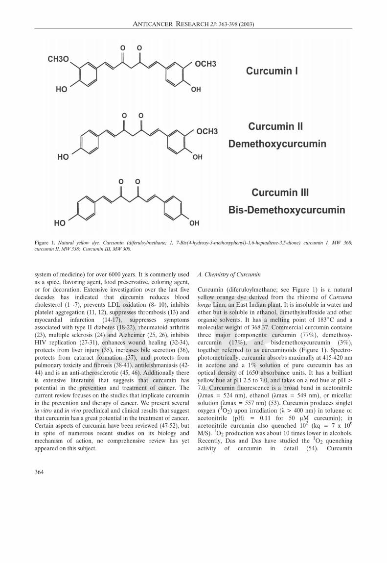
Curcumin (diferuloylmethane; see Figure 1) is a naturalyellow orange dye derived from the rhizome of Curcumalonga Linn, an East Indian plant. It is insoluble in water andether but is soluble in ethanol, dimethylsulfoxide and otherorganic solvents. It has a melting point of 183ÆC and amolecular weight of 368.37. Commercial curcumin containsthree major components: curcumin (77%), demethoxy-curcumin (17%), and bisdemethoxycurcumin (3%),together referred to as curcuminoids (Figure 1). Spectro-photometrically, curcumin absorbs maximally at 415-420 nmin acetone and a 1% solution of pure curcumin has anoptical density of 1650 absorbance units. It has a brilliantyellow hue at pH 2.5 to 7.0, and takes on a red hue at pH >7.0. Curcumin fluorescence is a broad band in acetonitrile(Ïmax = 524 nm), ethanol (Ïmax = 549 nm), or micellarsolution (Ïmax = 557 nm) (53). Curcumin produces singletoxygen (1O2) upon irradiation (Ï > 400 nm) in toluene oracetonitrile (pHi = 0.11 for 50 ÌM curcumin); inacetonitrile curcumin also quenched 102 (kq = 7 x 10⁶
M/S). 1O2 production was about 10 times lower in alcohols.Recently, Das and Das have studied the 1O2 quenchingactivity of curcumin in detail (54). Curcumin
364
ANTICANCER RESEARCH 23: 363-398 (2003)
Figure 1. Natural yellow dye, Curcumin (diferuloylmethane; 1, 7-Bis(4-hydroxy-3-methoxyphenyl)-1,6-heptadiene-3,5-dione) curcumin I, MW 368;curcumin II, MW 338; Curcumin III, MW 308.
A Rphotogenerates superoxide in toluene and ethanol.In contrast, it quenches superoxide ions in acetonitrile(55).
Curcumin is also phototoxic to mammalian cells, asdemonstrated in a rat basophilic leukemia cell model, and thisphototoxicity likewise requires the presence of oxygen (56).The spectral and photochemical properties of curcumin varywith environment, resulting in the potential for multiple oralternate pathways for the execution of photodynamic effects.For example, curcumin photogenerates singlet oxygen andreduced forms of molecular oxygen under several conditionsrelevant to cellular environments.
Tonnesen examined the kinetics of pH-dependentcurcumin degradation in aqueous solution (57). A plot ofthe rate constant against pH indicated the pKa values ofthe acid protons. The graph also indicated the complexityof curcumin degradation. The same investigators alsoinvestigated the stability of curcumin when exposed toUV/visible radiation (58). The main degradation productswere identified. The reaction mechanisms wereinvestigated and the order of the overall degradationreactions and the half-lives of curcumin in differentsolvents and in the solid state were determined. Theseworkers also examined the photobiological activity ofcurcumin using bacterial indicator systems (59). Onirradiation with visible light, curcumin proved to bephototoxic for Salmonella typhimurium and Escherichiacoli, even at very low concentrations. The observedphototoxicity makes curcumin a potential photosensitizingdrug, which might find application in the phototherapy of,
for example, psoriasis, cancer and bacterial and viraldiseases. Recently, the same group (60), complexedcurcumin with cyclodextrin to improve the water solubilityand the hydrolytic and photochemical stability of thecompound. Complex formation resulted in an increase inwater solubility at pH 5 by a factor of at least 104. Thehydrolytic stability of curcumin under alkaline conditionswas strongly improved by complex formation, while thephotodecomposition rate was increased compared to acurcumin solution in organic solvents. The cavity size andthe charge and bulkiness of the cyclodextrin side-chainsinfluenced the stability constant for complexation and thedegradation rate of the curcumin molecule. Wang et al.examined the degradation kinetics of curcumin undervarious pH conditions and the stability of curcumin inphysiological matrices (61). When curcumin was incubatedin 0.1 M phosphate buffer and serum-free medium, pH 7.2,at 37ÆC, about 90% decomposed within 30 minutes. Aseries of pH conditions ranging from 3 to 10 were testedand the result showed that decomposition was pH-dependent and occurred faster at neutral-basic conditions.It is more stable in cell culture medium containing 10%fetal calf serum and in human blood; less than 20% ofcurcumin decomposed within 1 hour and, after incubationfor 8 hours, about 50% of curcumin still remained. Trans-6-(4'-hydroxy-3'-methoxyphenyl)2,4-dioxo-5-hexenal waspredicted to be the major degradation product and vanillin,ferulic acid and feruloyl methane were identified as minordegradation products. The amount of vanillin increasedwith incubation time.
365
Aggarwal et al: Anticancer Potential of Curcumin (Review)
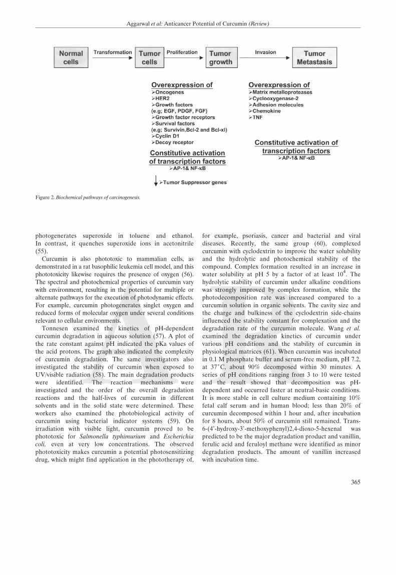
Figure 2. Biochemical pathways of carcinogenesis.
366
ANTICANCER RESEARCH 23: 363-398 (2003)
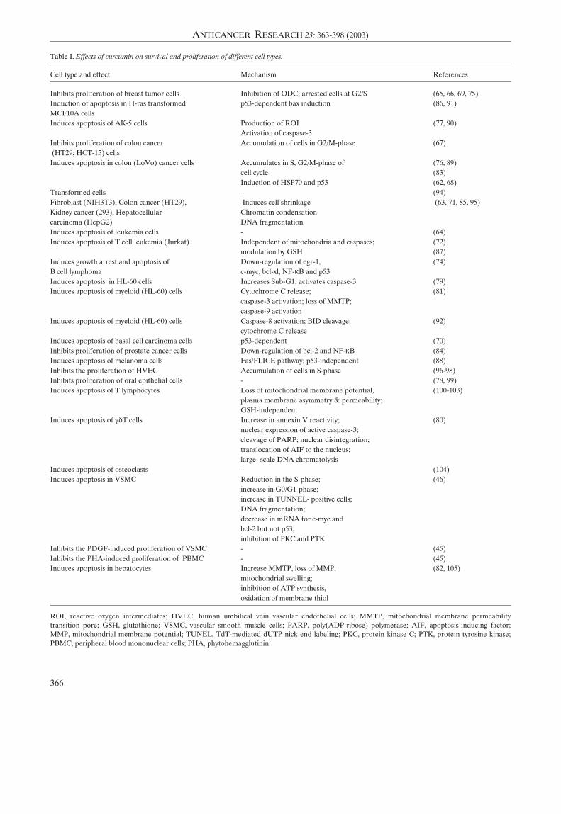
Table I. Effects of curcumin on survival and proliferation of different cell types.
Cell type and effect Mechanism References
Inhibits proliferation of breast tumor cells Inhibition of ODC; arrested cells at G2/S (65, 66, 69, 75)Induction of apoptosis in H-ras transformed p53-dependent bax induction (86, 91)MCF10A cellsInduces apoptosis of AK-5 cells Production of ROI (77, 90)
Activation of caspase-3Inhibits proliferation of colon cancer Accumulation of cells in G2/M-phase (67)(HT29; HCT-15) cellsInduces apoptosis in colon (LoVo) cancer cells Accumulates in S, G2/M-phase of (76, 89)
cell cycle (83)Induction of HSP70 and p53 (62, 68)
Transformed cells - (94)Fibroblast (NIH3T3), Colon cancer (HT29), Induces cell shrinkage (63, 71, 85, 95)Kidney cancer (293), Hepatocellular Chromatin condensationcarcinoma (HepG2) DNA fragmentationInduces apoptosis of leukemia cells - (64)Induces apoptosis of T cell leukemia (Jurkat) Independent of mitochondria and caspases; (72)
modulation by GSH (87)Induces growth arrest and apoptosis of Down-regulation of egr-1, (74)B cell lymphoma c-myc, bcl-xl, NF-κB and p53Induces apoptosis in HL-60 cells Increases Sub-G1; activates caspase-3 (79) Induces apoptosis of myeloid (HL-60) cells Cytochrome C release; (81)
caspase-3 activation; loss of MMTP;caspase-9 activation
Induces apoptosis of myeloid (HL-60) cells Caspase-8 activation; BID cleavage; (92)cytochrome C release
Induces apoptosis of basal cell carcinoma cells p53-dependent (70)Inhibits proliferation of prostate cancer cells Down-regulation of bcl-2 and NF-κB (84)Induces apoptosis of melanoma cells Fas/FLICE pathway; p53-independent (88)Inhibits the proliferation of HVEC Accumulation of cells in S-phase (96-98)Inhibits proliferation of oral epithelial cells - (78, 99)Induces apoptosis of T lymphocytes Loss of mitochondrial membrane potential, (100-103)
plasma membrane asymmetry & permeability;GSH-independent
Induces apoptosis of Á‰T cells Increase in annexin V reactivity; (80)nuclear expression of active caspase-3;cleavage of PARP; nuclear disintegration;translocation of AIF to the nucleus; large- scale DNA chromatolysis
Induces apoptosis of osteoclasts - (104)Induces apoptosis in VSMC Reduction in the S-phase; (46)
increase in G0/G1-phase;increase in TUNNEL- positive cells; DNA fragmentation;decrease in mRNA for c-myc andbcl-2 but not p53;inhibition of PKC and PTK
Inhibits the PDGF-induced proliferation of VSMC - (45)Inhibits the PHA-induced proliferation of PBMC - (45)Induces apoptosis in hepatocytes Increase MMTP, loss of MMP, (82, 105)
mitochondrial swelling; inhibition of ATP synthesis, oxidation of membrane thiol
ROI, reactive oxygen intermediates; HVEC, human umbilical vein vascular endothelial cells; MMTP, mitochondrial membrane permeabilitytransition pore; GSH, glutathione; VSMC, vascular smooth muscle cells; PARP, poly(ADP-ribose) polymerase; AIF, apoptosis-inducing factor;MMP, mitochondrial membrane potential; TUNEL, TdT-mediated dUTP nick end labeling; PKC, protein kinase C; PTK, protein tyrosine kinase;PBMC, peripheral blood mononuclear cells; PHA, phytohemagglutinin.
B. Preclinical Studies
Cancer is a hyperproliferative disorder that metastasize intothe vital organs in the body through invasion followed byangiogenesis and distant metastasis. Within the last 25 years,much has been learned about the biochemical pathway thatultimately leads to cancer (Figure 2). During the last decade,there has been extensive investigation of how curcuminaffects this overall process of tumorigenesis. Below is thedescription of the effects of curcumin first in vitro and then invivo and its relevance to cancer.
B1. In vitro effects
B1A. Antiproliferative effects of curcumin: Curcuminsuppresses the proliferation of a wide variety of tumor cells,including breast carcinoma, colon carcinoma, renal cellcarcinoma, hepatocellular carcinoma, T cell leukemia, Bcell lymphoma, acute myelogenous leukemia, basal cellcarcinoma, melanoma and prostate carcinoma (see Table I)(62-93). Additionally curcumin suppresses the proliferationof certain normal cells such as hepatocytes (105), epithelialcells (99), human vascular endothelial cells (HVEC) (96,97), human vascular smooth muscle cells (HVSMC) (45,46), osteoclasts (104), peripheral bood mononuclear cells(PBMC) (45), and T lymphocytes (45, 46, 80, 94, 96, 97, 99-105). Curcumin also inhibits the cell proliferation inducedby growth factors. For instance IL-2-induced proliferationof PBMC, PDGF-induced proliferation of VSMC andPHA-induced proliferation of PBMC were inhibited bytreatment with curcumin (45). The suppression of cellproliferation by curcumin usually occurs through its effectson the cell cycle. Depending on the cell type, the inhibitionof cell proliferation at different phases of the cell cycle hasbeen reported (46, 76, 83, 96, 99). Inhibition of cellproliferation could also be mediated through suppression ofornithine decarboxylase (ODC)(65).
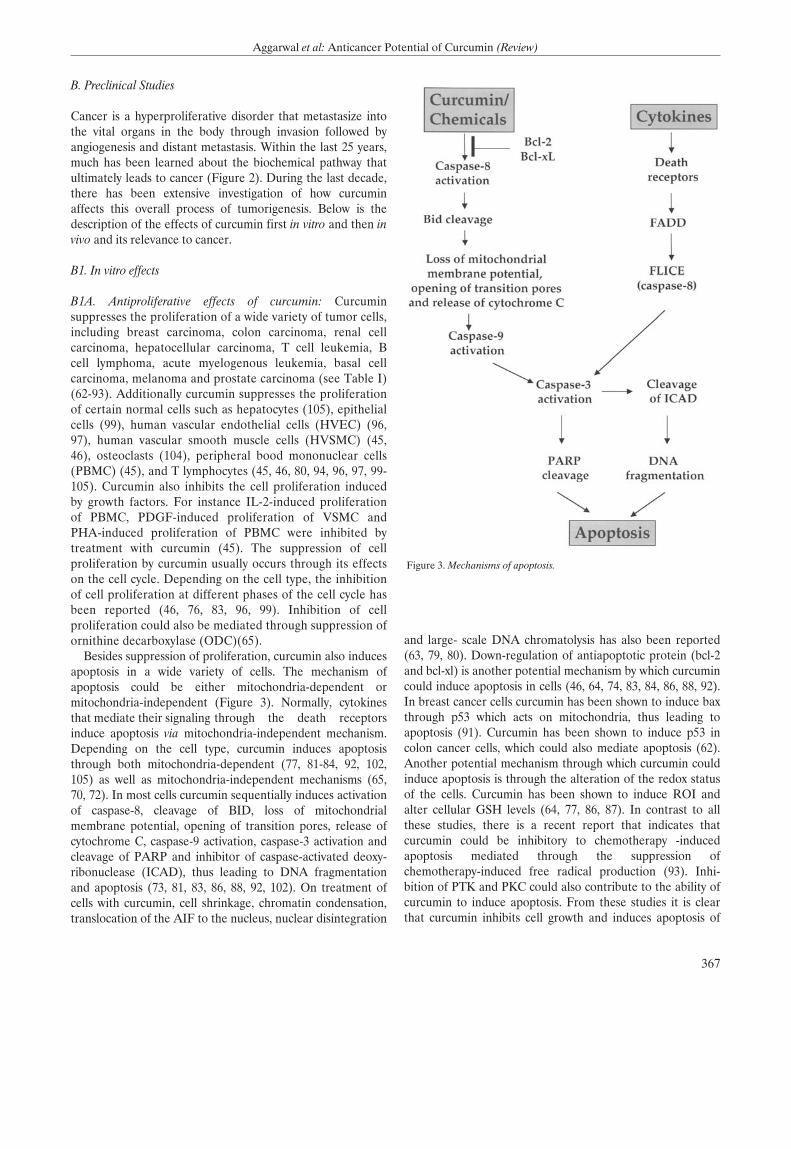
Besides suppression of proliferation, curcumin also inducesapoptosis in a wide variety of cells. The mechanism ofapoptosis could be either mitochondria-dependent ormitochondria-independent (Figure 3). Normally, cytokinesthat mediate their signaling through the death receptorsinduce apoptosis via mitochondria-independent mechanism.Depending on the cell type, curcumin induces apoptosisthrough both mitochondria-dependent (77, 81-84, 92, 102,105) as well as mitochondria-independent mechanisms (65,70, 72). In most cells curcumin sequentially induces activationof caspase-8, cleavage of BID, loss of mitochondrialmembrane potential, opening of transition pores, release ofcytochrome C, caspase-9 activation, caspase-3 activation andcleavage of PARP and inhibitor of caspase-activated deoxy-ribonuclease (ICAD), thus leading to DNA fragmentationand apoptosis (73, 81, 83, 86, 88, 92, 102). On treatment ofcells with curcumin, cell shrinkage, chromatin condensation,translocation of the AIF to the nucleus, nuclear disintegration
and large- scale DNA chromatolysis has also been reported(63, 79, 80). Down-regulation of antiapoptotic protein (bcl-2and bcl-xl) is another potential mechanism by which curcumincould induce apoptosis in cells (46, 64, 74, 83, 84, 86, 88, 92).In breast cancer cells curcumin has been shown to induce baxthrough p53 which acts on mitochondria, thus leading toapoptosis (91). Curcumin has been shown to induce p53 incolon cancer cells, which could also mediate apoptosis (62).Another potential mechanism through which curcumin couldinduce apoptosis is through the alteration of the redox statusof the cells. Curcumin has been shown to induce ROI andalter cellular GSH levels (64, 77, 86, 87). In contrast to allthese studies, there is a recent report that indicates thatcurcumin could be inhibitory to chemotherapy -inducedapoptosis mediated through the suppression ofchemotherapy-induced free radical production (93). Inhi-bition of PTK and PKC could also contribute to the ability ofcurcumin to induce apoptosis. From these studies it is clearthat curcumin inhibits cell growth and induces apoptosis of
367
Aggarwal et al: Anticancer Potential of Curcumin (Review)
Figure 3. Mechanisms of apoptosis.
368
ANTICANCER RESEARCH 23: 363-398 (2003)
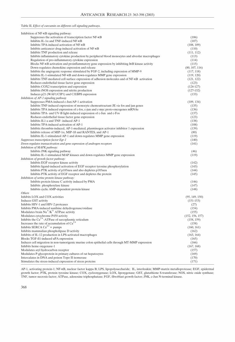
Table II. Effect of curcumin on different cell signaling pathways.
Inhibition of NF-κB signaling pathway: Suppresses the activation of transcription factor NF-κB (106)Inhibits IL-1· and TNF-induced NF-κB (107)Inhibits TPA-induced activation of NF-κB (108, 109)Inhibits anticancer drug-induced activation of NF-κB (110)Inhibits TNF production and release (111, 112)Inhibits inflammatory cytokine production by peripheral blood monocytes and alveolar macrophages (113)Regulation of pro-inflammatory cytokine expression (114)Blocks NF-κB activation and proinflammatory gene expression by inhibiting IκB kinase activity (115)Down-regulates chemokine expression and release (80, 107, 116)Inhibits the angiogenic response stimulated by FGF-2, including expression of MMP-9 (117, 118)Inhibits IL-1-stimulated NF-κB and down-regulates MMP gene expression (119, 120)Inhibits TNF-mediated cell surface expression of adhesion molecules and of NF-κB activation (121, 122)Reduces endothelial tissue factor gene expression (123)Inhibits COX2 transcription and expression (124-127)Inhibits iNOS expression and nitrite production (127-132)Induces p21 (WAF1/CIP1) and C/EBPß expression (133)
Inhibition of AP-1 signaling pathway Suppresses PMA-induced c-Jun/AP-1 activation (109, 134)Inhibits TNF-induced expression of monocyte chemoattractant JE via fos and jun genes (135)Inhibits TPA induced expression of c-fos, c-jun and c-myc proto-oncogenes mRNAs (136)Inhibits TPA- and UV-B light-induced expression of c-Jun and c-Fos (137)Reduces endothelial tissue factor gene expression (123)Inhibits IL1 a and TNF- induced AP-1 (138)Inhibits TPA-induced activation of AP-1 (108)Inhibits thrombin-induced, AP-1-mediated, plasminogen activator inhibitor 1 expression (139)Inhibits release of MIP-1·, MIP-1ß and RANTES, and AP-1 (80)Inhibits IL-1-stimulated AP-1 and down-regulates MMP gene expression (119)
Suppresses transcription factor Egr-1 (140)Down-regulates transactivation and gene expression of androgen receptors (141)Inhibition of MAPK pathway:
Inhibits JNK signaling pathway (46)Inhibits IL-1-stimulated MAP kinases and down-regulates MMP gene expression (119)
Inhibition of growth factor pathway: Inhibits EGF receptor kinase activity (142)Inhibits ligand-induced activation of EGF receptor tyrosine phosphorylation (143)Inhibits PTK activity of p185neu and also depletes p185neu (144)Inhibits PTK activity of EGF receptor and depletes the protein (145)
Inhibition of serine protein kinase pathway: Inhibits protein kinase C activity induced by PMA (146)Inhibits phosphorylase kinase (147)Inhibits cyclic AMP-dependent protein kinase (148)
OthersInhibits LOX and COX activities (95, 149, 150)Induces GST activity (151-153)Inhibits HIV-1 and HIV-2 proteases (27)Inhibits PMA-induced xanthine dehydrogenase/oxidase (154)Modulates brain Na+/K+ ATPase activity (155)Modulates cytochrome P450 activity (152, 156, 157)Inhibits the Ca2+-ATPase of sarcoplasmic reticulum (158, 159)Increases the rate of accumulation of Ca2+ (158)Inhibits SERCA Ca2++ pumps (160, 161)Inhibits mammalian phospholipase D activity (162)Inhibits of IL-12 production in LPS-activated macrophages (163, 164)Blocks TGF-ß1-induced uPA expression (165)Induces cell migration in non-tumorigenic murine colon epithelial cells through MT-MMP expression (166)Inhibits heme oxygenase-1 (167, 168)Modulates aryl hydrocarbon receptor (157)Modulates P-glycoprotein in primary cultures of rat hepatocytes (169)Intercalates in DNA and poison Topo II isomerase (170)Stimulates the stress-induced expression of stress proteins (171)
AP-1, activating protein-1; NF-κB, nuclear factor kappa B; LPS, lipopolysaccharide; IL, interleukin; MMP-matrix metalloprotease; EGF, epidermalgrowth factor; PTK, protein tyrosine kinase; COX, cyclooxygenase; LOX, lipoxygenase; GST, glutathione S-transferase; NOS, nitric oxide synthase;TNF, tumor necrosis factor; ATPase, adenosine triphosphatase; FGF, fibroblast growth factor; JNK, c-Jun N-terminal kinase.
A R
various tumor cells through a mechanism similar to that ofmost chemotherapeutic agents.
In addition curcumin could affect cellular proliferationthrough modulation of various cell-signaling pathways (seeTable II). These may include transcription factors (e.g, NF-ÎBand AP-1), mitogen-activated protein kinases, growth factorreceptor kinases and cyclooxygenases. The mechanism bywhich curcumin inhibits these pathways is further discussedbelow.
B1B. Curcumin inhibits farnesyl protein transferase (FPTase):Ras proteins must be isoprenylated at a conserved cysteineresidue near the carboxyl terminus (Cys- 186 in mammalianRas p21 proteins) in order to extend their biological activity.Previous studies indicate an intermediate in the mevalonatepathway, most likely farnesyl pyrophosphate, is the donor ofthis isoprenyl group, and that using inhibitors of themevalonate pathway could block the transforming propertiesof ras oncogene. Chen et al. examined the effects of curcuminon farnesyl protein transferase (FTPase) (172). They foundthat partially purified farnesyl protein transferase (FPTase)capable of catalyzing the farnesylation of unprocessed Rasp21 proteins in vitro was inhibited by curcumin and itsderivatives. This is another potential mechanism by whichcurcumin could suppress cellular growth.
B1C. Suppression of NF-κB activation by curcumin: Membersof the NF-κB transcription factor family play a central role invarious responses leading to host defense, activating a rapidprogression of gene expression. These transcription factorsare dimeric complexes composed of different members ofthe Rel/NF-κB family of polypeptides. This family isdistinguished by the presence of a Rel homology domain ofabout 300 amino acids that displays a 35% to 61 % identitybetween various family members (173). Although NF-κB is aubiquitous transcription factor, it plays its critical role in thecells of the immune system, where it controls the expressionof various cytokines and the major histocompatibility complexgenes. The inappropriate regulation of NF-κB and itsdependent genes have been associated with variouspathological conditions including toxic/septic shock, graft vshost reaction, acute inflammatory conditions, acute phaseresponse, viral replication, radiation damage, atherosclerosis,and cancer (173, 174).
Unlike other transcription factors, the NF-κB proteins andother members of the Rel family reside in the cytoplasm in aninactive state; upon activation, they are translocated to thenucleus. The nuclear translocation of Rel proteins is inducedby many agents, including inflammatory cytokines (e.g., tumornecrosis factor (TNF), lymphotoxin (LT), and interleukin(IL)-1), mitogens, bacterial products, protein synthesisinhibitors, oxidative stress (H2O2), ultraviolet light andphorbol esters (175, 176). Upon activation of NF-ÎB, a largenumber of genes are induced, including various inflammatorycytokines (e.g. TNF, IL-1, chemokines), adhesion molecules
(e.g. ICAM-1, VCAM I, ELAM-1), COX2, MMP-9 and NOS(175, 176).
Because of its intimate involvement in host defense againstdisease, this transcription factor is an important target fortherapeutic intervention. Our laboratory reported thatcurcumin is a potent inhibitor of NF-κB activated in responseto TNF, PMA and H2O2 (106). The suppression of NF-κBactivation by curcumin was found to be due to the inhibitionof IκB· degradation, thus abrogating nuclear translocation ofp65. These results indicated that curcumin inhibits the NF-κBactivation pathway at a step before IÎB· phosphorylation butafter the convergence of various stimuli. Other laboratorieshave reported that curcumin also suppresses the NF-κBactivated by agents such as IL-1, LPS and chemotherapeuticdrugs (108, 110, 115, 122-127, 131, 133, 138, 163, 164, 177-179). More recently, it was demonstrated that curcumininhibits NF-κB by inhibiting the kinase that induces thephosphorylation of IκB· (IκB· kinase; (115, 125, 131,180).The expression of several genes that are regulated by NF-κBhas also been shown to be suppressed by curcumin (80, 107,111, 112, 119, 121-124, 126, 129, 130, 132, 163-165). Theseinclude cell surface adhesion molecules, chemokines, TNF,MMP9, COX2 and NOS. Since these genes are criticalregulators of inflammation, the suppression of expression ofthese genes may explain the anti-inflammatory effects ofcurcumin. NF-κB has recently been implicated in the cellsurvival and proliferation pathways (181, 182), thus thesuppression of NF-ÎB may explain the antiproliferative andapoptosis-inducing effects of curcumin.
B1D. Suppression of AP-1 by curcumin: The role of varioustranscription factors in tumorigenesis (tumor initiation, tumorinduction and tumor promotion) have been described (183).Activating protein-1 (AP-1) is one such transcription factortransactivated by various tumor-promoting agents, such asphorbol ester, UV radiation, asbestos and crystalline silica(for references see (184, 185)). AP-1 complexes are formed bydimers of Jun proto-oncogene family members (c-Jun, JunB,and JunD) or heterodimers of the Jun family members withthe Fos proto-oncogene family members (c-Fos, FosB, Fra-1,and Fra-2). AP-1 binds to a specific target DNA site (alsoknown as the TRE) in the promoters of several cellular genesand mediates immediate early gene expression involved in adiverse set of transcriptional regulation processes (184, 186).Agents that activate NF-κB also activate the transcriptionfactor AP-1. Both of these factors are regulated by the redoxstatus of the cell. AP-1 activation has been implicated in cellproliferation and in chemical carcinogenesis. Studies in exvivo and in vivo models showed that the expression of variousgenes regulated by AP-1 play important roles in thetransformation from preneoplastic to neoplastic state (187).AP-1 is also known to be involved in tumor progression andmetastasis.
Curcumin has been shown to suppress the activation of AP-1 induced by tumor promoters (108, 119, 123, 134, 138, 178).
369
Aggarwal et al: Anticancer Potential of Curcumin (Review)
A R
In vitro experiments indicate that inhibition of c-Jun/AP-1binding to its cognate motif by curcumin may be responsiblefor the inhibition of c-Jun/AP-1-mediated gene expression.The expression of several AP-1-regulated genes has beenshown to be regulated by curcumin. These include c-fos, c-jun, c-myc, endothelial tissue factor, chemokines and MMP(107, 119, 123, 135-137). The mechanism by which curcumininhibits AP-1 is not fully understood, but there are threepotential mechanisms: first through alteration of the redoxstatus of the cells; second, through inhibition of JNK (188), akinase needed for AP-1 activation; and third, throughinhibition of the fos-jun-DNA complex (189). Thus down-regulation of AP-1 by curcumin may explain its ability tosuppress chemical carcinogenesis.
B1E. Suppression of Egr-1 by curcumin: The transcriptionfactor early growth response-1 gene product (Egr-1) is amember of the family of immediate early response genes andregulates a number of patho-physiologically relevant genes invasculature that are involved in growth, differentiation,immune response, wound healing and blood clotting.Pendurthi et al. investigated the effect of curcumin on Egr-1expression in endothelial cells and fibroblasts (140). Gelmobility shift assays showed that pretreatment of endothelialcells and fibroblasts with curcumin suppressed TPA andserum-induced Egr-1 binding to the consensus Egr-1 bindingsite and also to the Egr-1 binding site present in the promoterof the tissue factor gene. Western blot analysis revealed thatcurcumin inhibited TPA-induced de novo synthesis of Egr-1protein in endothelial cells. Suppression of Egr-1 proteinexpression in curcumin-treated cells stemmed from thesuppression of Egr-1 mRNA. Northern blot analysis showedthat curcumin inhibited serum and TPA-induced expressionof tissue factor and urokinase-type plasminogen activatorreceptor mRNA in fibroblasts. These results showed thatcurcumin suppresses the induction of Egr-1 and therebymodulates the expression of Egr-1-regulated genes inendothelial cells and fibroblasts. The down-regulation oftissue factor by curcumin has also been demonstrated byanother group (123).
B1F. Suppression of mitogen-activated protein kinases bycurcumin: Most inflammatory stimuli are known to activatethree independent mitogen-activated protein kinase(MAPK) pathways, leading to activation of p44/42 MAPK(also called ERK1/ERK2), JNK and p38 MAPK pathway.Chen et al. found that curcumin inhibits JNK activationinduced by various agonists including PMA plus ionomycin,anisomycin, UV-C, gamma radiation, TNF and sodiumorthovanadate (188). Although both JNK and ERKactivation by PMA plus ionomycin were suppressed bycurcumin, the JNK pathway was more sensitive. The IC50(50% inhibition concentration) of curcumin was between 5-10 ÌM for JNK activation and was 20 ÌM for ERKactivation. In transfection assays, curcumin moderately
suppressed MEKK1-induced JNK activation; however, iteffectively blocked JNK activation caused by co-transfection of TAK1, GCK, or HPK1. Curcumin did notdirectly inhibit JNK, SEK1, MEKK1 or HPK1 activity.Although curcumin suppressed TAK1 and GCK activities athigh concentrations, this inhibition cannot fully account forthe JNK inhibition by curcumin in vivo. Thus these resultssuggested that curcumin affected the JNK pathway byinterfering with the signaling molecule(s) at the same levelor proximally upstream of the MAPKKK level. Theinhibition of the MEKK1-JNK pathway reveals a possiblemechanism of suppression of AP-1 and NF-κB signaling bycurcumin and may explain the potent anti-inflammatoryand anti-carcinogenic effects of this chemical.
B1G. Suppression protein kinases by curcumin: Curcumincould also mediate its effects through inhibition of variousother serine/threonine protein kinases. Our group showedthat treatment of highly purified protein kinase A (PKA),protein kinase C (PKC), protamine kinase (cPK), phospho-rylase kinase (PhK), autophosphorylation- activated proteinkinase (AK), and pp60c-src tyrosine kinase with curcumininhibited all kinases. PhK was completely inhibited at lowconcentration of curcumin (147). At around 0.1 mMcurcumin, PhK, pp60c-src, PKC, PKA, AK and cPK wereinhibited by 98%, 40%,15%, 10%, 1% and 0.5%, respectively.Lineweaver-Burk plot analysis indicated that curcumin is anoncompetitive inhibitor of PhK with a Ki of 0.075 mM.
Other investigators have shown suppression of PMA-induced activation of cellular PKC by curcumin (146).Treatment of cells with 15 or 20 ÌM curcumin inhibited TPA-induced PKC activity in the particulate fraction by 26% or60%, respectively, and did not affect the level of PKC.Curcumin also inhibited PKC activity in both cytosolic andparticulate fractions in vitro by competing with phospha-tidylserine. However, the inhibitory effect of curcumin wasreduced after preincubation with the thiol compounds. Thesefindings suggested that the suppression of PKC activity maycontribute to the molecular mechanism of inhibition of TPA-induced tumor promotion by curcumin.
Besides in vitro suppression, curcumin could also inhibitPKC in the cells (148). Hasmeda et al. showed that curcumininhibits Ca2+-and phospholipid-dependent PKC and of thecatalytic subunit of cyclic AMP-dependent protein kinase(cAK; IC50 values 15 and 4.8 ÌM, respectively) (148).Curcumin inhibits plant Ca2+-dependent protein kinase(CDPK) (IC50 41 ÌM), but does not inhibit myosin light chainkinase or a high-affinity 3',5'-cyclic AMP-bindingphosphatase. Curcumin inhibits cAK, PKC and CDPK in afashion that is competitive with respect to both ATP and thesynthetic peptide substrate employed. The IC50 values forinhibition of cAK by curcumin are very similar whenmeasured with kemptide (LRRASLG) (in the presence orabsence of ovalbumin) or with casein or histone III-S assubstrates. However, the presence of bovine serum albumin
370
ANTICANCER RESEARCH 23: 363-398 (2003)
A R
(0.8 mg ml-1) largely overcomes inhibition of cAK bycurcumin.
µ1∏ Curcumin inhibits growth factor receptor protein tyrosinekinases: Besides serine protein kinases, curcumin has alsobeen shown to inhibit protein tyrosine kinase activity (PTK)of the EGF receptor (142, 143, 145). Korutla et al. showedthat the short term treatment of cells with curcumin inhibitedEGF receptor intrinsic kinase activity by up to 90%, and alsoinhibited EGF-induced tyrosine phosphorylation of EGFreceptors (142). Another study found that the treatment ofcells with a saturating concentration of EGF for 5-15 minutesinduced increased EGF-R tyrosine phosphorylation, and thisinduction was inhibited by up to 90% by curcumin, which alsoinhibited the growth of EGF-stimulated cells (143). Curcumintreatment had no effect on the amount of surface expressionof labeled EGF-R, and inhibition of EGF-mediated tyrosinephosphorylation of EGF-R by curcumin was mediated by areversible mechanism. In addition, curcumin also inhibitedEGF-induced, but not bradykinin-induced, calcium release.These findings demonstrate that curcumin is a potentinhibitor of a growth stimulatory pathway, the ligand-inducedactivation of EGF-R, and may be useful in developing anti-proliferative strategies to control tumor cell growth.
The erbB2/neu gene-encoded p185neu tyrosine kinase is apotent oncoprotein. Overexpression of p185neu in breastcancer is known as a prognostic factor. Hong et al.investigated the effect of curcumin on p185neu tyrosinekinase and on the growth of breast cancer cell lines (144).Curcumin inhibited p185neu autophosphorylation andtransphosphorylation in vitro and depleted p185neu protein invivo. It dissociated the binding of p185neu with GRP94(glucose-regulated protein), a molecular chaperone, andenhanced the depletion of p185neu. The amount of p185neuprotein on the cell membrane was drastically decreased aftercurcumin treatment. These data demonstrated a newmechanism of the anti-tyrosine kinase activity of curcumin.The growth of several breast cancer cell lines was inhibited;the IC50 ranged from 7 to 18 ÌM, which, however, did notcorrelate with the expression level of p185neu. Colonyformation in the soft agar assay, a hallmark of thetransformation phenotype, was preferentially suppressed inp185neu-overexpressing cell lines by 5 ÌM curcumin. Becausecurcumin effectively inhibited p185neu tyrosine kinase activityby depleting p185neu and potently suppressed the growth ofmultiple breast cancer cell lines, its therapeutic potential inadvanced breast cancer is worthy of further investigation.
Dorai and coworkers examined the effect of curcumin onEGF receptor signaling in the androgen- sensitive LNCaPand androgen-insensitive PC-3 cell lines (145). They foundthat curcumin was a potent inhibitor of EGF-R signaling, andit accomplished this effect by three different means: (1)down-regulating the EGF-R protein; (2) inhibiting theintrinsic EGF-R tyrosine kinase activity; and (3) inhibiting theligand-induced activation of the EGF-R. They concluded that
curcumin can induce apoptosis by interfering with the signaltransduction pathways of the prostate cancer cell.
B1 π. Suppression of COX2 and lipoxygenase (LOX) expressionby curcumin: COX2 and LOX are important enzymes thatmediate inflammation through production of prostaglandinsand leukotrienes, respectively. Curcumin has been shown tosuppress the expression of both COX2 and LOX protein aswell as their enzymatic activities. Huang et al., (134,149)showed that topical application of curcumin markedlyinhibited TPA- and arachidonic acid-induced epidermalinflammation (ear edema) in mice. The in vitro addition ofcurcumin to cytosol from homogenates of mouse epidermisinhibited the metabolic conversion of arachidonic acid to 5-hydroxyeicosatetraenoic acid (5-HETE), of arachidonic acidto 8-HETE, and of arachidonic acid to prostaglandin (PG)E2, PGF2·, and PGD2. The inhibitory effects of curcumin onTPA-induced tumor promotion in mouse epidermisparalleled their inhibitory effects on TPA-induced epidermalinflammation and epidermal LOX and COX activities.
Ammon et al. showed that curcumin acts as an anti-inflammatory in in vivo animal models through inhibition of5-LOX and 12-LOX activity in rat peritoneal neutrophils andCOX activity in human platelets (190). Zhang et al.investigated whether curcumin inhibits chenodeoxycholate -or PMA-mediated induction of COX-2 in severalgastrointestinal cell lines (124). Treatment with curcuminsuppressed chenodeoxycholate - and PMA-mediated indu-ction of COX2 protein and synthesis of PGE2. It alsosuppressed the induction of COX-2 mRNA by cheno-deoxycholate and PMA. Nuclear run-offs revealed increasedrates of COX2 transcription after treatment with cheno-deoxycholate or PMA, and these effects were inhibited bycurcumin. Treatment with chenodeoxycholate or PMAincreased binding of AP-1 to DNA. This effect was alsoblocked by curcumin. In addition to the above effects on geneexpression, Zhang et al. found that curcumin directlyinhibited the expression of COX2 (124).
Ramsewak and his group examined curcumin π, curcuminππ (demethoxycurcumin) and curcumin III (bisdemetho-xycurcumin) for their cytotoxicity and antioxidant and anti-inflammatory activities (95). These compounds were activeagainst leukemia, colon, CNS, melanoma, renal and breastcancer cell lines. Curcumins π, ππ and III at 100 Ìg/mlinhibited liposome peroxidation by 58%, 40% and 22%,respectively. Curcumins π, ππ and III were active againstCOX1 enzyme at 125 Ìg/ml and showed 32%, 38% and 39%inhibition of the enzyme, respectively. Curcumins π, ππ, and πππat 125 Ìg/ml inhibited the COX-ππ enzyme by 90%, 82% and59% inhibition of the enzyme, respectively.
Plummer et al. also examined the effect of curcumin onCOX2 levels in colorectal cancer (125). They found thatcurcumin inhibited COX2 induction by the colon tumorpromoters TNF or fecapentaene-12 through inhibition of NF-ÎB. Similar results were reported by Goel et al. in colon
371
Aggarwal et al: Anticancer Potential of Curcumin (Review)
A R
cancer cells (126). Surh et al. found that NF-κB is involved inthe regulation of COX2 expression by curcumin (127).
Skrzypczak-Jankun et al. discovered a novel mechanism forinhibition of LOX by curcumin (150). They investigated thefate of curcumin when used as a soybean lipoxygenase L3substrate. By use of X-ray diffraction and mass spectrometry,they found that curcumin can act as a LOX substrate:specifically, an unoccupied electron mass that appeared to bean unusual degradation product of curcumin (4-hydro-xyperoxy-2-methoxyphenol) was located near the soybean L3catalytic site. Understanding how curcumininhibits LOX mayhelp in the development of novel anti-cancer drugs.
B1J. Suppression of cyclin D1 by curcumin: Cyclins areessential for cell division, activating their partners, the cyclin-dependent kinases, and directing them to specific substrates.One of the subgroup of cyclins, called cyclin D, consists ofthree known subtypes, D1, D2 and D3, all of whichcollectively control cell cycle progression by activating theircyclin-dependent kinase partners, CDK4 and CDK6. Thesekinases then phosphorylate the retinoblastoma protein andthus advance the cell through the G1-phase of the cell cycle(191, 192), leading to stimulation of DNA synthesis. (192)
Cyclin D1 is a proto-oncogene that is overexpressed as aresult of gene amplification or translocation in many cancers,including breast, esophagus, lung, liver, head and neck, colonand prostate (193-203). For instance, the cyclinD1 gene isamplified in 20-50% of squamous cell carcinoma (SCC), andits protein is overexpressed in up to 80% of SCCs (198).Overexpression of cyclin D1 is also associated with metastaticprostate cancer to bone (202, 203). The cyclin D1 gene is alsoamplified in up to 20% of human breast cancers (197), whilecyclin D1 protein is overexpressed in some 50% of humanbreast cancers (194, 195) and is required for the proliferationof breast cancer cells in culture (204, 205). Transgenic miceengineered to overexpress cyclin D1 in mammary glandsdevelop breast cancer, thus suggesting a causative role for thisprotein (205). Cyclin D1 also has been associated withaggressive forms of hepatocellular carcinoma (196, 206).
The genetic deletion of cyclin D1 in mice impaired breastdevelopment during pregnancy (207-210), and these micewere found to be resistant to breast cancers induced by Ras,rac and neu oncogenes (211-213). In comparison, geneticdeletion of cyclin D2 and D3 had no effect. Thus tumor cellproliferation by Ras and Neu in breast cancer cells ismediated through cyclin D1 only. Furthermore, Ras and Neuhave been shown to regulate the promoter region of the cyclinD1 gene (211, 213). Thus drugs that can down-regulate cyclinD1 have potential as treatments for breast cancer and othertumors. Antisense to cyclin D1 has been used to down-regulate cyclin D1 and shown to induce apoptosis and tumorshrinkage in SCC (214).
Our laboratory found that inhibition of the proliferation ofprostate cancer, breast cancer and multiple myeloma cell linesby curcumin correlated with the down-regulation of the
expression of cyclin D1 protein (180, 215). The suppression ofcyclin D1 by curcumin led to inhibition of CDK4-mediatedphosphorylation of retinoblastoma protein. Curcumin-inducddown-regulation of cyclin D1 was inhibited by lactacystin, aninhibitor of 26S proteosome, suggesting that curcuminrepresses cyclin D1 expresion by promoting proteolysis.Curcumin also down-regulated mRNA expression andinhibited the activity of cyclin D1 promoter-dependentreporter gene expression. Thus curcumin down-regulatescyclin D1 expression through activation of both transcri-ptional and post-transcriptional mechanisms, and this maycontribute to the antiproliferative effects of curcumin.
B1K. Suppression of adhesion molecules by curcumin:Recruitment of leukocytes by endothelial cells (EC) and theirsubsequent migration is known to play a critical role ininflammation. TNF is a multifunctional cytokine shown to beinvolved in inflammation through the expression of variousinflammatory molecules (216). It induces the expression ofadhesion molecules on EC involved in transendothelial cellmigration of leukocytes (217-219). In particular TNF hasbeen shown to induce de novo synthesis of intracellularadhesion molecule-1 (ICAM-1, also called CD54), vascularcell adhesion molecule-1 (VCAM-1), and endothelialleukocyte adhesion molecule-1 (ELAM-1, also called E-selectin). ICAM-1 and VCAM-1 are 95-kDa and 110-kDaproteins, respectively, and both belong to the immunoglobulinsuperfamily. Besides EC, ICAM-1 is also expressed bymonocytes, B and T cells, keratinocytes, chondrocytes andepithelial cells. In addition to EC, monocytes, and dendriticcells, VCAM-1 is also expressed in myoblast and bonemarrow fibroblasts. The 115-kDa protein ELAM-1, whichbelongs to the selectin family, is expressed exclusively on EC.We investigated the effect of curcumin on adhesion ofmonocytes to human umbilical vein endothelial cells (EC)(121). Treatment of endothelial cells with tumor necrosisfactor (TNF) augmented the adhesion of monocytes toendothelial cells, and this adhesion was due to increasedexpression of ICAM-1, VCAM-1 and ELAM-1. Curcumincompletely blocked the adhesion of monocytes withendothelial cells as well as the cell surface expression ofICAM-1, VCAM-1 and ELAM-1 in EC. That curcumininhibits TNF-induced expression of adhesion molecules onhuman umbilical vein endothelial cells has also been reportedby others (122). Although curcumin inhibited adhesion evenwhen administered 1 hour after TNF treatment, maximuminhibition occurred when added either 1 hour before or at thesame time as TNF. The inhibition of TNF-induced adhesionmolecules was found to be due to abrogation of NF-κB,activation. Our results demonstrated that curcumin's anti-inflammatory properties may be attributable in part toinhibition of leukocyte recruitment. Since cell adhesionmolecules play a determining role in the tumor metastasis,their down-regulation by curcumin may contribute to its anti-cancer properties.
372
ANTICANCER RESEARCH 23: 363-398 (2003)
A R
B1L. Suppression of Ca2+ ATPase by curcumin: Logan-Smithet al. found that curcumin is a potent inhibitor of the Ca2+-ATPase of sarcoplasmic reticulum (SR) but increases the rateof accumulation of Ca2+ (158). Curcumin is an inhibitor ofthe ATPase activity of the Ca2+-ATPase of skeletal muscleSR. Inhibition by curcumin is structurally specific, requiringthe presence of a pair of -OH groups at the 4-position of therings. Inhibition is not competitive with ATP. Unexpectedly,addition of curcumin to SR vesicles leads to an increase inthe rate of accumulation of Ca2+, unlike otherinhibitors of Ca 2+-ATPase which reduce the rate ofaccumulation. An increase in the rate of accumulation ofCa2+ is seen in the presence of phosphate ion, which lowersthe concentration of free Ca2+ within the lumen of the SR,showing that the effect is not a passive leak across the SRmembrane. The effect is to reduce the rate of slippage onthe ATPase, a process in which a Ca2+-bound,phosphorylated intermediate releases its bound Ca2+ onthe cytoplasmic rather than on the lumenal side of themembrane. The structural specificity of the effects ofcurcumin on ATPase activity and on Ca2+ accumulation isthe same, and the apparent dissociation constants for thetwo effects are similar, suggesting that the two effects ofcurcumin could follow from binding to a single site on theATPase.
B1M. Suppression of production of inflammatory cytokine bycurcumin: Curcumin has been found to suppress theexpression of several inflammatory cytokines including TNF,IL-1, IL-12 and chemokines (107, 111-113, 163). Chen et al.showed that curcumin, at 5 ÌM, inhibited lipopolysaccharide(LPS)-induced production of TNF and IL-1 by a humanmonocytic macrophage cell line, Mono Mac 6 (111). Inaddition, curcumin inhibited LPS-induced activation of NF-κB and reduced the biological activity of TNF in L929fibroblast lytic assay. Hanazawa et al. showed that TNFstimulates the expression of the monocyte chemoattractantJE (MCP-1 /JE) gene in osteoblastic MC3T3-E1 cells (135).TNF stimulated this MCP-1/JE gene expression trans-criptionally through expression of the early protooncogenes c-fos and c-jun. Curcumin, markedly inhibited JE geneexpression and monocyte chemotactic activity induced by thecytokine. Xu et al. investigated the effect of curcumin on theexpression of MCP-1/JE and interferon inducible protein-10kDA (IP-10) in mouse bone marrow stromal cell line+/+-1.LDA11 (107). Both chemokines were readilyexpressed in stromal cells after stimulation IL-1·, IFN-Á,TNF-· and LPS. Curcumin attenuated the levels of MCP-1/JE and IP-10 mRNA expression by all of these stimulatoryagents. Curcumin inhibited both chemokine mRNAs in adose- and time-dependent manner. The suppressive effect ofcurcumin on both mRNAs was reversible with completerecovery from suppression within 24 hours after removal ofcurcumin. The suppression of mRNA by curcumin wasdependent on de novo synthesis of an intermediary protein(s),
since suppression was abrogated by concomitant treatmentwith cycloheximide (CHX). Destabilization of mRNAtranscripts was not the mechanism by which curcuminlowered the levels of mRNA; however, transcripts formed inthe presence of curcumin were more stable, as indicated bytheir slower degradation kinetics. Run-on transcriptionalassays demonstrated that curcumin inhibits the transcriptionalactivity of both genes. The attenuation of chemokine geneexpression was associated with decreased production ofchemotactic activity. These findings indicate that whilecurcumin may post-transcriptionally stabilize mRNAtranscripts formed in its presence, the overall reduction inmRNA levels by curcumin is mediated by inhibition of thetranscription of chemokine genes (135).
Abe et al. investigated the effect of curcumin oninflammatory cytokine production by PBMC and alveolarmacrophages (113). They examined the levels of IL-8,monocyte inflammatory protein-1 (MIP-1·), MCP-1, IL-10and TNF in the culture supernatants from PMA- or LPS-stimulated monocytes and alveolar macrophages in thepresence or absence of curcumin. Curcumin inhibited theproduction of IL-8, MIP-1 MCP-1, IL-10 and TNF by PMA-or LPS-stimulated monocytes and alveolar macrophages.
Literat et al. also examined the regulation of pro-inflammatory cytokine expression by curcumin in hyalinemembrane disease (HMD) (114). Persistent expression ofpro-inflammatory cytokines is believed to play a major role inthe pathogenesis of chronic lung disease (CLD) in prematureinfants. Inhibition of pro-inflammatory cytokine productionin the lungs of preterm newborns may result in theattenuation of CLD. Literat's group derived lung inflamma-tory cells from preterm newborns at risk for the developmentof CLD via modified broncho-alveolar lavage and stimulatedthem ex vivo with LPS (10 ng/ml). Curcumin was added tothese cultures at 0, 0.5 and 20 ÌM concentrations. Pro-inflammatory cytokine, TNF, IL-10 and IL-8 protein from theculture supernatants were measured 12 hours after culture.For control, PBMC were cultured under the same conditions.Both neonatal lung inflammatory cells and adult PBMCproduced high levels of pro-inflammatory cytokines inresponse to LPS. Curcumin significantly inhibited IL-10 andIL-8 but minimally inhibited TNF expression by preterm lunginflammatory cells at 20 ÌM concentration. Adult PBMCexpression of IL-8 was significantly inhibited by curcumin at a20 ÌM concentration. Therefore, curcumin inhibits pro-inflammatory cytokine production (TNF, IL-1ß and IL-8) bylung inflammatory cells ex vivo. Kang et al. examined theeffect of curcumin on IL-12 production from mousemacrophages stimulated with LPS (164). Curcumin potentlyinhibited the production of IL-12, The effect of curcumin onIL-12 p40 promoter activation was analyzed by transfectingRAW264.7 monocytic cells with p40 promoter/reporterconstructs. The repressive effect mapped to a region in thep40 promoter containing a binding site for NF-κB (p40-κB).Furthermore, activation of macrophages by LPS resulted in
373
Aggarwal et al: Anticancer Potential of Curcumin (Review)
A R
markedly enhanced binding activity to the κB site, whichsignificantly decreased upon addition of curcumin. The samegroup (163) also examined the effect of curcumin on IL-12production by mouse splenic macrophages and thesubsequent ability of these cells to regulate cytokineproduction by CD4+ T cells. Pretreatment with curcuminsignificantly inhibited IL-12 production by macrophagesstimulated with either LPS or heat-killed Listeriamonocytogenes (HKL). Curcumin pretreated macrophagesreduced their ability to induce IFN-Á and increased the abilityto induce IL-4 in Ag-primed CD4+ T cells. Addition ofrecombinant IL-12 to cultures of curcumin-pretreatedmacrophages and CD4+ T cells restored IFN-Á production inCD4+ T cells. The in vivo administration of curcumin resultedin the inhibition of IL-12 production by macrophagesstimulated in vitro with either LPS or HKL, leading to theinhibition of Thl cytokine profile (decreased IFN-Á andincreased IL-4 production) in CD4+ T cells. These findingssuggest that curcumin may inhibit the Th 1 cytokine profile inCD4+ T cells by suppressing IL-12 production in macro-phages and point to a possible therapeutic use of curcumin inTh 1 -mediated immune diseases.
Natarajan et al. found that curcumin inhibits experimentalallergic encephalomyelitis (EAE) in association with adecrease in IL-12 production from macrophage/microglialcells and differentiation of neural Ag-specific Th1 cells (24). Invitro treatment of activated T cells with curcumin inhibitedIL-12-induced tyrosine phosphorylation of Janus kinase 2,tyrosine kinase 2, and STAT3 and STAT4 transcriptionfactors. The inhibition of Janus kinase-STAT pathway bycurcumin decreased IL-12-induced T cell proliferation andTh1 differentiation. These findings highlight the fact thatcurcumin inhibits EAE by blocking IL-12 signaling in T cellsand suggest its use in the treatment of multiple sclerosis andother Th1 cell-mediated inflammatory diseases. Thus, overallthese studies suggests that cucumin has potent immuno-suppressive effects.
B1N. Suppression of angiogenesis by curcumin: Angiogenesis isa crucial step in the growth and metastasis of many cancers.The effect of curcumin on endothelial cell migration,attachment, and tube formation on Matrigel has beeninvestigated (220). Curcumin had no effect on endothelial cellmigration or attachment to either plastic or Matrigel.Curcumin inhibited tube formation and also caused thepreformed tubes to break down. Curcumin inhibited angioge-nesis in a subcutaneus Matrigel plug model in mice. The studyalso showed that curcumin inhibits the gelatinolytic activitiesof secreted 53- and 72-kDa MMP and suppresses theexpression and transcription of the 72kDa, MMP, indicatingits inhibitory effect at both the transcriptional and post-transcriptional level, thus suggesting that curcumin acts as anangiogenesis inhibitor by modulating MMP. Cell motility isessential for a wide range of cellular activities includinganigogenesis. In the highly invasive SK-Hep-1 cell line of
human hepatocellular carcinoma (HCC), Lin et al. found thatcurcumin inhibited cellular migration and invasion of SK-Hep-1 (117). Further, and parallel with its anti-invasionactivity, curcumin inhibited MMP-9 secretion in SK-Hep-1.Shin et al. studied the role of JNK in endothelial cell motilityusing stable transfectant (DAR-ECV) of ECV304 endothelialcells expressing previously established oncogenic H-Ras (leu61) and used curcumin to inhibit it (221). DAR-ECV cellsshowed an enhanced angiogenic potential and motility(approximately 2-fold) compared to ECV304 cells. Westernblot analysis revealed constitutive activation of JNK in DAR-ECV cells. Pretreatment of curcumin decreased the basalmotility of DAR-ECV cells in a dose-dependent manner andsuppressed the motility stimulated by known JNK agonistssuch as TNF· and anisomycin. These results suggest that theJNK pathway regulates the motility of endothelial cells duringangiogenesis and curcumin can inhibit it. Arbiser et al.investigated curcumin for its ability to inhibit theproliferation of primary endothelial cells in the presence andabsence of basic fibroblast growth factor (bFGF), as well asits ability to inhibit proliferation of an immortalizedendothelial cell line (222). They found that curcumineffectively inhibited endothelial cell proliferation, inhibitedbFGF-mediated corneal neovascularization in the mouse andhad no effect on phorbol ester-stimulated VEGF production.These results indicate that curcumin has direct antiangiogenicactivity in vitro and in vivo.
Using cultured corneal cells, Mohan et al. showed thatFGF-2 stimulates DNA binding activity of AP-1 but notNF-κB and that AP-1 stimulation is inhibited by curcu-minoids (118). The FGF-2-induced gelatinase B trans-criptional promoter activity was found to be dependent onAP-1 but not NF-κB response elements, and promoteractivity was also inhibited by curcuminoids. In rabbit corneas,the angiogenic response induced by implantation of an FGF-2pellet was inhibited by the co-implantation of a curcuminoidpellet and this correlated with inhibition of endogenousgelatinase B expression induced by FGF-2. Angiostaticefficacy in the cornea is also observed when curcuminoidswere provided to mice in the diet. Thus these results alsoprovide evidence that curcuminoids target the FGF-2angiogenic signaling pathway and inhibit expression ofgelatinase B in the angiogenic process.
TGF-ß1 stimulates migration/invasion of mouse trans-formed keratinocytes and increases urokinase (uPA)expression/secretion. Santibanez et al. found that curcuminabrogated the enhancement of uPA levels induced by TGF-ß1in transformed keratinocytes; inhibited the TGF-ß1-inducedsynthesis of fibronectin, an early response gene to the growthfactor; and reduced TGF-ß1-stimulated cell migration andinvasiveness (165). These results suggest that a tyrosinekinase- signaling pathway should be involved in TGF-ß1-mediated increase in malignancy of transformed keratinocytesand that curcumin could play an important role in inhibitingthis process.
374
ANTICANCER RESEARCH 23: 363-398 (2003)
A R
B1O. Curcumin inhibits xanthine oxidase: Lin et al. showedthat the treatment of cells with the PMA elevates xanthineoxidase (XO) activity, an enzyme capable of generatingreactive oxygen species such as superoxide and hydrogenperoxide (154). Simultaneous administration of 2 and 10 ÌMcurcumin with 100 ng/ml PMA inhibited PMA-inducedincreases in XO activity. The PMA-induced conversion ofxanthine dehydrogenase (XD) to XO was reduced bycurcumin to the basal level noted in untreated cells. Theactivity of XO is remarkably inhibited by curcumin in vitro,but not by its structurally related compounds caffeic acid,chlorogenic acid and ferulic acid. Based on these findings,induction of XO activity is thought to be one of the majorcauses of PMA-mediated tumor promotion, and the majormechanism by which curcumin inhibits PMA-inducedincreases in XD/XO enzyme activities is through directinactivation at the protein level.
B1P. Curcumin activates aryl hydrocarbon receptor andinduces cytochrome P450: Various carcinogens activate thepathway mediated by the aryl hydrocarbon receptor(AhR). Ciolino et al. examined the effect of curcumin onthe AhR and cytochrome P450 1A1 in MCF-7 cells.curcumin caused a rapid accumulation of cytochrome P4501A1 (CYP1A1) mRNA, and CYP1A1 monooxygenaseactivity increased as measured by ethoxyresorufin-O-deethylation (157). Curcumin activated the DNA-bindingcapacity of the AhR for the xenobiotic responsive elementof CYP1A1 as measured by electrophoratic mobility shiftassay. Curcumin was able to compete with the prototypicalAhR ligand 2,3,7,8-tetrachlorodibenzo-p-dioxin for bindingto the AhR in isolated MCF-7 cytosol, indicating that itinteracts directly with the receptor. Although curcumincould activate the AhR on its own, it partially inhibited theactivation of AhR, as measured by electrophoratic mobilityshift assay, and partially decreased the accumulation ofCYP1A1 mRNA caused by the mammary carcinogendimethylbenzanthracene (DMBA). Curcumin compe-titively inhibited CYP1A1 activity in DMBA-treated cellsand in microsomes isolated from DMBA-treated cells.Curcumin also inhibited the metabolic activation ofDMBA, as measured by the formation of DMBA-DNAadducts, and decreased DMBA-induced cytotoxicity. Theseresults suggest that the chemopreventive effect ofcurcumin may be due, in part, to its ability to compete witharyl hydrocarbons for both the AhR and CYP1A1 sites.Curcumin may thus be a natural ligand and substrate of theAhR pathway.
Oetari et al. investigated the interactions between curcuminand cytochrome P450s (P450s) in rat liver (152). Curcumin isrelatively unstable in phosphate buffer at pH 7.4. The stabilityof curcumin was strongly improved by lowering the pH or byadding glutathione (GSH), N-acetyl L-cysteine (NAC),ascorbic acid, rat liver microsomes, or rat liver cytosol.Curcumin was found to be a potent inhibitor of rat liver P450
lAl/lA2 measured as ethoxyresorufin deethylation (EROD)activity in ß-naphthoflavone (ßNF)-induced microsomes, aless potent inhibitor of P450 2B1/2µ2, measured aspentoxyresorufin depentylation (PROD) activity in pheno-barbital PB-induced microsomes, and a weak inhibitor ofP450 2EI, measured as p-nitrophenol (PNP) hydroxylationactivity in pyrazole-induced microsomes. Ki values were 0.14and 76.02 ÌM for the EROD and PROD activities, respe-ctively, and 30 ÌM curcumin inhibited PNP-hydroxylationactivity by only 9%. In EROD and PROD experiments,curcumin showed a competitive type of inhibition.
B1Q. Curcumin binds to P-glycoprotein and induces chemo-sensitivity: Curcumin has been endowed with beneficialbiological activities, including antioxidant, anticarcinogenicand hepatoprotective effects. Romiti et al. examined theeffects of curcumin on P-glycoprotein in primary cultures ofrat hepatocytes for its potential ability to interact in vitro withhepatic P-glycoprotein (Pgp) (169). In this system, sponta-neous overexpression of multidrug resistance (mdr) genesoccurs. In both freshly-plated hepatocytes, containing lowlevels of Pgp, and 72 hour-cultured hepatocytes, containinghigh levels of Pgp, the rhodamine-123 (R-123) efflux, whichrepresents a specific functional test for Pgp-mediatedtransport, was inhibited by curcumin. A 25 ÌM dose ofcurcumin significantly lowered the increase of mAb C219-immunoreactive protein that spontaneously occurs in cellsduring culture. Curcumin, at doses ranging from 50 to 150ÌM, was cytotoxic for freshly plated hepatocytes, as shown bythe strong decrease in the cell's ability to exclude trypan blue24 hours later, but it was significantly less cytotoxic whenadded to cells cultured for 24 or 48 hours. The resistance tocurcumin, progressively acquired by cells during culture wassignificantly reduced by high concentrations of dexa-methasone (DEX) or DMSO, culture conditions known toinhibit the spontaneous overexpression of Pgp. In addition, ina concentration-dependent manner, verapamil revertedcurcumin resistance in Pgp overexpressing hepatocytes. Inphotoaffinity labeling studies, curcumin competed withazidopine for binding to Pgp, suggesting a direct interactionwith glycoprotein. Recently, Anuchapreeda et al. also studiedthe effect of curcumin on the expression and function of Pgpin the multidrug-resistant human cervical carcinoma cell line∫µ-V1 (223). Curcumin lowered Pgp expression and MDRImRNA levels in ∫µ-V1 cells in a concentration-dependentmanner. The effect of curcumin on Pgp function wasdemonstrated by rhodamine 123 (Rh123) accumulation andefflux in Pgp-expressing ∫µ-V1 cells. Curcumin increasedRh123 accumulation in a concentration-dependent manner (1-55 microM) and inhibited the efflux of Rh123 from thesecells, but did not affect the efflux of Rh123 from the wild-typedrug-sensitive ∫µ-3-1 cells. Treatment of drug-resistant ∫µ-V1 cells with curcumin increased their sensitivity tovinblastine, which was consistent with an increasedintracellular accumulation of Rh123. In addition, curcumin
375
Aggarwal et al: Anticancer Potential of Curcumin (Review)
A Rinhibited verapamil-stimulated ATPase activity and thephotoaffinity labeling of Pgp with the prazosin analog [125π]iodoarylazidoprazosin in a concentration-dependent manner,demonstrating that curcumin interacts directly with thetransporter. These results suggest that in vitro curcumin isable to modulate both expression and function of Pgp. Thus,curcumin could also reveal itself to be a compound endowedwith chemosensitizing properties in cells with the mdrphenotype.
B1R. Curcumin interacts and inhibits glutathione s-transferase:Anticarcinogenic, antimutagenic, antioxidant and cytopro-tective effects of curcumin can be explained by its inhibitoryeffect on glutathione s-transferase (GST). Susan et al. studiedthe induction of GST activity by curcumin in mice (15 1). At adose of 250 mg/kg orally for 15 days, the enzyme activity inliver was increased by 1.8-fold. Its effect on the stomach,small intestine, lungs and kidney was not significant. curcuminalso depleted sulfhydryl levels in tissues, especially in thestomach, where 45% depletion was observed. Oetari et al.investigated the interactions between curcumin and GST inrat liver (152). curcumin is relatively unstable in phosphatebuffer at pH 7.4, but the stability was strongly improved bylowering the pH or by adding glutathione (GSH), N-acetyl L-cysteine (NAC), ascorbic acid, rat liver microsomes, or ratliver cytosol. Curcumin was found to be a potent inhibitor ofGST activity in cytosol from liver of rats treated withphenobarbital (PB), Pnaphthoflavone (PNF) and pyrazole(Pyr), when measured using 1-chloro-2,4-dinitrobenzene(CDNB) as substrate. In liver cytosol from rats treated withPB, curcumin inhibited GST activity with Ki of 5.75 ÌM and12.5 ÌM. In liver cytosol from rats treated with pyrazole (Pyr)or beta-naphthoflavone (beta NF), curcumin demonstrated acompetitive type of inhibition with Ki values of 1.79 ÌM and2.29 ̪, respectively.
πersel et al. reported that curcumin inhibits GST activitytowards 1-chloro-2,4dinitrobenzene in human melanoma cells(224). The major GST subunit expressed in these cells is thepi-class GST subunit P1. A 1-hour exposure of GST-treatedcells to 25 ÌM curcumin caused 96% inhibition. Up to about50% GSH-depletion was found after treatment withcurcumin. Curcumin inactivated GSTP1-1 by covalent modi-fication. This was clear from the fact that, depending on thedose, between 30% and 80% inhibition was still observedafter lysis of the cells, under which conditions inhibition is nolonger reversible.
Awasthi et al. also examined the interaction of glutathione(GSH) with curcumin (153). Curcumin contains two ele-ctrophilic ·, ß-unsaturated carbonyl groups, which can reactwith nucleophilic compounds such as GSH. Awasthi's grouptook advantage of this characteristic in investigating thereactions of curcumin with GSH and the effect ofrecombinant human GSTP1-1 on reaction kinetics. Gluta-thionylated products of curcumin identified by FAB-MS andMALDI-MS included mono- and di-glutathionyl adducts of
curcumin as well as cyclic rearrangement products of GSHadducts of feruloylmethylketone (FMK) and feruloylaldehyde(FAL). The presence of GSTP1-1 significantly accelerated theinitial rate of GSH-mediated consumption of curcumin in 10mM potassium phosphate, pH 7.0 and 1 mM GSH. GSTP1-1kinetics determined using HPLC indicated substrateinhibition (apparent Km for curcumin of 25 ± 11 ÌM, andapparent Ki for curcumin of 8 ± 3 ÌM). GSTP1-1 was alsoshown to catalyze the reverse reaction, leading to theformation of curcumin from GSH adducts of FMK and FAL.Thus the effects of curcumin on the GSH-GST system mayexplain the chemopreventive activities assigned to it.
µ1S. Suppression of inflammation by curcumin: Inflammationhas been implicated in carcinogenesis. Numerous lines ofevidence, both in vitro and in vivo, suggest that curcumin is apotent anti-inflammatory agent (47, 95, 113, 114, 128, 162,225-229). Joe et al. (229) examined the effect of curcumin onarachidonic acid metabolism and secretion of lysosomalenzymes by rat peritoneal macrophages. They found that 10ÌM curcumin treatment for 1 hour inhibited the incorpo-ration of arachidonic acid into the membrane lipids by 82%:prostaglandin E2 by 45%; leukotriene B4 by 61% andleukotriene C4 by 34%, respectively, but did not affect therelease of arachidonic acid from macrophages stimulated byPMA. However, the secretion of 6-keto PGF1a was enhancedby 40% from macrophages preincubated with 10 ÌMcurcumin. Curcumin also inhibited the secretion of colla-genase, elastase and hyaluronidase. These results thus showedthat curcumin can control the release of inflammatorymediators such as eicosanoids and hydrolytic enzymessecreted by macrophages, thus explaining its anti-inflamma-tory properties.
Reddy et al. examined the anti-inflammatory activity ofcurcumin on carrageenin-induced inflammation in rats (228).They found that curcumin, when given by gavage, loweredcarrageenin-induced edema in the foot pads of rats.Interestingly. supplementation of diets with 1% curcumin(w/w) did not affect the inflammatory responses of animals tocarrageenin injection. Yamamoto et al. examined the effect ofcurcumin on mammalian phospholipase D (PLD), phospha-tidylinositol- specific PLC and PLA2 from mouse macro-phage-like cell line J774.1 cells, sphingomyelinase frombovine brain and phosphatidylcholine-phospholipase C fromBacillus cereus (162). Curcumin inhibited several types ofphospholipases, most effectively PLD among those tested. Italso inhibited PMA-induced PLD activation in intact J774.1cells.
With respect to inflammation, curcumin has been found toinhibit the activation of free radical-activated transcriptionfactors in vitro, such as NF-κµ and AP-1, and to reduce theproduction of pro-inflammatory cytokines such as TNF, IL-1ß, and IL-8. The response to curcumin of inducible nitricoxide synthase (iNOS), an inflammationinduced enzyme thatcatalyzes the production of nitric oxide (NO) and a molecule
376
ANTICANCER RESEARCH 23: 363-398 (2003)
A Rthat may lead to carcinogenesis, was studied by Chan et al., invivo. (128). They found that in ex vivo cultured BALB/cmouse peritoneal macrophages, 1-20 ÌM curcumin reducedthe production of iNOS mRNA in a concentration-dependentmanner. Furthermore, they demonstrated that two oraltreatments of 0.5 mL of a 10 ÌM solution of curcumin (92ng/g of body weight) reduced iNOS mRNA expression in thelivers of LPS-injected mice by 50-70%. Although many holdthat curcumin needs to be given at dosages that areunattainable through diet to produce an in vivo effect, theseinvestigators obtained potency at concentrations ofnanomoles per gram of body weight. This efficacy wasassociated with two modifications in the preparation andfeeding regimen: 1) an aqueous solution of curcumin wasprepared by initially dissolving the compound in 0.5 N NaOHand then immediately diluting it in PBS; and 2) mice were fedcurcumin at dusk after fasting. Inhibition was not observed inmice that were fed ad libitum, suggesting that food intake mayinterfere with the absorption of curcumin.
B1T. Effect of curcumin on mutagenic effects of drugs:Numerous studies suggest that curcumin is a potentantimutagenic agent (230-247) that may contribute to itschemopreventive effects. Nagabhushan et al. examined theeffect of curcumin on the mutagenicity of several envi-ronmental mutagens in the Salmonella microsome test withor without Aroclor 1254-induced rat-liver homogenate (S-9mix) (230). With Salmonella typhimurium strain TA98 in thepresence of S-9 mix, curcumin inhibited the mutagenicity ofbidi (a rural cigarette where tobacco is wrapped in a driedleaf) and cigarette smoke condensates, tobacco and masheriextracts, benzo[a]pyrene and dimethyl benzo[a]anthracene.curcumin did not influence the mutagenicity without the S-9mix of sodium azide, monoacetyl hydrazine and streptozocinin strain TA100 nor of 4nitrophenylenediamine in strainTA98. These observations indicated that curcumin may alterthe metabolic activation and detoxification of mutagens.Shalini et al. examined the effect of curcumin on lipidperoxide-induced DNA damage (231). Curcumin was veryeffective in protection of DNA against peroxidative injury.
Shalini et al. also investigated the effect of curcumin on thefuel smoke condensate (FSC)-induced DNA damage inhuman lymphocytes using fluorescence analysis of DNAunwinding (232). Curcumin protected DNA against FSC andPMA. Donatus et al. examined the effect of curcumin onparacetamol-induced cytotoxicity, lipid peroxidation, andglutathione depletion in rat hepatocytes (233). Paracetamolwas selected as a model toxin because it is known to bebioactivated by 3-methylcholanthrene-inducible cytochromeP450, presumably to N-acetyl-p-benzoquinone imine, areactive metabolite that upon overdosage causes protein andnon-protein thioldepletion, lipid peroxidation and cytoto-xicity, the last measured as LDH leakage. At low con-centrations, curcumin protected hepatocytes significantlyagainst paracetamol-induced lipid peroxidation, without
protecting them against paracetamol-induced LDH leakageor paracetamol-induced GSH depletion. At 100 times thelow-dose concentration of curcumin, protection against onlipid peroxidation was accompanied by a tendency to increasecellular GSH depletion and LDH leakage. It was concludedthat curcumin's cytoprotective effects at low dose andcytotoxic effects at high dose may be explained by a stronganti-oxidant capacity of curcumin and the ability of curcuminto conjugate with GSH.
Abraham et al. investigated the effect of curcumin againstÁ-radiation-induced in vivo chromosomal damage using themouse bone marrow micronucleus test (236). Oraladministration of curcumin (5, 10, and 20 mg/kg b.w.) tomice significantly reduced the frequency of micronucleatedpolychromatic erythrocytes induced by whole body exposureto Á-radiation (L 15 Gy; 0.05 Gy/s). This effect wasobserved when curcumin was given after a singleadministration either 2 hours before or immediately afterirradiation. The protective effects were observed in bonemarrow cells sampled 24, 30 and 48 hours after exposure toradiation.
Soudamini et al. used S. typhimurium strains TA 100 andTA 1535 to examine the mutagenicity and anti-mutagenicityof curcumin (237). Turmeric extract was found to inhibitmicrosomal activation-dependent mutagenicity of 2-aceta-midofluorene. Similar results were also obtained usingcurcumin. Oda et al. reported inhibitory effects of curcuminon SOS functions induced by UV irradiation in S.typhimurium TA 1535/pSK1002 and E. coli K-12 strain (238).Induction of the SOS gene (umuC) expression was assayed bymeasuring accumulated ß-galactosidase activity. They foundthat curcumin blocked umuC induction promoted by UVirradiation. In regard to another SOS response, the Weiglereactivation, they observed that curcumin effectively inhibitedphage reactivation by UV irradiation. They also showed thatmutagenesis induced by UV irradiation was suppressed by theaddition of curcumin.
Recently, Grandhean-Laquerriere et al. have studied themodulatory effect of curcumin on the UVB-induced cytokineexpression by keratinocytes (248). Following UVB radiationtreatment, expression of TNF-·, IL-6 and IL-8 by NCTC 2544keratinocyte cell line was significantly enhanced both at themRNA and protein level. The UVB also increased the IL-10steady-state mRNAs level. Radiation-induced cytokineoverexpression was accompanied by NF-κB and AP-1 trans-cription factors. To investigate in keratinocytes the relativecontributions of those transcription factors on UVB-mediatedcytokine induction, cell cultures were supplemented withcurcumin. Curcumin significantly inhibited NF-κB activationby UVB, but AP-1 activation was unaffected. In parallel,curcumin decreased, in a dose-dependent manner, the UVB-mediated overexpression of all three pro-inflammatorycytokines and only exhibited a moderate enhancing influenceon IL-10 expression.
Deshpande et al. (239) investigated the effects of curcumin
377
Aggarwal et al: Anticancer Potential of Curcumin (Review)
A Ron the formation of benzo[a]pyrene-derived DNA adducts invitro employing mouse liver S9. A dose-dependent decrease inbinding of [3H]B(a)P metabolites to calf thymus DNA wasobserved in the presence of curcumins. Further studiesemploying mouse liver microsomes and individual compo-nents of curcumins, i.e. curcumin (C), demethoxycurcumin(dmC) and bisdemethoxycurcumin (bdmC), showed that allthree components brought about dose-dependent inhibitionof [3H]B(a)P-DNA adduct formation. Inhibitory activity wason the order C > dmC > bdmC. Investigations on theinhibitory effect of curcumin showed a dose-dependentdecrease in cytochrome P450 and aryl hydrocarbon hydro-xylase (AhH) activity, resulting in increased unmetabolizedB(a)P in the presence of curcumin. A comparison of thestructures of curcumins with their activity profile suggestedthe importance of both parahydroxy (p-OH) and methoxygroups (-OCH3) in the structure-activity relationship.Removal of curcumin restorated cytochrome P450 activityand the levels of [3H]-B(a)P-DNA adducts. Thus thesestudies demonstrated that curcumin is effective in inhibiting[3H]B(a)P-derived DNA adducts by interfering with themetabolic enzymes and that its physical presence is essentialfor this effect.
Anto et al. investigated the antimutagenic and anticarci-nogenic activity of natural and synthetic curcuminoids (240).They used five synthetic curcuminoids and three naturalcurcuminoids. The natural curcuminoids, curcumin I (diferu-loylmethane), curcumin ππ (feruloyl-p-hydroxycinnamoylme-thane) and curcumin III (bis-(phydroxycinnamoyl) methane),isolated from C longa were found to be potent inhibitors ofmutagenesis and crotan oil-induced tumor promotion.Curcumin III produced 87% inhibition of 2-acetami-dofluorene (2-AAF)-induced mutagenesis, at a concentrationof 100 Ìg/plate; curcumin ππ and curcumin I produced 70%and 68% inhibition at the same concentration. All thesynthetic curcuminoids inhibited 2-AAF-induced mutage-nicity; salicyl- and anisyl-curcuminoids were the most active.Curcumin III most effectively antagonized the tumorpromotor among the natural curcuminoids: while 90% ofcontrol animals had papillomas on the 10th week of tumorinitiation, only 10% of the curcumin Iππ-treated animals, 20%of the curcumin ππ-treated animals and 40% of the curcuminI-treated animals had papillomas at that time. Salicylcurcu-minoid, which completely blocked papilloma formation in the10th week, was the most potent anti-carcinogen among thesynthetic curcuminoids. Piperonal curcuminoid also exhibitedantipromotional activity.
Soni and his coworkers examined the protective effect ofcurcumin on aflatoxin-induced mutagenicity and hepato-carcinogenicity using Salmonella tester strains TA 98 and TA100 (241). Curcumin inhibited mutation frequency by morethan 80% at concentrations of 2 Ìg/plate. Dietary admi-nistration of curcumin to rats significantly reduced thenumber of aflatoxin-induced Á-glutamyl transpeptidase-positive foci, which are considered precursors of hepato-
cellular neoplasms. These results indicate the usefulness ofcurcumin in ameliorating aflatoxin-induced mutagenicity andcarcinogenicity.
Interestingly, Ahsan et al. showed that in the presence ofCu(II), curcumin caused breakage of calf thymus andsupercoiled plasmid pBR322 DNA (242). The products wererelaxed circles with no detectable linear forms. The metal ionstested, Mg(II), Ca(II), Fe(II) and Ni(II) were ineffective orless effective in the DNA breakage reaction. Cu(I) was shownto be an essential intermediate by using the Cu(I)-specificsequestering reagent neocuproine. The involvement ofreactive oxygen species, such as H2O2 and 1O2 wasestablished by the inhibition of DNA breakage by catalaseand azide. Curcumin is also able to directly produce O2
- andH2O2, and in the presence of Cu(II), OH is generated.Absorption spectra of curcumin in the presence of DNAindicated that a complex is formed between the two.Similarily, Antunes et al. examined the effects of curcumin onchromosomal damage induced by doxorubicin (DXR) inChinese hamster ovary (CHO) cells (243). The DXR is aknown free radical generator. Cells were treated with threeconcentrations of curcumin (2.5, 5, or 10 Ìg/ml), and thentreated with DXR (1 Ìg/ml) during different phases of thecell cycle. Curcumin induced chromosomal damage in CHOcells, and treatment with curcumin did not reduce theclastogenicity of DXR. Instead, a statistically significantincrease in the frequency of chromosomal damage wasobserved when curcumin was combined with DXR during theG1/S- S- and S/G2- phases of the cell cycle. The results clearlyindicated the potentiating effect of curcumin on DXR-induced chromosomal damage.
Kelly et al. examined the effect of curcumin on oxidativedamage to cellular DNA (244). They pretreated Jurkat T-lymphocytes for 30 minutes with 0-25 ÌM curcumin and thenchallenged them with 25 ÌM H2O2. Afterwards, the cells weresubjected to alkaline microgel electrophoresis (i.e. cometassay) to assess the extent of single-strand breaks in DNA.Curcumin alone actually induced DNA damage. This effectdid not appear to reflect the DNA fragmentation associatedwith apoptosis because there was no proteolytic cleavage ofpoly-(ADP-ribose)-polymerase, an early marker of apoptosis.Curcumin-induced damage to DNA was prevented by pre-treatment of the cells with the lipophilic antioxidant·-tocopherol, suggesting that curcumin damaged DNAthrough oxygen radicals. Therefore, it was concluded thatcurcumin has prooxidant activity in cultured cells based ontheir opposite effects on DNA.
Araujo et al. investigated the protective effect of thiourea, ahydroxyl-radical scavenger, on curcumin-induced chromo-somal aberrations in an in vitro mammalian cell system (245).Although known to scavenge free radicals and to inhibitclastogenesis in mammalian cells, curcumin has also beenreported to induce a significant increase in the frequency ofchromosomal aberrations in CHO cells. To investigatewhether the clastogenic activity of curcumin in CHO cells in
378
ANTICANCER RESEARCH 23: 363-398 (2003)
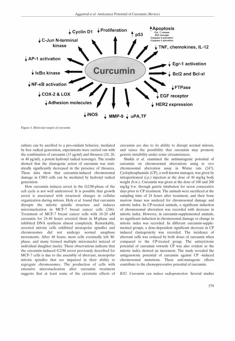
culture can be ascribed to a pro-oxidant behavior, mediatedby free radical generation, experiments were carried out withthe combination of curcumin (15 Ìg/ml) and thiourea (10, 20,or 40 Ìg/ml), a potent hydroxyl radical scavenger. The resultsshowed that the clastogenic action of curcumin was stati-stically significantly decreased in the presence of thiourea.These data show that curcumin-induced chromosomaldamage in CHO cells can be mediated by hydroxyl radicalgeneration.
How curcumin induces arrest in the G2/M-phase of thecell cycle is not well understood. It is possible that growtharrest is associated with structural changes in cellularorganization during mitosis. Holy et al. found that curcumindisrupts the mitotic spindle structure and inducesmicronucleation in MCF-7 breast cancer cells (246).Treatment of MCF-7 breast cancer cells with 10-20 ÌMcurcumin for 24-48 hÔurs arrested them in M-phase andinhibited DNA synthesis almost completely. Remarkably,arrested mitotic cells exhibited monopolar spindles andchromosomes did not undergo normal anaphasemovements. After 48 hÔurs, most cells eventually left M-phase, and many formed multiple micronuclei instead ofindividual daughter nuclei. These observations indicate thatthe curcumin-induced G2/M arrest previously described forMCF-7 cells is due to the assembly of aberrant, monopolarmitotic spindles that are impaired in their ability tosegregate chromosomes. The production of cells withextensive micronucleation after curcumin treatmentsuggests that at least some of the cytostatic effects of
curcumin are due to its ability to disrupt normal mitosis,and raises the possibility that curcumin may promotegenetic instability under some circumstances.
Shukla et al. examined the antimutagenic potential ofcurcumin on chromosomal aberrations using in vivochromosomal aberration assay in Wistar rats (247).Cyclophosphamide (CP), a well-known mutagen, was given byintraperitoneal (i.p.) injection at the dose of 40 mg/kg bodyweight (b.w.). Curcumin was given at the dose of 100 and 200mg/kg b.w. through gastric intubation for seven consecutivedays prior to CP treatment. The animals were sacrificed at thesampling time of 24 hÔurs after treatment, and their bonemarrow tissue was analyzed for chromosomal damage andmitotic index. In CP-treated animals, a significant inductionof chromosomal aberration was recorded with decrease inmitotic index. However, in curcumin-supplemented animals,no significant induction in chromosomal damage or change inmitotic index was recorded. In different curcumin-supple-mented groups, a dose-dependent significant decrease in CPinduced clastogenicity was recorded. The incidence ofaberrant cells was reduced by both doses of curcumin whencompared to the CP-treated group. The anticytotoxicpotential of curcumin towards CP was also evident as themitotic index showed an increment. The study revealed theantigenotoxic potential of curcumin against CP -inducedchromosomal mutations. These anti-mutagenic effectscontribute to the chemopreventive potential of curcumin.
B1U. Curcumin can induce radioprotection: Several studies
379
Aggarwal et al: Anticancer Potential of Curcumin (Review)
Figure 4. Molecular targets of curcumin.
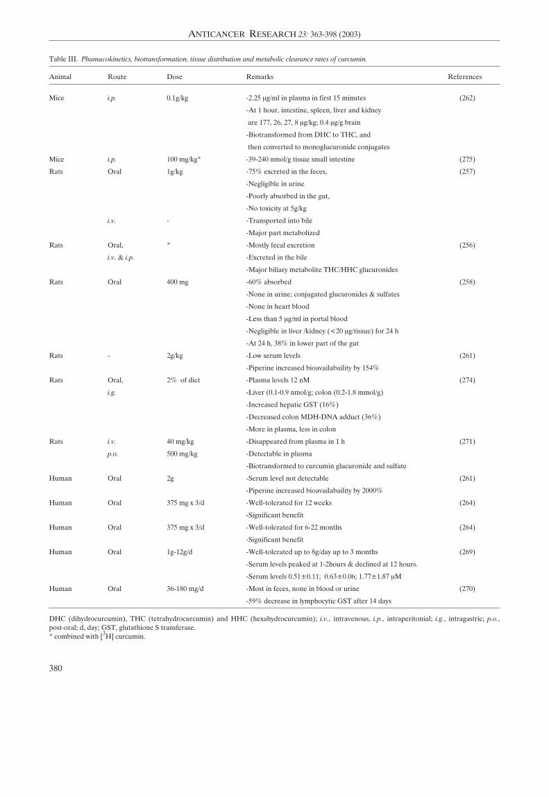
380
ANTICANCER RESEARCH 23: 363-398 (2003)
Table πππ. Phamacokinetics, biotransformation, tissue distribution and metabolic clearance rates of curcumin.
Animal Route Dose Remarks References
Mice i.p. 0.1g/kg -2.25 Ìg/ml in plasma in first 15 minutes (262)
-At 1 hour, intestine, spleen, liver and kidney
are 177, 26, 27, 8 Ìg/kg; 0.4 Ìg/g brain
-Biotransformed from DHC to THC, and
then converted to monoglucuronide conjugates
Mice i.p. 100 mg/kg* -39-240 nmol/g tissue small intestine (275)
Rats Oral 1g/kg -75% excreted in the feces, (257)
-Negligible in urine
-Poorly absorbed in the gut,
-No toxicity at 5g/kg
i.v. - -Transported into bile
-Major part metabolized
Rats Oral, * -Mostly fecal excretion (256)
i.v. & i.p. -Excreted in the bile
-Major biliary metabolite THC/HHC glucuronides
Rats Oral 400 mg -60% absorbed (258)
-None in urine; conjugated glucuronides & sulfates
-None in heart blood
-Less than 5 Ìg/ml in portal blood
-Negligible in liver /kidney (<20 Ìg/tissue) for 24 h
-At 24 h, 38% in lower part of the gut
Rats - 2g/kg -Low serum levels (261)
-Piperine increased bioavailabaility by 154%
Rats Oral, 2% of diet -Plasma levels 12 nM (274)
i.g. -Liver (0.1-0.9 nmol/g; colon (0.2-1.8 mmol/g)
-Increased hepatic GST (16%)
-Decreased colon MDH-DNA adduct (36%)
-More in plasma, less in colon
Rats i.v. 40 mg/kg -Disappeared from plasma in 1 h (271)
p.o. 500 mg/kg -Detectable in plasma
-Biotransformed to curcumin glucuronide and sulfate
Human Oral 2g -Serum level not detectable (261)
-Piperine increased bioavailabaility by 2000%
Human Oral 375 mg x 3/d -Well-tolerated for 12 weeks (264)
-Significant benefit
Human Oral 375 mg x 3/d -Well-tolerated for 6-22 months (264)
-Significant benefit
Human Oral 1g-12g/d -Well-tolerated up to 8g/day up to 3 months (269)
-Serum levels peaked at 1-2hours & declined at 12 hours.
-Serum levels 0.51±0.11; 0.63±0.06; 1.77±1.87 ÌM
Human Oral 36-180 mg/d -Most in feces, none in blood or urine (270)
-59% decrease in lymphocytic GST after 14 days
DHC (dihydrocurcumin), THC (tetrahydrocurcumin) and HHC (hexahydrocurcumin); i.v., intravenous, i.p., intraperitonial; i.g., intragastric; p.o.,post-oral; d, day; GST, glutathione S transferase.* combined with [3H] curcumin.
A R
suggest that curcumin is radioprotective. Thresiamma et al.showed curcumin protects rats from radiation- inducedtoxicity (249). Whole body irradiation of rats (10 Gy as fivefractions) produced lung fibrosis within 2 months, as assessedby increased lung collagen hydroxyproline and histo-pathology. Oral administration of curcumin (200 Ìmole/kgbody weight) significantly reduced hydroxyproline. In serumand liver, irradiation-induced increases in lipid peroxidationwere reduced significantly by curcumin treatment. The liverSOD and GSH peroxidase activity increases were alsoreduced significantly by curcumin, as was the frequency ofmicronucleated polychromatic erythrocytes in mice. Inanother study, Thresiamma et al. later investigated theprotective effect of curcumin on radiation-induced geno-toxicity (250). They showed that induction of micronuclei andchromosomal aberrations produced by whole-body exposureof Á-radiation (1.5-3.0 Gy) in mice was significantly inhibitedby oral administration of curcumin (400 Ìmoles/kg bodyweight), inhibited micronucleated polychromatic and normo-chromatic erythrocytes, significantly reduced the number ofbone marrow cells with chromosomal aberrations andchromosomal fragments, and inhibited the DNA strandbreaks produced in rat lymphocytes upon radiation as seenfrom the DNA unwinding studies. In contrast to these studies,Araujo et al. showed potentiation by curcumin of Á-radiation-induced chromosome aberrations in CHO cells (251). Theytreated the cells with curcumin (2.5, 5, and 10 Ìg/ml), andthen irradiated (2.5 Gy) them during different phases of thecell cycle. curcumin at 10 Ìg/ml increased the chromosomaldamage frequency. Curcumin did not have a protective effectagainst the clastogenicity of Á-radiation. Instead, an obviousincrease in the frequencies of chromosome aberrationsoccurred when curcumin was given at 10 Ìg/ml plusÁ-radiation during S- and G2/S- phases of the cell cycle. Whythere is a difference in the results of Thresiamma et al. andAraujo et al. is unclear.
Inano and Onoda investigated the radioprotective action ofcurcumin on the formation of urinary 8-hydroxy-2'-deoxy-guanosine (8-OHdG), tumorigenesis and mortality inducedby Á-irradiation. The rats were examined for mammary andpituitary tumors for 1 year. Virgin rats received whole-bodyirradiation with 9.6 Gy and were fed a diet containingcurcumin for 3 days before and/or 3 days after irradiation.After irradiation, all rats were assessed for 8-OHdG levelsand survival was assessed daily for 30 days. The invstigatorsfound that, in virgin rats irradiated with 3 Gy, the creati-nine-corrected concentration and total amount of 8-OHdG inthe 24-hours urine samples were higher (~1.3-fold) than inthe non-irradiated controls. Adding curcumin to the diet for 3days before and/or 2 days after irradiation reduced theelevated 8-OHdG levels by 50-70%. Dietary curcumin (1%,w/w) significantly decreased the incidence of mammary andpituitary tumors. However, curcumin did not improve survivalwhen administered for 3 days before and/or 3 days afterirradiation (9.6 Gy). These findings demonstrate that curcu-
min can be used as an effective radioprotective agent toinhibit acute and chronic effects, but not mortality, afterirradiation.
How curcumin provides radioprotection is not fullyunderstood. A few studies, however, indicate that curcumininhibits the radiation-induced damage of specific proteins.Kapoor and Priyadarsini examined the free radical reactionsof lysozyme (Lz), tryptophan and disulfides to curcumin inaqueous solution using a pulse radiolysis technique (252). Thebinding of curcumin with lysozyme was confirmed usingabsorption, fluorescence and stopped-flow techniques. Thefree radicals of curcumin generated after repairing radicals ofdisulfides, lysozyme and tryptophan absorbed meximally at500-510 nm. This interaction may play a role in the anti-oxidant behavior of curcumin in protecting proteins. Incontrast, Varadkar et al. examined the effect of curcumin onradiation-induced PKC activity isolated from the liver cytosoland the particulate fraction of unirradiated mice and miceirradiated at 5 Gy (253). Following irradiation, the PKCactivity was increased in both cytosolic and particulatefractions. Curcumin inhibited the activated cytosolic andparticulate PKC at very low concentrations. Since activationof PKC is one of the means conferring radioresistance on atumor cell, suppression of PKC activity by curcumin mightprevent the development of radioresistance.
Another potential mechanism of radioprotection involvessuppression of radiation-induced gene expression. Oguro andYoshida examined the effect of curcumin on UVA-inducedornithine decarboxylase (ODC) and metallothionein (MT)gene expression in mouse skin (254). They showed that UVAinduced MT mRNA in mouse skin; and 1,4-diazabicylo-[2,2,2]-octane (DABCO), a singlet oxygen scavenger, reducedUVA-mediated induction of MT mRNA (by 40%). UVAslightly enhanced TPA-mediated ODC mRNA induction,while it enhanced ODC enzyme activity by 70%. UVAadditively intensified TPA-mediated MT mRNA induction.Curcumin dramatically inhibited both TPA- and TPA +UVA-induced expression of ODC and MT genes.
B1V. Curcumin inhibits telomerase activity: Telomerase activityis linked to cellular proliferation, and its activation seems tobe a mandatory step in carcinogenesis. Ramachandran et al.analyzed the effect of curcumin on telomerase activity inhuman mammary epithelial (MCF-10A) and breast cancer(MCF-7) cells (255). Telomerase activity in MCF-7 cells was6.9-fold higher than that of human mammary epithelial cells.In MCF-7 cells, telomerase activity decreased with increasingconcentrations of curcumin, inhibiting about 93.4% activityat 100 ÌM concentration. The inhibition of telomerase activityin MCF-7 cells may be due to down-regulation of hTERTexpression. Increasing concentrations of curcumin caused asteady decrease in the level of hTERT mRNA in MCF-7 cells,whereas the level of c-myc mRNAs remained the same. Theseresults suggest that curcumin inhibits telomerase activity bydown-regulating hTERT expression in breast cancer cells and
381
Aggarwal et al: Anticancer Potential of Curcumin (Review)
A R
this down-regulation is not through the c-myc pathway. SincehTERT is known to be regulated by NF-κB, it is possible thatthe down-regulation of hTERT is mediated throughsuppression of NF-κB by curcumin.
From the studies described above it is clear that curcumininterferes with multiple signal transduction pathways (seeFigure 4), which could play a role in tumorigenesis. Thesuppression of these pathways may explain the antitumoreffects of curcumin.
B2. In vivo animal studies with curcumin
µ2A. Metabolism, pharmacokinetics, tissue distribution andclearance of curcumin: B2A1: Animal studies: Curcumin hasbeen administered to the animals via a variety of routesincluding topical, systemic, intravenous and oral routes ofadministration (Table πππ) (5, 61, 224, 256-274). Wahlstrom etal. examined the uptake, distribution and excretion ofcurcumin administered orally to Sprague-Dawley rats (257).When administered at a dose of 1 g/kg, about 75% ofcurcumin was excreted in the feces, while negligible amountsof curcumin appeared in the urine. Measurements of bloodplasma levels and biliary excretion showed that curcuminwas poorly absorbed from the gut. No apparent toxic effectswere seen after doses of up to 5 g/kg. When intravenouslyinjected or when added to the perfusate of the isolated liver,curcumin was actively transported into bile, against con-centration gradients of several hundred times. The major partof the drug was, however, metabolized. In suspensions ofisolated hepatocytes or liver microsomes 90% of the addedcurcumin was metabolized within 30 minutes. Holder et al.used curcumin labeled with deuterium and tritium to examinethe pharmacokinetics (256). Oral and intraperitoneal doses of[3H] curcumin led to the fecal excretion of most of theradioactivity. When administered via either the intravenousor intraperitoneal route, [3H] curcumin was excreted in thebile of cannulated rats. Chemical ionization mass spectro-metry demonstrated that the major biliary metabolites wereglucuronides of tetrahydrocurcumin and hexahydrocurcumin.A minor biliary metabolite was dihydroferulic acid togetherwith traces of ferulic acid.
Ravindranath et al. examined the absorption and tissuedistribution of curcumin (400 mg) after oral administration inrats (258). About 60% of the dose was absorbed; no curcuminwas detectable in urine. The urinary excretion of conjugatedglucuronides and sulfates significantly increased. Nocurcumin was present in heart blood. Only traces (less than 5Ìg/ml) in portal blood and negligible quantities in liver andkidney (< 20 Ìg/ tissue) were observed from 15 minutes up to24 hours after administration of curcumin. At the end of 24hours the concentration of curcumin remaining in the lowerpart of the gut, namely the cecum and large intestine,amounted to 38% of the quantity administered. The sameinvestigators also examined the in vitro intestinal absorptionof curcumin in rats (259). When everted sacs of rat intestines
were incubated with 50-750-Ìg of curcumin, 30-80% of theadded curcumin disappeared from the mucosal side. Nocurcumin was detectable in the serosal fluid. Less than 3% ofthe added curcumin was found in the tissue at the highestconcentration. In experiments with [3H] curcumin, 5-6% ofadded radioactivity was found in the serosal side. TLCexamination of the mucosal extract showed the presence of 2compounds, 1 corresponding to curcumin and the other to aless polar, colourless compound. The serosal fluid had nocurcumin, but there was a compound whose RF was identicalwith the colourless compound present in the mucosal side.These experiments indicated that curcumin undergoestransformation during absorption from the intestine.
Like Holder et al., Ravindranath examined the metabolismof curcumin using [3H] curcumin (256, 260). Radioactivitywas detectable in blood, liver and kidney following doses of400, 80, or 10 mg of [3H] curcumin. The major route ofelimination of the label was the feces; urinary excretion wasvery low regardless of the dose. At the lower doses of 80 mgand 10 mg of [3H]curcumin, most of the label was excretedwithin 72 hours, while with 400 mg, considerable amounts ofthe label was present in the tissues 12 days after dosage. Thepercentage of curcumin absorbed (60-66% of the given dose)remained constant regardless of the dose administered. Theabove mentioned studies indicate poor absorption, rapidmetabolism and excretion of curcumin. Therefore, it isunlikely that substantial concentrations of curcumin occur inthe body after ingestion. However, Pan et al. investigated thepharmacokinetic properties of curcumin in mice after i.p.administration (262). After a dose of 0.1 g/kg, about 2.25Ìg/ml of curcumin appeared in the plasma in the first 15minutes. One hour after administration, the levels ofcurcumin in the intestines, spleen, liver and kidneys were 177,26, 27 and 8 Ìg/g, respectively. Only traces (0.4 Ìg/g) wereobserved in the brain at 1 hour. When plasma was examinedfor the nature of the metabolites of curcumin, by reverse-phase HPLC, two putative conjugates were observed.Treatment of the plasma with P-glucuronidase decreased theconcentrations of these two putative conjugates andtetrahydrocurcumin (THC) and curcumin were detected. Toinvestigate the nature of these glucuronide conjugates in vivo,the plasma was analyzed by electrospray. The chemicalstructures of these metabolites, determined by mass spectro-metry analysis, suggested that curcumin was first bio-transformed to dihydrocurcumin and THC and that thesecompounds subsequently were converted to monoglucuronideconjugates. Because THC is one of the major metabolites ofcurcumin, its stability at different pH values was examined.THC was very stable in 0.1 M phosphate buffers of variouspH values. Moreover, THC was more stable than curcumin in0.1 M phosphate buffer, pH 7.2. These results suggestedthat curcumin-glucuronoside, dihydrocurcumin-glucurono-side, THC-glucuronoside and THC are major metabolites ofcurcumin in vivo.
Sharma et al. tested the hypothesis that 14 days of dietary
382
ANTICANCER RESEARCH 23: 363-398 (2003)
A R
curcumin (2%) affects biomarkers relevant to cancerchemoprevention in the rat (274). Levels of inducible COX2,as reflected by PGE2 production by blood leukocytes, weremeasured ex vivo. Total GST activity and adducts ofmalondialdehyde with DNA (M(1)G), which reflectendogenous lipid peroxidation, were measured in colonmucosa, liver and blood leukocytes. Curcumin and itsmetabolites were analyzed by HPLC in plasma and itspharmacokinetics were compared following a diet containing2% curcumin versus intragastric (i.g.) administration ofcurcumin suspended in an amphiphilic solvent. The curcumindiet did not alter any of the markers in the blood butincreased hepatic GST by 16% and decreased colon M(1)Glevels by 36% when compared with controls. Administrationof carbon tetrachloride during the treatment period increasedcolon M(1)G levels, and this increase was prevented bydietary curcumin. Dietary curcumin yielded low drug levels inthe plasma, between 0 and 12 nM, whereas tissue con-centrations of curcumin in liver and colon mucosa were0.1-0.9 nmol/g and 0.2-1.8 Ìmol/g, respectively. In comparisonwith dietary administration, suspended curcumin given i.g.resulted in more curcumin in the plasma but much less in thecolon mucosa. The results show that curcumin mixed with thediet achieves drug levels in the colon and liver sufficient toexplain the pharmacological activities observed and suggestthat this mode of administration may be preferable for thechemoprevention of colon cancer.
B2A2: Studies in humans: Animal studies have revealed thatcurcumin has poor bioavailability due to its rapid metabolismin the liver and intestinal wall. Therefore, Shoba et al. exa-mined the effect of combining piperine, a known inhibitor ofhepatic and intestinal glucuronidation, on the bioavailabilityof curcumin in rats and healthy human volunteers (261).When curcumin was given alone, in the dose 2 g/kg to rats,moderate serum concentrations were achieved over a periodof 4 hours. Concomitant administration of piperine 20 mg/kgincreased the serum concentration of curcumin for a shortperiod of 1 - 2 hours. The time to maximum concentrationwas significantly increased (p < 0.02), while elimination half-life and clearance significantly decreased (p < 0.02), and thebioavailability was increased by 154%. On the other hand inhumans after a dose of 2 g curcumin alone, serum levels wereeither undetectable or very low. Concomitant administrationof piperine 20 mg produced much higher concentrations from0.25 to 1 hour (p < 0.01 at 0.25 and 0.5 hour; p < 0.001 at 1hour), and the increase in bioavailability was 2000%.Investigators did not observe any adverse effect. The studyshowed that in the dosages used, piperine enhances the serumconcentration, extent of absorption and bioavailability ofcurcumin in both rats and humans with no adverse effects.
The systemic bioavailability of curcumin is low, so that itspharmacological activity may be mediated, in part, bycurcumin metabolites. To investigate this possibility, Ireson etal. compared curcumin metabolism in human and rat
hepatocytes in suspension with that in rats in vivo (271).Analysis by HPLC with detection at 420 and 280 nmpermitted characterization of metabolites with intactdiferoylmethane structure and increased saturation of theheptatrienone chain. Chromatographic inferences werecorroborated by mass spectrometry. The major metabolites insuspensions of human and rat hepatocytes were identified ashexahydrocurcumin and hexahydrocurcuminol. In rats, invivo, curcumin administered i.v. (40 mg/kg) disappeared fromthe plasma within 1 hour of dosing. After p.o. administration(500 mg/kg), the parent drug was present in plasma at levelsnear the detection limit. The major products of curcuminbiotransformation identified in rat plasma were curcuminglucuronide and curcumin sulfate, whereas hexahydro-curcumin, hexahydrocurcuminol and hexahydrocurcuminglucuronide were present in small amounts. To test thehypothesis that curcumin metabolites resemble their pro-genitor in that they can inhibit COX2 expression, curcuminand four of its metabolites at a concentration of 20 ÌM werecompared in terms of their ability to inhibit phorbol ester-induced PGE2 production in human colonic epithelial cells.Curcumin reduced PGE2 levels to preinduction levels,whereas tetrahydrocurcumin, previously shown to be amurine metabolite of curcumin, hexahydrocurcumin andcurcumin sulfate, had only weak PGE2 inhibitory activity andhexahydrocurcuminol was inactive. The results suggest that(a) the major products of curcumin biotransformation byhepatocytes occur only at low abundance in rat plasma aftercurcumin administration; and (b) metabolism of curcumin byreduction or conjugation generates species with reducedability to inhibit COX2 expression. Because the gastrointe-stinal tract seems to be exposed more prominently tounmetabolized curcumin than any other tissue, the resultssupported the clinical evaluation of curcumin as a colorectalcancer chemopreventive agent.
Indirect evidence suggests that curcumin is metabolized inthe intestinal tract. To investigate this notion further, Iresonet al. explored curcumin metabolism in subcellular fractionsof human and rat intestinal tissue, compared it withmetabolism in the corresponding hepatic fractions andstudied curcumin metabolism in situ in intact rat intestinalsacs (272). Curcumin glucuronide was identified in intestinaland hepatic microsomes and curcumin sulfate, tetrahydro-curcumin and hexahydrocurcumin were found as curcuminmetabolites in intestinal and hepatic cytosol from humans andrats. The extent of curcumin conjugation was much greater inintestinal fractions from humans than in those from rats,whereas curcumin conjugation was less extensive in hepaticfractions from humans than in those from rats. The curcumin-reducing ability of cytosol from human intestinal and livertissue exceeded that observed with the corresponding rattissue by factors of 18 and 5, respectively. Curcumin sulfatewas identified in incubations of curcumin with intact rat gutsacs. Curcumin was sulfated by human phenolsulfotransferase isoenzymes SULT1A1 and SULT1A3.
383
Aggarwal et al: Anticancer Potential of Curcumin (Review)
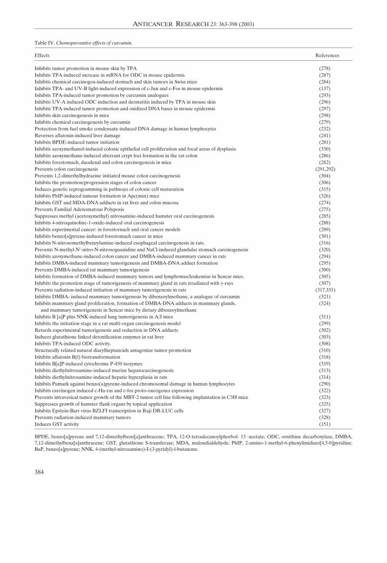
384
ANTICANCER RESEARCH 23: 363-398 (2003)
Table IV. Chemopreventive effects of curcumin.
Effects References
Inhibits tumor promotion in mouse skin by TPA (278)Inhibits TPA-induced increase in mRNA for ODC in mouse epidermis (287)Inhibits chemical carcinogen-induced stomach and skin tumors in Swiss mice (284)Inhibits TPA- and UV-B light-induced expression of c-Jun and c-Fos in mouse epidermis (137)Inhibits TPA-induced tumor promotion by curcumin analogues (293)Inhibits UV-A induced ODC induction and dermatitis induced by TPA in mouse skin (296)Inhibits TPA-induced tumor promotion and oxidized DNA bases in mouse epidermis (297)Inhibits skin carcinogenesis in mice (298)Inhibits chemical carcinogenesis by curcumin (279)Protection from fuel smoke condensate-induced DNA damage in human lymphocytes (232)Reverses aflatoxin-induced liver damage (241)Inhibits BPDE-induced tumor initiation (281)Inhibits azoxymethanol-induced colonic epithelial cell proliferation and focal areas of dysplasia (330)Inhibits azoxymethane-induced aberrant crypt foci formation in the rat colon (286)Inhibits forestomach, duodenal and colon carcinogenesis in mice (282)Prevents colon carcinogenesis (291,292)Prevents 1,2-dimethylhydrazine initiated mouse colon carcinogenesis (304)Inhibits the promotion/progression stages of colon cancer (306)Induces genetic reprogramming in pathways of colonic cell maturation (315)Inhibits PhIP-induced tumour formation in Apc(min) mice (326)Inhibits GST and MDA-DNA adducts in rat liver and colon mucosa (274)Prevents Familial Adenomatous Polyposis (275)Suppresses methyl (acetoxymethyl) nitrosamine-induced hamster oral carcinogenesis (285)Inhibits 4-nitroquinoline-1-oxide-induced oral carcinogenesis (288)Inhibits experimental cancer: in forestornach and oral cancer models (289)Inhibits benzo[a]pyrene-induced forestomach cancer in mice (301)Inhibits N-nitrosomethylbenzylamine-induced esophageal carcinogenesis in rats. (316)Prevents N-methyl-N'-nitro-N-nitrosoguanidine and NaCl-induced glandular stomach carcinogenesis (320)Inhibits azoxymethane-induced colon cancer and DMBA-induced mammary cancer in rats (294)Inhibits DMBA-induced mammary tumorigenesis and DMBA-DNA adduct formation (295)Prevents DMBA-induced rat mammary tumorigenesis (300)Inhibits formation of DMBA-induced mammary tumors and lymphomas/leukemias in Sencar mice. (305)Inhibits the promotion stage of tumorigenesis of mammary gland in rats irradiated with Á-rays (307)Prevents radiation-induced initiation of mammary tumorigenesis in rats (317,331)Inhibits DMBA- induced mammary tumorigenesis by dibenzoylmethane, a analogue of curcumin (321)Inhibits mammary gland proliferation, formation of DMBA-DNA adducts in mammary glands, (324)
and mammary tumorigenesis in Sencar mice by dietary dibenzoylmethaneInhibits B [a]P plus NNK-induced lung tumorigenesis in A/J mice (311)Inhibits the initiation stage in a rat multi-organ carcinogenesis model (299)Retards experimental tumorigenesis and reduction in DNA adducts (302)Induces glutathione linked detoxification enzymes in rat liver (303)Inhibits TPA-induced ODC activity. (308)Structurally related natural diarylheptanoids antagonize tumor promotion (310)Inhibits aflatoxin B(l) biotransformation (318)Inhibits B[a]P-induced cytochrome P-450 isozymes (319)Inhibits diethylnitrosamine-induced murine hepatocarcinogenesis (313)Inhibits diethylnitrosamine-induced hepatic hyperplasia in rats (314)Inhibits Pumark against benzo(a)pyrene-induced chromosomal damage in human lymphocytes (290)Inhibits carcinogen induced c-Ha-ras and c-fos proto-oncogenes expression (322)Prevents intravesical tumor growth of the MBT-2 tumor cell line following implantation in C3H mice (323)Suppresses growth of hamster flank organs by topical application (325)Inhibits Epstein-Barr virus BZLFI transcription in Raji DR-LUC cells (327)Prevents radiation-induced mammary tumors (328)Induces GST activity (151)
BPDE, benzo[a]pyrene and 7,12-dimethylbenz[a]anthracene; TPA, 12-O-tetradecanoylphorbol- 13 -acetate; ODC, ornithine decarboxylase, DMBA,7,12-dimethylbenz[a]anthracene; GST, glutathione S-transferase; MDA, malondialdehyde; PhIP, 2-amino-1-methyl-6-phenylimidazo[4,5-b]pyridine;BaP, benzo[a]pyrene; NNK, 4-(methyl-nitrosamino)-I-(3-pyridyl)-l-butanone.
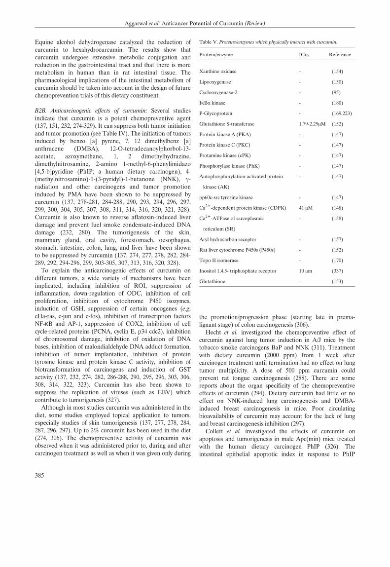
Equine alcohol dehydrogenase catalyzed the reduction ofcurcumin to hexahydrocurcumin. The results show thatcurcumin undergoes extensive metabolic conjugation andreduction in the gastrointestinal tract and that there is moremetabolism in human than in rat intestinal tissue. Thepharmacological implications of the intestinal metabolism ofcurcumin should be taken into account in the design of futurechemoprevention trials of this dietary constituent.
B2B. Anticarcinogenic effects of curcumin: Several studiesindicate that curcumin is a potent chemopreventive agent(137, 151, 232, 274-329). It can suppress both tumor initiationand tumor promotion (see Table IV). The initiation of tumorsinduced by benzo [a] pyrene, 7, 12 dimethylbenz [a]anthracene (DMBA), 12-O-tetradecanoylphorbol-13-acetate, azoxymethane, 1, 2 dimethylhydrazine,dimethylnitrosamine, 2-amino 1-methyl-6-phenylimidazo[4,5-b]pyridine (PhIP; a human dietary carcinogen), 4-(methylnitrosamino)-1-(3-pyridyl)-1-butanone (NNK), Á-radiation and other carcinogens and tumor promotioninduced by PMA have been shown to be suppressed bycurcumin (137, 278-281, 284-288, 290, 293, 294, 296, 297,299, 300, 304, 305, 307, 308, 311, 314, 316, 320, 321, 328).Curcumin is also known to reverse aflatoxin-induced liverdamage and prevent fuel smoke condensate-induced DNAdamage (232, 280). The tumorigenesis of the skin,mammary gland, oral cavity, forestomach, oesophagus,stomach, intestine, colon, lung, and liver have been shownto be suppressed by curcumin (137, 274, 277, 278, 282, 284-289, 292, 294-296, 299, 303-305, 307, 313, 316, 320, 328).
To explain the anticarcinogenic effects of curcumin ondifferent tumors, a wide variety of mechanisms have beenimplicated, including inhibition of ROI, suppression ofinflammation, down-regulation of ODC, inhibition of cellproliferation, inhibition of cytochrome P450 isozymes,induction of GSH, suppression of certain oncogenes (e.g;cHa-ras, c-jun and c-fos), inhibition of transcription factorsNF-κB and AP-1, suppression of COX2, inhibition of cellcycle-related proteins (PCNA, cyclin E, p34 cdc2), inhibitionof chromosomal damage, inhibition of oxidation of DNAbases, inhibition of malondialdehyde DNA adduct formation,inhibition of tumor implantation, inhibition of proteintyrosine kinase and protein kinase C activity, inhibition ofbiotransformation of carcinogens and induction of GSTactivity (137, 232, 274, 282, 286-288, 290, 295, 296, 303, 306,308, 314, 322, 323). Curcumin has also been shown tosuppress the replication of viruses (such as EBV) whichcontribute to tumorigenesis (327).
Although in most studies curcumin was administered in thediet, some studies employed topical application to tumors,especially studies of skin tumorigenesis (137, 277, 278, 284,287, 296, 297). Up to 2% curcumin has been used in the diet(274, 306). The chemopreventive activity of curcumin wasobserved when it was administered prior to, during and aftercarcinogen treatment as well as when it was given only during
the promotion/progression phase (starting late in prema-lignant stage) of colon carcinogenesis (306).
Hecht et al. investigated the chemopreventive effect ofcurcumin against lung tumor induction in A/J mice by thetobacco smoke carcinogens BaP and NNK (311). Treatmentwith dietary curcumin (2000 ppm) from 1 week aftercarcinogen treatment until termination had no effect on lungtumor multiplicity. A dose of 500 ppm curcumin couldprevent rat tongue carcinogenesis (288). There are somereports about the organ specificity of the chemopreventiveeffects of curcumin (294). Dietary curcumin had little or noeffect on NNK-induced lung carcinogenesis and DMBA-induced breast carcinogenesis in mice. Poor circulatingbioavailability of curcumin may account for the lack of lungand breast carcinogenesis inhibition (297).
Collett et al. investigated the effects of curcumin onapoptosis and tumorigenesis in male Apc(min) mice treatedwith the human dietary carcinogen PhIP (326). Theintestinal epithelial apoptotic index in response to PhIP
385
Aggarwal et al: Anticancer Potential of Curcumin (Review)
Table V. Proteins/enzymes which physically interact with curcumin.
Protein/enzyme IC50 Reference
Xanthine oxidase - (154)
Lipooxygenase - (150)
Cyclooxygenase-2 - (95)
IκB· kinase - (180)
P-Glycoprotein - (169,223)
Glutathione S-transferase 1.79-2.29ÌM (152)
Protein kinase A (PKA) - (147)
Protein kinase C (PKC) - (147)
Protamine kinase (cPK) - (147)
Phosphorylase kinase (PhK) - (147)
Autophosphorylation-activated protein - (147)
kinase (AK)
pp60c-src tyrosine kinase - (147)
Ca2+-dependent protein kinase (CDPK) 41 ÌM (148)
Ca2+-ATPase of sarcoplasmic - (158)
reticulum (SR)
Aryl hydrocarbon receptor - (157)
Rat liver cytochrome P450s (P450s) - (152)
Topo II isomerase - (170)
Inositol 1,4,5- triphosphate receptor 10 Ìm (337)
Glutathione - (153)
A R
treatment was approximately twice as great in the wild-typeC57BL/6 APC(+/+) strain as in Apc(min) mice. PhIPpromoted tumor formation in Apc(min) proximal smallintestine. Curcumin enhanced PhIP-induced apoptosis andinhibited PhIP-induced turnorigenesis in the proximal smallintestine of Apc(min) mice.
Mahmoud et al. investigated the effect of curcumin for theprevention of tumors in C57BL/6J-Min/+ (Min/+) micewhich bear germline mutation in the Apc gene andspontaneously develop numerous intestinal adenomas by 15weeks of age (332). At a dietary level of 0.15 %, curcumindecreased tumor formation in Min/+ mice by 63%.Examination of intestinal tissue from the treated animalsshowed that tumor prevention by curcumin was associatedwith increased enterocyte apoptosis and proliferation.Curcumin also decreased expression of the oncoprotein beta-catenin in the enterocytes of the Min/+ mouse, anobservation previously associated with an antitumor effect.
Recently, Perkins et al. also examined the preventive effectof curcumin on the development of adenomas in the intestinaltract of the C57B1/6J Min/+ mouse, a model of humanfamilial APC (275). These investigators explored the linkbetween its chemopreventive potency in the Min/+ mouseand levels of drug and metabolites in target tissue and plasma.Mice received dietary curcumin for 15 weeks, after whichadenomas were enumerated. Levels of curcumin andmetabolites were determined by high-performance liquidchromatography in plasma, tissues and feces of mice aftereither long-term ingestion of dietary curcumin or a singledose of [14C] curcumin (100 mg/kg) via the i.p. route.Whereas curcumin at 0.1 % in the diet was without effect, at0.2 and 0.5%, it reduced adenoma multiplicity by 39 and 40%,respectively, compared with untreated mice. Hematocritvalues in untreated Min/+ mice were drastically reducedcompared with those in wild-type C57BI/6J mice. Dietarycurcumin partially restored the suppressed hematocrit. Tracesof curcumin were detected in the plasma. Differences in itsconcentration in the small intestinal mucosa, between 39 and240 nmol/g of tissue, reflected differences in dietaryconcentration. [14C]Curcumin disappeared rapidly fromtissues and plasma within 2-8 hours after dosing. The groupconcluded that curcumin may be useful in the chemo-prevention of human intestinal malignancies related to Apcmutations. The comparison of dose, resulting curcumin levelsin the intestinal tract, and chemopreventive potencysuggested tentatively that a daily dose of 1.6 g of curcumin isrequired for efficacy in humans. A clear advantage ofcurcumin over nonsteroidal antiinflammatory drugs is itsability to decrease intestinal bleeding linked to adenomamaturation.
B2C: Antitumor effects of curcumin in animals: While thereare numerous studies on the prevention of cancer bycurcumin, there is very little known about the therapeuticeffects of curcumin. Busquets et al. showed that systemic
administration of curcumin (20 Ìg/kg body weight) for 6consecutive days to rats bearing the highly cachectic YoshidaAH-130 ascites hepatoma inhibited tumor growth (31 % oftotal cell number) (333). Dorai et al. examined the efficacy ofcurcumin in vivo for suppressing the growth of LNCaP cells asheterotopically implanted tumors in nude mice (334). TheLNCaP prostate cancer cells were grown, mixed withMatrigel, and injected subcutaneously into nude mice thatreceived a synthetic diet containing 2% curcumin for up to 6weeks. At the end point, sections taken from the excisedtumors were evaluated for pathology, cell proliferation,apoptosis and vascularity. The results showed that curcuminmarkedly decreases the extent of cell proliferation asmeasured by the BrdU incorporation assay and a significantincrease in the extent of apoptosis as measured by an in situcell death assay. Moreover, a significant decrease in themicrovessel density as measured by the CD31 antigen stainingwas also seen. These results thus suggest that curcumin couldbe a therapeutic anti-cancer agent, as it significantly inhibitsprostate cancer growth and has the potential to prevent theprogression of this cancer to its hormone refractory state.
Menon et al. examined the effect of curcumin on lungmetastasis induced by B16F-10 melanoma cells in femaleC57BL/6 mice (335). Curcumin significantly inhibited lungtumor formation (89%) and significantly increased the lifespan (144%). Moreover, lung collagen hydroxyproline andserum sialic acid levels were found to be significantly lower intreated animals compared to the untreated controls.Curcumin treatment (10 Ìg/ml) significantly inhibited theinvasion of B16F-10 melanoma cells across the collagenmatrix of the Boyden chamber. Gelatin Zymographic analysisof a trypsin-activated B16F-10 melanoma cell sonicaterevealed that curcumin did not have any metalloproteinaseactivity. Nor did curcumin treatment inhibit the motility ofB16F-10 melanoma cells across a polycarbonate filter in vitro.These findings suggest that curcumin inhibits the invasion ofB16F-10 melanoma cells by inhibition of metalloproteinases,thereby inhibiting lung metastasis.
C. Clinical Studies
C1. Phase I clinical trials: Sharma et al. investigated thepharmacodynamics and pharmacokinetics of curcumin inhumans, a dose-escalation pilot study at doses between 440and 2200 mg/day of Curcuma extracts, containing 36-180 mgof curcumin (270). Fifteen patients with advanced colorectalcancer refractory to standard chemotherapies receivedCurcuma extract daily for up to 4 months. The activity of GSTand levels of a DNA adduct (M(1)G) formed by malondial-dehyde, a product of lipid peroxidation and prostaglandinbiosynthesis, were measured in patients' blood cells. OralCurcuma extract was well tolerated and dose-limiting toxicitywas not observed. Neither curcumin nor its metabolites weredetected in blood or urine, but curcumin was recovered fromfeces. curcumin sulfate was identified in the feces of one
386
ANTICANCER RESEARCH 23: 363-398 (2003)
A R
patient. Ingestion of 440 mg of Curcuma extract for 29 dayswas accompanied by a 59% decrease in lymphocytic GSTactivity. At higher dose levels, this effect was not observed.Leukocytic M(l)G levels were constant within each patientand unaffected by treatment. Radiologically stable diseasewas demonstrated in five patients for 2-4 months oftreatment. The results suggested that (a) Curcuma extract canbe administered safely to patients at doses of up to 2.2 g daily,equivalent to 180 mg of curcumin; (b) curcumin has low oralbioavailability in humans and may undergo intestinalmetabolism; and (c) larger clinical trials of Curcuma extractare merited.
C2. Phase II clinical trials: Cheng et al. investigated the toxi-cology, pharmacokinetics and biologically effective dose ofcurcumin in humans (269). Their prospective phase -I/IIstudy evaluated the effect of curcumin in patients with oneof the following five high-risk conditions: 1) recentlyresected urinary bladder cancer; 2) arsenic Bowen's diseaseof the skin; 3) uterine cervical intraepithelial neoplasm(CIN); 4) oral leucoplakia; and 5) intestinal metaplasia ofthe stomach. Curcumin was given orally for 3 months.Biopsy of the lesion sites was done immediately before and3 months after starting curcumin treament. The startingdose was 500 mg/day. If no toxicity of grade ππ or higher wasnoted in at least 3 successive patients, the dose was thenescalated to another level in the order of 1,000, 2,000,4,000, 8,000 and 12,000 mg/day. The concentration ofcurcumin in serum and urine was determined by HPLC. Atotal of 25 patients were enrolled in this study. There wasno treatment-related toxicity up to 8,000 mg/day. Beyond8,000 mg/day, the bulky volume of the drug wasunacceptable to the patients. The serum concentration ofcurcumin usually peaked at 1 to 2 hour after oral intake ofcrucumin and gradually declined over the next 12 hours.The average peak serum concentrations after taking 4,000mg, 6,000 mg and 8,000 mg of curcumin were0.51±0.11 ÌM, 0.63 ± 0.06 ÌM and 1.77 ± 1.87 ̪,respectively. Urinary excretion of curcumin wasundetectable. One of 4 patients with CIN and 1 of 7patients with oral leucoplakia proceeded to develop frankmalignancies in spite of curcumin treatment. In contrast,histological improvement of precancerous lesions was seenin 1 of 2 patients with recently resected bladder cancer, 2 of7 patients with oral leucoplakia, 1 of 6 patients withintestinal metaplasia of the stomach, 1 of 4 patients withCIN and 2 of 6 patients with Bowen's disease. Inconclusion, this study demonstrated that curcumin is nottoxic to humans up to 8,000 mg/day when taken by mouthfor 3 months. These results also suggested a biologicaleffect of curcumin in the chemoprevention of cancer.
Curcumin has shown wide-ranging chemopreventiveactivity in preclinical carcinogenic models, in which itinhibits COX2 at the transcriptional level. COX2 has beenimplicated in the development of many human cancers.
Plummer et al. explored the inhibition of COX2 activity as asystemic biomarker of drug efficacy, a biomarker ofpotential use in clinical trials of many chemopreventivedrugs known to inhibit this enzyme (268). They measuredCOX2 protein induction and PGE2 production in humanblood after incubation with LPS. When 1 ÌM curcumin wasadded in vitro to blood from healthy volunteers, LPS-induced COX2 protein levels and concomitant PGE2production were reduced by 24% and 41%, respectively. Totest whether effects on COX2 activity could also bemeasured after oral dosing in humans, these workersconducted a dose escalation pilot study of a standardizedformulation of Curcuma extract in 15 patients withadvanced colorectal cancer. Basal and LPS-mediated PGE2production was measured in blood, twice before treatmentand on days 1, 2, 8 and 29 of treatment. Analysis of basaland LPS-induced PGE2 production during treatmentdemonstrated a trend toward dose-dependent inhibition(p<0.005 by regression analysis), but there was nosignificant difference compared with values frompretreatment time- points.
Lal et al. administered curcumin orally to patientssuffering from chronic anterior uveitis (CAU) at a dose of375 mg three times a day for 12 weeks. Of 53 patientsenrolled, 32 completed the 12-week study (264). They weredivided into two groups: one group of 18 patients receivedcurcumin alone, whereas the other group of 14 patients,who had a strong PPD reaction, in addition receivedantitubercular treatment. The patients in both the groupsstarted improving after 2 weeks of treatment. All thepatients who received curcumin alone improved, whereasthe group receiving antitubercular therapy along withcurcumin had a response rate of 86%. Follow-up of all thepatients for the next 3 years indicated a recurrence rate of55% in the first group and of 36% in the second group.Four of 18 (22%) patients in the first group and 3 of 14patients (21%) in the second group lost their vision in thefollow-up period due to various complications in the eyes,e.g. vitritis, macular edema, central venous block, cataractformation and glaucomatous optic nerve damage. None ofthe patients reported any side-effect of the drug. Theefficacy of curcumin and recurrences following treatmentare comparable to corticosteroid therapy, which is presentlythe only available standard treatment for this disease. Thelack of side-effects with curcumin is its greatest advantagecompared with cortico steroids.
Curcumin has also been used for the treatment ofpatients suffering from idiopathic inflammatory orbitalpseudotumors (267). curcumin was administered orally at adose of 375 mg/3 times/day orally for a period of 6-22months in eight patients. They were followed-up for aperiod of 2 years at 3-month intervals. Five patientscompleted the study, of which four recovered completelyand one experienced some persistent limitation ofmovement but the swelling regressed completely. No side-
387
Aggarwal et al: Anticancer Potential of Curcumin (Review)
A R
effect was noted in any patient and there was no recurrence.The study suggests that curcumin could be used as a safeand effective drug in the treatment of idiopathicinflammatory orbital pseudotumors.
C3. Epidemiological correlates of curcumin. It has beendocumented that cancer of the colon, breast, prostate andlung, while most common in the United States, is not asprevalent in countries such as India where curcumin isfrequently consumed (336). Mohandas et al. examined theepidemiology of digestive tract cancers within India (263).They found a low incidence of large bowel cancers in Indiansand attributed this to high intake of starch and the presenceof natural antioxidants such as curcumin in Indian cooking.The role of hereditary factors has been evaluated in a fewstudies. Some studies have reported the occurrence of bothFAP and HNPCC in India. Adenoma is rare in elderlyIndians undergoing colonoscopy, even in those with largebowel cancers. They also reported that small bowel cancersare extremely rare in India. In conclusion, the incidence oflarge and small bowel adenomas and cancers is low inIndians. An increase in the incidence of large bowel cancersin immigrants and urban Indians compared to ruralpopulations supports a role for environmental risk factorsincluding diet.
Conclusion
From numerous studies as described above, it is quite apparentthat curcumin has tremendous potential for prevention andtherapy of various cancers. Several cellular proteins have beenidentified with which curcumin directly interacts (see Table V).One of the major problem with curcumin is its rapid clearancefrom the body. Whether rapid clearance is a disadvantage or anadvantage is not clear at present. In spite of it, whetheradministered orally, i.p., or systemically, curcumin appears tohave biological effects both in animals and humans. No naturalagent has yet been described which modulates so many signaltransduction pathways as curcumin does. Is that to ourdisadvantage? Probably not. Most cancer biologists are of theopinion that tumor cells use multiple pathways to escape host-defense mechanisms. Thus the drug which is specific forinhibition of one signal transduction pathway in the tumor cellmay not be adequate. Finding a drug that attacks multiplepathways and yet is pharmacologically safe is difficult.Curcumin, however, is one such drug that meets this criterion.The inability to patent curcumin, because of its naturaloccurance and well-established, long history of dietary use,prevents the involvement of pharmaceutical industry toexamine this drug closely as a therapeutic or preventive agent.The structure of curcumin can serve as lead compound for thedesign of a better molecule that is also patentable. It is ourhope that other nonprofit organizations see the value in thiscompound and provide the kind of evidence needed to helpcancer patients.
Acknowledgements
This research was supported by The Clayton Foundation for Research(to BBA), by the Department of Defense of the US Army Breast CancerResearch Program BC010610 (to BBA), and by P50 Head and NeckSPORE grant from the National Institute of Health, U.S.A. (to BBA).We would like to thank Walter Pagel for his careful review of themanuscript.
References
1 Rao DS, Sekhara NC, Satyanarayana MN and Srinivasan M: Effectof curcumin on serum and liver cholesterol levels in the rat. J Nutr100: 1307-15, 1970.
2 Patil TN and Srinivasan M: Hypocholesteremic effect of curcuminin induced hypercholesteremic rats. Indian J Exp Biol 9: 167-9,1971.
3 Keshavarz K: The influence of turmeric and curcumin oncholesterol concentration of eggs and tissues. Poult Sci 55: 1077-83,1976.
4. Soudamini KK, Unnikrishnan MC, Soni KB and Kuttan R:Inhibition of lipid peroxidation and cholesterol levels in mice bycurcumin. Indian J Physiol Pharmacol 36: 239-43, 1992.
5 Soni KB and Kuttan R: Effect of oral curcumin administration onserum peroxides and cholesterol levels in human volunteers. IndianJ Physiol Pharmacol 36: 273-5, 1992.
6 Hussain MS and Chandrasekhara N: Effect on curcumin oncholesterol gall-stone induction in mice. Indian J Med Res 96: 288-91, 1992.
7 Asai A and Miyazawa T: Dietary curcuminoids prevent high-fatdiet-induced lipid accumulation in rat liver and epididymal adiposetissue. J Nutr 131: 2932-5, 2001.
8 Ramirez-Tortosa MC, Mesa MD, Aguilera MC, Quiles JL, Baro L,Ramirez- Tortosa CL, Martinez-Victoria E and Gil A: Oral admi-nistration of a turmeric extract inhibits LDL oxidation and hashypocholesterolemic effects in rabbits with experimental athe-rosclerosis. Atherosclerosis 147: 371-8, 1999.
9 Naidu KA and Thippeswamy NB: Inhibition of human low densitylipoprotein oxidation by active principles from spices. Mol CellBiochem 229: 19-23, 2002.
10 Patro BS, Rele S, Chintalwar GJ, Chattopadhyay S, Adhikari S andMukherjee T: Protective activities of some phenolic 1,3-diketonesagainst lipid peroxidation: possible involvement of the 1,3-diketonemoiety. Chembiochem 3: 364-70, 2002.
11 Srivastava R, Puri V, Srimal RC and Dhawan BN: Effect ofcurcumin on platelet aggregation and vascular prostacyclinsynthesis. Arzneimittelforschung 36: 715-7, 1986.
12 Srivastava KC, Bordia A and Verma SK: Curcumin, a majorcomponent of food spice turmeric (Curcuma longa) inhibitsaggregation and alters eicosanoid metabolism in human bloodplatelets. Prostaglandins Leukot Essent Fatty Acids 52: 223-7,1995.
13 Srivastava R, Dikshit M, Srimal RC and Dhawan BN: Anti-thrombotic effect of curcumin. Thromb Res 40: 413-7, 1985.
14 Dikshit M, Rastogi L, Shukla R and Srimal RC: Prevention ofischaemia-induced biochemical changes by curcumin & quinidinein the cat heart. Indian J Med Res 101: 31-5, 1995.
15 Nirmala C and Puvanakrishnan R: Effect of curcumin on certainlysosomal hydrolases in isoproterenol-induced myocardialinfarction in rats. Biochem Pharmacol 51: 47-51, 1996.
16 Nirmala C and Puvanakrishnan R: Protective role of curcuminagainst isoproterenol induced myocardial infarction in rats. MolCell Biochem 159: 85-93, 1996.
388
ANTICANCER RESEARCH 23: 363-398 (2003)
A R
17 Venkatesan N: curcumin attenuation of acute adriamycinmyocardial toxicity in rats. Br J Pharmacol 124: 425-7, 1998.
18 Srinivasan M: Effect of curcumin on blood sugar as seen in adiabetic subject. Indian J Med Sci 26: 269-70, 1972.
19 Babu PS and Srinivasan K: Influence of dietary curcumin andcholesterol on the progression of experimentally induced diabetesin albino rat. Mol Cell Biochem 152: 13-21, 1995.
20 Babu PS and Srinivasan K: Hypolipidernic action of curcumin, theactive principle of turmeric (Curcuma longa) in streptozotocininduced diabetic rats. Mol Cell Biochem 166: 169-75, 1997.
21 Suresh Babu P and Srinivasan K: Amelioration of renal lesionsassociated with diabetes by dietary curcumin in streptozotocindiabetic rats. Mol Cell Biochem 181: 87-96, 1998.
22 Arun N and Nalini N: Efficacy of turmeric on blood sugar andpolyol pathway in diabetic albino rats. Plant Foods Hum Nutr 57:41-52, 2002.
23 Deodhar SD, Sethi R and Srimal RC: Preliminary study onantirheumatic activity of curcumin (diferuloyl methane). Indian JMed Res 71: 632-4, 1980.
24 Natarajan C and Bright JJ: Curcumin inhibits experimental allergicencephalomyelitis by blocking IL-12 signaling through Januskinase-STAT pathway in T lymphocytes. J Immunol 168: 6506-13,2002.
25 Lim GP, Chu T, Yang F, Beech W, Frautschy SA and Cole GM:The curry spice curcumin reduces oxidative damage and amyloidpathology in an Alzheimer transgenic mouse. J Neurosci 21: 8370-7,2001.
26 Frautschy SA, Hu W, Kim P, Miller SA, Chu T, Harris-White MEand Cole GM: Phenolic anti-inflammatory antioxidant reversal of abeta-induced cognitive deficits and neuropathology. NeurobiolAging 22: 993-1005, 2001.
27 Sui Z, Salto R, Li J, Craik C and Ortiz de Montellano PR:Inhibition of the HIV-1 and HIV-2 proteases by curcumin andcurcumin boron complexes. Bioorg Med Chem 1: 415-22, 1993.
28 Li CJ, Zhang LJ, Dezube BJ, Crumpacker CS and Pardee AB:Three inhibitors of type 1 human immunodeficiency virus longterminal repeat-directed gene expression and virus replication.Proc Natl Acad Sci USA 90: 1839-42, 1993.
29 Jordan WC and Drew CR: Curcumin--a natural herb with anti-HIVactivity. J Natl Med Assoc 88: 333, 1996.
30 Mazumder A, Nearnati N, Sunder S, Schulz J, Pertz H, Eich E andPommier Y: curcumin analogs with altered potencies against HIV-1 integrase as probes for biochemical mechanisms of drug action. JMed Chem 40: 3057-63, 1997.
31 Barthelemy S, Vergnes L, Moynier M, Guyot D, Labidalle S andBahraoui E: Curcumin and curcumin derivatives inhibit Tat-mediated transactivation of type 1 human immunodeficiency viruslong terminal repeat. Res Virol 149: 43-52, 1998.
32 Sidhu GS, Singh AK, Thaloor D, Banaudha KK, Patnaik GK,Srimal RC and Maheshwari RK: Enhancement of wound healingby curcumin in animals. Wound Repair Regen 6: 167-77, 1998.
33 Phan TT, See P, Lee ST and Chan SY: Protective effects ofcurcumin against oxidative damage on skin cells in vitro: itsimplication for wound healing. J Trauma 51: 927-31, 2001.
34 Shahed AR, Jones E and Shoskes D: Quercetin and curcumin up-regulate antioxidant gene expression in rat kidney after ureteralobstruction or ischemia/reperfusion injury. Transplant Proc 33:2988, 2001.
35 Morikawa T, Matsuda H, Ninomiya K and Yoshikawa M: Medi-cinal foodstuffs. XXIX. Potent protective effects of sesquiterpenesand curcumin from Zedoariae Rhizoma on liver injury induced byD-galactosamine/lipopolysaccharide or tumor necrosis factor-alpha.Biol Pharm Bull 25: 627-31, 2002.
36 Ramprasad C and Sirsi M: Studies on Indian medicinal plants:
Curcuma longa Linn.- effect of curcumin & the essential oils of C.longa on bile secretion. J Sci Industr Res 15: 262-265, 1956.
37 Awasthi S, Srivatava SK, Piper JT, Singhal SS, Chaubey M andAwasthi YC: Curcumin protects against 4-hydroxy-2-trans-nonenal-induced cataract formation in rat lenses. Am J Clin Nutr 64: 761-6,1996.
38 Venkatesan N and Chandrakasan G: Modulation of cyclopho-sphamide-induced early lung injury by curcumin, an anti-inflammatory antioxidant. Mol Cell Biochem 142: 79-87, 1995.
39 Venkatesan N, Punithavathi V and Chandrakasan G: Curcuminprotects bleomycin-induced lung injury in rats. Life Sci 61: PL51-8,1997.
40 Venkatesan N: Pulmonary protective effects of curcumin againstparaquat toxicity. Life Sci 66: PL21-8, 2000.
41 Punithavathi D, Venkatesan N and Babu M: Curcumin inhibitionof bleomycin- induced pulmonary fibrosis in rats. Br J Pharmacol131: 169-72, 2000.
42 Saleheen D, Ali SA, Ashfaq K, Siddiqui AA, Agha A and YasinzaiMM: Latent activity of curcumin against leishmaniasis in vitro. BiolPharm Bull 25: 386-9, 2002.
43 Gomes Dde C, Alegrio LV, de Lima ME, Leon LL and Araujo CA:Synthetic derivatives of curcumin and their activity againstLeishmania amazonensis. Arzneimittelforschung 52: 120-4, 2002.
44 Koide T, Nose M, Ogihara Y, Yabu Y and Ohta N: Leishmanicidaleffect of curcumin in vitro. Biol Pharm Bull 25: 131-3, 2002.
45 Huang HC, Jan TR and Yeh SF: Inhibitory effect of curcumin, ananti-inflammatory agent, on vascular smooth muscle cellproliferation. Eur J Pharmacol 221: 381-4, 1992.
46 Chen HW and Huang HC: Effect of curcumin on cell cycleprogression and apoptosis in vascular smooth muscle cells. Br JPharmacol 124: 1029-40, 1998.
47 Srimal RC and Dhawan BN: Pharmacology of diferuloylmethane(curcumin), a non- steroidal anti-inflammatory agent. J PharmPharmacol 25: 447-52, 1973.
48 Conney AH, Lysz T, Ferraro T, Abidi TF, Manchand PS, LaskinJD and Huang MT: Inhibitory effect of curcumin and some relateddietary compounds on tumor promotion and arachidonic acidmetabolism in mouse skin. Adv Enzyme Regul 31: 385-96, 1991.
49 Majeed M, Badmaev V and Murray F: Turmeric and the healingcurcuminoids: their amazing antioxidant properties and protectivepowers. New Canaan: Keats Publishing Inc: 1-46. 1996.
50 Surh YJ: Anti-tumor promoting potential of selected spiceingredients with antioxidative and anti-inflammatory activities: ashort review. Food Chem Toxicol 40: 1091-7, 2002.
51 Bremner P and Heinrich M: Natural products as targetedmodulators of the nuclear factor-kappaB pathway. J PharmPharmacol 54: 453-72, 2002.
52 Gescher AJ, Sharma RA and Steward WP: Cancer chemo-prevention by dietary constituents: a tale of failure and promise.Lancet Oncol 2: 371-9, 2001.
53 Chignell CF, Bilski P, Reszka KJ, Motten AG, Sik RH and DahlTA: Spectral and photochemical properties of curcumin. Photo-chem Photobiol 59: 295-302, 1994.
54 Das KC and Das CK: curcumin (diferuloylmethane), a singletoxygen ((1)0(2)) quencher. Biochem Biophys Res Commun 295:62-6. 2002.
55 Toniolo R, Di Narda F, Susmel S, Martelli M, Martelli L andBontempelli G: Quenching of superoxide ions by curcumin. Amechanistic study in acetonitrile. Ann Chim 92: 281-8, 2002.
56 Dahl TA, Bilski P, Reszka KJ and Chignell CF: Photocytotoxicityof curcumin. Photochem Photobiol 59: 290-4, 1994.
57 Tonnesen HH and Karlsen J: Studies on curcumin andcurcuminoids. VI. Kinetics of curcumin degradation in aqueoussolution. Z Lebensm Unters Forsch 180: 402-4, 1985.
389
Aggarwal et al: Anticancer Potential of Curcumin (Review)
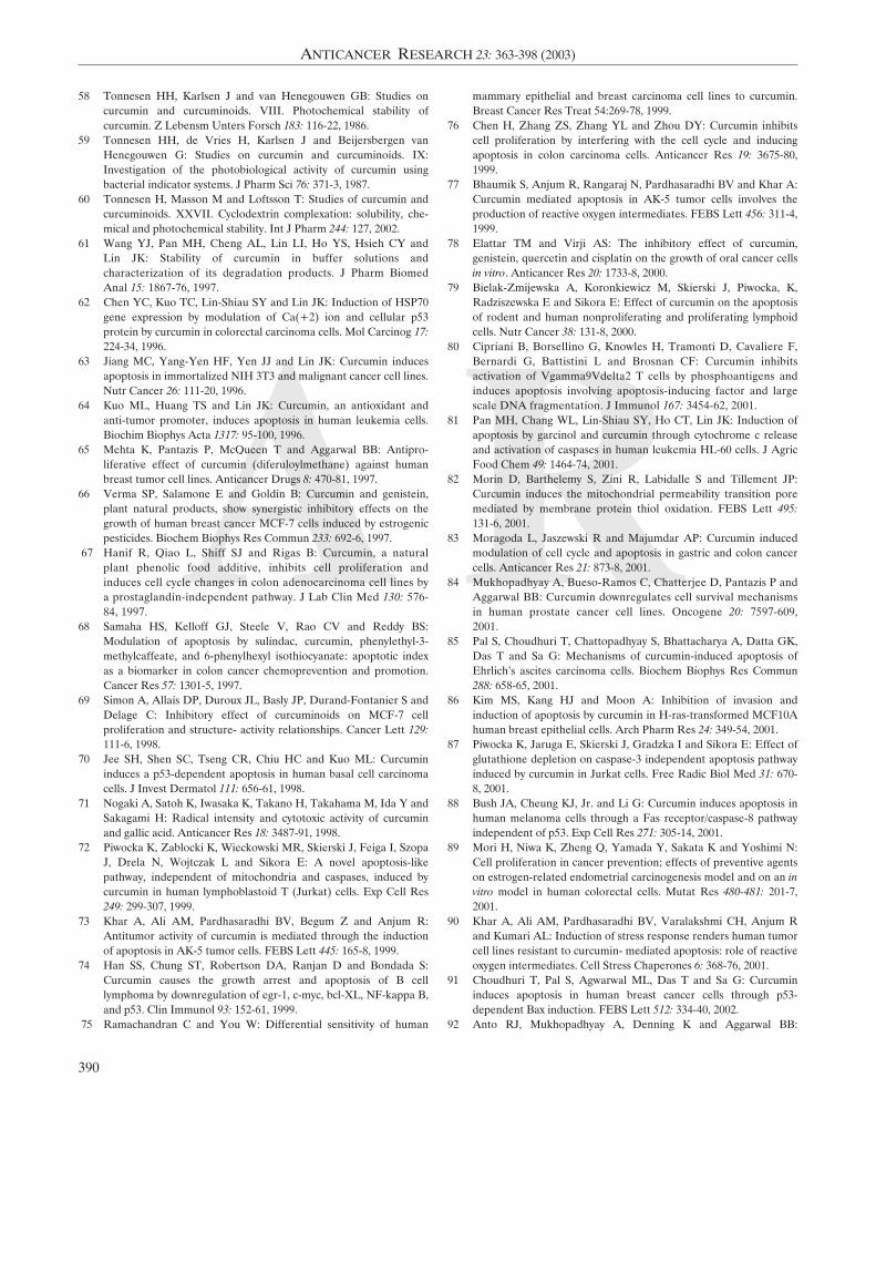
A R
58 Tonnesen HH, Karlsen J and van Henegouwen GB: Studies oncurcumin and curcuminoids. VIII. Photochemical stability ofcurcumin. Z Lebensm Unters Forsch 183: 116-22, 1986.
59 Tonnesen HH, de Vries H, Karlsen J and Beijersbergen vanHenegouwen G: Studies on curcumin and curcuminoids. IX:Investigation of the photobiological activity of curcumin usingbacterial indicator systems. J Pharm Sci 76: 371-3, 1987.
60 Tonnesen H, Masson M and Loftsson T: Studies of curcumin andcurcuminoids. XXVII. Cyclodextrin complexation: solubility, che-mical and photochemical stability. Int J Pharm 244: 127, 2002.
61 Wang YJ, Pan MH, Cheng AL, Lin LI, Ho YS, Hsieh CY andLin JK: Stability of curcumin in buffer solutions andcharacterization of its degradation products. J Pharm BiomedAnal 15: 1867-76, 1997.
62 Chen YC, Kuo TC, Lin-Shiau SY and Lin JK: Induction of HSP70gene expression by modulation of Ca(+2) ion and cellular p53protein by curcumin in colorectal carcinoma cells. Mol Carcinog 17:224-34, 1996.
63 Jiang MC, Yang-Yen HF, Yen JJ and Lin JK: Curcumin inducesapoptosis in immortalized NIH 3T3 and malignant cancer cell lines.Nutr Cancer 26: 111-20, 1996.
64 Kuo ML, Huang TS and Lin JK: Curcumin, an antioxidant andanti-tumor promoter, induces apoptosis in human leukemia cells.Biochim Biophys Acta 1317: 95-100, 1996.
65 Mehta K, Pantazis P, McQueen T and Aggarwal BB: Antipro-liferative effect of curcumin (diferuloylmethane) against humanbreast tumor cell lines. Anticancer Drugs 8: 470-81, 1997.
66 Verma SP, Salamone E and Goldin B: Curcumin and genistein,plant natural products, show synergistic inhibitory effects on thegrowth of human breast cancer MCF-7 cells induced by estrogenicpesticides. Biochem Biophys Res Commun 233: 692-6, 1997.
67 Hanif R, Qiao L, Shiff SJ and Rigas B: Curcumin, a naturalplant phenolic food additive, inhibits cell proliferation andinduces cell cycle changes in colon adenocarcinoma cell lines bya prostaglandin-independent pathway. J Lab Clin Med 130: 576-84, 1997.
68 Samaha HS, Kelloff GJ, Steele V, Rao CV and Reddy BS:Modulation of apoptosis by sulindac, curcumin, phenylethyl-3-methylcaffeate, and 6-phenylhexyl isothiocyanate: apoptotic indexas a biomarker in colon cancer chemoprevention and promotion.Cancer Res 57: 1301-5, 1997.
69 Simon A, Allais DP, Duroux JL, Basly JP, Durand-Fontanier S andDelage C: Inhibitory effect of curcuminoids on MCF-7 cellproliferation and structure- activity relationships. Cancer Lett 129:111-6, 1998.
70 Jee SH, Shen SC, Tseng CR, Chiu HC and Kuo ML: Curcumininduces a p53-dependent apoptosis in human basal cell carcinomacells. J Invest Dermatol 111: 656-61, 1998.
71 Nogaki A, Satoh K, Iwasaka K, Takano H, Takahama M, Ida Y andSakagami H: Radical intensity and cytotoxic activity of curcuminand gallic acid. Anticancer Res 18: 3487-91, 1998.
72 Piwocka K, Zablocki K, Wieckowski MR, Skierski J, Feiga I, SzopaJ, Drela N, Wojtczak L and Sikora E: A novel apoptosis-likepathway, independent of mitochondria and caspases, induced bycurcumin in human lymphoblastoid T (Jurkat) cells. Exp Cell Res249: 299-307, 1999.
73 Khar A, Ali AM, Pardhasaradhi BV, Begum Z and Anjum R:Antitumor activity of curcumin is mediated through the inductionof apoptosis in AK-5 tumor cells. FEBS Lett 445: 165-8, 1999.
74 Han SS, Chung ST, Robertson DA, Ranjan D and Bondada S:Curcumin causes the growth arrest and apoptosis of B celllymphoma by downregulation of egr-1, c-myc, bcl-XL, NF-kappa B,and p53. Clin Immunol 93: 152-61, 1999.
75 Ramachandran C and You W: Differential sensitivity of human
mammary epithelial and breast carcinoma cell lines to curcumin.Breast Cancer Res Treat 54:269-78, 1999.
76 Chen H, Zhang ZS, Zhang YL and Zhou DY: Curcumin inhibitscell proliferation by interfering with the cell cycle and inducingapoptosis in colon carcinoma cells. Anticancer Res 19: 3675-80,1999.
77 Bhaumik S, Anjum R, Rangaraj N, Pardhasaradhi BV and Khar A:Curcumin mediated apoptosis in AK-5 tumor cells involves theproduction of reactive oxygen intermediates. FEBS Lett 456: 311-4,1999.
78 Elattar TM and Virji AS: The inhibitory effect of curcumin,genistein, quercetin and cisplatin on the growth of oral cancer cellsin vitro. Anticancer Res 20: 1733-8, 2000.
79 Bielak-Zmijewska A, Koronkiewicz M, Skierski J, Piwocka, K,Radziszewska E and Sikora E: Effect of curcumin on the apoptosisof rodent and human nonproliferating and proliferating lymphoidcells. Nutr Cancer 38: 131-8, 2000.
80 Cipriani B, Borsellino G, Knowles H, Tramonti D, Cavaliere F,Bernardi G, Battistini L and Brosnan CF: Curcumin inhibitsactivation of Vgamma9Vdelta2 T cells by phosphoantigens andinduces apoptosis involving apoptosis-inducing factor and largescale DNA fragmentation. J Immunol 167: 3454-62, 2001.
81 Pan MH, Chang WL, Lin-Shiau SY, Ho CT, Lin JK: Induction ofapoptosis by garcinol and curcumin through cytochrome c releaseand activation of caspases in human leukemia HL-60 cells. J AgricFood Chem 49: 1464-74, 2001.
82 Morin D, Barthelemy S, Zini R, Labidalle S and Tillement JP:Curcumin induces the mitochondrial permeability transition poremediated by membrane protein thiol oxidation. FEBS Lett 495:131-6, 2001.
83 Moragoda L, Jaszewski R and Majumdar AP: Curcumin inducedmodulation of cell cycle and apoptosis in gastric and colon cancercells. Anticancer Res 21: 873-8, 2001.
84 Mukhopadhyay A, Bueso-Ramos C, Chatterjee D, Pantazis P andAggarwal BB: Curcumin downregulates cell survival mechanismsin human prostate cancer cell lines. Oncogene 20: 7597-609,2001.
85 Pal S, Choudhuri T, Chattopadhyay S, Bhattacharya A, Datta GK,Das T and Sa G: Mechanisms of curcumin-induced apoptosis ofEhrlich's ascites carcinoma cells. Biochem Biophys Res Commun288: 658-65, 2001.
86 Kim MS, Kang HJ and Moon A: Inhibition of invasion andinduction of apoptosis by curcumin in H-ras-transformed MCF10Ahuman breast epithelial cells. Arch Pharm Res 24: 349-54, 2001.
87 Piwocka K, Jaruga E, Skierski J, Gradzka I and Sikora E: Effect ofglutathione depletion on caspase-3 independent apoptosis pathwayinduced by curcumin in Jurkat cells. Free Radic Biol Med 31: 670-8, 2001.
88 Bush JA, Cheung KJ, Jr. and Li G: Curcumin induces apoptosis inhuman melanoma cells through a Fas receptor/caspase-8 pathwayindependent of p53. Exp Cell Res 271: 305-14, 2001.
89 Mori H, Niwa K, Zheng Q, Yamada Y, Sakata K and Yoshimi N:Cell proliferation in cancer prevention; effects of preventive agentson estrogen-related endometrial carcinogenesis model and on an invitro model in human colorectal cells. Mutat Res 480-481: 201-7,2001.
90 Khar A, Ali AM, Pardhasaradhi BV, Varalakshmi CH, Anjum Rand Kumari AL: Induction of stress response renders human tumorcell lines resistant to curcumin- mediated apoptosis: role of reactiveoxygen intermediates. Cell Stress Chaperones 6: 368-76, 2001.
91 Choudhuri T, Pal S, Agwarwal ML, Das T and Sa G: Curcumininduces apoptosis in human breast cancer cells through p53-dependent Bax induction. FEBS Lett 512: 334-40, 2002.
92 Anto RJ, Mukhopadhyay A, Denning K and Aggarwal BB:
390
ANTICANCER RESEARCH 23: 363-398 (2003)
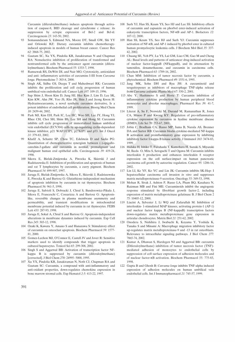
A RCurcumin (diferuloylmethane) induces apoptosis through activa-tion of caspase-8, BID cleavage and cytochrome c release: itssuppression by ectopic expression of Bel-2 and Bcl-xl.Carcinogenesis 23: 143-50, 2002.
93 Somasundaram S, Edmund NA, Moore DT, Small GW, Shi YYand Orlowski RZ: Dietary curcumin inhibits chemotherapy-induced apoptosis in models of human breast cancer. Cancer Res62: 3868-75, 2002.
94 Gautam SC, Xu YX, Pindolia KR, Janakiraman N and ChapmanRA: Nonselective inhibition of proliferation of transformed andnontransformed cells by the anticancer agent curcumin (diferu-loylmethane). Biochem Pharmacol 55: 1333-7, 1998.
95 Ramsewak RS, DeWitt DL and Nair MG: Cytotoxicity, antioxidantand anti- inflammatory activities of curcumins I-III from Curcumalonga. Phytomedicine 7: 303-8, 2000.
96 Singh AK, Sidhu GS, Deepa T and Maheshwari RK: Curcumininhibits the proliferation and cell cycle progression of humanumbilical vein endothelial cell. Cancer Lett 107: 109-15, 1996.
97 Sup Shim J, Hoon Kim D, Jung HJ, Hee Kim J, Lim D, Lee SK,Kim KW, Ahn JW, Yoo JS, Rho JR, Shin J and Jeong Kwon H:Hydrazinocurcumin, a novel synthetic curcumin derivative, Is apotent inhibitor of endothelial cell proliferation. Bioorg Med Chem10: 2439-44, 2002.
98 Park MJ, Kim EH, Park IC, Lee HC, Woo SH, Lee JY, Hong YJ,Rhee CH, Choi SH, Shim BS, Lee SH and Hong SI: Curcumininhibits cell cycle progression of immortalized human umbilicalvein endothelial (ECV304) cells by up-regulating cyclin-dependentkinase inhibitor, p21 WAF1/CIP1, p27KIP1 and p53. Int J Oncol21: 379-83, 2002.
99 Khafif A, Schantz SP, Chou TC, Edelstein D and Sacks PG:Quantitation of chemopreventive synergism between (-)-epigallo-catechin-3-gallate and curcumin in normal, premalignant andmalignant human oral epithelial cells. Carcinogenesis 19: 419-24,1998.
100 Sikora E, Bielak-Zmijewska A, Piwocka K, Skierski J andRadziszewska E: Inhibition of proliferation and apoptosis of humanand rat T lymphocytes by curcumin, a curry pigment. BiochemPharmacol 54: 899-907, 1997.
101 Jaruga E, Bielak-Zmijewska A, Sikora E, Skierski J, RadziszewskaE, Piwocka K and Bartosz G: Glutathione-independent mechanismof apoptosis inhibition by curcumin in rat thymocytes. BiochemPharmacol 56: 961-5, 1998.
102 Jaruga E, Salvioli S, Dobrucki J, Chrul S, Bandorowicz-Pikula J,Sikora E, Franceschi C, Cossarizza A and Bartosz G: Apoptosis-like, reversible changes in plasma membrane asymmetry andpermeability, and transient modifications in mitochondrialmembrane potential induced by curcumin in rat thymocytes. FEBSLett 433: 287-93, 1998.
103 Jaruga E, Sokal A, Chrul S and Bartosz G: Apoptosis-independentalterations in membrane dynamics induced by curcumin. Exp CellRes 245: 303-12, 1998.
104 Ozaki K, Kawata Y, Amano S and Hanazawa S: Stimulatory effectof curcumin on osteoclast apoptosis. Biochem Pharmacol 59: 1577-81, 2000.
105 Gomez-Lechon MJ, O'Connor E, Castell JV and Jover R: Sensitivemarkers used to identify compounds that trigger apoptosis incultured hepatocytes. Toxicol Sci 65: 299-308, 2002.
106 Singh S and Aggarwal BB: Activation of transcription factor NF-kappa B is suppressed by curcumin (diferuloylmethane)[corrected]. J Biol Chem 270: 24995- 5000, 1995.
107 Xu YX, Pindolia KR, Janakiraman N, Noth CJ, Chapman RA andGautam SC: Curcumin, a compound with anti-inflammatory andanti-oxidant properties, down-regulates chemokine expression inbone marrow stromal cells. Exp Hematol 2-5: 413-22, 1997.
108 Surh YJ, Han SS, Keum YS, Seo HJ and Lee SS: Inhibitory effectsof curcumin and capsaicin on phorbol ester-induced activation ofeukaryotic transcription factors, NF-κB and AP-1. Biofactors 12:107-12, 2000.
109 Han SS, Keum YS, Seo HJ and Surh YJ: Curcumin suppressesactivation of NF-κB, and AP-1 induced by phorbol ester in culturedhuman promyelocytic leukemia cells. J Biochem Mol Biol 35: 337-42, 2002.
110 Chuang SE, Yeh PY, Lu YS, Lai GM, Liao CM, Gao M and ChengAL: Basal levels and patterns of anticancer drug-induced activationof nuclear factor-kappaB (NFkappaB), and its attenuation bytamoxifen, dexamethasone, and curcumin in carcinoma cells.Biochem Pharmacol 63: 1709-16, 2002.
111 Chan MM: Inhibition of tumor necrosis factor by curcumin, aphytochemical. Biochem Pharmacol 49: 1551-6, 1995.
112 Jang MK, Sohn DH and Ryu JH: A curcuminoid andsesquiterpenes as inhibitors of macrophage TNF-alpha releasefrom Curcuma zedoaria. Planta Med 67: 550-2, 2001.
113 Abe Y, Hashimoto S and Horie T: Curcumin inhibition ofinflammatory cytokine production by human peripheral bloodmonocytes and alveolar macrophages. Pharmacol Res 39: 41-7,1999.
114 Literat A, Su F, Norwicki M, Durand M, Ramanathan R, JonesCA, Minoo P and Kwong KY: Regulation of pro-inflammatorycytokine expression by curcumin in hyaline membrane disease(HMD). Life Sci 70: 253-67, 2001.
115 Jobin C, Bradham CA, Russo MP, Juma B, Narula AS, BrennerDA and Sartor RB: Curcumin blocks cytokine-mediated NF-kappaB activation and proinflammatory gene expression by inhibitinginhibitory factor I-kappa B kinase activity. J Immunol 163: 3474-83,1999.
116 Hidaka H, Ishiko T, Furuhashi T, Kamohara H, Suzuki S, MiyazakiM, Ikeda O, Mita S, Setoguchi T and Ogawa M: Curcumin inhibitsinterleukin 8 production and enhances interleukin 8 receptorexpression on the cell surface:impact on human pancreaticcarcinoma cell growth by autocrine regulation. Cancer 95: 1206-14,2002.
117 Lin LI, Ke YF, Ko YC and Lin JK: Curcumin inhibits SK-Hep- Ihepatocellular carcinoma cell invasion in vitro and suppressesmatrix metalloproteinase-9 secretion. Oncology 55: 349-53, 1998.
118 Mohan R, Sivak J, Ashton P, Russo LA, Pham BQ, Kasahara N,Raizman MB and Fini ME: Curcuminoids inhibit the angiogenicresponse stimulated by fibroblast growth factor-2, includingexpression of matrix metalloproteinase gelatinase B. J Biol Chem 275: 10405-12, 2000.
119 Liacini A, Sylvester J, Li WQ and Zafarullah M: Inhibition ofinterleukin- 1-stimulated MAP kinases, activating protein-1 (AP-1)and nuclear factor kappa B (NF-kappaB) transcription factorsdown-regulates matrix metalloproteinase gene expression inarticular chondrocytes. Matrix Biol 21: 251-62, 2002.
120 Onodera S, Nishihira J, Iwabuchi K, Koyama Y, Yoshida K,Tanaka S and Minami A: Macrophage migration inhibitory factorup-regulates matrix metalloproteinase-9 and -13 in rat osteoblasts.Relevance to intracellular signaling pathways. J Biol Chem 277:7865-74, 2002.
121 Kumar A, Dhawan S, Hardegen NJ and Aggarwal BB: curcumin(Diferuloylmethane) inhibition of tumor necrosis factor (TNF)-mediated adhesion of monocytes to endothelial cells bysuppression of cell surface expression of adhesion molecules andof nuclear factor-κB activation. Biochem Pharmacol 55: 775-83,1998.
122 Gupta B and Ghosh B: Curcuma longa inhibits TNF-alpha inducedexpression of adhesion molecules on human umbilical veinendothelial cells. Int J Immunopharmacol 21: 745-57, 1999.
391
Aggarwal et al: Anticancer Potential of Curcumin (Review)
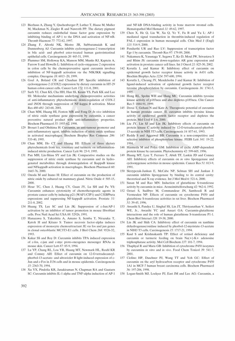
A R123 Bierhaus A, Zhang Y, Quehenberger P, Luther T, Haase M, Muller
M, Mackman N, Ziegler R and Nawroth PP: The dietary pigmentcurcumin reduces endothelial tissue factor gene expression byinhibiting binding of AP-1 to the DNA and activation of NF-κB.Thromb Haemost 77: 772-82, 1997.
124 Zhang F, Altorki NK, Mestre JR, Subbaramaiah K andDannenberg AJ: Curcumin inhibits cyclooxygenase-2 transcriptionin bile acid- and phorbol ester-treated human gastrointestinalepithelial cells. Carcinogenesis 20: 445-51, 1999.
125 Plummer SM, Holloway KA, Manson MM, Munks RJ, Kaptein A,Farrow S and Howells L: Inhibition of cyclo-oxygenase 2 expressionin colon cells by the chemopreventive agent curcumin involvesinhibition of NF-kappaB activation via the NIK/IKK signallingcomplex. Oncogene 18: 6013 -20, 1999.
126 Goel A, Boland CR and Chauhan DP: Specific inhibition ofcyclooxygenase-2 (COX2) expression by dietary curcumin in HT-29human colon cancer cells. Cancer Lett 172: 111-8, 2001.
127 Surh YJ, Chun KS, Cha HH, Han SS, Keum YS, Park KK and LeeSS: Molecular mechanisms underlying chemopreventive activitiesof anti-inflammatory phytochemicals: down-regulation of COX-2and iNOS through suppression of NF-kappa B activation. MutatRes 480-481: 243-68, 2001.
128 Chan MM, Huang HI, Fenton MR and Fong D: In vivo inhibitionof nitric oxide synthase gene expression by curcumin, a cancerpreventive natural product with anti-inflammatory properties.Biochem Pharmacol 55: 1955-62, 1998.
129 Brouet I and Ohshima H: Curcumin, an anti-tumour promoter andanti-inflammatory agent, inhibits induction of nitric oxide synthasein activated macrophages. Biochem Biophys Res Commun 206:533-40, 1995.
130 Chan MM, Ho CT and Huang HI: Effects of three dietaryphytochemicals from tea, rosemary and turmeric on inflammation-induced nitrite production. Cancer Lett 96: 23-9, 1995.
131 Pan MH, Lin-Shiau SY and Lin JK: Comparative studies on thesuppression of nitric oxide synthase by curcumin and its hydro-genated metabolites through downregulation of IkappaB kinaseand NFkappaB activation in macrophages. Biochem Pharmacol 60:1665-76, 2000.
132 Onoda M and Inano H: Effect of curcumin on the production ofnitric oxide by cultured rat mammary gland. Nitric Oxide 4: 505-15,2000.
133 Hour TC, Chen J, Huang CY, Guan JY, Lu SH and Pu YS:Curcumin enhances cytotoxicity of chemotherapeutic agents inprostate cancer cells by inducing p21 (WAF1/CIP1) and C/EBPbetaexpressions and suppressing NF-kappaB activation. Prostate 51:211-8, 2002.
134 Huang TS, Lee SC and Lin JK: Suppression of c-Jun/AP-1activation by an inhibitor of tumor promotion in mouse fibroblastcells. Proc Natl Acad Sci USA 88: 52926, 1991.
135 Hanazawa S, Takeshita A, Amano S, Semba T, Nirazuka T,Katoh H and Kitano S: Tumor necrosis factor-alpha inducesexpression of monocyte chemoattractant JE via fos and jun genesin clonal osteoblastic MC3T3-E1 cells. J Biol Chem 268: 9526-32,1993.
136 Kakar SS and Roy D: Curcumin inhibits TPA induced expressionof c-fos, c-jun and c-myc proto-oncogenes messenger RNAs inmouse skin. Cancer Lett 87: 85-9, 1994.
137 Lu YP, Chang RL, Lou YR, Huang MT, Newmark HL, Reuhl KRand Conney AH: Effect of curcumin on 12-O-tetradecanoyl-phorbol-13-acetate- and ultraviolet B light-induced expression of c-Jun and c-Fos in J136 cells and in mouse epidermis. Carcinogenesis15: 2363-70, 1994.
138 Xu YX, Pindolia KR, Janakiraman N, Chapman RA and GautarnSC: Curcumin inhibits IL-1 alpha and TNF-alpha induction of AP-1
and NF-kB DNA-binding activity in bone marrow stromal cells.Hematopathol Mol Hematol 11: 49-62, 1997.
139 Chen X, He Q, Liu W, Xu Q, Ye Y, Fu B and Yu L: AP-1mediated signal transduction in thrombin-induced regulation ofPAL-1 expression in human mesangial cells. Chin Med J (Engl)113: 514-9, 2000.
140 Pendurthi UR and Rao LV: Suppression of transcription factorEgr-1 by curcumin. Thromb Res 97: 179-89, 2000.
141 Nakamura K, Yasunaga Y, Segawa T, Ko D, Moul JW, Srivastava Sand Rhim JS: curcumin down-regulates AR gene expression andactivation in prostate cancer cell lines. Int J Oncol 21: 825-30, 2002.
142 Korutla L and Kumar R: Inhibitory effect of curcumin onepidermal growth factor receptor kinase activity in A431 cells.Biochim Biophys Acta 1224: 597-600, 1994.
143 Korutla L, Cheung JY, Mendelsohn J and Kumar R: Inhibition ofligand-induced activation of epidermal growth factor receptortyrosine phosphorylation by curcumin. Carcinogenesis 16: 1741-5,1995.
144 Hong RL, Spohn WH and Hung MC: Curcumin inhibits tyrosinekinase activity of p185neu and also depletes p185neu. Clin CancerRes 5: 1884-91, 1999.
145 Dorai T, Gehani N and Katz A: Therapeutic potential of curcuminin human prostate cancer. II. curcumin inhibits tyrosine kinaseactivity of epidermal growth factor receptor and depletes theprotein. Mol Urol 4: 1-6, 2000.
146 Liu JY, Lin SJ and Lin JK: Inhibitory effects of curcumin onprotein kinase C activity induced by 12-O-tetradecanoyl-phorbol-13-acetate in NIH 3T3 cells. Carcinogenesis 14: 857-61, 1993.
147 Reddy S and Aggarwal BB: Curcumin is a non-competitive andselective inhibitor of phosphorylase kinase. FEBS Lett 341: 19-22,1994.
148 Hasmeda M and Polya GM: Inhibition of cyclic AMP-dependentprotein kinase by curcumin. Phytochemistry 42: 599-605, 1996.
149 Huang MT, Lysz T, Ferraro T, Abidi TF, Laskin JD and ConneyAH: Inhibitory effects of curcumin on in vitro lipoxygenase andcyclooxygenase activities in mouse epidermis. Cancer Res 51: 813-9,1991.
150 Skrzypczak-Jankun E, McCabe NP, Selman SH and Jankun J:curcumin inhibits lipoxygenase by binding to its central cavity:theoretical and X-ray evidence. Int J Mol Med 6: 521-6, 2000.
151 Susan M and Rao MN: Induction of glutathione S-transferaseactivity by curcumin in mice. Arzneimittelforschung 42: 962-4, 1992.
152 Oetari S, Sudibyo M, Commandeur JN, Samhoedi R andVermeulen NP: Effects of curcumin on cytochrome P450 andglutathione S-transferase activities in rat liver. Biochem Pharmacol51: 39-45, 1996.
153 Awasthi S, Pandya U, Singhal SS, Lin JT, Thiviyanathan V, SeifertWE, Jr., Awasthi YC and Ansari GA: Curcumin-glutathioneinteractions and the role of human glutathione S-transferase P1-1.Chem Biol Interact 128: 19-38, 2000.
154 Lin JK and Shih CA: Inhibitory effect of curcumin on xanthinedehydrogenase/oxidase induced by phorbol-12-myristate-13-acetatein NIH3 T3 cells. Carcinogenesis 15: 1717-21, 1994.
155 Kaul S and Krishnakanth TP: Effect of retinol deficiency andcurcumin or turmeric feeding on brain Na(+)-K+ adenosinetriphosphatase activity. Mol Cell Biochem 137: 101-7, 1994.
156 Thaphyal R and Maru GB: Inhibition of cytochrome P450 isozymesby curcumins in vitro and in vivo. Food Chem Toxicol 39: 541-7,2001.
157 Ciolino HP, Daschner PJ, Wang TT and Yeh GC: Effect ofcurcumin on the aryl hydrocarbon receptor and cytochrome P4501A1 in MCF-7 human breast carcinoma cells. Biochem Pharmacol56: 197-206, 1998.
158 Logan-Smith MJ, Lockyer PJ, East JM and Lee AG: Curcumin, a
392
ANTICANCER RESEARCH 23: 363-398 (2003)
A R
molecule that inhibits the Ca2+-ATPase of sarcoplasmic reticulumbut increases the rate of accumulation of Ca2+. J Biol Chem 276:46905-11, 2001.
159 Logan-Smith MJ, East JM and Lee AG: Evidence for a globalinhibitor-induced conformation change on the Ca(2+)-ATPase ofsarcoplasmic reticulum, from paired inhibitor studies. Biochemistry41: 2869-75, 2002.
160 Bilmen JG, Khan SZ, Javed MH and Michelangeli F: Inhibition ofthe SERCA Ca2+ pumps by curcumin. Curcumin putativelystabilizes the interaction between the nucleotide-binding andphosphorylation domains in the absence of ATP. Eur J Biochem268: 6318-27, 2001.
161 Sumbilla C, Lewis D, Hammerschmidt T and Inesi G: The slippageof the Ca2+ pump and its control by anions and curcumin inskeletal and cardiac sarcoplasmic reticulum. J Biol Chem 277:13900-6, 2002.
162 Yamamoto H, Hanada K, Kawasaki K and Nishijima M: Inhibitoryeffect on curcumin on mammalian phospholipase D activity. FEBSLett 417: 196-8, 1997.
163 Kang BY, Song YJ, Kim KM, Choe YK, Hwang SY and Kim TS:Curcumin inhibits Thl cytokine profile in CD4+ T cells bysuppressing interleukin-12 production in macrophages. Br JPharmacol 128: 380-4, 1999.
164 Kang BY, Chung SW, Chung W, Im S, Hwang SY and Kim TS:Inhibition of interleukin-12 production in lipopolysaccharide-activated macrophages by curcumin. Eur J Pharmacol 384: 191-5,1999.
165 Santibanez JF, Quintanilla M and Martinez J: Genistein andcurcumin block TGF-beta 1-induced u-PA expression andmigratory and invasive phenotype in mouse epidermal kera-tinocytes. Nutr Cancer 37: 49-54, 2000.
166 Fenton JI, Wolff MS, Orth MW and Hord NG: Membrane-typematrix metalloproteinases mediate curcumin-induced cell migra-tion in non-tumorigenic colon epithelial cells differing in Apcgenotype. Carcinogenesis 23: 1065-70, 2002.
167 Motterlini R, Foresti R, Bassi R and Green CJ: Curcumin, anantioxidant and antiinflammatory agent, induces heme oxygenase-1and protects endothelial cells against oxidative stress. Free RadicBiol Med 28: 1303-12, 2000.
168 Scapagnini G, Foresti R, Calabrese V, Giuffrida Stella AM, GreenCJ and Motterlini R: Caffeic acid phenethyl ester and curcumin: anovel class of heme oxygenase-1 inducers. Mol Pharmacol 61: 554-61, 2002.
169 Romiti N, Tongiani R, Cervelli F and Chieli E: Effects of curcuminon P-glycoprotein in primary cultures of rat hepatocytes. Life Sci62: 2349-58, 1998.
170 Snyder RD and Arnone MR: Putative identification of functionalinteractions between DNA intercalating agents and topoisomeraseII using the V79 in vitro micronucleus assay. Mutat Res 503: 21-35,2002.
171 Kato K, Ito H, Kamei K and Iwamoto I: Stimulation of the stress-induced expression of stress proteins by curcumin in cultured cellsand in rat tissues in vivo. Cell Stress Chaperones 3: 152-60, 1998.
172 Chen X, Hasuma T, Yano Y, Yoshimata T, Morishima Y, Wang Yand Otani S: Inhibition of farnesyl protein transferase by mono-terpene, curcumin derivatives and gallotannin. Anticancer Res 17:2555-64, 1997.
173 Siebenlist U, Franzoso G and Brown K: Structure, regulation andfunction of NFkappa B. Annu Rev Cell Biol 10: 405-55, 1994.
174 Baeuerle PA and Henkel T: Function and activation of NF-kappaB in the immune system. Annu Rev Immunol 12: 141-79, 1994.
175 Grilli M, Chiu JJ and Lenardo MJ: NF-kappa B and Rel:participants in a multiform transcriptional regulatory system. IntRev Cytol 143: 1-62, 1993.
176 Baeuerle PA: The inducible transcription activator NF-kappa B:regulation by distinct protein subunits. Biochim Biophys Acta 1072:63-80, 1991.
177 Brennan P and O'Neill LA: Inhibition of nuclear factor kappaB bydirect modification in whole cells--mechanism of action ofnordihydroguaiaritic acid, curcumin and thiol modifiers. BiochemPharmacol 55: 965-73, 1998.
178 Ishii H, Takada K, Higuchi T and Sugiyama J: Verotoxin-1 inducestissue factor expression in human umbilical vein endothelial cellsthrough activation of NFkappaB/Rel and AP-1. Thromb Haemost84: 712-21, 2000.
179 Anto RJ, Maliekal TT and Karunagaran D: L-929 cells harboringectopically expressed ReIA resist curcumin-induced apoptosis. JBiol Chem 275: 15601-4, 2000.
180 Bharti AC, Donato N, Singh S and Aggarwal BB: Curcumin(diferuloylmethane) downregulates the constitutive activation ofnuclear factor-kappaB and I kappa B alpha kinase in humanmultiple myeloma cells leading to suppression of proliferation andinduction of apoptosis. Blood (in press).
181 Aggarwal BB: Apoptosis and transcription factor NF-kB: a tale ofassociation and dissociation. Biochem Pharmacol 60: 1033-1039,2000.
182 Shishodia S and Aggrwal BB: Nuclear factor-kappa B activation: aquestion of life and death. J Biochem Mol Biol 35: 28-40, 2002.
183 Karunagaran D and Aggarwal BB: Transcription factors as targetsfor the drug development. In: Keri G, Toth I, eds. MolecularPathomechanisms and New Trends in Drug Development. NewYork: Harwood Academic Publisher, 2002: 76-91.
184 Dhar A, Young MR, Colburn NH: The role of AP-1, NF-kappaBand ROS/NOS in skin carcinogenesis: the J136 model is predictive.Mol Cell Biochem 234235: 185-93, 2002.
185 Shaulian E and Karin M: AP-1 as a regulator of cell life and death.Nat Cell Biol 4: E 131-6, 2002.
186 Hsu TC, Young MR, Cmarik J and Colburn NH: Activatorprotein 1 (AP-1)- and nuclear factor kappaB (NF-kappaB)-dependent transcriptional events in carcinogenesis. Free Radic BiolMed 28: 1338-48, 2000.
187 Dong Z, Lavrovsky V and Colburn NH: Transformation reversioninduced in J136 RT101 cells by AP-1 inhibitors. Carcinogenesis 16:749-56, 1995.
188 Chen YR, Zhou G and Tan TH: c-Jun N-terminal kinase mediatesapoptotic signaling induced by N-(4-hydroxyphenyl)retinamide.Mol Pharmacol 56: 1271-9, 1999.
189 Hahm ER, Cheon G, Lee J, Kim B, Park C and Yang CH: New andknown symmetrical curcumin derivatives inhibit the formation ofFos-Jun-DNA complex. Cancer Lett 184: 89-96, 2002.
190 Ammon HP, Safayhi H, Mack T and Sabieraj J: Mechanism ofantiinflammatory actions of curcumin and boswellic acids. JEthnopharmacol 38: 113-9, 1993.
191 Sherr CJ: D-type cyclins. Trends Biochem Sci 20: 187-90, 1995. 192 Baldin V, Lukas J, Marcote MJ, Pagano M and Draetta G: Cyclin
D1 is a nuclear protein required for cell cycle progression in G1.Genes Dev 7: 812-21, 1993.
193 Bartkova J, Lukas J, Strauss M and Bartek J: The PRAD-1/cyclinD1 oncogene product accumulates aberrantly in a subset ofcolorectal carcinomas. Int J Cancer 58: 568-73, 1994.
194 Bartkova J, Lukas J, Muller H, Lutzhoft D, Strauss M and Bartek J:Cyclin D1 protein expression and function in human breast cancer.Int J Cancer 57: 353-61, 1994.
195 Gillett C, Fantl V, Smith R, Fisher C, Bartek J, Dickson C, BarnesD and Peters G: Amplification and overexpression of cyclin D1 inbreast cancer detected by immunohistochemical staining. CancerRes 54: 1812-7, 1994.
196 Nishida N, Fukuda Y, Komeda T, Kita R, Sando T, Furukawa
393
Aggarwal et al: Anticancer Potential of Curcumin (Review)
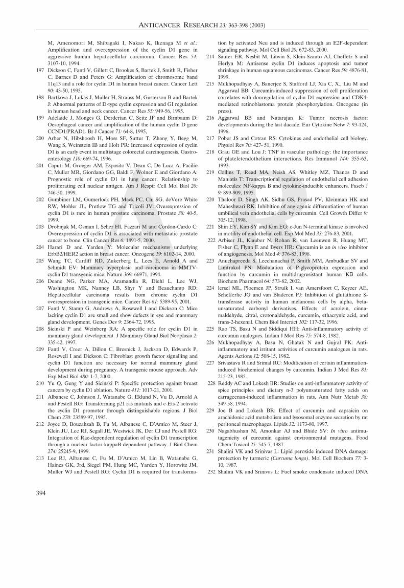
A R
M, Amenomori M, Shibagaki I, Nakao K, Ikenaga M et al.:Amplification and overexpression of the cyclin D1 gene inaggressive human hepatocellular carcinoma. Cancer Res 54:3107-10, 1994.
197 Dickson C, Fantl V, Gillett C, Brookes S, Bartek J, Smith R, FisherC, Barnes D and Peters G: Amplification of chromosome band11q13 and a role for cyclin D1 in human breast cancer. Cancer Lett90: 43-50, 1995.
198 Bartkova J, Lukas J, Muller H, Strauss M, Gusterson B and BartekJ: Abnormal patterns of D-type cyclin expression and GI regulationin human head and neck cancer. Cancer Res 55: 949-56, 1995.
199 Adelaide J, Monges G, Derderian C, Seitz JF and Birnbaum D:Oesophageal cancer and amplification of the human cyclin D geneCCND1/PRAD1. Br J Cancer 71: 64-8, 1995,
200 Arber N, Hibshoosh H, Moss SF, Sutter T, Zhang Y, Begg M,Wang S, Weinstein IB and Holt PR: Increased expression of cyclinD1 is an early event in multistage colorectal carcinogenesis. Gastro-enterology 110: 669-74, 1996.
201 Caputi M, Groeger AM, Esposito V, Dean C, De Luca A, PacilioC, Muller MR, Giordano GG, Baldi F, Wolner E and Giordano A:Prognostic role of cyclin D1 in lung cancer. Relationship toproliferating cell nuclear antigen. Am J Respir Cell Mol Biol 20:746-50, 1999.
202 Gumbiner LM, Gumerlock PH, Mack PC, Chi SG, deVere WhiteRW, Mohler JL, Pretlow TG and Tricoli JV: Overexpression ofcyclin D1 is rare in human prostate carcinoma. Prostate 38: 40-5,1999.
203 Drobnjak M, Osman I, Scher HI, Fazzari M and Cordon-Cardo C:Overexpression of cyclin D1 is associated with metastatic prostatecancer to bone. Clin Cancer Res 6: 1891-5, 2000.
204 Harari D and Yarden Y: Molecular mechanisms underlyingErbB2/HER2 action in breast cancer. Oncogene 19: 6102-14, 2000.
205 Wang TC, Cardiff RD, Zukerberg L, Lees E, Arnold A andSchmidt EV: Mammary hyperplasia and carcinoma in MMTV-cyclin D1 transgenic mice. Nature 369: 66971, 1994.
206 Deane NG, Parker MA, Aramandla R, Diehl L, Lee WJ,Washington MK, Nanney LB, Shyr Y and Beauchamp RD:Hepatocellular carcinoma results from chronic cyclin D1overexpression in transgenic mice. Cancer Res 61: 5389-95, 2001.
207 Fantl V, Stamp G, Andrews A, Rosewell I and Dickson C: Micelacking cyclin D1 are small and show defects in eye and mammarygland development. Genes Dev 9: 2364-72, 1995.
208 Sicinski P and Weinberg RA: A specific role for cyclin D1 inmammary gland development. J Mammary Gland Biol Neoplasia 2:335-42, 1997.
209 Fantl V, Creer A, Dillon C, Bresnick J, Jackson D, Edwards P,Rosewell I and Dickson C: Fibroblast growth factor signalling andcyclin D1 function are necessary for normal mammary glanddevelopment during pregnancy. A transgenic mouse approach. AdvExp Med Biol 480: 1-7, 2000.
210 Yu Q, Geng Y and Sicinski P: Specific protection against breastcancers by cyclin D1 ablation. Nature 411: 1017-21, 2001.
211 Albanese C, Johnson J, Watanabe G, Eklund N, Vu D, Arnold Aand Pestell RG: Transforming p21 ras mutants and c-Ets-2 activatethe cyclin D1 promoter through distinguishable regions. J BiolChem 270: 23589-97, 1995.
212 Joyce D, Bouzahzah B, Fu M, Albanese C, D'Amico M, Steer J,Klein JU, Lee RJ, Segall JE, Westwick JK, Der CJ and Pestell RG:Integration of Rac-dependent regulation of cyclin D1 transcriptionthrough a nuclear factor-kappaB-dependent pathway. J Biol Chem274: 25245-9, 1999.
213 Lee RJ, Albanese C, Fu M, D'Amico M, Lin B, Watanabe G,Haines GK, 3rd, Siegel PM, Hung MC, Yarden Y, Horowitz JM,Muller WJ and Pestell RG: Cyclin D1 is required for transforma-
tion by activated Neu and is induced through an E2F-dependentsignaling pathway. Mol Cell Biol 20: 672-83, 2000.
214 Sauter ER, Nesbit M, Litwin S, Klein-Szanto AJ, Cheffetz S andHerlyn M: Antisense cyclin D1 induces apoptosis and tumorshrinkage in human squamous carcinomas. Cancer Res 59: 4876-81,1999.
215 Mukhopadhyay A, Banerjee S, Stafford LJ, Xia C, X., Liu M andAggarwal BB: Curcumin-induced suppression of cell proliferationcorrelates with donregulation of cyclin D1 expression and CDK4-mediated retinoblastoma protein phosphorylation. Oncogene (inpress).
216 Aggarwal BB and Natarajan K: Tumor necrosis factor:developments during the last dacade. Eur Cytokine Netw 7: 93-124,1996.
217 Pober JS and Cotran RS: Cytokines and endothelial cell biology.Physiol Rev 70: 427- 51, 1990.
218 Grau GE and Lou J: TNF in vascular pathology: the importanceof plateletendothelium interactions. Res Immunol 144: 355-63,1993.
219 Collins T, Read MA, Neish AS, Whitley MZ, Thanos D andManiatis T: Transcriptional regulation of endothelial cell adhesionmolecules: NF-kappa B and cytokine-inducible enhancers. Faseb J9: 899-909, 1995.
220 Thaloor D, Singh AK, Sidhu GS, Prasad PV, Kleinman HK andMaheshwari RK: Inhibition of angiogenic differentiation of humanumbilical vein endothelial cells by curcumin. Cell Growth Differ 9:305-12, 1998.
221 Shin EY, Kim SY and Kim EG: c-Jun N-terminal kinase is involvedin motility of endothelial cell. Exp Mol Med 33: 276-83, 2001.
222 Arbiser JL, Klauber N, Rohan R, van Leeuwen R, Huang MT,Fisher C, Flynn E and Byers HR: Curcumin is an in vivo inhibitorof angiogenesis. Mol Med 4: 376-83, 1998.
223 Anuchapreeda S, Leechanachai P, Smith MM, Ambudkar SV andLimtrakul PN: Modulation of P-glycoprotein expression andfunction by curcumin in multidrugresistant human KB cells.Biochem Pharmacol 64: 573-82, 2002.
224 lersel ML, Ploemen JP, Struik I, van Amersfoort C, Keyzer AE,Schefferlie JG and van Bladeren PJ: Inhibition of glutathione S-transferase activity in human melanoma cells by alpha, beta-unsaturated carbonyl derivatives. Effects of acrolein, cinna-maldehyde, citral, crotonaldehyde, curcumin, ethacrynic acid, andtrans-2-hexenal. Chem Biol Interact 102: 117-32, 1996.
225 Rao TS, Basu N and Siddiqui HH: Anti-inflammatory activity ofcurcumin analogues. Indian J Med Res 75: 574-8, 1982.
226 Mukhopadhyay A, Basu N, Ghatak N and Gujral PK: Anti-inflammatory and irritant activities of curcumin analogues in rats.Agents Actions 12: 508-15, 1982.
227 Srivastava R and Srimal RC: Modification of certain inflammation-induced biochemical changes by curcumin. Indian J Med Res 81:215-23, 1985.
228 Reddy AC and Lokesh BR: Studies on anti-inflammatory activity ofspice principles and dietary n-3 polyunsaturated fatty acids oncarrageenan-induced inflammation in rats. Ann Nutr Metab 38:349-58, 1994.
229 Joe B and Lokesh BR: Effect of curcumin and capsaicin onarachidonic acid metabolism and lysosomal enzyme secretion by ratperitoneal macrophages. Lipids 32: 1173-80, 1997.
230 Nagabhushan M, Amonkar AJ and Bhide SV: In vitro antimu-tagenicity of curcumin against environmental mutagens. FoodChem Toxicol 25: 545-7, 1987.
231 Shalini VK and Srinivas L: Lipid peroxide induced DNA damage:protection by turmeric (Curcuma longa). Mol Cell Biochem 77: 3-10, 1987.
232 Shalini VK and Srinivas L: Fuel smoke condensate induced DNA
394
ANTICANCER RESEARCH 23: 363-398 (2003)
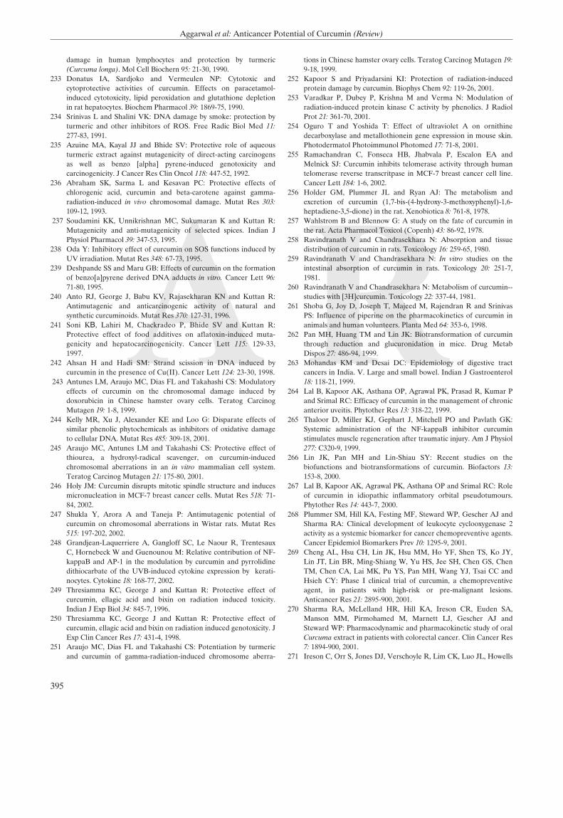
A R
damage in human lymphocytes and protection by turmeric(Curcuma longa). Mol Cell Biochern 95: 21-30, 1990.
233 Donatus IA, Sardjoko and Vermeulen NP: Cytotoxic andcytoprotective activities of curcumin. Effects on paracetamol-induced cytotoxicity, lipid peroxidation and glutathione depletionin rat hepatocytes. Biochem Pharmacol 39: 1869-75, 1990.
234 Srinivas L and Shalini VK: DNA damage by smoke: protection byturmeric and other inhibitors of ROS. Free Radic Biol Med 11:277-83, 1991.
235 Azuine MA, Kayal JJ and Bhide SV: Protective role of aqueousturmeric extract against mutagenicity of direct-acting carcinogensas well as benzo [alpha] pyrene-induced genotoxicity andcarcinogenicity. J Cancer Res Clin Oncol 118: 447-52, 1992.
236 Abraham SK, Sarma L and Kesavan PC: Protective effects ofchlorogenic acid, curcumin and beta-carotene against gamma-radiation-induced in vivo chromosomal damage. Mutat Res 303:109-12, 1993.
237 Soudamini KK, Unnikrishnan MC, Sukumaran K and Kuttan R:Mutagenicity and anti-mutagenicity of selected spices. Indian JPhysiol Pharmacol 39: 347-53, 1995.
238 Oda Y: Inhibitory effect of curcumin on SOS functions induced byUV irradiation. Mutat Res 348: 67-73, 1995.
239 Deshpande SS and Maru GB: Effects of curcumin on the formationof benzo[a]pyrene derived DNA adducts in vitro. Cancer Lett 96:71-80, 1995.
240 Anto RJ, George J, Babu KV, Rajasekharan KN and Kuttan R:Antimutagenic and anticarcinogenic activity of natural andsynthetic curcuminoids. Mutat Res 370: 127-31, 1996.
241 Soni KB, Lahiri M, Chackradeo P, Bhide SV and Kuttan R:Protective effect of food additives on aflatoxin-induced muta-genicity and hepatocarcinogenicity. Cancer Lett 115: 129-33,1997.
242 Ahsan H and Hadi SM: Strand scission in DNA induced bycurcumin in the presence of Cu(II). Cancer Lett 124: 23-30, 1998.
243 Antunes LM, Araujo MC, Dias FL and Takahashi CS: Modulatoryeffects of curcumin on the chromosomal damage induced bydoxorubicin in Chinese hamster ovary cells. Teratog CarcinogMutagen 19: 1-8, 1999.
244 Kelly MR, Xu J, Alexander KE and Loo G: Disparate effects ofsimilar phenolic phytochemicals as inhibitors of oxidative damageto cellular DNA. Mutat Res 485: 309-18, 2001.
245 Araujo MC, Antunes LM and Takahashi CS: Protective effect ofthiourea, a hydroxyl-radical scavenger, on curcumin-inducedchromosomal aberrations in an in vitro mammalian cell system.Teratog Carcinog Mutagen 21: 175-80, 2001.
246 Holy JM: Curcumin disrupts mitotic spindle structure and inducesmicronucleation in MCF-7 breast cancer cells. Mutat Res 518: 71-84, 2002.
247 Shukla Y, Arora A and Taneja P: Antimutagenic potential ofcurcumin on chromosomal aberrations in Wistar rats. Mutat Res515: 197-202, 2002.
248 Grandjean-Laquerriere A, Gangloff SC, Le Naour R, TrentesauxC, Hornebeck W and Guenounou M: Relative contribution of NF-kappaB and AP-1 in the modulation by curcumin and pyrrolidinedithiocarbate of the UVB-induced cytokine expression by kerati-nocytes. Cytokine 18: 168-77, 2002.
249 Thresiamma KC, George J and Kuttan R: Protective effect ofcurcumin, ellagic acid and bixin on radiation induced toxicity.Indian J Exp Biol 34: 845-7, 1996.
250 Thresiamma KC, George J and Kuttan R: Protective effect ofcurcumin, ellagic acid and bixin on radiation induced genotoxicity. JExp Clin Cancer Res 17: 431-4, 1998.
251 Araujo MC, Dias FL and Takahashi CS: Potentiation by turmericand curcumin of gamma-radiation-induced chromosome aberra-
tions in Chinese hamster ovary cells. Teratog Carcinog Mutagen 19:9-18, 1999.
252 Kapoor S and Priyadarsini KI: Protection of radiation-inducedprotein damage by curcumin. Biophys Chem 92: 119-26, 2001.
253 Varadkar P, Dubey P, Krishna M and Verma N: Modulation ofradiation-induced protein kinase C activity by phenolics. J RadiolProt 21: 361-70, 2001.
254 Oguro T and Yoshida T: Effect of ultraviolet A on ornithinedecarboxylase and metallothionein gene expression in mouse skin.Photodermatol Photoimmunol Photomed 17: 71-8, 2001.
255 Ramachandran C, Fonseca HB, Jhabvala P, Escalon EA andMelnick SJ: Curcumin inhibits telomerase activity through humantelomerase reverse transcritpase in MCF-7 breast cancer cell line.Cancer Lett 184: 1-6, 2002.
256 Holder GM, Plummer JL and Ryan AJ: The metabolism andexcretion of curcumin (1,7-bis-(4-hydroxy-3-methoxyphenyl)-1,6-heptadiene-3,5-dione) in the rat. Xenobiotica 8: 761-8, 1978.
257 Wahlstrom B and Blennow G: A study on the fate of curcumin inthe rat. Acta Pharmacol Toxicol (Copenh) 43: 86-92, 1978.
258 Ravindranath V and Chandrasekhara N: Absorption and tissuedistribution of curcumin in rats. Toxicology 16: 259-65, 1980.
259 Ravindranath V and Chandrasekhara N: In vitro studies on theintestinal absorption of curcumin in rats. Toxicology 20: 251-7,1981.
260 Ravindranath V and Chandrasekhara N: Metabolism of curcumin--studies with [3H]curcumin. Toxicology 22: 337-44, 1981.
261 Shoba G, Joy D, Joseph T, Majeed M, Rajendran R and SrinivasPS: Influence of piperine on the pharmacokinetics of curcumin inanimals and human volunteers. Planta Med 64: 353-6, 1998.
262 Pan MH, Huang TM and Lin JK: Biotransformation of curcuminthrough reduction and glucuronidation in mice. Drug MetabDispos 27: 486-94, 1999.
263 Mohandas KM and Desai DC: Epidemiology of digestive tractcancers in India. V. Large and small bowel. Indian J Gastroenterol18: 118-21, 1999.
264 Lal B, Kapoor AK, Asthana OP, Agrawal PK, Prasad R, Kumar Pand Srimal RC: Efficacy of curcumin in the management of chronicanterior uveitis. Phytother Res 13: 318-22, 1999.
265 Thaloor D, Miller KJ, Gephart J, Mitchell PO and Pavlath GK:Systemic administration of the NF-kappaB inhibitor curcuminstimulates muscle regeneration after traumatic injury. Am J Physiol277: C320-9, 1999.
266 Lin JK, Pan MH and Lin-Shiau SY: Recent studies on thebiofunctions and biotransformations of curcumin. Biofactors 13:153-8, 2000.
267 Lal B, Kapoor AK, Agrawal PK, Asthana OP and Srimal RC: Roleof curcumin in idiopathic inflammatory orbital pseudotumours.Phytother Res 14: 443-7, 2000.
268 Plummer SM, Hill KA, Festing MF, Steward WP, Gescher AJ andSharma RA: Clinical development of leukocyte cyclooxygenase 2activity as a systemic biomarker for cancer chemopreventive agents.Cancer Epidemiol Biomarkers Prev 10: 1295-9, 2001.
269 Cheng AL, Hsu CH, Lin JK, Hsu MM, Ho YF, Shen TS, Ko JY,Lin JT, Lin BR, Ming-Shiang W, Yu HS, Jee SH, Chen GS, ChenTM, Chen CA, Lai MK, Pu YS, Pan MH, Wang YJ, Tsai CC andHsieh CY: Phase I clinical trial of curcumin, a chemopreventiveagent, in patients with high-risk or pre-malignant lesions.Anticancer Res 21: 2895-900, 2001.
270 Sharma RA, McLelland HR, Hill KA, Ireson CR, Euden SA,Manson MM, Pirmohamed M, Marnett LJ, Gescher AJ andSteward WP: Pharmacodynamic and pharmacokinetic study of oralCurcuma extract in patients with colorectal cancer. Clin Cancer Res7: 1894-900, 2001.
271 Ireson C, Orr S, Jones DJ, Verschoyle R, Lim CK, Luo JL, Howells
395
Aggarwal et al: Anticancer Potential of Curcumin (Review)
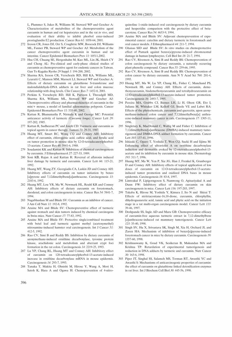
A R
L, Plummer S, Jukes R, Williams M, Steward WP and Gescher A:Characterization of metabolites of the chemopreventive agentcurcumin in human and rat hepatocytes and in the rat in vivo, andevaluation of their ability to inhibit phorbol ester-inducedprostaglandin E2 production. Cancer Res 61: 1058-64, 2001.
272 Ireson CR, Jones DJ, Orr S, Coughtrie MW, Boocock DJ, WilliamsML, Fanner PB, Steward WP and Gescher AJ: Metabolism of thecancer chemopreventive agent curcumin in human and ratintestine. Cancer Epidemiol Biomarkers Prev 11: 10511,2002.
273 Hsu CH, Chuang SE, Hergenhahn M, Kuo ML, Lin JK, Hsieh CYand Cheng AL: Pre-clinical and early-phase clinical studies ofcurcumin as chemopreventive agent for endemic cancers in Taiwan.Gan To Kagaku Ryoho 29 Suppl 1: 194-200, 2002.
274 Sharma RA, Ireson CR, Verschoyle RD, Hill KA, Williams ML,Leuratti C, Manson MM, Marnett LJ, Steward WP and Gescher A:Effects of dietary curcumin on glutathione S-transferase andmalondialdehyde-DNA adducts in rat liver and colon mucosa:relationship with drug levels. Clin Cancer Res 7: 1452-8, 2001.
275 Perkins S, Verschoyle RD, Hill K, Parveen π, Threadgill MD,Sharma RA, Williams ML, Steward WP and Gescher AJ:Chemopreventive efficacy and pharmacokinetics of curcumin in themin/+ mouse, a model of familial adenomatous polyposis. CancerEpiderniol Biomarkers Prev 11: 535-40, 2002.
276 Kuttan R, Bhanumathy P, Nirmala K and George MC: Potentialanticancer activity of turmeric (Curcuma longa). Cancer Lett 29:197-202, 1985.
277 Kuttan R, Sudheeran PC and Josph CD: Turmeric and curcumin astopical agents in cancer therapy. Tumori 73: 29-31, 1987.
278 Huang MT, Smart RC, Wong CQ and Conney AH: Inhibitoryeffect of curcumin, chlorogenic acid, caffeic acid, and ferulic acidon tumor promotion in mouse skin by 12-O-tetradecanoylphorbol-13-acetate. Cancer Res 48: 5941-6, 1988.
279 Soudamini KK and Kuttan R: Inhibition of chemical carcinogenesisby curcumin. J Ethnopharmacol 27: 227-33, 1989.
280 Soni KB, Rajan A and Kuttan R: Reversal of aflatoxin inducedliver damage by turmeric and curcumin. Cancer Lett 66: 115-21,1992.
281 Huang MT, Wang ZY, Georgiadis CA, Laskin JD and Conney AH:Inhibitory effects of curcumin on tumor initiation by benzo[a]pyrene and 7,12dimethylbenz[a]anthracene. Carcinogenesis 13:2183-6, 1992.
282 Huang MT, Lou YR, Ma W, Newmark HL, Reuhl KR and ConneyAH: Inhibitory effects of dietary curcumin on forestomach,duodenal, and colon carcinogenesis in mice. Cancer Res 54: 5841-7,1994.
283 Nagabhushan M and Bhide SV: Curcumin as an inhibitor of cancer.J Am Coll Nutr 11: 192-8, 1992.
284 Azuine MA and Bhide SV: Chemopreventive effect of turmericagainst stomach and skin tumors induced by chemical carcinogensin Swiss mice. Nutr Cancer 17: 77-83, 1992.
285 Azuine MA and Bhide SV: Protective single/combined treatmentwith betel leaf and turmeric against methyl (acetoxymethyl)nitrosamine-induced hamster oral carcinogenesis. Int J Cancer 51:412-5, 1992.
286 Rao CV, Simi B and Reddy BS: Inhibition by dietary curcumin ofazoxymethane-induced ornithine decarboxylase, tyrosine proteinkinase, arachidonic acid metabolism and aberrant crypt fociformation in the rat colon. Carcinogenesis 14: 2219-25, 1993.
287 Lu YP, Chang RL, Huang MT and Conney AH: Inhibitory effectof curcumin on 120-tetradecanoylphorbol-13-acetate-inducedincrease in ornithine decarboxylase mRNA in mouse epidermis.Carcinogenesis 14: 293-7, 1993.
288 Tanaka T, Makita H, Ohnishi M, Hirose Y, Wang A, Mori H,Satoh K, Hara A and Ogawa H: Chemoprevention of 4-nitro-
quinoline 1-oxide-induced oral carcinogenesis by dietary curcuminand hesperidin: comparison with the protective effect of beta-carotene. Cancer Res 54: 4653-9, 1994.
289 Azuine MA and Bhide SV: Adjuvant chemoprevention of expe-rimental cancer: catechin and dietary turmeric in forestomach andoral cancer models. J Ethnopharmacol 44: 211-7, 1994.
290 Ghaisas SID and Bhide SV: In vitro studies on chemoprotectiveeffect of Pumark against benzo(a)pyrene-induced chromosomaldamage in human lymphocytes. Cell Biol Int 18: 21-7, 1994.
291 Rao CV, Rivenson A, Simi B and Reddy BS: Chemoprevention ofcolon carcinogenesis by dietary curcumin, a naturally occurringplant phenolic compound. Cancer Res 55: 259-66, 1995.
292 Rao CV, Rivenson A, Simi B and Reddy BS: Chemoprevention ofcolon cancer by dietary curcumin. Ann N Y Acad Sci 768: 201-4,1995.
293 Huang MT, Ma W, Lu YP, Chang RL, Fisher C, Manchand PS,Newmark HL and Conney AH: Effects of curcumin, deme-thoxycurcumin, bisdemethoxycurcumin and tetrahydrocurcumin on12-O-tetradecanoylphorbol-13-acetate-induced tumor promotion.Carcinogenesis 16: 2493-7, 1995.
294 Pereira MA, Grubbs CJ, Barnes LH, Li H, Olson GR, Eto I,Juliana M, Whitaker LM, Kelloff GJ, Steele VE and Lubet RA:Effects of the phytochemicals, curcumin and quercetin, upon azoxy-methane-induced colon cancer and 7,12dimethylbenz[a] anthra-cene-induced mammary cancer in rats. Carcinogenesis 17: 1305-11,1996.
295 Singletary K, MacDonald C, Wallig M and Fisher C: Inhibition of7,12dimethylbenz[a]anthracene (DMBA)-induced mammary tumo-rigenesis and DMBA-DNA adduct formation by curcumin. CancerLett 103: 137-41, 1996.
296 Ishizaki C, Oguro T, Yoshida T, Wen CQ, Sueki H and Iijima M:Enhancing effect of ultraviolet A on ornithine decarboxylaseinduction and dermatitis evoked by 12-otetradecanoylphorbol-13-acetate and its inhibition by curcumin in mouse skin. Dermatology193: 311-7, 1996.
297 Huang MT, Ma W, Yen P, Xie JG, Han J, Frenkel K, GrunbergerD and Conney AH: Inhibitory effects of topical application of lowdoses of curcumin on 12-O-tetradecanoylphorbol-13-acetate-induced tumor promotion and oxidized DNA bases in mouseepidermis. Carcinogenesis 18: 83-8, 1997.
298 Limtrakul P, Lipigorngoson S, Namwong O, Apisariyakul A andDunn FW: Inhibitory effect of dietary curcumin on skincarcinogenesis in mice. Cancer Lett 116: 197-203, 1997.
299 Takaba K, Hirose M, Yoshida Y, Kimura J, Ito N and Shirai T:Effects of ntritriacontane-16,18-dione, curcumin, chlorphyllin,dihydroguaiaretic acid, tannic acid and phytic acid on the initiationstage in a rat multi-organ carcinogenesis model. Cancer Lett 113:39-46, 1997.
300 Deshpande SS, Ingle AD and Maru GB: Chemopreventive efficacyof curcumin-free aqueous turmeric extract in 7,12-dimethylbenz[a]anthracene-induced rat mammary tumorigenesis. Cancer Lett123: 35-40, 1998.
301 Singh SV, Hu X, Srivastava SK, Singh M, Xia H, Orchard JL andZaren HA: Mechanism of inhibition of benzo[a]pyrene-inducedforestomach cancer in mice by dietary curcumin. Carcinogenesis 19:1357-60, 1998.
302 Krishnaswamy K, Goud VK, Sesikeran B, Mukundan MA andKrishna TP: Retardation of experimental tumorigenesis andreduction in DNA adducts by turmeric and curcumin. Nutr Cancer30: 163-6, 1998.
303 Piper JT, Singhal SS, Salameh MS, Torman RT, Awasthi YC andAwasthi S: Mechanisms of anticarcinogenic properties of curcumin:the effect of curcumin on glutathione linked detoxification enzymesin rat liver. Int J Biochem Cell Biol 30: 445-56, 1998.
396
ANTICANCER RESEARCH 23: 363-398 (2003)
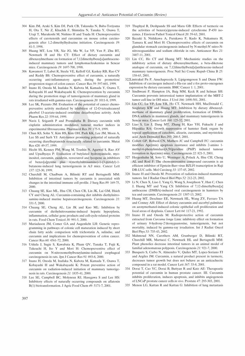
A R
304 Kim JM, Araki S, Kim DJ, Park CB, Takasuka N, Baba-ToriyamaH, Ota T, Nir Z, Khachik F, Shimidzu N, Tanaka Y, Osawa T,Uraji T, Murakoshi M, Nishino H and Tsuda H: Chemopreventiveeffects of carotenoids and curcumins on mouse colon carcino-genesis after 1,2-dimethylhydrazine initiation. Carcinogenesis 19:81-5, 1998.
305 Huang MT, Lou YR, Xie JG, Ma W, Lu YP, Yen P, Zhu BT,Newmark H and Ho CT: Effect of dietary curcumin anddibenzoylmethane on formation of 7,12dimethylbenz[a]anthracene-induced mammary tumors and lymphomas/leukemias in Sencarmice. Carcinogenesis 19: 1697-700, 1998.
306 Kawamori T, Lubet R, Steele VE, Kelloff GJ, Kaskey RB, Rao CVand Reddy BS: Chemopreventive effect of curcumin, a naturallyoccurring anti-inflammatory agent, during the promotion/progression stages of colon cancer. Cancer Res 59: 597-601, 1999.
307 Inano H, Onoda M, Inafuku N, Kubota M, Kamada Y, Osawa T,Kobayashi H and Wakabayashi K: Chemoprevention by curcuminduring the promotion stage of tumorigenesis of mammary gland inrats irradiated with gamma-rays. Carcinogenesis 20: 1011-8, 1999.
308 Lee SK, Pezzuto JM: Evaluation of the potential of cancer chemo-preventive activity mediated by inhibition of 12-O-tetradecanoylphorbol 13-acetate-induced ornithine decarboxylase activity. ArchPharm Res 22: 559-64, 1999.
309 Navis I, Sriganth P and Premalatha B: Dietary curcumin withcisplatin administration modulates tumour marker indices inexperimental fibrosarcoma. Pharmacol Res 39: 175-9, 1999.
310 Chun KS, Sohn Y, Kim HS, Kim OH, Park KK, Lee JM, Moon A,Lee SS and Surh YJ: Anti-tumor promoting potential of naturallyoccurring diarylheptanoids structurally related to curcumin. MutatRes 428: 49-57, 1999.
311 Hecht SS, Kenney PM, Wang M, Trushin N, Agarwal S, Rao AVand Upadhyaya P: Evaluation of butylated hydroxyanisole, myo-inositol, curcumin, esculetin, resveratrol and lycopene as inhibitorsof benzo[a]pyrene plus 4(methylnitrosamino)-1-(3-pyridyl)-1-butanone-induced lung turnorigenesis in A/J mice. Cancer Lett137: 123-30, 1999.
312 Churchill M, Chadbum A, Bilinski RT and Bertagnolli MM:Inhibition of intestinal tumors by curcumin is associated withchanges in the intestinal immune cell profile. J Surg Res 89: 169-75,2000.
313 Chuang SE, Kuo ML, Hsu CH, Chen CR, Lin JK, Lai GM, HsiehCY and Cheng AL: Curcumin-containing diet inhibits diethylnitro-samine-induced murine hepatocarcinogenesis. Carcinogenesis 21:331-5, 2000.
314 Chuang SE, Cheng AL, Lin JK and Kuo ML: Inhibition bycurcumin of diethylnitrosamine-induced hepatic hyperplasia,inflammation, cellular gene products and cell-cycle-related proteinsin rats. Food Chem Toxicol 38: 991-5, 2000.
315 Mariadason JM, Comer GA and Augenlicht LH: Genetic repro-gramming in pathways of colonic cell maturation induced by shortchain fatty acids: comparison with trichostatin A, sulindac, andcurcumin and implications for chemoprevention of colon cancer.Cancer Res 60: 4561-72, 2000.
316 Ushida J, Sugie S, Kawabata K, Pham QV, Tanaka T, Fujii K,Takeuchi H, Ito Y and Mori H: Chemopreventive effect ofcurcumin on N-nitrosomethylbenzylamine-induced esophagealcarcinogenesis in rats. Jpn J Cancer Res 91: 893-8, 2000.
317 Inano H, Onoda M, Inafuku N, Kubota M, Kamada Y, Osawa T,Kobayashi H and Wakabayashi K: Potent preventive action ofcurcumin on radiation-induced initiation of mammary tumorige-nesis in rats. Carcinogenesis 21: 1835-41, 2000.
318 Lee SE, Campbell BC, Molyneux RJ, Hasegawa S and Lee HS:Inhibitory effects of naturally occurring compounds on aflatoxinB(1) biotransformation. J Agric Food Chem 49: 5171-7, 2001.
319 Thaphyal R, Deshpande SS and Maru GB: Effects of turmeric onthe activities of benzo(a)pyrene-induced cytochrome P-450 iso-zymes. J Environ Pathol Toxicol Oncol 20: 59-63, 2001.
320 Ikezaki S, Nishikawa A, Furukawa F, Kudo K, Nakamura H,Tamura K and Mori H: Chemopreventive effects of curcumin onglandular stomach carcinogenesis induced by N-methyl-N'-nitro-N-nitrosoguanidine and sodium chloride in rats. Anticancer Res 21:3407-11, 2001.
321 Lin CC, Ho CT and Huang MT: Mechanistic studies on theinhibitory action of dietary dibenzoylmethane, a beta-diketoneanalogue of curcumin, on 7,12dimethylbenz[a]anthracene-inducedmammary tumorigenesis. Proc Natl Sci Counc Repub China B 25:158-65, 2001.
322Limtrakul Pn P, Anuchapreeda S, Lipigomgoson S and Dunn FW:Inhibition of carcinogen induced c-Ha-ras and c-fos proto-oncogenesexpression by dietary curcumin. BMC Cancer 1: 1, 2001.
323 Sindhwani P, Hampton JA, Baig MM, Keck R and Selman SH:Curcumin prevents intravesical tumor implantation of the MBT-2tumor cell line in OH mice. J Urol 166: 1498-501, 2001.
324 Lin CC, Lu YP, Lou YR, Ho CT, Newmark HH, MacDonald C,Singletary KW and Huang MT: Inhibition by dietary dibenzoyl-methane of mammary gland proliferation, formation of DMBA-DNA adducts in mammary glands, and mammary tumorigenesis inSencar mice. Cancer Lett 168: 125-32, 2001.
325 Liao S, Lin J, Dang MT, Zhang H, Kao YH, Fukuchi J andHiipakka RA: Growth suppression of hamster flank organs bytopical application of catechins, alizarin, curcumin, and myristoleicacid. Arch Dermatol Res 293: 200-5, 2001.
326 Collett GP, Robson CN, Mathers JC and Campbell FC: curcuminmodifies Apc(min) apoptosis resistance and inhibits 2-amino 1-methyl-6-phenylimidazo[4,5b]pyridine (PhIP) induced tumourformation in Apc(min) mice. Carcinogenesis 22: 821-5, 2001.
327 Hergenhahn M, Soto U, Weninger A, Polack A, Hsu CH, ChengAL and Rosl F: The chemopreventive compound curcumin is anefficient inhibitor of Epstein-Barr virus BZLFl transcription in RajiDR-LUC cells. Mol Carcinog 33: 137-45, 2002.
328 Inano H and Onoda M: Prevention of radiation-induced mammarytumors. Int J Radiat Oncol Biol Phys 52: 212-23, 2002.
329 Li N, Chen X, Liao J, Yang G, Wang S, Josephson Y, Han C, ChenJ, Huang MT and Yang CS: Inhibition of 7,12-dimethylbenz[a]anthracene (DMBA)-induced oral carcinogenesis in hamsters bytea and curcumin. Carcinogenesis 23: 1307-13, 2002.
330 Huang MT, Deschner EE, Newmark HL, Wang ZY, Ferraro TAand Conney AH: Effect of dietary curcumin and ascorbyl palmitateon azoxymethanol-induced colonic epithelial cell proliferation andfocal areas of dysplasia. Cancer Lett 64: 117-21, 1992.
331 Inano H and Onoda M: Radioprotective action of curcuminextracted from Curcuma longa Linn: inhibitory effect on formationof urinary 8-hydroxy-2'deoxyguanosine, tumorigenesis, but notmortality, induced by gamma-ray irradiation. Int J Radiat OncolBiol Phys 53: 735-43, 2002.
332 Mahmoud NN, Carothers AM, Grunberger D, Bilinski RT,Churchill MR, Martucci C, Newmark HL and Bertagnolli MM:Plant phenolics decrease intestinal tumors in an animal model offamilial adenomatous polyposis. Carcinogenesis 21: 921-7, 2000.
333 Busquets S, Carbo N, Almendro V, Quiles MT, Lopez-Soriano FJand Argiles JM: Curcumin, a natural product present in turmeric,decreases tumor growth but does not behave as an anticachecticcompound in a rat model. Cancer Lett 167: 33-8, 2001.
334 Dorai T, Cao YC, Dorai B, Buttyan R and Katz AE: Therapeuticpotential of curcumin in human prostate cancer. III. Curcumininhibits proliferation, induces apoptosis, and inhibits angiogenesisof LNCaP prostate cancer cells in vivo. Prostate 47: 293-303, 2001.
335 Menon LG, Kuttan R and Kuttan G: Inhibition of lung metastasis
397
Aggarwal et al: Anticancer Potential of Curcumin (Review)

in mice induced by B16F10 melanoma cells by polyphenoliccompounds. Cancer Lett 95: 221-5, 1995.
336 Food, Nutrition and the Prevention of Cancer: A GlobalPerspective. Washington DC: World Cancer Research Fund/American Institute for Cancer Research 1997.
337 Dyer JL, Khan SZ, Bilmen JG, Hawtin SR, Wheatley M, Javed MUand Michelangeli F: Curcumin: a new cell-permeant inhibitor of
the inositol 1,4,5-trisphosphate receptor. Cell Calcium 31: 45-52,2002.
Received October 10, 2002Accepted November 8, 2002
398
ANTICANCER RESEARCH 23: 363-398 (2003)