Inflammatory cytokines in aqueous humor of patients with choroidal
ORIGINAL ARTICLE
Anti-inflammatory mechanism of taurine against ischemicstroke is related to down-regulation of PARP and NF-jB
Ming Sun • Yumei Zhao • Yi Gu • Chao Xu
Received: 29 September 2010 / Accepted: 5 March 2011 / Published online: 16 March 2011
� Springer-Verlag 2011
Abstract Taurine is reported to reduce tissue damage
induced by inflammation and to protect the brain against
experimental stroke. The objective of this study was to
investigate whether taurine reduced ischemic brain damage
through suppressing inflammation related to poly (ADP-
ribose) polymerase (PARP) and nuclear factor-kappaB (NF-
jB) in a rat model of stroke. Rats received 2 h ischemia by
intraluminal filament and were then reperfused. Taurine
(50 mg/kg) was administered intravenously 1 h after ische-
mia. Treatment with taurine markedly reduced neurological
deficits, lessened brain swelling, attenuated cell death, and
decreased the infarct volume 72 h after ischemia. Our data
showed the up-regulation of PARP and NF-jB p65 in
cytosolic fractions in the core and nuclear fractions in the
penumbra and core, and the increases in the nuclear poly
(ADP-ribose) levels and the decreases in the intracellular
NAD? levels in the penumbra and core at 22 h of reperfu-
sion; these changes were reversed by taurine. Moreover,
taurine significantly reduced the levels of tumor necrosis
factor-a, interleukin-1b, inducible nitric oxide synthase, and
intracellular adhesion molecule-1, lessened the activities of
myeloperoxidase and attenuated the infiltration of neutro-
phils in the penumbra and core at 22 h of reperfusion. These
data demonstrate that suppressing the inflammatory reaction
related to PARP and NF-jB-driven expression of inflam-
matory mediators may be one mechanism of taurine against
ischemic stroke.
Keywords Experimental stroke � Taurine �PARP � NF-jB � Inflammation
Abbreviations
DTT Dithiothreitol
EDTA Ethylenediaminetetraacetic acid
EGTA Ethyleneglycol bis(2-aminoethyl
ether)tetraacetic acid
HE staining Hematoxylin and eosin staining
HEPES N-2-Hydroxyethylpiperazine-
N0-20-ethanesulfonic acid
HOCl Hypochlorous acid
IjB Inhibitory jB
ICAM-1 Intracellular adhesion molecule-1
IL-1b Interleukin-1biNOS Inducible nitric oxide synthase
MCAo Middle cerebral artery occlusion
MPO Myeloperoxidase
NAD? Nicotinamide adenine dinucleotide
NBT/BCIP Nitroblue tetrazolium/5-bromo-4-chloro-
3-inoloyl-phosphate
NF-jB Nuclear factor-kappaB
PAR Poly (ADP-ribose)
PARP Poly (ADP-ribose) polymerase
PMSF Phenylmethanesulfonyl fluoride
ROS Reactive oxygen species
SDS-PAGE Sodium dodecyl sulfate-polyacrylamide gel
electrophoresis
Tau-NHCl Taurine monochloramine
Tau-NCl2 Taurine dichloramine
TNF-a Tumor necrosis factor-aTTC 2,3,5-Triphenyltetrazolium chlorides
Introduction
The progression and extent of brain injury due to experi-
mental stroke are related to several reperfusion mechanisms,
M. Sun � Y. Zhao � Y. Gu � C. Xu (&)
Department of Neurochemistry, Beijing Neurosurgical Institute,
6 Tiantan Xili, Chongwen District, Beijing 100050, China
e-mail: [email protected]
123
Amino Acids (2012) 42:1735–1747
DOI 10.1007/s00726-011-0885-3
many of which involve post-injury inflammatory response
elements. These inflammatory mediators include the rapid
activation of resident microglial cells and the infiltration of
neutrophils and macrophages into the injured parenchyma.
Accompanying the early responses of neutrophil and
microglia is a significant accumulation of other inflamma-
tory elements such as cytokines, adhesion molecules, and
chemokines (Wang et al. 2007; Jordan et al. 2008; Tuttolo-
mondo et al. 2008; Amantea et al. 2009). Tumor necrosis
factor-a (TNF-a) and interleukin-1b (IL-1b) are two pleio-
tropic cytokines with many pro-inflammatory properties.
They are expressed by a variety of cell types and play
potentially noxious roles during experimental stroke. Intra-
cellular adhesion molecule-1 (ICAM-1), required for neu-
trophil adhesion and infiltration, is induced in endothelial
cells and neutrophils after cerebral ischemia. It is pro-
inflammatory in cerebral ischemia, contributing to the
no-reflow phenomenon and releasing cytotoxic mediators.
Inducible nitric oxide synthase (iNOS) is expressed in
inflammatory and vascular cells, and over-production of
nitric oxide due to iNOS is detrimental after brain ischemia.
These inflammatory elements exacerbate cerebral ischemic
injury, which has been demonstrated by the significant
neuroprotection observed after inhibition of neutrophil or
cytokine actions.
Nuclear factor-kappaB (NF-jB), a pivotal transcription
factor, is essential for immune and stress responses within
brain. It is composed of subunits p65 and p50. In the
dormant state, NF-jB exists in the cytoplasm as a complex
with its inhibitory protein, inhibitory jB (IjB). When cells
are stimulated, IjB is phosphorylated, ubiquitinylated, and
digested by proteasome, leading to a release of active
NF-jB. Active NF-jB is translocated into the nucleus and
stimulates transcriptional activation of potentially delete-
rious pro-inflammatory genes, such as TNF-a, IL-1b,
iNOS, and ICAM-1 (Kumar et al. 2004; Ridder and
Schwaninger 2009). In rodents, activation of NF-jB occurs
after experimental stroke, and inhibiting the NF-jB sig-
naling pathway due to pharmacological and genetic
approaches has been reported to be neuroprotective in the
model of experimental stroke (Schneider et al. 1999;
Williams et al. 2006; Ridder and Schwaninger 2009; Wang
et al. 2009).
Poly (ADP-ribose) polymerase (PARP) is a nuclear
enzyme, which catalyzes the formation of poly (ADP-
ribose) (PAR) through transferring ADP-ribose units from
nicotinamide adenine dinucleotide (NAD?) to a variety of
nuclear proteins under genotoxicity (Virag and Szabo
2002; Moroni 2008). It is involved in DNA repair in
response to moderate DNA damage. However, over-acti-
vation of PARP due to severe DNA damage depletes
NAD? and ATP stores, eventually leading to necrotic cell
death. Recently, PARP is reported to participate in the
regulation of gene expression through affecting transcrip-
tion factors; particularly, increasing evidences highlight the
central role of PARP in the regulation of NF-jB-driven
gene expression (Hassa and Hottiger 2002; Virag and
Szabo 2002). It is reported that NF-jB-driven transcription
of proinflammatory cytokines is reduced in PARP knock-
out animals and after the administration of PARP inhibitors
(Eliasson et al. 1997; Oliver et al. 1999; Ha et al. 2002;
Chiarugi and Moskowitz 2003; Koh et al. 2004; Haddad
et al. 2006).
Taurine, a major intracellular free b-amino acid present
in leukocytes (Fukuda et al. 1982; Huxtable 1992), is
reported to protect against tissue damage in a variety of
models that share inflammation as a common pathogenic
feature (Schuller-Levis and Park 2003, 2004). One possi-
bility is that taurine can react with hypochlorous acid
(HOCl) generated by the myeloperoxidase (MPO) pathway
to produce the more stable and less toxic taurine mono-
chloramine (Tau-NHCl). Tau-NHCl is a powerful regulator
of inflammation. Specifically, it has been reported to down-
regulate the production of proinflammatory mediators in
inflammatory cells, such as TNF-a, IL-1b, ICAM-1, and
iNOS (Schuller-Levis and Park 2003, 2004). In addition,
taurine can protect against a variety of pathological con-
ditions including hypoxia, neurotoxicity, oxidative stress,
and cardiomyocyte ischemia (Schurr et al. 1987; Hagar
2004; Takatani et al. 2004; El Idrissi 2008). Recently, we
had reported on the dose-dependent neuroprotection of
taurine against experimental stroke, and taurine at
5–50 mg/kg had significant protection (Sun and Xu 2008).
Therefore, we hypothesized that taurine could reduce
ischemic brain injury through suppressing the inflamma-
tory reaction after experimental stroke. This study was
designed to evaluate the effects of taurine at a dose of
50 mg/kg on the activation and expression of PARP and
NF-jB, the levels of TNF-a, IL-1b, ICAM-1, and iNOS,
and the infiltration of neutrophils in the penumbra and core
in a rat model of ischemic stroke.
Materials and methods
Rat model of focal cerebral ischemia
All animal procedures were in accordance with the Guide-
lines for Care and Use of Laboratory Animals and were
approved by the institutional animal care and use commit-
tee. Under chloral hydrate anesthesia (400 mg/kg, i.p.),
male adult Sprague–Dawley rats (weighing 315–340 g,
Beijing Vital River Experimental Animals Technology
Ltd.) were subjected to middle cerebral artery occlusion
(MCAo) using an intraluminal filament as described pre-
viously (Sun et al. 2009). Reperfusion was accomplished
1736 M. Sun et al.
123
by withdrawing the filament 2 h after MCAo. Sham-oper-
ated animals were subjected to the same surgical procedure
without MCAo.
Experimental protocols
For evaluating the effects of taurine on neurological defi-
cits, brain swelling, neutrophil infiltration, and infarct
volume, rats were randomly assigned to two groups treated
with taurine (Shanghai Chemical Reagents Company; dis-
solved in 0.9% saline, 50 mg/kg) or vehicle (0.9% saline).
In the experiments of assay of the NAD? levels and MPO
activities, Western blot analysis, and histopathology, rats
were randomly assigned to three groups treated with tau-
rine or vehicle: (1) taurine (50 mg/kg); (2) vehicle (0.9%
saline); and (3) sham (0.9% saline). Vehicle or taurine
(1 ml/kg) was administered intravenously 1 h after MCAo.
Rat neurological deficits, brain swelling, infarct volume,
and histopathology (n = 7 per group) were evaluated at
70 h of reperfusion after 2 h MCAo (R 70 h), and the
infiltration of neutrophils (n = 8 per group) was deter-
mined at 22 h of reperfusion after 2 h MCAo (R 22 h). In
the experiments of assay of the NAD? levels and MPO
activities, and Western blot analysis, the tissues of pen-
umbra and core in vehicle- or taurine-treated rats were
dissected at R 22 h, and regions from the right hemispheres
that corresponded to the penumbra and core in sham-
operated rats were dissected 24 h after operation. The
activities of MPO and the levels of NAD?, PARP, PAR,
NF-jB p65, IL-1b, TNF-a, iNOS, and ICAM-1 were
determined (n = 5 per group).
Evaluation of neurological deficits
The rat neurological deficits, including postural reflex,
forelimb placement, and beam balance, were tested at R
70 h by a person who was blind to the treatment conditions.
The postural reflex of rat was evaluated using a 6-point
score described by Schmid-Elsaesser et al.: 0, normal; 1,
contralateral forelimb flexion; 2, lowered resistance to
lateral push (and forelimb flexion) without circling; 3,
circling if pulled by tail; 4, spontaneous circling; and 5, no
spontaneous motor activity (Schmid-Elsaesser et al. 1998).
The limb placement test was employed to examine senso-
rimotor integration in the forelimb placing responses to
visual, tactile, and proprioceptive stimuli. The scores were
as follows: 0, complete immediate placing; 1, incomplete
and/or delayed placing (\2 s); and 2, absence of placing
(De Ryck et al. 1989). The modified beam balance test
examined vestibulomotor activity as the animal balanced
on a narrow beam (1,750 9 19 mm) for 60 s (Clifton et al.
1991). Scoring was as follows: 0, steady posture with paws
on top of beam; 1, paws on the side of beam or wavering;
2, one or two limbs slip off the beam; 3, three limbs slip off
the beam; 4, rat attempts to balance with paws on the beam
but falls; 5, rat drapes over the beam, then falls; and 6, rat
falls off the beam without attempting to stay on.
Measurement of volumes of infarction
and brain swelling
Rats were anesthetized with chloral hydrate (400 mg/kg,
i.p.) and decapitated at R 70 h. The brains were rapidly
removed and sliced into 2 mm-thick coronal sections. The
sections were immediately immersed in 1% 2,3,5-triphe-
nyltetrazolium chlorides (TTC) (Sigma Co., St Louis, MO,
USA) at 37�C for 15 min in the dark, and then fixed by
4% formaldehyde in phosphate-buffered solution. The
unstained area of the brain section was defined as infarc-
tion. The infarct volume was measured using an image
analysis program (Beijing Konghai Co., China). Since
brain edema might significantly affect the accuracy of
infarct estimation, the corrected infarct volume was cal-
culated (Lin et al. 1993; Swanson et al. 1990). Brain
swelling was determined by subtracting the total volume of
the nonischemic hemisphere from that of the ischemic
hemisphere (Lin et al. 1993).
Sample collection and preparation
The tissues of penumbra and core were dissected according
to the experimental protocols at 4�C (Ashwal et al. 1998;
Sun et al. 2009). The designation of these core and pen-
umbral regions was based on the thresholds of the cerebral
blood flow, biochemical changes, and the studies of phar-
macology and histopathology (Ginsberg 1997; Lipton 1999;
Graham and Chen 2001). For the measurement of cellular
NAD? levels, the tissue was weighed and homogenized in
0.5 ml of 0.4 M hyperchloric acid containing 1 mM ethy-
lenediaminetetraacetic acid (EDTA) and then neutralized
with 80 ll of 2.5 M KHCO3. Cellular debris was removed
by centrifugation at 10,000g at 4�C for 10 min. The
supernatant was used to determine the intracellular NAD?
levels (Bernofsky and Swan 1973; Nagayama et al. 2000).
For determining the activities of MPO, the tissue was
weighed and homogenized using the method provided by
the MPO activity assay kit (Nanjing Jiancheng Bioengi-
neering Institute, Nanjing, China), and the homogenate was
used to measure the activities of MPO.
For Western blot analysis, protein samples (n = 5
for each group) were prepared as described previously
(Solaroglu et al. 2006). Briefly, the tissue was homoge-
nized in 5 volumes of homogenization buffer A (20
Mm N-2-hydroxyethylpiperazine-N0-20-ethanesulfonic acid
(HEPES), 1.5 mM MgCl2, 10 mM KCl, 1 mM EDTA,
1 mM ethyleneglycol bis(2-aminoethyl ether)tetraacetic
Anti-inflammatory mechanism of taurine 1737
123

acid (EGTA), 250 mM sucrose, 0.1 mM phenylmethan-
esulfonyl fluoride (PMSF), 1 mM dithiothreitol (DTT), and
10 lg/ml of each of aprotinin, pepstatin A, and leupeptin,
pH 7.9). The sample was centrifuged at 750g at 4�C for
15 min to separate the sample into supernatant A and pellet
A. Pellet A, containing the nuclear fraction, was resus-
pended in 90 ll of buffer B (20 mM HEPES, 1.5 mM
MgCl2, 20 mM KCl, 0.2 mM EDTA, 0.5 mM EGTA,
0.2 mM PMSF, 0.5 mM DTT, and 10 lg/ml of each of
aprotinin, pepstatin A, and leupeptin, pH 7.9) and mixed
with 30 ll of buffer C (20 mM HEPES, 1.2 M KCl, 0.2 mM
EDTA, 0.2 mM PMSF, 0.5 mM DTT, and 10 lg/ml of each
of aprotinin, pepstatin A, and leupeptin, pH 7.9). The sample
was placed on ice for 30 min during the extraction and then
centrifuged at 12,000g for 30 min at 4�C. The supernatant
containing the nuclear fraction was transferred and stored at
-70�C. Supernatant A, containing the cytosolic/mitochon-
drial protein, was further centrifuged at 16,000g for 30 min at
4�C to separate supernatant B from pellet B. Supernatant B
was used as the cytosolic fraction and pellet B was discarded.
The protein concentrations in cytosolic and nuclear frac-
tions were determined by the method of Bradford (Bradford
1976).
Measurement of intracellular NAD? levels
Supernatant (50 ll) was added to 1,125 ll buffer (0.105 M
bicine, 0.527 M ethanol, 1.755 mM phenazine ethosulfate,
and 0.439 mM 3-(4,5-dimethylthiazole-2-yl)-2,5-diphe-
nyltetrazolium bromide, pH 7.8), and the mixture was
incubated at 37�C for 30 min after addition of 16 U alcohol
dehydrogenase. The reaction was stopped by addition of
1 ml of 12 mM sodium iodoacetate, and the absorbance
was measured at 570 nm (Bernofsky and Swan 1973;
Nagayama et al. 2000). The results were expressed as
percentage of the levels in sham-operated rats.
Western blot analysis
The proteins in the samples were separated by sodium
dodecyl sulfate-polyacrylamide gel electrophoresis (SDS-
PAGE) as described previously (Sun et al. 2008, 2009). As
much as 30 lg of proteins were separated by SDS-PAGE,
and molecular weight markers (New England Biolabs Inc.,
Ipswich, MA, USA) were loaded on each gel for protein
band identification. The proteins on the gel were subse-
quently transferred onto a PVDF membrane. The mem-
brane was then probed with antibody reactive with PARP
(1:400; Chemicon International Inc., Temecula, CA, USA),
PAR(1:3,000; Calbiochem, San Diego, CA, USA), NF-jB
p65 (1:1,000; Calbiochem), TNF-a (1:400; R&D Systems
Inc., Minneapolis, MN, USA), IL-1b (1:500; R&D Systems
Inc.), ICAM-1 (1:400; R&D Systems Inc.), or iNOS
(1:100; Santa Cruz Biotechnology, CA, USA) at 4�C
overnight and subsequently incubated with alkaline phos-
phatase-conjugated secondary antibody for 1.5 h at room
temperature. The color reaction was observed by incuba-
tion of membrane with nitroblue tetrazolium/5-bromo-4-
chloro-3-inoloyl-phosphate (NBT/BCIP) (Amresco, Solon,
OH, USA), and the integrated optical densities of the
protein bands were analyzed by gel image analyzer (Aal-
pha Innotech Co.). The membrane was then washed and
probed with antibody reactive with b-actin (1:400; Pro-
teinTech Group, Inc., Chicago, IL, USA) or histone H2A.X
(1:400; Signal way Antibody Co., Ltd., Pearland, TX,
USA), and the color reaction was observed by the method
described above. b-actin and histone H2A.X were used as
an internal control for the cytosolic and nuclear fractions,
respectively. The results were expressed as percentage of
the levels in sham-operated rats.
MPO activity assay
The MPO activity in the homogenate was determined by
MPO activity assay kit (Nanjing Jiancheng Bioengineering
Institute, Nanjing, China). One unit (U) of MPO activity is
defined as the amount that degrades 1 lmol hydrogen
peroxide at 37�C, and was normalized to the wet tissue
weight (U/g wet tissue). The results were expressed as
percentage of the levels in sham-operated rats.
Analysis of neutrophil infiltration
Animals were anesthetized with chloral hydrate at R 22 h,
and transcardially perfused with 100 ml heparinized nor-
mal saline followed by 200 ml 4% paraformaldehyde.
Brains were removed, fixed in 4% paraformaldehyde,
embedded in paraffin, and 8 lm-thick coronal sections
were collected through the anterior commissure. The sec-
tions were stained with hematoxylin and eosin (HE), and
the anatomical distribution of penumbra and core after
experimental stroke was demarcated (Ashwal et al. 1998;
Sun et al. 2009). The infiltration of neutrophils into the
penumbra and core was determined. Briefly, neutrophils
were counted in 12 random fields within the penumbra
and core under light microscopy at 4009 magnification,
and only intact, extravascular neutrophils were included
(Phillips et al. 2000).
Analysis of cell death
At 72 h after MCAo, animals were anesthetized with
chloral hydrate (400 mg/kg, i.p.). The brains were
removed, fixed, embedded in paraffin, and 8 lm-thick
coronal sections were collected through the anterior com-
missure, as in the above-mentioned method. The sections
1738 M. Sun et al.
123

were stained with HE or cresyl violet (Sun et al. 2009) and
examined with light microscopy. Pictures were taken with
a digital camera. The anatomical distribution of penumbra
and core after experimental stroke was demarcated (Ashwal
et al. 1998). Cell death showed the disappearance of Nissl’s
body in the cytoplasma, chromatolysis, nuclear pyknosis,
eosinophilic cytoplasm (red neuron), or lack of cellular
structure (ghost neuron). The following 5-point score was
used to evaluate the necrotic neurons in penumbra and core:
0, normal; 1, damaged neurons were \25%; 2, damaged
neurons were 25–50%; 3, damaged neurons were 50–75%;
and 4, damaged neurons were[75% (Sun et al. 2009).
Data expression and statistical analysis
Data were presented as mean ± SEM. Comparisons
between groups were statistically evaluated by Student’s
t test (infarct volume and brain swelling volume) or one-
way ANOVA with a post hoc Fisher’s test (the levels of
NAD?, PARP, PAR, NF-jB p65, TNF-a, IL-1b, ICAM-1,
and iNOS). Neurological deficits (postural reflex, forelimb
placement test, and beam balance test) and the cell death
were analyzed with a nonparametric Mann–Whitney U test.
A probability of \0.05 was considered to be statistically
significant.
Results
Effects of taurine on neurological deficits, infarct
volume, and brain swelling
Before ischemia, all animals showed no neurological def-
icits and performed normally in postural reflex, limb
placement, and beam balance tests. The vehicle-treated rats
displayed significant neurological dysfunction at R 70 h.
The abnormalities of postural reflex, limb placement, and
beam balance in taurine-treated rats were markedly
reduced versus vehicle-treated rats (Fig. 1a, b, c; P \ 0.05,
0.01, and 0.01, respectively). Representative coronal brain
sections from vehicle- and taurine-treated rats stained with
1% TTC at R 70 h are shown in Fig. 1d. Two-hour
ischemia following 70 h reperfusion resulted in an infarct
of 188 ± 23 mm3 and brain swelling of 108 ± 14 mm3 in
Fig. 1 Effects of taurine on the
neurological deficits, brain
swelling, and infarct volume at
70 h of reperfusion after 2 h
focal cerebral ischemia. Vehicle
or taurine was injected
intravenously 1 h after
ischemia. a Postural reflex.
b Forelimb placement. c Beam
balance. d The infarct zone was
displayed by TTC staining in
vehicle- or taurine-treated rats.
e, f The bar graph reflects the
volume of infarct zone and brain
swelling from TTC staining in
vehicle- or taurine-treated rats,
respectively. Data are presented
as mean ± SEM. n = 7.
*P \ 0.05 and **P \ 0.01
versus vehicle
Anti-inflammatory mechanism of taurine 1739
123
vehicle-treated rats (Fig. 1e, f). Treatment with taurine
decreased the infarct volume and lessened the brain
swelling significantly (both P \ 0.05 vs. vehicle-treated
rats).
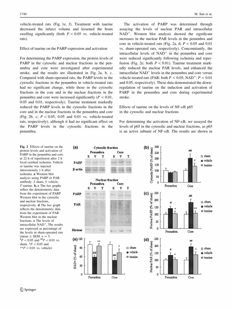
Effect of taurine on the PARP expression and activation
For determining the PARP expression, the protein levels of
PARP in the cytosolic and nuclear fractions in the pen-
umbra and core were investigated after experimental
stroke, and the results are illustrated in Fig. 2a, b, c.
Compared with sham-operated rats, the PARP levels in the
cytosolic fractions in the penumbra in vehicle-treated rats
had no significant change, while those in the cytosolic
fractions in the core and in the nuclear fractions in the
penumbra and core were increased significantly (P \ 0.01,
0.05 and 0.01, respectively). Taurine treatment markedly
reduced the PARP levels in the cytosolic fractions in the
core and in the nuclear fractions in the penumbra and core
(Fig. 2b, c; P \ 0.05, 0.05 and 0.01 vs. vehicle-treated
rats, respectively), although it had no significant effect on
the PARP levels in the cytosolic fractions in the
penumbra.
The activation of PARP was determined through
assaying the levels of nuclear PAR and intracellular
NAD?. Western blot analysis showed the significant
increases in the nuclear PAR levels in the penumbra and
core in vehicle-treated rats (Fig. 2a, d; P \ 0.05 and 0.01
vs. sham-operated rats, respectively). Concomitantly, the
intracellular levels of NAD? in the penumbra and core
were reduced significantly following ischemia and reper-
fusion (Fig. 2e; both P \ 0.01). Taurine treatment mark-
edly reduced the nuclear PAR levels, and enhanced the
intracellular NAD? levels in the penumbra and core versus
vehicle-treated rats (PAR: both P \ 0.05; NAD?: P \ 0.01
and 0.05, respectively). These data demonstrated the down-
regulation of taurine on the induction and activation of
PARP in the penumbra and core during experimental
stroke.
Effects of taurine on the levels of NF-jB p65
in the cytosolic and nuclear fractions
For determining the activation of NF-jB, we assayed the
levels of p65 in the cytosolic and nuclear fractions, as p65
is an active subunit of NF-jB. The results are shown in
Fig. 2 Effects of taurine on the
protein levels and activation of
PARP in the penumbra and core
at 22 h of reperfusion after 2 h
focal cerebral ischemia. Vehicle
or taurine was injected
intravenously 1 h after
ischemia. a Western blot
analysis using PARP or PAR
antibody. S sham, V vehicle,
T taurine. b, c The bar graphs
reflect the densitometric data
from the experiment of PARP
Western blot in the cytosolic
and nuclear fractions,
respectively. d The bar graph
reflects the densitometric data
from the experiment of PAR
Western blot in the nuclear
fractions. e The levels of
intracellular NAD?. The results
are expressed as percentage of
the levels in sham-operated rats
(mean ± SEM. n = 5.#P \ 0.05 and ##P \ 0.01 vs.
sham. *P \ 0.05 and
**P \ 0.01 vs. vehicle)
1740 M. Sun et al.
123

Fig. 3. The levels of p65 in the cytosolic fractions in the
penumbra in vehicle-treated rats had no significant change
compared with sham-operated rats. However, the levels of
p65 in cytosolic fractions in the core and in nuclear frac-
tions in the penumbra and core were increased significantly
(Fig. 3; P \ 0.01, 0.01 and 0.05, respectively). Treatment
with taurine markedly reduced the levels of p65 in cyto-
solic fractions in the core and in nuclear fractions in the
penumbra and core (P \ 0.01, 0.05 and 0.05 vs. vehicle-
treated rats, respectively), although it had no significant
effect on the levels of p65 in cytosolic fractions in the
penumbra. These data confirmed the suppression of taurine
on induction and activation of NF-jB in the penumbra and
core after experimental stroke.
Effects of taurine on the levels of inflammatory
mediators
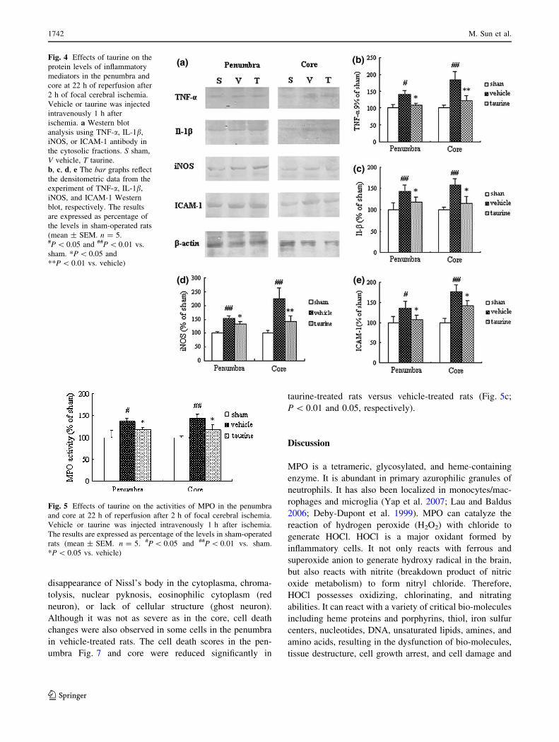
Western blot analysis was used to assay the protein levels
of TNF-a, IL-1b, iNOS, and ICAM-1. The representative
protein bands of TNF-a, IL-1b, iNOS, and ICAM-1 are
displayed in Fig. 4a. The protein levels of TNF-a, IL-1b,
iNOS, and ICAM-1 in the penumbra and core increased
significantly following experimental stroke versus sham-
operated rats (Fig. 4b, c, d, e; TNF-a or ICAM-1: P \ 0.05
and 0.01, respectively; IL-1b or iNOS: both P \ 0.01).
Taurine treatment markedly reduced the protein levels of
TNF-a, IL-1b, iNOS, and ICAM-1 in the penumbra and
core compared with vehicle-treated rats (TNF-a or iNOS:
P \ 0.05 and 0.01, respectively; IL-1b or ICAM-1: both
P \ 0.05).
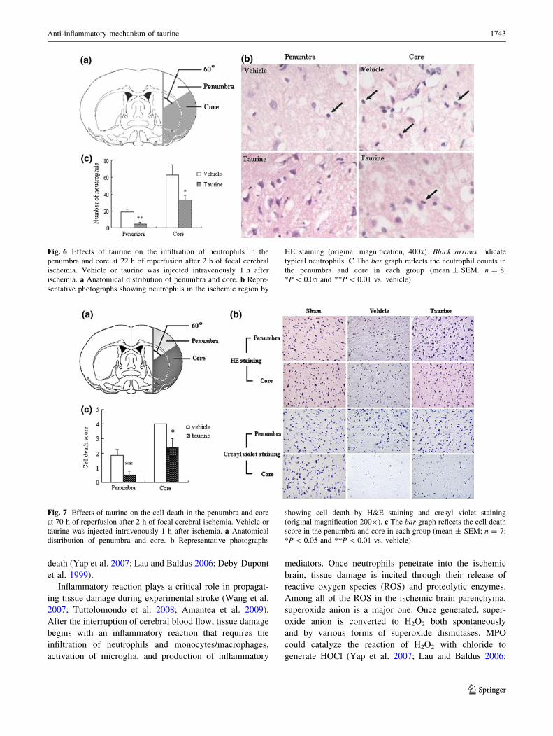
Effects of taurine on neutrophil infiltration
For determining the infiltration of neutrophils into the
penumbra and core, we measured the activities of MPO and
counted the number of neutrophils. As showed in Fig. 5,
the activities of MPO in the penumbra and core were
markedly increased at 22 h of reperfusion after 2 h of
ischemia (P \ 0.05 and 0.01 in the penumbra and core,
respectively). Administration of taurine reduced the activ-
ities of MPO in the penumbra and core significantly (both
P \ 0.05 vs. vehicle). Figure 6 describes the neutrophil
infiltration in the penumbra and core. There is rare infil-
tration of neutrophils in the contralateral hemispheres of
vehicle- or taurine-treated rats. Rats treated with vehicle
showed significant infiltration of neutrophils in the pen-
umbra and core. Treatment with taurine markedly reduced
the number of neutrophils in the penumbra and core
(P \ 0.01 and 0.05 vs. vehicle, respectively).
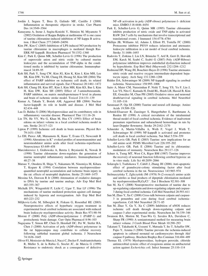
Effect of taurine on ischemic cell death
HE staining and cresyl violet staining were used to inves-
tigate the morphology of cell death, and the representative
photographs are shown in Fig. 5b. In sham-operated rats,
neurons in the cortex displayed intact morphology. In
ischemic core in vehicle-treated rats, most neurons showed
Fig. 3 Effects of taurine on the protein levels of NF-jB p65 in the
penumbra and core at 22 h of reperfusion after 2 h of focal cerebral
ischemia. Vehicle or taurine was injected intravenously 1 h after
ischemia. a, c Western blot analysis using NF-jB p65 antibody in the
cytosolic and nuclear fractions, respectively. S sham, V vehicle,
T taurine. b, d The bar graphs reflect the densitometric data from the
experiment of NF-jB p65 Western blot in the cytosolic and nuclear
fractions, respectively. The results are expressed as percentage of the
levels in sham-operated rats (mean ± SEM. n = 5. #P \ 0.05 and##P \ 0.01 vs. sham. *P \ 0.05 and **P \ 0.01 vs. vehicle)
Anti-inflammatory mechanism of taurine 1741
123
disappearance of Nissl’s body in the cytoplasma, chroma-
tolysis, nuclear pyknosis, eosinophilic cytoplasm (red
neuron), or lack of cellular structure (ghost neuron).
Although it was not as severe as in the core, cell death
changes were also observed in some cells in the penumbra
in vehicle-treated rats. The cell death scores in the pen-
umbra Fig. 7 and core were reduced significantly in
taurine-treated rats versus vehicle-treated rats (Fig. 5c;
P \ 0.01 and 0.05, respectively).
Discussion
MPO is a tetrameric, glycosylated, and heme-containing
enzyme. It is abundant in primary azurophilic granules of
neutrophils. It has also been localized in monocytes/mac-
rophages and microglia (Yap et al. 2007; Lau and Baldus
2006; Deby-Dupont et al. 1999). MPO can catalyze the
reaction of hydrogen peroxide (H2O2) with chloride to
generate HOCl. HOCl is a major oxidant formed by
inflammatory cells. It not only reacts with ferrous and
superoxide anion to generate hydroxy radical in the brain,
but also reacts with nitrite (breakdown product of nitric
oxide metabolism) to form nitryl chloride. Therefore,
HOCl possesses oxidizing, chlorinating, and nitrating
abilities. It can react with a variety of critical bio-molecules
including heme proteins and porphyrins, thiol, iron sulfur
centers, nucleotides, DNA, unsaturated lipids, amines, and
amino acids, resulting in the dysfunction of bio-molecules,
tissue destructure, cell growth arrest, and cell damage and
Fig. 4 Effects of taurine on the
protein levels of inflammatory
mediators in the penumbra and
core at 22 h of reperfusion after
2 h of focal cerebral ischemia.
Vehicle or taurine was injected
intravenously 1 h after
ischemia. a Western blot
analysis using TNF-a, IL-1b,
iNOS, or ICAM-1 antibody in
the cytosolic fractions. S sham,
V vehicle, T taurine.
b, c, d, e The bar graphs reflect
the densitometric data from the
experiment of TNF-a, IL-1b,
iNOS, and ICAM-1 Western
blot, respectively. The results
are expressed as percentage of
the levels in sham-operated rats
(mean ± SEM. n = 5.#P \ 0.05 and ##P \ 0.01 vs.
sham. *P \ 0.05 and
**P \ 0.01 vs. vehicle)
Fig. 5 Effects of taurine on the activities of MPO in the penumbra
and core at 22 h of reperfusion after 2 h of focal cerebral ischemia.
Vehicle or taurine was injected intravenously 1 h after ischemia.
The results are expressed as percentage of the levels in sham-operated
rats (mean ± SEM. n = 5. #P \ 0.05 and ##P \ 0.01 vs. sham.
*P \ 0.05 vs. vehicle)
1742 M. Sun et al.
123
death (Yap et al. 2007; Lau and Baldus 2006; Deby-Dupont
et al. 1999).
Inflammatory reaction plays a critical role in propagat-
ing tissue damage during experimental stroke (Wang et al.
2007; Tuttolomondo et al. 2008; Amantea et al. 2009).
After the interruption of cerebral blood flow, tissue damage
begins with an inflammatory reaction that requires the
infiltration of neutrophils and monocytes/macrophages,
activation of microglia, and production of inflammatory
mediators. Once neutrophils penetrate into the ischemic
brain, tissue damage is incited through their release of
reactive oxygen species (ROS) and proteolytic enzymes.
Among all of the ROS in the ischemic brain parenchyma,
superoxide anion is a major one. Once generated, super-
oxide anion is converted to H2O2 both spontaneously
and by various forms of superoxide dismutases. MPO
could catalyze the reaction of H2O2 with chloride to
generate HOCl (Yap et al. 2007; Lau and Baldus 2006;
Fig. 6 Effects of taurine on the infiltration of neutrophils in the
penumbra and core at 22 h of reperfusion after 2 h of focal cerebral
ischemia. Vehicle or taurine was injected intravenously 1 h after
ischemia. a Anatomical distribution of penumbra and core. b Repre-
sentative photographs showing neutrophils in the ischemic region by
HE staining (original magnification, 400x). Black arrows indicate
typical neutrophils. C The bar graph reflects the neutrophil counts in
the penumbra and core in each group (mean ± SEM. n = 8.
*P \ 0.05 and **P \ 0.01 vs. vehicle)
Fig. 7 Effects of taurine on the cell death in the penumbra and core
at 70 h of reperfusion after 2 h of focal cerebral ischemia. Vehicle or
taurine was injected intravenously 1 h after ischemia. a Anatomical
distribution of penumbra and core. b Representative photographs
showing cell death by H&E staining and cresyl violet staining
(original magnification 2009). c The bar graph reflects the cell death
score in the penumbra and core in each group (mean ± SEM; n = 7;
*P \ 0.05 and **P \ 0.01 vs. vehicle)
Anti-inflammatory mechanism of taurine 1743
123

Deby-Dupont et al. 1999). HOCl has been reported to
induce neuronal death and endothelial dysfunction in vitro
(Yap et al. 2006; Radovits et al. 2007). Moreover,
MPO activation is detrimental during experimental stroke
(Miljkovic-Lolic et al. 2003), and it is used to evaluate the
infiltration of neutrophils in the rodent model of ischemic
stroke (Barone et al. 1991; Matsuo et al. 1994). These data
suggest that the triad of MPO, chloride, and H2O2 could
involve in ischemic brain damage through producing
cytotoxic HOCl.
Taurine is one of the most abundant free amino acids in
mammalian tissue including leukocytes (Huxtable 1992;
Fukuda et al. 1982). It is reported to play an important role
as a regulatory molecule of the inflammatory reaction
(Schuller-Levis and Park 2003, 2004). Taurine can act as a
trap for HOCl forming the long-lived oxidant Tau-NHCl,
which is more stable and less toxic than HOCl. Moreover,
Tau-NHCl down-regulates the generation of proinflamma-
tory mediators by phagocytic cells, such as TNF-a, IL-1b,
and iNOS (Marcinkiewicz et al. 1998; Park et al. 1995; Kim
et al. 1996; Schuller-Levis and Park 2003, 2004). This
process suppresses the inflammatory reaction and protects
cells from the cytotoxic and cytolytic actions of HOCl.
During experimental stroke, the extracellular taurine is
significantly increased due to the release of intracellular
taurine (Lo et al. 1998), which could constitute an impor-
tant endogenous protective mechanism against neuronal
damage (Saransaari and Oja 2000). Meanwhile, the
depletion of intracellular taurine may result in the disrup-
tion of intracellular homeostasis or enantiostasis, leading to
neuronal damage (Huxtable 1992; Michalk et al. 1996).
Hence, the release of taurine may be an obligatory self-
protective mechanism under ischemic stress. Taurine either
in the extracellular space or in inflammatory cells could
scavenge HOCl generated in the ischemic brain, producing
Tau-NHCl (Thomas 1979; Weiss et al. 1982). Tau-NHCl
might then down-regulate the expression of proinflamma-
tory mediators (Marcinkiewicz et al. 1998; Park et al. 1995;
Kim et al. 1996), suppressing the inflammatory reaction
and protecting the brain against experimental stroke. The
present study shows that taurine depresses the activation of
MPO, reduces the production of TNF-a, IL-1b, iNOS, and
ICAM-1, attenuates the infiltration of neutrophils in the
penumbra and core, improves neurological functions,
lessens brain swelling, and decreases the infarct volumes in
the rat model of experimental stroke. These data suggest
that taurine may protect the brain against experimental
stroke through suppressing MPO activation, subsequently
reducing the formation of HOCl, scavenging HOCl, down-
regulating the expression of inflammatory mediators, and
attenuating the infiltration of neutrophils.
On the other hand, Tau-NHCl exerts prolonged oxida-
tive and chlorinating effects long after the initiation of
inflammation and at some distance from the cell of origin
(Zgliczynski et al. 1971). It is reported that Tau-NHCl
mediates HOCl-induced apoptosis (Englert and Shacter
2002). Moreover, Tau-NHCl can be oxidized to taurine
dichloramine (Tau-NCl2), which is more toxic than Tau-
NHCl (Stelmaszynska and Zgliczynski 1978). These data
suggest that Tau-NHCl and Tau-NCl2 produced at the site
of inflammation could suppress neutrophil-mediated
inflammation through inducing apoptotic cell death of
neutrophils, which may be protective during experimental
stroke. Contrarily, it may aggravate ischemic brain damage
through triggering neuronal apoptotic cell death in the
ischemic region. It is reported that a molar excess of tau-
rine increases the formation of Tau-NHCl, decreases the
generation of Tau-NCl2, and protects tissue against the
cytotoxicity of HOCl (Cantin 1994). Hence, it is possible
that exogenous administration of adequate amount of tau-
rine could reduce the release of intracellular taurine and
increase the levels of extracellular taurine, which would
contribute to maintaining intracellular homeostasis, scav-
enging HOCl, reducing the formation of Tau-NCl2,
enhancing Tau-NHCl, depressing the inflammatory reac-
tion, and finally leading to the reduction of ischemic brain
injury. This speculation has been supported by our results
in this study. Although in vivo, the generation of Tau-NCl2may be marginal, further study is necessary to evaluate the
production and the toxicity of Tau-NHCl and Tau-NCl2during experimental stroke, as Tau-NHCl at the concen-
tration of 1 mM and higher is cytotoxic, and Tau-NCl2 is
more toxic than Tan-NHCl (Cantin 1994; Kim and Kim
2005).
One mechanism for reducing inflammation after injury
is to block the inflammatory gene response of cells. Acti-
vation of the transcription factor NF-jB is largely
responsible for up-regulation of inflammatory genes after
ischemic brain injury (Kumar et al. 2004; Ridder and
Schwaninger 2009). Moreover, PARP is reported to act as a
coactivator of NF-jB, thereby contributing to the expres-
sion of NF-jB-driven inflammatory gene (Koh et al. 2004,
2005; Haddad et al. 2006). The present study shows that
taurine down-regulates the activation and expression of
PARP and NF-jB in the penumbra and core after experi-
mental stroke. Moreover, the activation of PARP is related
well to the expression of NF-jB-driven inflammatory
mediators. These data indicate that suppression of inflam-
matory reaction involved PARP and NF-jB is one mech-
anism of taurine against experimental stroke. Our results
are supported by previous studies that PARP-1 knockout or
PARP inhibitors suppress the expression of NF-jB-driven
genes (Chiarugi and Moskowitz 2003; Koh et al. 2004,
2005; Haddad et al. 2006). The possible mechanisms
concerning the effects of taurine on PARP and NF-jB
during experimental stroke may be explained by the
1744 M. Sun et al.
123

following: (1) taurine can down-regulate PARP-induced
NF-jB activation, as taurine can reduce the over-activation
of PARP due to ROS-induced DNA damage through
scavenging ROS or reducing ROS formation (Messina and
Dawson 2000; Virag and Szabo 2002; Chiarugi and
Moskowitz 2003; Saito et al. 2005); (2) taurine can reduce
oxidative stress-induced NF-jB activation, since oxidative
stress can induce the nuclear translocation of NF-jB
without degradation of IjB or promote the degradation of
IjB through phosphorylating IjB in the serine reside
(Redmond et al. 1996; Hanna et al. 2004; Traenckner et al.
1995; Canty et al. 1999); and (3) Tau-NHCl generated by
the reaction of taurine with HOCl can suppress NF-jB
activation through affecting NF-jB signal pathway,
including inhibiting the phosphorylation of IjB (Barua
et al. 2001; Kim and Kim 2005) or directly oxidizing IjB
(Kanayama et al. 2002).
Conclusions
This study demonstrates that treatment with taurine down-
regulates PARP and NF-jB, inhibits the expression of
NF-jB-driven inflammatory mediators, suppresses the
infiltration of neutrophils in the penumbra and core, and
reduces ischemic brain damage in the rat model against
experimental stroke. These data suggest that taurine may
protect the brain against experimental stroke through sup-
pressing inflammatory reaction, which may be related to its
capacity for scavenging HOCl and down-regulating PARP
and NF-jB-driven expression of inflammatory mediators.
Further study is needed to elucidate the detailed anti-
inflammatory mechanism of taurine against experimental
stroke.
Acknowledgments This research was supported by the Beijing
Natural Science Foundation (No. 7052018).
Conflict of interest The authors declare that they have no conflict
of interest.
References
Amantea D, Nappi G, Bernardi G, Bagetta G, Corasaniti MT (2009)
Post-ischemic brain damage: pathophysiology and role of
inflammatory mediators. FEBS J 276:13–26
Ashwal S, Tone B, Tian HR, Cole DJ, Pearce WJ (1998) Core and
penumbral nitric oxide synthase activity during cerebral ische-
mia and reperfusion. Stroke 29:1037–1047
Barone FC, Hillegass LM, Price WJ, White RF, Lee EV, Feuerstein
GZ, Sarau HM, Clark RK, Griswold DE (1991) Polymorphonu-
clear leukocyte infiltration into cerebral focal ischemic tissue:
myeloperoxidase activity assay and histologic verification.
J Neurosci Res 29:336–345
Barua M, Liu Y, Quinn MR (2001) Taurine chloramine inhibits
inducible nitric oxide synthase and TNF-alpha gene expression
in activated alveolar macrophages: decreased NF-kappaB acti-
vation and IkappaB kinase activity. J Immunol 167:2275–2281
Bernofsky C, Swan M (1973) An improved cycling assay for
nicotinamide adenine dinucleotide. Anal Biochem 53:452–458
Bradford MM (1976) A rapid and sensitive method for the
quantitation of microgram quantities of protein utilizing the
principle of protein–dye binding. Anal Biochem 72:248–254
Cantin AM (1994) Taurine modulation on hypochlorous acid-induced
lung epithelial cell injury in vitro. J Clin Invest 93:606–614
Canty TG Jr, Boyle EM Jr, Farr A, Morgan EN, Verrier ED, Pohlman
TH (1999) Oxidative stress induces NF-jB nuclear translocation
without degradation of Ij Ba. Circulation 100:II361–II364
(Suppl II)
Chiarugi A, Moskowitz MA (2003) Poly(ADP-ribose) polymerase-1
activity promotes NF-kappaB-driven transcription and microg-
lial activation: implication for neurodegenerative disorders.
J Neurochem 85:306–317
Clifton GL, Jiang JY, Lyeth BG, Jenkins LW, Hamm RJ, Hayes RL
(1991) Marked protection by moderate hypothermia after
experimental traumatic brain injury. J Cereb Blood Flow Metab
11:114–121
De Ryck M, Van Reempts J, Borgers M, Wauquier A, Janssen PA
(1989) Photochemical stroke model: flunarizine prevents senso-
rimotor deficits after neocortical infarcts in rats. Stroke
20:1383–1390
Deby-Dupont G, Deby C, Lamy M (1999) Neutrophil myeloperox-
idase revisited: it’s role in health and disease. Intensivmed
36:500–513
El Idrissi A (2008) Taurine increases mitochondrial buffering of
calcium: role in neuroprotection. Amino Acids 34:321–328
Eliasson MJ, Sampei K, Mandir AS, Hurn PD, Traystman RJ, Bao J,
Pieper A, Wang ZQ, Dawson TM, Snyder SH, Dawson VL
(1997) Poly (ADP-ribose) polymerase gene disruption renders
mice resistant to cerebral ischemia. Nat Med 3:1089–1095
Englert RP, Shacter E (2002) Distinct modes of cell death induced by
different reactive oxygen species: amino acyl chloramines
mediate hypochlorous acid-induced apoptosis. J Biol Chem 277:
20518–20526
Fukuda K, Hirai Y, Yoshida H, Nakajima T, Usui T (1982) Free
amino acid content of lymphocytes nd granulocytes compared.
Clin Chem 28:1758–1761
Ginsberg MD (1997) The new language of cerebral ischemia. AJNR
Am J Neuroradiol 18:1435–1445
Graham SH, Chen J (2001) Programmed cell death in cerebral
ischemia. Cereb Blood Flow Metab 21:99–109
Ha HC, Hester LD, Snyder SH (2002) Poly (ADP-ribose) polymerase-1
dependence of stress-induced transcription factors and associ-
ated gene expression in glia. Proc Natl Acad Sci USA 99:3270–
3275
Haddad M, Rhinn H, Bloquel C, Coqueran B, Szabo C, Plotkine M,
Scherman D, Margaill I (2006) Anti-inflammatory effects of
PJ34, a poly (ADP-ribose) polymerase inhibitor, in transient
focal cerebral ischemia in mice. Br J Pharmacol 149:23–30
Hagar HH (2004) The protective effect of taurine against cyclospor-
ine A-induced oxidative stress and hepatotoxicity in rats. Toxicol
Lett 151:335–343
Hanna J, Chahine R, Aftimos G, Nader M, Mounayar A, Esseily F,
Chamat S (2004) Protective effect of taurine against free radicals
damage in the rat myocardium. Exp Toxicol Pathol 56:189–194
Hassa PO, Hottiger MO (2002) The functional role of poly (ADP-
ribose)polymerase 1 as novel coactivator of NF-kappaB in
inflammatory disorders. Cell Mol Life Sci 59:1534–1553
Huxtable RJ (1992) Physiological action of taurine. Physiol Rev
72:101–163
Anti-inflammatory mechanism of taurine 1745
123
Jordan J, Segura T, Brea D, Galindo MF, Castillo J (2008)
Inflammation as therapeutic objective in stroke. Curr Pharm
Des 14:3549–3564
Kanayama A, Inoue J, Sugita-Konishi Y, Shimizu M, Miyamoto Y
(2002) Oxidation of Ikappa Balpha at methionine 45 is one cause
of taurine chloramine-induced inhibition of NF-kappa B activa-
tion. J Biol Chem 277:24049–24056
Kim JW, Kim C (2005) Inhibition of LPS-induced NO production by
taurine chloramine in macrophages is mediated though Ras-
ERK-NF-kappaB. Biochem Pharmacol 70:1352–1360
Kim C, Park E, Quinn MR, Schuller-Levis G (1996) The production
of superoxide anion and nitric oxide by cultured murine
leukocytes and the accumulation of TNF-alpha in the condi-
tioned media is inhibited by taurine chloramine. Immunophar-
macol 34:89–95
Koh SH, Park Y, Song CW, Kim JG, Kim K, Kim J, Kim MH, Lee
SR, Kim DW, Yu HJ, Chang DI, Hwang SJ, Kim SH (2004) The
effect of PARP inhibitor on ischaemic cell death, its related
inflammation and survival signals. Eur J Neurosci 20:1461–1472
Koh SH, Chang DI, Kim HT, Kim J, Kim MH, Kim KS, Bae I, Kim
H, Kim DW, Kim SH (2005) Effect of 3-aminobenzamide,
PARP inhibitor, on matrix metalloproteinase-9 level in plasma
and brain of ischemic stroke model. Toxicol 214:131–139
Kumar A, Takada Y, Boriek AM, Aggarwal BB (2004) Nuclear
factor-kappaB: its role in health and disease. J Mol Med
82:434–448
Lau D, Baldus S (2006) Myeloperoxidase and its contributory role in
inflammatory vascular disease. Pharmacol Ther 111:16–26
Lin TN, He YY, Wu G, Khan M, Hsu CY (1993) Effect of brain
edema on infarct volume in a focal cerebral ischemia model in
rats. Stroke 24:117–121
Lipton P (1999) Ischemic cell death in brain neurons. Physiol Rev
79:1431–1568
Lo EH, Pierce AR, Matsumoto K, Kano T, Evans CJ, Newcomb R
(1998) Alterations in K? evoked profiles of neurotransmitter and
neuromodulator amino acids after focal ischemia–reperfusion.
Neuroscience 83:449–458
Marcinkiewicz J, Grabowska A, Bereta J, Bryniarski K, Nowak B
(1998) Taurine chloramine down-regulates the generation of
murine neutrophil inflammatory mediators. Immunopharmacol
40:27–38
Matsuo Y, Onodera H, Shiga Y, Nakamura M, Ninomiya M, Kihara
T, Kogure K (1994) Correlation between myeloperoxidase-
quantified neutrophil accumulation and ischemic brain injury in
the rat: effects of neutrophil depletion. Stroke 25:1469–1475
Messina SA, Dawson R Jr (2000) Attenuation of oxidative damages
to DNA by taurine and taurine analogs. Adv Exp Med Biol
483:355–367
Michalk DV, Wingenfeld P, Licht C, Ugur T, Siar LF (1996) The
mechanisms of taurine mediated protection against cell damage
induced by hypoxia and reoxygenation. Adv Exp Med Biol
403:223–232
Miljkovic-Lolic M, Silbergleit R, Fiskum G, Rosenthal RE (2003)
Neuroprotective effects of hyperbaric oxygen treatment in
experimental focal cerebral ischemia are associated with reduced
brain leukocyte myeloperoxidase activity. Brain Res 971:90–94
Moroni F (2008) Poly (ADP-ribose)polymerase 1 (PARP-1) and
postischemic brain damage. Curr Opin Pharmacol 8:96–103
Nagayama T, Simon RP, Chen D, Henshall DC, Pei W, Stetler RA,
Chen J (2000) Activation of poly (ADP-ribose) polymerase in
the rat hippocampus may contribute to cellular recovery
following sublethal transient global ischemia. J Neurochem
74:1636–1645
Oliver FJ, Menissier-de Murcia J, Nacci C, Decker P, Andriantsitohania
R, Muller S, de la Rubia G, Stoclet JC, de Murcia G (1999)
Resistance to endotoxic shock as a consequence of defective
NF-jB activation in poly (ADP-ribose) polymerase-1- deficient
mice. EMBO J 18:4446–4454
Park E, Schuller-Levis G, Quinn MR (1995) Taurine chloramine
inhibits production of nitric oxide and TNF-alpha in activated
RAW 264.7 cells by mechanisms that involve transcriptional and
translational events. J Immunol 154:4778–4784
Phillips JB, Williams AJ, Adams J, Elliott PJ, Tortella FC (2000)
Proteasome inhibitor PS519 reduces infarction and attenuates
leukocyte infiltration in a rat model of focal cerebral ischemia.
Stroke 31:1686–1693
Radovits T, Zotkina J, Lin LN, Bomicke T, Arif R, Gero D, Horvath
EM, Karck M, Szabo C, Szabo G (2007) Poly (ADP-Ribose)
polymerase inhibition improves endothelial dysfunction induced
by hypochlorite. Exp Biol Med (Maywood) 232:1204–1212
Redmond HP, Wang JH, Bouchier-Hayes D (1996) Taurine attenuates
nitric oxide and reactive oxygen intermediate-dependent hepa-
tocyte injury. Arch Surg 131:1280–1288
Ridder DA, Schwaninger M (2009) NF-kappaB signaling in cerebral
ischemia. Neuroscience 158:995–1006
Saito A, Maier CM, Narasimhan P, Nishi T, Song YS, Yu F, Liu J,
Lee YS, Nito C, Kamada H, Dodd RL, Hsieh LB, Hassid B, Kim
EE, Gonzalez M, Chan PH (2005) Oxidative stress and neuronal
death/survival signaling in cerebral ischemia. Mol Neurobiol
31:105–116
Saransaari P, Oja SS (2000) Taurine and neural cell damage. Amino
Acids 19:509–526
Schmid-Elsaesser R, Zausinger S, Hungerhuber E, Baethmann A,
Reulen HJ (1998) A critical reevalution of the intraluminal
thread model of focal cerebral ischemia. Evidence of inadvertent
premature reperfusion and subarachnoid hemorrhage in rats by
laser-Doppler flowmetry. Stroke 29:2162–2170
Schneider A, Martin-Villalba A, Weih F, Vogel J, Wirth T,
Schwaninger M (1999) NF-kappaB is activated and promotes
cell death in focal cerebral ischemia. Nat Med 5:554–559
Schuller-Levis GB, Park E (2003) Taurine: new implications for an
old amino acid. FEMS Microbiol Lett 226:195–202
Schuller-Levis GB, Park E (2004) Taurine and its chloramine:
modulators of immunity. Neurochem Res 29:117–126
Schurr A, Tseng MT, West CA, Rigor BM (1987) Taurine improves
the recovery of neuronal function following cerebral hypoxia: an
in vitro study. Life Sci 40:2059–2066
Solaroglu I, Tsubokawa T, Cahill J, Zhang JH (2006) Anti-apoptotic
effect of granulocyte-colony stimulating factor after focal
cerebral ischemia in the rat. Neuroscience 143:965–974
Stelmaszynska T, Zgliczynski JM (1978) N-(2-oxoacyl) amino acids
and nitriles as final products of dipeptide chlorination mediated
by myeloperoxidase/H2O2/Cl-. Eur J Biochem 92:301–308
Sun M, Xu C (2008) Neuroprotective mechanism of taurine due to
up-regulating calpastatin and down-regulating calpain and caspase-
3 during focal cerebral ischemia. Cell Mol Neurobiol 28:593–611
Sun M, Zhao Y, Xu C (2008) Cross-talk between calpain and caspase-
3 in penumbra and core during focal cerebral ischemia–
reperfusion. Cell Mol Neurobiol 28:71–85
Sun M, Zhao Y, Gu Y, Xu C (2009) Inhibition of nNOS reduces
ischemic cell death through down-regulating calpain and
caspase-3 after experimental stroke. Neurochem Int 54:339–346
Swanson RA, Morton M, Tsao-Wu G, Savalos RA, Davidson C,
Sharp FR (1990) A semiautomated method for measuring brain
infarct volume. J Cereb Blood Flow Metab 10:290–293
Takatani T, Takahashi K, Uozumi Y, Matsuda T, Ito T, Schaffer SW,
Fujio Y, Azuma J (2004) Taurine prevents the ischemia-induced
apoptosis in cultured neonatal rat cardiomyocytes through Akt/
caspase-9 pathway. Biochem Biophys Res Commun 316:484–489
Thomas EL (1979) Myeloperoxidase, hydrogen peroxide, chloride
antimicrobial system: effect of exogenous amine on antibacterial
action against Escherichia coli. Infect Immune 25:110–114
1746 M. Sun et al.
123
Traenckner EB, Pahl HL, Henkel T, Schmidt KN, Wilk S, Baeuerle
PA (1995) Phosphorylation of human Ij B-a on serines 32 and
36 controls Ij B-a proteolysis and NF-jB activation in response
to diverse stimuli. EMBO J 14:2876–2883
Tuttolomondo A, Di Raimondo D, di Sciacca R, Pinto A, Licata G
(2008) Inflammatory cytokines in acute ischemic stroke. Curr
Pharm Des 14:3574–3589
Virag L, Szabo C (2002) The therapeutic potential of poly (ADPRibose)
polymerase inhibitors. Pharmacol Rev 54:375–429
Wang Q, Tang XN, Yenari MA (2007) The inflammatory response in
stroke. J Neuroimmunol 184:53–68
Wang Q, van Hoecke M, Tang XH, Lee H, Zheng Z, Swanson RA,
Yenari MA (2009) Pyruvate protects against experimental stroke
via an anti-inflammatory mechanism. Neurobiol Dis 36:223–231
Weiss SJ, Klein R, Slivka A, Wei M (1982) Chlorination of taurine by
human neutrophils. Evidence for hypochlorous acid generation.
J Clin Invest 170:598–607
Williams AJ, Dave JR, Tortella FC (2006) Neuroprotection with the
proteasome inhibitor MLN519 in focal ischemic brain injury:
relation to nuclear factor kappaB (NF-kappaB), inflammatory
gene expression, and leukocyte infiltration. Neurochem Int
49:106–112
Yap YW, Whiteman M, Bay BH, Li Y, Sheu FS, Qi RZ, Tan CH,
Cheung NS (2006) Hypochlorous acid induces apoptosis of
cultured cortical neurons through activation of calpains and
rupture of lysosomes. J Neurochem 98:1597–1609
Yap YW, Whiteman M, Cheung NS (2007) Chlorinative stress: an
under appreciated mediator of neurodegeneration? Cell Signal
19:219–228
Zgliczynski JM, Stelmaszynska T, Domanski J, Ostrowski W (1971)
Chloramines as intermediates of oxidation reaction of amino
acids by myeloperoxidase. Biochim Biophys Acta 235:419–424
Anti-inflammatory mechanism of taurine 1747
123


![From Stroke to Dementia: a Comprehensive Review Exposing ... · after both hemorrhagic and ischemic stroke, as observed in rodents and non-human primates [17, 18]. Abnormal perivascular](https://static.fdocument.org/doc/165x107/5e47cc033fa49928c25efa78/from-stroke-to-dementia-a-comprehensive-review-exposing-after-both-hemorrhagic.jpg)