

AN ATYPICAL CRM1-DEPENDENT NUCLEAR EXPORT SIGNAL
17
Regulated nuclear export of HIF-1α 1 AN ATYPICAL CRM1-DEPENDENT NUCLEAR EXPORT SIGNAL MEDIATES REGULATION OF HYPOXIA-INDUCIBLE FACTOR HIF-1α BY MAPK Ilias Mylonis 1,3 , Georgia Chachami 1,3 , Efrosyni Paraskeva 2,3 , and George Simos 1,3 1 Laboratory of Biochemistry and 2 Laboratory of Physiology, Department of Medicine, University of Thessaly; 3 Institute of Biomedical Research & Technology (BIOMED); Mezourlo, 41110 Larissa, Greece Running head: Regulated nuclear export of HIF-1α Address correspondence to: George Simos, Laboratory of Biochemistry, Department of Medicine, University of Thessaly, P.O. Box 1400, Mezourlo, 41110 Larissa, Greece; Fax: ++30- 2410-685545; E-Mail: [email protected] HIF-1 is the key transcriptional activator of hypoxia-inducible genes and an important anti- cancer target. Its regulated subunit, HIF-1α, is controlled by oxygen levels and major signaling pathways. We previously reported that phosphorylation of Ser 641/643 by p42/44 MAPK is essential for HIF-1α nuclear accumulation and activity. We now show that a fragment of HIF- 1α (aa 616-658), termed MAPK target domain (MTD), contains a nuclear export signal (NES) which has atypical hydrophobic residue spacing. Localization, reporter gene and co- immunoprecipitation assays demonstrate that the identified NES interacts with CRM1 in a phosphorylation-sensitive manner Further- more, disruption of the NES ( 637 ILI 639 to AAA) restores nuclear localization and activity of non-phosphorylated HIF-1α and renders it largely resistant to inhibition of MAPK, an effect reproduced by a phosphomimetic mutation (Ser 641 to Glu). As these data predict, over-expression of wild-type or mutant ( 641 SPSP 644 to APAP) MTD in HeLa cells modulates the activity and subcellular distribution of endogenous HIF-1α. We suggest that control of HIF-1α nuclear transport represents an important MAPK-dependent regulatory mechanism. Hypoxia-inducible factor 1 (HIF-1) is a transcriptional activator and the key mediator of cellular response to hypoxia. HIF-1 binds to regulatory DNA sequences called hypoxia response elements and controls the expression of genes involved in cell metabolism, erythropoiesis, angiogenesis, invasion and metastasis. It is, therefore, essential not only for cell survival under low oxygen but also for embryogenesis and tumor progression (1). HIF-1 is a heterodimer of HIF-1α and HIF-1β (or ARNT), both members of the bHLH-PAS family of transcription factors. Whereas ARNT is expressed constitutively, HIF- 1α is highly regulated and a major target of anti- cancer therapy (2). Under normal oxygen concentration, HIF- 1α protein levels are kept low by VHL-mediated polyubiquitination and subsequent degradation. Interaction with VHL requires the hydroxylation of two proline residues in the oxygen dependent degradation domain of HIF-1α (3). The proline hydroxylases that participate in this process depend on iron and require molecular oxygen and 2-oxoglutarate as substrates. When oxygen concentration is low, hydroxylation is impaired allowing HIF-1α to be stabilized, enter the nucleus, bind to ARNT and DNA and induce the expression of target genes. This latter process is also regulated by oxygen tension as HIF-1α hydroxylation at Asn803 by FIH-1 compromises its association with the transcriptional co-activator CBP/p300 (4). Reactive oxygen species produced under hypoxia as well as intermediate products of cell metabolism can also affect hydroxylation and, consequently, modulate HIF-1 activity (5-7). HIF-1α expression and transcriptional activity is additionally controlled, irrespective of oxygen levels, by major signaling pathways such as PI3-K and MAPK, which are induced by growth factors, cytokines and oncogenes (8,9). Activation of the PI3-K pathway leads to http://www.jbc.org/cgi/doi/10.1074/jbc.M803081200 The latest version is at JBC Papers in Press. Published on August 7, 2008 as Manuscript M803081200 Copyright 2008 by The American Society for Biochemistry and Molecular Biology, Inc. by guest on January 2, 2019 http://www.jbc.org/ Downloaded from
Transcript of AN ATYPICAL CRM1-DEPENDENT NUCLEAR EXPORT SIGNAL
Regulated nuclear export of HIF-1α 1
AN ATYPICAL CRM1-DEPENDENT NUCLEAR EXPORT SIGNAL MEDIATES REGULATION OF HYPOXIA-INDUCIBLE FACTOR
HIF-1α BY MAPK Ilias Mylonis1,3, Georgia Chachami1,3, Efrosyni Paraskeva2,3, and George Simos1,3
1Laboratory of Biochemistry and 2Laboratory of Physiology, Department of Medicine, University of Thessaly;
3Institute of Biomedical Research & Technology (BIOMED); Mezourlo, 41110 Larissa, Greece
Running head: Regulated nuclear export of HIF-1α Address correspondence to: George Simos, Laboratory of Biochemistry, Department of Medicine, University of Thessaly, P.O. Box 1400, Mezourlo, 41110 Larissa, Greece; Fax: ++30-2410-685545; E-Mail: [email protected] HIF-1 is the key transcriptional activator of hypoxia-inducible genes and an important anti-cancer target. Its regulated subunit, HIF-1α, is controlled by oxygen levels and major signaling pathways. We previously reported that phosphorylation of Ser641/643 by p42/44 MAPK is essential for HIF-1α nuclear accumulation and activity. We now show that a fragment of HIF-1α (aa 616-658), termed MAPK target domain (MTD), contains a nuclear export signal (NES) which has atypical hydrophobic residue spacing. Localization, reporter gene and co-immunoprecipitation assays demonstrate that the identified NES interacts with CRM1 in a phosphorylation-sensitive manner Further-more, disruption of the NES (637ILI639 to AAA) restores nuclear localization and activity of non-phosphorylated HIF-1α and renders it largely resistant to inhibition of MAPK, an effect reproduced by a phosphomimetic mutation (Ser641 to Glu). As these data predict, over-expression of wild-type or mutant (641SPSP644 to APAP) MTD in HeLa cells modulates the activity and subcellular distribution of endogenous HIF-1α. We suggest that control of HIF-1α nuclear transport represents an important MAPK-dependent regulatory mechanism.
Hypoxia-inducible factor 1 (HIF-1) is a
transcriptional activator and the key mediator of cellular response to hypoxia. HIF-1 binds to regulatory DNA sequences called hypoxia response elements and controls the expression of genes involved in cell metabolism, erythropoiesis,
angiogenesis, invasion and metastasis. It is, therefore, essential not only for cell survival under low oxygen but also for embryogenesis and tumor progression (1). HIF-1 is a heterodimer of HIF-1α and HIF-1β (or ARNT), both members of the bHLH-PAS family of transcription factors. Whereas ARNT is expressed constitutively, HIF-1α is highly regulated and a major target of anti-cancer therapy (2). Under normal oxygen concentration, HIF-1α protein levels are kept low by VHL-mediated polyubiquitination and subsequent degradation. Interaction with VHL requires the hydroxylation of two proline residues in the oxygen dependent degradation domain of HIF-1α (3). The proline hydroxylases that participate in this process depend on iron and require molecular oxygen and 2-oxoglutarate as substrates. When oxygen concentration is low, hydroxylation is impaired allowing HIF-1α to be stabilized, enter the nucleus, bind to ARNT and DNA and induce the expression of target genes. This latter process is also regulated by oxygen tension as HIF-1α hydroxylation at Asn803 by FIH-1 compromises its association with the transcriptional co-activator CBP/p300 (4). Reactive oxygen species produced under hypoxia as well as intermediate products of cell metabolism can also affect hydroxylation and, consequently, modulate HIF-1 activity (5-7). HIF-1α expression and transcriptional activity is additionally controlled, irrespective of oxygen levels, by major signaling pathways such as PI3-K and MAPK, which are induced by growth factors, cytokines and oncogenes (8,9). Activation of the PI3-K pathway leads to
http://www.jbc.org/cgi/doi/10.1074/jbc.M803081200The latest version is at JBC Papers in Press. Published on August 7, 2008 as Manuscript M803081200
Copyright 2008 by The American Society for Biochemistry and Molecular Biology, Inc.
by guest on January 2, 2019http://w
ww
.jbc.org/D
ownloaded from
Regulated nuclear export of HIF-1α 2
enhanced levels of HIF-1α protein synthesis (10) while MAPK activation promotes predominantly HIF-1 transcriptional activity via direct phosphorylation of HIF-1α (11). We have recently revealed that p44/42 MAPK (ERK1/2) modifies HIF-1α residues Ser641 and Ser643 and showed that inhibition of this phosphorylation by mutagenesis, MAPK-pathway inhibitors or treatment with flavonoids impairs activity and nuclear accumulation of HIF-1α (12,13). The degree of nuclear accumulation of a protein depends on its relative nuclear import and export rates (14). Nuclear import of HIF-1α has been previously suggested to be regulated (15,16). Although the ability of human HIF-1α to also exit the nucleus and be a shuttling protein has been documented (17,18), the transport signals or factors regulating its nuclear export have remained so far unknown. In this work we characterize an atypical but CRM1-dependent NES in human HIF-1α. We further show that the identified NES responds to MAPK-dependent phosphorylation and, therefore, provides additional means of HIF-1α regulation.
EXPERIMENTAL PROCEDURES Plasmid constructions - HIF-1α point mutants were constructed with the QuikChange® II site-directed mutagenesis kit (Stratagene) using as templates pGEX-HIF-1α (19) or pGEX-HIF-1α(S A) (12) and suitable primers (sequence available upon request) and were then sub-cloned as BamHI inserts into the mammalian expression vector pEGFP-C1 (Clontech). The cDNA fragments corresponding to HIF-1α MTD (amino acids 616-658) were obtained by PCR using as template pGEX-HIF-1α (wild type or mutant forms). The derived inserts were subcloned as BamHI fragments into pEGFP-C1 and pCMV-FLAG vectors. The DNA sequence of the point mutants was confirmed by sequencing performed by Lark Technologies Inc. Cell culture and transfection - Human HeLa, HEK293-T and Huh7 cells were cultured in DMEM containing 10% FBS and antibiotics. Transient transfections were performed as previously described (12). When required, 20 hours after transfection cells were treated for 4h with 50 µM PD98059, 10 ng/ml LMB (both from
Sigma-Aldrich) or 1 mM DMOG (Alexis Biohemicals). Reporter gene assays - Luciferase assays were performed in cells transiently co-transfected with plasmids expressing different forms of full length or MTD of HIF-1α (or the empty pEGFP-C1 parental vector as control) as previously reported (12). Protein purification and phosphorylation assays - GST-HIF-1α, GST-MTD and GST-MTD(S A) were expressed in E. coli and purified as GST-HIF-1α (19). Phosphorylation reactions were carried out according to previously published procedures (12). Immunoprecipitation - HeLa cells treated with DMOG or transfected with GFP-HIF-1α, GFP-MTD or their mutant forms were washed with cold PBS and lysed (20 min, 4°C) in buffer containing 25 mM Hepes pH 7.6, 150 mM NaCl, 2 mM MgCl2, 1 mM PMSF, 50 mM β-glycerol-phosphate and 10 mM Na3VO4. Immunoprecipitation of endogenous or GFP-tagged proteins was carried as previously described using an affinity purified polyclonal anti-HIF-1α antibody raised in rabbits against GST-HIF-1α(348-826) (20) or an anti-GFP antibody (12), respectively, and precipitates were analyzed by western blotting using anti-CRM1 and anti-HIF-1α mouse monoclonal antibodies (BD Transduction Laboratories, Franklin Lakes, NJ, USA) or the polyclonal anti-HIF-1α. Membranes were then incubated with horseradish peroxidase conjugated goat anti-mouse or goat anti-rabbit IgG (Cell Signaling) followed by ECL. Fluorescence microscopy – HeLa, HEK293-T and Huh7 cells grown on coverslips were transiently transfected with GFP-tagged full length HIF-1α, its MTD fragment or their mutant forms and 24 hours post-transfection were washed once with PBS and fixed with 3% formaldehyde in PBS for 5 min at room temperature. After washing twice with PBS, cells were counterstained with DAPI for 2 min and mounted on slides. Images were collected with a Leica DFC480 camera (LAS software ver. V2.3.1.R1) on an Axioscope 40 Zeiss microscope equipped with a 40x objective at RT. Immunofluorescence - Hela cells were grown on coverslips and transiently transfected with pCMV-FLAG vector expressing the MTD fragment or its S A mutant. 20 hours post-transfection cells
by guest on January 2, 2019http://w
ww
.jbc.org/D
ownloaded from
Regulated nuclear export of HIF-1α 3
were treated for 4 hours with 1mM DMOG and if required with 50 µΜ PD98059, washed once with PBS and fixed with 3% formaldehyde in PBS for 5 min at room temperature. Cells were washed twice with PBS and permeabilized with PBS containing 0.1% Triton X-100 for 15 min. After washing twice with PBS, cells were treated with 3% BSA in PBS for 1 h. Coverslips were incubated for 1h at room temperature with a polyclonal anti-HIF-1α antibody (20) and a monoclonal anti-FLAG antibody (Sigma-Aldrich), washed twice with PBS and incubated for 30 min at room temperature with FITC-conjugated anti-rabbit and Cy3-conjugated anti-mouse secondary antibodies (Jackson ImmunoResearch). After washing twice with PBS, cells were counterstained with DAPI, mounted on slides and viewed by a Leica DFC480 camera (LAS software ver. V2.3.1.R1) on an Axioscope 40 Zeiss microscope equipped with a 40x objective at RT.
RESULTS
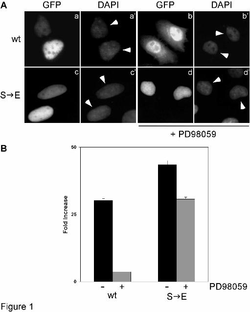
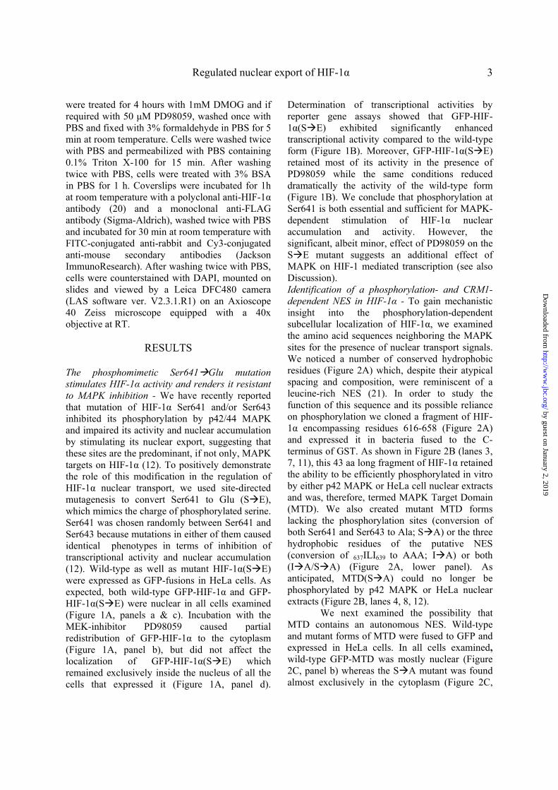
The phosphomimetic Ser641 Glu mutation stimulates HIF-1α activity and renders it resistant to MAPK inhibition - We have recently reported that mutation of HIF-1α Ser641 and/or Ser643 inhibited its phosphorylation by p42/44 MAPK and impaired its activity and nuclear accumulation by stimulating its nuclear export, suggesting that these sites are the predominant, if not only, MAPK targets on HIF-1α (12). To positively demonstrate the role of this modification in the regulation of HIF-1α nuclear transport, we used site-directed mutagenesis to convert Ser641 to Glu (S E), which mimics the charge of phosphorylated serine. Ser641 was chosen randomly between Ser641 and Ser643 because mutations in either of them caused identical phenotypes in terms of inhibition of transcriptional activity and nuclear accumulation (12). Wild-type as well as mutant HIF-1α(S E) were expressed as GFP-fusions in HeLa cells. As expected, both wild-type GFP-HIF-1α and GFP-HIF-1α(S E) were nuclear in all cells examined (Figure 1A, panels a & c). Incubation with the MEK-inhibitor PD98059 caused partial redistribution of GFP-HIF-1α to the cytoplasm (Figure 1A, panel b), but did not affect the localization of GFP-HIF-1α(S E) which remained exclusively inside the nucleus of all the cells that expressed it (Figure 1A, panel d).
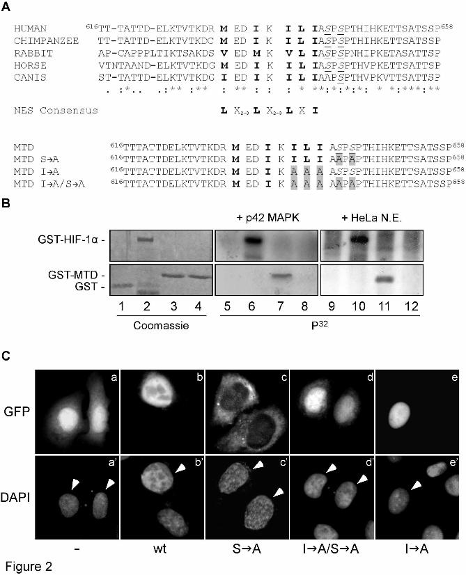
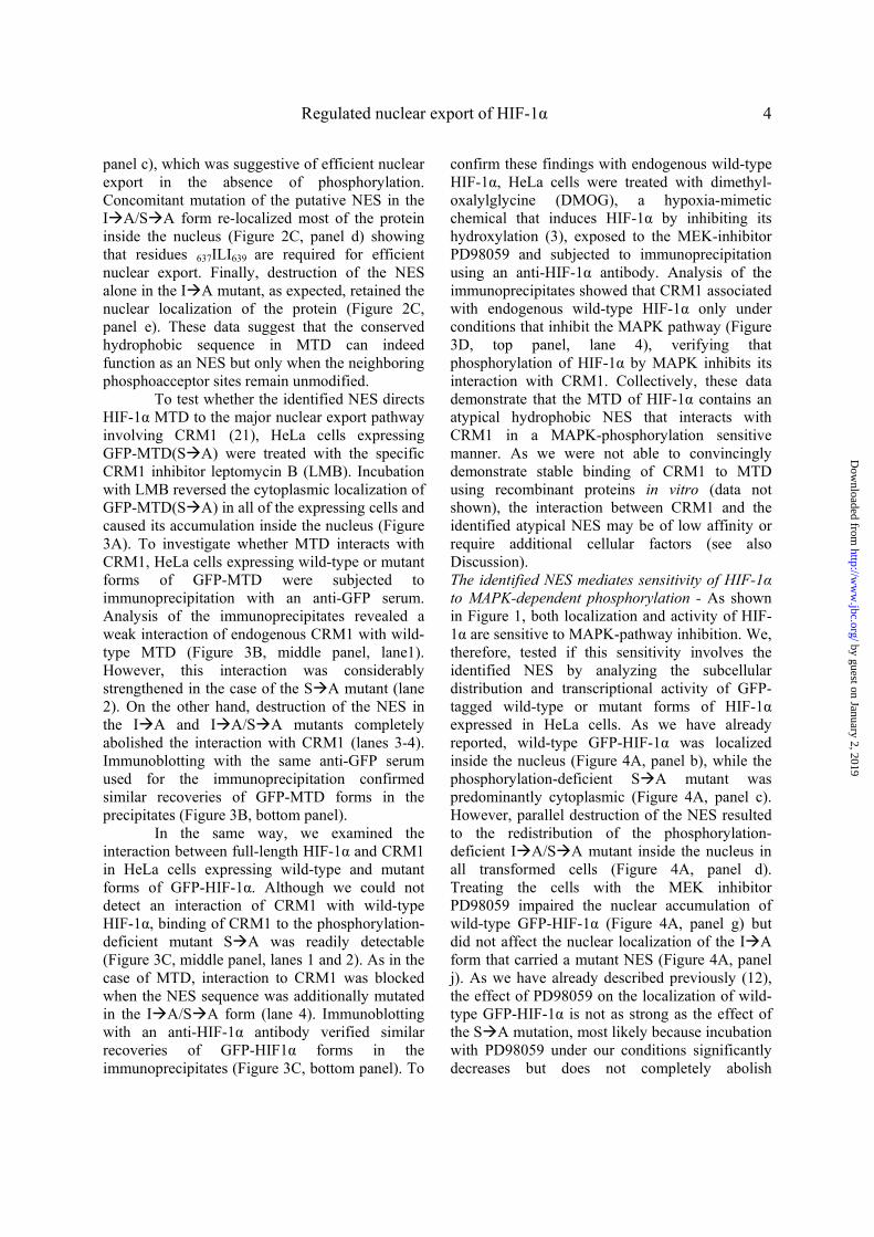
Determination of transcriptional activities by reporter gene assays showed that GFP-HIF-1α(S E) exhibited significantly enhanced transcriptional activity compared to the wild-type form (Figure 1B). Moreover, GFP-HIF-1α(S E) retained most of its activity in the presence of PD98059 while the same conditions reduced dramatically the activity of the wild-type form (Figure 1B). We conclude that phosphorylation at Ser641 is both essential and sufficient for MAPK-dependent stimulation of HIF-1α nuclear accumulation and activity. However, the significant, albeit minor, effect of PD98059 on the S E mutant suggests an additional effect of MAPK on HIF-1 mediated transcription (see also Discussion). Identification of a phosphorylation- and CRM1-dependent NES in HIF-1α - To gain mechanistic insight into the phosphorylation-dependent subcellular localization of HIF-1α, we examined the amino acid sequences neighboring the MAPK sites for the presence of nuclear transport signals. We noticed a number of conserved hydrophobic residues (Figure 2A) which, despite their atypical spacing and composition, were reminiscent of a leucine-rich NES (21). In order to study the function of this sequence and its possible reliance on phosphorylation we cloned a fragment of HIF-1α encompassing residues 616-658 (Figure 2A) and expressed it in bacteria fused to the C-terminus of GST. As shown in Figure 2B (lanes 3, 7, 11), this 43 aa long fragment of HIF-1α retained the ability to be efficiently phosphorylated in vitro by either p42 MAPK or HeLa cell nuclear extracts and was, therefore, termed MAPK Target Domain (MTD). We also created mutant MTD forms lacking the phosphorylation sites (conversion of both Ser641 and Ser643 to Ala; S A) or the three hydrophobic residues of the putative NES (conversion of 637ILI639 to AAA; I A) or both (I A/S A) (Figure 2A, lower panel). As anticipated, MTD(S A) could no longer be phosphorylated by p42 MAPK or HeLa nuclear extracts (Figure 2B, lanes 4, 8, 12).
We next examined the possibility that MTD contains an autonomous NES. Wild-type and mutant forms of MTD were fused to GFP and expressed in HeLa cells. In all cells examined, wild-type GFP-MTD was mostly nuclear (Figure 2C, panel b) whereas the S A mutant was found almost exclusively in the cytoplasm (Figure 2C,
by guest on January 2, 2019http://w
ww
.jbc.org/D
ownloaded from
Regulated nuclear export of HIF-1α 4
panel c), which was suggestive of efficient nuclear export in the absence of phosphorylation. Concomitant mutation of the putative NES in the I A/S A form re-localized most of the protein inside the nucleus (Figure 2C, panel d) showing that residues 637ILI639
are required for efficient nuclear export. Finally, destruction of the NES alone in the I A mutant, as expected, retained the nuclear localization of the protein (Figure 2C, panel e). These data suggest that the conserved hydrophobic sequence in MTD can indeed function as an NES but only when the neighboring phosphoacceptor sites remain unmodified.
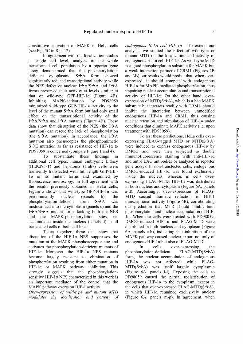
To test whether the identified NES directs HIF-1α MTD to the major nuclear export pathway involving CRM1 (21), HeLa cells expressing GFP-MTD(S A) were treated with the specific CRM1 inhibitor leptomycin B (LMB). Incubation with LMB reversed the cytoplasmic localization of GFP-MTD(S A) in all of the expressing cells and caused its accumulation inside the nucleus (Figure 3A). To investigate whether MTD interacts with CRM1, HeLa cells expressing wild-type or mutant forms of GFP-MTD were subjected to immunoprecipitation with an anti-GFP serum. Analysis of the immunoprecipitates revealed a weak interaction of endogenous CRM1 with wild-type MTD (Figure 3B, middle panel, lane1). However, this interaction was considerably strengthened in the case of the S A mutant (lane 2). On the other hand, destruction of the NES in the I A and I A/S A mutants completely abolished the interaction with CRM1 (lanes 3-4). Immunoblotting with the same anti-GFP serum used for the immunoprecipitation confirmed similar recoveries of GFP-MTD forms in the precipitates (Figure 3B, bottom panel).
In the same way, we examined the interaction between full-length HIF-1α and CRM1 in HeLa cells expressing wild-type and mutant forms of GFP-HIF-1α. Although we could not detect an interaction of CRM1 with wild-type HIF-1α, binding of CRM1 to the phosphorylation-deficient mutant S A was readily detectable (Figure 3C, middle panel, lanes 1 and 2). As in the case of MTD, interaction to CRM1 was blocked when the NES sequence was additionally mutated in the I A/S A form (lane 4). Immunoblotting with an anti-HIF-1α antibody verified similar recoveries of GFP-HIF1α forms in the immunoprecipitates (Figure 3C, bottom panel). To
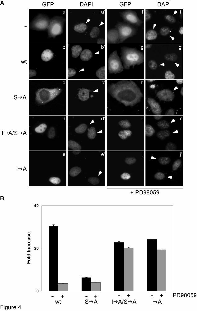
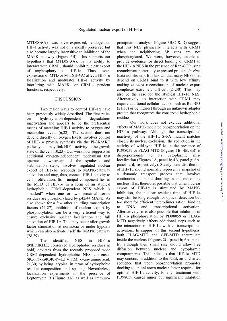
confirm these findings with endogenous wild-type HIF-1α, HeLa cells were treated with dimethyl-oxalylglycine (DMOG), a hypoxia-mimetic chemical that induces HIF-1α by inhibiting its hydroxylation (3), exposed to the MEK-inhibitor PD98059 and subjected to immunoprecipitation using an anti-HIF-1α antibody. Analysis of the immunoprecipitates showed that CRM1 associated with endogenous wild-type HIF-1α only under conditions that inhibit the MAPK pathway (Figure 3D, top panel, lane 4), verifying that phosphorylation of HIF-1α by MAPK inhibits its interaction with CRM1. Collectively, these data demonstrate that the MTD of HIF-1α contains an atypical hydrophobic NES that interacts with CRM1 in a MAPK-phosphorylation sensitive manner. As we were not able to convincingly demonstrate stable binding of CRM1 to MTD using recombinant proteins in vitro (data not shown), the interaction between CRM1 and the identified atypical NES may be of low affinity or require additional cellular factors (see also Discussion). The identified NES mediates sensitivity of HIF-1α to MAPK-dependent phosphorylation - As shown in Figure 1, both localization and activity of HIF-1α are sensitive to MAPK-pathway inhibition. We, therefore, tested if this sensitivity involves the identified NES by analyzing the subcellular distribution and transcriptional activity of GFP-tagged wild-type or mutant forms of HIF-1α expressed in HeLa cells. As we have already reported, wild-type GFP-HIF-1α was localized inside the nucleus (Figure 4A, panel b), while the phosphorylation-deficient S A mutant was predominantly cytoplasmic (Figure 4A, panel c). However, parallel destruction of the NES resulted to the redistribution of the phosphorylation-deficient I A/S A mutant inside the nucleus in all transformed cells (Figure 4A, panel d). Treating the cells with the MEK inhibitor PD98059 impaired the nuclear accumulation of wild-type GFP-HIF-1α (Figure 4A, panel g) but did not affect the nuclear localization of the I A form that carried a mutant NES (Figure 4A, panel j). As we have already described previously (12), the effect of PD98059 on the localization of wild-type GFP-HIF-1α is not as strong as the effect of the S A mutation, most likely because incubation with PD98059 under our conditions significantly decreases but does not completely abolish
by guest on January 2, 2019http://w
ww
.jbc.org/D
ownloaded from
Regulated nuclear export of HIF-1α 5
constitutive activation of MAPK in HeLa cells (see Fig. 5C in Ref. 12).
In agreement with the localization studies at single cell level, analysis of the whole transformed cell population by a reporter gene assay demonstrated that the phosphorylation-deficient cytoplasmic S A form showed significantly reduced transcriptional activity while the NES-defective nuclear I A/S A and I A forms preserved their activity at levels similar to that of wild-type GFP-HIF-1α (Figure 4B). Inhibiting MAPK-activation by PD98059 minimized wild-type GFP-HIF-1α activity to the level of the mutant S A form but had only small effect on the transcriptional activity of the I A/S A and I A mutants (Figure 4B). These data show that disruption of the NES (the I A mutation) can rescue the lack of phosphorylation (the S A mutation). In accordance, the I A mutation also phenocopies the phosphomimetic S E mutation as far as resistance of HIF-1α to PD98059 is concerned (compare Figure 1 and 4).
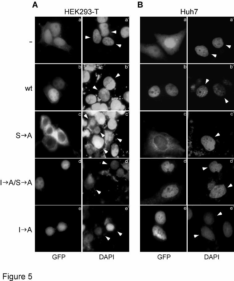
To substantiate these findings in additional cell types, human embryonic kidney (HEK293-T) and hepatoma (Huh7) cells were transiently transfected with full length GFP-HIF-1α or its mutant forms and examined by fluorescence microscopy. In full agreement with the results previously obtained in HeLa cells, Figure 5 shows that wild-type GFP-HIF-1α was predominantly nuclear (panels b), the phosphorylation-deficient form S A was mislocalized into the cytoplasm (panels c) and the I A/S A mutant form, lacking both the NES and the MAPK-phosphorylation sites, re-accumulated inside the nucleus (panels d) in all transfected cells of both cell lines.
Taken together, these data show that disruption of the HIF-1α NES suppresses the mutation at the MAPK phosphoacceptor site and activates the phosphorylation-deficient mutants of HIF-1α. Moreover, the HIF-1α NES mutants become largely resistant to elimination of phosphorylation resulting from either mutation in HIF-1α or MAPK pathway inhibition. This strongly suggests that the phosphorylation-sensitive HIF-1α NES characterized in this work is an important mediator of the control that the MAPK pathway exerts on HIF-1 activity. Over-expression of wild-type and mutant MTD modulates the localization and activity of
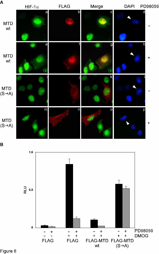
endogenous HeLa cell HIF-1α - To extend our analysis, we studied the effect of wild-type or mutant MTD on the localization and activity of endogenous HeLa cell HIF-1α. As wild-type MTD is a good phosphorylation substrate for MAPK but a weak interaction partner of CRM1 (Figures 2B and 3B) our results would predict that, when over-expressed, it should compete with endogenous HIF-1α for MAPK-mediated phosphorylation, thus impairing nuclear accumulation and transcriptional activity of HIF-1α. On the other hand, over-expression of MTD(S A), which is a bad MAPK substrate but interacts readily with CRM1, should inhibit the interaction between unmodified endogenous HIF-1α and CRM1, thus causing nuclear retention and stimulation of HIF-1α under conditions that eliminate MAPK activity (i.e. upon treatment with PD98059).
To test these predictions, HeLa cells over-expressing FLAG-tagged MTD or MTD(S A) were induced to express endogenous HIF-1α by DMOG and were then subjected to double immunofluorescence staining with anti-HIF-1α and anti-FLAG antibodies or analyzed in reporter gene assays. In non-transfected cells, endogenous DMOG-induced HIF-1α was found exclusively inside the nucleus, whereas in cells over-expressing FLAG-MTD, HIF-1α was distributed in both nucleus and cytoplasm (Figure 6A, panels a-d). Accordingly, over-expression of FLAG-MTD caused dramatic reduction of HIF-1 transcriptional activity (Figure 6B), corroborating our prediction that MTD should inhibit both phosphorylation and nuclear accumulation of HIF-1α. When the cells were treated with PD98059, DMOG-induced HIF-1α and FLAG-MTD were distributed in both nucleus and cytoplasm (Figure 6A, panels e-h), indicating that inhibition of the MAPK pathway caused nuclear export not only of endogenous HIF-1α but also of FLAG-MTD.
In cells over-expressing the phosphorylation-deficient FLAG-MTD(S A) form, the nuclear accumulation of endogenous HIF-1α was not affected, while FLAG-MTD(S A) was itself largely cytoplasmic (Figure 6A, panels i-l). Exposing the cells to PD98059 caused the partial redistribution of endogenous HIF-1α to the cytoplasm, except in the cells that over-expressed FLAG-MTD(S A), in which HIF-1α remained exclusively nuclear (Figure 6A, panels m-p). In agreement, when
by guest on January 2, 2019http://w
ww
.jbc.org/D
ownloaded from
Regulated nuclear export of HIF-1α 6
MTD(S A) was over-expressed, endogenous HIF-1 activity was not only mostly preserved but also became largely insensitive to inhibition of the MAPK pathway (Figure 6B). This supports our hypothesis that MTD(S A), by its ability to interact with CRM1, should inhibit nuclear export of unphosphorylated HIF-1α. Thus, over-expression of MTD or MTD(S A) affects HIF-1α localization and modulates HIF-1 activity by interfering with MAPK- or CRM1-dependent functions, respectively.
DISCUSSION
Two major ways to control HIF-1α have
been previously widely described. The first relies on hydroxylation-dependent degradation/ inactivation and appears to be the preferential means of matching HIF-1 activity to oxygen and metabolite levels (6,22). The second does not depend directly on oxygen levels, involves control of HIF-1α protein synthesis via the PI-3K/AKT pathway and may link HIF-1 activity to the growth state of the cell (10,23). Our work now suggests an additional oxygen-independent mechanism that operates downstream of the synthesis and stabilization steps, involves regulated nuclear export of HIF-1α, responds to MAPK-pathway activation and may, thus, connect HIF-1 activity to cell proliferation. Its principal component lies in the MTD of HIF-1α in a form of an atypical hydrophobic CRM1-dependent NES which is “masked” when one or two proximal serine residues are phosphorylated by p42/44 MAPK. As also shown for a few other shuttling transcription factors (24-27), inhibition of nuclear export by phosphorylation can be a very efficient way to ensure exclusive nuclear localization and full activation of HIF-1α. This may occur after growth factor stimulation at normoxia or under hypoxia which can also activate itself the MAPK pathway (28,29).
The identified NES in HIF-1α (MEDIKILI; conserved hydrophobic residues in bold) deviates from the recently proposed wide CRM1-dependent hydrophobic NES consensus (Φx2-3Φx2-3ΦxΦ; Φ=L,I,V,F,M; x=any amino acid; 21,30) by being atypical in terms of hydrophobic residue composition and spacing. Nevertheless, localization experiments in the presence of Leptomycin B (Figure 3A) as well as immuno-
precipitation analysis (Figure 3B,C & D) suggest that this NES physically interacts with CRM1 when the neighboring SP sites are not phosphorylated. We were, however, unable to provide evidence for direct binding of CRM1 to the HIF-1α NES in the presence of Ran-GTP using recombinant bacterially expressed proteins in vitro (data not shown). It is known that many NESs that depend on CRM1 bind to it with low affinity making in vitro reconstitution of nuclear export complexes extremely difficult (21,30). This may also be the case for the atypical HIF-1α NES. Alternatively, its interaction with CRM1 may require additional cellular factors, such as RanBP3 (21,30) or be indirect through an unknown adaptor protein that recognizes the conserved hydrophobic residues.
Our work does not exclude additional effects of MAPK-mediated phosphorylation on the HIF-1α pathway. Although the transcriptional inactivity of the HIF-1α S A mutant matches closely its nuclear exclusion, the reduction in the activity of wild-type HIF-1α in the presence of PD98059 or FLAG-MTD (Figures 1B; 4B; 6B) is disproportionate to its cytoplasmic mis-localization (Figures 1A, panel b; 4A, panel g; 6A, panels a-d; respectively). Steady-state distribution of HIF-1α should normally represent a snapshot of a dynamic transport process that involves continuous and rapid shuttling in and out of the nucleus. It is, therefore, possible that when nuclear export of HIF-1α is stimulated by MAPK-inhibition, the nuclear resident time of HIF-1α may still be long enough for optical detection but too short for efficient heterodimerization, binding to DNA and transcriptional activation. Alternatively, it is also possible that inhibition of HIF-1α phosphorylation by PD98059 or FLAG-MTD negatively affects additional steps such as the interaction of HIF-1α with co-transcriptional activators. In support of this second hypothesis, both FLAG-MTD and GFP-MTD accumulate inside the nucleus (Figures 2C, panel b; 6A, panel b), although their small size should allow free diffusion between nuclear and cytoplasmic compartments. This indicates that HIF-1α MTD may contain, in addition to the NES, an uncharted sequence that upon phosphorylation promotes docking to an unknown nuclear factor required for optimal HIF-1α activity. Finally, treatment with PD98059 causes minor but significant inhibition
by guest on January 2, 2019http://w
ww
.jbc.org/D
ownloaded from

Regulated nuclear export of HIF-1α 7
of activity even in the cases of the phosphomimetic S E (Figure 1B) or NES I A (Figure 4B) HIF-1α mutants suggesting indirect effects of MAPK on HIF-1 mediated transcription, such as phosphorylation of CBP/p300 and stimulation of its interaction with HIF-1α (31).
In any case, the characterization of the MAPK-targeted NES on HIF-1α opens up the way for further investigation of HIF-1α nuclear
transport and its significance for regulating the hypoxia response pathway, especially under conditions that stimulate the MAPK-pathway and cellular proliferation. Our findings also offer proof-of-principle that nucleocytoplasmic traf-ficking may be an intriguing target for controlling HIF-1 activity under pathological conditions involving tissue hypoxia.
REFERENCES 1. Semenza, G. L. (2007) Sci STKE 2007, cm8 2. Semenza, G. L. (2003) Nat Rev Cancer 3, 721-732 3. Schofield, C. J., and Ratcliffe, P. J. (2005) Biochem Biophys Res Commun 338, 617-626 4. Lancaster, D. E., McDonough, M. A., and Schofield, C. J. (2004) Biochem Soc Trans 32, 943-945 5. Kietzmann, T., and Gorlach, A. (2005) Semin Cell Dev Biol 6. Pan, Y., Mansfield, K. D., Bertozzi, C. C., Rudenko, V., Chan, D. A., Giaccia, A. J., and Simon,
M. C. (2007) Mol Cell Biol 27, 912-925 7. Chandel, N. S., and Budinger, G. R. (2007) Free Radic Biol Med 42, 165-174 8. Bardos, J. I., and Ashcroft, M. (2005) Biochim Biophys Acta 1755, 107-120 9. Dery, M. A., Michaud, M. D., and Richard, D. E. (2005) Int J Biochem Cell Biol 37, 535-540 10. Bernardi, R., Guernah, I., Jin, D., Grisendi, S., Alimonti, A., Teruya-Feldstein, J., Cordon-Cardo,
C., Simon, M. C., Rafii, S., and Pandolfi, P. P. (2006) Nature 442, 779-785 11. Richard, D. E., Berra, E., Gothie, E., Roux, D., and Pouyssegur, J. (1999) J Biol Chem 274,
32631-32637 12. Mylonis, I., Chachami, G., Samiotaki, M., Panayotou, G., Paraskeva, E., Kalousi, A., Georgatsou,
E., Bonanou, S., and Simos, G. (2006) J Biol Chem 281, 33095-33106 13. Triantafyllou, A., Mylonis, I., Simos, G., Bonanou, S., and Tsakalof, A. (2008) Free Radic Biol
Med 44, 657-670 14. Pemberton, L. F., and Paschal, B. M. (2005) Traffic 6, 187-198 15. Kallio, P. J., Okamoto, K., O'Brien, S., Carrero, P., Makino, Y., Tanaka, H., and Poellinger, L.
(1998) Embo J 17, 6573-6586 16. Kubis, H. P., Hanke, N., Scheibe, R. J., and Gros, G. (2005) Biochim Biophys Acta 1745, 187-195 17. Groulx, I., and Lee, S. (2002) Mol Cell Biol 22, 5319-5336 18. Berra, E., Roux, D., Richard, D. E., and Pouyssegur, J. (2001) EMBO Rep 2, 615-620 19. Chachami, G., Paraskeva, E., Georgatsou, E., Bonanou, S., and Simos, G. (2005) Biochem
Biophys Res Commun 331, 464-470 20. Lyberopoulou, A., Venieris, E., Mylonis, I., Chachami, G., Pappas, I., Simos, G., Bonanou, S.,
and Georgatsou, E. (2007) Cell Physiol Biochem 20, 995-1006 21. Hutten, S., and Kehlenbach, R. H. (2007) Trends Cell Biol 17, 193-201 22. Schofield, C. J., and Ratcliffe, P. J. (2004) Nat Rev Mol Cell Biol 5, 343-354 23. Deberardinis, R. J., Lum, J. J., Hatzivassiliou, G., and Thompson, C. B. (2008) Cell Metab 7, 11-
20 24. Zhang, Y., and Xiong, Y. (2001) Science 292, 1910-1915 25. Lee, H., and Bai, W. (2002) Mol Cell Biol 22, 5835-5845 26. Ikuta, T., Kobayashi, Y., and Kawajiri, K. (2004) J Biol Chem 279, 19209-19216 27. Sasaki, T., Kojima, H., Kishimoto, R., Ikeda, A., Kunimoto, H., and Nakajima, K. (2006) Mol
Cell 24, 63-75
by guest on January 2, 2019http://w
ww
.jbc.org/D
ownloaded from
Regulated nuclear export of HIF-1α 8
28. Bardos, J. I., and Ashcroft, M. (2004) Bioessays 26, 262-269 29. Bilton, R. L., and Booker, G. W. (2003) Eur J Biochem 270, 791-798 30. Kutay, U., and Guttinger, S. (2005) Trends Cell Biol 15, 121-124 31. Sang, N., Stiehl, D. P., Bohensky, J., Leshchinsky, I., Srinivas, V., and Caro, J. (2003) J Biol
Chem 278, 14013-14019
FOOTNOTES We are grateful to the other members of our Laboratories of Biochemistry and Physiology for their help and useful comments. This work was supported in part by a grant, co-funded by the European Social Fund & National Resources - EPEAEK II – PYTHAGORAS, from the Greek Ministry of National Education and Religious Affairs to G.S. The abbreviations used are: DMOG, dimethyl-oxalylglycine; HIF-1, hypoxia inducible factor 1; LMB, Leptomycin B; MTD, MAPK target domain; NES, nuclear export signal.
by guest on January 2, 2019http://w
ww
.jbc.org/D
ownloaded from

Regulated nuclear export of HIF-1α 9
FIGURE LEGENDS Figure 1. Phosphomimetic mutation Ser641 Glu stimulates HIF-1α activity and renders it resistant to MAPK inhibition. (A) Localization of wild-type (wt) GFP-HIF-1α or mutant form GFP-HIF-1α(S E) bearing the phosphomimetic Ser641 Glu mutation (as indicated) in Hela cells without (a and c) or after (b and d) treatment with 50 µΜ PD98059 for 4h. a’ – d’: nuclei stained with DAPI, white arrowheads indicate the nuclei of transfected cells. In all cases the cells shown in the micrographs of this figure are representative of greater than 95% of the cells examined in, at least, three repetitions of the experiment. (B) Transcriptional activity of wild-type (wt) GFP-HIF-1α or GFP-HIF-1α(S E) in HeLa cells, 24h post-transfection, determined by reporter gene assay as described in Materials and Methods. Where indicated (+), cells were treated with 50 µM PD98059 for 4h prior lysis. Values are expressed in relation to the values obtained from the empty pEGFP vector and represent the mean of three independent experiments performed in triplicate (± SE). Figure 2. Disruption of a putative hydrophobic NES causes nuclear relocation of phosphorylation-deficient HIF-1α MTD. (A) Top panel: Alignment of amino acid sequences of the HIF-1α MTD (aa 616-658) from vertebrate HIF-1α homologues. MAPK phosphorylation sites are in underlined italics and neighboring conserved hydrophobic residues are shown in bold. A consensus NES is also shown. Bottom panel: Wild-type and mutant forms of human HIF-1α MTD used in this work. Mutations introduced by site-directed mutagenesis are highlighted in grey boxes. (B) 2 µg of GST (lanes 1, 5, 9), GST-HIF-1α (lanes 2, 6, 10), GST-MTD (lanes 3, 7, 11) and GST-MTD(S A) (lanes 4, 8, 12) were subjected to in vitro phosphorylation by either 50 units of recombinant p42 MAPK (lanes 5-8) or 2 µg of HeLa nuclear protein extracts (N.E.; lanes 9-12) and analyzed by SDS-PAGE followed by Coomassie Blue staining (lanes 1-4) or autoradiography (lanes 5-12). Only the relevant parts of the gels are shown. (C) Localization of GFP (panel a) and wild-type (panel b) or indicated mutant forms (panels c-e) of GFP-MTD in Hela cells. Nuclei are stained with DAPI (a’-e’) and white arrowheads indicate the nuclei of transfected cells. In all cases the cells shown in the micrographs of this figure are representative of greater than 95% of the cells examined in, at least, three repetitions of the experiment Figure 3. The NES of HIF-1α MTD depends on and interacts with CRM1. (A) 24h post-transfection, HeLa cells expressing GFP-MTD(S A) were further incubated without (a) or with (b) 10 ng/ml LMB for 4h, fixed, stained with DAPI (a’, b’; white arrowheads indicate the nuclei of transfected cells) and subjected to fluorescence microscopy. In all cases, the cells shown in the micrographs of this figure are representative of greater than 95% of the cells examined in, at least, three repetitions of the experiment (B) and (C) Total cell extracts of HeLa cells expressing wild-type (lane 1) or mutant forms (lanes 2-4 as indicated) of GFP-MTD (B) or full-length GFP-HIF-1α (C) were subjected to immunoprecipitation with an anti-GFP serum. Extracts (Input; upper two panels) and precipitated proteins (I.P.; lower two panels) were analyzed by western blotting using anti-CRM1 and anti-GFP (B) or anti-CRM1 and anti-HIF-1α (C) antibodies as indicated. Only the relevant parts of the blots are shown. (D) Total cell extracts of HeLa cells treated for 4h with 1 mM DMOG alone (-) or in combination with 50 µM PD98059 (+) were subjected to immunoprecipitation with a rabbit anti-HIF-1α polyclonal antibody. Extracts (Input) and precipitated proteins (I.P.) were analyzed by western blotting using anti-CRM1 mouse monoclonal and an affinity purified anti-HIF-1α rabbit polyclonal antibodies.
by guest on January 2, 2019http://w
ww
.jbc.org/D
ownloaded from

Regulated nuclear export of HIF-1α 10
Figure 4. Mutation of the NES renders HIF-1α localization and activity insensitive to lack of phosphorylation. (A) Subcellular localization of GFP (a), wild-type (b, g) or mutant (c-e and h-j as indicated) forms of GFP-HIF-1α expressed in HeLa cells. 20h post-transfection cells were treated with (panels f-j) or without (panels a-e) 50 µM PD98059 for 4h, fixed and subjected to fluorescence microscopy. Nuclei are stained with DAPI (a’-j’) and white arrowheads indicate the nuclei of transfected cells. In all cases, the cells shown in the micrographs of this figure are representative of greater than 95% of the cells examined in, at least, three repetitions of the experiment. (B) Transcriptional activity of wild-type (wt) or mutant (as indicated) forms of GFP-HIF-1α in HeLa cells, determined as described in Materials & Methods. Where indicated (+), cells were treated with 50 µM PD98059 for 4h prior to lysis. Values are expressed in relation to the values obtained from the empty pEGFP vector and represent the mean of three independent experiments performed in triplicate (± SE). Figure 5. Mutation of the NES causes the nuclear accumulation of phosphorylation-deficient HIF-1α in HEK293-T and Huh7 cells. Subcellular localization of GFP (a), GFP-HIF-1α (b) and its mutant forms (c-e as indicated) expressed in HEK-293T (A) and Huh7 (B) cells. 24h post-transfection cells were fixed and subjected to fluorescence microscopy. Nuclei are stained with DAPI (a’-e’) and white arrowheads indicate the nuclei of transfected cells. In all cases, the cells shown in the micrographs of this figure are representative of greater than 95% of the cells examined in, at least, two repetitions of the experiment. Figure 6. Expression of MTD or MTD(S A) in HeLa cells modulates the activity of endogenous HIF-1α. (A) HeLa cells expressing FLAG-MTD (a-h) or FLAG-MTD(S A) (i-p) were treated for 4h with 1 mM DMOG alone (-) or in combination with 50 µM PD98059 (+). The cells were then fixed, immunostained with affinity purified anti-HIF-1α rabbit polyclonal (a, e, i, m) and anti-FLAG mouse monoclonal (b, f, j, n) antibodies, counterstained with DAPI (d, h, l, p) and subjected to immunofluorescence microscopy. Images of endogenous HIF-1α localization using an FITC-conjugated secondary antibody are shown in green color while images of FLAG-MTD localization using a Cy3-conjugated secondary antibody in red. Merged pictures (merge is represented by yellow) are shown in c, g, k and o. White arrowheads indicate the nuclei of transfected cells. In all cases, the cells shown in the micrographs in this figure are representative of greater than 95% of the cells examined in two repetitions of the experiment. (B) Determination of HIF-1 transcriptional activity in HeLa cells expressing FLAG, FLAG-MTD or FLAG-MTD(S A) and treated with 1 mM DMOG, 50 µM PD98059 or their combination for 4h (as indicated). Values are in abstract units (RLU, Relative Luciferase Activity) and represent the mean of two independent experiments performed in triplicate (± SE).
by guest on January 2, 2019http://w
ww
.jbc.org/D
ownloaded from

Ilias Mylonis, Georgia Chachami, Efrosyni Paraskeva and George Simos by MAPKαhypoxia-inducible factor HIF-1
An atypical CRM1-dependent nuclear export signal mediates regulation of
published online August 7, 2008J. Biol. Chem.
10.1074/jbc.M803081200Access the most updated version of this article at doi:
Alerts:
When a correction for this article is posted•
When this article is cited•
to choose from all of JBC's e-mail alertsClick here
by guest on January 2, 2019http://w
ww
.jbc.org/D
ownloaded from