Akt&Ezrin Tyr353&NF-κB pathway regulates EGF-induced EMT...
11
Akt/Ezrin Tyr353/NF-jB pathway regulates EGF-induced EMT and metastasis in tongue squamous cell carcinoma Y Wang 1,4 , Z Lin 1,4 , L Sun 1,2,4 , S Fan 1 , Z Huang 1 , D Zhang 1 , Z Yang 1 , J Li * ,1,2,3 and W Chen * ,1,3 1 Department of Oral and Maxillofacial Surgery, Sun Yat-sen Memorial Hospital, Sun Yat-sen University, 107 Yanjiang West Road, Guangzhou 510120, People’s Republic of China; 2 Key Laboratory of Malignant Tumor Gene Regulation and Target Therapy of Guangdong Higher Education Institutes, 107 Yanjiang West Road, Guangzhou 510120, People’s Republic of China and 3 Oral and Cranio-maxillofacial Surgery Center, 107 Yanjiang West Road, Guangzhou 510120, People’s Republic of China Background: Epithelial–mesenchymal transition (EMT) is a crucial programme in cancer metastasis. Epidermal growth factor (EGF) is a key inducer of EMT, and Ezrin has an important role in this process. However, how Ezrin is activated and whether it mediates EGF-induced EMT in tongue squamous cell carcinomas (TSCCs) through activating NF-kB remains obscure. Methods: We used two TSCC cell lines as a cell model to study invasion and EMT in vitro, and used nude mice xenografts model to evaluate metastasis of TSCC cells. Finally, we evaluated the level of pEzrin Tyr353, nuclear p65 and EMT markers in TSCC clinical samples. Results: Ezrin Tyr353 was phosphorylated through Akt (but not ERK1/2, ROCK1) pathway, and lead to the activation of NF-kB in EGF-treated TSCC cells. Akt and NF-kB inhibitors blocked EGF-induced EMT, and suppressed invasion and migration of TSCC cells. In vivo, silencing Ezrin significantly suppressed EGF-enhanced metastasis of TSCC xenografts. Finally, high levels of expression of pEzrin Tyr353, nuclear p65, vimentin and low level of expression of E-cadherin were correlated with cancer metastasis and poor patient prognosis. Conclusion: Our data suggest that Akt/Ezrin Tyr353/NF-kB pathway regulates EGF-induced EMT and metastasis inTSCC, and Ezrin may serve as a therapeutic target to reverse EMT in tongue cancers and prevent TSCC progression. Tongue squamous cell carcinoma (TSCC) is the most common oral cancer, which frequently leads to lymph node and distant metastasis, and metastasis is the most reliable adverse prognostic factor in TSCC patients (Sano and Myers, 2007; Jemal et al, 2009). Previous studies have demonstrated that tumour metastasis is associated with epithelial–mesenchymal transition (EMT), a biological process in which epithelial cells lose cell–cell contact, E-cadherin expression and develop mesenchymal properties, leading to enhanced motility, invasiveness, vimentin expression and resistance to apoptosis (Thiery and Sleeman, 2006; Polyak and Weinberg, 2009). Recent advances have fostered a more detailed understanding of molecular mechanisms and networks that govern EMT in tumour progression (Huber et al, 2005). The signalling pathways are always triggered by different members of the TGFb superfamily, Wnts, Notch, EGF, HGF, FGF and HIF (Kalluri and Weinberg, 2009; Thiery et al, 2009; Chaudhury et al, 2010; Shin et al, 2010). The transcription factors include Snail, Slug, Twist, ZEB1, ZEB2/SIP1, b-catenin and NF-kB (Huber et al, 2004; Kudo-Saito et al, 2009; Wellner et al, 2009; Wu et al, 2009; Sa ´nchez-Tillo ´ et al, 2010; Vuoriluoto et al, 2010; Yip and Seow, 2012). Besides, these studies also illustrate that blocking these signal networks can inhibit EMT and cancer metastasis in vitro and *Correspondence: Professor W Chen; E-mail: [email protected] or Professor J Li; E-mail: [email protected] 4 These authors contributed equally to this work. Received 12 July 2013; revised 16 October 2013; accepted 14 November 2013; published online 17 December 2013 & 2014 Cancer Research UK. All rights reserved 0007 – 0920/14 FULL PAPER Keywords: tongue squamous cell carcinoma; metastasis; Ezrin; EMT; NF-kB British Journal of Cancer (2014) 110, 695–705 | doi: 10.1038/bjc.2013.770 www.bjcancer.com | DOI:10.1038/bjc.2013.770 695
Transcript of Akt&Ezrin Tyr353&NF-κB pathway regulates EGF-induced EMT...
Akt/Ezrin Tyr353/NF-jB pathway regulatesEGF-induced EMT and metastasis in tonguesquamous cell carcinomaY Wang1,4, Z Lin1,4, L Sun1,2,4, S Fan1, Z Huang1, D Zhang1, Z Yang1, J Li*,1,2,3 and W Chen*,1,3
1Department of Oral and Maxillofacial Surgery, Sun Yat-sen Memorial Hospital, Sun Yat-sen University, 107 Yanjiang West Road,Guangzhou 510120, People’s Republic of China; 2Key Laboratory of Malignant Tumor Gene Regulation and Target Therapy ofGuangdong Higher Education Institutes, 107 Yanjiang West Road, Guangzhou 510120, People’s Republic of China and 3Oral andCranio-maxillofacial Surgery Center, 107 Yanjiang West Road, Guangzhou 510120, People’s Republic of China
Background: Epithelial–mesenchymal transition (EMT) is a crucial programme in cancer metastasis. Epidermal growth factor (EGF)is a key inducer of EMT, and Ezrin has an important role in this process. However, how Ezrin is activated and whether it mediatesEGF-induced EMT in tongue squamous cell carcinomas (TSCCs) through activating NF-kB remains obscure.
Methods: We used two TSCC cell lines as a cell model to study invasion and EMT in vitro, and used nude mice xenografts modelto evaluate metastasis of TSCC cells. Finally, we evaluated the level of pEzrin Tyr353, nuclear p65 and EMT markers in TSCCclinical samples.
Results: Ezrin Tyr353 was phosphorylated through Akt (but not ERK1/2, ROCK1) pathway, and lead to the activation of NF-kB inEGF-treated TSCC cells. Akt and NF-kB inhibitors blocked EGF-induced EMT, and suppressed invasion and migration of TSCCcells. In vivo, silencing Ezrin significantly suppressed EGF-enhanced metastasis of TSCC xenografts. Finally, high levels ofexpression of pEzrin Tyr353, nuclear p65, vimentin and low level of expression of E-cadherin were correlated with cancermetastasis and poor patient prognosis.
Conclusion: Our data suggest that Akt/Ezrin Tyr353/NF-kB pathway regulates EGF-induced EMT and metastasis inTSCC, andEzrin may serve as a therapeutic target to reverse EMT in tongue cancers and prevent TSCC progression.
Tongue squamous cell carcinoma (TSCC) is the most commonoral cancer, which frequently leads to lymph node and distantmetastasis, and metastasis is the most reliable adverse prognosticfactor in TSCC patients (Sano and Myers, 2007; Jemal et al, 2009).Previous studies have demonstrated that tumour metastasis isassociated with epithelial–mesenchymal transition (EMT), abiological process in which epithelial cells lose cell–cell contact,E-cadherin expression and develop mesenchymal properties,leading to enhanced motility, invasiveness, vimentin expressionand resistance to apoptosis (Thiery and Sleeman, 2006; Polyak andWeinberg, 2009). Recent advances have fostered a more detailed
understanding of molecular mechanisms and networks that governEMT in tumour progression (Huber et al, 2005). The signallingpathways are always triggered by different members of the TGFbsuperfamily, Wnts, Notch, EGF, HGF, FGF and HIF (Kalluri andWeinberg, 2009; Thiery et al, 2009; Chaudhury et al, 2010; Shinet al, 2010). The transcription factors include Snail, Slug, Twist,ZEB1, ZEB2/SIP1, b-catenin and NF-kB (Huber et al, 2004;Kudo-Saito et al, 2009; Wellner et al, 2009; Wu et al, 2009;Sanchez-Tillo et al, 2010; Vuoriluoto et al, 2010; Yip and Seow,2012). Besides, these studies also illustrate that blocking thesesignal networks can inhibit EMT and cancer metastasis in vitro and
*Correspondence: Professor W Chen; E-mail: [email protected] or Professor J Li; E-mail: [email protected] authors contributed equally to this work.
Received 12 July 2013; revised 16 October 2013; accepted 14 November 2013; published online 17 December 2013
& 2014 Cancer Research UK. All rights reserved 0007 – 0920/14
FULL PAPER
Keywords: tongue squamous cell carcinoma; metastasis; Ezrin; EMT; NF-kB
British Journal of Cancer (2014) 110, 695–705 | doi: 10.1038/bjc.2013.770
www.bjcancer.com | DOI:10.1038/bjc.2013.770 695
in vivo. Therefore, EMT in tumour cells is a crucial programme incancer metastasis, and determining the mechanisms that governEMT is essential for the development of novel therapeuticstrategies to overcome cancer metastasis.
Ezrin, the most important member of ERM (Ezrin/Radixin/Moesin) proteins, not only involves in cytoskeleton organisationbut also in transmission of signals in responses to extracellular cues(Louvet-Vallee, 2000; Arpin et al, 2011). Ezrin may function asmetastasis-related oncogene (Khanna et al, 2004) by modulatingmultiple cellular processes, including the formation of microvilli(Chiang et al, 2008), maintenance of cell shape (Baumgartner et al,2006), cell–cell adhesion (Srivastava et al, 2005), cell motility(Vanacker et al, 2011) and invasion (Chuan et al, 2009). Activationof Ezrin is a cellular mechanism to promote local alterations in cellmorphology in response to EGF and might have a role in tumourcell metastasis (Baumgartner et al, 2006). On the other hand, thecell morphology is an important phenotype transition of EMT andEGF can initiate and sustain various aspects of the EMT pathwayin normal and malignant epithelial cells (Hardy et al, 2010; Chaiet al, 2012). Activation of NF-kB by inflammatory cytokine hasbeen implicated in the control of EMT and motility andinvasiveness of tumour cells (Wu et al, 2009). More interestingly,a recent study has shown that the complex of NF-kB and Ezrin isessential for L1 (cell–neural adhesion molecule L1CAM)-mediatedmetastasis of colon cancer cells (Gavert et al, 2010). Furthermore,another study suggests that NF-kB activity is required for Ezrin-induced actin polymerisation (Lim et al, 2009). Accumulatingevidence has demonstrated that Ezrin may have an important rolein EMT through the activation of NF-kB pathway. However,whether Ezrin mediates EGF-induced EMT in TSCCs throughactivating NF-kB remains obscure.
In the present study, we investigated the role of Ezrin inregulating EGF-induced EMT in TSCC cells at first. Then, weexplored how Ezrin was activated in response to EGF, identified itsphosphorylated site and detected its effect on NF-kB activity. Wedefined the pathway through which EGF induced EMT in TSCCcells, and evaluated the effects of Ezrin expression on tumourgrowth and metastasis of TSCC xenografts. Finally, we correlatedthe activation of Ezrin and NF-kB with the clinicopathologicalstatus and prognosis of TSCC patients.
MATERIALS AND METHODS
Cell culture. Human tongue cancer cell lines SCC9 and SCC25,and a human embryonic kidney cell line (HEK-293) werepurchased from the American Type Culture Collection (Manassas,VA, USA). SCC9 and SCC25 were cultivated in Dulbecco’smodified Eagle’s medium-F12 (Gibco, Rockville, MD, USA)supplemented with 10% fetal bovine serum (Invitrogen, Carlsbad,CA, USA). Human embryonic kidney-293 cells were cultivated inDulbecco’s modified Eagle’s medium (Gibco) supplemented with10% fetal bovine serum (Invitrogen).
Transfection. Ezrin siRNAs were obtained from GenePharma(Shanghai, China) and their sequences were Ezrin siRNA1:50-GUGGGAUGCUCAAAGAUAATT-30 and Ezrin siRNA2:50-GGGCAACCAUGAGUUGUAUTT-50. Cells were transfectedwith 30 nM siRNA using Lipofectamine 2000 (Invitrogen).Lentivirus carrying Ezrin shRNA were also obtained fromGenePharma. An expression construct with wild-type Ezrin(Ez-WT) and the Y353F-Ezrin mutant (Ez-Y353F) are a kind giftfrom Dr Monique Arpin (Srivastava et al, 2005).
Quantitative RT–PCR. Real-time PCR was carried out usingLightCycler 480 (Roche, Basel, Switzerland). Reactions were run intriplicate in three independent experiments. The relative expres-sion of E-cadherin and vimentin were normalised to GAPDH. The
primer sequences were shown in our previous study (Sun et al,2012). All the relative expressions in control were set to 1.
Western blotting. Protein extracts were resolved through 10%SDS–polyacrylamide gel electrophoresis, transferred to polyviny-lidene difluoride membranes (Bio-Rad, Berkeley, CA, USA),probed with antibody against human E-cadherin, vimentin (SantaCruz, Santa Cruz, CA, USA), b-catenin, pEzrin Thr567 (Abcam,Cambridge, MA), Ezrin, pEzrin Tyr353, IkBa, p-IkBa, ERK1/2,p-ERK1/2, Akt, p-Akt (Ser473), ROCK1 (Cell Signaling Techno-logy, CST, Danvers, MA, USA) or GAPDH (Proteintech, Chicago,IL, USA), and then with peroxidase-conjugated secondary antibody(Proteintech) and visualised by chemiluminescence (GE, Fairfield,CT, USA). The band densities were quantified by Gel-Pro analyser4.0 (Media Cybernetics, Rockville, MD, USA). The band intensityvalues of E-cadherin, vimentin, Ezrin and ROCK1 were normalisedto those of GAPDH. The ratios of phosphorylated target to its totalprotein values were also calculated.
Immunofluorescence staining. Cells were stained for immuno-fluorescence on coverslips. After fixation and permeabilisation, thecells were incubated with primary antibodies against E-cadherin,vimentin (Santa Cruz) or p65 (CST) and then incubated withrhodamine- or FITC-conjugated secondary antibodies (Invitrogen).The coverslips were counterstained with 40,6-diamidino-2-phenylindole and imaged under a confocal microscope TCS SP5 (Leica,Solms, Germany). The images were merged with ImageJ software(National Institutes of Health, Betheseda, MD, USA).
Modified Boyden chamber assay. A total of 1� 105 cells wereplated into the upper chamber of a polycarbonate transwell filterchamber (Corning, NewYork, NY, USA) and incubated for 10 h.For invasion assay, the upper chamber was coated with Matrigel(R&D, Minneapolis, MN, USA) and incubated for 24 h. Cells onthe lower membrane surface were fixed in 4% paraformaldehyde,stained with crystal violet and counted (five random 100� fieldsper well). Three independent experiments were performed and thedata are presented as the average±s.d.
Wound healing assay. A total of 1� 106 cells were seeded in six-well plates and grew until reaching 90% confluence. Linear woundswere created using pipette tips and then cells were cultured withoutserum. Wounds were observed and photographed at 0 and 10 h.
Luciferase reporter assay. To evaluate the function of Ezrin inactivating NF-kB, Ezrin siRNA was transfected in TSCC cells, andthen pNF-kB-luc, together with pRL-TK control vector or pTAL-luc,together with pRL-TK, were transfected into these cells. Toconfirm which phosphosite of Ezrin regulated the activity of NF-kB, different Ezrin plasmids (WT or mutated) were co-transfectedwith pNF-kB-luc and pRL-TK control vector into HER-293 cells(low Ezrin expression). Luciferase activities were assayed using aluciferase assay kit (Promega, Madison, WI, USA).
Patients and tissue samples. Primary tongue carcinomas wereobtained from 63 patients who were admitted to the Department ofOral and Maxillofacial Surgery of the Sun Yat-sen MemorialHospital, Sun Yat-sen University, from January 2007 to January2008. All the patients recruited into the present study did notreceive radiotherapy or chemotherapy or any other treatmentbefore and after operation, following a protocol approved by theethics committee. All samples were collected with informedconsent. The pathological sections for immunohistochemical assaywere collected during surgery.
Immunohistochemistry. For immunohistochemistry, E-cadherin,vimentin (Santa Cruz), pEzrin Tyr353, p65 (CST) were used asprimary antibodies for overnight incubation at 4 1C. The sectionswere subsequently treated with secondary antibody, followed byfurther incubation with streptavidin–horseradish peroxidase complex.
BRITISH JOURNAL OF CANCER Akt/Ezrin Tyr353/NF-kB in EGF-induced EMT
696 www.bjcancer.com | DOI:10.1038/bjc.2013.770
Diaminobenzidine (Dako) was used as a chromogen and sectionswere lightly counterstained with haematoxylin. In total, 5� 1000tumour cells were counted in each section. High expression:positive cells 430%; low expression: positive cells o30%.
Tumour xenografts. Tongue cancer SCC9 cells that were infectedwith shGFP or shEzrin were injected subcutaneously into thearmpit of the 5-week-old BALB/c-nu mice (n¼ 8 per group),following a protocol approved by the ethics committee of Sun Yat-sen University. When the xenografts were palpable (around 0.5 cmin diameter), intratumour injection of PBS or EGF at 0.02 mg kg� 1
was performed bi-weekly for 5 consecutive weeks. Tumourgrowth was evaluated by monitoring tumour volume (TV¼length�width2� 0.5) bi-weekly for 8 weeks. Then, tumourxenografts, as well as whole lung and liver tissues, were harvested,weighed and snap-frozen in liquid nitrogen. To evaluate in vivometastasis, images of mice lungs and livers were acquired andportions of the lung and liver tissues were used for qRT–PCR forhuman hypoxanthine phosphorybosyl transferase expression(Sun et al, 2012). Cryosections (4mm) were stained with haematoxylinand eosin and used for immunohistochemistry.
Statistics. All statistical analyses were performed using SPSS 17.0(Armonk, NY, USA). The Student’s t-test and w2 test were used toanalyse the relationship between Ezrin or NF-kB activation(nuclear p65) and clinicopathological characteristics. To measurethe association between pairs of variables, Spearman’s ordercorrelations were run. Kaplan–Meier survival curves were plottedand log-rank test was performed. All experiments for cell cultureswere performed at least in triplicate. Results were expressed asmean±s.d. Po0.05 was considered statistically significant.
RESULTS
Ezrin is phosphorylated by EGF, and reduction of Ezrin inhibitsEGF-induced EMT in TSCC cells. The SCC9 and SCC25 cellsgrew in clusters and were round in shape with tight cell–celljunctions, whereas SCC9 and SCC25 cells treated with EGFdisplayed spindle shape and separated from one another(Supplementary Figure 1a). Immunofluorescence staining andwestern blotting demonstrated that the protein expression ofE-cadherin decreased, whereas that of vimentin markedlyincreased in TSCC cells treated with EGF (SupplementaryFigures 1b and c). Furthermore, E-cadherin and b-catenin werelocalised on and under the cell membrane in the EGF-untreatedTSCC cells and localised in the cytoplasm in the EGF-treatedTSCC cells (Supplementary Figures 1b). In addition, we examinedthe invasion and migration of EGF treated TSCC cells usingBoyden chamber assays. After 24 or 10 h of culture, the invasionand migration increased by 7–5.1- and 8.4–7.4-fold in EGF-treatedSCC9 and SCC25 cells, respectively, as compared with SCC9 andSCC25 cells (Supplementary Figure 1d). Taken together, ourobservation indicates that SCC9 and SCC25 cells treated with EGFhave undergone EMT with enhanced invasiveness and motility.
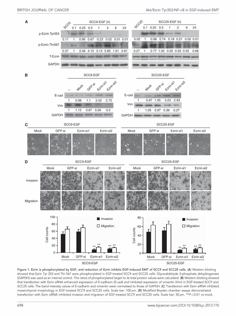
To explore the role of Ezrin in EGF-induced EMT, we firstdetected the phosphorylated site of Ezrin in EGF-treated SCC9 andSCC25 cells. Remarkable upregulation of p-Ezrin Tyr353 andThr567 were observed in EGF-treated SCC9 and SCC25 cells(Figure 1a). Then, we silenced Ezrin expression using RNAinterference (Supplementary Figure 2a), and found that EzrinsiRNAs enhanced expression of E-cadherin and inhibited expres-sion of vimentin in EGF-treated SCC9 and SCC25 cells, as shownby western blotting (Figure 1b). These were further confirmed atmRNA level by qRT–PCR (Supplementary Figures 2b). At thesame time, Ezrin siRNAs reserved mesenchymal morphology ofEGF-treated SCC9 and SCC25 cells. These cells began to grow incluster and were round in shape (Figure 1c). Modified Boyden
chamber assay demonstrated that Ezrin siRNAs suppressed theinvasion and migration of EGF-treated SCC9 and SCC25 cells(Figure 1d). Wound healing assay also showed that down-expression of Ezrin suppressed the motility of EGF-treated TSCCcells (Supplementary Figure 2c). These data suggest that reductionof Ezrin could inhibit EGF-induced EMT in TSCC cells.
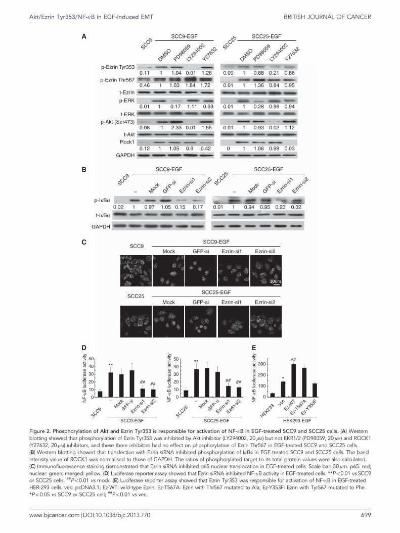
Phosphorylation of Akt and Ezrin Tyr353 is responsible foractivation of NF-kB in EGF-treated TSCC cells. It has beenreported that several signal pathways are involved in activatingEzrin, including Akt, ERK1/2 and ROCK1. We detected theactivations of Akt, ERK1/2 and ROCK1 in EGF-treated TSCC cellsand found that Akt and ERK1/2 were phosphorylated and theexpression of ROCK1 was upregulated (Supplementary Figure 3a).Then, we pretreated these TSCC cells with LY294002 (Aktinhibitor), PD98059 (ERK1/2 inhibitor) or Y27632 (ROCK1inhibitor) and investigated the phosphorylation of Ezrin Tyr353and Thr567. Phosphorylation of Ezrin Tyr353 was inhibited byLY294002, but not by PD98059 and Y27632. The three inhibitorshave no effects on phosphorylation of Ezrin Thr567 (Figure 2a).These data indicated that phosphorylation of Ezrin Tyr353 wasmediated by Akt in EGF-treated TSCC cells.
Immunofluorescence assay showed that p65 translocated toTSCC cell nuclear after EGF treatment (Supplementary Figure 3b),suggesting that NF-kB was activated by EGF. To explore the keymediator in activating NF-kB, we pretreated TSCC cells withLY294002, PD98059 or Y27632 before EGF treatment. As shownby immunofluorescence assay, p65 translocation to cell nuclear wasinhibited by Akt inhibitor and NF-kB inhibitor (BAY and JSH),but not by ERK and ROCK1 inhibitors (Supplementary Figure 3b).Taken together, Akt activated Ezrin Tyr353 and NF-kB in EGF-treated TSCC cells.
To demonstrate the relationship between Ezrin and NF-kB, wesilenced Ezrin expression in EGF-treated TSCC cells and examinedNF-kB activity. We found that Ezrin siRNAs significantlysuppressed phosphorylation of IkBa (Figure 2b) and p65translocation to nuclear (Figure 2c) in EGF-treated TSCC cells.At the same time, luciferase reporter assay showed that knockdownof Ezrin suppressed NF-kB activities in EGF-treated TSCC cells(Figure 2d; Po0.01). These results indicated that Ezrin regulatedthe activation of NF-kB in EGF-treated TSCC cells. To furtherconfirm the functional site of Ezrin, TSCC cells were highlyexpressed with Ezrin or mutated Ezrin (Ezrin Y353F, Ezrin T567A)(kind gifts from Dr Arpin M). Luciferase reporter assay showedthat NF-kB activity was enhanced when EGF-treated TSCC cellswere transfected with WT Ezrin or Ezrin T567A (Po0.05), butcould not be enhanced when cells were transfected with EzrinY353F (Supplementary Figure 3c). Furthermore, we transfectedthese plasmids into HER-293 cells with low Ezrin expression.Luciferase reporter assay also showed that Ezrin Y353F could notenchance NF-kB activity (Figure 2e). In conclusion, these resultsindicated that phosphorylation of Akt and Ezrin Tyr353 isresponsible for the activation of NF-kB in EGF-treated TSCC cells.
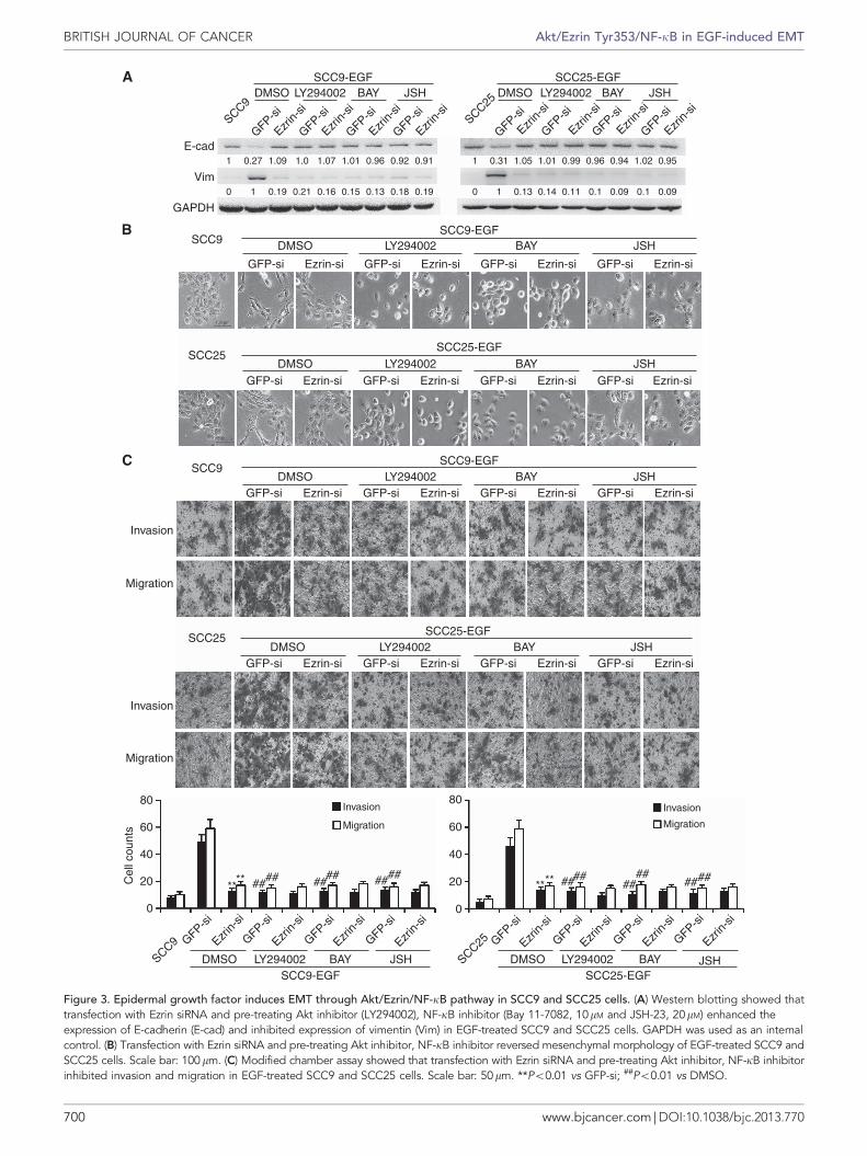
EGF induces EMT through Akt/Ezrin/NF-kB pathway in TSCCcells. To determine the signal pathway involved in EGF-inducedEMT in TSCC cells, we pretreated the cells with inhibitors of Aktor NF-kB and then transfected Ezrin siRNA. We found that Aktand NF-kB inhibitors, as well as Ezrin siRNA, blocked EGF-induced EMT in TSCC cells. Expression of E-cadherin in TSCCcells was not inhibited and the expression of vimentin was notenhanced by EGF (Figure 3a). However, ERK and ROCK1inhibitors could not block EGF-induced EMT in TSCC cells(Supplementary Figure 4a), suggesting that activation of the Akt/Ezrin/NF-kB pathway, but not ERK and ROCK1, was importantfor EGF-induced EMT in TSCC cells. At the same time, Aktand NF-kB inhibitors impaired the mesenchymal phenotype ofEGF-treated TSCC cells (Figure 3b), and suppressed invasion and
Akt/Ezrin Tyr353/NF-kB in EGF-induced EMT BRITISH JOURNAL OF CANCER
www.bjcancer.com | DOI:10.1038/bjc.2013.770 697
p-Ezrin Tyr353
0.1
0.11
0.37
0.96
0.46
0.67
4.15
0.23
3.14
0.02
5.85
0.01
1.91
0.01
0.61
0.02
0.27
1
1
10.1 0.25 0.5
0.98
2.77 1.02 0.55 0.93 0.55 0.66
0.74 0.18 0.21 0.02 0.01
62 240.25 0.5 1
1
1
1 1.12 0.87 0.26 0.2
2.722.521.10.981 1 0.97
0.97
3.23
0.36
2.93
0.271.05
1.05
1
2 6 24
SCC9-EGF (h)
SCC9-EGF SCC25-EGF
SCC25-EGF (h)
SCC9SCC25
p-Ezrin Thr567
T-Ezrin
Vim
E-cad
Mock
Mock
GFP-si
GFP-si
Ezrin-si1
Ezrin-si1
Ezrin-si2
Ezrin-si2
Mock
SCC9-EGF
GFP-si Ezrin-si1 Ezrin-si2
Mock GFP-si Ezrin-si1 Ezrin-si2
Mock GFP-si Ezrin-si1 Ezrin-si2
SCC9-EGF
SCC9-EGF
Invasion
Invasion
Migration
Migration
Invasion
Migration
********** ** ** **
100
80
80
60
20
0
Cel
l cou
nts
Cel
l cou
nts
40
60
20
0
40
SCC25-EGF
SCC25-EGF
GAPDH
– Moc
kGFP-s
i
Ezrin-
si1
Ezrin-
si2
– Moc
kGFP-s
i
Ezrin-
si1
Ezrin-
si2
GAPDH
Vim
E-cad
GAPDH
Mock
SCC25-EGF
GFP-si Ezrin-si2Ezrin-si1
Figure 1. Ezrin is phosphorylated by EGF, and reduction of Ezrin inhibits EGF-induced EMT of SCC9 and SCC25 cells. (A) Western blottingshowed that Ezrin Tyr 353 and Thr 567 were phosphorylated in EGF-treated SCC9 and SCC25 cells. Glyceraldehyde 3-phosphate dehydrogenase(GAPDH) was used as an internal control. The ratios of phosphorylated target to its total protein values were calculated. (B) Western blotting showedthat transfection with Ezrin siRNA enhanced expression of E-cadherin (E-cad) and inhibited expression of vimentin (Vim) in EGF-treated SCC9 andSCC25 cells. The band intensity values of E-cadherin and vimentin were normalised to those of GAPDH. (C) Transfection with Ezrin siRNA inhibitedmesenchymal morphology in EGF-treated SCC9 and SCC25 cells. Scale bar: 100mm. (D) Modified Boyden chamber assays demonstratedtransfection with Ezrin siRNA inhibited invasion and migration of EGF-treated SCC9 and SCC25 cells. Scale bar: 50mm. **Po0.01 vs mock.
BRITISH JOURNAL OF CANCER Akt/Ezrin Tyr353/NF-kB in EGF-induced EMT
698 www.bjcancer.com | DOI:10.1038/bjc.2013.770
p-Ezrin Tyr3530.11 0.09
0.01
0.01
0.88
1.36
0.28
0.93
1.06
0.02
0.98
1.12
0.03
0.940.96
0.84 0.95
0.21 0.86
0.01
1
1
1
1
1
1
1
1
10
0.46 1.03
0.01
0.08 2.33 0.01
1.110.17
1.84 1.72
0.93
1.66
0.12 1 1.05 0.9 0.42
1.04 0.01 1.28
SCC9-EGFA
SCC9-EGF
0.02 1 0.97 1.05 0.15 0.17 0.01 1 0.94 0.95 0.23 0.32
SCC9
SCC25
50
****
*## #### ##
##50300
200
100
0
40
30
20
10
0
40
30
20
10
0
NF
-κB
luci
fera
se a
ctiv
ity
NF
-κB
luci
fera
se a
ctiv
ity
NF
-κB
luci
fera
se a
ctiv
ity
SCC9-EGF SCC25-EGF HEK293-EGF
Mock GFP-si Ezrin-si1 Ezrin-si2
Mock GFP-si Ezrin-si1 Ezrin-si2
–
SCC9-EGF
SCC25-EGF
SCC9
SCC9
Moc
kGFP-s
i
Ezrin-
si1
Ezrin-
si2
–
SCC25
Moc
kGFP-s
i
Ezrin-
si1
Ezrin-
si2
–
SCC9
SCC25
HEK293 ve
c
Ez-W
T
Ez-T56
7A
Ez-Y35
3FM
ock
GFP-si
Ezrin-
si1
Ezrin-
si2–
Moc
k
GFP-si
Ezrin-
si1
Ezrin-
si2
SCC25
DMSO
DMSO
PD9805
9
PD9805
9
LY29
4002
LY29
4002
Y2763
2
Y2763
2
SCC25-EGF
SCC25-EGF
p-Ezrin Thr567
t-Ezrin
t-ERKp-Akt (Ser473)
Rock1
GAPDH
GAPDH
p-IκBα
t-IκBα
p-ERK
t-Akt
B
C
D E
Figure 2. Phosphorylation of Akt and Ezrin Tyr353 is responsible for activation of NF-kB in EGF-treated SCC9 and SCC25 cells. (A) Westernblotting showed that phosphorylation of Ezrin Tyr353 was inhibited by Akt inhibitor (LY294002, 20mM) but not EKR1/2 (PD98059, 20mM) and ROCK1(Y27632, 20mM) inhibitors, and these three inhibitors had no effect on phosphorylation of Ezrin Thr567 in EGF-treated SCC9 and SCC25 cells.(B) Western blotting showed that transfection with Ezrin siRNA inhibited phosphorylation of IkBa in EGF-treated SCC9 and SCC25 cells. The bandintensity value of ROCK1 was normalised to those of GAPDH. The ratios of phosphorylated target to its total protein values were also calculated.(C) Immunofluorescence staining demonstrated that Ezrin siRNA inhibited p65 nuclear translocation in EGF-treated cells. Scale bar: 30mm. p65: red;nuclear: green; merged: yellow. (D) Luciferase reporter assay showed that Ezrin siRNA inhibited NF-kB activity in EGF-treated cells. **Po0.01 vs SCC9or SCC25 cells. ##Po0.01 vs mock. (E) Luciferase reporter assay showed that Ezrin Tyr353 was responsible for activation of NF-kB in EGF-treatedHER-293 cells. vec: pcDNA3.1; Ez-WT: wild-type Ezrin; Ez-T567A: Ezrin with Thr567 mutated to Ala; Ez-Y353F: Ezrin with Tyr567 mutated to Phe.*Po0.05 vs SCC9 or SCC25 cell; ##Po0.01 vs vec.
Akt/Ezrin Tyr353/NF-kB in EGF-induced EMT BRITISH JOURNAL OF CANCER
www.bjcancer.com | DOI:10.1038/bjc.2013.770 699
SCC9
Invasion
Invasion
Invasion
Migration
Migration
80
60
****
#### #### ####
#### #### ####****C
ell c
ount
s
40
20
0
SCC9 GFP-si
Ezrin-
si
GFP-si
Ezrin-
si
GFP-si
Ezrin-
si
GFP-si
Ezrin-
si
GFP-si
Ezrin-
si
GFP-si
Ezrin-
si
GFP-si
Ezrin-
si
GFP-si
Ezrin-
si
80
60
40
20
0
Migration
Invasion
Migration
GFP-si GFP-siEzrin-si Ezrin-si GFP-si Ezrin-si GFP-si Ezrin-siDMSO LY294002 BAY JSH
SCC25
SCC9-EGF SCC25-EGF
GFP-si GFP-siEzrin-si Ezrin-si GFP-si Ezrin-si GFP-si Ezrin-siDMSO
DMSO DMSO
LY294002
LY294002 LY294002
SCC25-EGFBAY
BAY BAY
JSH
JSH JSH
SCC9
GFP-si GFP-siEzrin-si Ezrin-si GFP-si Ezrin-si GFP-si Ezrin-si
DMSO LY294002 BAY JSH
GFP-si GFP-siEzrin-si Ezrin-si GFP-si Ezrin-si GFP-si Ezrin-siDMSO LY294002 BAY JSH
SCC9-EGF
SCC9-EGF
SCC25-EGFSCC25
SCC25
E-cad
Vim
GAPDH
GFP-si
Ezrin-
si
GFP-si
Ezrin-
si
GFP-si
Ezrin-
si
GFP-si
Ezrin-
si
GFP-si
Ezrin-
si
GFP-si
Ezrin-
si
GFP-si
Ezrin-
si
GFP-si
Ezrin-
siSCC9
SCC25
SCC9-EGF SCC25-EGFDMSO LY294002 BAY JSHDMSO LY294002 BAY JSH
1 0.27 1.09 1.0 1.07 1.01 0.96 0.92 0.91 1
1
0.31 1.05
0.13 0.14 0.11 0.1 0.09 0.1 0.09
0.951.020.940.960.991.01
00.190.180.130.150.160.210.190 1
Figure 3. Epidermal growth factor induces EMT through Akt/Ezrin/NF-kB pathway in SCC9 and SCC25 cells. (A) Western blotting showed thattransfection with Ezrin siRNA and pre-treating Akt inhibitor (LY294002), NF-kB inhibitor (Bay 11-7082, 10mM and JSH-23, 20mM) enhanced theexpression of E-cadherin (E-cad) and inhibited expression of vimentin (Vim) in EGF-treated SCC9 and SCC25 cells. GAPDH was used as an internalcontrol. (B) Transfection with Ezrin siRNA and pre-treating Akt inhibitor, NF-kB inhibitor reversed mesenchymal morphology of EGF-treated SCC9 andSCC25 cells. Scale bar: 100mm. (C) Modified chamber assay showed that transfection with Ezrin siRNA and pre-treating Akt inhibitor, NF-kB inhibitorinhibited invasion and migration in EGF-treated SCC9 and SCC25 cells. Scale bar: 50mm. **Po0.01 vs GFP-si; ##Po0.01 vs DMSO.
BRITISH JOURNAL OF CANCER Akt/Ezrin Tyr353/NF-kB in EGF-induced EMT
700 www.bjcancer.com | DOI:10.1038/bjc.2013.770
migration of these cells (Figure 3c). Wound healing assay alsoshowed that blocking the Akt/Ezrin/NF-kB pathway couldsuppress the motility of EGF-treated SCC9 and SCC25 cells(Supplementary Figure 4b).
Reduction of Ezrin inhibits metastasis of EGF-treated TSCCxenografts. As reduction of Ezrin inhibited EMT of EGF-treatedTSCC cells in vitro, we further assessed its effect on tumour growthand metastasis in vivo. Ezrin was stably downregulated in SCC9cells by infected with lentivirus carrying Ezrin shRNA (shEzrin)(Supplementary Figure 5a). As shown in Supplementary Figures 5band c, downexpression of Ezrin did not significantly changetumour growth or body weight of the BALB/c-nu mice inoculatedwith SCC9 cells.
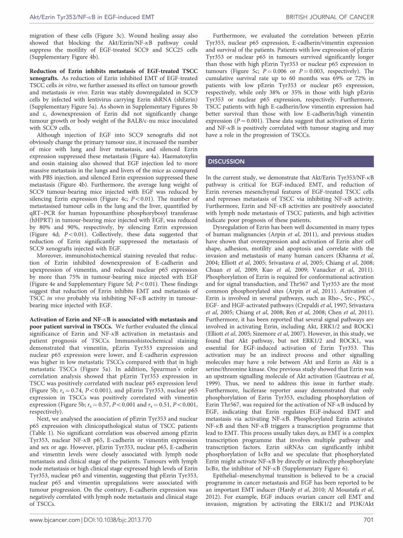
Although injection of EGF into SCC9 xenografts did notobviously change the primary tumour size, it increased the numberof mice with lung and liver metastasis, and silenced Ezrinexpression suppressed these metastasis (Figure 4a). Haematoxylinand eosin staining also showed that EGF injection led to moremassive metastasis in the lungs and livers of the mice as comparedwith PBS injection, and silenced Ezrin expression suppressed thesemetastasis (Figure 4b). Furthermore, the average lung weight ofSCC9 tumour-bearing mice injected with EGF was reduced bysilencing Ezrin expression (Figure 4c; Po0.01). The number ofmetastasised tumour cells in the lung and the liver, quantified byqRT–PCR for human hypoxanthine phosphorybosyl transferase(hHPRT) in tumour-bearing mice injected with EGF, was reducedby 80% and 90%, respectively, by silencing Ezrin expression(Figure 4d; Po0.01). Collectively, these data suggested thatreduction of Ezrin significantly suppressed the metastasis ofSCC9 xenografts injected with EGF.
Moreover, immunohistochemical staining revealed that reduc-tion of Ezrin inhibited downexpression of E-cadherin andupexpression of vimentin, and reduced nuclear p65 expressionby more than 75% in tumour-bearing mice injected with EGF(Figure 4e and Supplementary Figure 5d; Po0.01). These findingssuggest that reduction of Ezrin inhibits EMT and metastasis ofTSCC in vivo probably via inhibiting NF-kB activity in tumour-bearing mice injected with EGF.
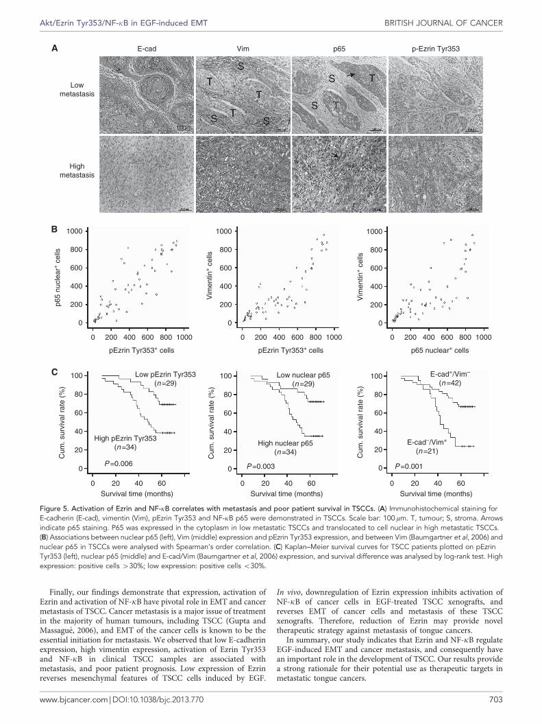
Activation of Ezrin and NF-kB is associated with metastasis andpoor patient survival in TSCCs. We further evaluated the clinicalsignificance of Ezrin and NF-kB activation in metastasis andpatient prognosis of TSCCs. Immunohistochemical stainingdemonstrated that vimentin, pEzrin Tyr353 expression andnuclear p65 expression were lower, and E-cadherin expressionwas higher in low metastatic TSCCs compared with that in highmetastatic TSCCs (Figure 5a). In addition, Spearman’s ordercorrelation analysis showed that pEzrin Tyr353 expression inTSCC was positively correlated with nuclear p65 expression level(Figure 5b; rs¼ 0.74, Po0.001), and pEzrin Tyr353, nuclear p65expression in TSCCs was positively correlated with vimentinexpression (Figure 5b; rs¼ 0.57, Po0.001 and rs¼ 0.51, Po0.001,respectively).
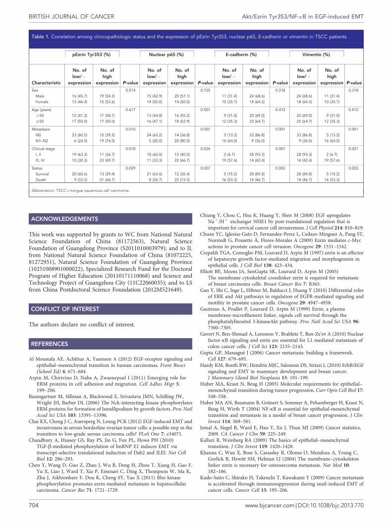
Next, we analysed the association of pEzrin Tyr353 and nuclearp65 expression with clinicopathological status of TSCC patients(Table 1). No significant correlation was observed among pEzrinTyr353, nuclear NF-kB p65, E-cadherin or vimentin expressionand sex or age. However, pEzrin Tyr353, nuclear p65, E-cadherinand vimentin levels were closely associated with lymph nodemetastasis and clinical stage of the patients. Tumours with lymphnode metastasis or high clinical stage expressed high levels of EzrinTyr353, nuclear p65 and vimentin, suggesting that pEzrin Tyr353,nuclear p65 and vimentin upregulations were associated withtumour progression. On the contrary, E-cadherin expression wasnegatively correlated with lymph node metastasis and clinical stageof TSCCs.
Furthermore, we evaluated the correlation between pEzrinTyr353, nuclear p65 expression, E-cadherin/vimentin expressionand survival of the patients. Patients with low expression of pEzrinTyr353 or nuclear p65 in tumours survived significantly longerthan those with high pEzrin Tyr353 or nuclear p65 expression intumours (Figure 5c; P¼ 0.006 or P¼ 0.003, respectively). Thecumulative survival rate up to 60 months was 69% or 72% inpatients with low pEzrin Tyr353 or nuclear p65 expression,respectively, while only 38% or 35% in those with high pEzrinTyr353 or nuclear p65 expression, respectively. Furthermore,TSCC patients with high E-cadherin/low vimentin expression hadbetter survival than those with low E-cadherin/high vimentinexpression (P¼ 0.001). These data suggest that activation of Ezrinand NF-kB is positively correlated with tumour staging and mayhave a role in the progression of TSCCs.
DISCUSSION
In the current study, we demonstrate that Akt/Ezrin Tyr353/NF-kBpathway is critical for EGF-induced EMT, and reduction ofEzrin reverses mesenchymal features of EGF-treated TSCC cellsand represses metastasis of TSCC via inhibiting NF-kB activity.Furthermore, Ezrin and NF-kB activities are positively associatedwith lymph node metastasis of TSCC patients, and high activitiesindicate poor prognosis of these patients.
Dysregulation of Ezrin has been well documented in many typesof human malignancies (Arpin et al, 2011), and previous studieshave shown that overexpression and activation of Ezrin alter cellshape, adhesion, motility and apoptosis and correlate with theinvasion and metastasis of many human cancers (Khanna et al,2004; Elliott et al, 2005; Srivastava et al, 2005; Chiang et al, 2008;Chuan et al, 2009; Kuo et al, 2009; Vanacker et al, 2011).Phosphorylation of Ezrin is required for conformational activationand for signal transduction, and Thr567 and Tyr353 are the mostcommon phosphorylated sites (Arpin et al, 2011). Activation ofEzrin is involved in several pathways, such as Rho-, Src-, PKC-,EGF- and HGF-activated pathways (Crepaldi et al, 1997; Srivastavaet al, 2005; Chiang et al, 2008; Ren et al, 2008; Chen et al, 2011).Furthermore, it has been reported that several signal pathways areinvolved in activating Ezrin, including Akt, ERK1/2 and ROCK1(Elliott et al, 2005; Sizemore et al, 2007). However, in this study, wefound that Akt pathway, but not ERK1/2 and ROCK1, wasessential for EGF-induced activation of Ezrin Tyr353. Thisactivation may be an indirect process and other signallingmolecules may have a role between Akt and Ezrin as Akt is aserine/threonine kinase. One previous study showed that Ezrin wasan upstream signalling molecule of Akt activation (Gautreau et al,1999). Thus, we need to address this issue in further study.Furthermore, luciferase reporter assay demonstrated that onlyphosphorylation of Ezrin Tyr353, excluding phosphorylation ofEzrin Thr567, was required for the activation of NF-kB induced byEGF, indicating that Ezrin regulates EGF-induced EMT andmetastasis via activating NF-kB. Phosphorylated Ezrin activatesNF-kB and then NF-kB triggers a transcription programme thatlead to EMT. This process usually takes days, as EMT is a complextranscription programme that involves multiple pathway andtranscription factors. Ezrin siRNAs can significantly inhibitphosphorylation of IkBa and we speculate that phosphorylatedEzrin might activate NF-kB by directly or indirectly phosphorylateIkBa, the inhibitor of NF-kB (Supplementary Figure 6).
Epithelial–mesenchymal transition is believed to be a crucialprogramme in cancer metastasis and EGF has been reported to bean important EMT inducer (Hardy et al, 2010; Al Moustafa et al,2012). For example, EGF induces ovarian cancer cell EMT andinvasion, migration by activating the ERK1/2 and PI3K/Akt
Akt/Ezrin Tyr353/NF-kB in EGF-induced EMT BRITISH JOURNAL OF CANCER
www.bjcancer.com | DOI:10.1038/bjc.2013.770 701
pathways and upregulating Snail, Slug and ZEB1 (Chai et al, 2012).However, another study demonstrated that EGF promoted EMT byactivating Akt pathway, but not ERK1/2 pathway (Gan et al, 2010).In the present study, our data also demonstrated that activation ofAkt, but not ERK1/2, mediated the EGF-induced EMT. Epithelial–mesenchymal transition-induced by EGF is mainly due to
Akt-mediated activation of Ezrin Tyr353 and NF-kB. Thus, weidentified Akt/Ezrin/NF-kB pathway as an important pathway inregulating EGF-induced EMT and metastasis in TSCC cells. This issupported by the fact that NF-kB can promote and maintaininvasive phenotype, repress epithelial marker expression andinduce mesenchymal marker expression (Min et al, 2008).
SCC9-P-PBS
shGFP
Lung
Liver
Lung
Liver
shEzrin
0.4 10
5
0
**
0.3
0.2
0.1
0
Wet
lung
(g)
hHP
RT
mR
NA
nor
mai
sed
to 1
8 S
rR
NA
shGFP shEzrin
SCC9-EGF SCC9-PBS
shGFP shEzrin shGFP shEzrin
SCC9-EGF
shGFP
shGFP
E-cad
Vim
p65
SCC9-PBS SCC9-EGF
SCC9-PBS SCC9-EGF
shEzrin
shEzrin shGFP shEzrin
shGFP shEzrin shGFP
SCC9-PBS SCC9-EGF
shEzrin shGFP shEzrin
Lung
Liver
****
Figure 4. Reduction of Ezrin inhibits metastasis of EGF-injected SCC9 xenografts of BALB/c-nu mice. (A) Tissue images and (B) haematoxylin andeosin staining of paraffin sections for the lungs (upper) and livers (lower) of the tumour-bearing mice. Arrows: tumour; scale bar: 100mm.(C) Mean±s.d. wet lung weight of tumour-bearing mice (n¼8 per group). (D) Expression of human HPRT mRNA relative to mouse 18 S rRNA,in the lungs and livers of the tumour-bearing mice, was determined by qRT–PCR. **Po0.01 vs shGFP. (E) Immunohistochemical staining illustratedthat reduction of Ezrin enhanced E-cadherin expression, inhibited vimentin expression and p65 nuclear expression of EGF-injected SCC9xenografts. Scale bar: 50mm. shEzrin, Ezrin shRNA.
BRITISH JOURNAL OF CANCER Akt/Ezrin Tyr353/NF-kB in EGF-induced EMT
702 www.bjcancer.com | DOI:10.1038/bjc.2013.770
Finally, our findings demonstrate that expression, activation ofEzrin and activation of NF-kB have pivotal role in EMT and cancermetastasis of TSCC. Cancer metastasis is a major issue of treatmentin the majority of human tumours, including TSCC (Gupta andMassague, 2006), and EMT of the cancer cells is known to be theessential initiation for metastasis. We observed that low E-cadherinexpression, high vimentin expression, activation of Ezrin Tyr353and NF-kB in clinical TSCC samples are associated withmetastasis, and poor patient prognosis. Low expression of Ezrinreverses mesenchymal features of TSCC cells induced by EGF.
In vivo, downregulation of Ezrin expression inhibits activation ofNF-kB of cancer cells in EGF-treated TSCC xenografts, andreverses EMT of cancer cells and metastasis of these TSCCxenografts. Therefore, reduction of Ezrin may provide noveltherapeutic strategy against metastasis of tongue cancers.
In summary, our study indicates that Ezrin and NF-kB regulateEGF-induced EMT and cancer metastasis, and consequently havean important role in the development of TSCC. Our results providea strong rationale for their potential use as therapeutic targets inmetastatic tongue cancers.
E-cad
Lowmetastasis
Highmetastasis
1000
1000
pEzrin Tyr353+ cells
Low pEzrin Tyr353(n=29)
Low nuclear p65(n=29)
High nuclear p65(n=34)
E-cad–/Vim+
(n=21)
E-cad+/Vim–
(n=42)
High pEzrin Tyr353(n=34)
P=0.006 P=0.003 P=0.001
Survival time (months)
pEzrin Tyr353+ cells p65 nuclear+ cells
800
800
600
600
400
400
200
200
p65
nucl
ear+
cel
ls
Vim
entin
+ c
ells
Vim
entin
+ c
ells
0
100
80
60
40
20
604020
Cum
. sur
viva
l rat
e (%
)
0
0
0
Vim p65 p-Ezrin Tyr353
1000
800
600
400
200
0
1000
800
600
400
200
0
10008006004002000 10008006004002000
100
80
60
40
20
Cum
. sur
viva
l rat
e (%
)
0
100
80
60
40
20
Cum
. sur
viva
l rat
e (%
)
0
Survival time (months)6040200
Survival time (months)6040200
Figure 5. Activation of Ezrin and NF-kB correlates with metastasis and poor patient survival in TSCCs. (A) Immunohistochemical staining forE-cadherin (E-cad), vimentin (Vim), pEzrin Tyr353 and NF-kB p65 were demonstrated in TSCCs. Scale bar: 100mm. T, tumour; S, stroma. Arrowsindicate p65 staining. P65 was expressed in the cytoplasm in low metastatic TSCCs and translocated to cell nuclear in high metastatic TSCCs.(B) Associations between nuclear p65 (left), Vim (middle) expression and pEzrin Tyr353 expression, and between Vim (Baumgartner et al, 2006) andnuclear p65 in TSCCs were analysed with Spearman’s order correlation. (C) Kaplan–Meier survival curves for TSCC patients plotted on pEzrinTyr353 (left), nuclear p65 (middle) and E-cad/Vim (Baumgartner et al, 2006) expression, and survival difference was analysed by log-rank test. Highexpression: positive cells 430%; low expression: positive cells o30%.
Akt/Ezrin Tyr353/NF-kB in EGF-induced EMT BRITISH JOURNAL OF CANCER
www.bjcancer.com | DOI:10.1038/bjc.2013.770 703
ACKNOWLEDGEMENTS
This work was supported by grants to WC from National NaturalScience Foundation of China (81172563), Natural ScienceFoundation of Guangdong Province (S2011010003979); and to JLfrom National Natural Science Foundation of China (81072225,81272951), Natural Science Foundation of Guangdong Province(10251008901000022), Specialized Research Fund for the DoctoralProgram of Higher Education (20110171110068) and Science andTechnology Project of Guangzhou City (11C22060035); and to LSfrom China Postdoctoral Science Foundation (2012M521649).
CONFLICT OF INTEREST
The authors declare no conflict of interest.
REFERENCES
Al Moustafa AE, Achkhar A, Yasmeen A (2012) EGF-receptor signaling andepithelial–mesenchymal transition in human carcinomas. Front Biosci(School Ed) 4: 671–684.
Arpin M, Chirivino D, Naba A, Zwaenepoel I (2011) Emerging role forERM proteins in cell adhesion and migration. Cell Adhes Migr 5:199–206.
Baumgartner M, Sillman A, Blackwood E, Srivastava JMN, Schilling JW,Wright JH, Barber DL (2006) The Nck-interacting kinase phosphorylatesERM proteins for formation of lamellipodium by growth factors. Proc NatlAcad Sci USA 103: 13391–13396.
Chai KX, Cheng J-C, Auersperg N, Leung PCK (2012) EGF-induced EMT andinvasiveness in serous borderline ovarian tumor cells: a possible step in thetransition to low-grade serous carcinoma cells? PLoS One 7: e34071.
Chaudhury A, Hussey GS, Ray PS, Jin G, Fox PL, Howe PH (2010)TGF-b-mediated phosphorylation of hnRNP E1 induces EMT viatranscript-selective translational induction of Dab2 and ILEI. Nat CellBiol 12: 286–293.
Chen Y, Wang D, Guo Z, Zhao J, Wu B, Deng H, Zhou T, Xiang H, Gao F,Yu X, Liao J, Ward T, Xia P, Emenari C, Ding X, Thompson W, Ma K,Zhu J, Aikhionbare F, Dou K, Cheng SY, Yao X (2011) Rho kinasephosphorylation promotes ezrin-mediated metastasis in hepatocellularcarcinoma. Cancer Res 71: 1721–1729.
Chiang Y, Chou C, Hsu K, Huang Y, Shen M (2008) EGF upregulatesNaþ /Hþ exchanger NHE1 by post-translational regulation that isimportant for cervical cancer cell invasiveness. J Cell Physiol 214: 810–819.
Chuan YC, Iglesias-Gato D, Fernandez-Perez L, Cedazo-Minguez A, Pang ST,Norstedt G, Pousette Å, Flores-Morales A (2009) Ezrin mediates c-Mycactions in prostate cancer cell invasion. Oncogene 29: 1531–1542.
Crepaldi TGA, Comoglio PM, Louvard D, Arpin M (1997) ezrin is an effectorof hepatocyte growth factor-mediated migration and morphogenesis inepithelial cells. J Cell Biol 138: 423–434.
Elliott BE, Meens JA, SenGupta SK, Louvard D, Arpin M (2005)The membrane cytoskeletal crosslinker ezrin is required for metastasisof breast carcinoma cells. Breast Cancer Res 7: R365.
Gan Y, Shi C, Inge L, Hibner M, Balducci J, Huang Y (2010) Differential rolesof ERK and Akt pathways in regulation of EGFR-mediated signaling andmotility in prostate cancer cells. Oncogene 29: 4947–4958.
Gautreau A, Poullet P, Louvard D, Arpin M (1999) Ezrin, a plasmamembrane-microfilament linker, signals cell survival through thephosphatidylinositol 3-kinaseAkt pathway. Proc Natl Acad Sci USA 96:7300–7305.
Gavert N, Ben-Shmuel A, Lemmon V, Brabletz T, Ben-Ze’ev A (2010) Nuclearfactor-kB signaling and ezrin are essential for L1-mediated metastasis ofcolon cancer cells. J Cell Sci 123: 2135–2143.
Gupta GP, Massague J (2006) Cancer metastasis: building a framework.Cell 127: 679–695.
Hardy KM, Booth BW, Hendrix MJC, Salomon DS, Strizzi L (2010) ErbB/EGFsignaling and EMT in mammary development and breast cancer.J Mammary Gland Biol Neoplasia 15: 191–199.
Huber MA, Kraut N, Beug H (2005) Molecular requirements for epithelial–mesenchymal transition during tumor progression. Curr Opin Cell Biol 17:548–558.
Huber MA AN, Baumann B, Grunert S, Sommer A, Pehamberger H, Kraut N,Beug H, Wirth T (2004) NF-kB is essential for epithelial-mesenchymaltransition and metastasis in a model of breast cancer progression. J ClinInvest 114: 569–581.
Jemal A, Siegel R, Ward E, Hao Y, Xu J, Thun MJ (2009) Cancer statistics,2009. CA Cancer J Clin 59: 225–249.
Kalluri R, Weinberg RA (2009) The basics of epithelial–mesenchymaltransition. J Clin Invest 119: 1420–1428.
Khanna C, Wan X, Bose S, Cassaday R, Olomu O, Mendoza A, Yeung C,Gorlick R, Hewitt SM, Helman LJ (2004) The membrane–cytoskeletonlinker ezrin is necessary for osteosarcoma metastasis. Nat Med 10:182–186.
Kudo-Saito C, Shirako H, Takeuchi T, Kawakami Y (2009) Cancer metastasisis accelerated through immunosuppression during snail-induced EMT ofcancer cells. Cancer Cell 15: 195–206.
Table 1. Correlation among clinicopathologic status and the expression of pEzrin Tyr353, nuclear p65, E-cadherin or vimentin in TSCC patients
pEzrin Tyr353 (%) Nuclear p65 (%) E-cadherin (%) Vimentin (%)
Characteristic
No. oflow/�
expression
No. ofhigh
expression P-value
No. oflow/�
expression
No. ofhigh
expression P-value
No. oflow/�
expression
No. ofhigh
expression P-value
No. oflow/�
expression
No. ofhigh
expression P-value
Sex 0.514 0.725 0.318 0.318Male 16 (45.7) 19 (54.3) 15 (42.9) 20 (57.1) 11 (31.4) 24 (68.6) 24 (68.6) 11 (31.4)Female 13 (46.4) 15 (53.6) 14 (50.0) 14 (50.0) 10 (35.7) 18 (64.3) 18 (64.3) 10 (35.7)
Age (years) 0.617 0.501 0.412 0.412o50 12 (41.3) 17 (58.7) 13 (44.8) 16 (55.2) 9 (31.0) 20 (69.0) 20 (69.0) 9 (31.0)X50 17 (50.0) 17 (50.0) 16 (47.1) 18 (52.9) 12 (35.3) 22 (64.7) 22 (64.7) 12 (35.3)
Metastasis 0.010 0.007 0.001 0.001N0 23 (60.5) 15 (39.5) 24 (63.2) 14 (36.8) 5 (13.2) 33 (86.8) 33 (86.8) 5 (13.2)N1–N2 6 (24.0) 19 (76.0) 5 (20.0) 20 (80.0) 16 (64.0) 9 (36.0) 9 (36.0) 16 (64.0)
Clinical stage 0.018 0.026 0.001 0.001I, II 19 (63.3) 11 (36.7) 18 (60.0) 12 (40.0) 2 (6.7) 28 (93.3) 28 (93.3) 2 (6.7)III, IV 10 (30.3) 23 (69.7) 11 (33.3) 22 (66.7) 19 (57.6) 14 (42.4) 14 (42.4) 19 (57.6)
Status 0.029 0.007 0.003 0.003Survival 20 (60.6) 13 (39.4) 21 (63.6) 12 (36.4) 5 (15.2) 28 (84.8) 28 (84.8) 5 (15.2)Death 9 (33.3) 21 (66.7) 8 (26.7) 22 (73.3) 16 (53.3) 14 (46.7) 14 (46.7) 16 (53.3)
Abbreviation: TSCC¼ tongue squamous cell carcinoma.
BRITISH JOURNAL OF CANCER Akt/Ezrin Tyr353/NF-kB in EGF-induced EMT
704 www.bjcancer.com | DOI:10.1038/bjc.2013.770
Kuo WC, Yang KT, Hsieh SL, Lai MZ (2009) Ezrin is a negative regulator ofdeath receptor-induced apoptosis. Oncogene 29: 1374–1383.
Lim S, Ryu J, Shin JA, Shin MJ, Ahn YK, Kim JJ, Han KH (2009) Tumornecrosis factor-a potentiates RhoA-mediated monocyte transmigratoryactivity in vivo at a picomolar level. Arterioscler Thromb Vasc Biol 29:2138–2145.
Louvet-Vallee S (2000) ERM proteins from cellular architecture to cellsignaling. Biol Cell 92: 305–316.
Min C, Eddy SF, Sherr DH, Sonenshein GE (2008) NF-kB and epithelial tomesenchymal transition of cancer. J Cell Biochem 104: 733–744.
Polyak K, Weinberg RA (2009) Transitions between epithelial andmesenchymal states: acquisition of malignant and stem cell traits. Nat RevCancer 9: 265–273.
Ren L, Hong SH, Cassavaugh J, Osborne T, Chou AJ, Kim SY, Gorlick R,Hewitt SM, Khanna C (2008) The actin–cytoskeleton linker protein ezrinis regulated during osteosarcoma metastasis by PKC. Oncogene 28: 792–802.
Sanchez-Tillo E, Lazaro A, Torrent R, Cuatrecasas M, Vaquero EC, Castells A,Engel P, Postigo A (2010) ZEB1 represses E-cadherin and induces an EMTby recruiting the SWI/SNF chromatin-remodeling protein BRG1.Oncogene 29: 3490–3500.
Sano D, Myers Jeffrey N. (2007) Metastasis of squamous cell carcinoma of theoral tongue. Cancer Metast Rev 26: 645–662.
Shin SY, Rath O, Zebisch A, Choo SM, Kolch W, Cho KH (2010) Functionalroles of multiple feedback loops in extracellular signal-regulated kinaseand Wnt signaling pathways that regulate epithelial–mesenchymaltransition. Cancer Res 70: 6715–6724.
Sizemore S, Cicek M, Sizemore N, Ng KP, Casey G (2007) Podocalyxinincreases the aggressive phenotype of breast and prostate cancer cellsin vitro through its interaction with ezrin. Cancer Res 67: 6183–6191.
Srivastava J, Elliott B, Louvard D, Arpin M (2005) Src-dependent Ezrinphosphorylation in adhesion-mediated signaling. Mol Biol Cell 16:1481–1490.
Sun L, Yao Y, Liu B, Lin Z, Lin L, Yang M, Zhang W, Chen W, Pan C, Liu Q,Song E, Li J (2012) MiR-200b and miR-15b regulate chemotherapy-induced
epithelial-mesenchymal transition in human tongue cancer cells bytargeting BMI1. Oncogene 31: 432–445.
Thiery JP, Acloque H, Huang RYJ, Nieto MA (2009) Epithelial–mesenchymaltransitions in development and disease. Cell 139: 871–890.
Thiery JP, Sleeman JP (2006) Complex networks orchestrate epithelial–mesenchymal transitions. Nat Rev Mol Cell Biol 7: 131–142.
Vanacker J-M, Zheng S, Huang J, Zhou K, Zhang C, Xiang Q, Tan Z, Wang T,Fu X (2011) 17b-Estradiol enhances breast cancer cell motility andinvasion via extra-nuclear activation of actin-binding protein Ezrin.PLoS One 6: e22439.
Vuoriluoto K, Haugen H, Kiviluoto S, Mpindi JP, Nevo J, Gjerdrum C,Tiron C, Lorens JB, Ivaska J (2010) Vimentin regulates EMT induction bySlug and oncogenic H-Ras and migration by governing Axl expression inbreast cancer. Oncogene 30: 1436–1448.
Wellner U, Schubert J, Burk UC, Schmalhofer O, Zhu F, Sonntag A,Waldvogel B, Vannier C, Darling D, Az Hausen, Brunton VG, Morton J,Sansom O, Schuler J, Stemmler MP, Herzberger C, Hopt U, Keck T,Brabletz S, Brabletz T (2009) The EMT-activator ZEB1 promotestumorigenicity by repressing stemness-inhibiting microRNAs. Nat CellBiol 11: 1487–1495.
Wu Y, Deng J, Rychahou PG, Qiu S, Evers BM, Zhou BP (2009) Stabilizationof Snail by NF-kB is required for inflammation-induced cell migrationand invasion. Cancer Cell 15: 416–428.
Yip MK, Seow HF (2012) Activation of phosphatidylinositol 3-kinase/Aktsignaling by EGF downregulates membranous E-cadherin and b-cateninand enhances invasion in nasopharyngeal carcinoma cells. Cancer lett 318:162–172.
This work is published under the standard license to publish agree-ment. After 12 months the work will become freely available andthe license terms will switch to a Creative Commons Attribution-NonCommercial-Share Alike 3.0 Unported License.
Supplementary Information accompanies this paper on British Journal of Cancer website (http://www.nature.com/bjc)
Akt/Ezrin Tyr353/NF-kB in EGF-induced EMT BRITISH JOURNAL OF CANCER
www.bjcancer.com | DOI:10.1038/bjc.2013.770 705

![Ivyspring International Publisher Theranostics · more than 5% of all cancer types and is the fifth leading cause of cancer mortality worldwide with an extremely poor prognosis [1].](https://static.fdocument.org/doc/165x107/5f96143682877907366fc9c7/ivyspring-international-publisher-more-than-5-of-all-cancer-types-and-is-the-fifth.jpg)