Proliferation Cinetics and Prognosis of Renal Cell Carcinoma. · renzell-Karzinom wird beschrieben....
7
Internationale Zeitschrift für Krebsforschung und -behandlung Band 14, Heft 4, August 1991 Inhalt Contents Übersichtsarbeiten Proliferationskinetik und Prognose eines Nierenzellkarzinoms* de Riese, W., Allhoff, E., Werner, M., Stief, C G . Atzpodien, J., Kirchner, Η Zur Bewertung von Therapiestudien beim Mammakarzinom Sauerbrei, W., Schmoor, C , Schumacher, M. 297 303 Review Articles Proliferation Kinetics and Prognosis of Renal Cell Carcinoma* de Riese, W., Allhoff, E., Werner. M.. Stief, CG., Atzpodien, J.. Kirchner, Η On the Assessment of Quality in Breast Cancer Clinical Trials Sauerbrei, W., Schmoor, C , Schumacher, Μ 297 303 Originalarbeiten Etoposid, Leukovorin und 5-Flourouracil (ELF) beim fortgeschrittenen Magenkarzinom: Abschließende Ergebnisse einer Phase-lI-Studie bei älteren Patienten oder Patienten mit kardialem Risiko* Stahl, M., Wilke, H., Preusser, P., Fink, U., Achterrath, W., Schöber. C, Köhne-Wömpner, C.H., Harstrick, Α., Meyer, H J . , Meyer, J., Lenaz, L., Schmoll, H.J 314 Einfluß von Rekombinantem Granulozyten-Makro- phagen Kolonien-stimulierender Faktor (GM-CSF) auf Leukopenie bei AIDS: Eine Untersuchung bei sieben Patienten* Ottmann, O.G., Goebel, F.-D., Ganser, Α., Bogner, J.R., Seipelt. G., Chatterjee, M . , Helm. E.B., Hoelzer, D 319 Chirurgische Therapie bei großen Knochenmetastasen im Becken Spring, W., Dittmer, Η 322 Prospektive multizentrische Phase-III-Studie mit Doxifluridin (5'dFUR) versus 5-Flourouracil bei Patienten mit fortgeschrittenem kolorektalen Karzinom* Schuster, D., Heim, M.E., Dombernowski, P., Wood, C, Queißer,W 333 Original Papers Etoposide, Leukovorin and 5-Flourouracil (ELF) in Advanced Gastric Carcinoma - Final Results of a Phase-Ii Study in Elderly Patients or Patients with Cardiac Risk* Stahl, Μ., Wilke. Η., Preusser, P.. Fink, U., Achterrath, W., Schöber, C, Köhne-Wömpner, C.H., Harstrick, Α.. Meyer, H.J., Meyer, J., Lenaz, L., Schmoll, H.J 314 Effect of Recombinant Granulocyte-Macrophage Colony-Stimulating Factor (GM-CSF) on Leukopenia in AIDS: A Study of Seven Cases* Ottmann, O.G., Goebel, F.-D., Ganser, Α., Bogner, J.R., Seipelt. G., Chatterjee, M., Helm. E.B., Hoelzer, D 319 Surgical Therapie in Extensive Pelvic Bone Metastases Spring, W., Dittmer, Η 322 Prospective Multicenter Phase-Ill Trial of Doxifluridin (5'dFUR) versus 5-FIourouracil in Patients with Advanced Colorectal Carcinoma* Schuster. D., Heim. Μ.Ε., Dombernowski, Ρ, Wood, C, Queißer, W 333 Kurzmitteilungen Ansprechraten und Toxizität von Mitomycin-C, Mitoxantron und Methotrexat (3M) bei metastiertem Mammakarzinom* Porzsolt, F.. Meuret, G 338 Schmerztherapie: Vergleich zweier Bestrahlungs- schemata bei schmerzhaften Knochenmetastasen* Karstens, J.H.. Blach, M.. Ammon. J 341 Imniunzv to logische Differenzierung zwischen Leberzellkarzinom und metastasierendem extragonadalem Keimzelltumor* Hastka, J., Bohrer. M.H., Hartum*, G., Verbeke, CS 344 Short Communications Response Rates and Toxicity of Mitomycin-C, Mitoxantrone, and Methotrexate (3M) in Advanced Breast Cancer* Porzsolt, F., Meuret, G 338 Pain Management Policy: Comparison of Two Irradiation Schedules in Metastatic Bone Pain* Karstens. J.H.. Blach. M.. Ammon. J 341 Immunocytological Differentiation of Hepatocellular Carcinoma versus Estragonadal Germ Cell Tumor* Hastka. J.. Bohrer. Μ.Η., Hamme. G.. Verbeke. CS 344
Transcript of Proliferation Cinetics and Prognosis of Renal Cell Carcinoma. · renzell-Karzinom wird beschrieben....
Internationale Ze i tschr i f t für Krebsforschung und -behandlung Band 14, Heft 4, August 1991
Inhalt Contents
Übersichtsarbeiten Proliferationskinetik und Prognose eines Nierenzellkarzinoms* de Riese, W., Allhoff, E . , Werner, M . , Stief, C G . Atzpodien, J., Kirchner, Η
Zur Bewertung von Therapiestudien beim Mammakarzinom Sauerbrei, W., Schmoor, C , Schumacher, M .
297
303
Review Articles Proliferation Kinetics and Prognosis of Renal Cell Carcinoma* de Riese, W., Allhoff , E . , Werner. M . . Stief, C G . , Atzpodien, J.. Kirchner, Η
On the Assessment of Quality in Breast Cancer Clinical Trials Sauerbrei, W., Schmoor, C , Schumacher, Μ
297
303
Originalarbeiten Etoposid, Leukovorin und 5-Flourouracil ( E L F ) beim fortgeschrittenen Magenkarzinom: Abschl ießende Ergebnisse einer Phase-lI-Studie bei älteren Patienten oder Patienten mit kardialem Risiko* Stahl, M . , Wilke, H . , Preusser, P., Fink, U . , Achterrath, W. , Schöber. C , Köhne-Wömpner , C .H . , Harstrick, Α . , Meyer, H J . , Meyer, J., Lenaz, L . , Schmoll, H.J 314
Einfluß von Rekombinantem Granulozyten-Makrophagen Kolonien-stimulierender Faktor ( G M - C S F ) auf Leukopenie bei A I D S : Eine Untersuchung bei sieben Patienten* Ottmann, O.G. , Goebel, F . -D. , Ganser, Α . , Bogner, J.R., Seipelt. G. , Chatterjee, M . , Helm. E .B . , Hoelzer, D 319
Chirurgische Therapie bei großen Knochenmetastasen im Becken Spring, W., Dittmer, Η 322
Prospektive multizentrische Phase-III-Studie mit Doxifluridin (5 'dFUR) versus 5-Flourouracil bei Patienten mit fortgeschrittenem kolorektalen Karzinom* Schuster, D . , Heim, M . E . , Dombernowski, P., Wood, C , Q u e i ß e r , W 333
Original Papers Etoposide, Leukovorin and 5-Flourouracil ( E L F ) in Advanced Gastric Carcinoma - Final Results of a Phase-Ii Study in Elderly Patients or Patients with Cardiac Risk* Stahl, Μ. , Wilke. Η . , Preusser, P.. Fink, U . , Achterrath, W. , Schöber, C , Köhne-Wömpner , C . H . , Harstrick, Α . . Meyer, H.J . , Meyer, J., Lenaz, L . , Schmoll, H.J 314
Effect of Recombinant Granulocyte-Macrophage Colony-Stimulating Factor ( G M - C S F ) on Leukopenia in A I D S : A Study of Seven Cases* Ottmann, O.G. , Goebel, F . -D. , Ganser, Α . , Bogner, J.R., Seipelt. G. , Chatterjee, M . , He lm. E .B . , Hoelzer, D 319
Surgical Therapie in Extensive Pelvic Bone Metastases Spring, W., Dittmer, Η 322
Prospective Multicenter Phase-Il l Trial of Doxifluridin (5 'dFUR) versus 5-FIourouracil in Patients with Advanced Colorectal Carcinoma* Schuster. D . , Heim. Μ . Ε . , Dombernowski, Ρ , Wood, C , Queißer , W 333
Kurzmitteilungen Ansprechraten und Toxizität von Mitomycin-C, Mitoxantron und Methotrexat (3M) bei metastiertem Mammakarzinom* Porzsolt, F. . Meuret, G 338
Schmerztherapie: Vergleich zweier Bestrahlungsschemata bei schmerzhaften Knochenmetastasen* Karstens, J .H. . Blach, M . . Ammon. J 341
Imniunzv to logische Differenzierung zwischen Leberzellkarzinom und metastasierendem extragonadalem Keimzelltumor* Hastka, J., Bohrer. M . H . , Hartum*, G. , Verbeke, C S 344
Short Communications Response Rates and Toxicity of Mitomycin-C, Mitoxantrone, and Methotrexate (3M) in Advanced Breast Cancer* Porzsolt, F., Meuret, G 338
Pain Management Policy: Comparison of Two Irradiation Schedules in Metastatic Bone Pain* Karstens. J .H. . Blach. M . . Ammon. J 341
Immunocytological Differentiation of Hepatocellular Carcinoma versus Estragonadal Germ Cell Tumor* Hastka. J.. Bohrer. Μ . Η . , Hamme. G. . Verbeke. C S 344
Übersichtsarbeiten · Review Articles
Onkologie 1991;14:297-302
Proliferation Kinetics and Prognosis of Renal Cell Carcinoma
W. de Riese", E. Allhoff1, M. Werner0, C. G. Stief, J. Atzpodienc\ H. Kirchner"
"Dept. of Urology.
' 'Laboratory of Immunohistochemistry and Cytogenetics, Dept. of Pathology. c Dept . of Hematology and Oncology, Hannover Medical School
Summary and Key Words
The historical development of different techniques for the study of proliferative patterns of normal and malignant tissue is explained. For more than 100 years, pathologists and clinicians have attempted to clarify the prognostic impact of this tumor-biological parameter. One relevant result of these investigations is the 'Grading-System', a histomorphological description of tumor cell differentiation. The autoradiographic method using tritiated thymidine (thymidine incorporation assay) detects a portion of DNA-sythesizing cells directly. However, radioactivity and long autoradiographic exposure times are the main disadvantages. D N A cytophotometry and immunohistochemical labeling are currently the most frequently used techniques for direct assessment of proliferating cells in normal and malignant renal tissue as well as in other human tumor tissues. Concerning renal cell carcinoma (RCC). our own experience and the results of the literature are compared and discussed: In contrast to tumor staging, tumor grading correlates with the tumor-specific proliferation rates. According to the present data, the measurement of the proliferative kinetics seems to be a relevant prognostic parameter for detection of high-risk patients, especially in the early stages showing identical histological findings for staging and grading.
Renal cell carcinoma · Proliferation kinetics · Autoradiographic technique · DNA-cytometry · Immunostaining
Zusammenfassung und Schlüsselwörter
Die Entwicklung der verschiedenen Techniken zur Erfassung des proliferativen Verhaltens von Normal- und Tumorgewebe beim Nierenzell-Karzinom wird beschrieben. Seit mehr als 100 Jahren beschäftigen sich Pathologen und Kliniker mit der prognostischen Relevanz dieses tumor-biologischen Parameters. Ein Ergebnis dieser Forschung ist das «Grading-System», eine histomorphologische Beschreibung der Tumorzell-Differenzierung. Die autoradiographische Methode unter Verwendung von radiomarkiertem Thymidin (Thymidineinbaurate) erfaßt direkt die Rate der DNA-synthetisierenden Zellen. Radioaktivität und lange Expositionszeiten sind die Hauptnachteile dieser Methode. Die DNA-Zytophotometrie sowie immunhistochemische Markierungsmethoden sind gegenwärtig die am häufigsten angewandten Methoden zur direkten Bestimmung des Anteils proliferierender Zellen sowohl im Normal- als auch im Tumorgewebe der Niere sowie auch anderer Tumoren des Menschen. Die eigenen Erfahrungen sowie die Ergebnisse verschiedener Autoren werden verglichen und diskutiert: Im Gegensatz zum Tumor-Staging zeigt das Tumor-Grading eine Korrelation gegenüber den tumor-spezifischen Proliferationsraten. G e m ä ß den Daten aus der Literatur erscheint die Bestimmung der tumor-spezifischen Proliferationskinetik als ein relevanter prognostischer Faktor zur Erfassung von Hoch-Risiko-Patienten, insbesondere in Frühstadien mit identischen histologischen Befunden im Staging und Grading
Nierenzellkarzinom • Proliferationskinetik · Autoradiographische Technik · DNS-Zytometrie · Immunhistochemie
Historical Survey
One of the main characteristics of cancer is the haphazard
proliferation of malignant cells. The fraction of proliferating
cells strongly influences tumor growth and is believed to be a
major parameter for prognosis and treatment selection [ 1 , 2],
For more than 100 years pathologists and clinicians have been
engaged in examining proliferative patterns and behavior of
human malignancies, comparing these results with normal
tissue findings. This is necessary for a better understanding of
pathogenesis, therapeutic principles and individual prognosis.
Flamming (1882) and Waldeyer (1888) were the first to de
scribe the histomorphological conception of 'cell division,
mitosis and cell proliferation' in human malignancies [3]. In
different malignant tumors, pathologists found a correlation
between the mitosis rate determined in histological specimens
and individual clinical course of patients with neoplasms [2, 4].
Mitosis, however, represents only a short phase in the active
cell cycle. Microscopically, only 1% - 5% of all DNA-synthe-
sizing cells are detectable in histological specimens [5, 6, 7].
Therefore, this can only be regarded as an indirect parameter
of the proliferative pattern of the tissue.
It took a long time before a technique for the direct detection
of tumor specific proliferation rates was made available. By
autoradiographic studies using tritiated thymidine ( [ 3 H]-Thd)
(=thymidine incorporation assay), direct measurement of the
portion of DNA-synthesizing cells was achieved for the first
time. In 1979, Rabes et al. [5] presented a clinical report of
proliferation rates in renal cell carcinoma (RCC) using this
technique. The authors found low proliferation rates (<20%)
and a correlation to the recurrence rate in a series of RCC
patients. The main disadvantages of this method, however,
involve the extracorporal organ perfusion (ex vivo) required
after tumor nephrectomy, the radioactive contamination and
waste disposal as well as personal safety, long autoradiogra
phic exposure times and, thus, the lack of practicability of this
technique in routine diagnosis [6, 8]. The successful immuno-
298 de Riese/Allhoff/Werner/Stief/Atzpodien/Kirchner
logical preparation of monoclonal antibodies represented a relevant advance in this field [9].
DNA-Cytometry for R C C
Single-cell and flow DNA-cyto(photo)metry (e.g. combined with the Feulgen staining) are commonly used techniques for the evaluation DNA-content of malignant of tissue tumors [2]. Formalin-fixed and paraffin-embedded tissue [10] as well as fresh tissue can be analyzed for DNA-cytophotometry [2]. This technique has been used for more than ten years in basic and clinical research. Extensive experience has been achieved by means of this technique: In 1985, Ljungberg was one of the first authors using D N A flow-cytometry on RCC [11]. In 196 analysed tissue samples of 25 cases of RCC, a correlation between D N A distribution (ploidy) and morphologic grading was demonstrated. Tumor proliferation, as determined by the fraction of cells in S-phase, was significantly higher in aneu-ploid samples compared to normal kidney tissue samples and diploid tumor samples. This paper was the first to describe D N A heterogeneity in RCC caused by different tumor cell clones. To exclude a so-called "sampling error' several biopsies depending on the tumor size must be taken from each tumor [11]. About 40% of the analyzed RCC were aneuploid with a D N A content > 5 C. Different studies confirmed the prognostic importance of this parameter in RCC [12, 13]. The results of both techniques, single-cell and flow D N A -cytometry, showed no relevant differences in ploidy heterogeneity and prognostic value of D N A content for RCC patients [14]. The main disadvantage is that this technology requires equipment for photometric imaging. Due to the expense this equipment is only available in research and medical centers. A l though it is a valid and reliable parameter in RCC, D N A -cytometry did not prove successful in routine diagnostics [15].
Immunohistochemical Techniques
Bromodeoxyuridine (BrdU) Technique
In order to avoid the problems of the thymidine incorporation assay, several investigators used S-bromo^-deoxyuridine (BrdU), a non-radioactive thymidine analogue which is also incorporated into DNA-synthesizing cells [8 ,16,17,18,19] . In 1982, Gratzner et al. isolated and described a monoclonal antibody (mab) against B r d U [20] which allows for the rapid detection of BrdU-laden cells in vivo and in vitro as well as by immunohistochemical techniques [21]. Although BrdU immunohistochemistry solves many of the problems associated with [ 3 H]-Thd in cell kinetics studies (as outlined above), its use generates the following problems: B r d U is a cytotoxic substance. Administrations of B r d U at high doses have been shown to be mutagenic in vitro and carcinogenic in vivo in an animal model [22]. B rdU also inhibits cell differentiation and thymidine uptake in vitro, as well as D N A synthesis and cell replication in both in vitro and in vivo settings [8]. Whether or not these negative aspects of B r d U administration affect the results compiled by in-vivo cell kinetic studies, remains un
clear. Due to the mutagenic and carcinogenic potentials, B r d U can only be used for animal and in-vitro studies, but not for direct in-vivo determination of proliferative cell fractions in human malignancies. Therefore, the BrdU immunohistochemistry represents an indirect method only to assess the proliferation rates in human neoplasms.
Ki-67 Staining
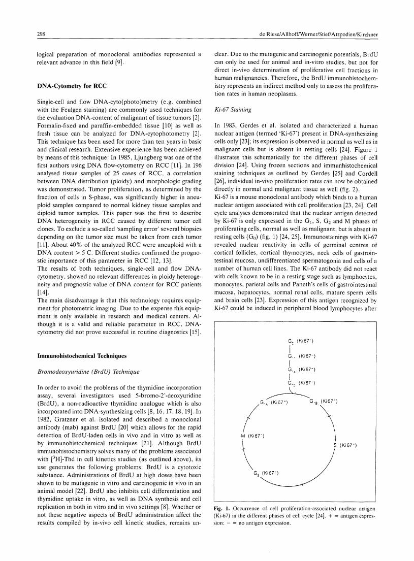
In 1983, Gerdes et al. isolated and characterized a human nuclear antigen (termed kKi-67') present in DNA-synthesizing cells only [23]; its expression is observed in normal as well as in malignant cells but is absent in resting cells [24]. Figure 1 illustrates this schematically for the different phases of cell division [24]. Using frozen sections and immunhistochemical staining techniques as outlined by Gerdes [25] and Cordell [26], individual in-vivo proliferation rates can now be obtained directly in normal and malignant tissue as well (fig. 2). Ki-67 is a mouse monoclonal antibody which binds to a human nuclear antigen associated with cell proliferation [23, 24]. Cell cycle analyses demonstrated that the nuclear antigen detected by Ki-67 is only expressed in the G{, S, G 2 and Μ phases of proliferating cells, normal as well as malignant, but is absent in resting cells (G 0 ) (fig. 1) [24, 25]. Immunostainings with Ki-67 revealed nuclear reactivity in cells of germinal centres of cortical follicles, cortical thymocytes, neck cells of gastrointestinal mucosa, undifferentiated spermatogonia and cells of a number of human cell lines. The Ki-67 antibody did not react with cells known to be in a resting stage such as lymphocytes, monocytes, parietal cells and Panetlrs cells of gastrointestinal mucosa, hepatocytes, normal renal cells, mature sperm cells and brain cells [23]. Expression of this antigen recognized by Ki-67 could be induced in peripheral blood lymphocytes after
G c ( Κ Ϊ 6 7 - )
G . . ( Κ Ϊ 6 7 - )
Fig. 1. Occurrence of cell proliferation-associated nuclear antigen (Ki-67) in the different phases of cell cycle [24]. -I- = antigen expression: - = no antigen expression.
Proliferation Kinetics and Prognosis of Renal Cell Carcinoma 299
Fast Red Monoclonal mouse antialkaline phosphatase
m Calf intestinal
alkaline phosphatase
— Rabbit antimouse IgG
m Primary mouse monoclonal antibody Antigen
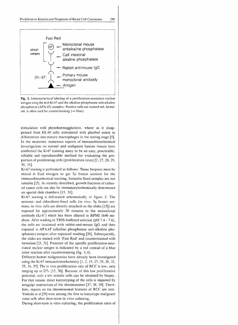
Fig. 2. Immunochemical labeling of a proliferation-associated nuclear antigen using the mab Ki-67 and the alkaline phosphatase anti-alkaline phosphatase (APAAP) complex: Positive cells are stained red, hemal-um is often used for counterstaining (—> blue).
stimulation with phytohaemagglutinin, where as it disappeared from HL-60 cells stimulated with phorbol esters to differentiate into mature macrophages in the resting stage* [5]. In the meantime, numerous reports of immunohistochemical investigations on normal and malignant human tissues have confirmed die Ki-67 staining assay to be an easy, practicable, reliable and reproduceable method for evaluating the proportion of proliferating cells (proliferation rates) [2, 27, 28, 29, 30, 31]. Ki-67 staining is performed as follows: Tissue biopsies must be stored in fluid nitrogen to get 5μ frozen sections for the immunohisiochemical staining; formalin fixed samples are not suitable [25]. As recently described, growth fractions of cultured tumor cells can also be immunocytochemically determined on special slide chambers [15, 31]. Ki-67 staining is delineated schematically in figure 2: The acetone- and chloroform-fixed cells (in vivo: 5μ frozen sections; in vitro: eel's are directly attached on the slides [15]) are exposed for approximately 30 minutes to the monoclonal antibody (Ki-67) which has been diluted in R P M I 1640 medium. Afier washing in TRIS-buffered solution (pH 7.4 - 7.6), the cells are incubated with rabbit-anti-mouse IgG and then exposed :o APAAP (alkaline phosphatase anti-alkaline phosphatase) complex after repeated washing [26]. Subsequently, the slides are stained with 'Fast Red' and counter/stained with hemalum [25, 31]. Presence of the specific proliferation-associated nuclear antigen is indicated by a red instead of a blue color reaction after counterstaining (fig. 3,4). Different human malignancies have already been investigated using the Ki-67 immunohistochemistry [1 ,2 , 15, 27, 29, 30, 32, 33, 34, 35]: The in vivo proliferation rate of RCC is low, only ranging up to 25% [15, 36]. Because of this low proliferative potential, only a few mitotic cells can be obtained by biopsy. For that reason, direct karyotyping of the cells is impaired by irregular contractions of the chromosomes [37, 38, 39]. Therefore, reports on the chromosomal features of RCC are rare. Yoshida et al [39] were among the first to karyotype malignant renal cells after short-term in vitro culturing. During short-term in vitro culturing, the proliferation rates of
300 Riese/Allhoff/Werner/Stief/Atzpodien/Kirchner
• Γ. fr--* .
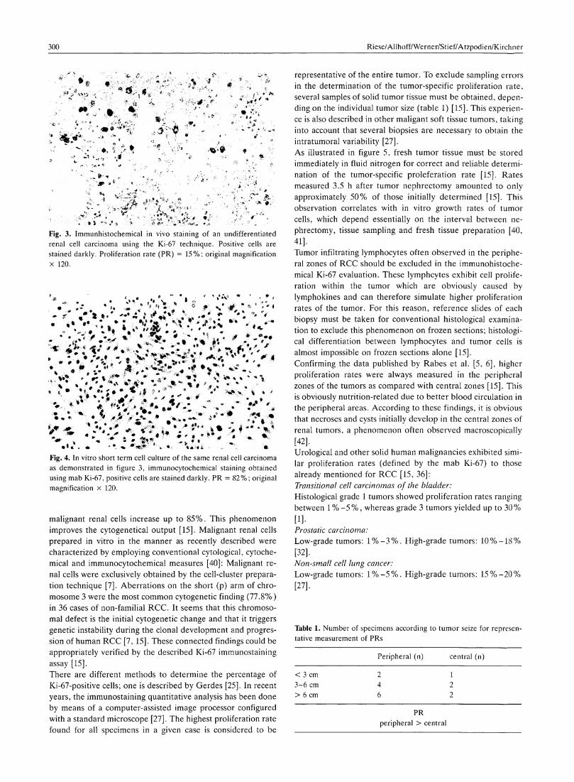
Fig. 3. Immunhistochemical in vivo staining of an undifferentiated renal cell carcinoma using the Ki-67 technique. Positive cells are stained darkly. Proliferation rate (PR) = 15%; original magnification x 120.
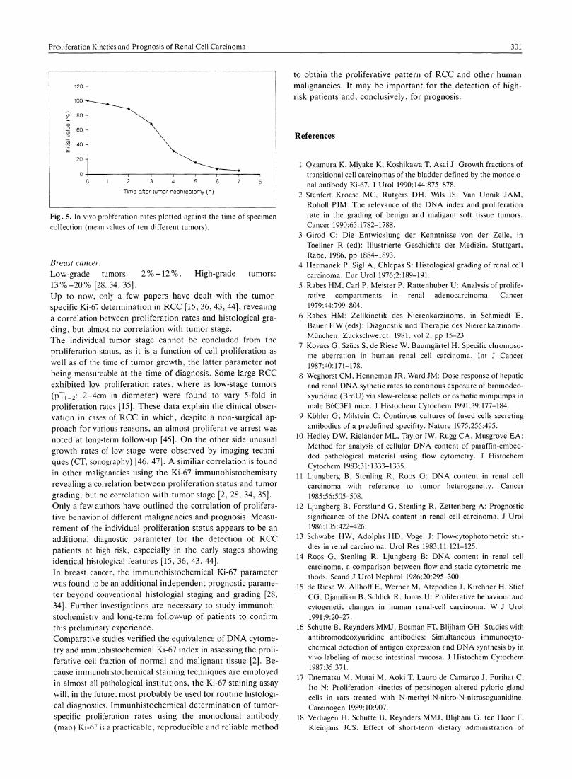
Fig. 4. In vitro short term cell culture of the same renal cell carcinoma as demonstrated in figure 3, immunocytochemical staining obtained using mab Ki-67, positive cells are stained darkly. PR = 82%; original magnification x 120.
malignant renal cells increase up to 85%. This phenomenon improves the cytogenetical output [15]. Malignant renal cells prepared in vitro in the manner as recently described were characterized by employing conventional cytological, cytoche-mical and immunocytochemical measures [40]: Malignant renal cells were exclusively obtained by the cell-cluster preparation technique [7]. Aberrations on the short (p) arm of chromosome 3 were the most common cytogenetic finding (77.8%) in 36 cases of non-familial RCC. I t seems that this chromosomal defect is the initial cytogenetic change and that it triggers genetic instability during the clonal development and progression of human RCC [7, 15]. These connected findings could be appropriately verified by the described Ki-67 immunostaining assay [15]. There are different methods to determine the percentage of Ki-67-positive cells; one is described by Gerdes [25]. In recent years, the immunostaining quantitative analysis has been done by means of a computer-assisted image processor configured with a standard microscope [27]. The highest proliferation rate found for all specimens in a given case is considered to be
representative of the entire tumor. To exclude sampling errors in the determination of the tumor-specific proliferation rate, several samples of solid tumor tissue must be obtained, depending on the individual tumor size (table 1) [15]. This experience is also described in other maligant soft tissue tumors, taking into account that several biopsies are necessary to obtain the intratumoral variability [27]. As illustrated in figure 5, fresh tumor tissue must be stored immediately in fluid nitrogen for correct and reliable determination of the tumor-specific proleferation rate [15]. Rates measured 3.5 h after tumor nephrectomy amounted to only approximately 50% of those initially determined [15]. This observation correlates with in vitro growth rates of tumor cells, which depend essentially on the interval between nephrectomy, tissue sampling and fresh tissue preparation [40. 41]. Tumor infiltrating lymphocytes often observed in the peripheral zones of RCC should be excluded in the immunohistoche-mical Ki-67 evaluation. These lymphcytes exhibit cell proliferation within the tumor which are obviously caused by lymphokines and can therefore simulate higher proliferation rates of the tumor. For this reason, reference slides of each biopsy must be taken for conventional histological examination to exclude this phenomenon on frozen sections; histological differentiation between lymphocytes and tumor cells is almost impossible on frozen sections alone [15]. Confirming the data published by Rabes et al. [5, 6], higher proliferation rates were always measured in the peripheral zones of the tumors as compared with central zones [15]. This is obviously nutrition-related due to better blood circulation in the peripheral areas. According to these findings, it is obvious that necroses and cysts initially develop in the central zones of renal tumors, a phenomenon often observed macroscopically [42]. Urological and other solid human malignancies exhibited similar proliferation rates (defined by the mab Ki-67) to those already mentioned for RCC [15, 36]: Transitional cell carcinomas of the bladder: Histological grade 1 tumors showed proliferation rates ranging between 1 % - 5 % , whereas grade 3 tumors yielded up to 30% [1]· Prostatic carcinoma: Low-grade tumors: l % - 3 % . High-grade tumors: 10% - 1 8 % [32]. Non-small cell lung cancer: Low-grade tumors: l % - 5 % . High-grade tumors: 15%-20% [27].
Table 1. Number of specimens according to tumor seize for representative measurement of PRs
Peripheral (n) central (n)
< 3 cm 2 1 3-6 cm 4 2 > 6 cm 6 2
peripheral PR
I > central
Proliferation Kinetics and Prognosis of Renal Cell Carcinoma 301
120 η
loo -*
80 -CD
CO 60 ->
TO 40 -C
20 -
0 -
Time after tumor nephrectomy (h)
Fig. 5. In vivo proliferation rates plotted against the time of specimen
collection (mean values of ten different tumors).
Breast cancer: Low-grade tumors: 2 % - 1 2 % . High-grade tumors: 13%-20% [28. 54, 35]. Up to now, only a few papers have dealt with the tumor-specific Ki-67 determination in RCC [15, 36, 43, 44], revealing a correlation between proliferation rates and histological grading, but almost no correlation with tumor stage. The individual tumor stage cannot be concluded from the proliferation status, as it is a function of cell proliferation as well as of the time of tumor growth, the latter parameter not being measureable at the time of diagnosis. Some large RCC exhibited low proliferation rates, where as low-stage tumors (pT |_2 : 2-4cm in diameter) were found to vary 5-fold in proliferation rates [15]. These data explain the clinical observation in cases of RCC in which, despite a non-surgical approach for various reasons, an almost proliferative arrest was noted at long-term follow-up [45]. On the other side unusual growth rates of low-stage were observed by imaging techniques (CT, sonography) [46, 47]. A similiar correlation is found in other malignancies using the Ki-67 immunohistochemistry revealing a correlation between proliferation status and tumor grading, but no correlation with tumor stage [2, 28, 34, 35]. Only a few authors have outlined the correlation of proliferative behavior of different malignancies and prognosis. Measurement of the individual proliferation status appears to be an additional diagnostic parameter for the detection of RCC patients at high risk, especially in the early stages showing identical histological features [15, 36, 43, 44]. In breast cancer, the immunohistochemical Ki-67 parameter was found to be an additional independent prognostic parameter beyond conventional histologial staging and grading [28, 34]. Further investigations are necessary to study immunohistochemistry and long-term follow-up of patients to confirm this preliminary experience.
Comparative studies verified the equivalence of D N A cytometry and immunhistochemical Ki-67 index in assessing the proliferative cell fraction of normal and malignant tissue [2]. Because immunohistochemical staining techniques are employed in almost all pathological institutions, the Ki-67 staining assay wil l , in the future, most probably be used for routine histological diagnostics. Immunhistochemical determination of tumor-specific proliferation rates using the monoclonal antibody (mab) K i -6 T is a practicable, reproducible and reliable method
to obtain the proliferative pattern of RCC and other human malignancies. I t may be important for the detection of high-risk patients and, conclusively, for prognosis.
References
1 Okamura K , Miyake K , Koshikawa T, Asai J: Growth fractions of transitional cell carcinomas of the bladder defined by the monoclonal antibody Ki-67. J Uro l 1990;144:875-878.
2 Stenfert Kroese M C , Rutgers D H , Wils IS, Van Unnik J A M , Roholl PJM: The relevance of the D N A index and proliferation rate in the grading of benign and maligant soft tissue tumors. Cancer 1990;65:1782-1788.
3 Girod C: Die Entwicklung der Kenntnisse von der Zelle, in Toellner R (ed): Illustrierte Geschichte der Medizin. Stuttgart, Rabe, 1986, pp 1884-1893.
4 Hermanek P, Sigl A , Chlepas S: Histological grading of renal cell carcinoma. Eur U r o l 1976;2:189-191.
5 Rabes H M , Carl Ρ , Meister Ρ , Rattenhuber U : Analysis of proliferative compartments in renal adenocarcinoma. Cancer 1979:44:799-804.
6 Rabes H M : Zellkinetik des Nierenkarzinoms, in Schmiedt Ε , Bauer H W (eds): Diagnostik und Therapie des Nierenkarzinom ν München, Zuckschwerdt, 1981, vol 2, pp 15-23.
7 Kovacs G , Szücs S, de Riese W, Baumgärte l H : Specific chromosome aberration in human renal cell carcinoma. Int J Cancer 1987:40:171-178.
8 Weghorst C M , Henneman JR, Ward JM: Dose response of hepatic and renal D N A sythetic rates to continous exposure of bromodeo-xyuridine (BrdU) via slow-release pellets or osmotic minipumps in male B6C3F1 mice. J Histochem Cytochem 1991:39:177-184.
9 Köhler G , Milstein C: Continous cultures of fused cells secreting antibodies of a predefined specifity. Nature 1975;256:495.
10 Hedley D W , Rielander M L , Taylor IW, Rugg C A , Musgrove E A : Method for analysis of cellular D N A content of paraffin-embedded pathological material using flow cytometry. J Histochem Cytochem 1983;31:1333-1335.
11 Ljungberg B , Stenling R, Roos G: D N A content in renal cell carcinoma with reference to tumor heterogeneity. Cancer 1985:56:505-508.
12 Ljungberg B , Forsslund G, Stenling R, Zettenberg A : Prognostic significance of the D N A content in renal cell carcinoma. J Uro l 1986;135:422-426.
13 Schwabe H W , Adolphs H D , Vogel J: Flow-cytophotometric studies in renal carcinoma. Uro l Res 1983;11:121-125.
14 Roos G, Stenling R, Ljungberg B: D N A content in renal cell carcinoma, a comparison between flow and static cytometric methods. Scand J Uro l Nephrol 1986;20:295-300.
15 de Riese W, Al lhoff Ε , Werner Μ, Atzpodien J, Kirchner Η , Stief CG, Djamilian B , Schlick R, Jonas U : Proliferative behaviour and cytogenetic changes in human renal-cell carcinoma. W J Uro! 1991:9:20-27.
16 Schutte B , Reynders M M J , Bosman FT, Blijham G H : Studies with antibromodeoxyuridine antibodies: Simultaneous immunocyto-chemical detection of antigen expression and D N A synthesis by in vivo labeling of mouse intestinal mucosa. J Histochem Cytochem 1987:35:371.
17 Tatematsu M . Mutai M . A o k i T, La uro de Camargo J, Furihat C. Ito N : Proliferation kinetics of pepsinogen altered pyloric gland cells in rats treated with N-methyl.N-nitro-N-nitrosoguanidine. Carcinogen 1989; 10:907.
18 Verhagen H . Schutte Β , Reynders M M J , Blijham G. ten Hoor F, Kleinjans JCS: Effect of short-term dietary administration of
302 Riese/Allhoff/Werner/Stief/Atzpoclien/Kirchner
butylated hydroxyanisole on cell kinetc parameters in rat gastrointestinal tract, assessed by immunicytochemistry and flow cytometry. Carcinogen 1988:9:1107.
19 Ward J M , Hagiwara A , Anderson L M , Lindsey K, Diwan Β Α : The chronic hepatic or renal toxicity of di(2-ethylhexyl)-phthalate. acetaminophen, sodium barbital, and phenobarbital in male B6C3F1 mice: autoradiographic, immunohistochemical. and biochemical evidence for levels of D N A synthesis not associated with carcinogenesis or tumor promotion. Toxicol Appl Pharmacol 1988:96:494.
20 Gratzer H G : Monoclonal antibody to 5-bromo- and 5-iododeoxyu-ridine: a new reagent for detection of DNA-replication. Science 1982:218:474.
21 Sugihara H , Hattori T, Fukuda M : Immunohistochemical detection of bromodeoxyuridine in formalin-fixed tissues. Histochem 1986:85:193.
22 Napalkov NP, Anisimov V N , Likhachev A J , Tomatis L : 5-Bromo-deoxyuridine-induced carcinogenesis and its modification my persistent estrus syndrome, unilateral nephrectomy, and X-irradiation in rats. Cancer Res 1989:49:318.
23 Gerdes J, Schwab U , Lemke Η , Stein Η: Production of a mouse monoclonal antibody reactive with a human nuclear antigen associated with cell proliferation. Int J Cancer 1983:31:13—20.
24 Gerdes J. Lemke Η , Baisch Η . Wacker H H , Schwab U . Stein H : Cell cycle analysis of a cell proliferation-associated human nuclear antigen defined by the monoclonal antibody Ki-67 J Immunol 1984:133:1710-1715.
25 Gerdes J, Dallenbach F, Lennert K: Growth fractions in malignant non-Hodgkin's lymphomas ( N H L ) as determined in situ with the monoclonal antibody Ki-67. Hemat Oncol 1984:2:365-371.
26 Cordeil JL, Falini B , Erber NW, Ghosh A K , Abdulaziz Z . Mac-Donald S, Palford K A F , Stein H , Mason D Y : Immunoenzymatic labeling of monoclonal antibodies using immune complexes of alkaline phosphatase and monoclonal antialkaline phosphatase ( A P A A P complexes). J Histochem Cytochem 1984:32:219-229.
27 Simony J, Pujol JL, Radal M , Ursule E, Michel FB, Pujol H : In situ evaluation of growth fractions determined by monoclonal antibody Ki-67 and ploidy in surgically resected non-small cell lung cancers. Cancer Res 50 1990:50:4382-4387.
28 Lelle RJ, Heidenreich W, Stauch G, Gerdes J: The correlation of growth fractions with histologic grading and lymph node status in human mammary carcinoma. Cancer 1987;59:83-88.
29 Isola J, Heiin HJ, Helle MJ , Kallioniemi OP: Evaluation of cell proliferation in breast carcinoma. Cancer 1990;65:1180-1184.
30 Yonemura Y, Ooyama S, Sugiyama K , Ninomiya I , Kamata T, Yamaguchi A , Matsumoto H . Miyazaki F: Growth fractions in gastric carcinomas determined with monoclonal antibody Ki-67. Cancer 1990:65:1130-1134.
31 de Riese W, Allhoff Ε , Lenis G, Liedke S, Feretos P, Jonas U : Bestimmung der proliferationsrate in-vivo sowie in-vitro beim Nierenzellkarzinom mittels des Ki-67-Assays. (In vivo and in vitro determination of the proliferation rates in renal cell carcinoma with the Ki-67-assay). A k t Urol 1989;20:79-84.
32 Oomens E H G M , van Steenbrugge, van der Kwast T H , Schröder F H : Application of the monoclonal antibody Ki-67 on prostate biopsies to assess the proliferative cell fraction of human prostatic carcinoma. J Urol 1991:145:81-85.
33 Zehr RJ, Bauer TW, Marks K E , Weltevreden A: Ki-67 and grading of malignant fibrous histiocytomas. Cancer 1990:66:1984-1990.
34 Wintzer H O , Zipfel I . Schulte-Mönting J, Hellerich U . von Kleist S: Ki-67 immunostaining in human breast tumors and its relationship to prognosis. Cancer 1991;67:421-428.
35 D i Stefano D , Mingazzini PL, Scucchi L . Donnetti M , Marinozzi V : A comparative study of histopathology, hormone receptors, peanut lectin binding. Ki-67 immunostaining. and nuclear organizer region-associated proteins in human breast cancer. Cancer 1991:67:463-471.
36 Loy V, Krech R, Gerdes J, Kramer W, Stein H : Malignitätsgrad und Wachstumsfraktion beim Nierenzellkarzinom. Verh Dtsch Ges Path 1986:70:633.
37 Berger CS, Sandberg A , Todd I A : Chromosomes in kidney cancer. Cancer Genet Cytosenet 1986:23:1—24.
38 Yunis JJ: The chromosomal basis of human neoplasia. Science 1983:221:227.
39 Yoshida M A , Ohyashiki K , Ochi H , Gibas Z , Pontes JE. Prout GR. Sandberg A A : Cytogenetic studies of tumor tissue from patients with nonfamilial renal cell carcinoma. Cancer Res 1986;46:2139-2147.
40 de Riese W, Allhoff Ε , Pohl U , Lenis G, Liedke S. Atay Z , Jonas U . Warnaar SO: Comparison of human normal and malignant renal cells in vivo and in vitro using cytological, cytochemical and immunocytochemical methods. Invest Uro l 1989:3:8-16.
41 von Hoff D D : In-vitro predictive testing. Int J Cell Cloning 1987;5:179-190.
42 de Kernion JB: Renal Tumors, in Walsh PC, Gittes RF, Permutter A D , Stamey T A (eds): CampelPs Urology. Fifth edition. Philadelphia. PA. Saunders, 1986. vol 2. pp 1297-1342.
43 Kühne Ρ, Dieckmann KP, Baisch H . Loy V , Huland H : Wertung der weitergehenden Tumoruntersuchung beim Nierenzellkarzinom. Urologe A 1990:29 (suppl):A 33.
44 de Riese W, Al lhoff E, Lenis G . Schlick R: Die In-vivo-Prolifera-tionsrate des Nierenzellkarzinoms als neuer prognostischer Faktor. Urologe A 1990:29 (suppl):A 33.
45 Schorn A , Marberger M : Long-term survival of untreated bilateral renal cell carcinoma with supradiaphragmatic vena caval thrombus. J Urol 1984:131:108-109.
46 Sehlen S. Uhlenbrock D : Unusual size and growth rate of a hypernephroma. Roentgen-Bl 1988:41:145-146.
47 Birnbaum Β Α , Bosniak M A , Megibow A J , Lubat E. Gordon RB: Observations on the growth of rani neoplasms. Radiol 1990;176:695-701.
Requests for reprints to: PD Dr . Werner de Riese Urologische Klinik Medizinische Hochschule Hannover Konstanty-Gutschow-Straße 8 D-3000 Hannover 61 (FRG)


![Die Untersuchung der Zerfallsrate und der ...philipsen/theses/alikhani...Der β−Zerfall konnte zun¨achst gut durch die Fermi-Theorie beschrieben werden [4][12][13]. In dieser Theorie](https://static.fdocument.org/doc/165x107/61289eb387b1fe0e690fc1e6/die-untersuchung-der-zerfallsrate-und-der-philipsenthesesalikhani-der-azerfall.jpg)

![Ivyspring International Publisher Theranostics · more than 5% of all cancer types and is the fifth leading cause of cancer mortality worldwide with an extremely poor prognosis [1].](https://static.fdocument.org/doc/165x107/5f96143682877907366fc9c7/ivyspring-international-publisher-more-than-5-of-all-cancer-types-and-is-the-fifth.jpg)