Enhanced anti-inflammatory effects of DHA and quercetin in ...
UNIVERSITÀ DEGLI STUDI DI MILANO
DOTTORATO DI RICERCA IN CHIMICA DEL FARMACO XXIV CICLO
Dipartimento di Scienze Molecolari Applicate ai Biosistemi
ESTERIFICATION OF NATURAL COMPOUNDS WITH FATTY ACIDS
CHIM 03/C1 (ex CHIM 06)
Tesi di Dottorato di Francesca Mainini Matricola R08333
Tutor: Prof. Riccardo Stradi Dott.ssa Elena Pini
Coordinatore: Prof. Ermano Valoti
Anno Accademico 2010/2011
ABSTRACT
In this PhD project we dealt with β-sitosterol, resveratrol and quercetin, three natural substances featuring
ascertained biological activities. These compounds are characterized by a limited bioavailability and a low
stability: these features reduce their application in pharmaceutical, nutraceutical and dermocosmetic areas.
In order to improve the above mentioned properties, we synthesized esters of these compounds, following
either a chemical approach (resveratrol, quercetin, and β-sitosterol) and a enzymatic approach (resveratrol
and β-sitosterol). The esterification was performed with saturated (palmitic and stearic) and unsaturated
(oleic, linoleic and linolenic) fatty acids: this synthesis introduced in a single molecule (prodrug) two
moieties both pharmacologically active.
Chemical synthesis was used to obtain resveratrol triesters, but failed with diesters: so these derivatives
were synthesized by enzymatic approach. 1H-NMR and
13C-NMR analyses allowed to define the structure of
these derivatives.
The synthesis of quercetin penta, tetra and triesters was obtained by modulating the acid/quercetin molar
ratio; mono- and bi-dimensional NMR techniques were used to determine the structure of all quercetin
esters, while for triester computational studies were needed.
In addition, for resveratrol and quercetin esters the antioxidant activity was evaluated: this property was
not increased by esterification. Also the bioavailability was studied for resveratrol and its trioleoyl ester,
but the resveratrol resulted more bioavailable in comparison with its ester.
In order to improve drug topical delivery, quercetin and its derivatives were also incorporated in liposome
formulations.
I
INDEX
1 – INTRODUCTION 1
1.1 - β-SITOSTEROL 3
1.1.1 - BIOLOGICAL ACTIVITIES 3
1.1.2 - METABOLISM AND BIOAVAILABILITY 5
1.2-RESVERATROL 5
1.2.1 - BIOLOGICAL ACTIVITIES 6
1.2.2 - METABOLISM AND BIOAVAILABILITY 13
1.3 – QUERCETIN 14
1.3.1 - BIOLOGICAL ACTIVITY 15
1.3.2 - METABOLISM AND BIOAVAILABILITY 19
1.4-FATTY ACIDS 20
1.4.1 - PHARMACOLOGICAL ACTIVITIES 21
2 – AIM 24
3 - MATERIALS AND METHODS 25
3.1 – MATERIALS 25
3.2 – METHODS 25
3.2.1 - INFRARED SPECTROSCOPY ANALYSIS (FT-IR) 25
3.2.2 - NUCLEAR MAGNETIC RESONANCE ANALYSIS (NMR) 26
3.2.3 - ULTRAVIOLET-VISIBLE SPECTROSCOPY ANALYSIS (UV-Vis) 26
3.2.4 - HIGH PERFORMANCE LIQUID CHROMATOGRAPHY (HPLC) 26
3.2.5 - MASS SPECTROMETRY (MS) 27
3.2.6 - ANTIOXIDANT ACTIVITY 27
4 - EXPERIMENTAL PART 30
4.1 - β-SITOSTEROL 30
4.1.1 - CHEMICAL SYNTHESIS OF β-SITOSTEROL FATTY ESTERS 30
4.1.2 - HPLC METHOD DEVELOPMENT 39
4.1.3 - BIOCATALYTIC SYNTHESIS OF β-SITOSTEROL FATTY ESTERS 42
II
4.2 – RESVERATROL 44
4.2.1 - CHEMICAL SYNTHESIS OF RESVERATROL FATTY ESTERS 44
4.2.2 - HPLC METHOD DEVELOPMENT 57
4.2.3 - BIOCATALYTIC SYNTHESIS OF RESVERATROL FATTY ESTERS 60
4.2.4 - ORAL BIOAVALABILITY STUDIES OF RESVERATROL AND 3,5,4’-
TRIOLEOYL RESVARATROL IN THE RAT 62
4.2.4.1 – STUDY PROTOCOL 62
4.2.4.2 – EXTRACTION METHOD DEVELOPMENT 62
4.3 – QUERCETIN 64
4.3.1 - CHEMICAL SYNTHESIS OF QUERCETIN FATTY ESTERS 64
4.3.2 - HPLC METHOD DEVELOPMENT 95
4.3.3 – LIPOSOMES 98
4.3.3.1 – VESICLE PREPARATION 99
4.3.3.2 – VESICLE CHARATERIZATION 99
5 – RESULTS 100
5.1 - β-SITOSTEROL 100
5.1.1 - BIOCATALYTIC SYNTHESIS OF β-SITOSTEROL FATTY ESTERS 100
5.2 – RESVERATROL 103
5.2.1 - BIOCATALYTIC SYNTHESIS OF RESVERATROL FATTY ESTERS 103
5.2.2 – ORAL BIOAVAILABILITY STUDIES OF RESVERATROL AND 3,5,4’-
TRIOLEOYL RESVERATROL IN THE RAT 105
5.3 – QUERCETIN 106
5.3.1 - CHARACTERIZZATION OF QUERCETIN POLYESTERS 106
5.3.2 – LIPOSOMES 108
5.4. - ANTIOXIDANT ACTIVITY 109
6 - REFERENCES 112
1
1 - INTRODUCTION
In recent years, the interest for natural substances with beneficial activity in human, and in particular for
the ones that are useful to counteract aging, has sharply risen. In fact, there is a significant increase in skin
cosmetic, nutraceuticals and even pharmaceutical products, based on natural compounds or on their semi-
synthetic derivatives. The main interest has been observed for natural substances with strong anti-oxidant
activity, because oxidative stress induced by multiple factors is the main cause of many pathological
conditions: inflammation, cancer, coronary heart disease and even skin aging.
In this PhD work we dealt with three natural compounds: β-sitosterol, resveratrol and quercetin,
substances featuring biological activities which are ascertained and described in literature.
β-sitosterol is the most abundant phytosterol; plant sterols are structurally similar to cholesterol and a
recent epidemiological study have shown an inverse relation between phytosterol consumption and serum
cholesterol. In addition, growing evidence suggests that phytosterols have preventive effects against cancer
of the lung, stomach, ovary and estrogen-dependent human breast cancer. Phytosterols consumption may
also increase the activity of antioxidant enzymes and thereby reduce oxidative stress.
Resveratrol, a natural polyphenol present in several plants, especially in grapes, has been shown to have a
number of physiological properties such as antioxidant, anti-inflammatory and antitumor activities. In
addition, resveratrol was identified as the most potent activator of SIRT1, a protein possessing NAD+-
dependent deacetylase activity. SIRT1 plays a context-dependent role in health span regulation, for
instance by mediating effects in metabolic stress situations, such as high-fat-diet-induced obesity, glucose
intolerance and cancer. Resveratrol has been shown to have a preventive effect on photoaging and
photocarcinogenesis as well, in fact the topical application of this polyphenol was found to inhibit skin
edema, inflammation, peroxidation, protein expression induced by UVB irradiation.
Quercetin is a natural polyphenol, but unlike resveratrol, it belongs to the flavonoid family. Flavonoids are
found in a wide variety of fruits and vegetables, either as glycosides (bound to a sugar moiety) or as
aglycones (without sugar groups). Quercetin has been shown to possess several biological properties, such
as antioxidant, antithrombotic, antihypertensive, antitumor and antiallergic activities. In addition, topical
application of quercetin inhibits oxidative skin damage and the inflammatory process induced by solar UV
radiation.
Although these compounds are present in significant amounts in foods, their dietary intake is generally
characterized by a low bioavailability, either due to their low absorption and to the high rate with which
they are metabolized, mainly by methylation, sulfation and glucuronidation of -OH groups. These factors
reduce the potential preventive and therapeutic activities of these classes of molecules, hampering their
2
use in pharmaceutical and nutraceutical areas. Their dermocosmetic application is reduced by their low
skin permeability and poor solubility in water media, making the development of pharmaceutical
formulations difficult.
The aim of this PhD project is to modify the structure of the three investigated compounds in order to
improve their solubility, bioavailability and stability, leading to the development of a number of semi-
synthetic compounds. In drugs containing carboxylic and hydroxylic groups, esterification represents one of
the main reactions in organic synthesis used to improve the above mentioned properties.
We decided to esterify these natural substances with saturated and unsaturated fatty acids, especially ω-3
and ω-6. This would introduce in a single molecule (prodrug) two moieties both pharmacologically active.
Fatty acids are a class of compounds with interesting pharmacological properties, such as cardiovascular
activity and structural function especially in the skin. In fact, palmitic acid (C16:0), stearic acid (C18:0), oleic
acid (C18:1), linoleic acid (C18:2), and (all-cis)-11,14,17-eicosatrienoic acid (ETA, C20:3n-3) were
determined as major fatty acid components in human epidermis; the levels of saturated fatty acids (such as
palmitic and stearic acids) and unsaturated fatty acids (such as linoleic acid and ETA) result to be decreased
in aged skin compared with those in young skin.
In this PhD project we looked at the development of esterification processes of β-sitosterol, resveratrol and
quercetin with fatty acids and the analytical characterization of the compounds obtained.
The esterification of β-sitosterol with fatty acids was performed either by chemical and enzymatic
approach. The β-sitosterol esters chemically synthesized were used as reference standards for the
determination of the esters enzymatically obtained.
Every attempt of chemical esterification of resveratrol led indistinctly to the synthesis of the triester, even
by modulating the molar ratio of acid vs. resveratrol. To obtain the mono and di derivatives we went to a
biosynthetic approach: starting from the triester, in appropriate conditions and using suitable enzymes, we
were able to synthesize and to characterize partially hydrolyzed derivatives of resveratrol.
Following a similar approach, the chemical esterification of quercetin led to the formation of completely
esterified quercetin. By modulating the molar ratios between quercetin and fatty acid, on the contrary, a
mixture of tri, tetraester was obtained. To identify non-esterified groups of quercetin, mono- and bi-
dimensional NMR techniques were used: this allowed us to determine the structure of tetraester, while the
structure of triester was determined by computational studies.
In addition, for the esters obtained from resveratrol and quercetin some pharmacological properties such
as antioxidant activity and bioavailability were assessed, and liposome formulation trials were carried out.
3
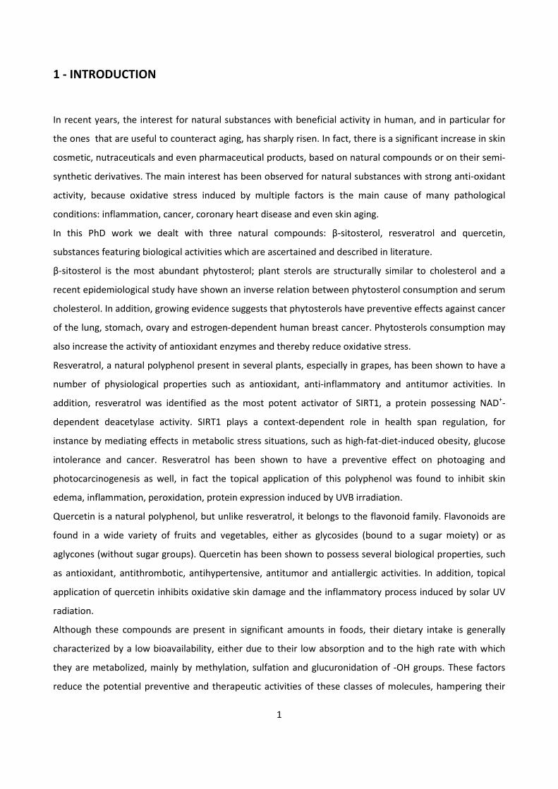
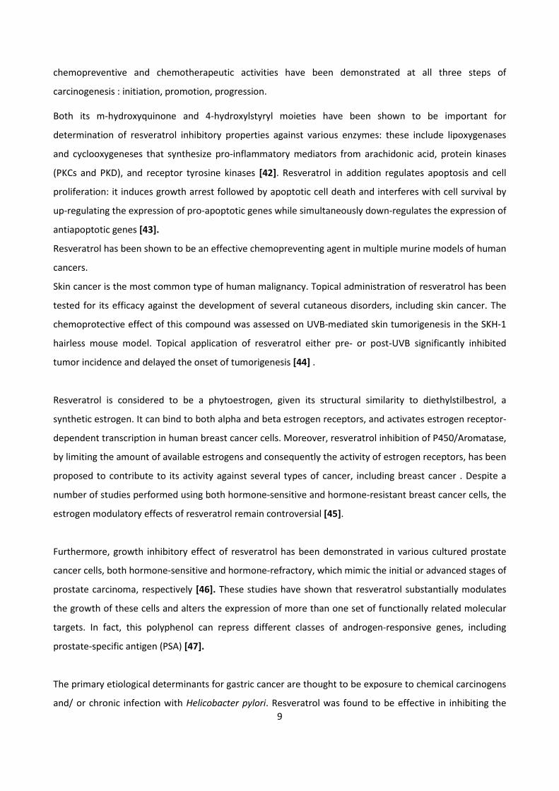
1.1 - β-SITOSTEROL
Sterols in plants have cellular functions analogous to those of cholesterol in animals. β-sitosterol (Fig. 1) is
the most common sterol in plants; it differs from cholesterol for the ethyl group at C-21. Hydrogenation of
the 6, 7 double bond of β-sitosterol converts it into sitostanol.
Figure 1.
In plants, sterols have both a structural function and a metabolic role. They are integral membrane
components which regulate the membrane fluidity and permeability, affecting various functions such as
simple diffusion, carrier-mediated diffusion and active transport across the membrane. They also modulate
the activities of membrane-associated proteins including enzymes, receptors and signal transduction
components. In addition, they are precursors of other bioactive steroids, such as the so-called
brassinosteroids (a special class of growth substances), and substrates for the synthesis of numerous
secondary plant metabolites [1].
Vegetable oils and cereals are generally known to be the best natural sources of dietary plant sterols [2].
Plant sterols occur both as free sterols and as bound conjugates, i.e., fatty acid esters (mainly C-16 and C-18
fatty acids), esters of phenolic acids, glycosides (most commonly with β-D-glucose) and acylated glycosides
(esterified at the 6-hydroxy group of the sugar moiety) [3]. The sterol content in a given plant may vary
depending on many factors, such as genetic background, growing conditions, tissue maturity and
postharvest changes [2].
1.1.1 - BIOLOGICAL ACTIVITIES
Cardiovascular activity
There is a scientific consensus that reduction of low density lipoprotein (LDL) cholesterol is important for
coronary risk decrease. Dietary cholesterol intake is variable but is often less than 300 mg/day and
currently 200 mg/day is recommended. Approximately 25% of the plasma cholesterol is due to dietary
HO
H
21
3
4
67
8
910
11
12
13 17
16
1514
1819
20
21
2223
25
26
24
27
28
29
5
4
intake, while 75% is accounted by endogenous synthesis [4]. However, dietary cholesterol appears to be
quite important because it is inversely correlated with endogenously synthesized cholesterol, suggesting
that they are co-regulated [5]. Phytosterols are thought to act primarily in the intestinal lumen. As
cholesterol analogs, they compete with it in absorptive micelles resulting in a reduced solubility of
cholesterol [6]. The affinity of plant sterols for micelles is greater than that of cholesterol. Human
physiological studies showed that the inclusion of phytosterols in a test meal resulted in the reduction of
absorbable micellar cholesterol in duodenal aspirates [7]. Even if phytosterols have a primary mechanism
of action in the intestine, their effects cause indirect changes in circulating LDL cholesterol. Reduced
transportation of absorbed cholesterol to the liver results in increased tissue LDL receptors [8]. Further
evidence of relative cholesterol deficiency after phytosterol treatment is the measured increase in whole
body cholesterol biosynthesis of 38–53% [9]. These results also demonstrates the rationale for using
phytosterols with statin drugs.
Then, phytosterols are recognized as an important component of healthy diets and diets designed to
reduce hypercholesterolemia. The United States National Cholesterol Education Program recommends
dietary phytosterol supplementation of 2 g/day as a lifestyle modification for cholesterol reduction.
Antioxidant and anti-inflammatory activity
Reactive oxygen species (ROS) produced by stressed cells can damage DNA. Vivancos [10] reported that β-
sitosterol increases the activities of antioxidant enzymes, superoxide dismutase and glutathione peroxidase
in cultured macrophage cells with oxidative stress induced by phorbol 12-myristate 13-acetate, indicating
that phytosterols can protect cells from ROS damage. In another study [11], cultured lipopolysaccharide-
activated macrophage cells were treated with β-sitosterol and campesterol: the results showed a
decreased production of prostaglandin E and prostaglandin I of series 2, by 68% and 67% (for sitosterol),
and by 55% and 52% (for campesterol), respectively. These studies suggest that phytosterols are able to
counteract the oxidative and inflammatory processes.
Anticancerogenic activity
Phytosterols seem to inhibit the development of various cancers mainly by inhibiting growth and promoting
apoptosis of cancer cells by the activation of caspase enzymes. The increased activity of caspase enzymes
could be attributed to the incorporation of phytosterols into cell membranes, which results in changes in
membrane structure and function; furthermore, these changes increase the activities of proteins involved
in extra- and intracellular signal-transduction pathways that activate caspase enzymes. Phytosterols could
also inhibit cancer development by lowering blood cholesterol, as high blood cholesterol levels, and hence
5
the concentration of cholesterol in lipid rafts of cell membranes, are associated with reduced apoptosis of
cancer cells. This combined evidence strongly supports an anticarcinogenic action of phytosterols and
hence advocates their dietary inclusion as an important strategy in prevention and treatment of cancer
[12].
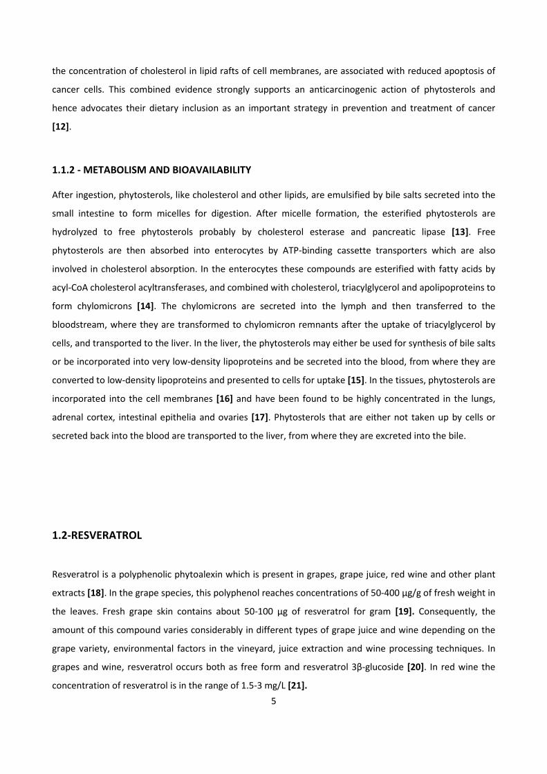
1.1.2 - METABOLISM AND BIOAVAILABILITY
After ingestion, phytosterols, like cholesterol and other lipids, are emulsified by bile salts secreted into the
small intestine to form micelles for digestion. After micelle formation, the esterified phytosterols are
hydrolyzed to free phytosterols probably by cholesterol esterase and pancreatic lipase [13]. Free
phytosterols are then absorbed into enterocytes by ATP-binding cassette transporters which are also
involved in cholesterol absorption. In the enterocytes these compounds are esterified with fatty acids by
acyl-CoA cholesterol acyltransferases, and combined with cholesterol, triacylglycerol and apolipoproteins to
form chylomicrons [14]. The chylomicrons are secreted into the lymph and then transferred to the
bloodstream, where they are transformed to chylomicron remnants after the uptake of triacylglycerol by
cells, and transported to the liver. In the liver, the phytosterols may either be used for synthesis of bile salts
or be incorporated into very low-density lipoproteins and be secreted into the blood, from where they are
converted to low-density lipoproteins and presented to cells for uptake [15]. In the tissues, phytosterols are
incorporated into the cell membranes [16] and have been found to be highly concentrated in the lungs,
adrenal cortex, intestinal epithelia and ovaries [17]. Phytosterols that are either not taken up by cells or
secreted back into the blood are transported to the liver, from where they are excreted into the bile.
1.2-RESVERATROL
Resveratrol is a polyphenolic phytoalexin which is present in grapes, grape juice, red wine and other plant
extracts [18]. In the grape species, this polyphenol reaches concentrations of 50-400 µg/g of fresh weight in
the leaves. Fresh grape skin contains about 50-100 µg of resveratrol for gram [19]. Consequently, the
amount of this compound varies considerably in different types of grape juice and wine depending on the
grape variety, environmental factors in the vineyard, juice extraction and wine processing techniques. In
grapes and wine, resveratrol occurs both as free form and resveratrol 3β-glucoside [20]. In red wine the
concentration of resveratrol is in the range of 1.5-3 mg/L [21].
6
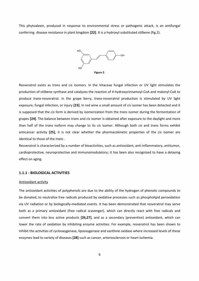
This phytoalexin, produced in response to environmental stress or pathogenic attack, is an antifungal
conferring disease resistance in plant kingdom [22]. It is a hydroxyl substituted stilbene (fig.2).
Figure 2.
Resveratrol exists as trans and cis isomers. In the Vitaceae fungal infection or UV light stimulates the
production of stilbene synthase and catalyzes the reaction of 4-hydroxycinnamoyl-CoA and malonyl-CoA to
produce trans-resveratrol. In the grape berry, trans-resveratrol production is stimulated by UV light
exposure, fungal infection, or injury [23]. In red wine a small amount of cis isomer has been detected and it
is supposed that the cis form is derived by isomerization from the trans isomer during the fermentation of
grapes [24]. The balance between trans and cis isomer is obtained after exposure to the daylight and more
than half of the trans isoform may change to its cis isomer. Although both cis and trans forms exhibit
anticancer activity [25], it is not clear whether the pharmacokinetic properties of the cis isomer are
identical to those of the trans .
Resveratrol is characterized by a number of bioactivities, such as antioxidant, anti-inflammatory, antitumor,
cardioprotective, neuroprotective and immunomodulatory; it has been also recognized to have a delaying
effect on aging.
1.1.1 - BIOLOGICAL ACTIVITIES
Antioxidant activity
The antioxidant activities of polyphenols are due to the ability of the hydrogen of phenolic compounds to
be donated, to neutralize free radicals produced by oxidative processes such as phospholipid peroxidation
via UV radiation or by biologically-mediated events. It has been demonstrated that resveratrol may serve
both as a primary antioxidant (free radical scavenger), which can directly react with free radicals and
convert them into less active products [26,27], and as a secondary (preventive) antioxidant, which can
lower the rate of oxidation by inhibiting enzyme activities. For example, resveratrol has been shown to
inhibit the activities of cyclooxygenase, lipooxygenase and xanthine oxidase where increased levels of these
enzymes lead to variety of diseases [28] such as cancer, arteriosclerosis or heart ischemia.
HO
HO
OH
7
Cardiovascular activity
Coronary artery disease remains the most deadly illness in western countries. Atherosclerosis,
hypertension, obesity, endothelial dysfunction, alteration in platelet function and oxidative stress all
contribute to the onset and progression of coronary artery disease. Many of the risk factors for this
pathology such as age, sex and family history are uncontrollable; hence, emphasis must be placed on the
modifiable risk factors. In particular, interest in dietary modification and even supplementation is at an all-
time high: examples of compounds that can be supplemented are omega-3 fatty acids, natural antioxidants
and plant sterols, all recognized as “heart-healthy” foods [29]. Red wine was found to be one of these
heart-healthy foods. In fact, the term “French paradox” refers to the observation that mortality due to
coronary diseases in France, where red wine consumption is considerable, is significantly lower compared
to that in other countries despite similarities in other risk factors such as dietary fat intake, obesity and
cigarette smoking: resveratrol contributes to the antioxidant potential of red wine, and may be related with
the decrease in coronary heart disease among wine drinkers.
Atherosclerosis lies at the root of coronary artery disease. The generation of atherosclerotic plaque can be
thought of as a stepwise process, initiated by the concentration-dependent transport of LDLs into the
arterial wall followed by modification of lipids and proteins by oxidation and inflammation invasion of
magrophages and their transformation into foam cells, which infiltrate smooth muscle cells with
subsequent production of fibrin and extracellular matrix [30]. Resveratrol has been shown to attenuate
each of these processes.
LDL and HDL play quite different roles in the genesis of atherosclerosis. LDL is the cholesterol compound
that is responsible for mural deposition and initiating atherosclerotic lesion, whereas HDL transports
cholesterol back to the liver where it is metabolized. Treatment with resveratrol has been shown
repeatedly to produce an anti-atherogenic serum lipid profile. It had been demonstrated that
apolipoprotein E-deficient mice treated with daily oral resveratrol for 20 weeks had significantly lower
circulating LDL and higher HDL compared to untreated controls [31]. Moreover, most studies show that this
compound, administered at a wide range of doses and for even short periods of time, exerts a favorable
effect on lipids profile in hypercholesterolemic animals. One possible explanation for this beneficial effect
of resveratrol on lipids was proposed by Cho et al [32], who showed that HGM-CoA mRNA was up-
regulated by resveratrol supplementation in hamsters fed with a high-fat diet.
8
Intramural lipid accumulation is followed by LDL oxidation, which promotes macrophage migration into the
vessel wall. Macrophages in turn take up oxidized LDL in large quantities and transform it into the
cholesterol-rich foam cells, that characterize early atherosclerotic lesion [33]. Resveratrol by scavenging
free radicals, decreases the oxidation of LDL and increases serum HDL, which not only has antioxidant
activity but also promotes the excretion of excess cholesterol from macrophages, transporting it back to
the liver [34]. Resveratrol has also been shown to inhibit monocyte recruitment and macrophage
activation. Studies by Deng et al. [35] showed that this compound inhibits tumor necrosis factor-α (TNF-α)-
mediated adhesion of monocytes to endothelial cells and reduces protein and gene expression of the
intercellular adhesion molecule 1 (ICAM-1) and vascular cell adhesion molecule 1 (VCAM-1), by inhibiting
activation of NF-kB; ICAM-1 and VCAM-1 are essential to the localization of monocytes to early
atherosclerotic lesion. Other studies also demonstrated that resveratrol down-regulates the production of
other cytokines that contribute to the inflammatory environment, that is essential for the progression of
atherosclerosis [36].
Later in the course of atherosclerosis, smooth muscle cells, lured by the cytokines and growth factors
secreted by macrophages, migrate into the atherosclerotic plaque, proliferate, and begin to secrete
components of extracellular matrix, maturing the plaque into a capped, fibrous lesion. This process is
mediated by various chemotactic factor proteins and receptors, and resveratrol may oppose multiple steps
in this process. In fact, the estrogen receptor has also been shown to play a role in smooth muscle cells
proliferation, and resveratrol has been found to modulate this receptor, leading to increased inducible
nitric oxide synthase activity and subsequent decrease in smooth muscle cell proliferation [37].
The fibrotic plaque formed by migrating smooth muscle cells predisposes to the final step in
atherosclerosis, which is thrombogenesis. Several hypotheses have been proposed to explain the inhibitory
effect of resveratrol on platelet activation. The initial step in platelet activation is adhesion to type I
collagen. Resveratrol decreased adhesion to collagen and fibrinogen after activation with
lipopolysaccharide or thrombin [38]. Moreover, the protein kinase C (PKC) plays a key role in platelet
activation, and resveratrol has been shown to depress the activity of PKC in platelet membranes [39]. Each
of these mechanisms may contribute to the antithrombotic action of resveratrol.
Antitumor activity
Recent studies have documented that resveratrol has cancer-preventive properties [40], and this
compound is currently at the stage of preclinical studies for human cancer prevention [41]. Its potential
9
chemopreventive and chemotherapeutic activities have been demonstrated at all three steps of
carcinogenesis : initiation, promotion, progression.
Both its m-hydroxyquinone and 4-hydroxylstyryl moieties have been shown to be important for
determination of resveratrol inhibitory properties against various enzymes: these include lipoxygenases
and cyclooxygeneses that synthesize pro-inflammatory mediators from arachidonic acid, protein kinases
(PKCs and PKD), and receptor tyrosine kinases [42]. Resveratrol in addition regulates apoptosis and cell
proliferation: it induces growth arrest followed by apoptotic cell death and interferes with cell survival by
up-regulating the expression of pro-apoptotic genes while simultaneously down-regulates the expression of
antiapoptotic genes [43].
Resveratrol has been shown to be an effective chemopreventing agent in multiple murine models of human
cancers.
Skin cancer is the most common type of human malignancy. Topical administration of resveratrol has been
tested for its efficacy against the development of several cutaneous disorders, including skin cancer. The
chemoprotective effect of this compound was assessed on UVB-mediated skin tumorigenesis in the SKH-1
hairless mouse model. Topical application of resveratrol either pre- or post-UVB significantly inhibited
tumor incidence and delayed the onset of tumorigenesis [44] .
Resveratrol is considered to be a phytoestrogen, given its structural similarity to diethylstilbestrol, a
synthetic estrogen. It can bind to both alpha and beta estrogen receptors, and activates estrogen receptor-
dependent transcription in human breast cancer cells. Moreover, resveratrol inhibition of P450/Aromatase,
by limiting the amount of available estrogens and consequently the activity of estrogen receptors, has been
proposed to contribute to its activity against several types of cancer, including breast cancer . Despite a
number of studies performed using both hormone-sensitive and hormone-resistant breast cancer cells, the
estrogen modulatory effects of resveratrol remain controversial [45].
Furthermore, growth inhibitory effect of resveratrol has been demonstrated in various cultured prostate
cancer cells, both hormone-sensitive and hormone-refractory, which mimic the initial or advanced stages of
prostate carcinoma, respectively [46]. These studies have shown that resveratrol substantially modulates
the growth of these cells and alters the expression of more than one set of functionally related molecular
targets. In fact, this polyphenol can repress different classes of androgen-responsive genes, including
prostate-specific antigen (PSA) [47].
The primary etiological determinants for gastric cancer are thought to be exposure to chemical carcinogens
and/ or chronic infection with Helicobacter pylori. Resveratrol was found to be effective in inhibiting the
10
replication of H. Pylori [48]: this provides a reason for the intervention studies using resveratrol to
counteract gastric cancer [49].
A number of in vitro studies have also demonstrated the antiproliferative effect of resveratrol in various
leukemic cell lines [50].
Anti-inflammatory activity
One of the possible protective activities of antioxidants is the down-regulation of inflammatory responses,
which includes inhibition of synthesis and release of pro-inflammatory mediators, modification of
eicosanoid synthesis, inhibition of enzymes such as ciclooxygenase-1 (COX-1) and ciclooxygenase-2 (COX-2)
[51]. In mouse macrophages, resveratrol also has antioxidant activity, decreasing the production of reactive
oxygen species (ROS) and reactive nitrogen species (RNS) and inhibiting nitric oxygen synthetase (NOS)-2
and COX-2 synthesis as well as prostaglandin E2 production [52]. Furthermore, this compound inhibits the
production of TNF-induced monocyte chemo-attractant protein which plays an essential role in early events
during macrophage infiltration into adipose tissue, which results in chronic low-grade inflammation, a key
feature of obesity in type 2 diabetes characterized by adipose tissue macrophage infiltration and abnormal
cytokine production [53]. Finally, it is also established that some anti-inflammatory effects of resveratrol
arise from its capability to up-regulate histone deacetylase sirtuin (SIRT1) activity, that also shows
antitumor and anti-inflammatory effects [54]. Altogether, it is clear that key antitumor properties of
resveratrol are linked to its anti-inflammatory effects.
Resveratrol and SIRT-1
The sirtuin family of proteins possesses NAD+-dependent deacetylase activity and/or ADP
ribosyltransferase activity. The seven mammalian sirtuins (Sirt1–7) are localized differently within the cell
and have a variety of functions [55]. Sirt1 is the most extensively studied member of the family and
regulates diverse biological processes ranging from cell proliferation, differentiation and apoptosis, to
metabolism [56].
In fact, SIRT-1 plays a context-dependent role in metabolic regulation, for instance by mediating effects in
metabolic stress situations, such as high-fat-diet-induced obesity [57]. Furthermore, it confers protection
against aging-associated metabolic diseases such as glucose intolerance and cancer. In light of the growing
number of patients suffering from metabolic diseases, compounds that activate SIRT-1 directly or indirectly
might offer protection against the onset of metabolic damage and promote healthy aging.
Resveratrol was identified as the most potent activator of SIRT-1 [58]. Recently, however, it was shown that
resveratrol may not activate SIRT-1 directly, but rather exerts its effects on SIRT-1 through activation of
11
AMPK, although additional direct SIRT-1 activation is not completely excluded [59]. Resveratrol treatment
in mice fed a high-calorie diet consistently improved various health parameters, including glucose
homeostasis, endurance, and survival [57] and it has therefore been suggested to act as a calorie restriction
mimetic. In Timmers et al. study [60] a dietary supplementation of trans-resveratrol was given to obese
volunteers over 30 days. Whole-body energy expenditure, mitochondrial function and other parameter
have been examined: the results showed that resveratrol supplementation lowers energy expenditure and
improves metabolic profile as well as general health parameters.
Anti-diabetes activity
Diabetes mellitus is a complex metabolic disease and is divided into different types: type 1 and type 2
diabetes. Type 1 diabetes results from the autoimmune destruction of β cells and patients with this kind of
diabetes are dependent on exogenous insulin. Type 2 diabetes is characterized by defects in insulin
secretion and action.
In the last few years, rodent studies and experiments in vitro provided evidences that resveratrol may be
helpful in preventing and treating some metabolic diseases, including diabetes [61]. In general, the
management of diabetes involves in three main aspects: reduction of blood glucose, preservation of β cells,
and, in the case of type 2 diabetes, improvement in insulin action. Data in literature indicate that the
beneficial effects of resveratrol in relation to this pathology comprise all of these aspects.
The maintenance of blood glucose in the physiological range is pivotal in diabetes, since increased glycemia
causes numerous diabetic complications. The anti-hyperglycemic effect of resveratrol observed in diabetes
animals is thought to result from its stimulatory action on intracellular glucose transport. Increased glucose
uptake by different cells isolated from diabetic rats was found in the presence of resveratrol. Interestingly,
in experiments on isolated cells, resveratrol was able to stimulate glucose uptake in absence of insulin [62].
The stimulation of glucose uptake induced by resveratrol seems to be due to increased action of glucose
transporter in the plasma membrane (GLUT4) [63].
Type 2 diabetes develops slowly and is usually accompanied by insulin resistance. Initially, blood glucose is
maintained in the physiological range because of the compensatory increase in insulin secretion. This
compensatory mechanism impedes and delays the diagnosis of diabetes. Moreover, chronic
overstimulation of β cells causes their exhaustion and degradation, leading over time to insufficient
secretion of insulin [64]. In animals with hyperinsulinemia, resveratrol was established to effectively reduce
blood insulin and this inhibition was found to result from metabolic changes in β cells [65].
Since chronic overstimulation of β cells is known to induce their degradation, inhibition of insulin secretion
by resveratrol may attenuate these unfavorable effects.
12
The protective action of this polyphenol on the endocrine pancreas may also involve other mechanisms.
One of them is the inhibitory influence of resveratrol on cytokine action. Studies in vitro and in vivo
confirmed the important role of the inhibition of cytokine action in the mechanism activated by resveratrol
in order to protects pancreatic β cells [66].
Moreover, animal studies provided evidence that resveratrol may be useful to improve insulin action in
type 2 diabetes. The improvement in insulin action caused by resveratrol seems to result from different
effects. One of them is reduced adiposity. Resveratrol-induced reduction in body fat content was
demonstrated in mice and rats on a hypercaloric diet [67]. Moreover, resveratrol ingestion was found to
cause effects that are similar to those induced by calorie reduction [68]. Consistent with animal studies,
experiments in vitro revealed reduced ATP content and decreased accumulation of triglycerides in isolated
rat adipocytes exposed to resveratrol [69].
In conclusion, the preventive and therapeutic action of this compound in relation to diabetes is complex
and involves in different effects. Elucidation of these beneficial properties of resveratrol is necessary to
enable clinical human studies.
Resveratrol and skin
UV radiation, in particular UVB, is known to elicit a variety of adverse effects, such as erythema, sunburn,
inflammation, hyperplasia, hyperpigmentation, immunosuppression, premature skin aging, and
photocarcinogenesis [70]. UVB exposure is known to cause excessive generation of ROS, which if remains
uncounteracted creates a situation of oxidative stress in the skin[71]. Topical application of resveratrol to
SKH-1 hairless mice was found to inhibit UVB-mediated: skin edema, inflammation, cyclooxygenase (COX),
and ornithine decarboxyase (ODC), enzyme activity, ODC protein expression, lipid peroxidation and
generation of hydrogen peroxide (H2O2) [72].
UV radiation is also known to cause significant leukocyte infiltration, which may be responsible for an
additional outburst of ROS in the system. Some data demonstrated that resveratrol significantly inhibits
UVB-mediated infiltration of leucocytes, suggesting the photochemopreventive potential of this compound
and indicating that the antioxidant property may be responsible for the biological effects of resveratrol.
Antiviral activity
Resveratrol has been shown to be a potent antiviral molecule against various types of DNA and RNA
viruses. However, even if the antiviral activity of resveratrol is becoming evident, the cellular pathways that
lead to its protective activity are still far from being elucidated. Interestingly, some of the molecular
pathways regulated by resveratrol such as p53, NF-κB or PML (promyelocytic leukemia protein) are also
13
main players in the control of virus infection. In addition, the activation of SIRT1 by resveratrol considered
to be one of the main mediators of its activity, may also contribute to this regulation [73]. Further studies
are required to clarify the contribution of SIRT1 in the regulation of the mentioned molecular pathways
activated by resveratrol and their roles in the control of virus infection.
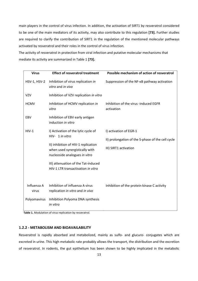
The activity of resveratrol in protection from viral infection and putative molecular mechanisms that
mediate its activity are summarized in Table 1 [73].
Virus Effect of resveratrol treatment Possible mechanism of action of resveratrol
HSV-1, HSV-2 Inhibition of virus replication in
vitro and in vivo
Suppression of the NF-κB pathway activation
VZV Inhibition of VZV replication in vitro
HCMV Inhibition of HCMV replication in
vitro
Inhibition of the virus -induced EGFR
activation
EBV Inhibition of EBV early antigen
induction in vitro
HIV-1 I) Activation of the lytic cycle of
HIV- 1 in vitro
II) inhibition of HIV-1 replication
when used synergistically with
nucleoside analogues in vitro
III) attenuation of the Tat-induced
HIV-1 LTR transactivation in vitro
I) activation of EGR-1
II) prolongation of the S-phase of the cell cycle
III) SIRT1 activation
Influenza A
virus
Inhibition of influenza A virus
replication in vitro and in vivo
Inhibition of the protein kinase C activity
Polyomavirus Inhibition Polyoma DNA synthesis
in vitro
Table 1. Modulation of virus replication by resveratrol.
1.2.2 - METABOLISM AND BIOAVAILABILITY
Resveratrol is rapidly absorbed and metabolized, mainly as sulfo- and glucuro- conjugates which are
excreted in urine. This high metabolic rate probably allows the transport, the distribution and the excretion
of resveratrol. In rodents, the gut epithelium has been shown to be highly implicated in the metabolic
14
process, resulting in polar resveratrol metabolism products which then need specific transporters to cross
cell membranes [74].
In a study by Walle et al. [75], 14
C-labeled resveratrol has been administered both orally and intravenously.
Measurement of total radioactivity demonstrated high absorption after oral intake of resveratrol, without
distinguishing resveratrol from its metabolites. The maximum plasma concentration was observed 1 h after
oral intake and a second peak was present after 6 h, probably as a result of enteric recirculation of
conjugate metabolites following reabsorption after intestinal hydrolysis. There was no second peak in any
subject after the intravenous administration, showing no enterohepatic cycle.
1.3 - QUERCETIN
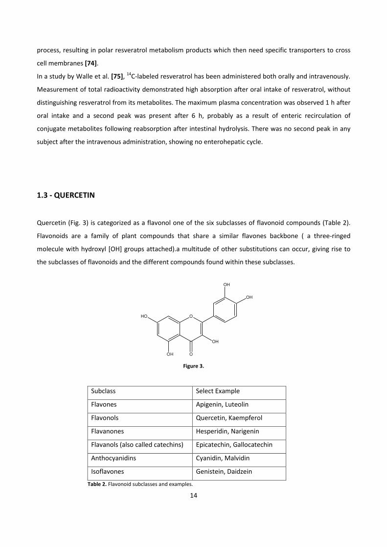
Quercetin (Fig. 3) is categorized as a flavonol one of the six subclasses of flavonoid compounds (Table 2).
Flavonoids are a family of plant compounds that share a similar flavones backbone ( a three-ringed
molecule with hydroxyl [OH] groups attached).a multitude of other substitutions can occur, giving rise to
the subclasses of flavonoids and the different compounds found within these subclasses.
Figure 3.
Subclass Select Example
Flavones Apigenin, Luteolin
Flavonols Quercetin, Kaempferol
Flavanones Hesperidin, Narigenin
Flavanols (also called catechins) Epicatechin, Gallocatechin
Anthocyanidins Cyanidin, Malvidin
Isoflavones Genistein, Daidzein
Table 2. Flavonoid subclasses and examples.
HO
OH
O
O
OH
OH
OH
15
Flavonoids also occur as either glycosides (with attached sugar) or as aglicones (without attached sugars)
[76]. Quercetin glucosides are found in a variety of foods including apples, berries, grapes, onion, shallots,
tea and tomatoes. the aglycone form of quercetin is found in much lesser amounts in diet. Two of the
better food sources are onion and shallots, but depending upon which part of these foods is eaten. For
example, quercetin in shallots flesh is about 99.2 % quercetin glucosides and 0.8 % quercetin aglycone. In
dry shallot skin the composition is almost the opposite 83,3% quercetin aglycone and 16.7% quercetin
glucoside [77]. Similar differences exist with onions. The flesh of onions contains mostly quercetin
glucosides, with only trace amounts of quercetin aglycone. Like shallots, the skin and outermost layers of
an onion have much more quercetin aglycone [78]. Growing conditions might significantly influence the
amount of quercetin in food, with evidence indicating that organically grown tomatoes have significantly
higher quercetin aglycone content than conventionally produced tomatoes.
1.3.1 - BIOLOGICAL ACTIVITY
Antioxidant activity
Quercetin has been shown to be an excellent in vitro antiossidant. Within the flavonoid family, quercetin is
the most potent scavenger of ROS, including O2.- and RNS like NO
. and ONOO
-[79]. These antioxidant
capacities of this compound are attributed to the presence of two antioxidant pharmacophores, cathecol
group and OH group in position 3, within the molecule that have the optimal configuration for free radical
scavenging [80]. In fact, when quercetin reacts with a free radical, it donates a proton and becomes a
radical itself, but the resulting unpaired electron is localized by resonance, making the quercetin radical too
low in energy to be reactive [81].
Quercetin can also protect against the more obvious environmental causes of free radicals, such as
smoking. Cigarette tar is a source of free radicals, which have been found to damage erythrocyte
membranes. Begum and Terao [82] found that the quercetin aglycone and its conjugate metabolites
(quercetin-3-O-β-glucuronide and quercetin-3-O-β-glucoside) could protect erythrocytes from the
membrane damage that is caused by smoking.
New evidence supports that quercetin not only has antioxidant capabilities but more importantly may act
as a signaling molecule. In fact, it has been shown that this compound can act as antioxidant leading to the
formation of quinones and prooxidants [83-84]. Furthermore, quercetin can be metabolized to
quinone/quinone methide metabolite such as o-semiquinone and o-quinone. O-semiquinone has the
capability to generate O2- in cell culture whereas o-quinone depletes glutathione (GSH) in the presence of
excess ascorbate [85-86]. The depletion of GSH and generation of O2- through these quercetin metabolites
suggest a prooxidant effect. The formation of quinone/quinone methide metabolites may lead to a low
16
level production of oxidants, which may in turn activate certain signaling cascades. By acting as a sensor as
well as a signaling molecule, H2O2 is an important regulator of signal transduction. An additional mechanism
of action of quercetin then is through regulating H2O2 levels: H2O2 regulates endothelial cell function such
as proliferation, inflammatory responses, apoptosis, and endothelium-dependent vasorelaxation [87].
Anti-inflammatory activity
Quercetin is known to possess strong anti-inflammatory capacities. Several in vitro studies have shown that
flavonoids are capable of inhibiting LPS-induced cytokine production. For instance, quercetin inhibits LPS-
induced TNFα production in macrophages and LPS-induced IL-8 production in lung cell [88].
A possible explanation for these anti-inflammatory effects of quercetin may be found in the interplay
between oxidative stress and inflammation: ROS are not only involved in the occurrence of oxidative stress,
but also in the promotion of inflammatory processes via activation of transcription factors such as NF-κB
and activator proteins (AP)-1 which induce the production of cytokines like TNFα [89]. Consequently,
scavenging ROS would not only prevent the occurrence of oxidative stress but also help mitigate
inflammation.
In addition quercetin, in according with several other in vitro studies, inhibits the production of
inflammation-production enzymes (cyclooxygenase and lipoxygenase) [90], NO production and nitric oxide
synthase (NOS) expression [91].
Cardiovascular activity
Consumption of flavonoids found in fruits, vegetables, and beverages such as tea and wine are inversely
correlated with mortality from coronary heart disease. The several studies demonstrated an inverse
relationship between flavonoid intake and occurrence of myocardial infarction [92]. The cardiovascular
benefits of flavonoid-rich foods have been attributed to their antioxidant effects (inhibiting the oxidation of
low density lipoproteins (LDL) [93]), NO mediated vasodilatory effects, reducing platelet aggregation [94],
and activating Nrf2-induced phase II detoxification through antioxidant response element (ARE)-mediated
gene expression [95]. Quercetin is the most prevalent flavonoid in the human diet and it has been shown to
engage in redox cycling reactions in cellular system thereby endowing both antioxidant and prooxidant (by
generating H2O2) properties [96-97]. The effect of quercetin on vessel function are not clear and especially
whether the proposed generation of H2O2 can modulated eNOS activity and vessel relaxation. In fact,
vascular endothelial cells have the capacity to release H2O2 and endothelium-derived H2O2 stimulates
EDHF. The endothelium produces and releases several vasoactive mediators involved in vessel tone, such as
NO, prostacyclins, and EDHF.
17
Moreover, quercetin, in human, inhibits platelet aggregation and thrombus formation [98]. Quercetin
appears to be effective in improving blood pressure and might have an effect on cholesterol in human.
Antitumor activity
Apart from scavenging ROS, another important effect of quercetin is to regulate cell cycle by modulating
several molecular targets, including p21, cyclin B, p27, cyclin-dependent kinases and topoisomerase II, even
if the mechanisms involved have not been elucidated yet. Depending on the cell type and tumor origin,
quercetin is able to block the cell cycle at G2/M or at the G1/S transition. In particular, quercetin causes
G2/M arrest in human esophageal squamous cell carcinoma cell line through up-regulation of p73 and
p21waf1 and subsequent down-regulation of cyclin B1, both at the mRNA and protein levels [99].In human
breast carcinoma cell lines low doses of quercetin inhibit proliferation. Cell-cycle arrest occurs at the G1
phase through the induction of p21 and through the concomitant decrease of phosphorylation of the
retinoblastoma protein (pRb) [100].In the same cell model, quercetin downregulates the cyclin B1 and
cyclin-dependent kinase (CDK) 1, which are essential in the progression to the G2/M phases of the cell cycle
[101]. Similarly, in the human lung cancer cells, quercetin glucuronides induce cell-cycle arrest at G2/M
phase by increasing the expressions of proteins such as cyclin B [102]. A similar antiproliferative effect has
also been observed both for highly or moderately aggressive prostate cancer cell lines, whereas no effect
has been found for poorly aggressive prostate cancer cells [103].
In add, quercetin has shown a pro-apoptotic activity. The capability of quercetin to induce apoptosis in
cancer cells (via both the intrinsic and extrinsic pathways) undoubtedly renders this molecule an interesting
tool in the oncology field. The pro-apoptotic effects of quercetin may result from multiple pathways. First,
in several kind of cell lines, quercetin treatment increases cytosolic Ca2+
levels and reduces the
mitochondrial membrane potential (MMP), thus promoting activation of caspase-3, -8 and -9 [104].
Second, quercetin is a potent enhancer of TNF-related [105]. Third, the anti-proliferative and pro-apoptotic
effects could be related to the capability of quercetin to directly bind tubulin, provoking the
depolymerization of cellular microtubules [106].
Several studies have investigated the role of p53 in the antiproliferative and proapoptotic action of
quercetin on tumor cell lines. In fact, quercetin causes cell-cycle arrest and apoptosis by inducing p53
phosphorylation and by stabilizing p53 both at the mRNA and protein level [107]. The presence of p53
limits the effect of quercetin, since when p53 is inhibited, cells become more sensitive to quercetin related
cytotoxicity and quercetin-related apoptosis. The effect of the presence or absence of p53 on quercetin
induced cytotoxicity and apoptosis is consistent with the involvement of this polyphenol in the oxidative
cell balance.
18
The studies of quercetin on cellular models offer an almost exhaustive explanation of the mechanisms that
link this compound to the oxidative cell balance and to the control of cell-cycle phases; these results invite
major attention to study quercetin in more complex and sophisticated animal models.
Allergy, Asthma, and Atopic Disease
In vitro quercetin inhibits histamine release by mast cells and basophils [108] suggesting an antiallergy
effect. Animal evidence indicates that quercetin might have therapeutic potential for allergic airway
disease. Several studies conducted in guinea pigs have reported that quercetin, provided orally or
administered via inhalation, has anti-asthmatic activity [109]. In murine models of allergic airway
inflammation and asthma, quercetin had pronounced anti-inflammatory effects [110], reduced eosinophil
and neutrophil counts and infiltration in in lung tissue, and inhibited asthmatic reactions [111].
In addition, quercetin inhibits anaphylactic contraction of giunea pig ileum smooth muscle in vitro [112].
Human epidemiological research reports an inverse association between intakes of quercetin and asthma
incidence [113]. Human intervention studies investigating quercetin for asthma and atopic disease are
currently lacking. However, two studies have investigated the effects of an enzymatically-modified
isoquercitrin (a quercetin glycoside) on allergic symptoms. In these studies, this specific quercetin glycoside
provided a statistically significant relief of ocular symptoms, but no statistically significant relief of nasal
symptoms caused by pollen [114].
In addition, other studies reported that quercetin might reduce erythema and burning sensation.
Presumably this occurred in part because quercetin inhibited niacin-induced human mast cell prostaglandin
D2 release [115].
Antiviral and antibacterial activity
Quercetin has in vitro antiviral activity against reverse transcriptase of HIV and other retroviruses like
Herpes simplex virus type 1, polio-virus type 1, respiratory syncytial virus (RSV) [116], and hepatitis C [117].
Quercetin has in vitro antibacterial activity against five microorganisms (Actinobacillus
actinomycetemcomitans, Actinomyces viscosus, Porphyromonas gingivalis, Fusobacterium nucleatum, and
Actinomyces naeslundii) associated with onset and progression of periodontal disease [118]. It also has in
vitro and in vivo activity against H. pylori [119].
Quercetin exerts immune and inflammation modulating activity in several murine models of autoimmunity.
For example, in experimental allergic encephalomyelitis a T-helper 1 (Th1) cell-mediated inflammatory
demyelinating autoimmune disease model of multiple sclerosis, quercetin ameliorated this patology by
blocking IL-12 signaling and Th1 differentiation [120].
19
Skin and quercetin
As mentioned above, quercetin has the highest antiradical activity compared to other flavonoids and its
properties include ROS scavenging, inhibition of peroxidation and metal ions chelation. These evidences
suggest the possible effectiveness of quercetin topical administration to prevent UVB radiation-induced
skin damage [121]. Acute UV skin exposure induces an immediate inflammatory response with erythema
and leukocyte infiltration. It has been suggested that the modulation of redox-sensitive transcription
factors such as nuclear factor (NF)-κB by ROS is a central and early event in the induction of inflammatory
reactions [122]. The NF-κB is an oxidative stress sensitive factor that activates multiple target genes
involved in the expression of several pro-inflammatory mediators. In agreement with the inflammatory
character of the UVB irradiation, it induces a dose-dependent increase in myeloperoxidase (MPO) activity
[123]. The topical application of a formulation containing quercetin significantly decreased the UVB-
induced increase of MPO in the skin [124].
The antioxidants are mainly concentrated in the epidermis compared to the dermis; in fact, the UVB
radiation exposure induces a greater decrease in the epidermis antioxidant systems compared to the
dermis [125]. Therefore, the epidermis can be considered the first line of skin defense and the glutathione
redox status has been confirmed as an early and sensitive sensor of UVB-induced epidermal oxidative stress
[126]. In fact, a dose-dependent depletion of GSH was detected in the skin after UVB irradiation. In
Casagrande et al. study [127] it has been demonstrated that the topical treatment with a formulation
containing quercetin significantly prevented the UVB irradiation-induced GSH depletion.
During multiple intermittent UV exposure, repeated processes induce the formation of severely damaged
collagen and ultimately skin wrinkling and reduced elasticity. The degradation of the extracellular matrix is
a consequence of metalloproteinases activity in epidermal keratinocytes and fibroblasts. This process is
followed by formation of imperfectly repaired collagen [128]. Furthermore, the UVB irradiation induction of
proteinases expression in the epidermis has been recently demonstrated; it also induces gene transcription
factors for metalloproteinases. The proteinases may be produced by a variety of cells including
macrophages and neutrophils; Casagrande et al. study [127] also demonstrated that the formulations
containing quercetin inhibited the proteinases secretion/activity in the skin. These evidences further
strengthen the possible use of topical formulations containing quercetin to prevent UVB radiation skin
damages.
1.3.2 - METABOLISM AND BIOAVAILABILITY
Quercetin is found in far greater amounts in the diet as glycosides than as quercetin aglycone. When
quercetin glycosides are ingested, glycosyl groups can be released during chewing, digestion, and
20
absorption. As an example, enzymes in the mouth and intestine can hydrolyze quercetin glycosides to
aglycones. [129]. Several human studies have been conducted to compare the bioavailability of quercetin
aglycone and glycosides. A study of absorption in ileostomy patients revealed absorption of 24 % of the
pure aglycone and 52 % of quercetin glycosides [130]. Other studies have confirmed that the absorpition of
quercetin is considerably enhanced by its conjugation with a sugar group [131].
After absorption, quercetin is metabolized in various organs, such as the small intestine, colon, liver and
kidney [132]. Metabolites formed in the small intestine and liver are mainly the result of phase II
metabolism by biotransformation enzymes and therefore include methylated, sulfated and glucuronidated
forms [133]. Moreover, bacterial ring fission of the aglycone occurs both in the small intestine and in colon,
resulting in breakdown of the backbone structure of quercetin and the subsequent formation of smaller
phenolics [132]. Normally, human quercetin plasma concentration is in low nanomolar range, but upon
quercetin supplementation it may increase to high nanomolar or low micromolar range [134]. A recent
study regarding the tissue distribution in rats and pigs has shown that, upon quercetin supplementation,
the highest accumulation of the flavonoid and its metabolites is found in lungs (rats) and liver and kidney
(pigs) [135]. It has been shown that the half-lives of quercetin metabolites are rather high (from 11 to 28 h).
This indicates that, upon repeated quercetin supplementation, they could attain a considerable plasma
level and pharmacological function of dietary quercetin, including antioxidant activity, should be exerted
exclusively by its conjugated metabolites [136].
1.4-FATTY ACIDS
Fatty acids are classified as saturated fatty acid (SFA), monounsaturated fatty acid (MUPA), and
polyunsaturated fatty acid (PUFA). Omega-3 (n-3), omega-6 (n-6), and omega-9 (n-9) unsaturated fatty acid
structures are based on the position of the first double bond at the third, sixth or ninth position from the
methyl (omega) terminal of the aliphatic carbon chain.
Humans and other mammals can synthesize saturated fatty acids and some monounsaturated fatty acids
from carbon groups in carbohydrates and proteins; they lack the enzymes necessary to insert a cis double
bond at the n-6 or the n-3 position of a fatty acid [137]. Consequently, omega-6 and omega-3 fatty acids
are essential nutrients. The parent fatty acid of the omega-6 series is linoleic acid (18:2n-6), and the parent
fatty acid of the omega-3 series is linolenic acid (18:3n-3.). Humans can synthesize long-chain (20 carbons
21
or more) omega-6 fatty acids, such as dihomo-gamma-linolenic acid (DGLA; 20:3n-6) and arachidonic acid
(AA; 20:4n-6) from linoleic acid and long-chain omega-3 fatty acids, such as eicosapentaenoic acid (EPA;
20:5n-3) and docosahexaenoic acid (DHA; 22:6n-3) from linolenic acid through a series of desaturation
(addition of a double bond) and elongation (addition of two carbon atoms) reactions.
Unlike the linolenic and linoleic acid, oleic acid (18:1n-9), is consumed in substantial amounts in the typical
Western diet and is not an essential fatty acid. There is little eicosatrienoic acid (ETA; 20:3n-9) in cell
membranes, however, probably because of the overwhelming competition from dietary linoleic acid for the
relevant desaturase and elongase enzymes.
1.4.1 - PHARMACOLOGICAL ACTIVITIES
Omega-6 and omega-3 PUFA are important structural components of cell membranes. When incorporated
into phospholipids, they affect cell membrane properties such as fluidity, flexibility, permeability and the
activity of membrane bound enzymes [138].
DHA is selectively incorporated into retinal cell membranes and postsynaptic neuronal cell membranes,
suggesting it plays important roles in vision and nervous system function. DHA is found at very high
concentrations in the cell membranes of the retina; the retina conserves and recycles DHA even when
omega-3 fatty acid intake is low [139]. Animal studies indicate that DHA is required for the normal
development and function of the retina. Moreover, these studies suggest that there is a critical period
during retinal development when inadequate DHA will result in permanent abnormalities in retinal
function.
The phospholipids of the brain's gray matter contain high proportions of DHA and AA, suggesting they are
important to central nervous system function [140]. Brain DHA content may be particularly important,
since animal studies have shown that depletion of DHA in the brain can result in learning deficits. It is not
clear how DHA affects brain function, but changes in DHA content of neuronal cell membranes could alter
the function of ion channels or membrane-associated receptors, as well as the availability of
neurotransmitters [141].
Eicosanoids, derived from 20-carbon PUFA, are potent chemical messengers that play critical roles in
immune and inflammatory responses. During an inflammatory response, DGLA, AA, and EPA in cell
membranes can be metabolized by enzymes known as cyclooxygenases and lipoxygenases to form
prostaglandins and leukotrienes [142].
AA is the progenitor of both PGE2 and LTB4 via the cyclooxygenase and 5-lipoxygenase enzymatic pathways,
respectively. PGE2 and LTB4 have pro-inflammatory biological actions. PGE2 can cause pain and vasodilation
22
and LTB4 is a chemoattractant and activator of neutrophils; together they can cause vascular leakage and
extravasation of fluid.
EPA, the n-3 homologue of AA, can inhibit AA metabolism competitively via the cyclooxygenase and 5-
lipoxygenase enzymatic pathways and, thus, can suppress production of the n-6 eicosanoid inflammatory
mediators. EPA is a potential cyclooxygenase substrate for the synthesis of PGE3, which also has
inflammatory activity [143, 144]. EPA is also a 5-lipoxygenase substrate and can lead to the formation of
LTB5, but LTB5 has little inflammatory activity compared with LTB4 [145].
In those who consume typical Western diets, the amount of AA in cell membranes is much greater than the
amount of EPA, resulting in the formation of more eicosanoids derived from AA than EPA. However,
increasing omega-3 fatty acid intake enhances the EPA content of cell membranes, resulting in higher
proportions of eicosanoids derived from EPA [146].
Many antiinflammatory pharmacotherapies are directed at inhibiting the production of these inflammatory
mediators and thus possibilities exist for therapies that incorporate n-3 and n-9 dietary fatty acids.
However, there are competitive interactions between dietary PUFAs; thus, in any examination of the
effects of dietary n-3 or n-9 PUFAs, it is important to consider also the background dietary n-6 PUFAs.
The results of epidemiological studies and randomized controlled trials suggest that replacing dietary SFAs
with omega-6 and omega-3 PUFA lowers LDL cholesterol and decreases cardiovascular disease risk.
A reduction in omega-6 fatty acids leads to skin lesions, anemia, increased platelet aggregation,
thrombocytopenia, fatty liver, delayed wound healing, increased susceptibility to infections, diarrhea,
growth delay in childhood.
The lack of omega-3 fatty acids is characterized by neurological symptoms, reduced visual acuity, skin
lesions, developmental delay, compromised learning ability, abnormal electroretinogram.
Maintaining an optimal omega-3/omega-6 ratio is essential in the prevention of some pathologic conditions
as well as in the therapy of diseases due to immuno-allergic causes and to lipid metabolism errors. In
particular, a correct omega-3/omega-6 ratio is important: to prevent cardiovascular diseases and to control
their risk factors [147, 148]; to prevent and treat skin and immuno-allergic affections [149]; in congenital
metabolic pathologies, such as adreno-leukodystrophy and phenylketonuria.
In addition, fatty acids are essential components of natural lipids, which determine the physiological
structure and function of the human skin [150]. They are present in the epidermis, especially in the stratum
corneum, the outermost layer, and cell membranes [151]. Many effects of fatty acids can be linked to
changes in membrane lipid composition affecting cell signaling mechanisms originating from membranes
[152]. Skin aging may influence epidermal lipids and free fatty acid composition, and their physiological
functions may be involved in aging process. In fact, the levels of SFAs such as palmitic acid and stearic acid,
23
PUFAs such as linoleic acid and 11,14,17-eicosatrienoic acid , one of the omega-3 polyunsaturated acids,
were decreased
in aged skin by 15%, 31%, 7%, and 56%, compared with those in young skin, respectively [153].Moreovere,
also photoaging process may affect the fatty acids composition in human skin [153].
24
2 - AIM
This PhD project aims to overcome the problems related to poor bioavailability of some natural
polyphenols and sterols, through the synthesis of the corresponding esters with saturated and unsaturated
fatty acids.
The limitation of pharmaceutical, cosmetic and dietary application of polyphenols and sterols is due to their
poor solubility and stability in lipophilic media: these features are also undermining their oral and dermal
bioavailability. The synthesis of more lipophilic derivatives, especially esters, could lead to an increase in
lipophilicity, and then in the bioavailability and permeability, in particular in the skin.
The selected natural compounds have all proven biological activities and are widely described in literature.
The choice of saturated and unsaturated fatty acids as acid components for the esterification is due to their
beneficial activities: thus, the synthesis of such esters would introduce in a single molecule (prodrug) two
moieties both pharmacologically active.
25
3 - MATERIALS AND METHODS
3.1 - MATERIALS
β-sitosterol (purity grade 60%), quercetin, oleic acid (purity grade 90%), linolenic acid (purity grade 70%),
linoleic acid (purity grade 95%), palmitoyl chloride, stearoyl chloride were purchased from Sigma Aldrich
(Milan, Italy); resveratrol was purchased from Polichimica (Bologna, Italy). All solvents, deuterated solvents,
reagents and enzymes used were purchased from Sigma Aldrich.
3.2 - METHODS
3.2.1 - INFRARED SPECTROSCOPY ANALYSIS (FT-IR)
Fourier Transform Infrared Spectroscopy (FT-IR) is an analytical technique used to identify functional
groups in the examined molecules.
Measurements have been performed by using a SPECTRUM ONE FT-IR spectrometer (Perkin Elmer).
Spectra have been acquired by DATA MANAGER 2 software (Perkin Elmer).
Sample preparation:
Samples in solid form and samples in liquid/semi-solid form have been treated differently:
• Solid form sample:
The sample is mixed at a ratio of 1:100 w/w with potassium bromide and homogenized in an agate
mortar; the powder is then pressed, without applying vacuum, under a 9 tons pressure for 4
minutes, to form a thin tablet that is gently put on a suitable support for the analysis. The
acquisition of spectra have been performed using the following parameters:
Wavelenght: 4000 - 450 cm-1
Resolution: 4 cm-1
Scan repetitions: 8
Background: KBr tablet
26
• Liquid/semi-solid form sample:
The substance is dropped between two sodium chloride windows. The acquisition of spectra have
been performed using the following parameters:
Wavelenght: 4000 - 600 cm-1
Resolution: 4 cm-1
Scan repetitions: 4
Background: NaCl window
3.2.2 - NUCLEAR MAGNETIC RESONANCE ANALYSIS (NMR)
NMR is a technique used to determine the structure of the investigated compounds.
NMR spectra have been registered, depending on different needs, by using two different instruments:
• Varian Mercury Plus 200, operating at 200 MHz, coupled with Sun software
• Bruker Advance 500, operating at 500 MHz, coupled with Bruker X-Win NMR 3.0 software.
Sample preparation:
8-10 mg (1H-NMR) or 50 mg (
13C-NMR) of substance have been dissolved in chloroform (CDCl3) or dimethyl
sulfoxide (DMSO) depending on their solubility.
3.2.3 - ULTRAVIOLET-VISIBLE SPECTROSCOPY ANALYSIS (UV-Vis)
UV-vis allow the determination of the maximum absorption wavelength of the analyzed molecule. This
value helps the identification of the analytes. Measurement have been performed by using a Jasco V-530
UV-vis spectrophotometer in different wavelength ranges, depending on the solvent:
• Methanol: 210-600 nm
• Chloroform: 235-600nm
Sample preparation:
Weighted quantities of the samples have been dissolved in the appropriate solvent (methanol or
chloroform) and spectra have been registered using quartz cuvettes. Solvent backgrounds have been
subtracted.
3.2.4 - HIGH PERFORMANCE LIQUID CHROMATOGRAPHY (HPLC)
HPLC is a chromatographic technique which allows to separate two or more components in an analyte,
based on differential affinity of the compounds for a stationary phase, placed inside the chromatographic
column, and a mobile phase, flowing through it. The stationary phase and the mobile phase are chosen
27
according to the sample to be analyzed. Depending on the stationary phase used, HPLC analysis can be
carried out in normal phase or reversed phase:
� Normal phase: in this case the column inner packing is polar, commonly made of silica
� Reversed phase: the column packing is apolar, commonly made of derivatized silica
The choice of the mobile phase is closely related to the stationary phase employed: usually in normal phase
HPLC apolar solvents are used, such as hexane and ter-butanol, whereas in reversed phase HPLC polar
solvents such as methanol, acetonitrile and water are used. In addition, HPLC can be performed using one
or more mobile phases: if the composition of the mobile phase is constant, the elution is performed in
isocratic conditions, while if the composition of the mobile phase changes during the analysis the
chromatographic separation is described as a gradient elution.
The samples to be analyzed are dissolved in a suitable solvent, usually similar or identical to the mobile
phase.
After leaving the column, the mobile phase enters a detector, needed to provide qualitative and
quantitative information on the analyte components. The most common detectors are based on UV-vis
light absorption. DAD (Diode Array Detector) allows to record the absorbance spectrum of the analyte in a
specified range of wavelenghts.
β-sitosterol, resveratrol and their respective derivatives analyses were performed by using an HPLC system
equipped with a quaternary pump Waters Delta Prep 600E, an injector Rheodyne 7125 with a 20 μl loop, a
thermostat Column Block Heater Mod. 7940 Hichrom Ltd., a detector DAD 2996. The acquired data were
processed by software Empawer 2.
Quercetin and its derivatives were performed by using an HPLC system equipped with a quaternary pump
Merck Hitachi L-7100, an injector Rheodyne 7125 with a 20 μl loop, a thermostat Column Block Heater
Mod. 7940 Hichrom Ltd., a detector DAD Hewlett Packard HP 1050. The acquired data were processed by
software HP CHEM.
3.2.5 - MASS SPECTROMETRY (MS)
All synthesized derivatives were analyzed by using a LCQ Advantage mass spectrometer, equipped with ion
trap and ESI (Electrospray Ionization) source.
3.2.6 - ANTIOXIDANT ACTIVITY
In collaboration with Dr. Angela Maria Rizzo from Dept. “Scienze Molecolari Applicate ai Biosistemi”,
Università degli Studi, Milan, the antioxidant activity of resveratrol and its tri-esters was tested using ABTS
(2,2’-azinobis-[3-ethylbenzthiazoline-6-sulphonic acid]) and DPPH (1,1-diphenyl-2-picrylhydrazyl) assays.
28
TEAC (Trolox Equivalent Antioxidant Capacity) and IC50 values of the compounds were calculated and
compared.
ABTS assay
The ABTS assay involves the oxidation of ABTS (2,2’-azinobis[3-ethylbenzothiazoline-6-sulphonate]) to a
radical cation, ABTS•+
. ABTS•+
is enzymatically pre-generated and the antioxidant or sample to be analyzed
is added to the reaction mixture. This results in the disappearance of ABTS•+
, which is measured by the
decrease in absorbance; the ABTS radical cation has an absorption maximum at 735 nm.
This method is based on the capacity of different components to scavenge ABTS radical cation compared to
a standard antioxidant (Trolox, vitamin E analogue) in a dose-response curve.
Stock solutions:
• Solution 1: potassium persulfate (K2S2O8) 2.5 mM in water
• Solution 2: ABTS 7.4 mM in water
• Solution 3: obtained by mixing Solution 1 and Solution 2 in equal quantities and allowing them to
react for 12 h at room temperature in the dark.
• Solution 4: Trolox 1 mM in methanol, used to prepare standard solutions
• Solution 5: obtained by dilution of 10 ml of Solution 3 to 100 ml of methanol (Abs735 =
approximately 0.700)
• Solution 6: sample solution
Standard solutions preparation (Table 3)
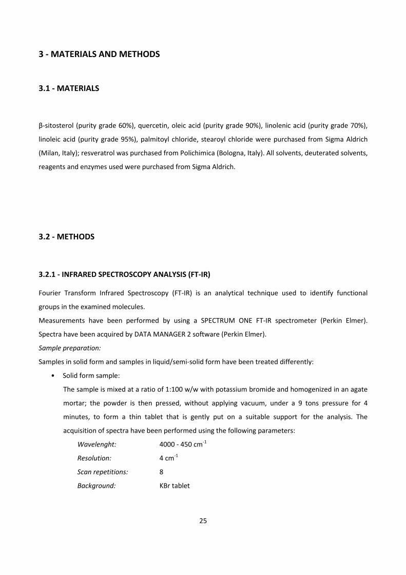
Standard solution Solution 4 vol. (μl) Solution 5 vol. (μl) Solvent vol. (μl) Trolox conc. (μM)
1 0 950 50 0
2 5 950 45 5
3 10 950 40 10
4 15 950 35 15
5 20 950 30 20
6 25 950 25 25
7 30 950 20 30
Table 3.
29
Results acquisition:
• for each acquisition Solution 5, solvent and Solution 4 (in the prescribed order) were added in a
cuvette, then the mixture was stirred and after 3 minutes its absorbance at 735 nm was acquired
using a UV-vis spectrophotometer;
• the calibration curve was plotted inserting the data in a graph (absorbance vs. Trolox
concentration);
• the samples were analyzed and the results were interpolated with the calibration curve and the
TEAC (Trolox Equivalent Antioxidant Capacity) values were obtained.
DPPH assay
The DPPH assay makes use of a stable free radical DPPH• (1,1-diphenyl-2-picrylhydrazyl). The reaction
involves a colour change from violet to yellow that can be easily monitored using a spectrophotomer at 520
nm.
DPPH solution preparation: prepared by dissolving 27 mg of DPPH with 25 ml of ethanol 70%, stirred until
complete dissolution and then stored in the dark.
Samples preparation: an increasing volume of analyte (50 µl by 50 µl) was added to different test tubes;
ethanol 70% was added to a final volume of 1 ml and then 500 µl di DPPH was added. The mixture was
stirred and stored for 30 minutes in the dark.
Results acquisition: the samples were analyzed using an UV-vis spectrophotometer (520 nm), obtaining
absorbance values relevant to the residual amounts of DPPH•: then IC50 value was obtained by
interpolation, plotting a graph with the calculated residual amounts of DPPH• vs. sample concentration.
IC50 value (µg/ml) represents the concentration of sample leading to a 50% decrease of DPPH•: the higher
the IC50 value, the lower the antioxidant activity.
30
4 - EXPERIMENTAL PART
4.1 - β-SITOSTEROL
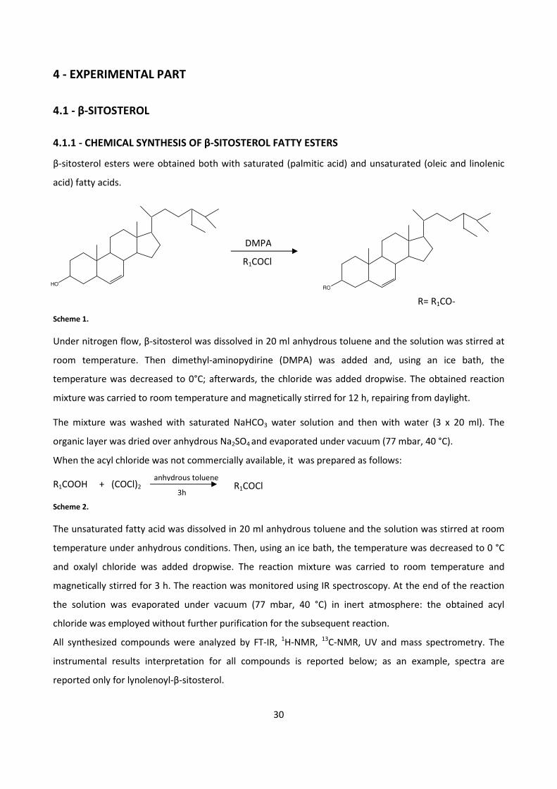
4.1.1 - CHEMICAL SYNTHESIS OF β-SITOSTEROL FATTY ESTERS
β-sitosterol esters were obtained both with saturated (palmitic acid) and unsaturated (oleic and linolenic
acid) fatty acids.
R= R1CO-
Scheme 1.
Under nitrogen flow, β-sitosterol was dissolved in 20 ml anhydrous toluene and the solution was stirred at
room temperature. Then dimethyl-aminopydirine (DMPA) was added and, using an ice bath, the
temperature was decreased to 0°C; afterwards, the chloride was added dropwise. The obtained reaction
mixture was carried to room temperature and magnetically stirred for 12 h, repairing from daylight.
The mixture was washed with saturated NaHCO3 water solution and then with water (3 x 20 ml). The
organic layer was dried over anhydrous Na2SO4 and evaporated under vacuum (77 mbar, 40 °C).
When the acyl chloride was not commercially available, it was prepared as follows:
R1COOH + (COCl)2
Scheme 2.
The unsaturated fatty acid was dissolved in 20 ml anhydrous toluene and the solution was stirred at room
temperature under anhydrous conditions. Then, using an ice bath, the temperature was decreased to 0 °C
and oxalyl chloride was added dropwise. The reaction mixture was carried to room temperature and
magnetically stirred for 3 h. The reaction was monitored using IR spectroscopy. At the end of the reaction
the solution was evaporated under vacuum (77 mbar, 40 °C) in inert atmosphere: the obtained acyl
chloride was employed without further purification for the subsequent reaction.
All synthesized compounds were analyzed by FT-IR, 1H-NMR,
13C-NMR, UV and mass spectrometry. The
instrumental results interpretation for all compounds is reported below; as an example, spectra are
reported only for lynolenoyl-β-sitosterol.
anhydrous toluene
3h
HO
DMPA
R1COCl
RO
R1COCl

31
PALMITOYL β-SITOSTEROL
R=
Reaction parameters:
Molar ratio palmitoyl chloride:β-sitosterol 1:1
β-sitosterol 1.21x10⁻³
mol, 0.50 g,
Palmitoyl chloride 1.21x10⁻³
mol, 0.33 g, 0.36 ml
DMPA 1.73x10⁻³
mol, 0.21 g,
Reaction temperature Room temperature
Reaction time 12 h
Yield 70%
C45H80O2 MW = 653.2
TLC: Hexane/Ethyl acetate/Ethyl Ether 5/2/1and spot visualization with iodine vapor
UV/Vis in MeOH λ (nm) =261; 280
1H-NMR (CDCl3): δ = 5.38-5.36 (m, 1 H, H7); 4.69-4.55 (m, 1 H, H2); 2.35-2.22 (m, 4 H, H1, H30); 2.04-0.80 (m,
74 H, CH2 and CH3).
HO
H
21
3
4
67
8
910
11
12
13 17
16
1514
1819
20
21
2223
25
26
24
27
28
29
5
O
30
31
32
33
34
35
36
37
38
39
40
41
42
43
44
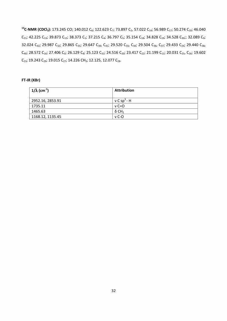
32
13C-NMR (CDCl3): 173.245 CO; 140.012 C6; 122.623 C7; 73.897 C2, 57.022 C14; 56.989 C17; 50.274 C10; 46.040
C21; 42.225 C13; 39.873 C12; 38.373 C1; 37.215 C4; 36.797 C5; 35.154 C18; 34.828 C19; 34.528 C30;; 32.089 C9;
32.024 C42; 29.987 C32; 29.865 C35; 29.647 C39, C41; 29.520 C33, C34; 29.504 C36, C37; 29.433 C24; 29.440 C38,
C40; 28.572 C16; 27.406 C3; 26.129 C8; 25.123 C31; 24.516 C20; 23.417 C22; 21.199 C11; 20.031 C25, C26; 19.602
C23; 19.243 C29; 19.015 C27; 14.226 CH3; 12.125, 12.077 C28.
FT-IR (KBr)
1/λλλλ (cm-1
) Attribution
2952.16, 2853.91 ν C sp3
- H
1735.11 ν C=O
1465.63 δ CH2
1168.12, 1135.45 ν C-O
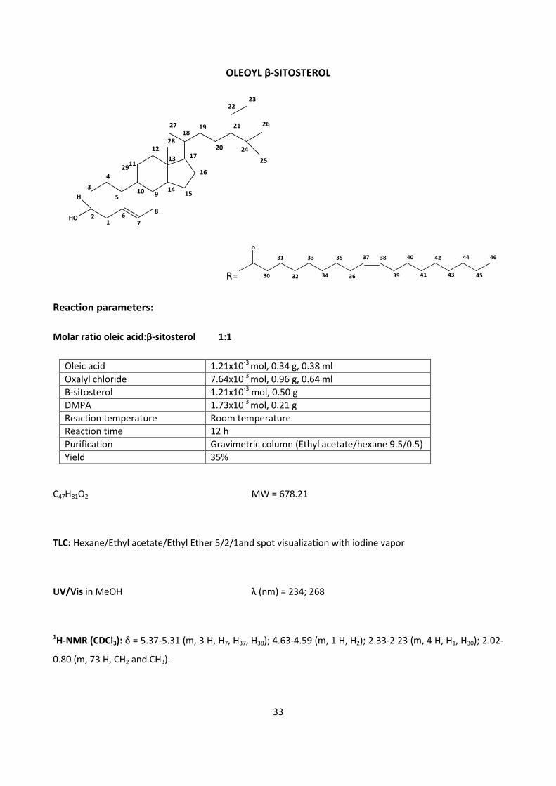
33
OLEOYL β-SITOSTEROL
R=
O
30
31
32
33
34
35
36
37 38
39
40
41
42
43
44
45
46
Reaction parameters:
Molar ratio oleic acid:β-sitosterol 1:1
Oleic acid 1.21x10⁻³
mol, 0.34 g, 0.38 ml
Oxalyl chloride 7.64x10⁻³
mol, 0.96 g, 0.64 ml
Β-sitosterol 1.21x10⁻³
mol, 0.50 g
DMPA 1.73x10⁻³
mol, 0.21 g
Reaction temperature Room temperature
Reaction time 12 h
Purification Gravimetric column (Ethyl acetate/hexane 9.5/0.5)
Yield 35%
C47H81O2 MW = 678.21
TLC: Hexane/Ethyl acetate/Ethyl Ether 5/2/1and spot visualization with iodine vapor
UV/Vis in MeOH λ (nm) = 234; 268
1H-NMR (CDCl3): δ = 5.37-5.31 (m, 3 H, H7, H37, H38); 4.63-4.59 (m, 1 H, H2); 2.33-2.23 (m, 4 H, H1, H30); 2.02-
0.80 (m, 73 H, CH2 and CH3).
HO
H
21
3
4
67
8
910
11
12
13 17
16
1514
1819
20
21
2223
25
26
24
27
28
29
5
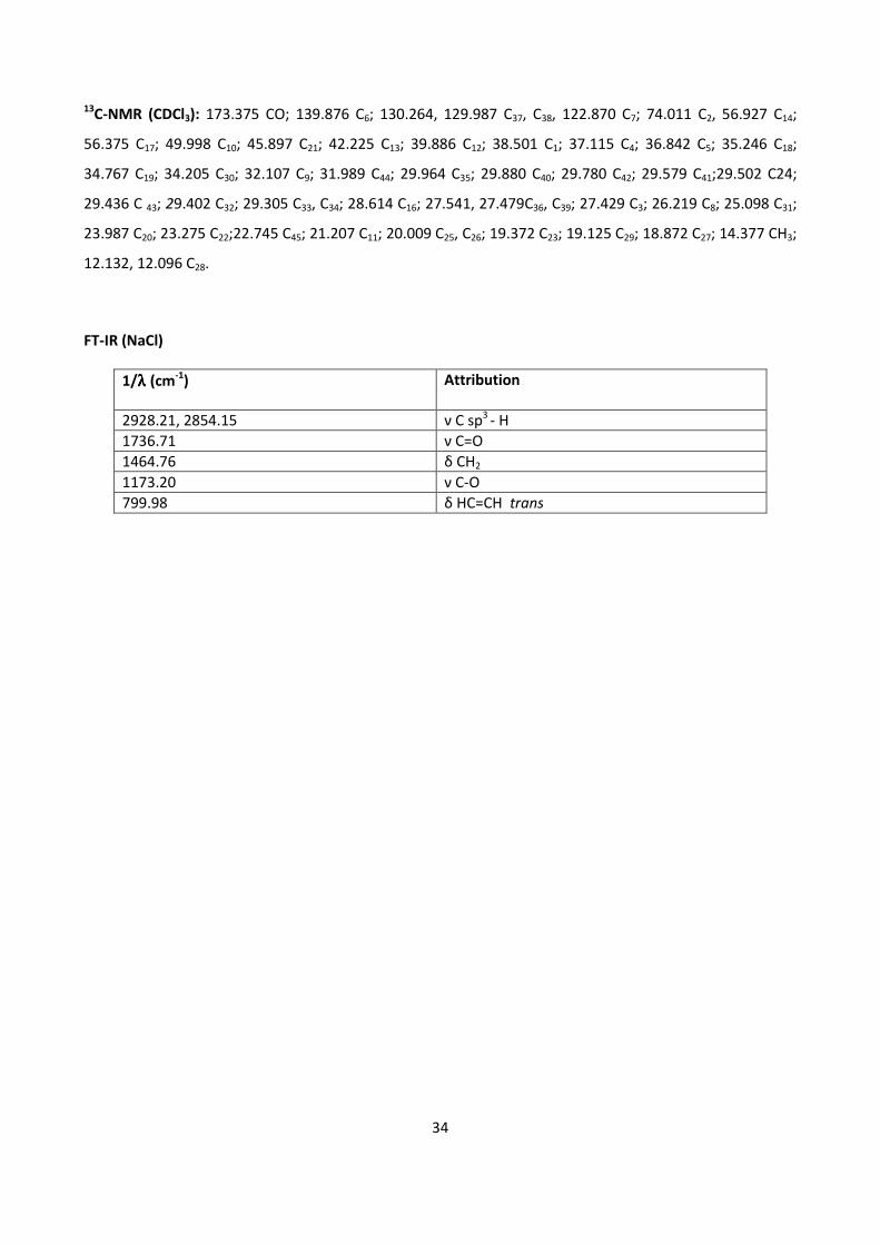
34
13C-NMR (CDCl3): 173.375 CO; 139.876 C6; 130.264, 129.987 C37, C38, 122.870 C7; 74.011 C2, 56.927 C14;
56.375 C17; 49.998 C10; 45.897 C21; 42.225 C13; 39.886 C12; 38.501 C1; 37.115 C4; 36.842 C5; 35.246 C18;
34.767 C19; 34.205 C30; 32.107 C9; 31.989 C44; 29.964 C35; 29.880 C40; 29.780 C42; 29.579 C41;29.502 C24;
29.436 C 43; 29.402 C32; 29.305 C33, C34; 28.614 C16; 27.541, 27.479C36, C39; 27.429 C3; 26.219 C8; 25.098 C31;
23.987 C20; 23.275 C22;22.745 C45; 21.207 C11; 20.009 C25, C26; 19.372 C23; 19.125 C29; 18.872 C27; 14.377 CH3;
12.132, 12.096 C28.
FT-IR (NaCl)
1/λλλλ (cm-1
) Attribution
2928.21, 2854.15 ν C sp3
- H
1736.71 ν C=O
1464.76 δ CH2
1173.20 ν C-O
799.98 δ HC=CH trans
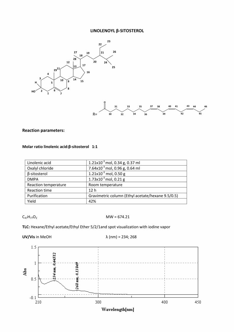
35
LINOLENOYL β-SITOSTEROL
R=
O
30
31
32
33
34
35
36
37 38
39
40 41
42
4443
45
46
Reaction parameters:
Molar ratio linolenic acid:β-sitosterol 1:1
Linolenic acid 1.21x10⁻³
mol, 0.34 g, 0.37 ml
Oxalyl chloride 7.64x10⁻³
mol, 0.96 g, 0.64 ml
β-sitosterol 1.21x10⁻³
mol, 0.50 g
DMPA 1.73x10⁻³
mol, 0.21 g
Reaction temperature Room temperature
Reaction time 12 h
Purification Gravimetric column (Ethyl acetate/hexane 9.5/0.5)
Yield 42%
C47H77O2 MW = 674.21
TLC: Hexane/Ethyl acetate/Ethyl Ether 5/2/1and spot visualization with iodine vapor
UV/Vis in MeOH λ (nm) = 234; 268
HO
H
21
3
4
67
8
910
11
12
13 17
16
1514
1819
20
21
2223
25
26
24
27
28
29
5
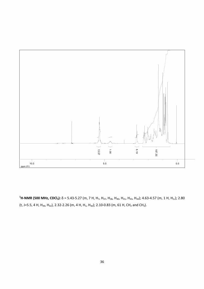
36
1H-NMR (500 MHz, CDCl3): δ = 5.43-5.27 (m, 7 H, H7, H37, H38, H40, H41, H43, H44); 4.63-4.57 (m, 1 H, H2,); 2.80
(t, J=5.5, 4 H, H39, H42), 2.32-2.26 (m, 4 H, H1, H30); 2.10-0.83 (m, 61 H, CH2 and CH3).
ppm (f1)
0.05.010.0
13.0
7
1.0
0
6.1
9
187
.33
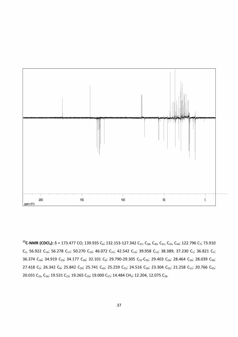
37
13C-NMR (CDCl3): δ = 173.477 CO; 139.935 C6; 132.153-127.342 C37, C38, C40, C41, C43, C44; 122.796 C7; 73.910
C2, 56.922 C14; 56.278 C17; 50.270 C10; 46.072 C21; 42.542 C13; 39.958 C12; 38.389; 37.230 C1; 36.821 C5;
36.374 C18; 34.919 C19; 34.177 C30; 32.101 C9; 29.790-29.305 C32-C35; 29.403 C24; 28.464 C16; 28.039 C36;
27.418 C3; 26.342 C8; 25.842 C39; 25.741 C42; 25.259 C31; 24.516 C20; 23.304 C22; 21.258 C11; 20.766 C45;
20.031 C25, C26; 19.531 C23; 19.265 C29; 19.000 C27; 14.484 CH3; 12.204, 12.075 C28.
ppm (f1)
050100150200
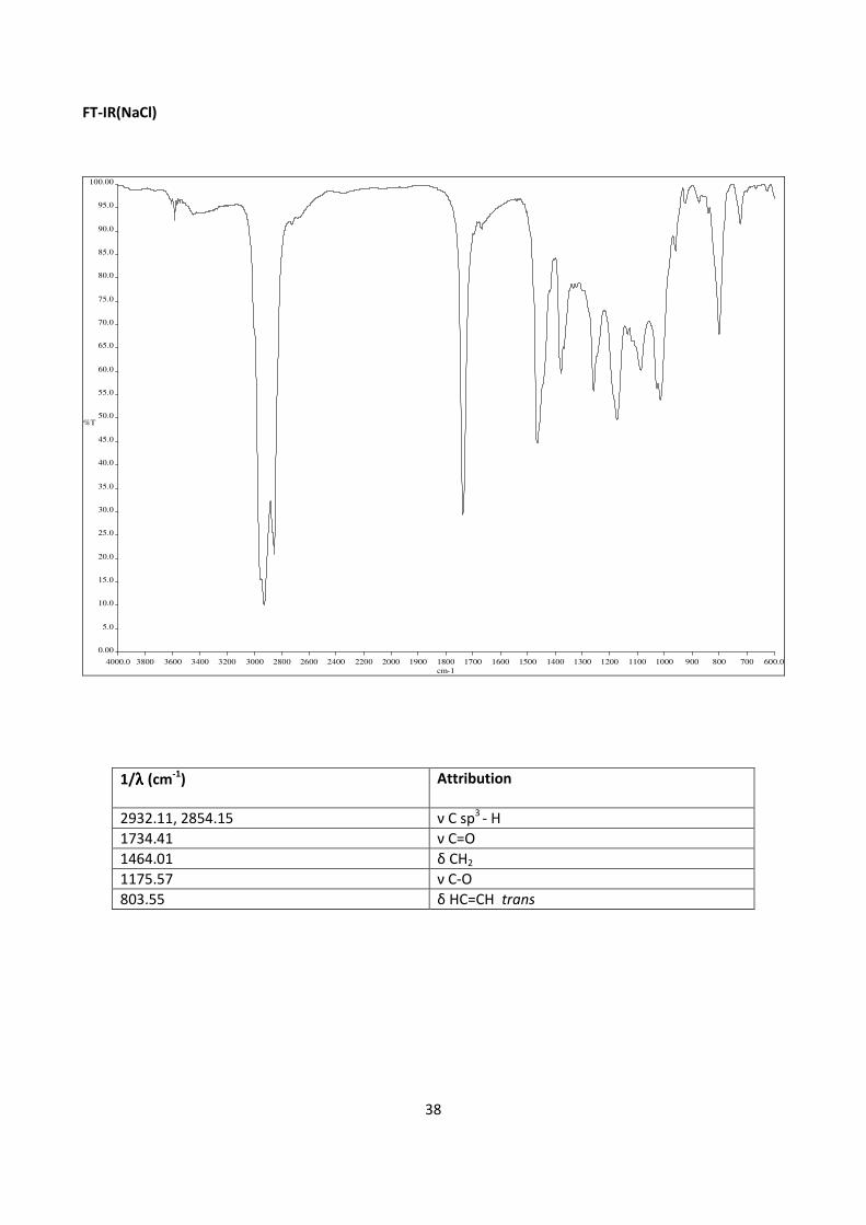
38
FT-IR(NaCl)
4000.0 3800 3600 3400 3200 3000 2800 2600 2400 2200 2000 1900 1800 1700 1600 1500 1400 1300 1200 1100 1000 900 800 700 600.0
0.00
5.0
10.0
15.0
20.0
25.0
30.0
35.0
40.0
45.0
50.0
55.0
60.0
65.0
70.0
75.0
80.0
85.0
90.0
95.0
100.00
cm-1
%T
1/λλλλ (cm-1
) Attribution
2932.11, 2854.15 ν C sp3
- H
1734.41 ν C=O
1464.01 δ CH2
1175.57 ν C-O
803.55 δ HC=CH trans
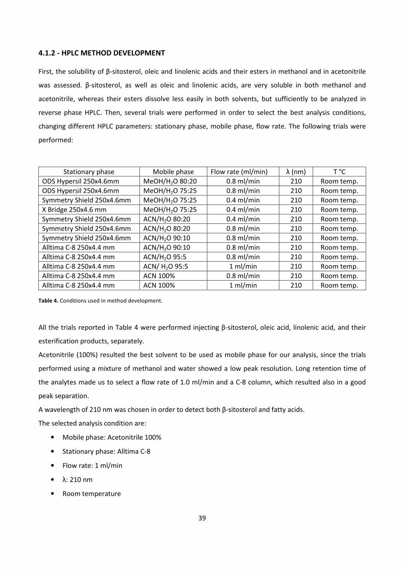
39
4.1.2 - HPLC METHOD DEVELOPMENT
First, the solubility of β-sitosterol, oleic and linolenic acids and their esters in methanol and in acetonitrile
was assessed. β-sitosterol, as well as oleic and linolenic acids, are very soluble in both methanol and
acetonitrile, whereas their esters dissolve less easily in both solvents, but sufficiently to be analyzed in
reverse phase HPLC. Then, several trials were performed in order to select the best analysis conditions,
changing different HPLC parameters: stationary phase, mobile phase, flow rate. The following trials were
performed:
Stationary phase Mobile phase Flow rate (ml/min) λ (nm) T °C
ODS Hypersil 250x4.6mm MeOH/H2O 80:20 0.8 ml/min 210 Room temp.
ODS Hypersil 250x4.6mm MeOH/H2O 75:25 0.8 ml/min 210 Room temp.
Symmetry Shield 250x4.6mm MeOH/H2O 75:25 0.4 ml/min 210 Room temp.
X Bridge 250x4.6 mm MeOH/H2O 75:25 0.4 ml/min 210 Room temp.
Symmetry Shield 250x4.6mm ACN/H2O 80:20 0.4 ml/min 210 Room temp.
Symmetry Shield 250x4.6mm ACN/H2O 80:20 0.8 ml/min 210 Room temp.
Symmetry Shield 250x4.6mm ACN/H2O 90:10 0.8 ml/min 210 Room temp.
Alltima C-8 250x4.4 mm ACN/H2O 90:10 0.8 ml/min 210 Room temp.
Alltima C-8 250x4.4 mm ACN/H2O 95:5 0.8 ml/min 210 Room temp.
Alltima C-8 250x4.4 mm ACN/ H2O 95:5 1 ml/min 210 Room temp.
Alltima C-8 250x4.4 mm ACN 100% 0.8 ml/min 210 Room temp.
Alltima C-8 250x4.4 mm ACN 100% 1 ml/min 210 Room temp.
Table 4. Conditions used in method development.
All the trials reported in Table 4 were performed injecting β-sitosterol, oleic acid, linolenic acid, and their
esterification products, separately.
Acetonitrile (100%) resulted the best solvent to be used as mobile phase for our analysis, since the trials
performed using a mixture of methanol and water showed a low peak resolution. Long retention time of
the analytes made us to select a flow rate of 1.0 ml/min and a C-8 column, which resulted also in a good
peak separation.
A wavelength of 210 nm was chosen in order to detect both β-sitosterol and fatty acids.
The selected analysis condition are:
• Mobile phase: Acetonitrile 100%
• Stationary phase: Alltima C-8
• Flow rate: 1 ml/min
• λ: 210 nm
• Room temperature
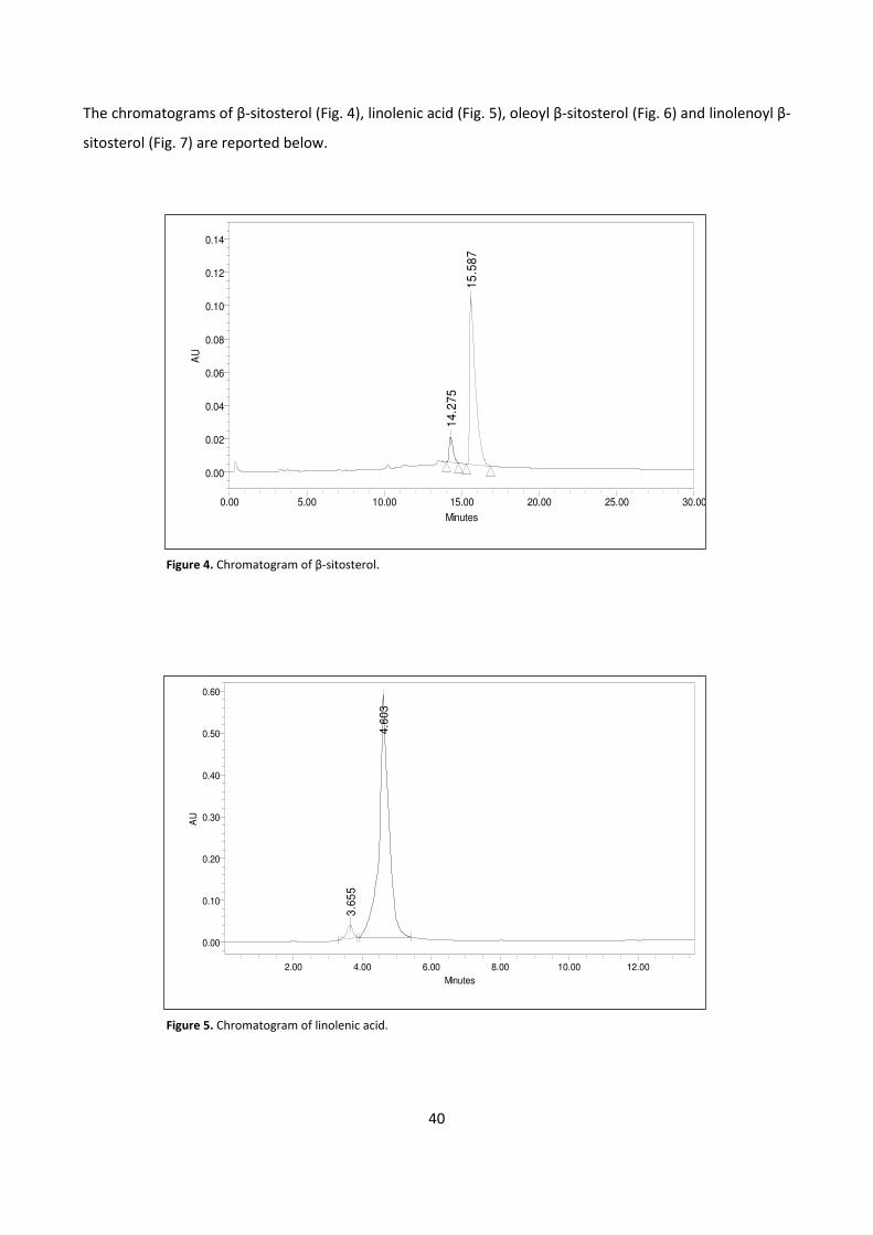
40
The chromatograms of β-sitosterol (Fig. 4), linolenic acid (Fig. 5), oleoyl β-sitosterol (Fig. 6) and linolenoyl β-
sitosterol (Fig. 7) are reported below.
Figure 4. Chromatogram of β-sitosterol.
Figure 5. Chromatogram of linolenic acid.
14
.27
5
15
.58
7
AU
0.00
0.02
0.04
0.06
0.08
0.10
0.12
0.14
Minutes
0.00 5.00 10.00 15.00 20.00 25.00 30.00
3.6
55
4.6
03
AU
0.00
0.10
0.20
0.30
0.40
0.50
0.60
Minutes
2.00 4.00 6.00 8.00 10.00 12.00
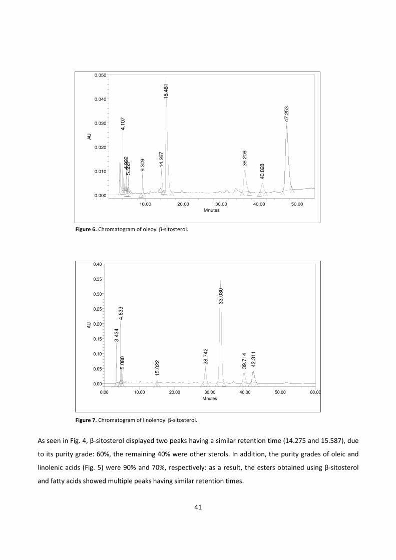
41
Figure 6. Chromatogram of oleoyl β-sitosterol.
Figure 7. Chromatogram of linolenoyl β-sitosterol.
As seen in Fig. 4, β-sitosterol displayed two peaks having a similar retention time (14.275 and 15.587), due
to its purity grade: 60%, the remaining 40% were other sterols. In addition, the purity grades of oleic and
linolenic acids (Fig. 5) were 90% and 70%, respectively: as a result, the esters obtained using β-sitosterol
and fatty acids showed multiple peaks having similar retention times.
4.1
07
4.9
92
5.5
53
9.3
09
14.2
67
15.4
81
36.2
06
40.8
28
47.2
53
AU
0.000
0.010
0.020
0.030
0.040
0.050
Minutes
10.00 20.00 30.00 40.00 50.00
3.4
34
4.6
33
5.0
80
15.0
22 28.7
42
33.0
30
39.7
14
42.3
11
AU
0.00
0.05
0.10
0.15
0.20
0.25
0.30
0.35
0.40
Minutes
0.00 10.00 20.00 30.00 40.00 50.00 60.00
42
4.1.3 - BIOCATALYTIC SYNTHESIS OF β-SITOSTEROL FATTY ESTERS
In the first phase of the study, different enzymes were screened in order to select the most suitable for the
proposed aim, namely the esterification of β-sitosterol with oleic acid and linolenic acid.
In this phase different trials were performed in screw-cap test tubes, with a high amount of enzyme (140
U/mg), a low concentration of substrates (1 mg/ml), used with a 1:1 molar ratio, and using heptane as
reaction solvent (2 ml), since the bio-catalysts used prefer hydrophobic solvents: such conditions were
shown to be the best ones in order to promote the full development of the reaction.
The enzymes used in this phase were:
• Amano lipase
• Lipase from Candida cylindracea
• Lipase from Rhizopus oryzae
• Lipase from Candida rugosa
• Lipase from Aspergillum oryzae
TLC analyses were performed at different times of the reactions, showing that lipase from Candida
cylindracea promotes the reaction with oleic acid, whereas lipase from Candida rugosa promotes the
esterification of β-sitosterol with both oleic and linolenic acids.
The second phase of this study was the optimization of the reaction conditions. Different parameters were
screened: solvent, reaction temperature, lipase units, reaction time. Several trials in 2 ml of solvent were
performed, changing one parameter at a time, and keeping the other variables constant. TLC technique was
used to assess the results of the trials. The following parameters were changed:
• Solvent: heptane, toluene, acetone, acetonitrile, hexane, isopropyl ether
• Reaction temperature: 25 °C, 30 °C, 40 °C
• Enzyme units : 140 U/mg, 70 U/mg , 35 U/mg, 28 U/mg, 14 U/mg
The conditions found in this phase of the study are shown in Tables 5, 6, 7.
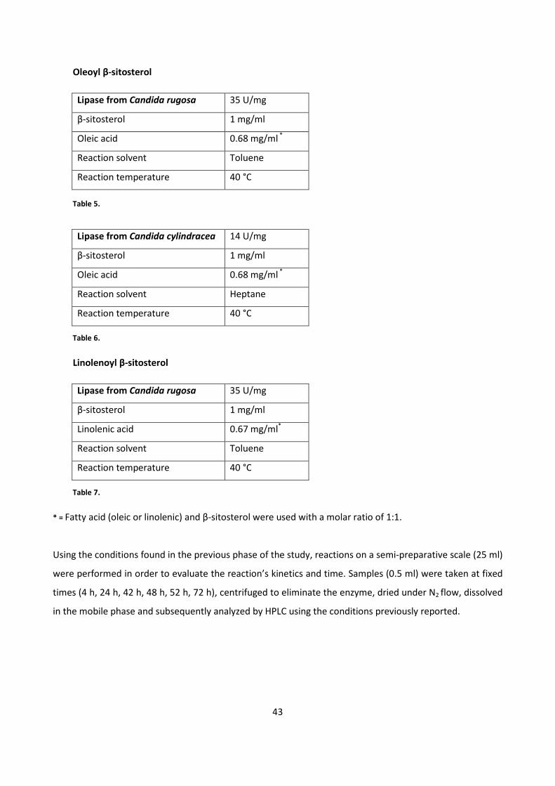
43
Oleoyl β-sitosterol
Lipase from Candida rugosa 35 U/mg
β-sitosterol 1 mg/ml
Oleic acid 0.68 mg/ml *
Reaction solvent Toluene
Reaction temperature 40 °C
Table 5.
Lipase from Candida cylindracea 14 U/mg
β-sitosterol 1 mg/ml
Oleic acid 0.68 mg/ml *
Reaction solvent Heptane
Reaction temperature 40 °C
Table 6.
Linolenoyl β-sitosterol
Lipase from Candida rugosa 35 U/mg
β-sitosterol 1 mg/ml
Linolenic acid 0.67 mg/ml*
Reaction solvent Toluene
Reaction temperature 40 °C
Table 7.
* = Fatty acid (oleic or linolenic) and β-sitosterol were used with a molar ratio of 1:1.
Using the conditions found in the previous phase of the study, reactions on a semi-preparative scale (25 ml)
were performed in order to evaluate the reaction’s kinetics and time. Samples (0.5 ml) were taken at fixed
times (4 h, 24 h, 42 h, 48 h, 52 h, 72 h), centrifuged to eliminate the enzyme, dried under N2 flow, dissolved
in the mobile phase and subsequently analyzed by HPLC using the conditions previously reported.
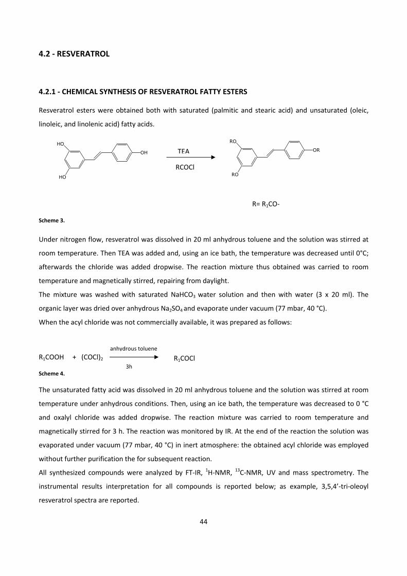
44
4.2 - RESVERATROL
4.2.1 - CHEMICAL SYNTHESIS OF RESVERATROL FATTY ESTERS
Resveratrol esters were obtained both with saturated (palmitic and stearic acid) and unsaturated (oleic,
linoleic, and linolenic acid) fatty acids.
R= R1CO-
Scheme 3.
Under nitrogen flow, resveratrol was dissolved in 20 ml anhydrous toluene and the solution was stirred at
room temperature. Then TEA was added and, using an ice bath, the temperature was decreased until 0°C;
afterwards the chloride was added dropwise. The reaction mixture thus obtained was carried to room
temperature and magnetically stirred, repairing from daylight.
The mixture was washed with saturated NaHCO3 water solution and then with water (3 x 20 ml). The
organic layer was dried over anhydrous Na2SO4 and evaporate under vacuum (77 mbar, 40 °C).
When the acyl chloride was not commercially available, it was prepared as follows:
R1COOH + (COCl)2
Scheme 4.
The unsaturated fatty acid was dissolved in 20 ml anhydrous toluene and the solution was stirred at room
temperature under anhydrous conditions. Then, using an ice bath, the temperature was decreased to 0 °C
and oxalyl chloride was added dropwise. The reaction mixture was carried to room temperature and
magnetically stirred for 3 h. The reaction was monitored by IR. At the end of the reaction the solution was
evaporated under vacuum (77 mbar, 40 °C) in inert atmosphere: the obtained acyl chloride was employed
without further purification the for subsequent reaction.
All synthesized compounds were analyzed by FT-IR, 1H-NMR,
13C-NMR, UV and mass spectrometry. The
instrumental results interpretation for all compounds is reported below; as example, 3,5,4’-tri-oleoyl
resveratrol spectra are reported.
3h
anhydrous toluene
R1COCl
HO
HO
OH TEA
RCOCl RO
RO
OR
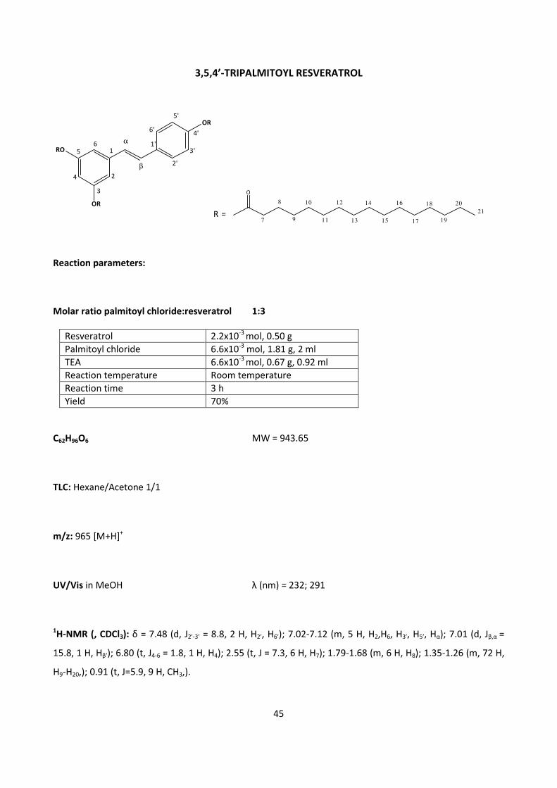
45
3,5,4’-TRIPALMITOYL RESVERATROL
Reaction parameters:
Molar ratio palmitoyl chloride:resveratrol 1:3
Resveratrol 2.2x10⁻³
mol, 0.50 g
Palmitoyl chloride 6.6x10⁻³
mol, 1.81 g, 2 ml
TEA 6.6x10⁻³
mol, 0.67 g, 0.92 ml
Reaction temperature Room temperature
Reaction time 3 h
Yield 70%
C62H96O6 MW = 943.65
TLC: Hexane/Acetone 1/1
m/z: 965 [M+H]+
UV/Vis in MeOH λ (nm) = 232; 291
1H-NMR (, CDCl3): δ = 7.48 (d, J2’-3’ = 8.8, 2 H, H2’, H6’); 7.02-7.12 (m, 5 H, H2,H6, H3’, H5’, Hα); 7.01 (d, Jβ,α =
15.8, 1 H, Hβ’); 6.80 (t, J4-6 = 1.8, 1 H, H4); 2.55 (t, J = 7.3, 6 H, H7); 1.79-1.68 (m, 6 H, H8); 1.35-1.26 (m, 72 H,
H9-H20,); 0.91 (t, J=5.9, 9 H, CH3,).
RO
OR
OR5'
4'
3'
2'
6'
1'
β
α
2
3
4
56
1
R =
O
8
7 9
10
11
12
13
14
15
16
17
18
19
2021
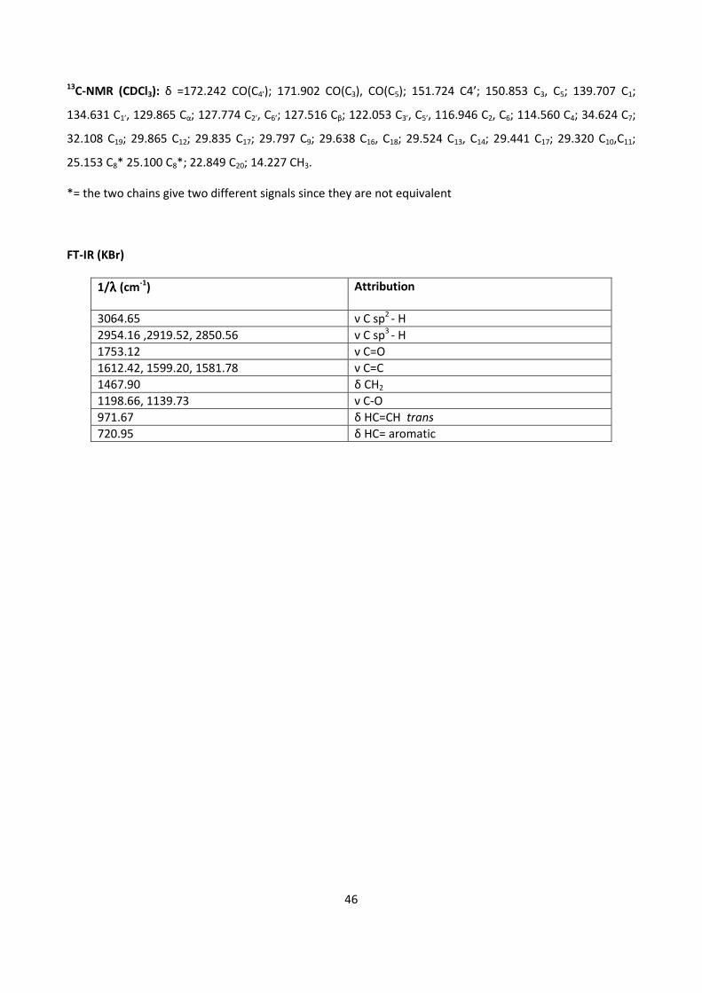
46
13C-NMR (CDCl3): δ =172.242 CO(C4’); 171.902 CO(C3), CO(C5); 151.724 C4’; 150.853 C3, C5; 139.707 C1;
134.631 C1’, 129.865 Cα; 127.774 C2’, C6’; 127.516 Cβ; 122.053 C3’, C5’, 116.946 C2, C6; 114.560 C4; 34.624 C7;
32.108 C19; 29.865 C12; 29.835 C17; 29.797 C9; 29.638 C16, C18; 29.524 C13, C14; 29.441 C17; 29.320 C10,C11;
25.153 C8* 25.100 C8*; 22.849 C20; 14.227 CH3.
*= the two chains give two different signals since they are not equivalent
FT-IR (KBr)
1/λλλλ (cm-1
) Attribution
3064.65 ν C sp2
- H
2954.16 ,2919.52, 2850.56 ν C sp3
- H
1753.12 ν C=O
1612.42, 1599.20, 1581.78 ν C=C
1467.90 δ CH2
1198.66, 1139.73 ν C-O
971.67 δ HC=CH trans
720.95 δ HC= aromatic
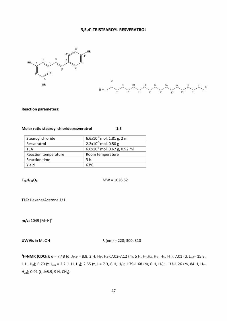
47
3,5,4’-TRISTEAROYL RESVERATROL
Reaction parameters:
Molar ratio stearoyl chloride:resveratrol 1:3
Stearoyl chloride 6.6x10⁻³
mol, 1.81 g, 2 ml
Resveratrol 2.2x10⁻³
mol, 0.50 g
TEA 6.6x10⁻³
mol, 0.67 g, 0.92 ml
Reaction temperature Room temperature
Reaction time 3 h
Yield 63%
C68H114O6 MW = 1026.52
TLC: Hexane/Acetone 1/1
m/z: 1049 [M+H]+
UV/Vis in MeOH λ (nm) = 228; 300; 310
1H-NMR (CDCl3): δ = 7.48 (d, J2’-3’ = 8.8, 2 H, H2’, H6’);7.02-7.12 (m, 5 H, H2,H6, H3’, H5’, Hα); 7.01 (d, Jα-β= 15.8,
1 H, Hβ); 6.79 (t, J4-6 = 2.2, 1 H, H4); 2.55 (t, J = 7.3, 6 H, H7); 1.79-1.68 (m, 6 H, H8); 1.33-1.26 (m, 84 H, H9-
H22); 0.91 (t, J=5.9, 9 H, CH3).
R =
O
8
7 9
10
11
12
13
14
15
16
17
18
19 21
222023
RO
OR
OR5'
4'
3'
2'
6'
1'
β
α
2
3
4
56
1
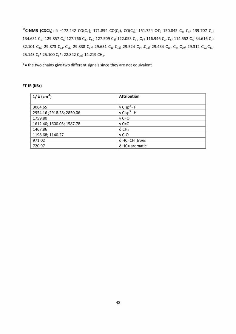
48
13C-NMR (CDCl3): δ =172.242 CO(C4’); 171.894 CO(C3), CO(C5); 151.724 C4’; 150.845 C3, C5; 139.707 C1;
134.631 C1’; 129.857 Cα; 127.766 C2’, C6’; 127.509 Cβ; 122.053 C3’, C5’; 116.946 C2, C6; 114.552 C4; 34.616 C7;
32.101 C21; 29.873 C12, C13; 29.838 C17; 29.631 C19 C16; 29.524 C14 ,C13; 29.434 C18, C9, C20; 29.312 C10,C11;
25.145 C8* 25.100 C8*; 22.842 C22; 14.219 CH3.
*= the two chains give two different signals since they are not equivalent
FT-IR (KBr)
1/ λλλλ (cm-1
) Attribution
3064.65 ν C sp2
- H
2954.16 ;2918.28; 2850.06 ν C sp3
- H
1759.80 ν C=O
1612.40; 1600.05; 1587.78 ν C=C
1467.86 δ CH2
1198.68; 1140.27 ν C-O
971.02 δ HC=CH trans
720.97 δ HC= aromatic
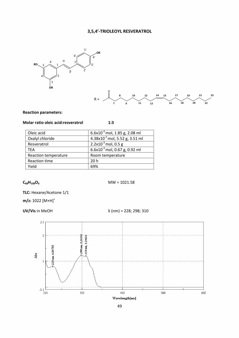
49
3,5,4’-TRIOLEOYL RESVERATROL
Reaction parameters:
Molar ratio oleic acid:resveratrol 1:3
Oleic acid 6.6x10⁻³
mol, 1.85 g, 2.08 ml
Oxalyl chloride 4.38x10⁻²
mol, 5.52 g, 3.51 ml
Resveratrol 2.2x10⁻³
mol, 0.5 g
TEA 6.6x10⁻³
mol, 0.67 g, 0.92 ml
Reaction temperature Room temperature
Reaction time 20 h
Yield 69%
C68H108O6 MW = 1021.58
TLC: Hexane/Acetone 1/1
m/z: 1022 [M+H]+
UV/Vis in MeOH λ (nm) = 228; 298; 310
RO
OR
OR5'
4'
3'
2'
6'
1'
β
α
2
3
4
56
1
O
7
8
9
10
11
12
13
14 15
16
17
18
19
20
21
22
23
R =
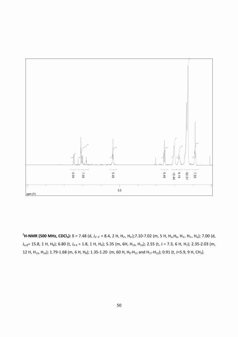
50
ppm (f1)5.0
2.0
0
7.0
5
5.4
6
5.4
6
6.1
9
10
.40
52
.31
7.5
0
1H-NMR (500 MHz, CDCl3): δ = 7.48 (d, J2’-3’ = 8.4, 2 H, H2’, H6’);7.10-7.02 (m, 5 H, H2,H6, H3’, H5’, Hα); 7.00 (d,
Jα-β= 15.8, 1 H, Hβ); 6.80 (t, J4-6 = 1.8, 1 H, H4); 5.35 (m, 6H, H14, H16); 2.55 (t, J = 7.3, 6 H, H7); 2.35-2.03 (m,
12 H, H13, H16); 1.79-1.68 (m, 6 H, H8); 1.35-1.20 (m, 60 H, H9-H12 and H17-H22); 0.91 (t, J=5.9, 9 H, CH3).
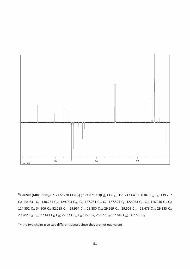
51
ppm (f1)
50100150
13C-NMR (MHz, CDCl3): δ =172.220 CO(C4’) ; 171.871 CO(C3), CO(C5); 151.717 C4’; 150.845 C3, C5; 139.707
C1; 134.631 C1’; 130.251 C14; 129.903 C15, Cα; 127.781 C2’, C6’; 127.524 Cβ; 122.053 C3’, C5’; 116.946 C2, C6;
114.552 C4; 34.906 C7; 32.085 C21; 29.964 C12, 29.880 C17; 29.669 C19; 29.509 C18 ; 29.479 C20; 29.335 C9;
29.282 C10, C11; 27.441 C13-C16; 27.373 C16-C13 ; 25.137, 25.077 C8*; 22.840 C22; 14.277 CH3.
*= the two chains give two different signals since they are not equivalent
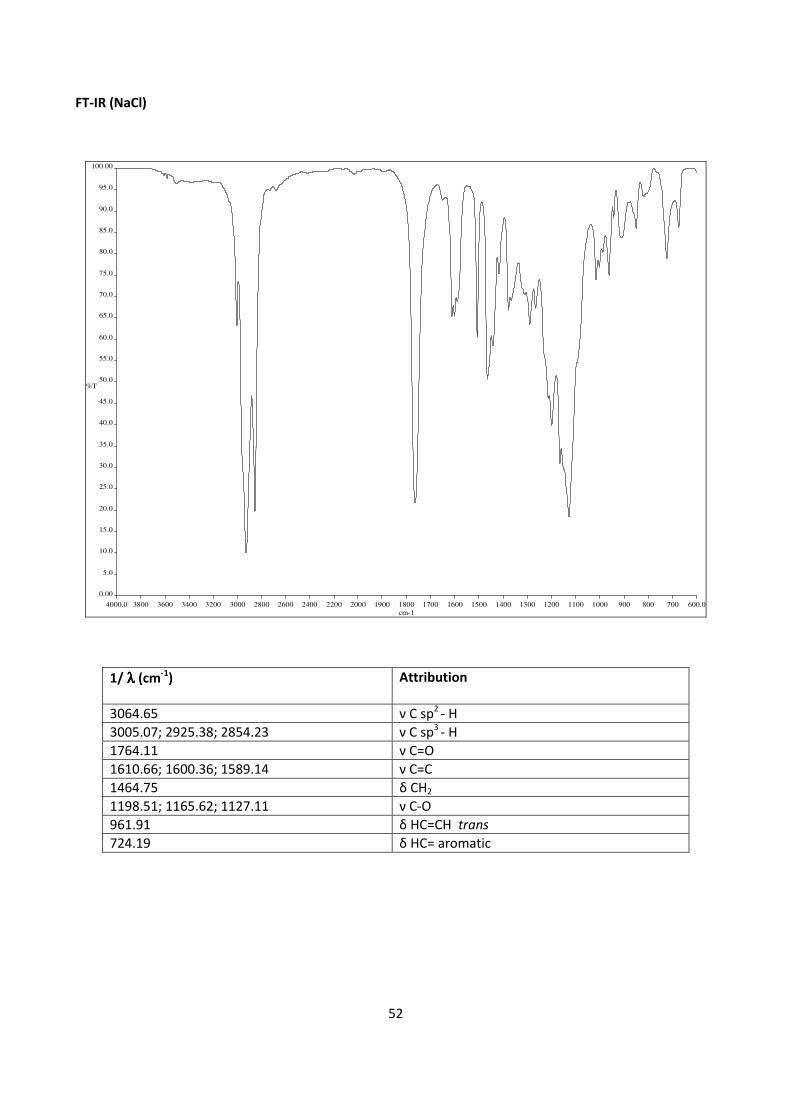
52
FT-IR (NaCl)
4000.0 3800 3600 3400 3200 3000 2800 2600 2400 2200 2000 1900 1800 1700 1600 1500 1400 1300 1200 1100 1000 900 800 700 600.0
0.00
5.0
10.0
15.0
20.0
25.0
30.0
35.0
40.0
45.0
50.0
55.0
60.0
65.0
70.0
75.0
80.0
85.0
90.0
95.0
100.00
cm-1
%T
1/ λλλλ (cm-1
) Attribution
3064.65 ν C sp2
- H
3005.07; 2925.38; 2854.23 ν C sp3
- H
1764.11 ν C=O
1610.66; 1600.36; 1589.14 ν C=C
1464.75 δ CH2
1198.51; 1165.62; 1127.11 ν C-O
961.91 δ HC=CH trans
724.19 δ HC= aromatic
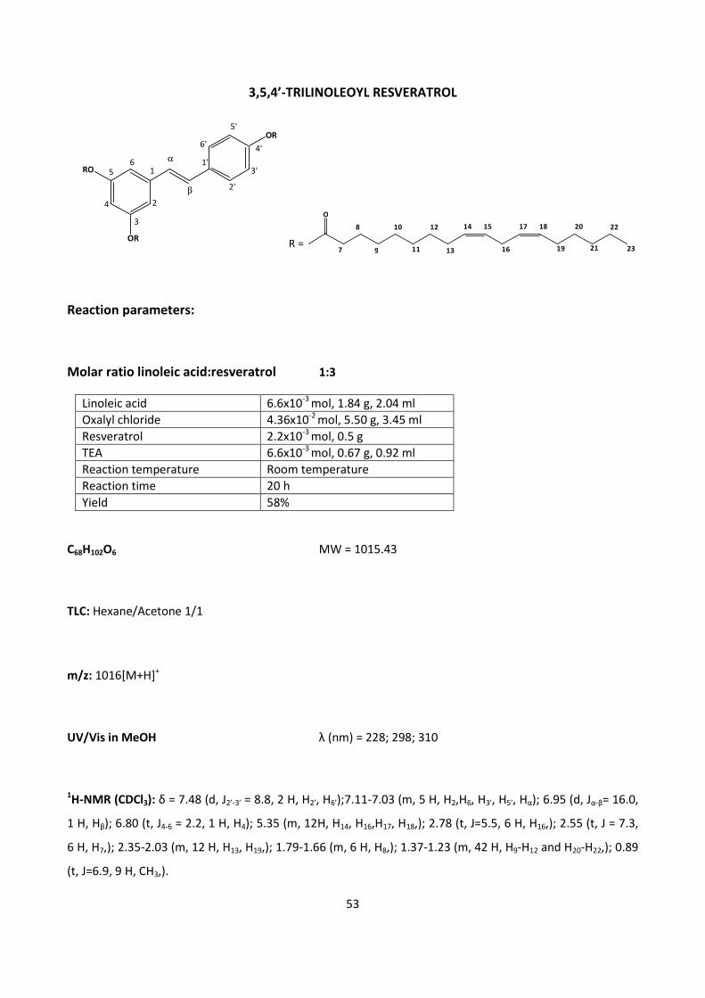
53
3,5,4’-TRILINOLEOYL RESVERATROL
O
7
8
9
10
11
12
13
14 15
16
17 18
19
20
21
22
23R =
Reaction parameters:
Molar ratio linoleic acid:resveratrol 1:3
Linoleic acid 6.6x10⁻³
mol, 1.84 g, 2.04 ml
Oxalyl chloride 4.36x10⁻²
mol, 5.50 g, 3.45 ml
Resveratrol 2.2x10⁻³
mol, 0.5 g
TEA 6.6x10⁻³
mol, 0.67 g, 0.92 ml
Reaction temperature Room temperature
Reaction time 20 h
Yield 58%
C68H102O6 MW = 1015.43
TLC: Hexane/Acetone 1/1
m/z: 1016[M+H]+
UV/Vis in MeOH λ (nm) = 228; 298; 310
1H-NMR (CDCl3): δ = 7.48 (d, J2’-3’ = 8.8, 2 H, H2’, H6’);7.11-7.03 (m, 5 H, H2,H6, H3’, H5’, Hα); 6.95 (d, Jα-β= 16.0,
1 H, Hβ); 6.80 (t, J4-6 = 2.2, 1 H, H4); 5.35 (m, 12H, H14, H16,H17, H18,); 2.78 (t, J=5.5, 6 H, H16,); 2.55 (t, J = 7.3,
6 H, H7,); 2.35-2.03 (m, 12 H, H13, H19,); 1.79-1.66 (m, 6 H, H8,); 1.37-1.23 (m, 42 H, H9-H12 and H20-H22,); 0.89
(t, J=6.9, 9 H, CH3,).
RO
OR
OR5'
4'
3'
2'
6'
1'
β
α
2
3
4
56
1
54
13C-NMR (CDCl3): δ =172.331 CO(C4’) ; 171.995 CO(C3), CO(C5); 151.671 C4’; 150.792 C3, C5; 139.707 C1;
134.623 C1’; 130.441 C14; 130.221 C17; 129.850 Cα; 128.342 C18; 128.137 C15; 127.812 C2’, C6’; 127.478 Cβ;
122.084 C3’, C5’; 116.984 C2, C6; 114.598 C4; 34.609 C7; 31.745 C21; 29.812 C12; 29.555 C19; 29.365 C9 C20;
29.305 C10, C11; 27.418 C13; 25.872 C16 ; 25.137, 25.084 C8*; 22.773 C22; 14.250 CH3.
*= the two chains give two different signals since they are not equivalent
FT-IR (NaCl)
1/ λλλλ (cm-1
) Attribution
3064.65 ν C sp2
- H
3008.07; 2927.64; 2855.64 ν C sp3
- H
1764.11 ν C=O
1611.15; 1600.59, 1588.92 ν C=C
1464.72 δ CH2
1198.85; 1165.83, 1127.41 ν C-O
961.92 δ HC=CH trans
728.86 δ HC= aromatic
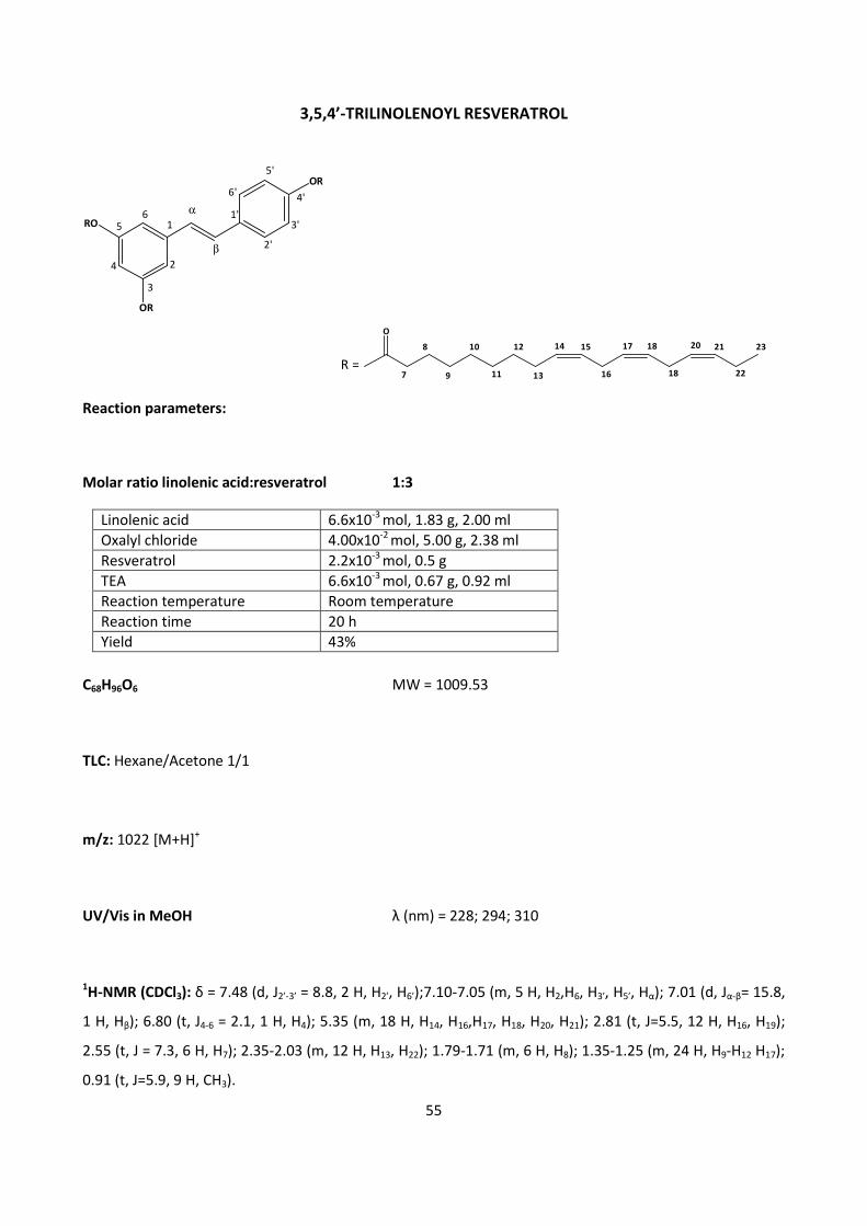
55
3,5,4’-TRILINOLENOYL RESVERATROL
O
7
8
9
10
11
12
13
14 15
16
17 18
18
2120
22
23
R =
Reaction parameters:
Molar ratio linolenic acid:resveratrol 1:3
Linolenic acid 6.6x10⁻³
mol, 1.83 g, 2.00 ml
Oxalyl chloride 4.00x10⁻²
mol, 5.00 g, 2.38 ml
Resveratrol 2.2x10⁻³
mol, 0.5 g
TEA 6.6x10⁻³
mol, 0.67 g, 0.92 ml
Reaction temperature Room temperature
Reaction time 20 h
Yield 43%
C68H96O6 MW = 1009.53
TLC: Hexane/Acetone 1/1
m/z: 1022 [M+H]+
UV/Vis in MeOH λ (nm) = 228; 294; 310
1H-NMR (CDCl3): δ = 7.48 (d, J2’-3’ = 8.8, 2 H, H2’, H6’);7.10-7.05 (m, 5 H, H2,H6, H3’, H5’, Hα); 7.01 (d, Jα-β= 15.8,
1 H, Hβ); 6.80 (t, J4-6 = 2.1, 1 H, H4); 5.35 (m, 18 H, H14, H16,H17, H18, H20, H21); 2.81 (t, J=5.5, 12 H, H16, H19);
2.55 (t, J = 7.3, 6 H, H7); 2.35-2.03 (m, 12 H, H13, H22); 1.79-1.71 (m, 6 H, H8); 1.35-1.25 (m, 24 H, H9-H12 H17);
0.91 (t, J=5.9, 9 H, CH3).
RO
OR
OR5'
4'
3'
2'
6'
1'
β
α
2
3
4
56
1
56
13C-NMR (MHz, CDCl3): δ =172.227 CO(C4’) ; 171.879 CO(C3), CO(C5); 151.717 C4’; 150.838 C3, C5; 139.715
C1; 134.631 C1’; 132.161 C21; 130.426 C14; 129.873 Cα; 128.531 C17-C18;128.471 C17-C18; 128.031 C15; 127.789
C2’, C6’; 127.516 Cβ; 127.365 C20; 122.061 C3’, C5’; 116.954 C2, C6; 114.552 C4; 34.601 C7; 29.775 C12; 29.335
C9; 29.282 C10,C11; 27.411 C13; 25.835 C16; 25.759 C19; 25.130-25.069 C8*; 20.743 C22; 14.401 CH3.
*= the two chains give two different signals since they are not equivalent
FT-IR (NaCl)
1/ λλλλ (cm-1
) Attribution
3064.65 ν C sp2
- H
3005.07; 2925.38; 2854.23 ν C sp3
- H
1764.11 ν –C=O
1610.66; 1600.36; 1589.14 ν –C=C aromatic
1464.75 δ CH2
1198.51; 1165.62; 1127.11 ν C-O
961.91 δ HC=CH trans
724.19 δ HC= aromatic
3064.65 ν C sp2
- H
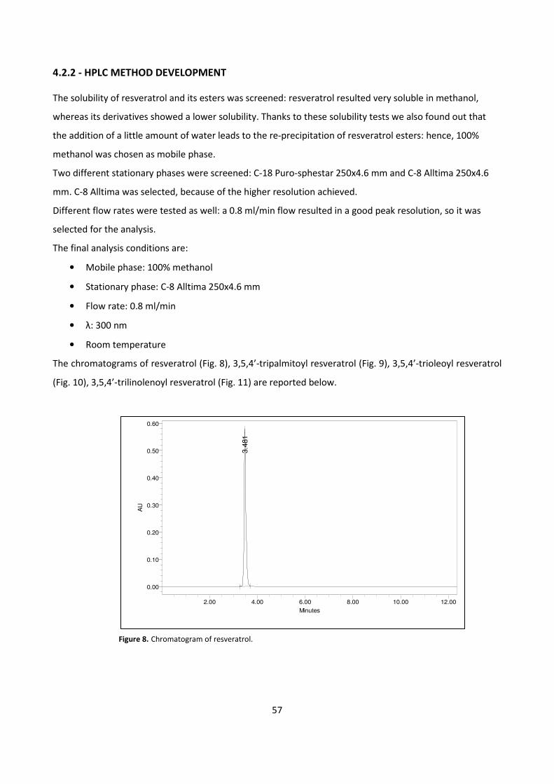
57
4.2.2 - HPLC METHOD DEVELOPMENT
The solubility of resveratrol and its esters was screened: resveratrol resulted very soluble in methanol,
whereas its derivatives showed a lower solubility. Thanks to these solubility tests we also found out that
the addition of a little amount of water leads to the re-precipitation of resveratrol esters: hence, 100%
methanol was chosen as mobile phase.
Two different stationary phases were screened: C-18 Puro-sphestar 250x4.6 mm and C-8 Alltima 250x4.6
mm. C-8 Alltima was selected, because of the higher resolution achieved.
Different flow rates were tested as well: a 0.8 ml/min flow resulted in a good peak resolution, so it was
selected for the analysis.
The final analysis conditions are:
• Mobile phase: 100% methanol
• Stationary phase: C-8 Alltima 250x4.6 mm
• Flow rate: 0.8 ml/min
• λ: 300 nm
• Room temperature
The chromatograms of resveratrol (Fig. 8), 3,5,4’-tripalmitoyl resveratrol (Fig. 9), 3,5,4’-trioleoyl resveratrol
(Fig. 10), 3,5,4’-trilinolenoyl resveratrol (Fig. 11) are reported below.
Figure 8. Chromatogram of resveratrol.
3.4
81
AU
0.00
0.10
0.20
0.30
0.40
0.50
0.60
Minutes
2.00 4.00 6.00 8.00 10.00 12.00
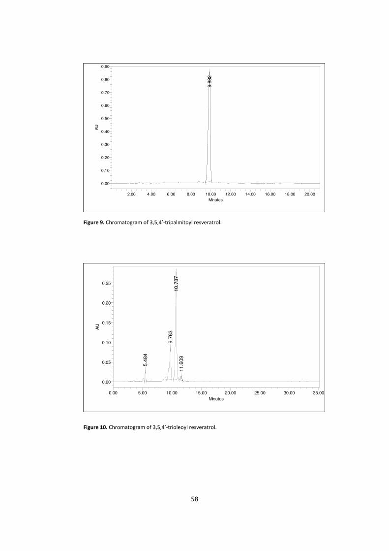
58
Figure 9. Chromatogram of 3,5,4’-tripalmitoyl resveratrol.
Figure 10. Chromatogram of 3,5,4’-trioleoyl resveratrol.
5.4
84
9.7
63
10.7
37
11.6
09
AU
0.00
0.05
0.10
0.15
0.20
0.25
Minutes
0.00 5.00 10.00 15.00 20.00 25.00 30.00 35.00
9.8
82
AU
0.00
0.10
0.20
0.30
0.40
0.50
0.60
0.70
0.80
0.90
Minutes
2.00 4.00 6.00 8.00 10.00 12.00 14.00 16.00 18.00 20.00
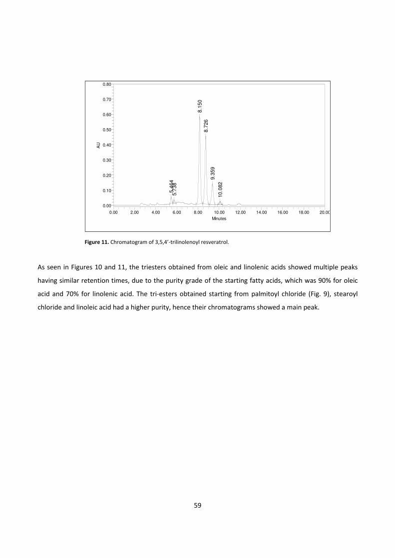
59
Figure 11. Chromatogram of 3,5,4’-trilinolenoyl resveratrol.
As seen in Figures 10 and 11, the triesters obtained from oleic and linolenic acids showed multiple peaks
having similar retention times, due to the purity grade of the starting fatty acids, which was 90% for oleic
acid and 70% for linolenic acid. The tri-esters obtained starting from palmitoyl chloride (Fig. 9), stearoyl
chloride and linoleic acid had a higher purity, hence their chromatograms showed a main peak.
5.4
64
5.7
38
8.1
50
8.7
26
9.3
59
10
.082
AU
0.00
0.10
0.20
0.30
0.40
0.50
0.60
0.70
0.80
Minutes
0.00 2.00 4.00 6.00 8.00 10.00 12.00 14.00 16.00 18.00 20.00
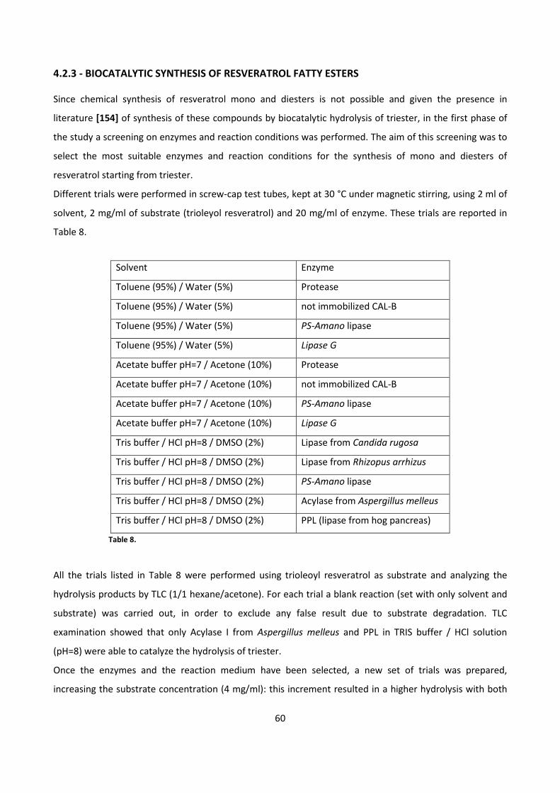
60
4.2.3 - BIOCATALYTIC SYNTHESIS OF RESVERATROL FATTY ESTERS
Since chemical synthesis of resveratrol mono and diesters is not possible and given the presence in
literature [154] of synthesis of these compounds by biocatalytic hydrolysis of triester, in the first phase of
the study a screening on enzymes and reaction conditions was performed. The aim of this screening was to
select the most suitable enzymes and reaction conditions for the synthesis of mono and diesters of
resveratrol starting from triester.
Different trials were performed in screw-cap test tubes, kept at 30 °C under magnetic stirring, using 2 ml of
solvent, 2 mg/ml of substrate (trioleyol resveratrol) and 20 mg/ml of enzyme. These trials are reported in
Table 8.
Table 8.
All the trials listed in Table 8 were performed using trioleoyl resveratrol as substrate and analyzing the
hydrolysis products by TLC (1/1 hexane/acetone). For each trial a blank reaction (set with only solvent and
substrate) was carried out, in order to exclude any false result due to substrate degradation. TLC
examination showed that only Acylase I from Aspergillus melleus and PPL in TRIS buffer / HCl solution
(pH=8) were able to catalyze the hydrolysis of triester.
Once the enzymes and the reaction medium have been selected, a new set of trials was prepared,
increasing the substrate concentration (4 mg/ml): this increment resulted in a higher hydrolysis with both
Solvent Enzyme
Toluene (95%) / Water (5%) Protease
Toluene (95%) / Water (5%) not immobilized CAL-B
Toluene (95%) / Water (5%) PS-Amano lipase
Toluene (95%) / Water (5%) Lipase G
Acetate buffer pH=7 / Acetone (10%) Protease
Acetate buffer pH=7 / Acetone (10%) not immobilized CAL-B
Acetate buffer pH=7 / Acetone (10%) PS-Amano lipase
Acetate buffer pH=7 / Acetone (10%) Lipase G
Tris buffer / HCl pH=8 / DMSO (2%) Lipase from Candida rugosa
Tris buffer / HCl pH=8 / DMSO (2%) Lipase from Rhizopus arrhizus
Tris buffer / HCl pH=8 / DMSO (2%) PS-Amano lipase
Tris buffer / HCl pH=8 / DMSO (2%) Acylase from Aspergillus melleus
Tris buffer / HCl pH=8 / DMSO (2%) PPL (lipase from hog pancreas)
61
enzymes. The subsequent step was to perform tests on a larger scale (25 ml), in order to assess the time
course of the reaction. At fixed times (4 h, 24 h, 48 h, 72 h, 96 h, 120 h), samples (0.5 ml) were taken,
extracted with chloroform and then with ethyl acetate, treated with anhydrous Na2SO4, dried under N2
flow, dissolved in the mobile phase and subsequently analyzed by HPLC using the conditions previously
reported. These analyses showed that both enzymes promote the maximum production of mono and
diesters at 96 h. A further increase of reaction time resulted in the formation of resveratrol, and thus in the
complete hydrolysis of triester.
In the case of biotransformation with Acylase I from Aspergillus melleus the reaction was set up on a semi-
preparative scale (100 ml) in order to isolate the final products in a sufficient amount for the structural
characterization. To 400 mg of trioleoyl resveratrol dispersed in 4 ml of DMSO, 2 g of Acylase I from
Aspergillus melleus and 100 ml of TRIS buffer/HCl pH=8 (prepared dissolving 1.2114 g of TRIS in 100 ml
deionized water and adding HCl dropwise until pH 8 is reached) were added.
After 96 hours the reaction mix was extracted with chloroform (2 x 40ml) and with ethyl acetate (1 x 40 ml).
The organic layers were pooled and washed with saturated NaHCO3 water solution and then with water (3 x
20 ml). The new organic layer was dried over anhydrous Na2SO4 and evaporated under vacuum; then, the
evaporation residue was analyzed by HPLC.
In order to characterize each analyte, the evaporation residue was separated using a chromatographic
column with 7/3 hexane/acetone as mobile phase and a 1/60 product/silica ratio. Four different fractions
were obtained in this separation, and subsequently analyzed.
62
4.2.4 - ORAL BIOAVALABILITY STUDIES OF RESVERATROL AND 3,5,4’-TRIOLEOYL RESVARATROL
IN THE RAT
4.2.4.1 - STUDY PROTOCOL
In order to check the oral bioavailability of 3,5,4’-trioleoyl resveratrol, preliminary tests were performed
on rats, in collaboration with Dr. Giuseppe Rossoni from Dept. of Pharmacology, Chemotherapy and
Medical Toxicology, Università degli Studi, Milan.
For these studies 32 male Wistar rats were purchased from Charles River Italy (Calco, LC, Italy). Their initial
weight was 250 ± 10 g and they were kept under automatically controlled conditions of temperature (21 ±
1 °C), humidity (55 ± 5%) and light (12 h light + 12 h dark). Both water (Milan water supply network) and
food (Mucedola Srl, Settimo Milanese, MI, Italia) were freely accessible to animals.
All of the tests were performed in accordance with the Italian Government guidelines for laboratory
animals and conformed with European Community Directives drawn up in November 1986 (86/609/EEC).
Seven days after their arrival, the rats were randomly divided in two different experimental groups of 16
animals (2 control rats + 14 treated rats), then starved for 24 hours before each treatment. Resveratrol
was administered to the first group (20 mg/ml), whereas 3,5,4’-trioleoyl resveratrol was administered to
the second group (90 mg/ml, corresponding to 20 mg/ml of resveratrol). Both resveratrol and 3,5,4’-
trioleoyl resveratrol were suspended in carboxymethyl cellulose (0.5 w/v) and administered to the animals
orally, by using a gastric probe. The rats were anesthetized (sodium thiopental, 50 mg/kg i.p.) at fixed
times (0.5, 1, 2, 3, 4, 6, 24 hours) in order to take blood samples, that were subsequently used to assess
the bioavailability of resveratrol and its ester. Blood was collected from vena cava by using heparinized
syringes, then placed in plastic tubes and centrifuged at 2000 x g for 15 minutes at 4 °C, in order to allow
the separation of plasma. The plasma thus obtained was stored at -80 °C until it was analyzed (less than
one week). Subsequently, plasma was extracted by using a suitable solvent and analyzed by HPLC/DAD.
4.4.2.2- EXTRACTION METHOD DEVELOPMENT
Before analyzing plasma samples by HPLC, the extraction method was developed.
Two samples of plasma from control rats were added with solutions of resveratrol and 3,5,4’-trioleoyl
resveratrol (at the same concentration), respectively. These samples were extracted using different
procedures:
Method 1: 70 µl of each sample were added with 10 µl of caffeine solution in methanol, then the resulting
solutions were stirred for 30 seconds. Subsequently, the samples were added with 100 µl of acetonitrile,
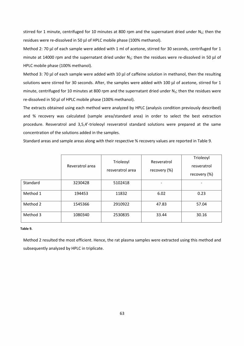
63
stirred for 1 minute, centrifuged for 10 minutes at 800 rpm and the supernatant dried under N2; then the
residues were re-dissolved in 50 µl of HPLC mobile phase (100% methanol).
Method 2: 70 µl of each sample were added with 1 ml of acetone, stirred for 30 seconds, centrifuged for 1
minute at 14000 rpm and the supernatant dried under N2; then the residues were re-dissolved in 50 µl of
HPLC mobile phase (100% methanol).
Method 3: 70 µl of each sample were added with 10 µl of caffeine solution in methanol, then the resulting
solutions were stirred for 30 seconds. After, the samples were added with 100 µl of acetone, stirred for 1
minute, centrifuged for 10 minutes at 800 rpm and the supernatant dried under N2; then the residues were
re-dissolved in 50 µl of HPLC mobile phase (100% methanol).
The extracts obtained using each method were analyzed by HPLC (analysis condition previously described)
and % recovery was calculated (sample area/standard area) in order to select the best extraction
procedure. Resveratrol and 3,5,4’-trioleoyl resveratrol standard solutions were prepared at the same
concentration of the solutions added in the samples.
Standard areas and sample areas along with their respective % recovery values are reported in Table 9.
Reveratrol area Trioleoyl
resveratrol area
Resveratrol
recovery (%)
Trioleoyl
resveratrol
recovery (%)
Standard 3230428 5102418 - -
Method 1 194453 11832 6.02 0.23
Method 2 1545366 2910922 47.83 57.04
Method 3 1080340 2530835 33.44 30.16
Table 9.
Method 2 resulted the most efficient. Hence, the rat plasma samples were extracted using this method and
subsequently analyzed by HPLC in triplicate.
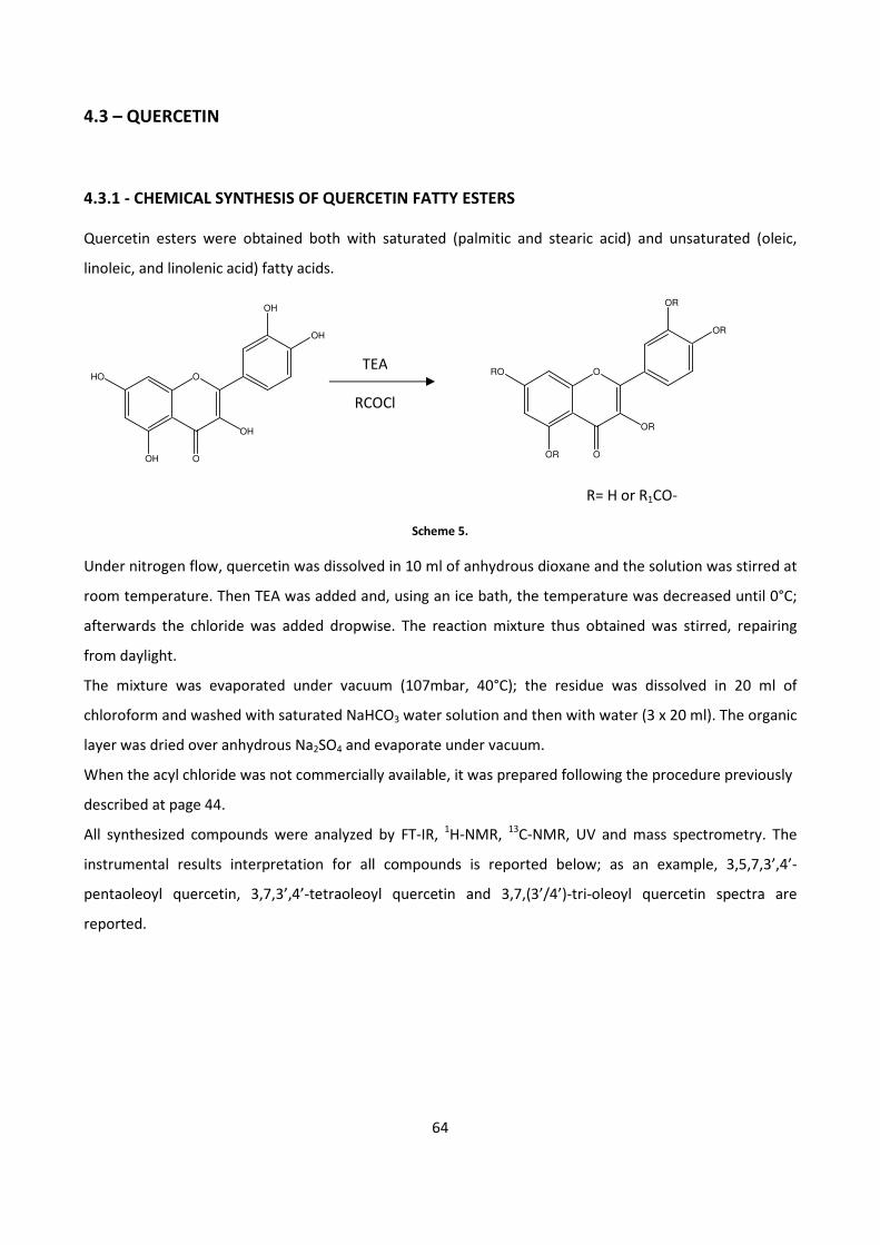
64
4.3 – QUERCETIN
4.3.1 - CHEMICAL SYNTHESIS OF QUERCETIN FATTY ESTERS
Quercetin esters were obtained both with saturated (palmitic and stearic acid) and unsaturated (oleic,
linoleic, and linolenic acid) fatty acids.
R= H or R1CO-
Scheme 5.
Under nitrogen flow, quercetin was dissolved in 10 ml of anhydrous dioxane and the solution was stirred at
room temperature. Then TEA was added and, using an ice bath, the temperature was decreased until 0°C;
afterwards the chloride was added dropwise. The reaction mixture thus obtained was stirred, repairing
from daylight.
The mixture was evaporated under vacuum (107mbar, 40°C); the residue was dissolved in 20 ml of
chloroform and washed with saturated NaHCO3 water solution and then with water (3 x 20 ml). The organic
layer was dried over anhydrous Na2SO4 and evaporate under vacuum.
When the acyl chloride was not commercially available, it was prepared following the procedure previously
described at page 44.
All synthesized compounds were analyzed by FT-IR, 1H-NMR,
13C-NMR, UV and mass spectrometry. The
instrumental results interpretation for all compounds is reported below; as an example, 3,5,7,3’,4’-
pentaoleoyl quercetin, 3,7,3’,4’-tetraoleoyl quercetin and 3,7,(3’/4’)-tri-oleoyl quercetin spectra are
reported.
HO
OH
O
O
OH
OH
OH
TEA RO
OR
O
O
OR
OR
OR
RCOCl
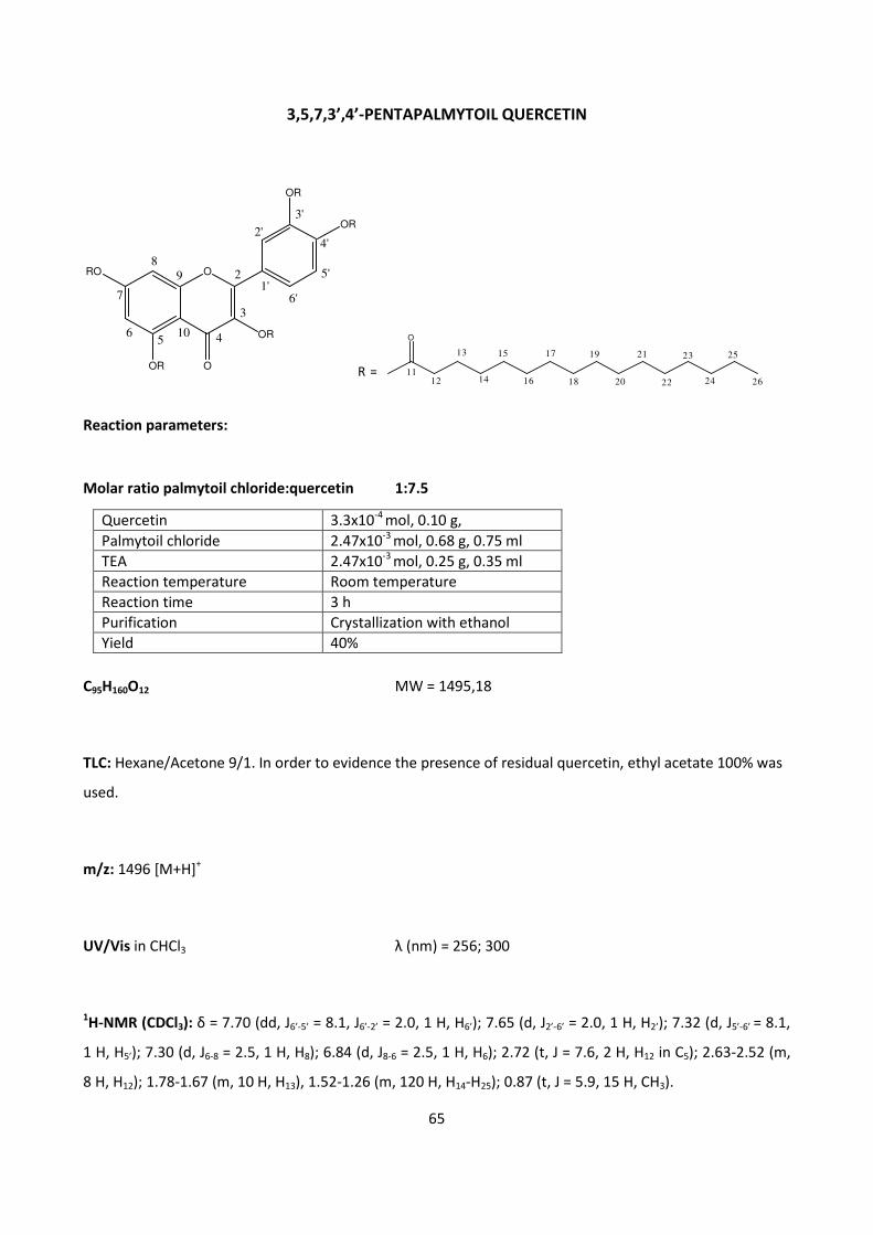
65
3,5,7,3’,4’-PENTAPALMYTOIL QUERCETIN
Reaction parameters:
Molar ratio palmytoil chloride:quercetin 1:7.5
Quercetin 3.3x10⁻⁴
mol, 0.10 g,
Palmytoil chloride 2.47x10⁻³
mol, 0.68 g, 0.75 ml
TEA 2.47x10⁻³
mol, 0.25 g, 0.35 ml
Reaction temperature Room temperature
Reaction time 3 h
Purification Crystallization with ethanol
Yield 40%
C95H160O12 MW = 1495,18
TLC: Hexane/Acetone 9/1. In order to evidence the presence of residual quercetin, ethyl acetate 100% was
used.
m/z: 1496 [M+H]+
UV/Vis in CHCl3 λ (nm) = 256; 300
1H-NMR (CDCl3): δ = 7.70 (dd, J6’-5’ = 8.1, J6’-2’ = 2.0, 1 H, H6’); 7.65 (d, J2’-6’ = 2.0, 1 H, H2’); 7.32 (d, J5’-6’ = 8.1,
1 H, H5’); 7.30 (d, J6-8 = 2.5, 1 H, H8); 6.84 (d, J8-6 = 2.5, 1 H, H6); 2.72 (t, J = 7.6, 2 H, H12 in C5); 2.63-2.52 (m,
8 H, H12); 1.78-1.67 (m, 10 H, H13), 1.52-1.26 (m, 120 H, H14-H25); 0.87 (t, J = 5.9, 15 H, CH3).
ORO
OR
OR
OR
OR
O
89
56
7
4
3
2
2'
3'
5'
4'
6'1'
10
R =
O
11
13
12 14
15
16
17
18
19
20
21
22
23
24 26
25
66
13C-NMR (CDCl3): δ = 172.09 CO(C5) ; 170.92, 170.76, 170.73, 170.66 CO; 170.11 C4; 157.11 C9; 154.56 C7;
153,79 C2; 150.88 C5, 144.74 C4’; 142.56 C3’; 134.39 C3; 128.02 C1’; 126.49 C6’; 123.99 C5’, C2’; 115.08 C10;
114,04 C6, 108.95 C8, 34.62, 34.33, 34.27, 34.01 C12; 31.12 C24; 29.88 -29.26 C14-C23, 25.12, 24.95, 24.87,
24.67 C13; 22.86 C25; 13.23 CH3.
FT-IR (KBr)
1/λλλλ (cm-1
) Attribution
2956.78; 2917.82; 2850.03 ν C sp3- H
1769.36 ν C=O
1641.86 ν C=O α,β unsaturated
1468.42; 1439.79 δ C=C
1268.31; 1242.04; 1208.39; 1189.18; 1114.42 ν C-O
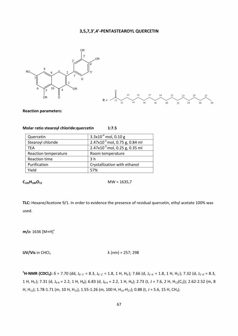
67
3,5,7,3’,4’-PENTASTEAROYL QUERCETIN
Reaction parameters:
Molar ratio stearoyl chloride:quercetin 1:7.5
Quercetin 3.3x10⁻⁴
mol, 0.10 g
Stearoyl chloride 2.47x10⁻³
mol, 0.75 g, 0.84 ml
TEA 2.47x10⁻³
mol, 0.25 g, 0.35 ml
Reaction temperature Room temperature
Reaction time 3 h
Purification Crystallization with ethanol
Yield 57%
C105H180O12 MW = 1635,7
TLC: Hexane/Acetone 9/1. In order to evidence the presence of residual quercetin, ethyl acetate 100% was
used.
m/z: 1636 [M+H]+
UV/Vis in CHCl3 λ (nm) = 257; 298
1H-NMR (CDCl3): δ = 7.70 (dd, J6’-5’ = 8.3, J6’-2’ = 1.8, 1 H, H6’); 7.66 (d, J2’-6’ = 1.8, 1 H, H2’); 7.32 (d, J5’-6’ = 8.3,
1 H, H5’); 7.31 (d, J6-8 = 2.2, 1 H, H8); 6.83 (d, J8-6 = 2.2, 1 H, H6); 2.73 (t, J = 7.6, 2 H, H12(C5)); 2.62-2.52 (m, 8
H, H12); 1.78-1.71 (m, 10 H, H13), 1.55-1.26 (m, 100 H, H14-H27); 0.88 (t, J = 5.6, 15 H, CH3).
ORO
OR
OR
OR
OR
O
89
56
7
4
3
2
2'
3'
5'
4'
6'1'
10
R =
O
11
13
12 14
15
16
17
18
19
20
21
22
23
24 26
27
28
25
68
13C-NMR (CDCl3): δ = : 172.25 (CO)C5 ; 171.04, 170.87 , 170.79 CO; 170.17 C4; 157.09 C9; 154.53 C7; 153,80
C2; 150.81 C5, 144.71 C4’; 142.52 C3’; 134.43 C3; 128.00 C1’; 126.57 C6’; 124.05 C5’, C2’; 115.04 C10; 114,10 C6,
109.07 C8, 34.63, 34.35, 33.40, 34.27, 34.02 C12; 32.16 C26; 29.95-29.28 C14- C25; 25.13, 24.97, 24.88, 24.69
C13; 22.91 C27; 13.34 CH3.
FT-IR (KBr)
1/λλλλ (cm-1
) Attribution
2956.62; 2918.33; 2850.34 ν C sp3- H
1764.58 ν C=O
1654.61 ν C=O α,β unsaturated
1468.24; 1437.97 δ C=C
1266.03; 1233.10; 1208.25; 1178.50; 1112.17 ν C-O
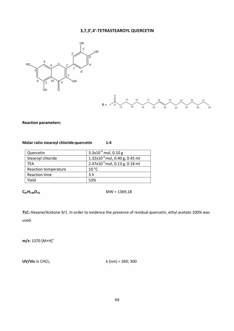
69
3,7,3’,4’-TETRASTEAROYL QUERCETIN
Reaction parameters:
Molar ratio stearoyl chloride:quercetin 1:4
Quercetin 3.3x10⁻⁴
mol, 0.10 g
Stearoyl chloride 1.32x10⁻³
mol, 0.40 g, 0.45 ml
TEA 2.47x10⁻³
mol, 0.13 g, 0.18 ml
Reaction temperature 10 °C
Reaction time 3 h
Yield 53%
C87H146O10 MW = 1369,18
TLC: Hexane/Acetone 9/1. In order to evidence the presence of residual quercetin, ethyl acetate 100% was
used.
m/z: 1370 [M+H]+
UV/Vis in CHCl3 λ (nm) = 260; 300
ORO
OH
OR
OR
OR
O
89
56
7
4
3
2
2'
3'
5'
4'
6'1'
10
R =
O
11
13
12 14
15
16
17
18
19
20
21
22
23
24 26
27
28
25
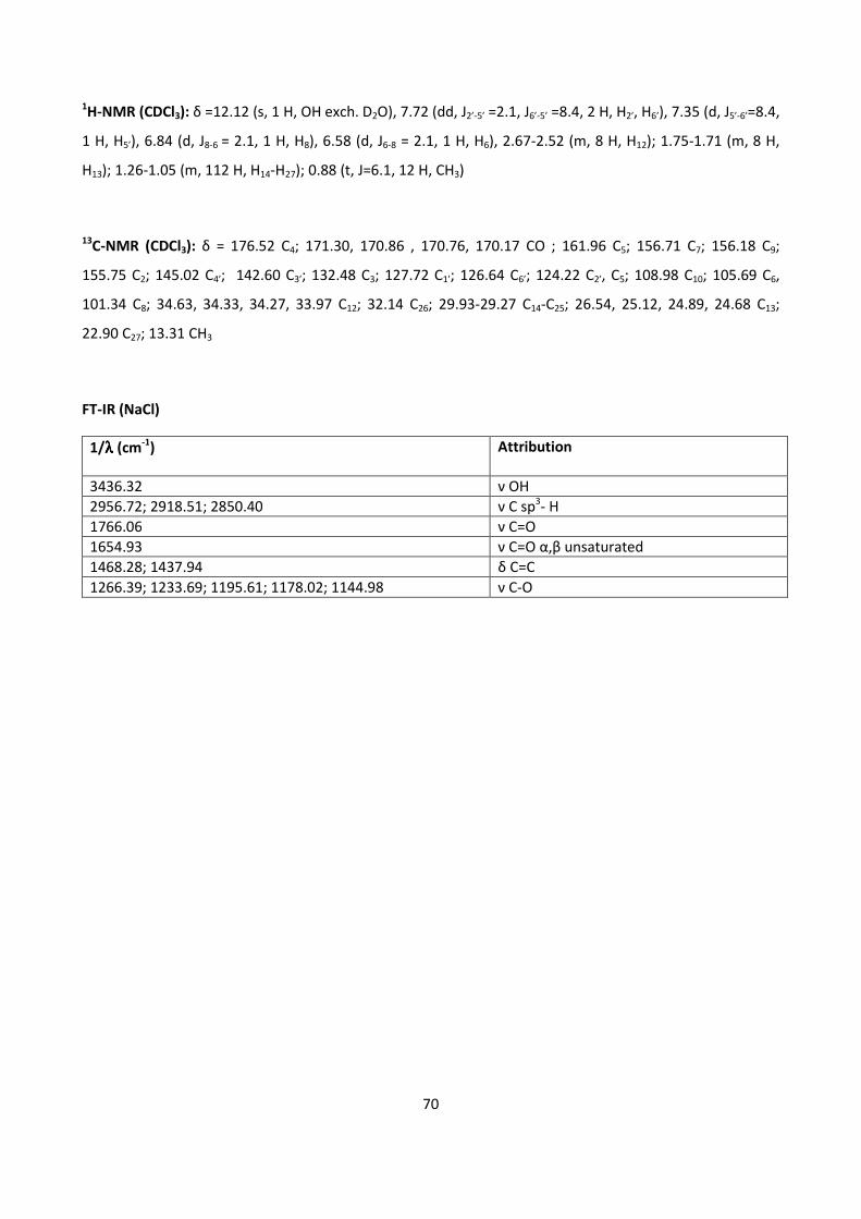
70
1H-NMR (CDCl3): δ =12.12 (s, 1 H, OH exch. D2O), 7.72 (dd, J2’-5’ =2.1, J6’-5’ =8.4, 2 H, H2’, H6’), 7.35 (d, J5’-6’=8.4,
1 H, H5’), 6.84 (d, J8-6 = 2.1, 1 H, H8), 6.58 (d, J6-8 = 2.1, 1 H, H6), 2.67-2.52 (m, 8 H, H12); 1.75-1.71 (m, 8 H,
H13); 1.26-1.05 (m, 112 H, H14-H27); 0.88 (t, J=6.1, 12 H, CH3)
13C-NMR (CDCl3): δ = 176.52 C4; 171.30, 170.86 , 170.76, 170.17 CO ; 161.96 C5; 156.71 C7; 156.18 C9;
155.75 C2; 145.02 C4’; 142.60 C3’; 132.48 C3; 127.72 C1’; 126.64 C6’; 124.22 C2’, C5; 108.98 C10; 105.69 C6,
101.34 C8; 34.63, 34.33, 34.27, 33.97 C12; 32.14 C26; 29.93-29.27 C14-C25; 26.54, 25.12, 24.89, 24.68 C13;
22.90 C27; 13.31 CH3
FT-IR (NaCl)
1/λλλλ (cm-1
) Attribution
3436.32 ν OH
2956.72; 2918.51; 2850.40 ν C sp3- H
1766.06 ν C=O
1654.93 ν C=O α,β unsaturated
1468.28; 1437.94 δ C=C
1266.39; 1233.69; 1195.61; 1178.02; 1144.98 ν C-O
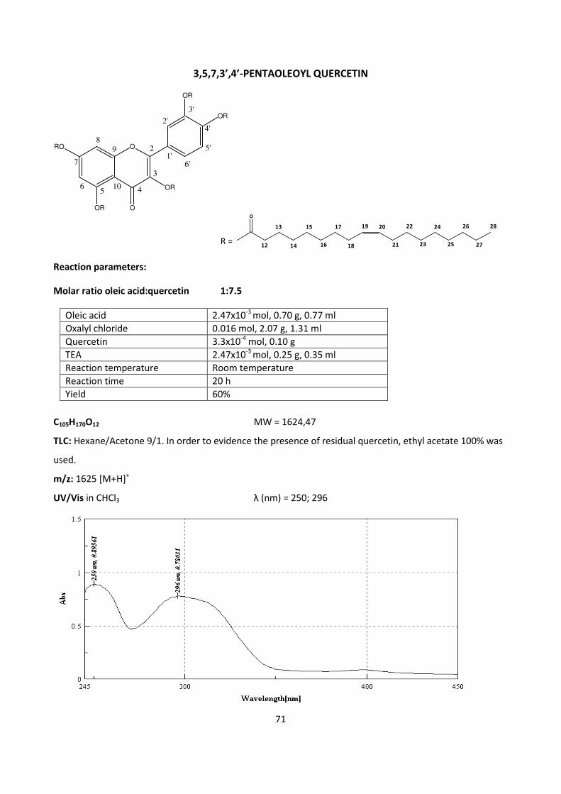
71
3,5,7,3’,4’-PENTAOLEOYL QUERCETIN
ORO
OR
OR
OR
OR
O
89
56
7
4
3
2
2'
3'
5'
4'
6'1'
10
O
12
13
14
15
16
17
18
19 20
21
22
23
24
25
26
27
28
R =
Reaction parameters:
Molar ratio oleic acid:quercetin 1:7.5
Oleic acid 2.47x10⁻³
mol, 0.70 g, 0.77 ml
Oxalyl chloride 0.016 mol, 2.07 g, 1.31 ml
Quercetin 3.3x10⁻⁴
mol, 0.10 g
TEA 2.47x10⁻³
mol, 0.25 g, 0.35 ml
Reaction temperature Room temperature
Reaction time 20 h
Yield 60%
C105H170O12 MW = 1624,47
TLC: Hexane/Acetone 9/1. In order to evidence the presence of residual quercetin, ethyl acetate 100% was
used.
m/z: 1625 [M+H]+
UV/Vis in CHCl3 λ (nm) = 250; 296
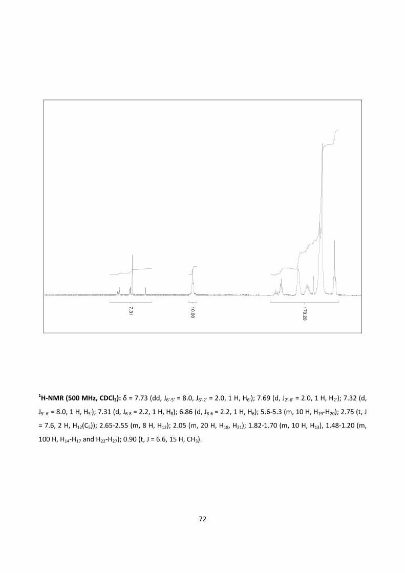
72
7.3
1
10.0
0
170
.20
1H-NMR (500 MHz, CDCl3): δ = 7.73 (dd, J6’-5’ = 8.0, J6’-2’ = 2.0, 1 H, H6’); 7.69 (d, J2’-6’ = 2.0, 1 H, H2’); 7.32 (d,
J5’-6’ = 8.0, 1 H, H5’); 7.31 (d, J6-8 = 2.2, 1 H, H8); 6.86 (d, J8-6 = 2.2, 1 H, H6); 5.6-5.3 (m, 10 H, H19-H20); 2.75 (t, J
= 7.6, 2 H, H12(C5)); 2.65-2.55 (m, 8 H, H12); 2.05 (m, 20 H, H18, H21); 1.82-1.70 (m, 10 H, H13), 1.48-1.20 (m,
100 H, H14-H17 and H22-H27); 0.90 (t, J = 6.6, 15 H, CH3).
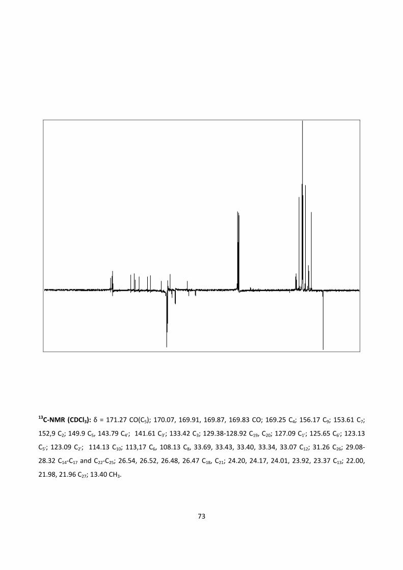
73
13C-NMR (CDCl3): δ = 171.27 CO(C5); 170.07, 169.91, 169.87, 169.83 CO; 169.25 C4; 156.17 C9; 153.61 C7;
152,9 C2; 149.9 C5, 143.79 C4’; 141.61 C3’; 133.42 C3; 129.38-128.92 C19, C20; 127.09 C1’; 125.65 C6’; 123.13
C5’; 123.09 C2’; 114.13 C10; 113,17 C6, 108.13 C8, 33.69, 33.43, 33.40, 33.34, 33.07 C12; 31.26 C26; 29.08-
28.32 C14-C17 and C22-C25; 26.54, 26.52, 26.48, 26.47 C18, C21; 24.20, 24.17, 24.01, 23.92, 23.37 C13; 22.00,
21.98, 21.96 C27; 13.40 CH3.
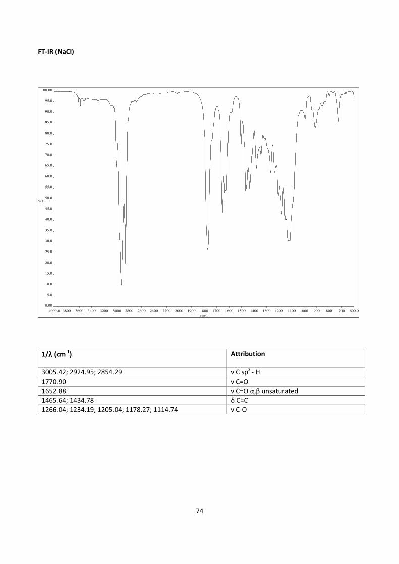
74
FT-IR (NaCl)
4000.0 3800 3600 3400 3200 3000 2800 2600 2400 2200 2000 1900 1800 1700 1600 1500 1400 1300 1200 1100 1000 900 800 700 600.0
0.00
5.0
10.0
15.0
20.0
25.0
30.0
35.0
40.0
45.0
50.0
55.0
60.0
65.0
70.0
75.0
80.0
85.0
90.0
95.0
100.00
cm-1
%T
1/λλλλ (cm-1
) Attribution
3005.42; 2924.95; 2854.29 ν C sp3
- H
1770.90 ν C=O
1652.88 ν C=O α,β unsaturated
1465.64; 1434.78 δ C=C
1266.04; 1234.19; 1205.04; 1178.27; 1114.74 ν C-O
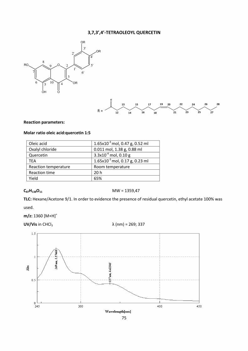
75
3,7,3’,4’-TETRAOLEOYL QUERCETIN
ORO
OH
OR
OR
OR
O
89
56
7
4
3
2
2'
3'
5'
4'
6'1'
10
O
12
13
14
15
16
17
18
19 20
21
22
23
24
25
26
27
28
R =
Reaction parameters:
Molar ratio oleic acid:quercetin 1:5
Oleic acid 1.65x10⁻³
mol, 0.47 g, 0.52 ml
Oxalyl chloride 0.011 mol, 1.38 g, 0.88 ml
Quercetin 3.3x10⁻⁴
mol, 0.10 g
TEA 1.65x10⁻³
mol, 0.17 g, 0.23 ml
Reaction temperature Room temperature
Reaction time 20 h
Yield 65%
C87H138O11 MW = 1359,47
TLC: Hexane/Acetone 9/1. In order to evidence the presence of residual quercetin, ethyl acetate 100% was
used.
m/z: 1360 [M+H]+
UV/Vis in CHCl3 λ (nm) = 269; 337
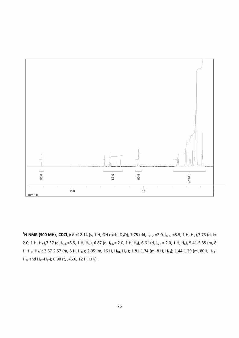
76
ppm (f1)
0.05.010.0
0.9
5
5.8
3
8.0
0
13
6.0
7
1H-NMR (500 MHz, CDCl3): δ =12.14 (s, 1 H, OH exch. D2O), 7.75 (dd, J2’-5’ =2.0, J6’-5’ =8.5, 1 H, H6’),7.73 (d, J=
2.0, 1 H, H2’),7.37 (d, J5’-6’=8.5, 1 H, H5’), 6.87 (d, J8-6 = 2.0, 1 H, H8), 6.61 (d, J6-8 = 2.0, 1 H, H6), 5.41-5.35 (m, 8
H, H19-H20); 2.67-2.57 (m, 8 H, H12); 2.05 (m, 16 H, H18, H21); 1.81-1.74 (m, 8 H, H13); 1.44-1.29 (m, 80H, H14-
H17 and H22-H27); 0.90 (t, J=6.6, 12 H, CH3).
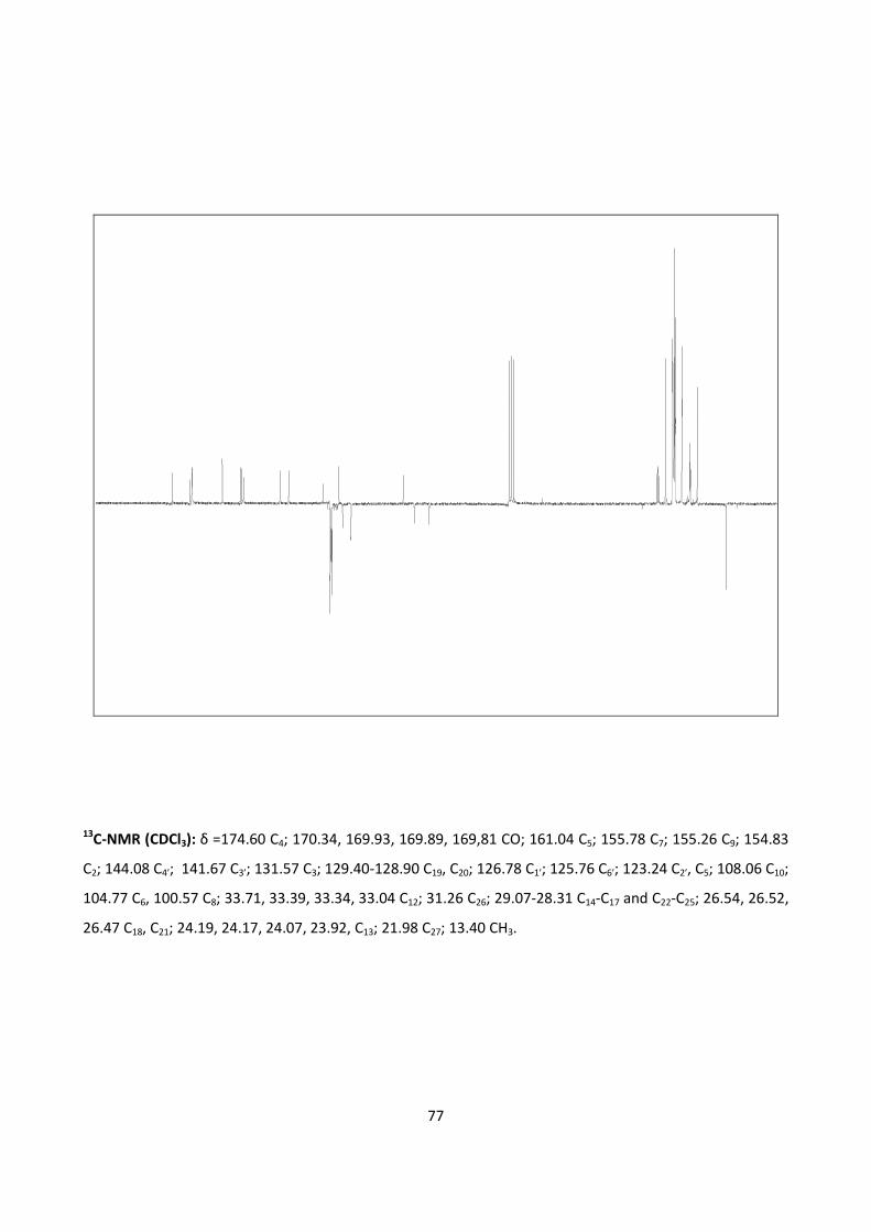
77
13C-NMR (CDCl3): δ =174.60 C4; 170.34, 169.93, 169.89, 169,81 CO; 161.04 C5; 155.78 C7; 155.26 C9; 154.83
C2; 144.08 C4’; 141.67 C3’; 131.57 C3; 129.40-128.90 C19, C20; 126.78 C1’; 125.76 C6’; 123.24 C2’, C5; 108.06 C10;
104.77 C6, 100.57 C8; 33.71, 33.39, 33.34, 33.04 C12; 31.26 C26; 29.07-28.31 C14-C17 and C22-C25; 26.54, 26.52,
26.47 C18, C21; 24.19, 24.17, 24.07, 23.92, C13; 21.98 C27; 13.40 CH3.
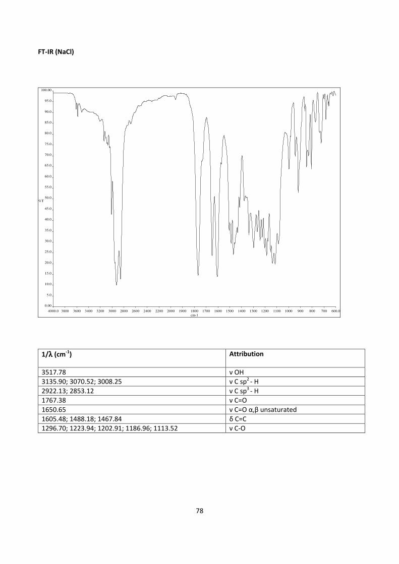
78
FT-IR (NaCl)
4000.0 3800 3600 3400 3200 3000 2800 2600 2400 2200 2000 1900 1800 1700 1600 1500 1400 1300 1200 1100 1000 900 800 700 600.0
0.00
5.0
10.0
15.0
20.0
25.0
30.0
35.0
40.0
45.0
50.0
55.0
60.0
65.0
70.0
75.0
80.0
85.0
90.0
95.0
100.00
cm-1
%T
1/λλλλ (cm-1
) Attribution
3517.78 ν OH
3135.90; 3070.52; 3008.25 ν C sp2
- H
2922.13; 2853.12 ν C sp3
- H
1767.38 ν C=O
1650.65 ν C=O α,β unsaturated
1605.48; 1488.18; 1467.84 δ C=C
1296.70; 1223.94; 1202.91; 1186.96; 1113.52 ν C-O
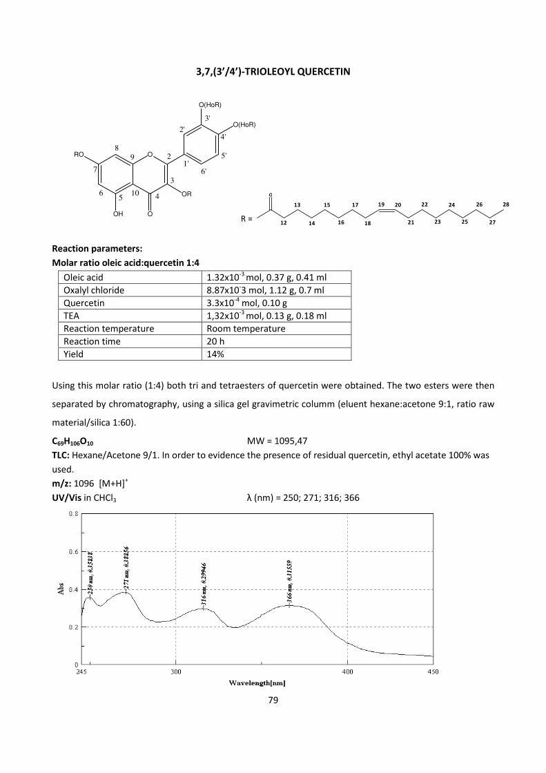
79
3,7,(3’/4’)-TRIOLEOYL QUERCETIN
Reaction parameters:
Molar ratio oleic acid:quercetin 1:4
Oleic acid 1.32x10⁻³
mol, 0.37 g, 0.41 ml
Oxalyl chloride 8.87x10⁻³ mol, 1.12 g, 0.7 ml
Quercetin 3.3x10⁻⁴
mol, 0.10 g
TEA 1,32x10⁻³
mol, 0.13 g, 0.18 ml
Reaction temperature Room temperature
Reaction time 20 h
Yield 14%
Using this molar ratio (1:4) both tri and tetraesters of quercetin were obtained. The two esters were then
separated by chromatography, using a silica gel gravimetric columm (eluent hexane:acetone 9:1, ratio raw
material/silica 1:60).
C69H106O10 MW = 1095,47
TLC: Hexane/Acetone 9/1. In order to evidence the presence of residual quercetin, ethyl acetate 100% was
used.
m/z: 1096 [M+H]+
UV/Vis in CHCl3 λ (nm) = 250; 271; 316; 366
ORO
OH
OR
O(HoR)
O(HoR)
O
89
56
7
4
3
2
2'
3'
5'
4'
6'1'
10 O
12
13
14
15
16
17
18
19 20
21
22
23
24
25
26
27
28
R =
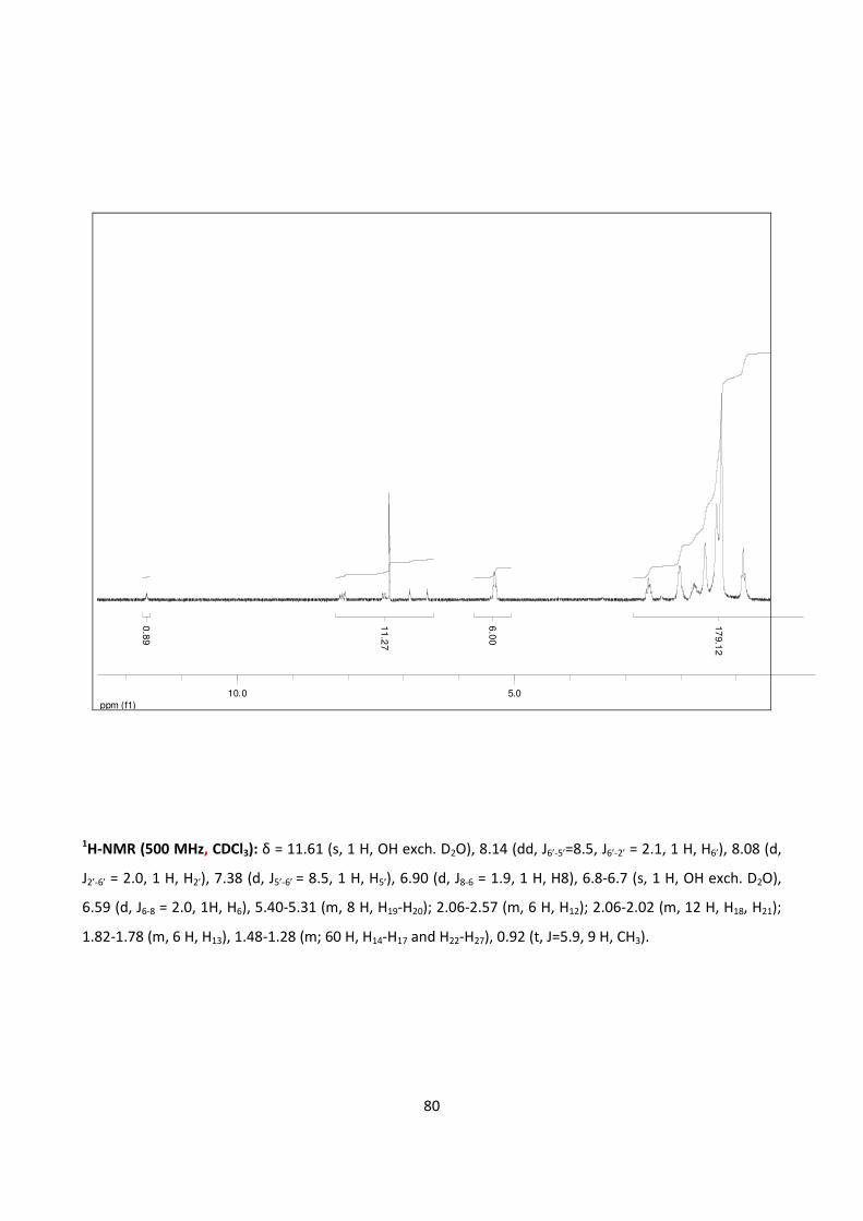
80
ppm (f1)
5.010.0
0.8
9
11.2
7
6.0
0
17
9.1
2
1H-NMR (500 MHz, CDCl3): δ = 11.61 (s, 1 H, OH exch. D2O), 8.14 (dd, J6’-5’=8.5, J6’-2’ = 2.1, 1 H, H6’), 8.08 (d,
J2’-6’ = 2.0, 1 H, H2’), 7.38 (d, J5’-6’ = 8.5, 1 H, H5’), 6.90 (d, J8-6 = 1.9, 1 H, H8), 6.8-6.7 (s, 1 H, OH exch. D2O),
6.59 (d, J6-8 = 2.0, 1H, H6), 5.40-5.31 (m, 8 H, H19-H20); 2.06-2.57 (m, 6 H, H12); 2.06-2.02 (m, 12 H, H18, H21);
1.82-1.78 (m, 6 H, H13), 1.48-1.28 (m; 60 H, H14-H17 and H22-H27), 0.92 (t, J=5.9, 9 H, CH3).
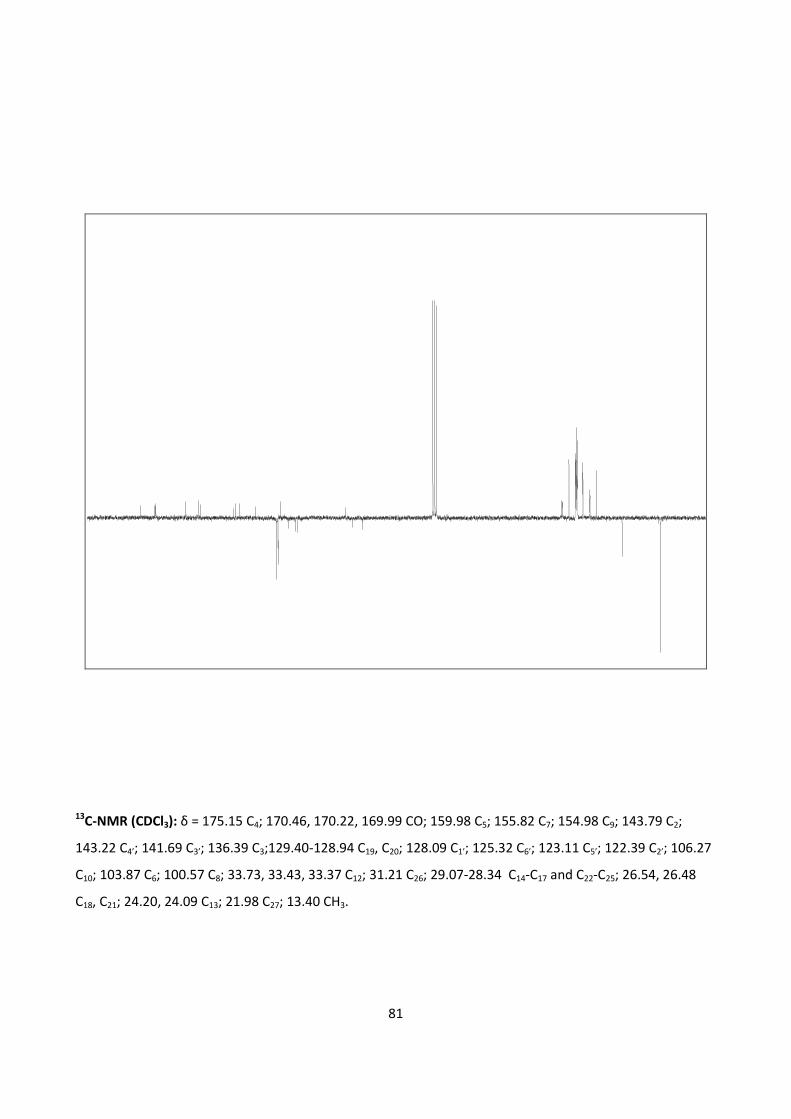
81
13C-NMR (CDCl3): δ = 175.15 C4; 170.46, 170.22, 169.99 CO; 159.98 C5; 155.82 C7; 154.98 C9; 143.79 C2;
143.22 C4’; 141.69 C3’; 136.39 C3;129.40-128.94 C19, C20; 128.09 C1’; 125.32 C6’; 123.11 C5’; 122.39 C2’; 106.27
C10; 103.87 C6; 100.57 C8; 33.73, 33.43, 33.37 C12; 31.21 C26; 29.07-28.34 C14-C17 and C22-C25; 26.54, 26.48
C18, C21; 24.20, 24.09 C13; 21.98 C27; 13.40 CH3.
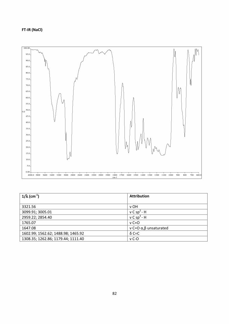
82
FT-IR (NaCl)
4000.0 3800 3600 3400 3200 3000 2800 2600 2400 2200 2000 1900 1800 1700 1600 1500 1400 1300 1200 1100 1000 900 800 700 600.0
0.00
5.0
10.0
15.0
20.0
25.0
30.0
35.0
40.0
45.0
50.0
55.0
60.0
65.0
70.0
75.0
80.0
85.0
90.0
95.0
100.00
cm-1
%T
1/λλλλ (cm-1
) Attribution
3321.56 ν OH
3099.91; 3005.01 ν C sp2
- H
2959.22; 2854.40 ν C sp3
- H
1765.07 ν C=O
1647.08 ν C=O α,β unsaturated
1602.99; 1562.62; 1488.98; 1465.92 δ C=C
1308.35; 1262.86; 1179.44; 1111.40 ν C-O
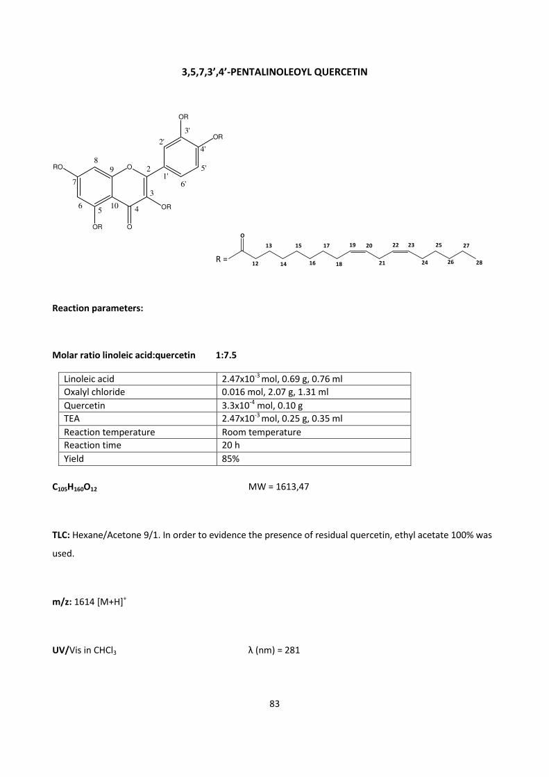
83
3,5,7,3’,4’-PENTALINOLEOYL QUERCETIN
ORO
OR
OR
OR
OR
O
89
56
7
4
3
2
2'
3'
5'
4'
6'1'
10
O
12
13
14
15
16
17
18
19 20
21
22 23
24
25
26
27
28R =
Reaction parameters:
Molar ratio linoleic acid:quercetin 1:7.5
Linoleic acid 2.47x10⁻³
mol, 0.69 g, 0.76 ml
Oxalyl chloride 0.016 mol, 2.07 g, 1.31 ml
Quercetin 3.3x10⁻⁴
mol, 0.10 g
TEA 2.47x10⁻³
mol, 0.25 g, 0.35 ml
Reaction temperature Room temperature
Reaction time 20 h
Yield 85%
C105H160O12 MW = 1613,47
TLC: Hexane/Acetone 9/1. In order to evidence the presence of residual quercetin, ethyl acetate 100% was
used.
m/z: 1614 [M+H]+
UV/Vis in CHCl3 λ (nm) = 281
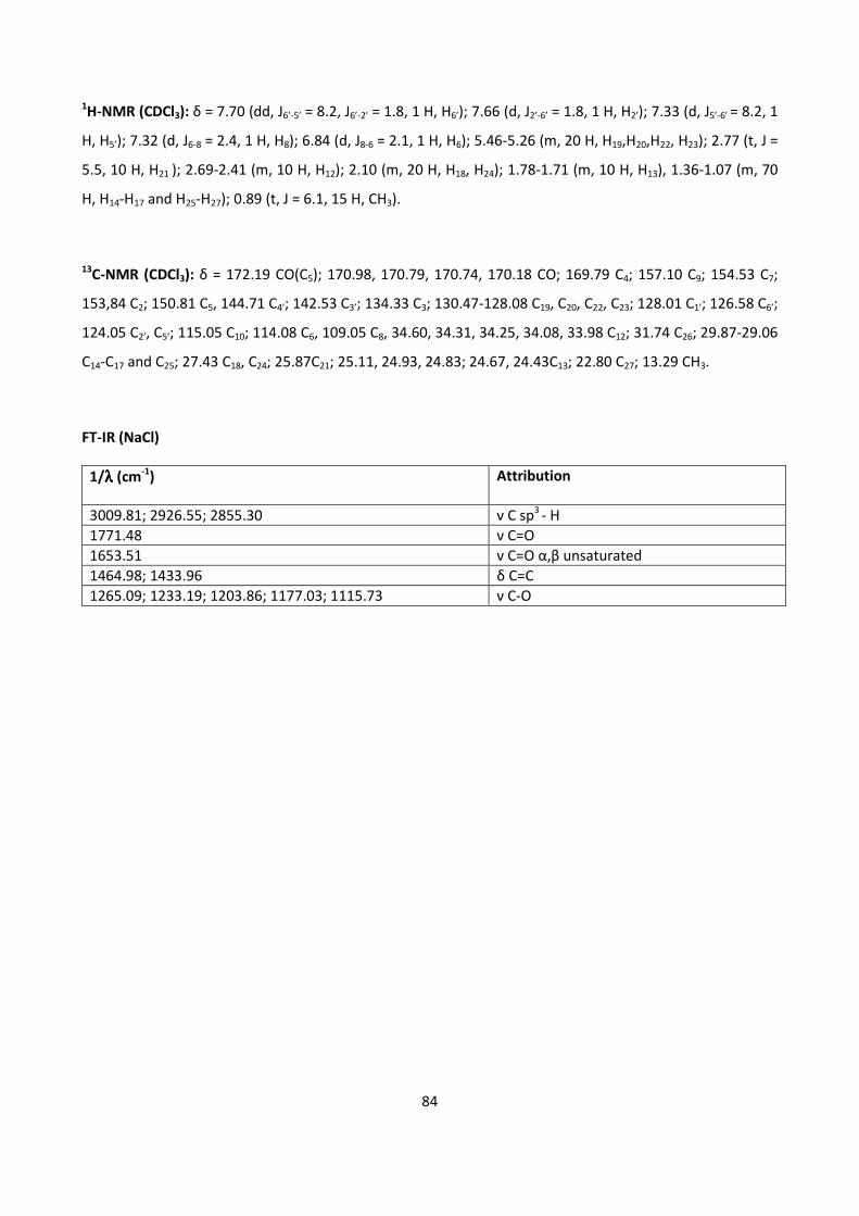
84
1H-NMR (CDCl3): δ = 7.70 (dd, J6’-5’ = 8.2, J6’-2’ = 1.8, 1 H, H6’); 7.66 (d, J2’-6’ = 1.8, 1 H, H2’); 7.33 (d, J5’-6’ = 8.2, 1
H, H5’); 7.32 (d, J6-8 = 2.4, 1 H, H8); 6.84 (d, J8-6 = 2.1, 1 H, H6); 5.46-5.26 (m, 20 H, H19,H20,H22, H23); 2.77 (t, J =
5.5, 10 H, H21 ); 2.69-2.41 (m, 10 H, H12); 2.10 (m, 20 H, H18, H24); 1.78-1.71 (m, 10 H, H13), 1.36-1.07 (m, 70
H, H14-H17 and H25-H27); 0.89 (t, J = 6.1, 15 H, CH3).
13C-NMR (CDCl3): δ = 172.19 CO(C5); 170.98, 170.79, 170.74, 170.18 CO; 169.79 C4; 157.10 C9; 154.53 C7;
153,84 C2; 150.81 C5, 144.71 C4’; 142.53 C3’; 134.33 C3; 130.47-128.08 C19, C20, C22, C23; 128.01 C1’; 126.58 C6’;
124.05 C2’, C5’; 115.05 C10; 114.08 C6, 109.05 C8, 34.60, 34.31, 34.25, 34.08, 33.98 C12; 31.74 C26; 29.87-29.06
C14-C17 and C25; 27.43 C18, C24; 25.87C21; 25.11, 24.93, 24.83; 24.67, 24.43C13; 22.80 C27; 13.29 CH3.
FT-IR (NaCl)
1/λλλλ (cm-1
) Attribution
3009.81; 2926.55; 2855.30 ν C sp3
- H
1771.48 ν C=O
1653.51 ν C=O α,β unsaturated
1464.98; 1433.96 δ C=C
1265.09; 1233.19; 1203.86; 1177.03; 1115.73 ν C-O

85
3,7,3’,4’-TETRALINOLEOYL QUERCETIN
ORO
OH
OR
OR
OR
O
89
56
7
4
3
2
2'
3'
5'
4'
6'1'
10
O
12
13
14
15
16
17
18
19 20
21
22 23
24
25
26
27
28R =
Reaction parameters:
Molar ratio linoleil acid:quercetin 1:5
Linoleic acid 1.65x10⁻³
mol, 0.46 g, 0.51 ml
Oxalyl chloride 0.011 mol, 1.38 g, 0.88 ml
Quercetin 3.3x10⁻⁴
mol, 0.10 g
TEA 1.65x10⁻³
mol, 0.17 g, 0.23 ml
Reaction temperature Room temperature
Reaction time 20 h
Yield 56%
C87H140O11 MW = 1351,47
TLC: Hexane/Acetone 9/1. In order to evidence the presence of residual quercetin, ethyl acetate 100% was
used.
m/z: 1352 [M+H]+
UV/Vis in CHCl3 λ (nm) = 273; 339

86
1H-NMR (CDCl3): δ = 12.12 (s, 1 H, OH exch. D2O), 7.72 (dd, J2’-5’ =2.1, J6’-5’ =8.2, 2 H, H2’ , H6’), 7.34 (d, J5’-
6’=8.2, 1 H, H5’), 6.84 (d, J8-6 = 1.8, 1 H, H8), 6.58 (d, J6-8 = 1.8, 1 H, H6), 5.45-5.29 (m, 16 H, H19-H20,H22-H23);
2.77 (t, J=5.5, 8 H, H21,); 2.66-2.53 (m, 8 H, H12); 2.00 (m, 16 H, H18, H24); 1.75-1.56 (m, 8 H, H13); 1.29-1.25
(m, 56H, H14-H17 and H25-H27); 0.88 (t, J=6.8, 12 H, CH3).
13C-NMR (CDCl3): δ = 176.52 C4; 171.29 ,170.85, 170.81, 170.74 CO; 161.96 C5; 156.70 C7; 156.19 C9; 155.74
C2; 145.00 C4’; 142.60 C3’; 133.48 C3; 130.48-128.08 C19, C20, C22, C23; 127.74 C1’; 126.69 C6’; 124.21 C2’, C5;
108.98 C10; 105.70 C6, 101.35 C8; 34.63, 34.31, 34.26, 33.96 C12; 31.75 C26; 29.85-29.25 C14-C17 and C25; 27.43
C18, C24;25.87 C21; 25.11, 24.99, 24.89 C13; 22.80 C27; 14.30 CH3.
FT-IR (NaCl)
1/λλλλ (cm-1
) Attribution
3009.59; 2927.35; 2855.56 ν C sp3
- H
1772.46 ν C=O
1654.85 ν C=O α,β unsaturated
1465.42; 1433.99 δ C=C
1266.25; 1235.50; 1196.53; 1179.28; 1127.48 ν C-O
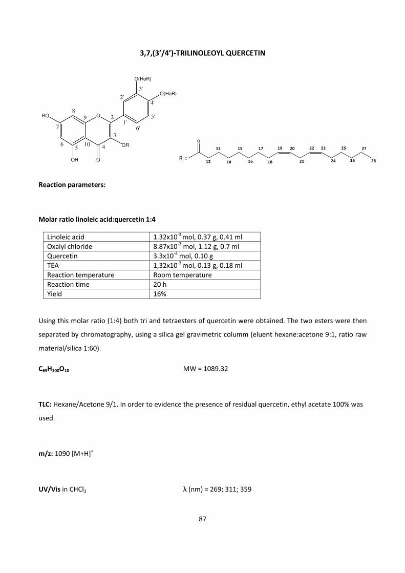
87
3,7,(3’/4’)-TRILINOLEOYL QUERCETIN
Reaction parameters:
Molar ratio linoleic acid:quercetin 1:4
Linoleic acid 1.32x10⁻³
mol, 0.37 g, 0.41 ml
Oxalyl chloride 8.87x10⁻³
mol, 1.12 g, 0.7 ml
Quercetin 3.3x10⁻⁴
mol, 0.10 g
TEA 1,32x10⁻³
mol, 0.13 g, 0.18 ml
Reaction temperature Room temperature
Reaction time 20 h
Yield 16%
Using this molar ratio (1:4) both tri and tetraesters of quercetin were obtained. The two esters were then
separated by chromatography, using a silica gel gravimetric columm (eluent hexane:acetone 9:1, ratio raw
material/silica 1:60).
C69H100O10 MW = 1089.32
TLC: Hexane/Acetone 9/1. In order to evidence the presence of residual quercetin, ethyl acetate 100% was
used.
m/z: 1090 [M+H]+
UV/Vis in CHCl3 λ (nm) = 269; 311; 359
ORO
OH
OR
O(HoR)
O(HoR)
O
89
56
7
4
3
2
2'
3'
5'
4'
6'1'
10O
12
13
14
15
16
17
18
19 20
21
22 23
24
25
26
27
28R =
88
1H-NMR (CDCl3): δ = 11.64 (s, 1 H, OH exch. D2O), 8.12 (dd, J6’-5’=8.5, J6’-2’ = 2.1, 1 H, H6’), 8.05 (d, J2’-6’ = 1.8, 1
H, H2’), 7.36 (d, J5’-6’ = 8.5, 1 H, H5’), 6.88 (d, J8-6 = 2.1, 1 H, H8), 6.85-6.70 (s, 1 H, OH exch. D2O), 6.57 (d, J6-8 =
2.1, 1H, H6), 5.45-5.26 (m, 12 H, H19-H20,H22-H23); 2.77 (t, J=5.5, 6 H, H21); 2.66-2.53 (m, 6 H, H12); 2.07-2.00
(m, 12 H, H18, H24); 1.75-1.59 (m, 6 H, H13), 1.30-1.21 (m, 42H, H14-H17 and H25-H27), 0.88 (t, J=6.7, 9 H, CH3).
13C-NMR (CDCl3): δ = 176.52 C4; 171.39, 171.15, 170.91 CO; 160.90 C5; 156.71 C7; 155.88 C9; 144.71 C2;
144.11 C4’; 142.59 C3’; 137.37 C3; 130.47-128.10 C19, C20, C22, C23; 129.03 C1’; 126.27 C6’; 124.02 C5; 123.29
C2’; 107.20 C10; 104.78 C6, 101.47 C8; 34.64, 34.34, 34.28 C12; 31.75 C26; 29.85-29.26 C14-C17 and C25; 27.43
C18,;25.87 C21, C24; 25.16, 25.00 C13; 22.80 C27; 14.30 CH3.
FT-IR (NaCl)
1/λλλλ (cm-1
) Attribution
3351.56 ν OH
3009.50; 3005.01 ν C sp2
- H
2927.46; 2855.61 ν C sp3
- H
1771.37 ν C=O
1651.79 ν C=O α,β unsaturated
1605.85; 1562.62; 1492.00; 1465.84 δ C=C
1307.00; 1268.23; 1194.05; 1133.49 ν C-O
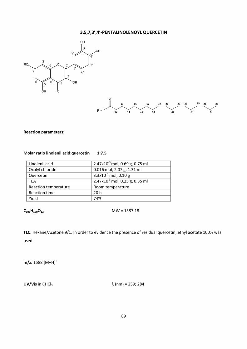
89
3,5,7,3’,4’-PENTALINOLENOYL QUERCETIN
ORO
OR
OR
OR
OR
O
89
56
7
4
3
2
2'
3'
5'
4'
6'1'
10
O
12
13
14
15
16
17
18
19 20
21
22 23
24
2625
27
28
R =
Reaction parameters:
Molar ratio linolenil acid:quercetin 1:7.5
Linolenil acid 2.47x10⁻³
mol, 0.69 g, 0.75 ml
Oxalyl chloride 0.016 mol, 2.07 g, 1.31 ml
Quercetin 3.3x10⁻⁴
mol, 0.10 g
TEA 2.47x10⁻³
mol, 0.25 g, 0.35 ml
Reaction temperature Room temperature
Reaction time 20 h
Yield 74%
C105H150O12 MW = 1587.18
TLC: Hexane/Acetone 9/1. In order to evidence the presence of residual quercetin, ethyl acetate 100% was
used.
m/z: 1588 [M+H]+
UV/Vis in CHCl3 λ (nm) = 259; 284
90
1H-NMR (CDCl3): δ = 7.70 (dd, J6’-5’ = 8.5, J6’-2’ = 2.1, 1 H, H6’); 7.65 (d, J2’-6’ = 1.8, 1 H, H2’); 7.32 (d,
J5’-6’ = 8.5, 1 H, H5’); 7.31 (d, J6-8 = 2.1, 1 H, H8); 6.84 (d, J8-6 = 2.1, 1 H, H6); 5.58-5.24(m, 30 H, H19-H20,H22-H23,
H25-H26); 2.84 (t, J=5.8, 20 H, H21, H24); 2.65-2.55 (m, 10 H, H12); 2.26-2.00 (m, 20 H, H18, H27,); 1.77-1.59 (m,
10 H, H13,), 1.36-1.21 (m, 40 H, H14-H17,); 0.94 (t, J = 7.6, 15 H, CH3).
13C-NMR (CDCl3): δ = 172.20 CO(C5); 170.98, 170.83, 170.74 CO; 169.79 C4; 157.09 C9; 154.53 C7; 153.85 C2;
150.80 C5; 144.70 C4’; 142.53 C3’; 134.33 C3; 133.18-127.93 C19, C20, C22, C23, C25, C26; 127.33 C1’; 126.58 C6’;
124.05 C2’, C5’; 115.05 C10; 114.10 C6, 109.07 C8; 35.49, 34.60, 34.30, 34.25, 33.99 C12; 29.84-29.05 C14-C17;
27.42 C18; 25.75 C24; 25.85 C21; 25.10, 24.91, 24.82, 24.67, 24.43 C13; 22.79, 20.77 C27; 14.48, 14.28 CH3.
FT-IR (NaCl)
1/λλλλ (cm-1
) Attribution
3011.02; 2928.57; 2855.35 ν C sp3
- H
1772.17 ν C=O
1654.88 ν C=O α,β unsaturated
1463.34; 1433.80 δ C=C
1265.47; 1232.57; 1203.31; 1177.68; 1123.19 ν C-O
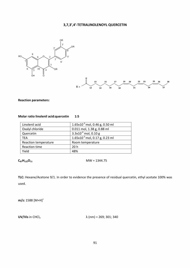
91
3,7,3’,4’-TETRALINOLENOYL QUERCETIN
ORO
OH
OR
OR
OR
O
89
56
7
4
3
2
2'
3'
5'
4'
6'1'
10
Reaction parameters:
Molar ratio linolenil acid:quercetin 1:5
Linolenil acid 1.65x10⁻³
mol, 0.46 g, 0.50 ml
Oxalyl chloride 0.011 mol, 1.38 g, 0.88 ml
Quercetin 3.3x10⁻⁴
mol, 0.10 g
TEA 1.65x10⁻³
mol, 0.17 g, 0.23 ml
Reaction temperature Room temperature
Reaction time 20 h
Yield 48%
C87H122O11 MW = 1344.75
TLC: Hexane/Acetone 9/1. In order to evidence the presence of residual quercetin, ethyl acetate 100% was
used.
m/z: 1588 [M+H]+
UV/Vis in CHCl3 λ (nm) = 269; 301; 340
O
12
13
14
15
16
17
18
19 20
21
22 23
24
2625
27
28
R =
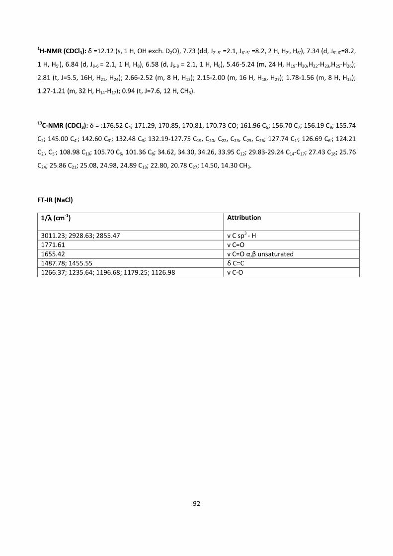
92
1H-NMR (CDCl3): δ =12.12 (s, 1 H, OH exch. D2O), 7.73 (dd, J2’-5’ =2.1, J6’-5’ =8.2, 2 H, H2’, H6’), 7.34 (d, J5’-6’=8.2,
1 H, H5’), 6.84 (d, J8-6 = 2.1, 1 H, H8), 6.58 (d, J6-8 = 2.1, 1 H, H6), 5.46-5.24 (m, 24 H, H19-H20,H22-H23,H25-H26);
2.81 (t, J=5.5, 16H, H21, H24); 2.66-2.52 (m, 8 H, H12); 2.15-2.00 (m, 16 H, H18, H27); 1.78-1.56 (m, 8 H, H13);
1.27-1.21 (m, 32 H, H14-H17); 0.94 (t, J=7.6, 12 H, CH3).
13C-NMR (CDCl3): δ = :176.52 C4; 171.29, 170.85, 170.81, 170.73 CO; 161.96 C5; 156.70 C7; 156.19 C9; 155.74
C2; 145.00 C4’; 142.60 C3’; 132.48 C3; 132.19-127.75 C19, C20, C22, C23, C25, C26; 127.74 C1’; 126.69 C6’; 124.21
C2’, C5’; 108.98 C10; 105.70 C6, 101.36 C8; 34.62, 34.30, 34.26, 33.95 C12; 29.83-29.24 C14-C17; 27.43 C18; 25.76
C24; 25.86 C21; 25.08, 24.98, 24.89 C13; 22.80, 20.78 C27; 14.50, 14.30 CH3.
FT-IR (NaCl)
1/λλλλ (cm-1
) Attribution
3011.23; 2928.63; 2855.47 ν C sp3
- H
1771.61 ν C=O
1655.42 ν C=O α,β unsaturated
1487.78; 1455.55 δ C=C
1266.37; 1235.64; 1196.68; 1179.25; 1126.98 ν C-O
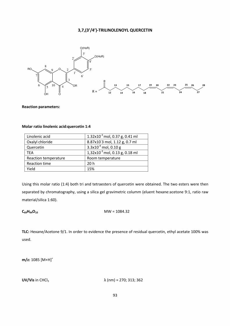
93
3,7,(3’/4’)-TRILINOLENOYL QUERCETIN
Reaction parameters:
Molar ratio linolenic acid:quercetin 1:4
Linolenic acid 1.32x10⁻³
mol, 0.37 g, 0.41 ml
Oxalyl chloride 8.87x10⁻³ mol, 1.12 g, 0.7 ml
Quercetin 3.3x10⁻⁴
mol, 0.10 g
TEA 1,32x10⁻³
mol, 0.13 g, 0.18 ml
Reaction temperature Room temperature
Reaction time 20 h
Yield 15%
Using this molar ratio (1:4) both tri and tetraesters of quercetin were obtained. The two esters were then
separated by chromatography, using a silica gel gravimetric columm (eluent hexane:acetone 9:1, ratio raw
material/silica 1:60).
C69H94O10 MW = 1084.32
TLC: Hexane/Acetone 9/1. In order to evidence the presence of residual quercetin, ethyl acetate 100% was
used.
m/z: 1085 [M+H]+
UV/Vis in CHCl3 λ (nm) = 270; 313; 362
O
12
13
14
15
16
17
18
19 20
21
22 23
24
2625
27
28
R =
ORO
OH
OR
O(HoR)
O(HoR)
O
89
56
7
4
3
2
2'
3'
5'
4'
6'1'
10
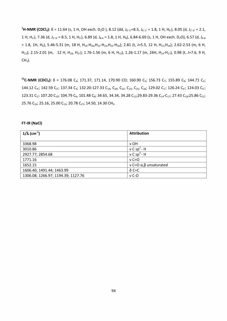
94
1H-NMR (CDCl3): δ = 11.64 (s, 1 H, OH exch. D2O ), 8.12 (dd, J6’-5’=8.5, J6’-2’ = 1.8, 1 H, H6’), 8.05 (d, J2’-6’ = 2.1,
1 H, H2’), 7.36 (d, J5’-6’ = 8.5, 1 H, H5’), 6.89 (d, J8-6 = 1.8, 1 H, H8), 6.84-6.69 (s, 1 H, OH exch. D2O), 6.57 (d, J6-8
= 1.8, 1H, H6), 5.46-5.31 (m, 18 H, H19-H20,H22-H23,H25-H26); 2.81 (t, J=5.5, 12 H, H21,H24); 2.62-2.53 (m, 6 H,
H12); 2.15-2.01 (m, 12 H, H18, H27); 1.76-1.56 (m, 6 H, H13), 1.26-1.17 (m, 24H, H14-H17), 0.98 (t, J=7.6, 9 H,
CH3).
13C-NMR (CDCl3): δ = 176.08 C4; 171.37, 171.14, 170.90 CO; 160.90 C5; 156.73 C7; 155.89 C9; 144.71 C2;
144.12 C4’; 142.59 C3’; 137.34 C3; 132.20-127.33 C19, C20, C22, C23, C25, C26; 129.02 C1’; 126.24 C6’; 124.03 C5’;
123.31 C2’; 107.20 C10; 104.79 C6, 101.48 C8; 34.65, 34.34, 34.28 C12;29.83-29.36 C14-C17; 27.43 C18;25.86 C21;
25.76 C24; 25.16, 25.00 C13; 20.78 C27; 14.50, 14.30 CH3.
FT-IR (NaCl)
1/λλλλ (cm-1
) Attribution
3368.98 ν OH
3010.86 ν C sp2
- H
2927.77; 2854.68 ν C sp3
- H
1771.16 ν C=O
1652.15 ν C=O α,β unsaturated
1606.40; 1491.44; 1463.99 δ C=C
1306.08; 1266.97; 1194.39; 1127.76 ν C-O
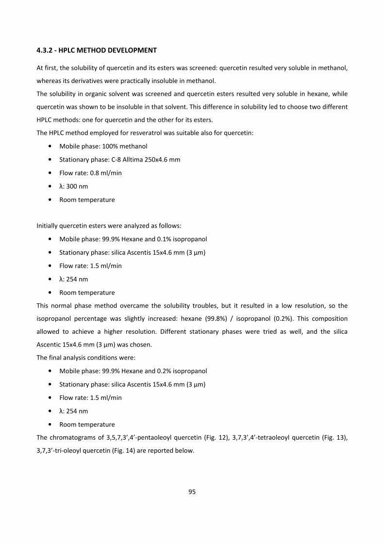
95
4.3.2 - HPLC METHOD DEVELOPMENT
At first, the solubility of quercetin and its esters was screened: quercetin resulted very soluble in methanol,
whereas its derivatives were practically insoluble in methanol.
The solubility in organic solvent was screened and quercetin esters resulted very soluble in hexane, while
quercetin was shown to be insoluble in that solvent. This difference in solubility led to choose two different
HPLC methods: one for quercetin and the other for its esters.
The HPLC method employed for resveratrol was suitable also for quercetin:
• Mobile phase: 100% methanol
• Stationary phase: C-8 Alltima 250x4.6 mm
• Flow rate: 0.8 ml/min
• λ: 300 nm
• Room temperature
Initially quercetin esters were analyzed as follows:
• Mobile phase: 99.9% Hexane and 0.1% isopropanol
• Stationary phase: silica Ascentis 15x4.6 mm (3 μm)
• Flow rate: 1.5 ml/min
• λ: 254 nm
• Room temperature
This normal phase method overcame the solubility troubles, but it resulted in a low resolution, so the
isopropanol percentage was slightly increased: hexane (99.8%) / isopropanol (0.2%). This composition
allowed to achieve a higher resolution. Different stationary phases were tried as well, and the silica
Ascentic 15x4.6 mm (3 μm) was chosen.
The final analysis conditions were:
• Mobile phase: 99.9% Hexane and 0.2% isopropanol
• Stationary phase: silica Ascentis 15x4.6 mm (3 μm)
• Flow rate: 1.5 ml/min
• λ: 254 nm
• Room temperature
The chromatograms of 3,5,7,3’,4’-pentaoleoyl quercetin (Fig. 12), 3,7,3’,4’-tetraoleoyl quercetin (Fig. 13),
3,7,3’-tri-oleoyl quercetin (Fig. 14) are reported below.

96
Figure 12. Chromatogram of 3,5,7,3’,4’-pentaoleoyl quercetin (RT= 2.933 min).
Figure 13. Chromatogram of 3,7, 3’,4’-tetraoleoyl quercetin (RT=2.490 min).
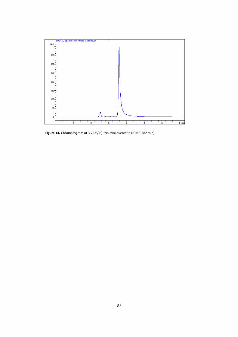
97
Figure 14. Chromatogram of 3,7,(3’/4’)-trioleyol quercetin (RT= 3.582 min).
98
4.3.3 - LIPOSOMES
The topical application of quercetin inhibits oxidative skin damage and the inflammatory processes induced
by UV radiation. Low skin permeability and poor solubility of quercetin in aqueous media reduce its topical
use: these features make the development of pharmaceutical formulations difficult. In addition to synthesis
of prodrugs (quercetin esters), other strategies can be used in order to improve quercetin topical delivery,
such as liposome formulation.
In collaboration with Dipartimento Farmaco Chimico Tecnologico, University of Cagliari, quercetin and its
derivatives were incorporated into phospholipid vesicles. The incorporation of quercetin esters into
liposomes would allow the delivery not only of quercetin, but also of fatty acids, a class of molecules known
to be active in the skin.
In order to better incorporate quercetin derivatives into phospholipid vesicles, two different hydrophilic
penetration enhancers (PEs), propylene glycol (PG) and polyethylene glycol 400 (PEG), were added to the
formulations. Two different quercetin derivatives-loaded Penetration Enhancer containing Vesicles (PEVs)
formulations (Formulation A e B) were produced, as reported below:
• Formulation A:
- Soybean lecithin (0.15 g)
- PEG 400 (1.0 g)
- Quercetin / quercetin derivative (0.005 g)
- Water (1.5 ml)
• Formulation B:
- Soybean lecithin (0.15 g)
- Propylene glycol (1.0 g)
- Quercetin / Quercetin derivative (0.005 g)
- Water (1.5 ml)
The esters of quercetin used in these experimental formulations were: 3,5,7,3’,4’-pentastearoyl quercetin,
3,5,7,3’,4’-pentaoleoyl quercetin, 3,7,3’,4’-tetraoleoyl quercetin, 3,5,7,3’,4’-pentalinoleoyl quercetin,
3,7,3’,4’-tetralinoleoyl quercetin, 3,5,7,3’,4’-pentalinolenoyl quercetin and 3,7,3’,4’-tetralinolenoyl
quercetin.
The use of a mixture of PE/water (40% v/v) as hydrophilic phase improved the quercetin and derivatives
solubility in water, due to their high solubilizing power, and enabled the incorporation of ~2 mg/ml of drug
in PEVs without any sign of drug precipitation during or after fabrication. Previous findings demonstrated
99
that PEVs are powerful enhancers for dermal delivery, due to the synergistic effect of phospholipid vesicles
and PE.
4.3.3.1 - VESICLE PREPARATION
The quercetin ester was dissolved in a PE/water solution (40% v/v) and added to a flask containing soybean
lecithin. Lipids were left swelling in the solution overnight. Vesicles were obtained by sonicating for 3
minutes (5 seconds on and 5 seconds off) the dispersions with a Soniprep 150 ultrasonic disintegrator (MSE
Crowley, UK).
4.3.3.2 - VESICLE CHARACTERIZATION
Each vesicle suspension was purified from the non-incorporated drug by exhaustive dialysis against distilled
water at 4 °C for 1 hour, using dialysis tubing (Spectra/Por® membranes: 12–14 kDa MW cut-off, 3 nm pore
size; Spectrum Laboratories Inc., USA). Incorporation efficiency (E%), expressed as the percentage of the
amount of drug initially used, was determined by high performance liquid chromatography (HPLC) after
disruption of vesicles with 0.025% non-ionic Triton X-100. Quercetin content was quantified at 255 and 367
nm using a chromatograph Alliance 2690 (Waters, Italy). The column was a SunFire C18 (3.5 µm, 4.6 × 150
mm). The mobile phase was a mixture of acetonitrile, water and acetic acid (94.8:5:0.2, v/v), delivered at a
flow rate of 1.0 mL/min.
The average diameter and polydispersity index (P.I.) of the samples (6 replicates) were determined by
Photon Correlation Spectroscopy (PCS) using a Zetasizer nano-ZS (Malvern Instrument, UK). Samples were
backscattered by a helium–neon laser (633 nm) at an angle of 173° and a constant temperature of 25 °C.
The P.I. was used as a measure of the width of the size distribution: P.I. less than 0.4 indicates a
homogenous and monodisperse population. Zeta potential was estimated using the Zetasizer nano-ZS by
means of the M3-PALS (Phase Analysis Light Scattering) technique, which measures the particle
electrophoretic mobility in a thermostated cell. All the samples were analyzed 24 h after their preparation.
100
5 - RESULTS
5.1 - β-SITOSTEROL
5.1.1 - BIOCATALYTIC SYNTHESIS OF β-SITOSTEROL FATTY ESTERS
The esters of β-sitosterol and fatty acid were already obtained by chemical synthesis; nevertheless, we
synthesized these compounds using the enzymatic approach, which represents a natural way to obtain
them.
In the first phase of this study, five enzymes were screened: only lipases from Candida rugosa and Candida
cylindracea resulted active. The subsequent step was to evaluate the suitability between these enzymes
with different organic solvents, such as heptane, toluene, acetone, acetonitrile, hexane and isopropyl
ether: among them, heptane and toluene resulted to be the most convenient. Then, different trials were
set up at 25, 30 and 40 °C, evidencing that the esterification rate was higher at 40 °C. Finally, different
amounts of enzyme were screened: lipase from Candida rugosa resulted more efficient at 35 U/mg,
whereas lipase from Candida cylindracea at 14 U/mg.
After setting up these reaction conditions, we assessed that lipase from Candida rugosa was able to esterify
β-sitosterol with both oleic and linolenic acids, while Candida cylindracea esterified β-sitosterol with only
oleic acid. In order to evaluate the reaction kinetics and time, three reactions in semi-preparative scale
were performed and at fixed times (4 h, 24 h, 42 h, 48 h, 52 h, 72 h) samples were taken and analyzed using
HPLC.
In this step, the synthetic esters were used as reference standards for HPLC analysis. The courses of the
biotrasformations are reported in the figures that show the amount of ester formed, calculated as area
under the peaks in relation to time. The resulting reaction kinetics are reported below (Figures 15, 16 and
17).
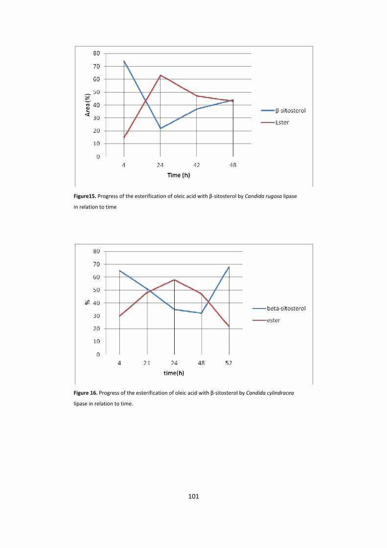
101
Figure15. Progress of the esterification of oleic acid with β-sitosterol by Candida rugosa lipase
in relation to time
Figure 16. Progress of the esterification of oleic acid with β-sitosterol by Candida cylindracea
lipase in relation to time.
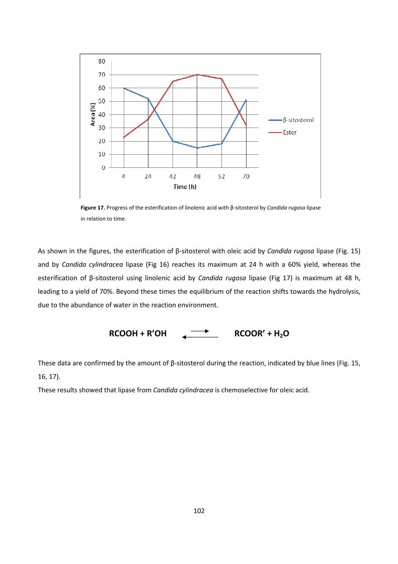
102
Figure 17. Progress of the esterification of linolenic acid with β-sitosterol by Candida rugosa lipase
in relation to time.
As shown in the figures, the esterification of β-sitosterol with oleic acid by Candida rugosa lipase (Fig. 15)
and by Candida cylindracea lipase (Fig 16) reaches its maximum at 24 h with a 60% yield, whereas the
esterification of β-sitosterol using linolenic acid by Candida rugosa lipase (Fig 17) is maximum at 48 h,
leading to a yield of 70%. Beyond these times the equilibrium of the reaction shifts towards the hydrolysis,
due to the abundance of water in the reaction environment.
RCOOH + R’OH RCOOR’ + H2O
These data are confirmed by the amount of β-sitosterol during the reaction, indicated by blue lines (Fig. 15,
16, 17).
These results showed that lipase from Candida cylindracea is chemoselective for oleic acid.
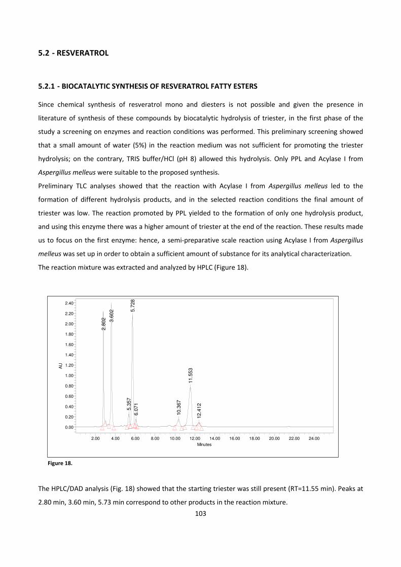
103
5.2 - RESVERATROL
5.2.1 - BIOCATALYTIC SYNTHESIS OF RESVERATROL FATTY ESTERS
Since chemical synthesis of resveratrol mono and diesters is not possible and given the presence in
literature of synthesis of these compounds by biocatalytic hydrolysis of triester, in the first phase of the
study a screening on enzymes and reaction conditions was performed. This preliminary screening showed
that a small amount of water (5%) in the reaction medium was not sufficient for promoting the triester
hydrolysis; on the contrary, TRIS buffer/HCl (pH 8) allowed this hydrolysis. Only PPL and Acylase I from
Aspergillus melleus were suitable to the proposed synthesis.
Preliminary TLC analyses showed that the reaction with Acylase I from Aspergillus melleus led to the
formation of different hydrolysis products, and in the selected reaction conditions the final amount of
triester was low. The reaction promoted by PPL yielded to the formation of only one hydrolysis product,
and using this enzyme there was a higher amount of triester at the end of the reaction. These results made
us to focus on the first enzyme: hence, a semi-preparative scale reaction using Acylase I from Aspergillus
melleus was set up in order to obtain a sufficient amount of substance for its analytical characterization.
The reaction mixture was extracted and analyzed by HPLC (Figure 18).
Figure 18.
The HPLC/DAD analysis (Fig. 18) showed that the starting triester was still present (RT=11.55 min). Peaks at
2.80 min, 3.60 min, 5.73 min correspond to other products in the reaction mixture.
2.8
02 3.6
02
5.3
57
5.7
28
6.0
71
10.3
67
11.5
53
12.4
12
AU
0.00
0.20
0.40
0.60
0.80
1.00
1.20
1.40
1.60
1.80
2.00
2.20
2.40
Minutes
2.00 4.00 6.00 8.00 10.00 12.00 14.00 16.00 18.00 20.00 22.00 24.00
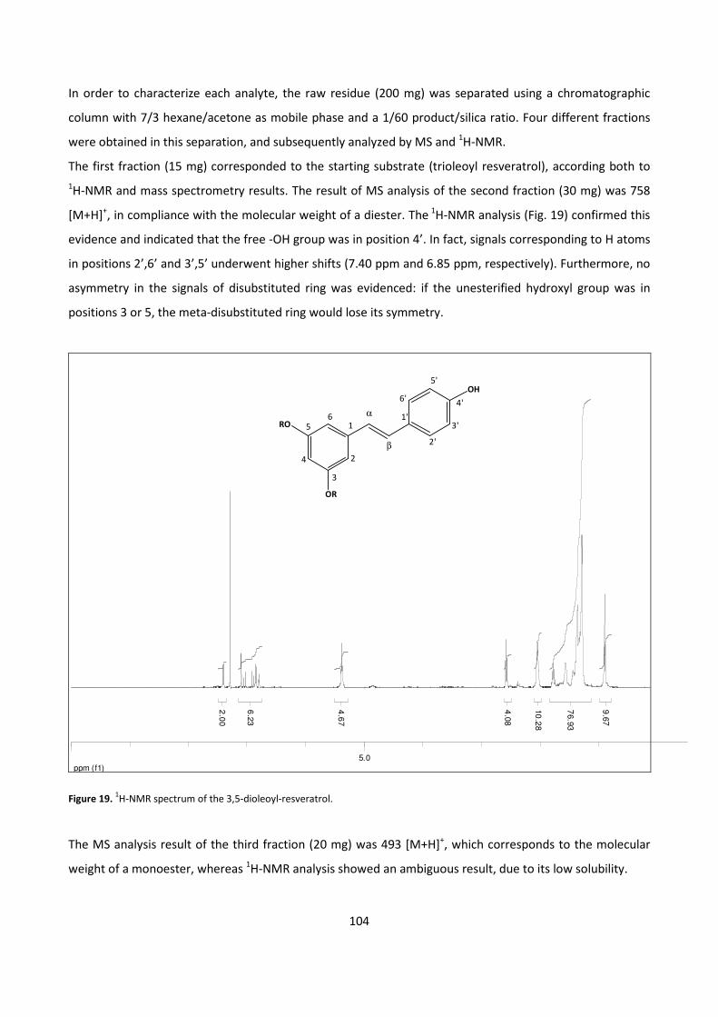
104
RO
OR
OH5'
4'
3'
2'
6'
1'
β
α
2
3
4
56
1
In order to characterize each analyte, the raw residue (200 mg) was separated using a chromatographic
column with 7/3 hexane/acetone as mobile phase and a 1/60 product/silica ratio. Four different fractions
were obtained in this separation, and subsequently analyzed by MS and 1H-NMR.
The first fraction (15 mg) corresponded to the starting substrate (trioleoyl resveratrol), according both to
1H-NMR and mass spectrometry results. The result of MS analysis of the second fraction (30 mg) was 758
[M+H]+, in compliance with the molecular weight of a diester. The
1H-NMR analysis (Fig. 19) confirmed this
evidence and indicated that the free -OH group was in position 4’. In fact, signals corresponding to H atoms
in positions 2’,6’ and 3’,5’ underwent higher shifts (7.40 ppm and 6.85 ppm, respectively). Furthermore, no
asymmetry in the signals of disubstituted ring was evidenced: if the unesterified hydroxyl group was in
positions 3 or 5, the meta-disubstituted ring would lose its symmetry.
ppm (f1)
5.0
2.0
0
6.2
3
4.6
7
4.0
8
10
.28
76.9
3
9.6
7
Figure 19. 1H-NMR spectrum of the 3,5-dioleoyl-resveratrol.
The MS analysis result of the third fraction (20 mg) was 493 [M+H]+, which corresponds to the molecular
weight of a monoester, whereas 1H-NMR analysis showed an ambiguous result, due to its low solubility.
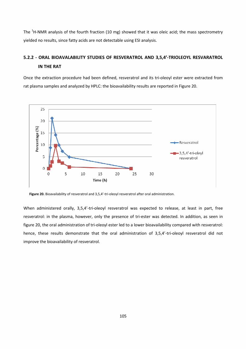
105
The 1H-NMR analysis of the fourth fraction (10 mg) showed that it was oleic acid; the mass spectrometry
yielded no results, since fatty acids are not detectable using ESI analysis.
5.2.2 - ORAL BIOAVALABILITY STUDIES OF RESVERATROL AND 3,5,4’-TRIOLEOYL RESVARATROL
IN THE RAT
Once the extraction procedure had been defined, resveratrol and its tri-oleoyl ester were extracted from
rat plasma samples and analyzed by HPLC: the bioavailability results are reported in Figure 20.
Figure 20. Bioavailability of resveratrol and 3,5,4’-tri-oleoyl resveratrol after oral administration.
When administered orally, 3,5,4’-tri-oleoyl resveratrol was expected to release, at least in part, free
resveratrol: in the plasma, however, only the presence of tri-ester was detected. In addition, as seen in
figure 20, the oral administration of tri-oleoyl ester led to a lower bioavailability compared with resveratrol:
hence, these results demonstrate that the oral administration of 3,5,4’-tri-oleoyl resveratrol did not
improve the bioavailability of resveratrol.
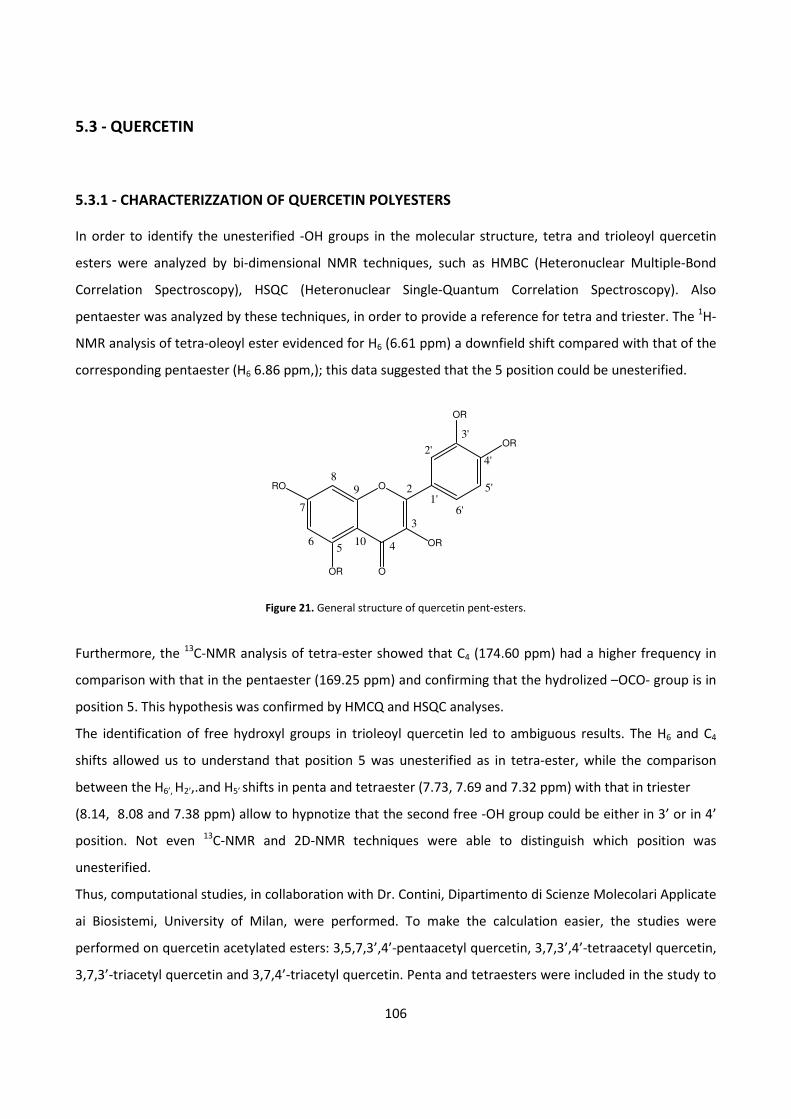
106
5.3 - QUERCETIN
5.3.1 - CHARACTERIZZATION OF QUERCETIN POLYESTERS
In order to identify the unesterified -OH groups in the molecular structure, tetra and trioleoyl quercetin
esters were analyzed by bi-dimensional NMR techniques, such as HMBC (Heteronuclear Multiple-Bond
Correlation Spectroscopy), HSQC (Heteronuclear Single-Quantum Correlation Spectroscopy). Also
pentaester was analyzed by these techniques, in order to provide a reference for tetra and triester. The 1H-
NMR analysis of tetra-oleoyl ester evidenced for H6 (6.61 ppm) a downfield shift compared with that of the
corresponding pentaester (H6 6.86 ppm,); this data suggested that the 5 position could be unesterified.
Figure 21. General structure of quercetin pent-esters.
Furthermore, the 13
C-NMR analysis of tetra-ester showed that C4 (174.60 ppm) had a higher frequency in
comparison with that in the pentaester (169.25 ppm) and confirming that the hydrolized –OCO- group is in
position 5. This hypothesis was confirmed by HMCQ and HSQC analyses.
The identification of free hydroxyl groups in trioleoyl quercetin led to ambiguous results. The H6 and C4
shifts allowed us to understand that position 5 was unesterified as in tetra-ester, while the comparison
between the H6’, H2’,.and H5’ shifts in penta and tetraester (7.73, 7.69 and 7.32 ppm) with that in triester
(8.14, 8.08 and 7.38 ppm) allow to hypnotize that the second free -OH group could be either in 3’ or in 4’
position. Not even 13
C-NMR and 2D-NMR techniques were able to distinguish which position was
unesterified.
Thus, computational studies, in collaboration with Dr. Contini, Dipartimento di Scienze Molecolari Applicate
ai Biosistemi, University of Milan, were performed. To make the calculation easier, the studies were
performed on quercetin acetylated esters: 3,5,7,3’,4’-pentaacetyl quercetin, 3,7,3’,4’-tetraacetyl quercetin,
3,7,3’-triacetyl quercetin and 3,7,4’-triacetyl quercetin. Penta and tetraesters were included in the study to
ORO
OR
OR
OR
OR
O
89
56
7
4
3
2
2'
3'
5'
4'
6'1'
10
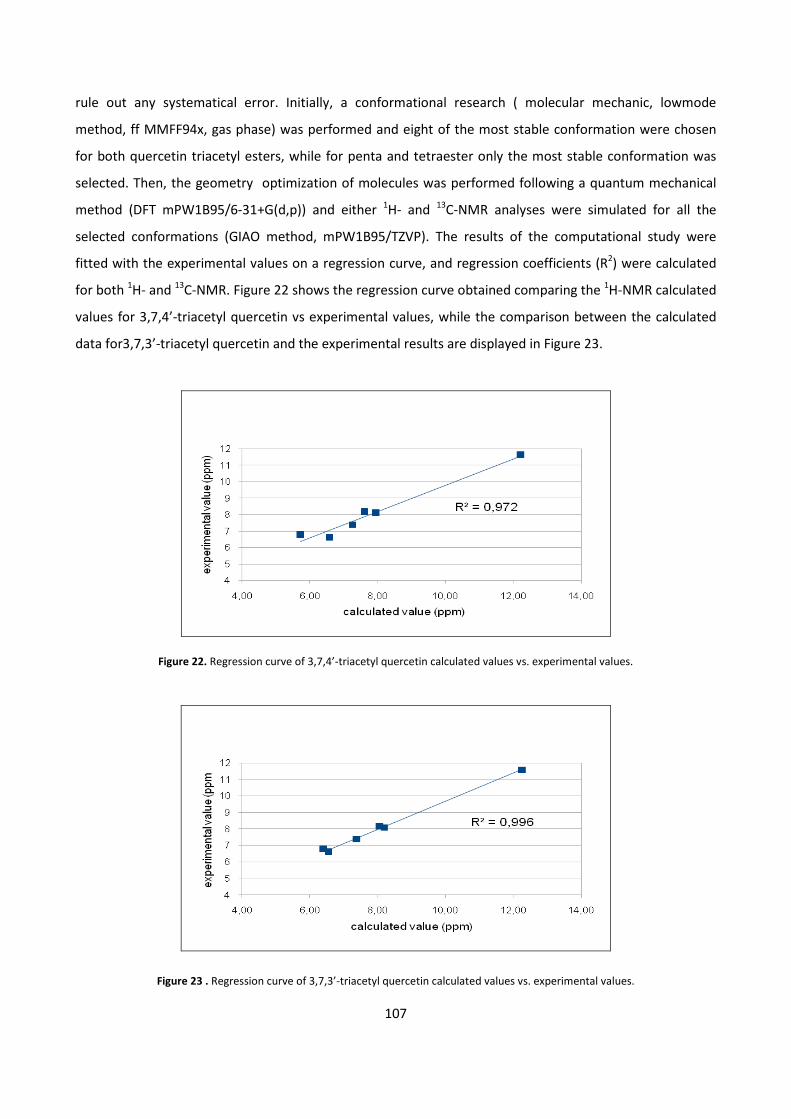
107
rule out any systematical error. Initially, a conformational research ( molecular mechanic, lowmode
method, ff MMFF94x, gas phase) was performed and eight of the most stable conformation were chosen
for both quercetin triacetyl esters, while for penta and tetraester only the most stable conformation was
selected. Then, the geometry optimization of molecules was performed following a quantum mechanical
method (DFT mPW1B95/6-31+G(d,p)) and either 1H- and
13C-NMR analyses were simulated for all the
selected conformations (GIAO method, mPW1B95/TZVP). The results of the computational study were
fitted with the experimental values on a regression curve, and regression coefficients (R2) were calculated
for both 1H- and
13C-NMR. Figure 22 shows the regression curve obtained comparing the
1H-NMR calculated
values for 3,7,4’-triacetyl quercetin vs experimental values, while the comparison between the calculated
data for3,7,3’-triacetyl quercetin and the experimental results are displayed in Figure 23.
Figure 22. Regression curve of 3,7,4’-triacetyl quercetin calculated values vs. experimental values.
Figure 23 . Regression curve of 3,7,3’-triacetyl quercetin calculated values vs. experimental values.
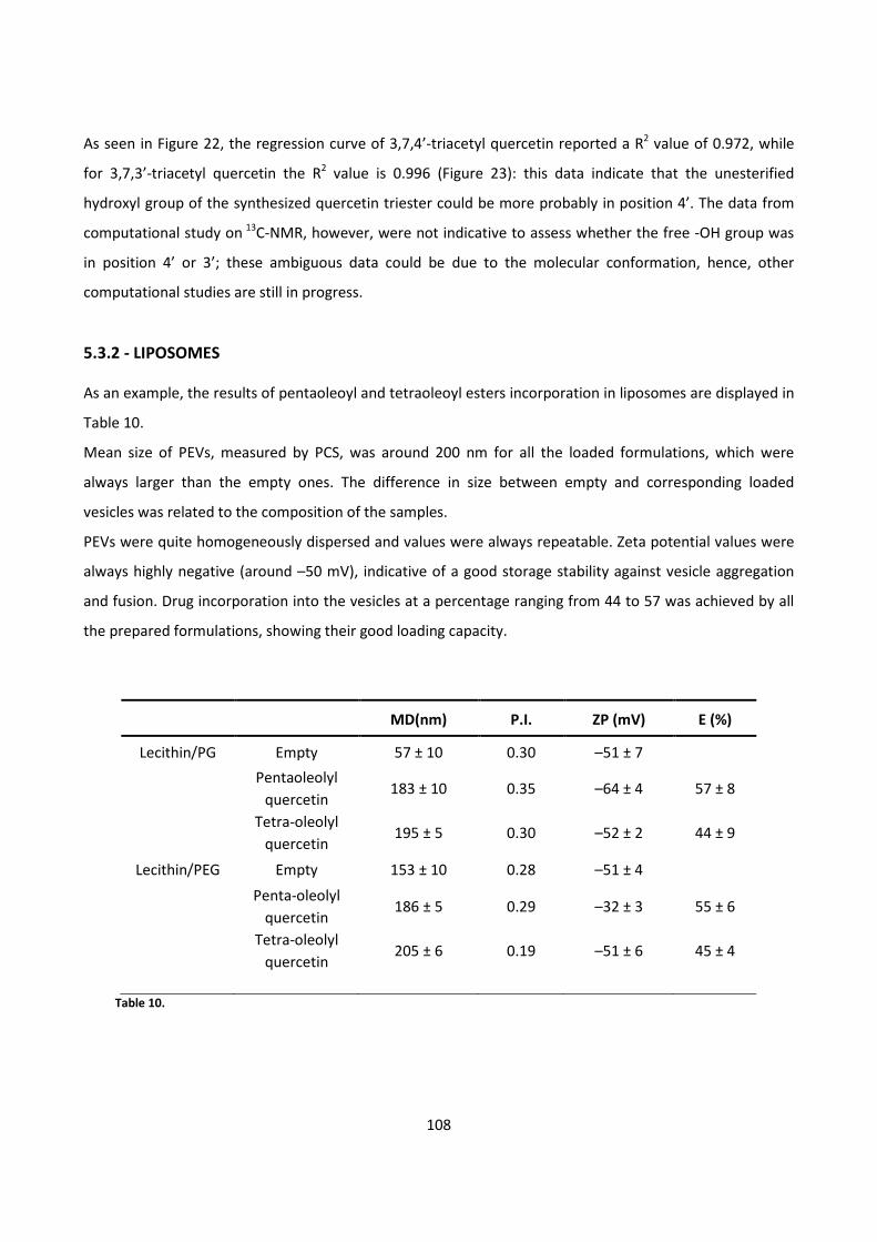
108
As seen in Figure 22, the regression curve of 3,7,4’-triacetyl quercetin reported a R2 value of 0.972, while
for 3,7,3’-triacetyl quercetin the R2 value is 0.996 (Figure 23): this data indicate that the unesterified
hydroxyl group of the synthesized quercetin triester could be more probably in position 4’. The data from
computational study on 13
C-NMR, however, were not indicative to assess whether the free -OH group was
in position 4’ or 3’; these ambiguous data could be due to the molecular conformation, hence, other
computational studies are still in progress.
5.3.2 - LIPOSOMES
As an example, the results of pentaoleoyl and tetraoleoyl esters incorporation in liposomes are displayed in
Table 10.
Mean size of PEVs, measured by PCS, was around 200 nm for all the loaded formulations, which were
always larger than the empty ones. The difference in size between empty and corresponding loaded
vesicles was related to the composition of the samples.
PEVs were quite homogeneously dispersed and values were always repeatable. Zeta potential values were
always highly negative (around –50 mV), indicative of a good storage stability against vesicle aggregation
and fusion. Drug incorporation into the vesicles at a percentage ranging from 44 to 57 was achieved by all
the prepared formulations, showing their good loading capacity.
MD(nm) P.I. ZP (mV) E (%)
Lecithin/PG Empty 57 ± 10 0.30 –51 ± 7
Pentaoleolyl
quercetin 183 ± 10 0.35 –64 ± 4 57 ± 8
Tetra-oleolyl
quercetin 195 ± 5 0.30 –52 ± 2 44 ± 9
Lecithin/PEG Empty 153 ± 10 0.28 –51 ± 4
Penta-oleolyl
quercetin 186 ± 5 0.29 –32 ± 3 55 ± 6
Tetra-oleolyl
quercetin 205 ± 6 0.19 –51 ± 6 45 ± 4
Table 10.
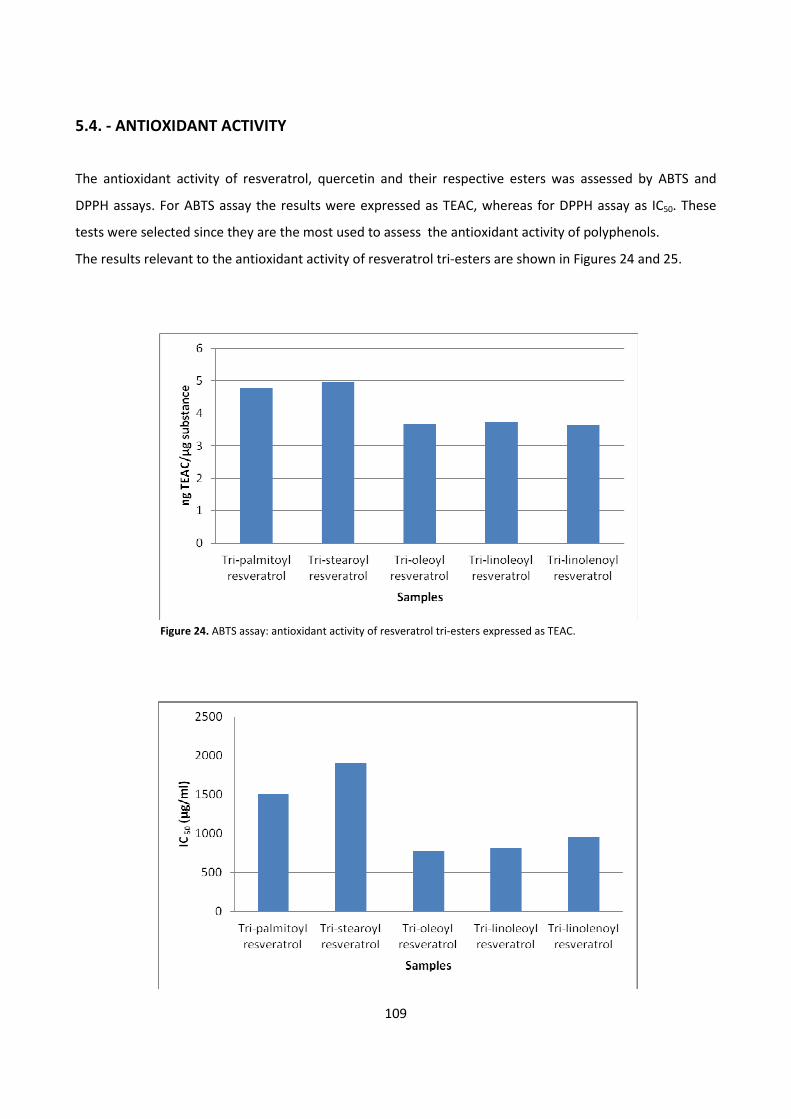
109
5.4. - ANTIOXIDANT ACTIVITY
The antioxidant activity of resveratrol, quercetin and their respective esters was assessed by ABTS and
DPPH assays. For ABTS assay the results were expressed as TEAC, whereas for DPPH assay as IC50. These
tests were selected since they are the most used to assess the antioxidant activity of polyphenols.
The results relevant to the antioxidant activity of resveratrol tri-esters are shown in Figures 24 and 25.
Figure 24. ABTS assay: antioxidant activity of resveratrol tri-esters expressed as TEAC.
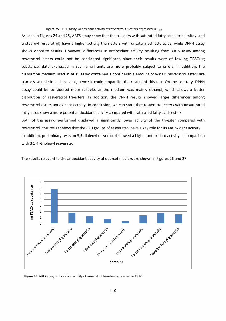
110
Figure 25. DPPH assay: antioxidant activity of resveratrol tri-esters expressed in IC50.
As seen in Figures 24 and 25, ABTS assay show that the triesters with saturated fatty acids (tripalmitoyl and
tristearoyl resveratrol) have a higher activity than esters with unsaturated fatty acids, while DPPH assay
shows opposite results. However, differences in antioxidant activity resulting from ABTS assay among
resveratrol esters could not be considered significant, since their results were of few ng TEAC/μg
substance: data expressed in such small units are more probably subject to errors. In addition, the
dissolution medium used in ABTS assay contained a considerable amount of water: resveratrol esters are
scarcely soluble in such solvent, hence it could jeopardize the results of this test. On the contrary, DPPH
assay could be considered more reliable, as the medium was mainly ethanol, which allows a better
dissolution of resveratrol tri-esters. In addition, the DPPH results showed larger differences among
resveratrol esters antioxidant activity. In conclusion, we can state that resveratrol esters with unsaturated
fatty acids show a more potent antioxidant activity compared with saturated fatty acids esters.
Both of the assays performed displayed a significantly lower activity of the tri-ester compared with
resveratrol: this result shows that the -OH groups of resveratrol have a key role for its antioxidant activity.
In addition, preliminary tests on 3,5-dioleoyl resveratrol showed a higher antioxidant activity in comparison
with 3,5,4’-trioleoyl resveratrol.
The results relevant to the antioxidant activity of quercetin esters are shown in Figures 26 and 27.
Figure 26. ABTS assay: antioxidant activity of resveratrol tri-esters expressed as TEAC.
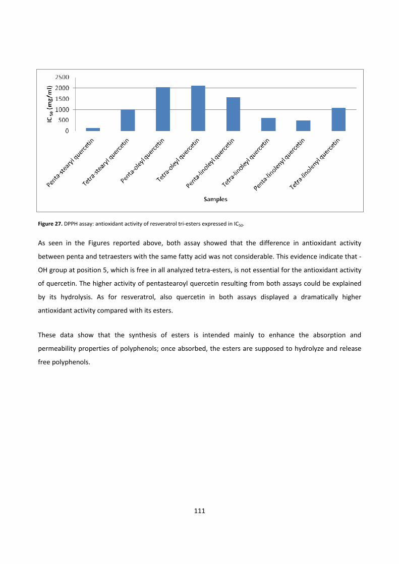
111
Figure 27. DPPH assay: antioxidant activity of resveratrol tri-esters expressed in IC50.
As seen in the Figures reported above, both assay showed that the difference in antioxidant activity
between penta and tetraesters with the same fatty acid was not considerable. This evidence indicate that -
OH group at position 5, which is free in all analyzed tetra-esters, is not essential for the antioxidant activity
of quercetin. The higher activity of pentastearoyl quercetin resulting from both assays could be explained
by its hydrolysis. As for resveratrol, also quercetin in both assays displayed a dramatically higher
antioxidant activity compared with its esters.
These data show that the synthesis of esters is intended mainly to enhance the absorption and
permeability properties of polyphenols; once absorbed, the esters are supposed to hydrolyze and release
free polyphenols.
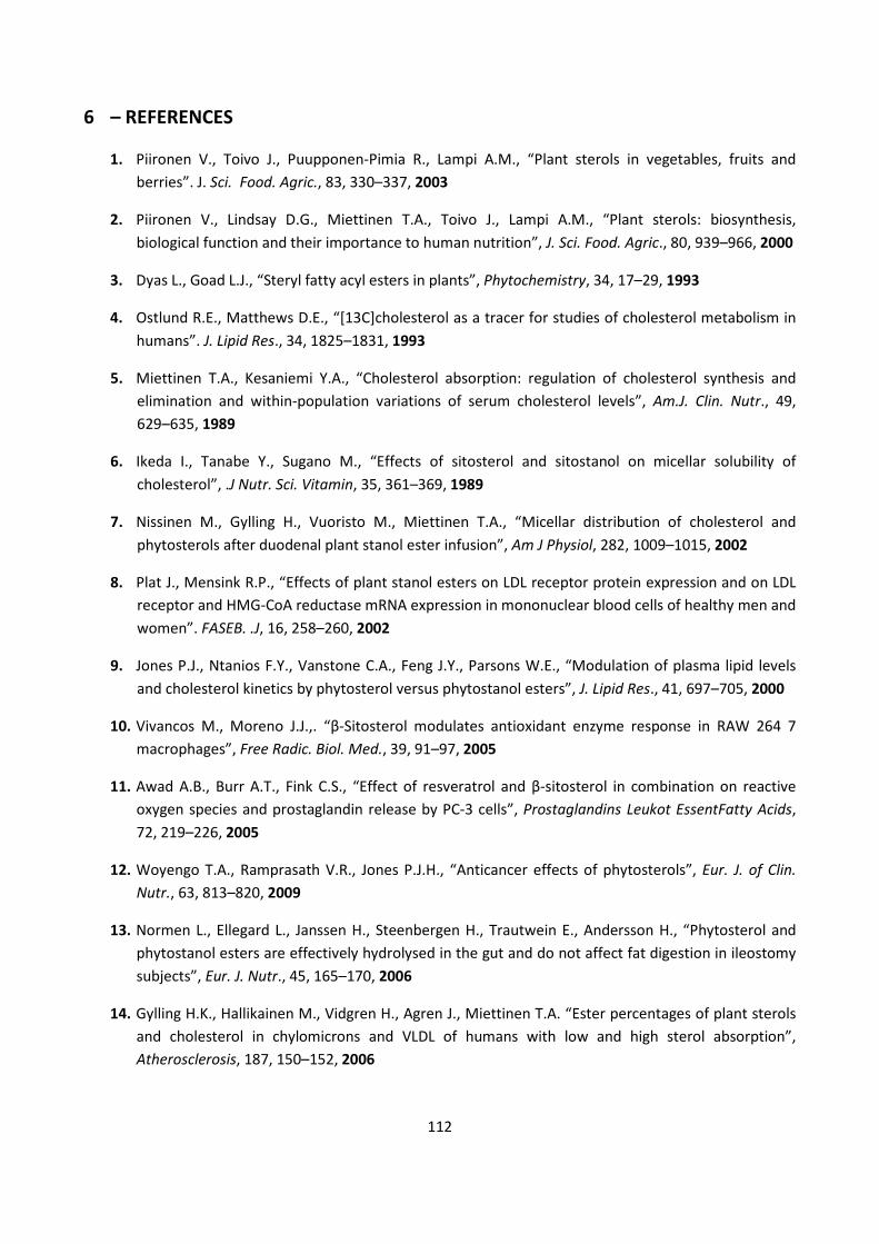
112
6 – REFERENCES
1. Piironen V., Toivo J., Puupponen-Pimia R., Lampi A.M., “Plant sterols in vegetables, fruits and
berries”. J. Sci. Food. Agric., 83, 330–337, 2003
2. Piironen V., Lindsay D.G., Miettinen T.A., Toivo J., Lampi A.M., “Plant sterols: biosynthesis,
biological function and their importance to human nutrition”, J. Sci. Food. Agric., 80, 939–966, 2000
3. Dyas L., Goad L.J., “Steryl fatty acyl esters in plants”, Phytochemistry, 34, 17–29, 1993
4. Ostlund R.E., Matthews D.E., “[13C]cholesterol as a tracer for studies of cholesterol metabolism in
humans”. J. Lipid Res., 34, 1825–1831, 1993
5. Miettinen T.A., Kesaniemi Y.A., “Cholesterol absorption: regulation of cholesterol synthesis and
elimination and within-population variations of serum cholesterol levels”, Am.J. Clin. Nutr., 49,
629–635, 1989
6. Ikeda I., Tanabe Y., Sugano M., “Effects of sitosterol and sitostanol on micellar solubility of
cholesterol”, .J Nutr. Sci. Vitamin, 35, 361–369, 1989
7. Nissinen M., Gylling H., Vuoristo M., Miettinen T.A., “Micellar distribution of cholesterol and
phytosterols after duodenal plant stanol ester infusion”, Am J Physiol, 282, 1009–1015, 2002
8. Plat J., Mensink R.P., “Effects of plant stanol esters on LDL receptor protein expression and on LDL
receptor and HMG-CoA reductase mRNA expression in mononuclear blood cells of healthy men and
women”. FASEB. .J, 16, 258–260, 2002
9. Jones P.J., Ntanios F.Y., Vanstone C.A., Feng J.Y., Parsons W.E., “Modulation of plasma lipid levels
and cholesterol kinetics by phytosterol versus phytostanol esters”, J. Lipid Res., 41, 697–705, 2000
10. Vivancos M., Moreno J.J.,. “β-Sitosterol modulates antioxidant enzyme response in RAW 264 7
macrophages”, Free Radic. Biol. Med., 39, 91–97, 2005
11. Awad A.B., Burr A.T., Fink C.S., “Effect of resveratrol and β-sitosterol in combination on reactive
oxygen species and prostaglandin release by PC-3 cells”, Prostaglandins Leukot EssentFatty Acids,
72, 219–226, 2005
12. Woyengo T.A., Ramprasath V.R., Jones P.J.H., “Anticancer effects of phytosterols”, Eur. J. of Clin.
Nutr., 63, 813–820, 2009
13. Normen L., Ellegard L., Janssen H., Steenbergen H., Trautwein E., Andersson H., “Phytosterol and
phytostanol esters are effectively hydrolysed in the gut and do not affect fat digestion in ileostomy
subjects”, Eur. J. Nutr., 45, 165–170, 2006
14. Gylling H.K., Hallikainen M., Vidgren H., Agren J., Miettinen T.A. “Ester percentages of plant sterols
and cholesterol in chylomicrons and VLDL of humans with low and high sterol absorption”,
Atherosclerosis, 187, 150–152, 2006
113
15. Hamada H., Egashira N., Nishizono S., Tomoyori H., Nakagiri H.,Imaizumi K., Ikeda I. “Lymphatic
absorption and deposition of various plant sterols in stroke-prone spontaneously hypertensive rats,
a strain saving a mutation in ATP binding cassette transporter G5”. Lipids, 42, 241–248, 2007
16. Awad A.B., Toczek J., Fink C.S., “Phytosterols decrease prostaglandin release in cultured
P388D1/MAB macrophages”, Prostaglandins Leukot Essent Fatty Acids, 70, 511–520, 2004
17. Sanders D.J., Minter H.J., Howes D., Hepburn P.A., “The safety evaluation of phytosterol esters Part
6 The comparative absorption and tissue distribution of phytosterols in the rat”, Food Chem Toxicol,
38, 485–491, 2000
18. He H., Chen X.,Wang G.,Wang J., Davey A.K.,”High-performance liquid chromatography
spectrometric analysis of trans-resveratrol in rat plasma”, J. Chromatography B, 832, 177-180, 2006
19. Jeandet P., Bessis R., Gautheron B., “ The production of resveratrol (3,5,4’-trihydroxystilbene) by
grape berries in different developmental stages”, Am. J.Enol. Vitic., 42, 41-46, 1991
20. Romero-perez A.I., Lamuela-Raventos R.M., Waterhouse A.L., Delatorreboranat M.C.,”Level of cis
and trans-resveratrol and their glucosides in white and rose Vitis Vinifera wines of Spain”, J. Agric.
Food Chem, 44, 2124-2128, 1996
21. Goldberg D.M., Yan J., Ng E., Diamandis E.P., “A global survey of trans-resveratrol concentrations in
commercial wines” Am. J. Enol. Vitic.46, 159-165, 1995
22. Derckes W., Creasy L.L., “ The significance or stilbene phytoalexins in Plasmapara viticola-grapevine
interactions” , Physiol. Mol. Plant Pathol., 34, 189-202, 1989
23. Brenta C.,Waterhouse T., Waterhouse L., “Resveratol: isomeric molar absorptivities and stability”,
J. Agric. Food Chem., 44, 1253-1257, 1996
24. Goldberg D.M., Tsang E., Karumanchiri A., Diamandis E.P., Soleas G.J., Ng E., “Method to assay the
concentrations of phenolic constituents of biological interest in wines ”, Analytical Chemistry, 68,
1688-1694, 1996
25. Jayatilake G.S., Jayasuriya H., Lee E.S., Koonchanik N.M., Geahlen R.L., Ashendel C.L., McLaughlin
J.L., Chang C.J., “Kinase inhibitors from Polygonum cuspidatum” , J. of Natural Products, 56, 1805-
1810, 1993
26. Aquaviva R., Russo A., Campisi A., Sorrenti V., “Antioxidant activity and protective effect on DNA
cleavage of resveratrol”, J. Food Technol., 67, 137-140, 2002
27. Li Z.D., Ma Q.Y., Wang C.A., “Effect of resveratrol on pancreatic oxygen free radicals in rats with
severe acute pancreatitis”, 12, 137-140, 2006
28. Bhat P.L.K., Pezzuto J.M., “Cancer chemopreventive activity of resveratrol”, Ann. N.Y. Acad. Sci.,
957, 210-229, 2002
29. Petrovski G., Gurusamy N., Das D.K., “Resveratrol in cardiovascular health and disease”, Ann. N.Y.
Acad. Sci., 125, 22-33, 2011
114
30. Berliner J.A., Navab M., Fogelman A.M., “Atherosclerosis: basic mechenisms oxidation inflammation
and genetics circulation”, 9, 2488-2496, 1995
31. Do G.M., Kwon E.Y., Kim H.J., et al., “Long-term effects of resveratrol supplemention on
suppression of atherogenic lesion formation and cholesterol synthesis in apo E-deficient mice”,
Biochem Biophys Res Commun., 374(1), 55-59, 2008
32. Robich M., Osipov R., Nezafat R. et al., “Resvaratrol improves myocardial perfusion in swine model
of hypercholesterolemia and chronic myocardial ischemia”, Circulation, 122(11 suppl), 142-149,
2010
33. Luis A.L., “Atherosclerosis”, Nature, 407, 233-241, 2000
34. Sviridov D., Nestel P., “Dynamics of reverse cholesterol transport: protection against
atherosclerosis”, Atherosclerosis, 161(2), 245-254, 2002
35. Deng Y.H., Alex D., Huang H.Q., et al., “Inhibition of TNF-α-mediated endothelial cell-monocyte cell
adhesion and adhesion molecules expression by the resveratrol derivative,trans-3,5,4’-
trimethoxystilbene”, Phytother Res., 25, 451-457, 2011
36. Park D.W., Baek K., Kim J.R., et al., “Resveratrol inhibits foam cell formation via NADPH oxidase 1
mediated reactive oxygen species and monocytechemotactic protein-1”, Exp. Mol. Med., 41, 171-
179, 2009
37. Ekshyyan V.P., Herbert V.Y., Khandelwal A., et al., “Resveratrol inhibits rat aortic vascular smooth
muscle cell proliferation vis estrogenic receptor dependent nitric oxide production”, J. Cardiovasc.
Pharmacol, 50, 83-93, 2007
38. Olas B., Wachowicz B., Saluk-Juszczak J, et al., “Effect of resveratrol, a natural polyphenolic
compound, on platelet activation induced by endotoxin or thrombin”, Thromb. Res., 107, 141-145,
2002
39. Yang Y., Wang X., Zhang L., et al., “Inhibitory effects of resveratrol on platelet activation induced by
thromboxane a(2) receptor agonist in human platelets”, Am. J. Chin. Med., 39, 145-159, 2011
40. Shankar S., Singh g., Srivastava R.K., “Chemoprevention by resveratrol molecular mechanism and
therapeutic potential”, Frontiers in Bioscience, 12, 4839-4854, 2007
41. Bishayee A., “Cancer prevention and treatment with resveratrol: from rodent studies to clinical
trial”, Cancer Prevention Research, 2(5), 409-418, 2009
42. Pirola L., Frojdo S., “Resveratrol: one molecule many targets”, IUBMB Life, 60(5), 323-332, 2008
43. Delmas D., Rèbè C., Micheau o., et al., “Redidtribution of CD95, DR4 and DR% in rafts accounts for
the synergistic toxicity of resveratrol and death receptor ligans in colon carcinoma cells”,
Oncogenes, 23, 8979-8986, 2004
115
44. Reagan-Shaw S., Afaq F., Aziz M.H., Ahmad N., “ Modulations of critical cell cycle regulatory events
during chemoprevention of ultraviolet B-mediated responsas by resveratrol in SKH-1 hairless
mouse skin”, Oncogene, 23, 5151-5160, 2004
45. Le corre L., Chaabi N., Delort L., Bignon Y.J., Bernard-Gallon D.J., “Resveratrol and breast cancer
chemoprevention molecular meecchanisms”, Mol. Nutr. Food Res., 49, 462-471, 2005
46. Aziz M.H., Nihal M., Fu V. X., Jarrard D.F., Ahamad N., “Resveratrol caused apoptosis of human
prostate carcinoma LNCaP cells is mediated via modulation of phosphatidylinositol 3’-kinase/Akt
pathway and Bcl-2 family proteins”, Mol. Cancer Ther., 5, 1335-1341, 2006
47. Narayanan B. A., Narayanan N.K., Re G.G., Nixon D.W., “Differential expression of genes induced by
resveratrol in LNCaP cells P53-mediated molecular targets”, Int. J. Cancer, 104, 204-212, 2003
48. Mahady G.B., Pendland S.L., “Resveratrol inhibits the growth of Helicobacter pylori in vitro”, Am. J.
Gastroenterol, 95, 1849, 2000
49. Atten M. J., Godoy-Romero E., Attar B.M., Milson T., Zopel M., Holian O., “Resveratrol regulates
cellular PKC alpha and delta to inhibit growth and induce apoptosis in gastric cancer cells”, Invest
New drugs, 23, 111-119, 2005
50. Gautam S.C., Xu Y.X., Dumaguin M., Janakiraman N., Chapman R.A., “Resveratrol selectively inhibits
leukemia cells a prospective agent for ex vivo bone marrow purging “, Bone Marrow Transplant, 25,
639-645, 2000
51. Das S., Das D.K., “Anti-inflammatory responses of resveratrol”, Inflammation and Allergy-Drug
Target, 6, 168-173, 2007
52. Leiro J., Alvarez E, Arranz J.A., Laguna R., Uriarte E., Orallo F., “Effect of cis-resveratrol on
inflammatory murine macrophage antioxidant activity and down regulation of inflammatory
genes”, J. of Leukocyte Biology, 75, 1156-1165, 2004
53. Zhu J., Yong W., Wu X, et al , “ Anti-inflammatory effect of resveratrol on TNF-α-induced MCP-1
expression in adipocites”, Biochemical and Biophysical Research Communication, 369, 471-477,
2008
54. Chung S., Yao H., Caito S., Hwang J.W., Arunachalam G., Rahman I., “ Regulation of SIRT1 in cellular
functions: role of polyphenols”, Archives of Biochemistry and Biophysics, 501, 79-90, 2010
55. Michishita E, et al, “Evolutionarily conserved and nonconserved cellular localizations and fuctions of
human SIRT proteins”, Mol. Biol. Cell, 16, 4623-4635, 2005
56. Haigis M.C., Guarente L.P., “Mammalian surtuins-emerging roles in physiology, aging and calorie
restriction”, Genes. Dev., 20, 2913-2921, 2006
57. Baur J.A., Pearson K.J., Price N.L., Jamieson H.A., Lerin C., Kaira A., Prabhu V.V., Allard J.S., Lopez-
Lluch G., Lewis K., “Resveratrol improved health and survival of mice on a high-calorie diet”,
Nature, 444, 337-342, 2006
116
58. Howitz K.T., Bitterman K.J., Cohen H.Y., Lamming D.W., Lavu S., Wood J.G., Zipkin R.E., Chung P.,
Kisielewski A., Zhang L.L., “Small molecule activators of sirtuins extend Saccharomyces cerevisiae
lifespan”, Nature, 425, 191-169, 2003
59. Dai H., Kustigian L., Carney D., Case A., Considine T., Hubbard B.P., Perni R.B., Riera T.V.,
Szczepankiewicz B., Vlasuk G.P., Stein R.L., “SIRT-1 activation by small molecules: kinetic and
biophysical evidence for direct interaction of enzyme and activator”, J. Biol. Chem., 285, 32695-
32703, 2010
60. Timmers S., Konings E., Bilet L., Houtkooper R.H., Van de Weijer T., Goossens G.H., Hoeks J., Van de
Krieken S., Ryu D., Kersten S., Moonen-Kornips E., Hesselink M.K.C., Kunz I., Schrauwen-Hinderling
V.B., Blaak E.E., Auwerx J., “Calorie restriction-like effects of 30 days of resveratrol supplementation
on energy metabolism and metabolic profile in obese humans”, Cell Metabolism, 14, 612-622, 2011
61. Szkudelska K., Szkudelski T., “Resveratrol, obesity and diabetes”, Eur. J. Pharmacol., 635, 1-8, 2010
62. Su H.C., Hung L.M., Cheng J.K., “Resveratrol, a red wine antioxidant, possesses an insulin-like effect
in streptozotocin-induced diabetic rats”, Am. J. Physiol. Endocrinol Metab., 290, 139-1346, 2006
63. Penumathsa S.V., Thirunavukkarasu M., Zhan L., “ Resveratrol enhances GLUT4 traslocation to the
caveolae lipid raft fraction through AMPK/Akt/eNOS signaling pathway in diabetic myocardium”,
J.Cell.Mol. Med., 12, 2350-2361, 2008
64. Hansen J.B., Arkhammar P.O., Bodvardottir T.B., Wahl P., “Inhibition of insulinsecretion as a new
drug target in the treatment of metabolic disorders”, Curr.Med.Chem., 11, 1595-1615, 2004
65. Szkudelski T, “Resveratrol inhibits insulin secretion from rat pancreatic islets”, Eur.J.Pharmacol, 55,
176-181, 2006
66. Lee J.H., Song M.Y., Song E.K., “Overexpression of by suppressing the nuclear factor-kappaB
signaling pathway”, Diabetes, 55, 344-351, 2009
67. Um J.H., Park S.J., Kang H., “AMP-activated protein kinase-deficient mice are resistant to the
metabolic effect or resveratrol”, Diabetes, 59, 554-563, 2010
68. Barger J.L., Kayo T, Vann J.M., “Alow dose of dietary resveratrol partially mimics caloric restriction
and retards again parameters in mice”, PloS ONE, 3, 2264, 2008
69. Szkudelska K., Nogowski L., Szkudelski T, “ Resveratrol, a naturally occurring diphenolic compound
affects lipogenesis, lipolysis and antilipolytic action of insulin in isolated rat adipocytes”, J.Steroid
Biochem. Mol.Biol., 113, 17-24, 2009
70. Goihman-Yahr M., “Skin aging and photoaging : an outlook”, Clin.Dermatol, 14, 153-160, 1996
71. Black H.S., DeGruijl F.R., Forbes P.D., Cleaver J.E., Anathaswamy H,N. deFabo E.C., Ullrich S.E.,
Tyrrell R.M., “Photocarcinogenesis: an overview”, J.Photochem Photobiol. B, 40, 29-47, 1997
117
72. Afaq F., Adhami V.M., Ahmad N., “Prevention of short-term ultraviolet B radiation.mediated
damages by resveratrol in SKH-1 hairless mice”, Toxicology and Applied Pharmacology, 186, 28-37,
2003
73. Campagna M., Rivas C., “ Antiviral activity of resveratrol”, Biochemical Society Transactions, 38, 50-
53, 2010
74. Wenzel E., Somoza V., “metabolism and bioavailability of trans-resveratrol”, Mol. Nutr. Food Res.,
49, 472-481, 2005
75. Walle T., Hsieh F., DeLegge M.H., Oatis J.E., Walle U.K., “High absorption but very low bioavailability
of oral resveratrol in human”, Drug Metab. Dispos., 32, 1377-1382, 2004
76. Ross J.A., Kasum C.M., “Dietary flavonoids bioavailability, metabolic effects, and safety”, Annu. Rev.
Nutr., 22, 19-34, 2002
77. Wiczkowski W., Romaszko J., Bucinski A., “Quercetin from shallots (Allium cepa L. var. aggregatum)
is more bioavailable than its glucosides”, J. Nutr., 138, 885-888, 2008
78. Nemeth K., Piskula M.K., “Food content, processing, absorption and metabolism of onion
flavonoids”, Crit Rev Food Sci. Nutr., 47, 397-409, 2007
79. Cushnie T.P, Lamb A.J., “Antimicrobial activity of flavonoids”, Int. J. Antimicrob. Agents, 26, 343-
356, 2005
80. Heijnen C.G., Heanen G.R.M.M., Oostveen R.M., Stalpers E.M.,Bast A., “Protection of flavonoids
against lipid peroxidation: the structure activity relationship revisited”, Free Radic. Res., 36, 575-
581, 2002
81. Mariani C. et al, “Flavonoid characterization and in vitro antioxidant activity of Aconitum anthora L.
(Ranunculaceae)”, Phytochemistry, 69, 1220-1226, 2008
82. Begum A.N.,Terao J., “Protective effect of quercetin against cigarette tar extract-induced
impairment of erythrocyte deformability”, Journal of Nutritional Biochemistry, 13, 265-272, 2002
83. Williams R.J., Spencer J.P., Rice-Evans C., “ Flavonoids: antioxidants or signalling molecules? Free
Radic”, Biol. Med., 36, 838–849, 2004
84. Choi E.J., Chee K.M., Lee B.H., “Anti- and prooxidant effects of chronic quercetin administration in
rats”, Eur. J. Pharmacol., 482, 281–285, 2003
85. Boots A.W., Kubben N., Haenen G.R., Bast A., “Oxidized quercetin reacts with thiols rather than
with ascorbate: implication for quercetin supplementation”, Biochem. Biophys. Res. Commun., 308,
560–565, 2003
86. Metodiewa D., Jaiswal A.K., Cenas N., Dickancaite E., Segura-Aguilar J., “Quercetin may act as
acytotoxic prooxidant after its metabolic activation to semiquinone and quinoidal product”, Free
Radic. Biol. Med, 26, 107–116, 1999
118
87. Cai H., “Hydrogen peroxide regulation of endothelial function: Origins, mechanisms, and
Consequences”, Cardiovasc. Res., 68, 26–36, 2005
88. Geraets L., Moonen H.J.J., Brauers K., Wouter E.F.M., Bast A., Hageman G.J., “Dietary flavones and
flavonoles are inhibiting of poly(ADP-ribose)polymerase-1in pulmonary epithelial cells”, J. Nutr.,
137, 2190-2195, 2007
89. Rahman I., “Oxidative stress, transcription factors and chromation remodeling in lung
inflammation”, Biochem. Pharmacol., 64, 935-942, 2002
90. Lee K.M., Hwang M.K., Lee D.E., et al., “Protective effect of quercetin against arsenite-induced COX-
2 expression by targeting PI3K in rat liver ephitelial cells”. J Agric Food Chem, 58, 5815-5820, 2010;
91. Ortega MG, Saragusti AC, Cabrera JL,Chiabrando G.A., “Quercetin tetracetyl derivates inhibitis LPS-
induced nitric oxide synthase (iNOS) expression in J774A 1 cells”, Arch. Biochem. Biophys, 498, 105-
110, 2010
92. Geleijnse J.M., Launer L.J., Van der Kuip D.A.M., Hofman A., Witteman J.C.M., “Inverse association
of tea and flavonoid intakes with incident myocardial infarction: the Rotterdam Study”, Am. J. Clin.
Nutr., 75, 880–886, 2002
93. De Whalley C.V., Rankin S.M., Hoult J.R., Jessup W., Leake D.S., “Flavonoids inhibit the oxidative
modification of low density lipoproteins by macrophages”, Biochem. Pharmacol., 39, 1743–1750,
1990
94. Pignatelli P., Pulcinelli F.M., Celestini A., Lenti L., Ghiselli A., Gazzaniga P.P., Violi F., “The flavonoids
quercetin and catechin synergistically inhibit platelet function by antagonizing the intracellular
production of hydrogen peroxide”, Am. J. Clin. Nutr., 72, 1150–1155, 2000
95. Oak M.H., El Bedoui J., Schini-Kerth V.B.,. “Antiangiogenic properties of natural polyphenols from
red wine and green tea”, J. Nutr. Biochem., 16, 1–8, 2005
96. Cao G., Sofic E., Prior R.L., “Antioxidant and prooxidant behavior of flavonoids: structure-activity
relationships”, Free Radic. Biol. Med.,22, 749–760, 1997
97. Zhang C., Patel R., Eiserich J.P., Zhou F., Kelpke S., Ma W., Parks D.A., Darley-Usmar V., White C.R.,
“Endothelial dysfunction is induced by proinflammatory oxidant hypochlorous acid”, Am. J. Physiol.
Heart Circ. Physiol., 281, H1469–H1475, 2001
98. Hubbard G.P., Wolffram S., Lovegrove J.A, Gibbins J.M., “Ingestion of quercetin inhibits platelet
aggregation and essential components of the collagen stimulated platelet activation pathway in
humans”, J. Thromb. Haemost., 2, 2138-2145, 2004
99. Zhang Q., Zhao X.H., Wang Z.J., “Cytotoxicity of flavones and flavonols to a human esophageal
squamous cell carcinoma cell line (KYSE-510) by induction of G2/M arrest and apoptosis”,
100. Jeong J.H., An J.Y., Kwon Y.T., Rhee J.G.,.Lee Y.J., “Effects of low dose quercetin: cancer cell-
specific inhibition of cell cycle progression”, Journal of Cellular Biochemistry,.106, 73–82, 2009.
119
101. Jeong J.H., An J.Y., Kwon Y.T., Rhee J.G., Lee Y.J., “Effects of low dose quercetin: cancer cell-
specific inhibition of cell cycle progression,” Journal of Cellular Biochemistry, 106, 73–82, 2009
102. Yang J.H., T.-C. Hsia T.C., Kuo H.M., et al., “Inhibition of lung cancer cell growth by quercetin
glucuronides via G 2/M arrest and induction of apoptosis,” Drug Metabolism and Disposition, 34,
296–304, 2006
103. Nair H.K., Rao K.V., Aalinkeel R., Mahajan S., Chawda R., Schwartz S.A., “Inhibition of
prostate cancer cell colony formation by the flavonoid quercetin correlates with modulation of
specific regulatory genes,” Clinical and Diagnostic Laboratory Immunology, 11, 63–69, 2004
104. Chien Y.S, Wu Y.C., J.-G. Chung J.G., et al., “Quercetin induced apoptosis acts through
mitochondrial- and caspase-3-dependent pathways in human breast cancer MDA-MB-231 cells”,
Human and Experimental Toxicology, 28, 493–503, 2009
105. Jung Y.H., Heo J., Lee Y.J., Kwon T.K., Kim Y.H., “Quercetin enhances TRAIL-induced
apoptosis in prostate cancer cells via increased protein stability of death receptor 5”, Life Sciences,
86, 351–357, 2010
106. Gupta K., Panda D., “Perturbation of microtubule polymerization by quercetin through
tubulin binding: a novel mechanism of its antiproliferative activity,” Biochemistry, 41, 13029–
13038, 2002
107. Tanigawa S, Fujii M, Hou D.X., “Stabilization of p53 is involved in quercetin-induced cell
cycle arrest and apoptosis in HepG2 cells”, Bioscience Biotechnology and Biochemistry, 72, 797–
804, 2008
108. Bronner C., Landry Y., “Kinetics of the inhibitory effect of flavonoids on histamine secretion
from mast cells”, Agents Actions, 16, 147-151, 1985
109. Moon H, Choi HH, Lee JY, et al., “Quercetin inhalation inhibits the asthmatic responses by
exposure to aerosolized-ovalbumin in conscious guinea-pigs”, Arch Pharm Res, 31, 771-778, 2008
110. Rogerio A.P., Dora C.L., Andrade E.L., et al., “Anti-inflammatory effect of quercetin loaded
microemulsion in the airways allergic inflammatory model in mice”, Pharmacol. Res., 61, 288-297,
2010
111. Park H.J., Lee C.M., Jung I.D., et al., “Quercetin regulates Th1/Th2 balance in a murine
model of asthma”, Int. Immunopharmacol.”, 9, 261-267, 2009
112. Fanning M.J., Macander P., Drzewiecki G., Middleton E.J., “Quercetin inhibits anaphylactic
contraction of guinea pig ileum smooth muscle”, Int. Arch. Allergy Appl. Immunol.,71, 371-373,
1983
113. Knekt P., Kumpulainen J., Järvinen R., et al., “Flavonoid intake and risk of chronic Diseases”,
Am J Clin. Nutr., 76, 560-568, 2002
120
114. Hirano T., Kawai M., Arimitsu J., et al. “Preventative effect of a flavonoid, enzymatically
modified isoquercitrin on ocular symptoms of Japanese cedar pollinosis”, Allergol Int, 58, 373-382,
2009
115. Kalogeromitros D., Makris M., Chliva C., et al. “A quercetin containing supplement reduces
niacin-induced flush in humans”, Int. J. Immunopathol. Pharmacol., 21, 509-514, 2008
116. Kaul T.N., Middleton E. Jr., Ogra P.L., “Antiviral effect of flavonoids on human viruses”, J.
Med. Virol., 15, 71-79, 1985
117. Gonzalez O., Fontanes V., Raychaudhuri S., et al., “The heat shock protein inhibitor
Quercetin attenuates hepatitis C virus production”, Hepatology, 50, 1756-1764, 2009
118. Geoghegan F., Wong R.W., Rabie A.B., “Inhibitory effect of quercetin on periodontal
pathogens in vitro”, Phytothe.r Res., 24, 817-820, 2010
119. Shin J.E., Kim J.M., Bae E.A., et al., “In vitro inhibitory effect of flavonoids on growth,
infection and vacuolation of Helicobacter pylori”, Planta Med., 71, 197-201, 2005
120. Muthian G., Bright J.J., “Quercetin, a flavonoid phytoestrogen, ameliorates experimental
allergic encephalomyelitis by blocking IL-12 signaling through JAK-STAT pathway in T lymphocyte”,
J. Clin. Immunol., 24, 542-552, 2005
121. Bonina F., Lanza M., Montenegro L., Puglisi C., Tomaino A., Trombetta D., Castelli F., Saija
A., “Flavonoids as potential protective agents photo-oxidative skin damage”, Int. J. Pharm., 145, 87-
94, 1996
122. Fuchs J.V., Zollner T.M., Kaufmann R., Podda M., “Redox-modulated pathways in
inflammatory skin diseases”, Free Radic Biol. Med, 30, 337-353, 2001
123. Xia Y., Zweier L., “Measurement of myeloperoxidase in leukocyte containing tissues”, Anal.
Biochem., 245, 93-96, 1997
124. Pincemail J., Deby C., Thirion A., Bruyn-Dister M., Goutier R., “Human myeloperoxidase
activity is inhibited in vitro by quercetin comparison with three related compounds”, Experientia,
44, 450-453, 1988
125. Podda M., Traber M.G., Weber C., Yan L.J., Packer L., “UV-irradiation depletes antioxidants
and causes oxidative damage in model of human skin”, Free Radic, Biol. Med., 24, 55-65, 1998
126. Meloni M., Nicolay J.F., “Dynamic monitoring of glutathione redox status in Uv-B
irradiatedreconstituted epidermis effect of antioxidant activity on skin homeostasis”, Toxicol. in
vitro, 17, 609-613, 2003
127. Casagrande R., Georgetti S.R., Verri Jr. W.A., Dorta J.D., dos Santos A.C., Fonseca M.J.V.,
“Protection effect of topical formulation containg quercetin against UVB-induced oxidative stress in
hirless mice”, J. Photochem. Photobiol. B:Biology, 84, 21-27, 2006
121
128. Rieger M., “Participation of metalloproteins in photoaging”, Cosmet. Toiletries, 114, 65-70,
1999
129. Tamura G., Gold C., Ferro-Luzzi A., Ames B.N., “Fecalase: a model for activation of dietary
glycosides to mutagens by intestinal flora”, Proc. Natl. Acad. Sci. U.S.A., 77, 4961-4965, 1980
130. Hollman P.C., de VriesJ.H., van Leeuwen S.D., Mengelers M.J., Katan M.B., “Absorption on
dietary quercetin glycosides and quercetin in ileostomy volunteers”, Am. J. Clin. Nutr., 62, 1276-
1282, 1995
131. Erlund I., Kosonen T., Alfthan G., Maenpaa J., Perttunen K., Kenraali J., Parantainen J., Aro
A., “Pharmacokinetics of quercetin from aglycone and rutin in healthy volunteers ”, Eur. .J. Clin.
Pharmacol., 56, 545-553, 2000
132. Hollman P.C., Katan M.B., “Absorption, metabolism and health effects of dietary flavonoids
in man ”, Biomed. Pharmacother.., 51, 305-310, 1997
133. Day A.J., Canada F.J., Diaz J.C., Kroon P.A., McLauchlan R., Faulds C.B., Plumb G.W., Morgan
M.R., Williamson G., “Dietary flavonoids and isoflavone glycosides are hydrolyses by lactase site of
lactase phlorizin hydrolase”, FEBS lett., 468, 166-170, 2000
134. Conquer J.A., Maiani G., Azzini E., Raguzzini A., Holub B.J., “Supplementation with quercetin
markedly increases plasma quercetin concentration without effect on selected risk factors for heart
disease in healthy subjects”, J. Nutr.,128, 593-597, 1998
135. de Boer V.C., Dihal A.A., van der Woude H., Arts I.C., Wolffram S., Alink G.M., Rietjens I.M.,
Keijer J, Hollman P.C., “Tissue distribution of quercetin in rats and pigs”, J. Nutr., 135, 1718-1725,
2005
136. Manach C., Williamson G., Morand C., Scalbert A., Remesy C., “Bioavailability and
bioefficacy of polyphenols in human”, Am. J. Clin. Nutr., 81, 230-242, 2005
137. Simopoulos A.P., Leaf A., Salem N.Jr., “Workshop statement on the essentiality of and
recommended dietary intakes for Omega-6 and Omega-3 fatty acids”, Prostaglandins Leukot Essent
Fatty Acids, 63(3), 119-121, 2000
138. Stillwell W., Wassall S.R., “Docosahexaenoic acid: membrane properties of a unique fatty
acid”, Chem Phys Lipids, 126, 1-27, 2003
139. Jeffrey B.G., Weisingerb H.S., Neuringer M., Mitcheli D.C., “The role of docosahexaenoic
acid in retinal function”, Lipids, 36, 859-871, 2001
140. Innis S.M., “Perinatal biochemistry and physiology of long-chain polyunsaturated fatty
acids”, J. Pediatr., 143, 1-8. 2003
141. Chalon S., Vancassel S., Zimmer L., Guilloteau D., Durand G., “Polyunsaturated fatty acids
and cerebral function: focus on monoaminergic neurotransmission”, Lipids, 36, 937-944, 2001
122
142. Calder P.C., “Dietary modification of inflammation with lipids”, Proc.Nutr. Soc.;61, 345-358,
2002
143. Hawkes J.S., James M.J., Cleland L.G.. “Biological activity of prostaglandin E3 with regard to
oedema formation in mice”, Agents Actions, 35, 85–87, 1992
144. Hawkes J.S., James M.J., Cleland L.G., “Separation and quantification of PGE3 following
derivatization with panacyl bromide by high pressure liquid chromatography with fluorometric
detection”, Prostaglandins, 42:355–68, 1991
145. James M.J., Cleland L.G., Gibson R.A., Hawkes J.S., “Interaction between fish and vegetable
oils in relation to rat leucocyte leukotriene production”, Nutr, 121:631–637, 1991
146. Lerner R., Lindstrom P., Berg A., Johansson E., Rosendahl K., Palmblad J., “Development and
characterization of essential fatty acid deficiency in human endothelial cells”, Proc Natl Acad Sci U S
A, 92, 1147–1151, 1995
147. Agostoni C., Bruzzese M.G., “Fatty acids: classification, biochemistry and function”, Ped.
Med. Chir., 14, 473-479, 1992
148. Agostoni C., Riva E., Biasucci G., “Fatty acids in prevention and therapy in pediatric”, Ped.
Med. Chir., 14, 489-494, 1992
149. Allert C.M., Hennekens C.H., O’Donnel C.J., “Fish consumption and risk of sudden cardiac
death”, JAMA; 279, 23-28., 1998
150. Trommer H., Wagner J., Graener H., Neubert R.H., “The examination of skin lipid model
systems stressed by ultraviolet irradiation in the presence of transition metal ions”, Eur. J. Pharm.
Biopharm., 51, 207-14, 2001
151. Hansen H.S., Jensen B., “Essential function of linoleic acid esterified in acylglucosylceramide
and acylceramide in maintaining the epidermal water permeability barrier. Evidence from feeding
studies with oleate, linoleate, arachidonate, columbinate and alpha-linolenate”, Biochim. Biophys.
Acta., 834: 357-63, 1985
152. Jump D.B, “Fatty acid regulation of gene transcription”, Crit. Rev. Clin. Lab. Sci., 41, 41-78,
2004
153. Kim E.J., Kim M.K., Jin X.J., Oh J.H., Kim J.E., Ch J.H., “Skin Aging and Photoaging alter Fatty
Acids Composition”, J. Korean Med. Sci., 25,980-983, 2010
154. Nicolosi G., Spatafora C., Trigali C., “Chemo-enzymatic preparation of resveratrol
derivatives”,J. Molecular Catalysis B: Enzymatic, 16, 223-229, 2002