Acetic Acid-Induced Ulcerative Colitis in Sprague Dawley ...
11
Research Article Acetic Acid-Induced Ulcerative Colitis in Sprague Dawley Rats Is Suppressed by Hydroethanolic Extract of Cordia vignei Leaves through Reduced Serum Levels of TNF-α and IL-6 George Owusu, 1,2 David D. Obiri , 1 George K. Ainooson, 1 Newman Osafo , 1 Aaron O. Antwi , 1 Babatunde M. Duduyemi , 3 and Charles Ansah 1 1 Department of Pharmacology, Faculty of Pharmacy and Pharmaceutical Sciences, College of Health Sciences, Kwame Nkrumah University of Science and Technology (KNUST), Kumasi, Ghana 2 Department of Pharmacology, School of Medicine and Health Sciences, University for Development Studies, Tamale, Ghana 3 Department of Pathology, School of Medical Sciences, Kwame Nkrumah University of Science and Technology (KNUST), Kumasi, Ghana Correspondence should be addressed to David D. Obiri; [email protected] Received 28 September 2019; Accepted 18 December 2019; Published 6 February 2020 Academic Editor: Tadeusz Robak Copyright © 2020 George Owusu et al. This is an open access article distributed under the Creative Commons Attribution License, which permits unrestricted use, distribution, and reproduction in any medium, provided the original work is properly cited. Background. Ulcerative colitis (UC) is a recurrent inflammatory bowel disease (IBD) that causes long-lasting inflammation on the innermost lining of the colon and rectum. Leaf decoctions of Cordia vignei have been used in traditional medicine either alone or in combination with other plant preparations to treat the disease. Aim. In this study, we investigated the effect of hydroethanolic extract of Cordia vignei leaves (CVE) on acetic acid-induced UC in rats. Method. Male Sprague Dawley rats received oral treatment of either saline (10 ml/kg), sulfasalazine (500 mg/kg), or CVE (30-300 mg/kg) daily for 7 days. On day 4, colitis was induced by a single intrarectal administration of 500 μl of acetic acid (4% v/v). Rats were sacrificed on day 8 and colons were collected for histopathological examination. Blood was also collected for haematological assessment. Results. CVE significantly (P < 0.05) prevented colonic ulceration and reduced the inflammatory score. Serum levels of TNF-α and IL-6 were significantly reduced. Depletion of superoxide dismutase (SOD) and catalase (CAT) activities by acetic acid was significantly inhibited while lipid peroxidation indexed as malondialdehyde (MDA) level in the colon was reduced. However, loss of body weight was not significantly affected by treatment with CVE. Conclusion. This data suggest that CVE has a potential antiulcerative effect. 1. Introduction Ulcerative colitis is a recurrent inflammation of the colon. The clinical symptoms often presented by this disease are diarrhoea, loss of weight, nausea, and abdominal pain which affect quality of life [1]. The pathological features include inflammatory cell infiltration and activation, downstream expression of nuclear factor kappa B- (NF-κB-) dependent proinflammatory mediators such as tumor necrosis factor alpha (TNF-α) and interleukin 6 (IL-6), excessive generation of free radicals such as reactive oxygen species (ROS) and reactive nitrogen species (RNS), depletion of antioxidant capacity of the colon, and loss of mucosal integrity [2]. Although genetic, immunological, and environmental factors are implicated in the pathogenesis of ulcerative colitis, the exact aetiology of the disease still remains elusive [3]. Studies in this area contribute to the attempt to elucidate the causes and development of the disease. Also, the increasing number of cases of ulcerative colitis shows the importance of search- ing for new compounds that can control or eliminate the dis- ease that severely affects the quality of life of these patients. Due to resemblance of experimentally induced colitis in animals and ulcerative colitis in humans, the former has become an indispensable tool to study the pathomechanism of the disease and also for screening of novel compounds for their antiulcerative activity [4]. An acetic acid-induced ulcerative colitis model involving intrarectal administration of acetic acid in rats was employed in this study. Currently, Hindawi International Journal of Chronic Diseases Volume 2020, Article ID 8785497, 11 pages https://doi.org/10.1155/2020/8785497
Transcript of Acetic Acid-Induced Ulcerative Colitis in Sprague Dawley ...
Research ArticleAcetic Acid-Induced Ulcerative Colitis in Sprague DawleyRats Is Suppressed by Hydroethanolic Extract of Cordia vigneiLeaves through Reduced Serum Levels of TNF-α and IL-6
George Owusu,1,2 David D. Obiri ,1 George K. Ainooson,1 Newman Osafo ,1
Aaron O. Antwi ,1 Babatunde M. Duduyemi ,3 and Charles Ansah 1
1Department of Pharmacology, Faculty of Pharmacy and Pharmaceutical Sciences, College of Health Sciences, Kwame NkrumahUniversity of Science and Technology (KNUST), Kumasi, Ghana2Department of Pharmacology, School of Medicine and Health Sciences, University for Development Studies, Tamale, Ghana3Department of Pathology, School of Medical Sciences, Kwame Nkrumah University of Science and Technology (KNUST),Kumasi, Ghana
Correspondence should be addressed to David D. Obiri; [email protected]
Received 28 September 2019; Accepted 18 December 2019; Published 6 February 2020
Academic Editor: Tadeusz Robak
Copyright © 2020 George Owusu et al. This is an open access article distributed under the Creative Commons Attribution License,which permits unrestricted use, distribution, and reproduction in any medium, provided the original work is properly cited.
Background. Ulcerative colitis (UC) is a recurrent inflammatory bowel disease (IBD) that causes long-lasting inflammation on theinnermost lining of the colon and rectum. Leaf decoctions of Cordia vignei have been used in traditional medicine either alone or incombination with other plant preparations to treat the disease. Aim. In this study, we investigated the effect of hydroethanolicextract of Cordia vignei leaves (CVE) on acetic acid-induced UC in rats. Method. Male Sprague Dawley rats received oraltreatment of either saline (10ml/kg), sulfasalazine (500mg/kg), or CVE (30-300mg/kg) daily for 7 days. On day 4, colitis wasinduced by a single intrarectal administration of 500 μl of acetic acid (4% v/v). Rats were sacrificed on day 8 and colons werecollected for histopathological examination. Blood was also collected for haematological assessment. Results. CVE significantly(P< 0.05) prevented colonic ulceration and reduced the inflammatory score. Serum levels of TNF-α and IL-6 were significantlyreduced. Depletion of superoxide dismutase (SOD) and catalase (CAT) activities by acetic acid was significantly inhibited whilelipid peroxidation indexed as malondialdehyde (MDA) level in the colon was reduced. However, loss of body weight was notsignificantly affected by treatment with CVE. Conclusion. This data suggest that CVE has a potential antiulcerative effect.
1. Introduction
Ulcerative colitis is a recurrent inflammation of the colon.The clinical symptoms often presented by this disease arediarrhoea, loss of weight, nausea, and abdominal pain whichaffect quality of life [1]. The pathological features includeinflammatory cell infiltration and activation, downstreamexpression of nuclear factor kappa B- (NF-κB-) dependentproinflammatory mediators such as tumor necrosis factoralpha (TNF-α) and interleukin 6 (IL-6), excessive generationof free radicals such as reactive oxygen species (ROS) andreactive nitrogen species (RNS), depletion of antioxidantcapacity of the colon, and loss of mucosal integrity [2].Although genetic, immunological, and environmental factors
are implicated in the pathogenesis of ulcerative colitis, theexact aetiology of the disease still remains elusive [3]. Studiesin this area contribute to the attempt to elucidate the causesand development of the disease. Also, the increasing numberof cases of ulcerative colitis shows the importance of search-ing for new compounds that can control or eliminate the dis-ease that severely affects the quality of life of these patients.
Due to resemblance of experimentally induced colitis inanimals and ulcerative colitis in humans, the former hasbecome an indispensable tool to study the pathomechanismof the disease and also for screening of novel compoundsfor their antiulcerative activity [4]. An acetic acid-inducedulcerative colitis model involving intrarectal administrationof acetic acid in rats was employed in this study. Currently,
HindawiInternational Journal of Chronic DiseasesVolume 2020, Article ID 8785497, 11 pageshttps://doi.org/10.1155/2020/8785497
conventional drugs that are commonly used in the treat-ment of colitis include 5-aminosalicylic acid, systemic cor-ticosteroids, immunomodulators, and vitamin E [5]. Sinceexcessive production of reactive oxygen species seems toplay an important role in tissue damage and inflammatoryprocess of the disease, administration of natural substancesas treatment may be a valuable option for new therapies[6–8]. One plant of interest is Cordia vignei. It is a woodyplant belonging to the family Boraginacaea. It grows toabout 30 cm in diameter and 10m tall. In traditional med-icine, decoctions of the leaves of the plant are used eitheralone or in combination with other plants to treat inflam-matory disorders [9]. An ethnopharmacological surveyconducted by Agyare et al. (2017) revealed that the leavesof the plant are traditionally used in Ghana for treatmentof prostate cancer [10]. Despite the usefulness of the plantin traditional medicine, scientific investigation of its effectshas not been conducted.
This study evaluates the antiulcerative effect of hydro-ethanolic leaf extract of Cordia vignei in rats to provide ascientific data and validate the traditional use. We alsodetermined whether the underlying mechanisms involveprotection of antioxidant potential of the colon and/or reduc-tion of the serum levels of principal proinflammatory media-tors such as TNF-α and IL-6.
2. Materials
2.1. Plant Collection and Extraction. Fresh leaves of Cordiavignei were collected from the Diabaa Forest Reserve (locatedat longitudes 3°W and 3° 30′ W and latitudes 7°N and 7° 30′N) in the Dormaa West District of Brong Ahafo region ofGhana. The leaves were authenticated at the herbarium ofthe Department of Herbal Medicine, Faculty of Pharmacyand Pharmaceutical Sciences, Kwame Nkrumah Universityof Science and Technology (KNUST), Kumasi, Ghana. Avoucher specimen (KNUST/HM1/2017/L003) was kept atthe herbarium. The plant material was air dried for sevendays. Three and half kilograms (3.5 kg) of the dried materialwas pulverized into fine powder using a heavy-duty blender(37BL85 (240CB6) Waring Commercial, USA). The finepowder was macerated with 5 l of 70% (v/v) ethanol for72 h, filtered and concentrated in a rotary evaporator (Rota-vapor R-210, BUCHI, Switzerland) at 50°C, and furthersolidified in an oven (Gallenkamp OMT Oven, Sanyo,Japan). The moist gummy solid extract with a total yield of11.62% (w/w) was kept in a desiccator for 72 h. For oraladministration, the extract was reconstituted as an emulsionin 2% tragacanth and referred to as Cordia vignei extract(CVE) in this study.
2.2. Animals. Male Sprague Dawley rats (9-11 weeks old,weighing 200-220 g) were obtained from the Animal Houseof the Department of Pharmacology, KNUST, and main-tained under standard laboratory conditions. All animalswere carefully handled in accordance with National Instituteof Health Guidelines for the Care and Use of LaboratoryAnimals (NIH, Department of Health and Human Services;Publication No. 85-23, revised 2011) [11]. All protocols
involving the use of animals were approved by the AnimalEthics Committee of Kwame Nkrumah University of Scienceand Technology.
2.3. Drugs and Chemicals. Sulfasalazine (KAR LABS LTD,New Delhi, India) and ELISA Kit for rat TNF-α and IL-6(Boster Biological Technology Ltd., CA, USA), acetic acid,and ethyl alcohol (British Drug House, Poole, UK) werepurchased.
3. Method
3.1. Induction of Colitis. Rats were randomly selected into 6groups (n = 5) and received daily oral administration ofeither normal saline (10ml/kg), sulfasalazine (500mg/kg),or CVE (30, 100, and 300mg/kg) from day 0 to day 7. Onday 4, colitis was induced by intrarectal administration of500μl of acetic acid (4% v/v). Body weights of rats were takendaily. Rats were euthanized on day 8 by cervical dislocationunder anaesthesia with 50mg/kg pentobarbitone (i.p.).
3.1.1. Assessment of Colon Weight-to-Length Ratio. Rats weredissected and their colons were extirpated, opened longitudi-nally, and rinsed gently under running water to remove thefaeces. Colons were placed on nonabsorbent surfaces andweight-to-length ratios were blindly assessed. Extirpatedcolons were stored at –80°C.
3.1.2. Macroscopic Assessment of Colonic Damage. Macro-scopically visible injuries such as thickening, shortening,hyperemia, and necrosis were blindly scored from 0-100%based on increasing order of severity as described by Ballesteret al. [12] and Motavallian-Naeini et al. [13]. The meanscores were presented as the Disease Activity Index (DAI)of the colon.
3.1.3. Microscopic Assessment of Colonic Damage. Samples ofthe distal colons were fixed immediately in 10% formalde-hyde, embedded in liquid paraffin, cut into transverse sec-tions of 5μm thick using a Leica RM 2125 Microtome(Leica Biosystems, Wetzlar, Germany), and then mountedon glass slides and stained with haematoxylin and eosin(H&E). Microscopic changes such as necrosis, fibrosis,hyperemia, epithelial damage, ulceration, infiltration, andsubmucosal abscesses were scored on a 0-4 scale where 0denotes no detectable damage and 4 denotes most severedamage [1].
3.1.4. Assessment of Antioxidant Enzymes in the Colon.Colons were homogenized using a Potter-Elvehjem homog-enizer (Ultra-Turrax T25, Janke and Kunkel IKA-Labortech-nik, Staufen, Germany) on ice-cold Tris-HCl buffer (0.01M,pH7.4) to give a 10% homogenate which was used for assaysof superoxide dismutase and catalase activity in the colons asdescribed below.
(1) Superoxide Dismutase (SOD). Activity of SOD in thecolon was determined as described by Misra and Fridovich(1972) [14] with a slight modification. Briefly, 750μl ethanol(96% v/v) and 150μl of ice-cold chloroform were added to
2 International Journal of Chronic Diseases
500μl of the tissue homogenate and centrifuged at 2000 rpmfor 20min at 25°C. To 500μl of the supernatant, 500μlEDTA (0.6mM) and 1ml carbonate bicarbonate buffer(0.1M, pH10.2) were added. The reaction was initiated bythe addition of 50μl of adrenaline (1.3mM). Absorbancewas measured against a blank at 480 nm for 4min usingthe Cecil ultraviolet-visible spectrophotometer (CE 2041,Milton, England).
Percentage inhibition of autoxidation of adrenaline wascalculated using the formula:
Percentage inhibition =Absorbancetest −Absorbanceblank
Absorbancetest
� �
× 100:ð1Þ
The enzyme activity was expressed as unit per mg pro-tein. One unit of the enzyme activity was defined as theamount of enzyme that inhibits the autooxidation of adrena-line by 50% at 25°C.
Unit of SOD activity/mg protein =% inhibition
50 × weight of protein
� �
× 100:ð2Þ
(2) Catalase. Activity of catalase in the colon was determinedby using the procedure described by Sinha [15] with a slightmodification. The assay mixture containing 1ml (0.01M)phosphate buffer (pH7.0), 500μl H2O2 (1.18M), and 400μlof water was added to 100μl of the aliquot of the tissue super-natant and incubated at 28°C for 5min to initiate the reac-tion. The reaction was terminated by adding 2ml aceticacid-dichromate mixture comprising a 3 : 1 ratio of glacialacetic acid and 5% potassium dichromate. Absorbance ofthe chromic acetate formed was measured at 620 nm withCecil ultraviolet-visible spectrophotometer (CE 2041, Mil-ton, England). One unit of catalase activity was defined asthe amount of enzyme needed to cause decomposition of1μmol H2O2 per min per mg protein at 25°C and pH7.0.The enzyme activity was expressed in terms of its molarextinction coefficient of 39.4M-1 cm-1:
mUnitCATmg/protein =Absorbance620 nm
3:94 × weight of protein
� �× 1000:
ð3Þ
(3) Malondialdehyde (MDA) Level. Levels of malondialde-hyde a byproduct of lipid peroxidation in the colon was esti-mated as described by Heath and Parker [16]. Briefly, 1ml ofthe tissue extract was added to a 3ml mixture of 20% trichlo-roacetic acid (TCA) and 0.5% thiobarbituric acid (TBA). Themixture was heated at 95°C for 30min, cooled in an ice bath,and centrifuged at 2,000 rpm for 10min. Absorbance of theMDA-TBA complex formed was read at 532nm against the
blank using a UV mini-1240 single beam spectrophotometer(Shimadzu Scientific Instrument, SSI, Kyoto, Japan). Theconcentration (nmol/mg protein) of MDA was calculatedusing the MDA extinction coefficient of 1:56 × 10-5M-1 cm-1.
3.2. Assay of Serum TNF-α and IL-6. Colitis was inducedin rats as described earlier and the rats were euthanizedon day 8. Blood samples were collected in clot activatortubes (Add Surgifield Medicals, Middlessex, England) andcentrifuged (Heraeus Megafuge 16R, Thermo Scientific)at 3000 rpm for 30min at 4°C to obtain the serum. Theserum was assayed quantitatively for TNF-α and IL-6 byusing their respective Picokine ELISA kits as instructed bythe manufacturer.
3.3. Haematology. Blood samples were also collected intovacutainer sterile tubes coated with EDTA as an anticoagu-lant. Full blood count was conducted using the AutomatedCell Analyzer (YSTE880-Guangzhou Yueshen MedicalEquipment Co. Ltd., China).
3.4. Data Analyses. Body weight change values were normal-ized as percentage of the body weight taken at the start of theexperiment. Time-course curves for body weight were sub-jected to two-way (treatment × time) ANOVA followed byBonferroni’s post hoc test. GraphPad Prism for Windowsversion 6.01 was used for all statistical analyses and plottingof graphs. Results were presented as mean ± SEM. P < 0:05was considered statistically significant.
4. Results
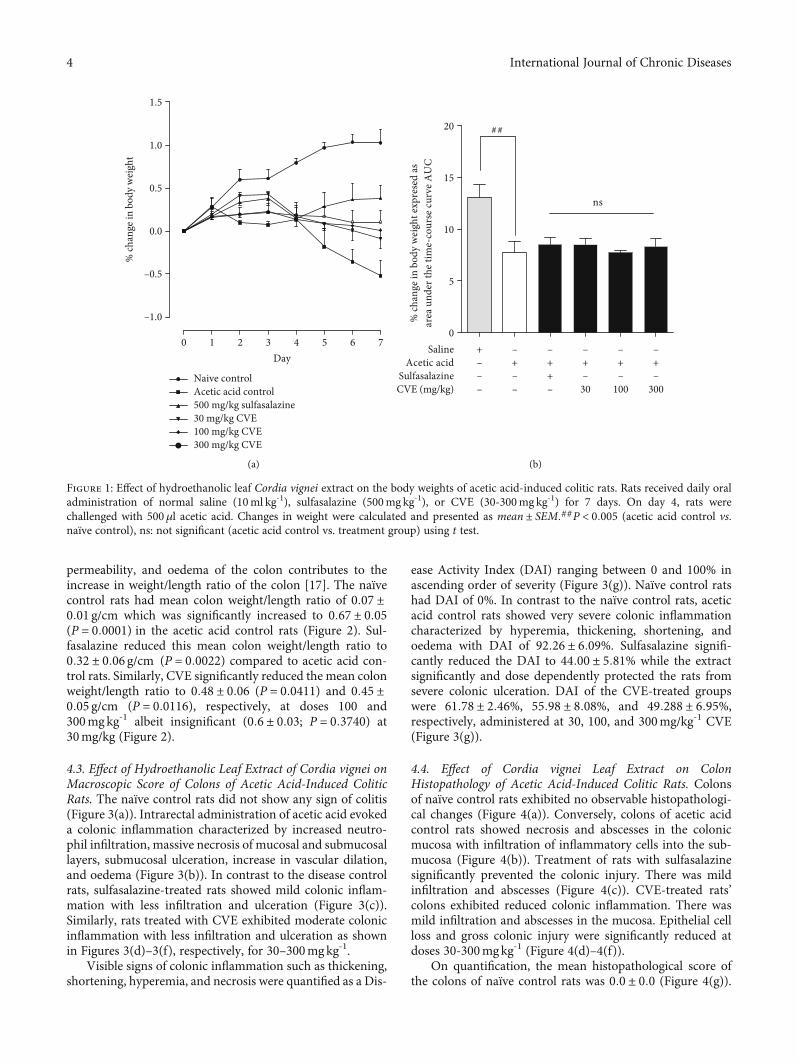
4.1. Effect of Hydroethanolic Leaf Extract of Cordia vignei onBody Weight of Acetic Acid-Induced Colitic Rats. Intrarectalinjection of acetic acid in rats evoked colonic inflammatoryresponse which became evident as passing of loose stool(with or without occult blood) and loss of body weight. Thenaïve control rats showed a gradual increase in weight (0-1.07%) throughout the 7 days (Figure 1(a)). In contrast tothe naïve control, acetic acid control rats exhibited decreasein body weight from day 2 to day 7 (Figure 1(a)). Body weightsof sulfasalazine-treated rats decreased from day 3 to day 4 andthen slightly increased from day 5 to day 7 (Figure 1(a)). Onthe other hand, body weights of CVE-treated rats slightlydeclined from day 3 to day 7 (Figure 1(a)).
The total change in body weight of rats over the 7 dayswas calculated as area under the curve (AUC) as shown inFigure 1(b). The AUC for the naïve control over the 7 dayswas 12:57 ± 1:079. Acetic acid significantly reduced theAUC to 7:495 ± 0:8371 (P = 0:0059) compared to naïve con-trol (Figure 1(b)). The AUC of sulfasalazine-treated rats was8:956 ± 0:7331 (P = 0:2258) compared to acetic acid controlwhile the AUC of CVE-treated rats were 8:257 ± 0:56(P = 0:4700), 7:858 ± 0:14 (P = 0:6800), and 8:086 ± 0:63(P = 0:5885), respectively, at 30, 100, and 300mgkg-1 com-pared to acetic acid control (Figure 1(b)).
4.2. Effect of Hydroethanolic Leaf Extract of Cordia vignei onColon Weight-to-Length Ratio of Acetic Acid-Induced ColiticRats. Increase in inflammatory cell infiltration, vascular
3International Journal of Chronic Diseases
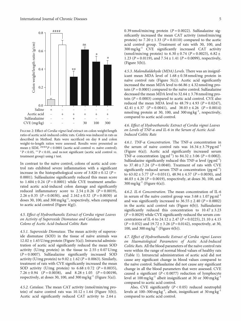
permeability, and oedema of the colon contributes to theincrease in weight/length ratio of the colon [17]. The naïvecontrol rats had mean colon weight/length ratio of 0:07 ±0:01 g/cm which was significantly increased to 0:67 ± 0:05(P = 0:0001) in the acetic acid control rats (Figure 2). Sul-fasalazine reduced this mean colon weight/length ratio to0:32 ± 0:06 g/cm (P = 0:0022) compared to acetic acid con-trol rats. Similarly, CVE significantly reduced the mean colonweight/length ratio to 0:48 ± 0:06 (P = 0:0411) and 0:45 ±0:05 g/cm (P = 0:0116), respectively, at doses 100 and300mgkg-1 albeit insignificant (0:6 ± 0:03; P = 0:3740) at30mg/kg (Figure 2).
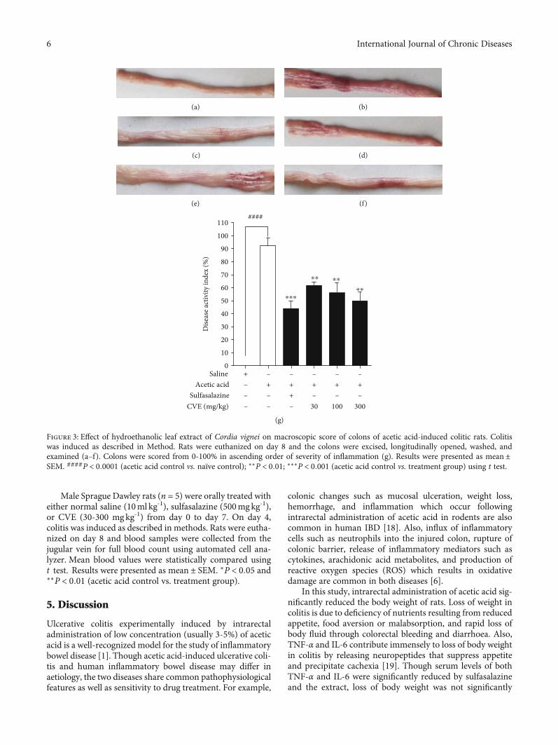
4.3. Effect of Hydroethanolic Leaf Extract of Cordia vignei onMacroscopic Score of Colons of Acetic Acid-Induced ColiticRats. The naïve control rats did not show any sign of colitis(Figure 3(a)). Intrarectal administration of acetic acid evokeda colonic inflammation characterized by increased neutro-phil infiltration, massive necrosis of mucosal and submucosallayers, submucosal ulceration, increase in vascular dilation,and oedema (Figure 3(b)). In contrast to the disease controlrats, sulfasalazine-treated rats showed mild colonic inflam-mation with less infiltration and ulceration (Figure 3(c)).Similarly, rats treated with CVE exhibited moderate colonicinflammation with less infiltration and ulceration as shownin Figures 3(d)–3(f), respectively, for 30–300mgkg-1.
Visible signs of colonic inflammation such as thickening,shortening, hyperemia, and necrosis were quantified as a Dis-
ease Activity Index (DAI) ranging between 0 and 100% inascending order of severity (Figure 3(g)). Naïve control ratshad DAI of 0%. In contrast to the naïve control rats, aceticacid control rats showed very severe colonic inflammationcharacterized by hyperemia, thickening, shortening, andoedema with DAI of 92:26 ± 6:09%. Sulfasalazine signifi-cantly reduced the DAI to 44:00 ± 5:81% while the extractsignificantly and dose dependently protected the rats fromsevere colonic ulceration. DAI of the CVE-treated groupswere 61:78 ± 2:46%, 55:98 ± 8:08%, and 49:288 ± 6:95%,respectively, administered at 30, 100, and 300mg/kg-1 CVE(Figure 3(g)).
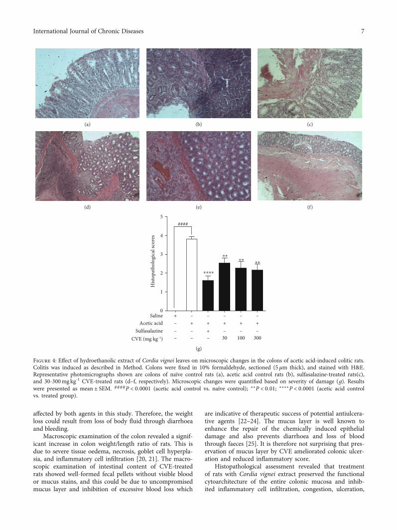
4.4. Effect of Cordia vignei Leaf Extract on ColonHistopathology of Acetic Acid-Induced Colitic Rats. Colonsof naïve control rats exhibited no observable histopathologi-cal changes (Figure 4(a)). Conversely, colons of acetic acidcontrol rats showed necrosis and abscesses in the colonicmucosa with infiltration of inflammatory cells into the sub-mucosa (Figure 4(b)). Treatment of rats with sulfasalazinesignificantly prevented the colonic injury. There was mildinfiltration and abscesses (Figure 4(c)). CVE-treated rats’colons exhibited reduced colonic inflammation. There wasmild infiltration and abscesses in the mucosa. Epithelial cellloss and gross colonic injury were significantly reduced atdoses 30-300mgkg-1 (Figure 4(d)–4(f)).
On quantification, the mean histopathological score ofthe colons of naïve control rats was 0:0 ± 0:0 (Figure 4(g)).
% ch
ange
in b
ody w
eigh
t
Day0 1 2 3 4 5 6 7
–1.0
–0.5
0.0
0.5
1.0
1.5
Naive controlAcetic acid control500 mg/kg sulfasalazine30 mg/kg CVE100 mg/kg CVE300 mg/kg CVE
(a)
0
5
10
15
20
% ch
ange
in b
ody
wei
ght e
xpre
sed
as ar
ea u
nder
the t
ime-
cour
se cu
rve A
UC
Acetic acidSulfasalazineCVE (mg/kg) 30 100 300
Saline– + + + + +– – + – – –– – –
+ – – – – –
ns
##
(b)
Figure 1: Effect of hydroethanolic leaf Cordia vignei extract on the body weights of acetic acid-induced colitic rats. Rats received daily oraladministration of normal saline (10ml kg-1), sulfasalazine (500mg kg-1), or CVE (30-300mg kg-1) for 7 days. On day 4, rats werechallenged with 500μl acetic acid. Changes in weight were calculated and presented as mean ± SEM.##P < 0:005 (acetic acid control vs.naïve control), ns: not significant (acetic acid control vs. treatment group) using t test.
4 International Journal of Chronic Diseases
In contrast to the naïve control, colons of acetic acid con-trol rats exhibited severe inflammation with a significantincrease in the histopathological score of 3:820 ± 0:12 (P =0:0001). Sulfasalazine significantly reduced this mean scoreto 1:604 ± 0:24 (P = 0:0001) while CVE treatment amelio-rated acetic acid-induced colon damage and significantlyreduced inflammatory score to 2:54 ± 0:26 (P = 0:0019),2:26 ± 0:35 (P = 0:0030), and 2:162 ± 0:32 (P = 0:0030) atdoses 30, 100, and 300mgkg-1, respectively, when comparedto acetic acid control (Figure 4(g)).
4.5. Effect of Hydroethanolic Extract of Cordia vignei Leaveson Activity of Superoxide Dismutase and Catalase onColons of Acetic Acid-Induced Colitic Rats
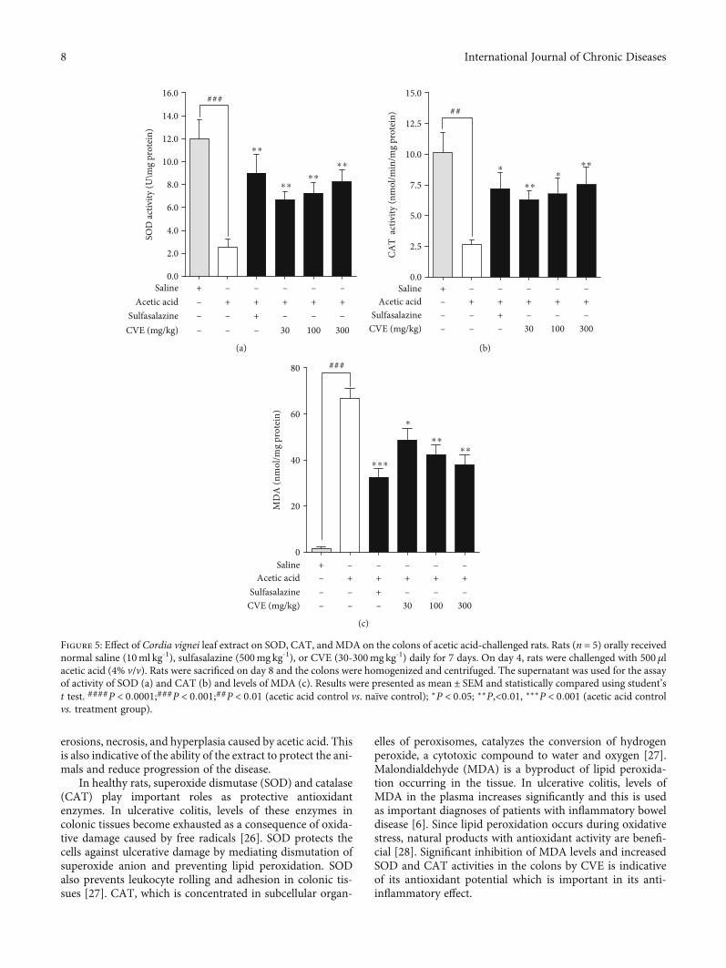
4.5.1. Superoxide Dismutase. The mean activity of superox-ide dismutase (SOD) in the tissue of naïve animals was12:02 ± 1:65U/mg protein (Figure 5(a)). Intrarectal adminis-tration of acetic acid significantly reduced the mean SODactivity (U/mg protein) in the tissue to 2:55 ± 0:07U/mg(P = 0:0007). Sulfasalazine significantly increased SODactivity (U/mg protein) to 9:02 ± 1:62 (P = 0:0063). Similarly,treatment of rats with CVE significantly increased the meanSOD activity (U/mg protein) to 6:68 ± 0:72 (P = 0:0035),7:26 ± 0:94 (P = 0:0038), and 8:28 ± 1:05 (P = 0:00190,respectively, at doses 30, 100, and 300mgkg-1 (Figure 5(a)).
4.5.2. Catalase. The mean CAT activity (nmol/min/mg pro-tein) of naive control rats was 10:12 ± 1:64 (Figure 5(b)).Acetic acid significantly reduced CAT activity to 2:64 ±
0:39nmol/min/mg protein (P = 0:0022). Sulfasalazine sig-nificantly increased the mean CAT activity (nmol/min/mgprotein) to 7:20 ± 1:33 (P = 0:0110) compared to the aceticacid control group. Treatment of rats with 30, 100, and300mgkg-1 CVE significantly increased CAT activity(nmol/min/mg protein) to 6:30 ± 0:74 (P = 0:0023), 6:82 ±1:23 (P = 0:0119), and 7:54 ± 1:41 (P = 0:0099), respectively,(Figure 5(b)).
4.5.3. Malondialdehyde (MDA) Levels. There was an insignif-icant mean MDA level of 1:68 ± 0:58nmol/mg protein innaïve control rats (Figure 5(c)). Acetic acid significantlyincreased the mean MDA level to 66:86 ± 4:32nmol/mg pro-tein (P = 0:0001) compared to the naïve control. Sulfasalazinedecreased the mean MDA level to 32:64 ± 3:78nmol/mg pro-tein (P = 0:0003) compared to acetic acid control. CVE alsoreduced the mean MDA level to 48:79 ± 4:93 (P = 0:0247),42:41 ± 4:37 (P = 0:0041), and 38:03 ± 4:26 (P = 0:0014)nmol/mg protein at 30, 100, and 300mgkg-1, respectively,compared to acetic acid control.
4.6. Effect of Hydroethanolic Extract of Cordia vignei Leaveson Levels of TNF-α and IL-6 in the Serum of Acetic Acid-Induced Colitic Rats
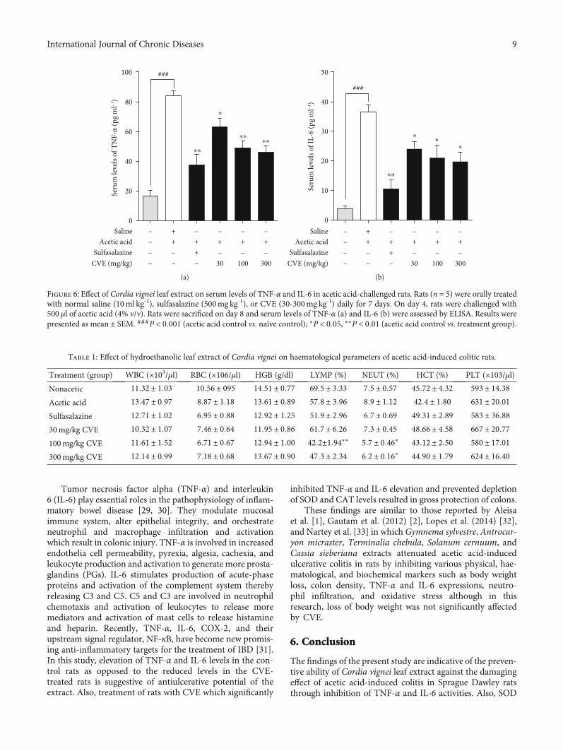
4.6.1. TNF-α Concentration. The TNF-α concentration inthe serum of naive control rats was 16:34 ± 3:79pgml-1
(Figure 6(a)). Acetic acid significantly increased serumTNF-α concentration (pgml-1) to 84:32 ± 3:06 (P = 0:0002).Sulfasalazine significantly reduced this TNF-α level (pgml-1)to 37:40 ± 7:24 (P = 0:0040). Treatment of rats with CVEsignificantly reduced serum TNF-α concentration (pgml-1)to 63:02 ± 5:77 (P = 0:0311), 48:94 ± 4:57 (P = 0:0030), and46:03 ± 4:26 (P = 0:0019), respectively, at doses 30, 100, and300mgkg-1 (Figure 6(a)).
4.6.2. IL-6 Concentration. The mean concentration of IL-6in serum of the naïve control group was 3:68 ± 1:07pgml-1
and was significantly increased to 36:55 ± 2:40 (P = 0:0002)in the acetic acid control rats (Figure 6(b)). Sulfasalazinesignificantly reduced this concentration to 10:47 ± 3:23(P = 0:0029) while CVE significantly reduced the serum con-centrations of IL-6 to 24:12 ± 2:47 (P = 0:0225), 21:10 ± 4:15(P = 0:032) and 19:72 ± 3:26 (P=0.0142), respectively, at 30,100, and 300mgkg-1 (Figure 6(b)).
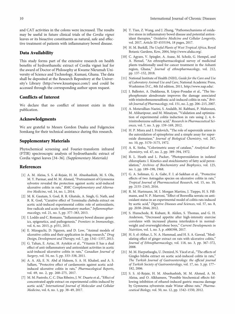
4.7. Effect of Hydroethanolic Extract of Cordia vignei Leaveson Haematological Parameters of Acetic Acid-InducedColitic Rats.All the blood parameters of the naïve control ratswere within the range of normal blood values of healthy rats(Table 1). Intrarectal administration of acetic acid did notcause any significant change in blood values compared tothe naïve control. Sulfasalazine did not cause any significantchange in all the blood parameters that were assessed. CVEcaused a significant (P < 0:0077) reduction of lymphocytelevel at 100mgkg-1 albeit insignificant at 30 or 300mgkg-1
compared to acetic acid control.Also, CVE significantly (P < 0:05) reduced neutrophil
levels at 100–300mgkg-1, albeit, insignificant at 30mgkg-1
compared to acetic acid control.
0.0
0.2
Colo
n w
eigh
t/len
gth
ratio
(g/c
m)
0.4
0.6
0.8
Saline + – – – – –Acetic acid – + + + + +
Sulfasalazine – – + – – –CVE (mg/kg) – – – 30 100 300
ns
####
⁎
⁎
⁎⁎
ns
Figure 2: Effect of Cordia vignei leaf extract on colon weight/lengthratio of acetic acid-induced colitic rats. Colitis was induced in rats asdescribed in Method. Rats were sacrificed on day 8 and colonweight-to-length ratios were assessed. Results were presented asmean ± SEM. ####P < 0:0001 (acetic acid control vs. naïve control);∗P < 0:05, ∗∗P < 0:01, and ns not significant (acetic acid control vs.treatment group) using t test.
5International Journal of Chronic Diseases
Male Sprague Dawley rats (n = 5) were orally treated witheither normal saline (10ml kg-1), sulfasalazine (500mgkg-1),or CVE (30-300 mgkg-1) from day 0 to day 7. On day 4,colitis was induced as described in methods. Rats were eutha-nized on day 8 and blood samples were collected from thejugular vein for full blood count using automated cell ana-lyzer. Mean blood values were statistically compared usingt test. Results were presented as mean ± SEM. ∗P < 0:05 and∗∗P < 0:01 (acetic acid control vs. treatment group).
5. Discussion
Ulcerative colitis experimentally induced by intrarectaladministration of low concentration (usually 3-5%) of aceticacid is a well-recognized model for the study of inflammatorybowel disease [1]. Though acetic acid-induced ulcerative coli-tis and human inflammatory bowel disease may differ inaetiology, the two diseases share common pathophysiologicalfeatures as well as sensitivity to drug treatment. For example,
colonic changes such as mucosal ulceration, weight loss,hemorrhage, and inflammation which occur followingintrarectal administration of acetic acid in rodents are alsocommon in human IBD [18]. Also, influx of inflammatorycells such as neutrophils into the injured colon, rupture ofcolonic barrier, release of inflammatory mediators such ascytokines, arachidonic acid metabolites, and production ofreactive oxygen species (ROS) which results in oxidativedamage are common in both diseases [6].
In this study, intrarectal administration of acetic acid sig-nificantly reduced the body weight of rats. Loss of weight incolitis is due to deficiency of nutrients resulting from reducedappetite, food aversion or malabsorption, and rapid loss ofbody fluid through colorectal bleeding and diarrhoea. Also,TNF-α and IL-6 contribute immensely to loss of body weightin colitis by releasing neuropeptides that suppress appetiteand precipitate cachexia [19]. Though serum levels of bothTNF-α and IL-6 were significantly reduced by sulfasalazineand the extract, loss of body weight was not significantly
(a) (b)
(c) (d)
(e) (f)
0
10
20
30
40
50
60
70
80
90
100
110####
⁎⁎
⁎⁎
⁎⁎
⁎⁎⁎
Dise
ase a
ctiv
ity in
dex
(%)
Saline + – – – – –Acetic acid – + + + + +
Sulfasalazine – – + – – –CVE (mg/kg) – – – 30 100 300
(g)
Figure 3: Effect of hydroethanolic leaf extract of Cordia vignei on macroscopic score of colons of acetic acid-induced colitic rats. Colitiswas induced as described in Method. Rats were euthanized on day 8 and the colons were excised, longitudinally opened, washed, andexamined (a–f). Colons were scored from 0-100% in ascending order of severity of inflammation (g). Results were presented as mean ±SEM. ####P < 0:0001 (acetic acid control vs. naïve control); ∗∗P < 0:01; ∗∗∗P < 0:001 (acetic acid control vs. treatment group) using t test.
6 International Journal of Chronic Diseases
affected by both agents in this study. Therefore, the weightloss could result from loss of body fluid through diarrhoeaand bleeding.
Macroscopic examination of the colon revealed a signif-icant increase in colon weight/length ratio of rats. This isdue to severe tissue oedema, necrosis, goblet cell hyperpla-sia, and inflammatory cell infiltration [20, 21]. The macro-scopic examination of intestinal content of CVE-treatedrats showed well-formed fecal pellets without visible bloodor mucus stains, and this could be due to uncompromisedmucus layer and inhibition of excessive blood loss which
are indicative of therapeutic success of potential antiulcera-tive agents [22–24]. The mucus layer is well known toenhance the repair of the chemically induced epithelialdamage and also prevents diarrhoea and loss of bloodthrough faeces [25]. It is therefore not surprising that pres-ervation of mucus layer by CVE ameliorated colonic ulcer-ation and reduced inflammatory score.
Histopathological assessment revealed that treatmentof rats with Cordia vignei extract preserved the functionalcytoarchitecture of the entire colonic mucosa and inhib-ited inflammatory cell infiltration, congestion, ulceration,
(a) (b) (c)
(d) (e) (f)
Hist
opat
holo
gica
l sco
res
0
1
2
3
4
5####
⁎⁎⁎⁎
⁎⁎⁎⁎
⁎⁎
Saline + – – – – –Acetic acid – + + + + +
Sulfasalazine – – + – – –CVE (mg kg–1) – – – 30 100 300
(g)
Figure 4: Effect of hydroethanolic extract of Cordia vignei leaves on microscopic changes in the colons of acetic acid-induced colitic rats.Colitis was induced as described in Method. Colons were fixed in 10% formaldehyde, sectioned (5 μm thick), and stained with H&E.Representative photomicrographs shown are colons of naïve control rats (a), acetic acid control rats (b), sulfasalazine-treated rats(c),and 30-300mg kg-1 CVE-treated rats (d–f, respectively). Microscopic changes were quantified based on severity of damage (g). Resultswere presented as mean ± SEM. ####P < 0:0001 (acetic acid control vs. naïve control); ∗∗P < 0:01; ∗∗∗∗P < 0:0001 (acetic acid controlvs. treated group).
7International Journal of Chronic Diseases
erosions, necrosis, and hyperplasia caused by acetic acid. Thisis also indicative of the ability of the extract to protect the ani-mals and reduce progression of the disease.
In healthy rats, superoxide dismutase (SOD) and catalase(CAT) play important roles as protective antioxidantenzymes. In ulcerative colitis, levels of these enzymes incolonic tissues become exhausted as a consequence of oxida-tive damage caused by free radicals [26]. SOD protects thecells against ulcerative damage by mediating dismutation ofsuperoxide anion and preventing lipid peroxidation. SODalso prevents leukocyte rolling and adhesion in colonic tis-sues [27]. CAT, which is concentrated in subcellular organ-
elles of peroxisomes, catalyzes the conversion of hydrogenperoxide, a cytotoxic compound to water and oxygen [27].Malondialdehyde (MDA) is a byproduct of lipid peroxida-tion occurring in the tissue. In ulcerative colitis, levels ofMDA in the plasma increases significantly and this is usedas important diagnoses of patients with inflammatory boweldisease [6]. Since lipid peroxidation occurs during oxidativestress, natural products with antioxidant activity are benefi-cial [28]. Significant inhibition of MDA levels and increasedSOD and CAT activities in the colons by CVE is indicativeof its antioxidant potential which is important in its anti-inflammatory effect.
0.0
2.0
4.0
6.0
SOD
activ
ity (U
\mg
prot
ein)
8.0
10.0
12.0
14.0
16.0 ###
⁎⁎
⁎⁎
⁎⁎
⁎⁎
–
Saline + – – – – –Acetic acid – + + + + +
Sulfasalazine – – + – –CVE (mg/kg) – – – 30 100 300
(a)
CAT
activ
ity (n
mol
/min
/mg
prot
ein)
0.0
2.5
5.0
7.5
10.0
12.5
15.0
–
##
⁎
⁎⁎
⁎⁎
⁎
Saline + – – – – –Acetic acid – + + + + +
Sulfasalazine – – + – –CVE (mg/kg) – – – 30 100 300
(b)
MD
A (n
mol
/mg
prot
ein)
0
20
40
60
80
–
Saline + – – – – –Acetic acid – + + + + +
Sulfasalazine – – + – –CVE (mg/kg) – – – 30 100 300
###
⁎⁎
⁎⁎
⁎
⁎⁎⁎
(c)
Figure 5: Effect of Cordia vignei leaf extract on SOD, CAT, and MDA on the colons of acetic acid-challenged rats. Rats (n = 5) orally receivednormal saline (10ml kg-1), sulfasalazine (500mg kg-1), or CVE (30-300mg kg-1) daily for 7 days. On day 4, rats were challenged with 500μlacetic acid (4% v/v). Rats were sacrificed on day 8 and the colons were homogenized and centrifuged. The supernatant was used for the assayof activity of SOD (a) and CAT (b) and levels of MDA (c). Results were presented asmean ± SEM and statistically compared using student’st test. ####P < 0:0001;###P < 0:001;##P < 0:01 (acetic acid control vs. naïve control); ∗P < 0:05; ∗∗P,<0:01, ∗∗∗P < 0:001 (acetic acid controlvs. treatment group).
8 International Journal of Chronic Diseases
Tumor necrosis factor alpha (TNF-α) and interleukin6 (IL-6) play essential roles in the pathophysiology of inflam-matory bowel disease [29, 30]. They modulate mucosalimmune system, alter epithelial integrity, and orchestrateneutrophil and macrophage infiltration and activationwhich result in colonic injury. TNF-α is involved in increasedendothelia cell permeability, pyrexia, algesia, cachexia, andleukocyte production and activation to generate more prosta-glandins (PGs). IL-6 stimulates production of acute-phaseproteins and activation of the complement system therebyreleasing C3 and C5. C5 and C3 are involved in neutrophilchemotaxis and activation of leukocytes to release moremediators and activation of mast cells to release histamineand heparin. Recently, TNF-α, IL-6, COX-2, and theirupstream signal regulator, NF-κB, have become new promis-ing anti-inflammatory targets for the treatment of IBD [31].In this study, elevation of TNF-α and IL-6 levels in the con-trol rats as opposed to the reduced levels in the CVE-treated rats is suggestive of antiulcerative potential of theextract. Also, treatment of rats with CVE which significantly
inhibited TNF-α and IL-6 elevation and prevented depletionof SOD and CAT levels resulted in gross protection of colons.
These findings are similar to those reported by Aleisaet al. [1], Gautam et al. (2012) [2], Lopes et al. (2014) [32],and Nartey et al. [33] in which Gymnema sylvestre, Antrocar-yon micraster, Terminalia chebula, Solanum cernuum, andCassia sieberiana extracts attenuated acetic acid-inducedulcerative colitis in rats by inhibiting various physical, hae-matological, and biochemical markers such as body weightloss, colon density, TNF-α and IL-6 expressions, neutro-phil infiltration, and oxidative stress although in thisresearch, loss of body weight was not significantly affectedby CVE.
6. Conclusion
The findings of the present study are indicative of the preven-tive ability of Cordia vignei leaf extract against the damagingeffect of acetic acid-induced colitis in Sprague Dawley ratsthrough inhibition of TNF-α and IL-6 activities. Also, SOD
0
20
40
60
80
100
Seru
m le
vels
of T
NF-𝛼
(pg
ml–1
)
###
⁎⁎
⁎
⁎⁎⁎⁎
––
Saline – + – – – –Acetic acid +– + + + +
Sulfasalazine – + – – –CVE (mg/kg) – – 30 100 300
(a)
Seru
m le
vels
of IL
-6 (p
g m
l–1)
0
10
20
30
40
50
––
Saline – + – – – –Acetic acid +– + + + +
Sulfasalazine – + – – –CVE (mg/kg) – – 30 100 300
###
⁎⁎
⁎⁎
⁎
(b)
Figure 6: Effect of Cordia vignei leaf extract on serum levels of TNF-α and IL-6 in acetic acid-challenged rats. Rats (n = 5) were orally treatedwith normal saline (10ml kg-1), sulfasalazine (500mg kg-1), or CVE (30-300mg kg-1) daily for 7 days. On day 4, rats were challenged with500μl of acetic acid (4% v/v). Rats were sacrificed on day 8 and serum levels of TNF-α (a) and IL-6 (b) were assessed by ELISA. Results werepresented asmean ± SEM. ###P < 0:001 (acetic acid control vs. naïve control); ∗P < 0:05, ∗∗P < 0:01 (acetic acid control vs. treatment group).
Table 1: Effect of hydroethanolic leaf extract of Cordia vignei on haematological parameters of acetic acid-induced colitic rats.
Treatment (group) WBC (×103/μl) RBC (×106/μl) HGB (g/dl) LYMP (%) NEUT (%) HCT (%) PLT (×103/μl)Nonacetic 11:32 ± 1:03 10:56 ± 095 14:51 ± 0:77 69:5 ± 3:33 7:5 ± 0:57 45:72 ± 4:32 593 ± 14:38
Acetic acid 13:47 ± 0:97 8:87 ± 1:18 13:61 ± 0:89 57:8 ± 3:96 8:9 ± 1:12 42:4 ± 1:80 631 ± 20:01
Sulfasalazine 12:71 ± 1:02 6:95 ± 0:88 12:92 ± 1:25 51:9 ± 2:96 6:7 ± 0:69 49:31 ± 2:89 583 ± 36:88
30mg/kg CVE 10:32 ± 1:07 7:46 ± 0:64 11:95 ± 0:86 61:7 ± 6:26 7:3 ± 0:45 48:66 ± 4:58 667 ± 20:77
100mg/kg CVE 11:61 ± 1:52 6:71 ± 0:67 12:94 ± 1:00 42:2±1:94∗∗ 5:7 ± 0:46∗ 43:12 ± 2:50 580 ± 17:01
300mg/kg CVE 12:14 ± 0:99 7:18 ± 0:68 13:67 ± 0:90 47:3 ± 2:34 6:2 ± 0:16∗ 44:90 ± 1:79 624 ± 16:40
9International Journal of Chronic Diseases
and CAT activities in the colons were increased. The resultsmay be useful in future clinical trials of the Cordia vigneileaves or its bioactive constituents as natural, safe and effec-tive treatment of patients with inflammatory bowel disease.
Data Availability
This study forms part of the extensive research on healthbenefits of hydroethanolic extract of Cordia vignei leaf forthe award of Doctor of Philosophy at Kwame Nkrumah Uni-versity of Science and Technology, Kumasi, Ghana. The datashall be deposited at the Research Repository at the Univer-sity’s Library (http://www.knustspace.com/) and could beaccessed through the corresponding author upon request.
Conflicts of Interest
We declare that no conflict of interest exists in thispublication.
Acknowledgments
We are grateful to Messrs Gordon Daaku and FulgenciosSomkang for their technical assistance during this research.
Supplementary Materials
Phytochemical screening and Fourier-transform infrared(FTIR) spectroscopic analysis of hydroethanolic extract ofCordia vignei leaves [34–36]. (Supplementary Materials)
References
[1] A. M. Aleisa, S. S. al-Rejaie, H. M. Abuohashish, M. S. Ola,M. Y. Parmar, and M. M. Ahmed, “Pretreatment of Gymnemasylvestre revealed the protection against acetic acid-inducedulcerative colitis in rats,” BMC Complementary and Alterna-tive Medicine, vol. 14, no. 1, 2014.
[2] M. K. Gautam, S. Goel, R. R. Ghatule, A. Singh, G. Nath, andR. K. Goel, “Curative effect of Terminalia chebula extract onacetic acid-induced experimental colitis: role of antioxidants,free radicals and acute inflammatory marker,” Inflammophar-macology, vol. 21, no. 5, pp. 377–383, 2013.
[3] I. Loddo and C. Romano, “Inflammatory bowel disease: genet-ics, epigenetics, and pathogenesis,” Frontiers in Immunology,vol. 6, no. 2015, p. p551, 2015.
[4] E. Mizoguchi, D. Nguyen, and D. Low, “Animal models ofulcerative colitis and their application in drug research,” DrugDesign, Development and Therapy, vol. 7, pp. 1341–1357, 2013.
[5] G. Tahan, E. Aytac, H. Aytekin et al., “Vitamin E has a dualeffect of anti-inflammatory and antioxidant activities in aceticacid-induced ulcerative colitis in rats,” Canadian Journal ofSurgery, vol. 54, no. 5, pp. 333–338, 2011.
[6] A. A. Ali, E. N. Abd al Haleem, S. A. H. Khaleel, and A. S.Sallam, “Protective effect of cardamonin against acetic acid-induced ulcerative colitis in rats,” Pharmacological Reports,vol. 69, no. 2, pp. 268–275, 2017.
[7] M. M. Pastrelo, C. C. Dias Ribeiro, J. W. Duarte et al., “Effect ofconcentrated apple extract on experimental colitis induced byacetic acid,” International Journal of Molecular and CellularMedicine, vol. 6, no. 1, pp. 38–49, 2017.
[8] T. Tian, Z. Wang, and J. Zhang, “Pathomechanisms of oxida-tive stress in inflammatory bowel disease and potential antiox-idant therapies,” Oxidative Medicine and Cellular Longevity,vol. 2017, Article ID 4535194, 18 pages, 2017.
[9] H. M. Burkill, The Useful Plants of West Tropical Africa, RoyalBotanic Gardens, Kew, 2004, http://www.aluka.org/.
[10] C. Agyare, V. Spiegler, A. Asase, M. Scholz, G. Hempel, andA. Hensel, “An ethnopharmacological survey of medicinalplants traditionally used for cancer treatment in the Ashantiregion, Ghana,” Journal of ethnopharmacology, vol. 212,pp. 137–152, 2018.
[11] National Institute of Health (NIH), Guide for the Care and Useof Laboratory Animal Use and Care, National Academic Press,Washinton D.C., 8th Ed edition, 2011, http://www.nap.edu/.
[12] I. Ballester, A. Daddaoua, R. López-Posadas et al., “The bis-phosphonate alendronate improves the damage associatedwith trinitrobenzenesulfonic acid-induced colitis in rats,” Brit-ish Journal of Pharmacology, vol. 151, no. 2, pp. 206–215, 2007.
[13] A. Motavallian-Naeini, S. Andalib, M. Rabbani, P. Mahzouni,M. Afsharipour, and M. Minaiyan, “Validation and optimiza-tion of experimental colitis induction in rats using 2, 4, 6-trinitrobenzene sulfonic acid,” Research in Pharmaceutical Sci-ences, vol. 7, no. 3, pp. 159–169, 2012.
[14] H. P. Misra and I. Fridovich, “The role of superoxide anion inthe autoxidation of epinephrine and a simple assay for super-oxide dismutase,” Journal of Biological Chemistry, vol. 247,no. 10, pp. 3170–3175, 1972.
[15] A. K. Sinha, “Colorimetric assay of catalase,” Analytical Bio-chemistry, vol. 47, no. 2, pp. 389–394, 1972.
[16] R. L. Heath and L. Packer, “Photoperoxidation in isolatedchloroplasts: I. Kinetics and stoichiometry of fatty acid perox-idation,” Archives of Biochemistry and Biophysics, vol. 125,no. 1, pp. 189–198, 1968.
[17] G. A. Soliman, G. A. Gabr, F. I. al-Saikhan et al., “Protectiveeffects of two Astragalus species on ulcerative colitis in rats,”Tropical Journal of Pharmaceutical Research, vol. 15, no. 10,pp. 2155–2163, 2016.
[18] R. M. Hartmann, M. I. Morgan Martins, J. Tieppo, H. S. Fill-mann, and N. P. Marroni, “Effect of Boswellia serrata on anti-oxidant status in an experimental model of colitis rats inducedby acetic acid,” Digestive Diseases and Sciences, vol. 57, no. 8,pp. 2038–2044, 2012.
[19] S. Hunschede, R. Kubant, R. Akilen, S. Thomas, and G. H.Anderson, “Decreased appetite after high-intensity exercisecorrelates with increased plasma interleukin-6 in normal-weight and overweight/obese boys,” Current Developments inNutrition, vol. 1, no. 3, p. e000398, 2017.
[20] H. S. el-Abhar, L. N. A. Hammad, and H. S. A. Gawad, “Mod-ulating effect of ginger extract on rats with ulcerative colitis,”Journal of Ethnopharmacology, vol. 118, no. 3, pp. 367–372,2008.
[21] M.M. Harputluoglu, U. Demirel, N. Yücel et al., “The effects ofGingko biloba extract on acetic acid-induced colitis in rats,”The Turkish Journal of Gastroenterology: the official journalof Turkish Society of Gastroenterology, vol. 17, no. 3, pp. 177–182, 2006.
[22] S. S. Al-Rejaie, H. M. Abuohashish, M. M. Ahmed, A. M.Aleisa, and O. Alkhamees, “Possible biochemical effects fol-lowing inhibition of ethanol-induced gastric mucosa damageby Gymnema sylvestrein male Wistar albino rats,” Pharma-ceutical Biology, vol. 50, no. 12, pp. 1542–1550, 2012.
10 International Journal of Chronic Diseases
[23] R. H. Duerr, “Update on the genetics of inflammatory boweldisease,” Journal of Clinical Gastroenterology, vol. 37, no. 5,pp. 358–367, 2003.
[24] B. E. Sands, “Therapy of inflammatory bowel disease,” Gastro-enterology, vol. 118, no. 2, pp. S68–S82, 2000.
[25] A. S. Awaad, A. M. Alafeefy, F. A. S. Alasmary, R. M. el-Meligy,and S. I. Alqasoumi, “Anti-ulcerogenic and anti-ulcerativecolitis (UC) activities of seven amines derivatives,” Saudi Phar-maceutical Journal, vol. 25, no. 8, pp. 1125–1129, 2017.
[26] S. V. Rana, S. Sharma, K. K. Prasad, S. K. Sinha, and K. Singh,The Indian Journal of Medical Research, vol. 139, no. 4,pp. 568–571, 2014.
[27] D. E. B. Baldo and J. E. Serrano, “Screening for intestinal anti-inflammatory activity of Alpinia galanga against acetic acid-induced colitis in mice (Mus musculus),” Journal of MedicinalPlants Studies, vol. 4, no. 1, pp. 72–77, 2017.
[28] C. E. Rosas, L. B. Correa, andM. G. Henriques, “Neutrophils inrheumatoid arthritis: a target for discovering new therapiesbased on natural products,” in Role of Neutrophils in DiseasePathogenesis, pp. 89–118, IntechOpen Limited, London, UK,2017.
[29] Q. Guan and J. Zhang, “Recent advances: the imbalance ofcytokines in the pathogenesis of inflammatory bowel disease,”Mediators of Inflammation, vol. 2017, Article ID 4810258, 8pages, 2017.
[30] Z. H. Nemeth, D. A. Bogdanovski, P. Barratt-Stopper, S. R.Paglinco, L. Antonioli, and R. H. Rolandelli, “Crohn’s diseaseand ulcerative colitis show unique cytokine profiles,” Cureus,vol. 9, no. 4, p. e1177, 2017.
[31] M. F. Neurath, “Cytokines in inflammatory bowel disease,”Nature Reviews Immunology, vol. 14, no. 5, pp. 329–342, 2014.
[32] L. C. Lopes, J. E. de Carvalho, M. Kakimore et al., “Pharmaco-logical characterization of Solanum cernuum Vell.: 31-norcycloartanones with analgesic and anti-inflammatoryproperties,” Inflammopharmacology, vol. 22, no. 3, pp. 179–185, 2013.
[33] E. T. Nartey, M. Ofosuhene, and C. M. Agbale, “Anti-ulcero-genic activity of the root bark extract of the African laburnum“Cassia sieberiana” and its effect on the anti-oxidant defencesystem in rats,” BMC Complementary and Alternative Medi-cine, vol. 247, no. 1, p. 247, 2012.
[34] A. Sofowora, Medicinal Plants and Traditional Medicine inAfrica, Spectrum Books Ltd., Ibadan, 2nd edn edition, 1993.
[35] D. L. Pavia, M. G. Lampman, and G. S. Kriz, Introduction toSpectroscopy, Thomson Learning Berkshire House, London,UK, 3 ed edition, 2001.
[36] N. D. M. Lakshmi, B. Mahitha, and T. Madhavi, “Phytochem-ical screening and FTIR analysis of Clitoria ternatea leaves,”International Journal of Scientific & Engineering Research,vol. 6, no. 2, pp. 287–290, 2015.
11International Journal of Chronic Diseases