MicroRNA-615-3p promotes progression of neonatal ARDS · icro-615-3p promotes progression of...
9
4625 Abstract. – OBJECTIVE: To explore whether microRNA-615-3p participates in the progression of neonatal acute respiratory distress syndrome (ARDS) by inhibiting differentiation of mesenchy- mal stem cells (MSCs) to alveolar type II epithelial cells (ATII) via Wnt/β-catenin pathway. PATIENTS AND METHODS: Expression levels of microRNA-615-3p and inflammatory factors (IL- 1, IL-6, IL-8, and TNF-α) in peripheral blood of 24 neonatal ARDS patients and 14 healthy newborns were detected by qRT-PCR (quantitative Re- al-Time Polymerase Chain Reaction). MSCs were isolated from bone marrow of mice and identified by flow cytometry. The effect of microRNA-615-3p on regulating the differentiation of MSCs to ATII was analyzed. After altering expressions of mi- croRNA-615-3p and DKK1 by plasmids transfec- tion, Wnt/β-catenin pathway-related genes were detected by Western blot. RESULTS: Higher expression levels of microR- NA-615-3p and inflammatory factors (IL-1, IL- 6, IL-8, and TNF-α) were observed in peripheral blood of neonatal ARDS patients than those of healthy newborns. ATII-specific genes were up- regulated, and inflammatory factors were down- regulated after the microRNA-615-3p knockdown in MSCs. Moreover, expressions of Wnt/β-catenin pathway-related genes were downregulated after the microRNA-615-3p overexpression, which was partially reserved by the DKK1 knockdown. CONCLUSIONS: Overexpressed microR- NA-615-3p promoted ARDS development through inhibiting differentiation of MSCs to ATII via Wnt/β-catenin pathway. Key Words: MicroRNA-615-3p, Mesenchymal stem cells, Alveo- lar type II epithelial cells, Wnt/β-catenin pathway. Introduction Acute respiratory distress syndrome (ARDS) is a progressive hypoxemic respiratory failure that occurs in various primary diseases. ARDS is manifested as the diffuse alveolar epithelium and microvascular endothelium injuries caused by pulmonary parenchyma inflammatory response. Current studies have demonstrated that ARDS is the consequence of the interaction of various in- flammatory factors and cytokines. The dynamic balance between pro-inflammatory cytokines and anti-inflammatory cytokines is an essential factor that remarkably affects ARDS prognosis 1 . The- refore, it is of great significance to explore the specific pathogenesis of ARDS, so as to better improve the clinical treatment. MicroRNAs are a group of endogenous sin- gle-stranded, non-coding small RNAs with 18-24 nt in length. Functionally, microRNAs interfe- re with protein synthesis by directly degrading or inhibiting translation of target genes at the post-transcriptional level. Stable microRNAs have been detected in serum and plasma, that are, cir- culating microRNAs 2,3 . Circulating microRNAs are differentially expressed under different phy- siological and pathological conditions, which are closely related to human health 4-6 . As an expres- sion regulator, microRNAs can target approxima- tely 60% of protein-coding genes 7 , which exert a crucial role in biological processes, such as cell proliferation, differentiation, apoptosis, immunity, and inflammation 8 . Studies have found that circu- lating microRNAs secreted by tumor cells possess European Review for Medical and Pharmacological Sciences 2018; 22: 4625-4633 Y.-Q. WU, Y.-J. DING Department of Pediatrics, The Affiliated Yantai Yuhuangding Hospital of Qingdao University, Yantai, China Corresponding Author: Yanjie Ding, MD; e-mail: [email protected] Overexpressed microRNA-615-3p promotes progression of neonatal acute respiratory distress syndrome by inhibiting differentiation of mesenchymal stem cells to alveolar type II epithelial cells
Transcript of MicroRNA-615-3p promotes progression of neonatal ARDS · icro-615-3p promotes progression of...
4625
Abstract. – OBJECTIVE: To explore whether microRNA-615-3p participates in the progression of neonatal acute respiratory distress syndrome (ARDS) by inhibiting differentiation of mesenchy-mal stem cells (MSCs) to alveolar type II epithelial cells (ATII) via Wnt/β-catenin pathway.
PATIENTS AND METHODS: Expression levels of microRNA-615-3p and inflammatory factors (IL-1, IL-6, IL-8, and TNF-α) in peripheral blood of 24 neonatal ARDS patients and 14 healthy newborns were detected by qRT-PCR (quantitative Re-al-Time Polymerase Chain Reaction). MSCs were isolated from bone marrow of mice and identified by flow cytometry. The effect of microRNA-615-3p on regulating the differentiation of MSCs to ATII was analyzed. After altering expressions of mi-croRNA-615-3p and DKK1 by plasmids transfec-tion, Wnt/β-catenin pathway-related genes were detected by Western blot.
RESULTS: Higher expression levels of microR-NA-615-3p and inflammatory factors (IL-1, IL-6, IL-8, and TNF-α) were observed in peripheral blood of neonatal ARDS patients than those of healthy newborns. ATII-specific genes were up-regulated, and inflammatory factors were down-regulated after the microRNA-615-3p knockdown in MSCs. Moreover, expressions of Wnt/β-catenin pathway-related genes were downregulated after the microRNA-615-3p overexpression, which was partially reserved by the DKK1 knockdown.
CONCLUSIONS: Overexpressed microR-NA-615-3p promoted ARDS development through inhibiting differentiation of MSCs to ATII via Wnt/β-catenin pathway.
Key Words:MicroRNA-615-3p, Mesenchymal stem cells, Alveo-
lar type II epithelial cells, Wnt/β-catenin pathway.
Introduction
Acute respiratory distress syndrome (ARDS) is a progressive hypoxemic respiratory failure that occurs in various primary diseases. ARDS is manifested as the diffuse alveolar epithelium and microvascular endothelium injuries caused by pulmonary parenchyma inflammatory response. Current studies have demonstrated that ARDS is the consequence of the interaction of various in-flammatory factors and cytokines. The dynamic balance between pro-inflammatory cytokines and anti-inflammatory cytokines is an essential factor that remarkably affects ARDS prognosis1. The-refore, it is of great significance to explore the specific pathogenesis of ARDS, so as to better improve the clinical treatment.
MicroRNAs are a group of endogenous sin-gle-stranded, non-coding small RNAs with 18-24 nt in length. Functionally, microRNAs interfe-re with protein synthesis by directly degrading or inhibiting translation of target genes at the post-transcriptional level. Stable microRNAs have been detected in serum and plasma, that are, cir-culating microRNAs2,3. Circulating microRNAs are differentially expressed under different phy-siological and pathological conditions, which are closely related to human health4-6. As an expres-sion regulator, microRNAs can target approxima-tely 60% of protein-coding genes7, which exert a crucial role in biological processes, such as cell proliferation, differentiation, apoptosis, immunity, and inflammation8. Studies have found that circu-lating microRNAs secreted by tumor cells possess
European Review for Medical and Pharmacological Sciences 2018; 22: 4625-4633
Y.-Q. WU, Y.-J. DING
Department of Pediatrics, The Affiliated Yantai Yuhuangding Hospital of Qingdao University, Yantai, China
Corresponding Author: Yanjie Ding, MD; e-mail: [email protected]
Overexpressed microRNA-615-3p promotes progression of neonatal acute respiratory distress syndrome by inhibiting differentiation of mesenchymal stem cells to alveolar type II epithelial cells
Y.-Q. Wu, Y.-J. Ding
4626
their own regulatory effects9. For example, mi-croRNA-21 and microRNA-29 secreted by tumor cells can activate TLR-mediated inflammatory re-sponses by binding to Toll-like receptors (TLRs), thereby regulating tumor growth and metastasis10. MicroRNA-615-3p not only participates in the development of many tumors, but also is related to the immune and inflammatory responses11-13. The role of microRNA-615-3p in neonatal ARDS, however, has not been reported yet.
Bone marrow mesenchymal stem cells are the origin of osteoblasts, which present the poten-tial of multi-directional differentiation and strong proliferation. Under some circumstances, MSCs are capable of differentiating to ATII14. In the present study, we detected the differentiation ability of MSCs regulated by microRNA-615-3p, which provides new directions in treating neona-tal ARDS.
Patients and Methods
Patients and Sample Collection24 neonatal ARDS patients and 14 healthy
newborns in our hospital from June 2016 to December 2017 were selected. No significant differences in age, gender and body weight were found between neonatal ARDS patients and he-althy newborns. The investigation was approved by the Hospital Ethics Committee and patients’ families signed the informed consent. 1 ml of peripheral blood samples of all enrolled subjects were collected under fasting state in the morning and preserved at -80°C.
RNA Extraction From Peripheral Blood0.25 mL of peripheral blood was added to 0.75
mL of TRI Reagent LS and 0.2 ml of chloroform. The mixture was gently shaken, incubated for 2-5 min, and centrifuged at 4°C, 12,000 g for 15 min. The aqueous complex was transferred to anoth-er tube and isodose isopropanol was added for RNA extraction. After centrifugation, RNA was washed and air dried for 5 min. RNA was then dissolved in diethyl pyrocarbonate (DEPC) water without RNase, followed by preservation at -80°C for the following experiments.
Enzyme-Linked Immunosorbent Assay (ELISA)
According to the instructions of ELISA kit (EBiosciences, San Diego, CA, USA), corre-sponding antibodies were diluted at a density
of 1-10 μg/mL with the coating buffer. Briefly, cells were blocked in 5% bovine serum for 40 min. After washing for three times, the sample and enzyme-labeled antibody were added to each well. The substrate solution was used to terminate the reaction and ELISA results were determined within 20 min. Absorbance at the wavelength of 450 nm was detected using ELI-SA detector.
Quantitative Reverse Transcriptase-Polymerase Chain Reaction (qRT-PCR)
The mRNAs were reversely transcribed to cD-NAs using PrimeScript RT reagent kit (TaKaRa, Tokyo, Japan), followed by the qRT-PCR reaction according to the instructions of miScript SYBR Green PCR kit. The reaction conditions were as follows: denaturation at 94°C for 30 s, followed by annealing at 55°C for 30 s, and extension at 72°C for 90 s, for a total of 40 cycles. Each sample was performed for 3 times. Primers used in this ex-periment were as the follows: MicroRNA-615-3p, F: 5’-ACACTCCAGCTGGGTCCGAGCCTGG-GTCTC-3’, R: 5’-TGGTGTCGTGGAGTCG-3’; IL-1, F: 5’-CTCAGCAACACTCCTAT-3’, R: 5’-TCCTGGTCTGCAGGTAA-3’; IL-6, F: 5’-TC-CAATCTGGGTTCAATCAGGCGA-3’, R: 5’-TTCCCTCATACTCGTTCTGGAGGT-3’; IL-8, F: 5’-CTTGTCATTGCCAGCTGTGT-3’, R: 5’-TGACTGTGGAGTTTTGGCTG-3’; TNF-α, F: 5’-CACGTTGTAGCCGACATCAACTCT-3’, R: 5’-GTTGTCTTCCAGCTTCACACCGTT-3’; Occludin, F: 5’-TGAAAGTCCACCTCCTTACAGA-3’, R: 5’-CCGGATAAAAAGAGTACGCTGG-3’; KGF, F: 5’-TGGGCACTATATCTCTAGCTTGC-3’, R: 5’-GGGTGCGACAGAACAGTCT-3’; CK18, F: 5’-CAGCCAGCGTCTATGCAGG-3’, R: 5’- CCTTCTCGGTCTGGATTCCAC-3’; SpA, F: 5’-CTTGGCCTTTGGTGGCTACTT -3’, R: 5’-GAGAGGTCTTAGGGAATGTCACT-3’; SpB, F: 5’-CAGCGGGTAGGAAGCAGTTTC-3’, R: 5’-CCCTGCACCTCATCCCTGA-3’; SpC, F: 5’-GGTGAGCAACTGGCTACTGAG-3’, R:5’-CCCTGGCGCGTACATTCTTT-3’; Wnt3a, F: 5’- CTCCTCTCGGATACCTCTTAGTG-3’, R: 5’-CCAAGGACCACCAGATCGG-3’; β-catenin, F: 5’-ATGGAGCCGGACAGAAAAGC-3’, R: 5’- TGGGAGGTGTCAACATCTTCTT-3’; DKK1, F: 5’- CAGTGCCACCTTGAACTCAGT-3’, R: 5’-CCGCCCTCATAGAGAACTCC-3’; GAPDH, F: 5’-ACCCACTCCTCCACCTTTGA-3’, R: 5’- CTGTTGCTGTAGCCAAATTCGT-3’; U6, F: 5’-CTCGCTTCGGCAGCAGCACATATA-3’, R: 5’- AAATATGGAACGCTTCACGA-3’.
MicroRNA-615-3p promotes progression of neonatal ARDS
4627
Isolation and Culture of MSCsSprague Dawley rats (Model Animal Research
Center of Nanjing University, Nanjing, China) were executed with dislocation of the cervical vertebra. The femur and tibia were collected under aseptic condition. The marrow cavity was washed with L-DMEM (L-Dulbecco’s Modified Eagle Medium). After centrifugation at 1000 r/min for 5 min, MSCs were re-suspended in L-DMEM containing 10% fetal bovine serum (FBS, Gibco, Grand Island, NY, USA). MSCs were then seeded in 6-well plates at a density of 2.5×10⁶/L. Culture medium was replaced every 3-4 days. After cell culture for 10 days, MSCs were digested with trypsin and centrifuged at 4°C, 1500 rpm for 5 min. First-passage MSCs (P1 MSCs) were seeded in 6-well plates at a density of 5×10⁵/L.
MSCs Identification Phenotype identification of MSCs was carried
out by flow cytometry. MSCs were re-suspended with phosphate buffered saline (PBS) at a density of 1×10⁶/mL. 500 μL of cell suspension was incu-bated with 5 μL of CD34 and CD44 without light for 30 min. Cells were washed and re-suspended with PBS, followed by flow cytometry detection of MSCs phenotypes.
Cell Culture of ATIIATII cells were obtained from ATCC, Manas-
sas, VA, USA. Briefly, ATII cells were cultured in DMEM (Gibco, Grand Island, NY, USA) contain-ing 5% FBS and incubated in a 5% CO2 incubator at 37°C. Cell passage was performed with trypsin when cell confluence was up to 80-90%.
Co-Culture of MSCs and ATIIThe cell density of ATII was adjusted to 1×10⁴/
mL with DMEM containing 10% FBS. 1.5 mL of cell suspension was added to the transwell cham-ber. The cell density of MSCs was adjusted to 8×10⁴/mL with DMEM containing 10% FBS and third-passage MSCs were seeded into the 6-well plates. After culturing for 24 h, transwell cham-ber was put into the 6-well plate with the culture medium containing 10% FBS, 1% non-essential amino-acid, and DMEM with 1% L-glucose.
Western BlotTotal protein was extracted from treated cells
by radioimmunoprecipitation assay (RIPA) solu-tion (Beyotime, Shanghai, China). The protein sample was separated by electrophoresis on 10%
SDS-PAGE (sodium dodecyl sulphate-polyacryl-amide gel electrophoresis) and then transferred to PVDF (polyvinylidene difluoride) membrane (Millipore, Billerica, MA, USA). After mem-branes were blocked with skimmed milk, the membranes were incubated with primary anti-bodies (Cell Signaling Technology, Danvers, MA, USA) overnight at 4°C. The membranes were then washed with TBS-T (Tris-buffered Saline with Tween 20) and followed by the incubation of secondary antibody. The protein blot on the membrane was exposed by chemiluminescence.
Cell TransfectionCells in good growth condition were selected
and seeded in the 6-well plates. Cell transfection was performed when the cell confluence was up to 60%-70% according to the instructions of Lipo-fectamine2000 (Invitrogen, Carlsbad, CA, USA). Transfection plasmids (microRNA-615-3p mimic, microRNA-615-3p inhibitor, si-NCDKK1-1, and si-NCDKK1-2) were purchased from GenePhar-ma (Shanghai, China).
Statistical AnalysisWe used Statistical Product and Service Solu-
tions (SPSS) 20.0 software (IBM, Armonk, NY, USA) for statistical analysis. The quantitative da-ta were represented as mean ± standard deviation (x– ± s). The t-test and Chi-square analysis were used for comparing measurement and categorical data, respectively. p<0.05 was considered statisti-cally significant.
Results
MicroRNA-615-3p Was Overexpressed in Neonatal ARDS Patients
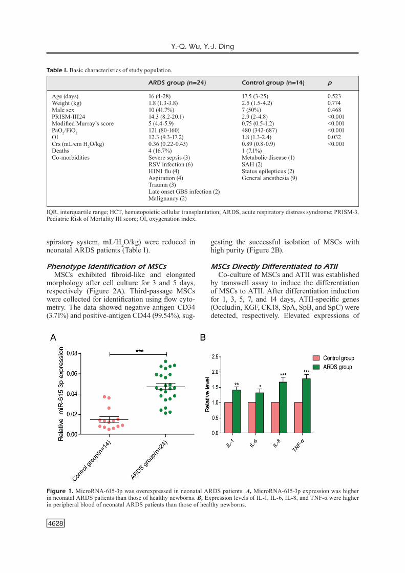
We detected expression levels of microR-NA-615-3p and inflammatory factors (IL-1, IL-6, IL-8, and TNF-α) in peripheral blood of 24 neo-natal ARDS patients and 14 healthy newborns by qRT-PCR. The results indicated higher expres-sions of microRNA-615-3p and inflammatory factors in neonatal ARDS patients than those of healthy newborns (Figure 1A and 1B). Besides, no significant differences in age, gender, and body weight were found between the two groups. Routine examinations showed that PRISM-III score, Modified Murray’s score, and oxygenation index (OI) were remarkably elevated, whereas PaO2/FiO2 (partial pressure of artery/fraction of inspired oxygen) and Crs (compliance of the re-
Y.-Q. Wu, Y.-J. Ding
4628
spiratory system, mL/H2O/kg) were reduced in neonatal ARDS patients (Table I).
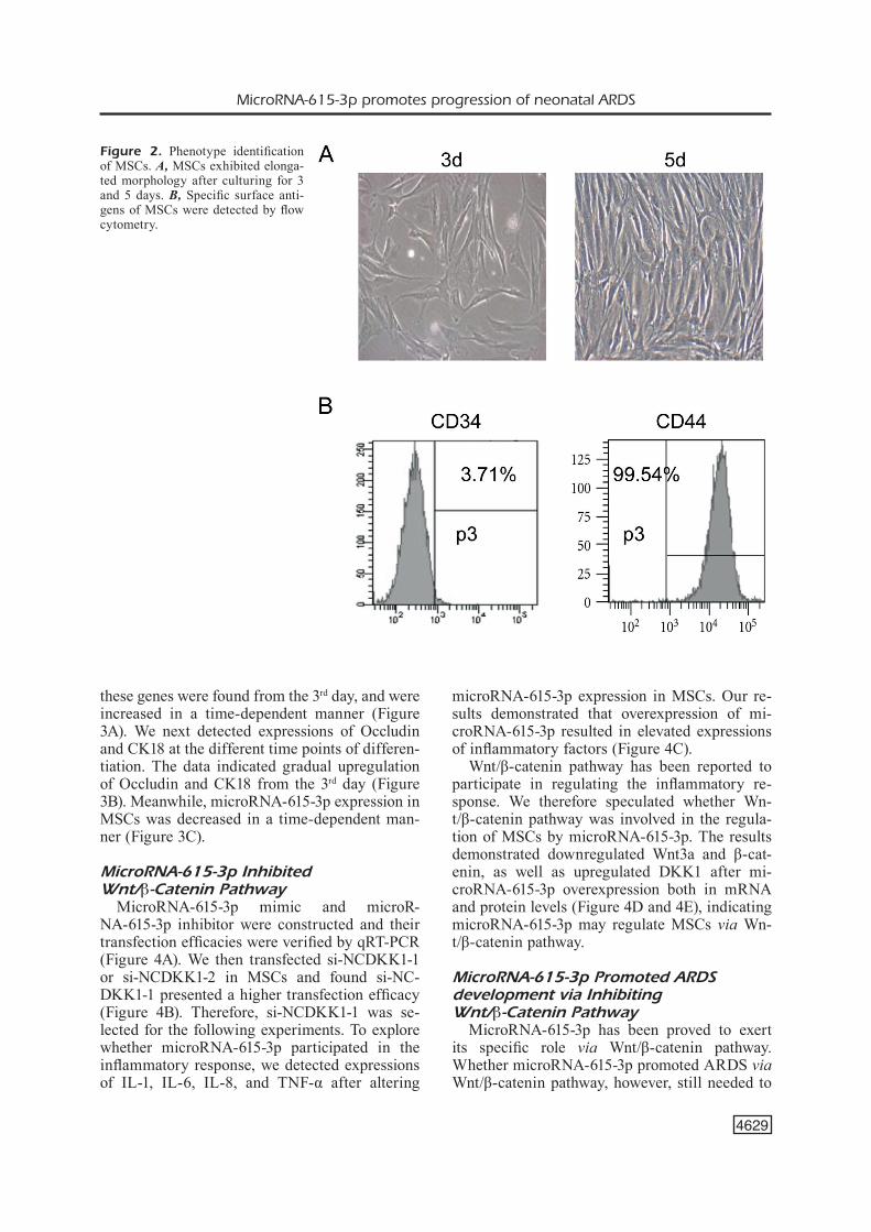
Phenotype Identification of MSCsMSCs exhibited fibroid-like and elongated
morphology after cell culture for 3 and 5 days, respectively (Figure 2A). Third-passage MSCs were collected for identification using flow cyto-metry. The data showed negative-antigen CD34 (3.71%) and positive-antigen CD44 (99.54%), sug-
gesting the successful isolation of MSCs with high purity (Figure 2B).
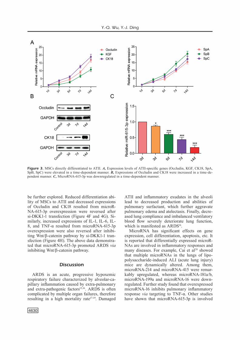
MSCs Directly Differentiated to ATIICo-culture of MSCs and ATII was established
by transwell assay to induce the differentiation of MSCs to ATII. After differentiation induction for 1, 3, 5, 7, and 14 days, ATII-specific genes (Occludin, KGF, CK18, SpA, SpB, and SpC) were detected, respectively. Elevated expressions of
Figure 1. MicroRNA-615-3p was overexpressed in neonatal ARDS patients. A, MicroRNA-615-3p expression was higher in neonatal ARDS patients than those of healthy newborns. B, Expression levels of IL-1, IL-6, IL-8, and TNF-α were higher in peripheral blood of neonatal ARDS patients than those of healthy newborns.
Table I. Basic characteristics of study population.
ARDS group (n=24) Control group (n=14) p
Age (days) 16 (4-28) 17.5 (3-25) 0.523Weight (kg) 1.8 (1.3-3.8) 2.5 (1.5-4.2) 0.774Male sex 10 (41.7%) 7 (50%) 0.468PRISM-III24 14.3 (8.2-20.1) 2.9 (2-4.8) <0.001Modified Murray’s score 5 (4.4-5.9) 0.75 (0.5-1.2) <0.001PaO2/FiO2 121 (80-160) 480 (342-687) <0.001OI 12.3 (9.3-17.2) 1.8 (1.3-2.4) 0.032Crs (mL/cm H2O/kg) 0.36 (0.22-0.43) 0.89 (0.8-0.9) <0.001Deaths 4 (16.7%) 1 (7.1%) Co-morbidities Severe sepsis (3) Metabolic disease (1) RSV infection (6) SAH (2) H1N1 flu (4) Status epilepticus (2) Aspiration (4) General anesthesia (9) Trauma (3) Late onset GBS infection (2) Malignancy (2)
IQR, interquartile range; HCT, hematopoietic cellular transplantation; ARDS, acute respiratory distress syndrome; PRISM-3, Pediatric Risk of Mortality III score; OI, oxygenation index.
MicroRNA-615-3p promotes progression of neonatal ARDS
4629
these genes were found from the 3rd day, and were increased in a time-dependent manner (Figure 3A). We next detected expressions of Occludin and CK18 at the different time points of differen-tiation. The data indicated gradual upregulation of Occludin and CK18 from the 3rd day (Figure 3B). Meanwhile, microRNA-615-3p expression in MSCs was decreased in a time-dependent man-ner (Figure 3C).
MicroRNA-615-3p Inhibited Wnt/β-Catenin Pathway
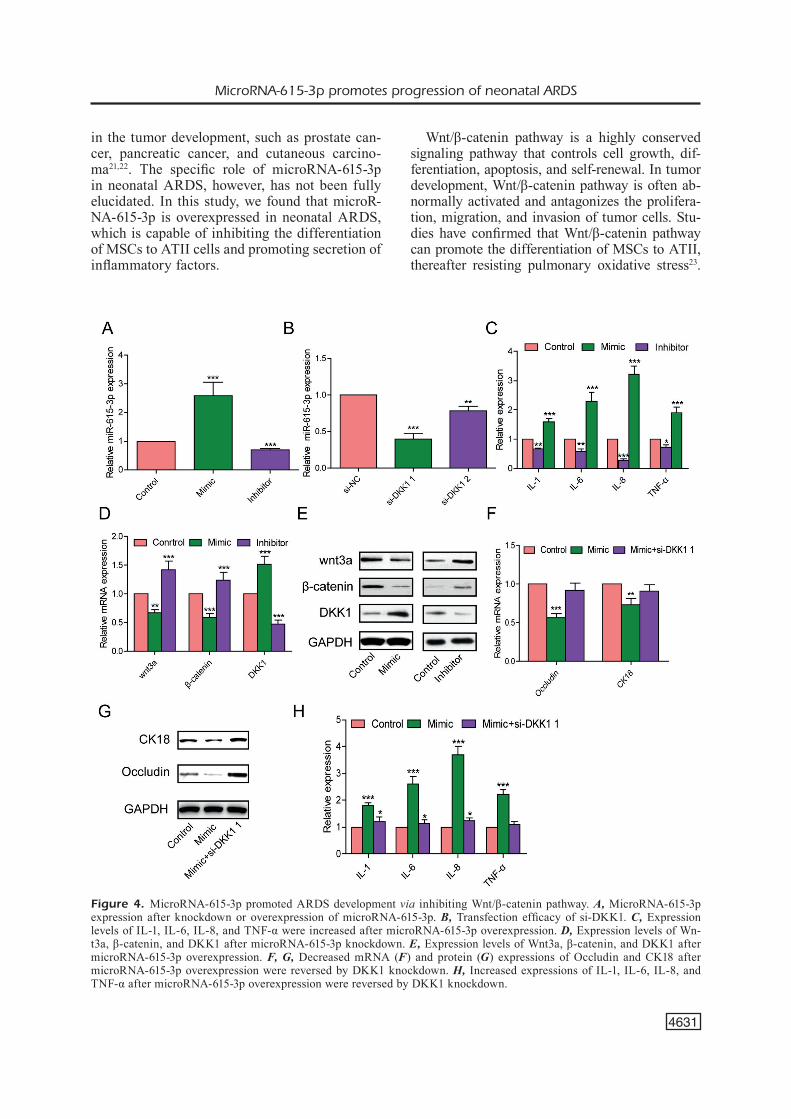
MicroRNA-615-3p mimic and microR-NA-615-3p inhibitor were constructed and their transfection efficacies were verified by qRT-PCR (Figure 4A). We then transfected si-NCDKK1-1 or si-NCDKK1-2 in MSCs and found si-NC-DKK1-1 presented a higher transfection efficacy (Figure 4B). Therefore, si-NCDKK1-1 was se-lected for the following experiments. To explore whether microRNA-615-3p participated in the inflammatory response, we detected expressions of IL-1, IL-6, IL-8, and TNF-α after altering
microRNA-615-3p expression in MSCs. Our re-sults demonstrated that overexpression of mi-croRNA-615-3p resulted in elevated expressions of inflammatory factors (Figure 4C).
Wnt/β-catenin pathway has been reported to participate in regulating the inflammatory re-sponse. We therefore speculated whether Wn-t/β-catenin pathway was involved in the regula-tion of MSCs by microRNA-615-3p. The results demonstrated downregulated Wnt3a and β-cat-enin, as well as upregulated DKK1 after mi-croRNA-615-3p overexpression both in mRNA and protein levels (Figure 4D and 4E), indicating microRNA-615-3p may regulate MSCs via Wn-t/β-catenin pathway.
MicroRNA-615-3p Promoted ARDS development via Inhibiting Wnt/β-Catenin Pathway
MicroRNA-615-3p has been proved to exert its specific role via Wnt/β-catenin pathway. Whether microRNA-615-3p promoted ARDS via Wnt/β-catenin pathway, however, still needed to
Figure 2. Phenotype identification of MSCs. A, MSCs exhibited elonga-ted morphology after culturing for 3 and 5 days. B, Specific surface anti-gens of MSCs were detected by flow cytometry.
Y.-Q. Wu, Y.-J. Ding
4630
be further explored. Reduced differentiation abi-lity of MSCs to ATII and decreased expressions of Occludin and CK18 resulted from microR-NA-615-3p overexpression were reversed after si-DKK1-1 transfection (Figure 4F and 4G). Si-milarly, increased expressions of IL-1, IL-6, IL-8, and TNF-α resulted from microRNA-615-3p overexpression were also reversed after inhibi-ting Wnt/β-catenin pathway by si-DKK1-1 tran-sfection (Figure 4H). The above data demonstra-ted that microRNA-615-3p promoted ARDS via inhibiting Wnt/β-catenin pathway.
Discussion
ARDS is an acute, progressive hypoxemic respiratory failure characterized by alveolar-ca-pillary inflammation caused by extra-pulmonary and extra-pathogenic factors15,16. ARDS is often complicated by multiple organ failures, therefore resulting in a high mortality rate17-19. Damaged
ATII and inflammatory exudates in the alveoli lead to decreased production and abilities of pulmonary surfactant, which further aggravate pulmonary edema and atelectasis. Finally, decre-ased lung compliance and imbalanced ventilatory blood flow severely deteriorate lung function, which is manifested as ARDS16.
MicroRNA has significant effects on gene expression, cell differentiation, apoptosis, etc. It is reported that differentially expressed microR-NAs are involved in inflammatory responses and many diseases. For example, Cai et al20 showed that multiple microRNAs in the lungs of lipo-polysaccharide-induced ALI (acute lung injury) mice are dynamically altered. Among them, microRNA-214 and microRNA-415 were remar-kably upregulated, whereas microRNA-181a/b, microRNA-199a and microRNA-16 were down-regulated. Further study found that overexpressed microRNA-16 inhibits pulmonary inflammatory response via targeting to TNF-α. Other studies have shown that microRNA-615-3p is involved
Figure 3. MSCs directly differentiated to ATII. A, Expression levels of ATII-specific genes (Occludin, KGF, CK18, SpA, SpB, SpC) were elevated in a time-dependent manner. B, Expressions of Occludin and CK18 were increased in a time-de-pendent manner. C, MicroRNA-615-3p was downregulated in a time-dependent manner.
MicroRNA-615-3p promotes progression of neonatal ARDS
4631
in the tumor development, such as prostate can-cer, pancreatic cancer, and cutaneous carcino-ma21,22. The specific role of microRNA-615-3p in neonatal ARDS, however, has not been fully elucidated. In this study, we found that microR-NA-615-3p is overexpressed in neonatal ARDS, which is capable of inhibiting the differentiation of MSCs to ATII cells and promoting secretion of inflammatory factors.
Wnt/β-catenin pathway is a highly conserved signaling pathway that controls cell growth, dif-ferentiation, apoptosis, and self-renewal. In tumor development, Wnt/β-catenin pathway is often ab-normally activated and antagonizes the prolifera-tion, migration, and invasion of tumor cells. Stu-dies have confirmed that Wnt/β-catenin pathway can promote the differentiation of MSCs to ATII, thereafter resisting pulmonary oxidative stress23.
Figure 4. MicroRNA-615-3p promoted ARDS development via inhibiting Wnt/β-catenin pathway. A, MicroRNA-615-3p expression after knockdown or overexpression of microRNA-615-3p. B, Transfection efficacy of si-DKK1. C, Expression levels of IL-1, IL-6, IL-8, and TNF-α were increased after microRNA-615-3p overexpression. D, Expression levels of Wn-t3a, β-catenin, and DKK1 after microRNA-615-3p knockdown. E, Expression levels of Wnt3a, β-catenin, and DKK1 after microRNA-615-3p overexpression. F, G, Decreased mRNA (F) and protein (G) expressions of Occludin and CK18 after microRNA-615-3p overexpression were reversed by DKK1 knockdown. H, Increased expressions of IL-1, IL-6, IL-8, and TNF-α after microRNA-615-3p overexpression were reversed by DKK1 knockdown.
Y.-Q. Wu, Y.-J. Ding
4632
Similarly, overexpressed catenin in MSCs could activate Wnt/β-catenin pathway, so as to improve the pulmonary epithelial permeability and attenua-te pulmonary edema. Overexpressed catenin could also improve pulmonary fibrosis, thereby repairing the lung damage resulted from ARDS24. In the present study, expression levels of Wnt/β-catenin pathway-related genes were detected after altering microRNA-615-3p expression. The results elucida-ted that microRNA-615-3p inhibits the differentia-tion of MSCs to ATII by inhibiting Wnt/β-catenin pathway, so as to improve ARDS development.
Conclusions
We demonstrated that overexpressed microR-NA-615-3p promoted ARDS development throu-gh inhibiting differentiation of MSCs to ATII via Wnt/β-catenin pathway.
Conflict of InterestThe Authors declare that they have no conflict of interest.
References
1) Cabezón nDL, Castro Is, UrIarte UXb, Casanova MPr, Peña JMG, CeLorrIo La. Síndrome de distrés respiratorio agudo: revisión a propósito de la definición de Berlín. Revista Española De Ane-stesiología Y Reanimación 2014; 61: 319-327.
2) MItCheLL Ps, ParkIn rk, kroh eM, FrItz br, WyMan sk, PoGosova-aGaDJanyan eL, Peterson a, notebooM J, o’brIant kC, aLLen a, LIn DW, Urban n, DresCher CW, knUDsen bs, stIreWaLt DL, GentLeMan r, vesseL-La rL, neLson Ps, MartIn Db, teWarI M. Circulating microRNAs as stable blood-based markers for cancer detection. Proc Natl Acad Sci U S A 2008; 105: 10513-10518.
3) Chen X, ba y, Ma L, CaI X, yIn y, WanG k, GUo J, zhanG y, Chen J, GUo X, LI Q, LI X, WanG W, zhanG y, WanG J, JIanG X, XIanG y, XU C, zhenG P, zhanG J, LI r, zhanG h, shanG X, GonG t, nInG G, WanG J, zen k, zhanG J, zhanG Cy. Characterization of microRNAs in serum: a novel class of biomarkers for diagnosis of cancer and other diseases. Cell Res 2008; 18: 997-1006.
4) shIFenG h, DannI W, PU C, PInG y, JU C, LIPInG z. Cir-culating liver-specific miR-122 as a novel potential biomarker for diagnosis of cholestatic liver injury. PLoS One 2013; 8: e73133.
5) XIn zC, yanG hQ, WanG XW, zhanG Q. Diagnostic value of microRNAs in breast cancer: A meta-a-nalysis. Eur Rev Med Pharmacol Sci 2017; 21: 284-291.
6) abUe M, yokoyaMa M, shIbUya r, taMaI k, yaMaGU-ChI k, sato I, tanaka n, haMaDa s, shIMoseGaWa t, sUGaMUra k, satoh k. Circulating miR-483-3p and miR-21 is highly expressed in plasma of pancre-atic cancer. Int J Oncol 2015; 46: 539-547.
7) zhenG n, yanG P, WanG z, zhoU Q. OncomicroR-NAs-mediated tumorigenesis: Implication in can-cer diagnosis and targeted therapy. Curr Cancer Drug Targets 2017; 17: 40-47.
8) barteL DP. MicroRNAs: target recognition and regulatory functions. Cell 2009; 136: 215-233.
9) Ma r, JIanG t, kanG X. Circulating microRNAs in cancer: origin, function and application. J Exp Clin Cancer Res 2012; 31: 38.
10) FabbrI M, Paone a, CaLore F, GaLLI r, GaUDIo e, santhanaM r, Lovat F, FaDDa P, Mao C, nUovo GJ, zanesI n, CraWForD M, ozer Gh, WernICke D, aLDer h, CaLIGIUrI Ma, nana-sInkaM P, PerrottI D, CroCe CM. MicroRNAs bind to Toll-like receptors to in-duce prometastatic inflammatory response. Proc Natl Acad Sci U S A 2012; 109: E2110-E2116.
11) JIanG a, zhanG s, LI z, LIanG r, ren s, LI J, PU y, yanG J. MiR-615-3p promotes the phagocytic capacity of splenic macrophages by targeting ligand-dependent nuclear receptor corepressor in cirrhosis-related portal hypertension. Exp Biol Med (Maywood) 2011; 236: 672-680.
12) yIn J, zhUanG G, zhU y, hU X, zhao h, zhanG r, GUo h, Fan X, Cao y. MiR-615-3p inhibits the osteoge-nic differentiation of human lumbar ligamentum flavum cells via suppression of osteogenic regu-lators GDF5 and FOXO1. Cell Biol Int 2017; 41: 779-786.
13) PU hy, XU r, zhanG My, yUan LJ, hU Jy, hUanG GL, WanG hy. Identification of microRNA-615-3p as a novel tumor suppressor in non-small cell lung cancer. Oncol Lett 2017; 13: 2403-2410.
14) hessert D, tanzer D, brUnstetter t, kaUPP s, MUr-DoCh D, MIrzaoFF M. Topical cyclosporine a for postoperative photorefractive keratectomy and laser in situ keratomileusis. J Cataract Refract Surg 2013; 39: 539-547.
15) WanG CC, WU WL, WU et, ChoU hC, LU FL. High frequency oscillatory ventilation in children: expe-rience of a medical center in Taiwan. J Formos Med Assoc 2008; 107: 311-315.
16) MIDDLeton ea, ronDIna Mt, sChWertz h, zIMMerMan Ga. Amicus or adversary revisited: platelets in acute lung injury & acute respiratory distress syndrome. Am J Respir Cell Mol Biol 2018 Mar 19. doi: 10.1165/rcmb.2017-0420TR. [Epub ahead of print].
17) Matthay Ma, zIMMerMan Ga, esMon C, bhattaCharya J, CoLLer b, DoersChUk CM, FLoros J, GIMbrone MJ, hoFFMan e, hUbMayr rD, LePPert M, MataLon s, MUnForD r, Parsons P, sLUtsky as, traCey kJ, WarD P, GaIL Db, harabIn aL. Future research directions in acute lung injury: summary of a National Heart, Lung, and Blood Institute working group. Am J Respir Crit Care Med 2003; 167: 1027-1035.
18) rUbenFeLD GD, CaLDWeLL e, PeaboDy e, Weaver J, MartIn DP, neFF M, stern eJ, hUDson LD. Incidence
MicroRNA-615-3p promotes progression of neonatal ARDS
4633
and outcomes of acute lung injury. N Engl J Med 2005; 353: 1685-1693.
19) Ueno h, MatsUDa t, hashIMoto s, aMaya F, kItaMUra y, tanaka M, kobayashI a, MarUyaMa I, yaMaDa s, haseGaWa n, soeJIMa J, koh h, IshIzaka a. Con-tributions of high mobility group box protein in experimental and clinical acute lung injury. Am J Respir Crit Care Med 2004; 170: 1310-1316.
20) CaI zG, zhanG sM, zhanG y, zhoU yy, WU hb, XU XP. MicroRNAs are dynamically regulated and play an important role in LPS-induced lung injury. Can J Physiol Pharmacol 2012; 90: 37-43.
21) hULF t, sIbbrItt t, WIkLUnD eD, bert s, strbenaC D, stathaM aL, robInson MD, CLark sJ. Discovery pi-peline for epigenetically deregulated miRNAs in cancer: Integration of primary miRNA transcrip-tion. Bmc Genomics 2011; 12: 54.
22) soares Mr, hUber J, rIos aF, raMos es. Investiga-tion of IGF2/ApaI and H19/RsaI polymorphisms in patients with cutaneous melanoma. Growth Horm Igf Res 2010; 20: 295-297.
23) LIU ar, LIU L, Chen s, yanG y, zhao hJ, LIU L, GUo FM, LU XM, QIU hb. Activation of canonical wnt pathway promotes differentiation of mouse bone marrow-derived MSCs into type II alveolar epithe-lial cells, confers resistance to oxidative stress, and promotes their migration to injured lung tis-sue in vitro. J Cell Physiol 2013; 228: 1270-1283.
24) CaI sX, LIU ar, Chen s, he hL, Chen Qh, XU Jy, Pan C, yanG y, GUo FM, hUanG yz, LIU L, QIU hb. Acti-vation of Wnt/beta-catenin signalling promotes mesenchymal stem cells to repair injured alveolar epithelium induced by lipopolysaccharide in mice. Stem Cell Res Ther 2015; 6: 65.