A specific immunological method to detect and quantify bacterial 2-substituted (1,3)-β-d-glucan
7
Carbohydrate Polymers 113 (2014) 39–45 Contents lists available at ScienceDirect Carbohydrate Polymers j ourna l ho me page: www.elsevier.com/locate/carbpol A specific immunological method to detect and quantify bacterial 2-substituted (1,3)--d-glucan María Laura Werning a , Adrián Pérez-Ramos a , Pilar Fernández de Palencia a , María Luz Mohedano a , María Teresa Due ˜ nas b , Alicia Prieto a , Paloma López a,∗ a Centro de Investigaciones Biológicas, C.S.I.C., Ramiro de Maeztu 9, 28040 Madrid, Spain b Departamento de Química Aplicada, Facultad de Ciencias Químicas, Box 1072, 20080 San Sebastian, Spain a r t i c l e i n f o Article history: Received 23 April 2014 Received in revised form 25 June 2014 Accepted 27 June 2014 Available online 6 July 2014 Keywords: Exopolysaccharide Lactic acid bacteria -d-Glucan Probiotic Prebiotic a b s t r a c t Exopolysaccharides synthesized by lactic acid bacteria have prebiotic properties and contribute to the rheology and texture of fermented foods. Here, we have standardized an immunological method for the specific detection of 2-substituted (1,3)--d-glucans. The method allows direct detection and quantifi- cation of this exopolysaccharide in culture supernatants containing other mono- and poly-saccharides. Moreover, it allows specific detection of the biomolecules synthesized in vitro in enzymatic reactions. Thus, this method allows the fast identification of producing bacteria, as well as biochemical characteri- zation of the glycosyltransferases responsible for their synthesis. © 2014 Elsevier Ltd. All rights reserved. 1. Introduction Most commercialized probiotic microorganisms are lactic acid bacteria (LAB) and some produce exopolysaccharides (EPS) (Ruas- Madiedo, Abraham, Mozzi, & de los Reyes-Gavilán, 2008; Salminen et al., 1998; Ventura, Canchaya, Fitzgerald, Gupta, & van Sinderen, 2007). In addition, some LAB strains belonging to the Pediococcus, Lactobacillus and Oenococcus genera, isolated from cider and wine, are able to produce a 2-substituted (1,3)--d-glucan EPS (Dols- Lafargue et al., 2008; Due ˜ nas-Chasco et al., 1997, 1998; Ibarburu et al., 2007; Llaubères, Richard, Lonvaud, Dubourdieu, & Fournet, 1990). Analysis of the rheological properties of the 2-substituted (1,3)--d-glucan synthesized by Pediococcus parvulus 2.6R showed that it has potential utility as a biothickener (Lambo-Fodje et al., 2007; Velasco et al., 2009). Pediococcus (Fernández de Palencia et al., 2009) and Lactobacillus (Garai-Ibabe et al., 2010) strains that produce this EPS have increased adherence to Caco-2 cells. Abbreviations: DM, defined medium; ELISA, enzyme linked immuno- sorbent assay; EPS, exopolysaccharide; GLC, gas–liquid chromatography; GLC–MS, gas–liquid chromatography–mass spectrometry; HP-SEC, high-performance size exclusion chromatography; LAB, lactic acid bacteria; PBS, Phosphate buffered saline; SDM, semi-defined medium. ∗ Corresponding author. Tel.: +34 918373112; fax: +34 915360432. E-mail address: [email protected] (P. López). P. parvulus 2.6R is able to immunomodulate macrophages (Fernández de Palencia et al., 2009) and production of the 2- substituted (1,3)--d-glucan confers to the intestinal Lactobacillus paracasei NFBC 338 higher resistance to gastrointestinal and technological stresses (Stack et al., 2010). In addition, human con- sumption of oat-based food prepared with P. parvulus 2.6R resulted in a decrease of serum cholesterol levels, boosting the effect previously demonstrated for (1,3)(1,4)--d-glucans of oat-based products (Mårtensson et al., 2005). Moreover, the production of yogurt and various beverages with 2-substituted (1,3)--d-glucan producing LAB indicates the advantageous techno-functional prop- erties of these strains (Elizaquível et al., 2011; Kearney et al., 2011). Furthermore, the purified EPS from P. parvulus 2.6R increased the growth, viability and adherence capability of probiotic LAB (Russo et al., 2012). Therefore, LAB producing this EPS could have poten- tial as probiotic strains and the 2-substituted (1,3)--d-glucan itself appears to have prebiotic properties. The specific detection and quantification of EPS produced by LAB, especially in a background that contains other car- bohydrates, is laborious and time-consuming. The colorimetric phenol–sulphuric method (Dubois, Gilles, Hamilton, Rebers, & Smith, 1956) to quantify the total polysaccharide content of sam- ples, requires the removal of any disaccharides or monosaccharides present in the media by ultrafiltration, dialysis or ethanol precip- itation (Ruas-Madiedo & de los Reyes-Gavilán, 2005; Notararigo et al., 2013). A complete characterization of the polysaccharide http://dx.doi.org/10.1016/j.carbpol.2014.06.072 0144-8617/© 2014 Elsevier Ltd. All rights reserved.
Transcript of A specific immunological method to detect and quantify bacterial 2-substituted (1,3)-β-d-glucan
A2
MMa
b
a
ARRAA
KEL�PP
1
bMe2LaLe1(t2et
sgeS
h0
Carbohydrate Polymers 113 (2014) 39–45
Contents lists available at ScienceDirect
Carbohydrate Polymers
j ourna l ho me page: www.elsev ier .com/ locate /carbpol
specific immunological method to detect and quantify bacterial-substituted (1,3)-�-d-glucan
aría Laura Werninga, Adrián Pérez-Ramosa, Pilar Fernández de Palenciaa,aría Luz Mohedanoa, María Teresa Duenasb, Alicia Prietoa, Paloma Lópeza,∗
Centro de Investigaciones Biológicas, C.S.I.C., Ramiro de Maeztu 9, 28040 Madrid, SpainDepartamento de Química Aplicada, Facultad de Ciencias Químicas, Box 1072, 20080 San Sebastian, Spain
r t i c l e i n f o
rticle history:eceived 23 April 2014eceived in revised form 25 June 2014ccepted 27 June 2014vailable online 6 July 2014
a b s t r a c t
Exopolysaccharides synthesized by lactic acid bacteria have prebiotic properties and contribute to therheology and texture of fermented foods. Here, we have standardized an immunological method for thespecific detection of 2-substituted (1,3)-�-d-glucans. The method allows direct detection and quantifi-cation of this exopolysaccharide in culture supernatants containing other mono- and poly-saccharides.Moreover, it allows specific detection of the biomolecules synthesized in vitro in enzymatic reactions.
eywords:xopolysaccharideactic acid bacteria-d-Glucanrobiotic
Thus, this method allows the fast identification of producing bacteria, as well as biochemical characteri-zation of the glycosyltransferases responsible for their synthesis.
© 2014 Elsevier Ltd. All rights reserved.
rebiotic
. Introduction
Most commercialized probiotic microorganisms are lactic acidacteria (LAB) and some produce exopolysaccharides (EPS) (Ruas-adiedo, Abraham, Mozzi, & de los Reyes-Gavilán, 2008; Salminen
t al., 1998; Ventura, Canchaya, Fitzgerald, Gupta, & van Sinderen,007). In addition, some LAB strains belonging to the Pediococcus,actobacillus and Oenococcus genera, isolated from cider and wine,re able to produce a 2-substituted (1,3)-�-d-glucan EPS (Dols-afargue et al., 2008; Duenas-Chasco et al., 1997, 1998; Ibarburut al., 2007; Llaubères, Richard, Lonvaud, Dubourdieu, & Fournet,990). Analysis of the rheological properties of the 2-substituted1,3)-�-d-glucan synthesized by Pediococcus parvulus 2.6R showedhat it has potential utility as a biothickener (Lambo-Fodje et al.,
007; Velasco et al., 2009). Pediococcus (Fernández de Palenciat al., 2009) and Lactobacillus (Garai-Ibabe et al., 2010) strainshat produce this EPS have increased adherence to Caco-2 cells.Abbreviations: DM, defined medium; ELISA, enzyme linked immuno-orbent assay; EPS, exopolysaccharide; GLC, gas–liquid chromatography; GLC–MS,as–liquid chromatography–mass spectrometry; HP-SEC, high-performance sizexclusion chromatography; LAB, lactic acid bacteria; PBS, Phosphate buffered saline;DM, semi-defined medium.∗ Corresponding author. Tel.: +34 918373112; fax: +34 915360432.
E-mail address: [email protected] (P. López).
ttp://dx.doi.org/10.1016/j.carbpol.2014.06.072144-8617/© 2014 Elsevier Ltd. All rights reserved.
P. parvulus 2.6R is able to immunomodulate macrophages(Fernández de Palencia et al., 2009) and production of the 2-substituted (1,3)-�-d-glucan confers to the intestinal Lactobacillusparacasei NFBC 338 higher resistance to gastrointestinal andtechnological stresses (Stack et al., 2010). In addition, human con-sumption of oat-based food prepared with P. parvulus 2.6R resultedin a decrease of serum cholesterol levels, boosting the effectpreviously demonstrated for (1,3)(1,4)-�-d-glucans of oat-basedproducts (Mårtensson et al., 2005). Moreover, the production ofyogurt and various beverages with 2-substituted (1,3)-�-d-glucanproducing LAB indicates the advantageous techno-functional prop-erties of these strains (Elizaquível et al., 2011; Kearney et al., 2011).Furthermore, the purified EPS from P. parvulus 2.6R increased thegrowth, viability and adherence capability of probiotic LAB (Russoet al., 2012). Therefore, LAB producing this EPS could have poten-tial as probiotic strains and the 2-substituted (1,3)-�-d-glucan itselfappears to have prebiotic properties.
The specific detection and quantification of EPS producedby LAB, especially in a background that contains other car-bohydrates, is laborious and time-consuming. The colorimetricphenol–sulphuric method (Dubois, Gilles, Hamilton, Rebers, &Smith, 1956) to quantify the total polysaccharide content of sam-
ples, requires the removal of any disaccharides or monosaccharidespresent in the media by ultrafiltration, dialysis or ethanol precip-itation (Ruas-Madiedo & de los Reyes-Gavilán, 2005; Notararigoet al., 2013). A complete characterization of the polysaccharide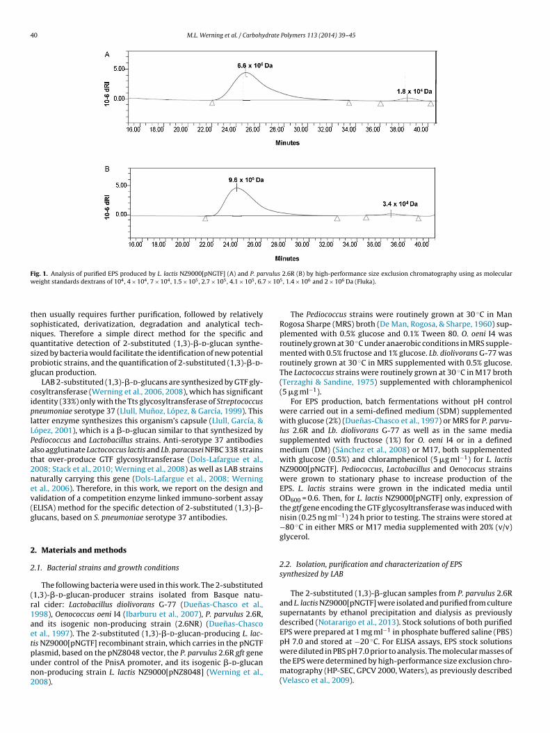
40 M.L. Werning et al. / Carbohydrate Polymers 113 (2014) 39–45
Fig. 1. Analysis of purified EPS produced by L. lactis NZ9000[pNGTF] (A) and P. parvulus 2.6R (B) by high-performance size exclusion chromatography using as molecularw 7 × 10
tsnqspg
ciplLPat2nev(g
2
2
(r1aetpun2
eight standards dextrans of 104, 4 × 10⁴, 7 × 10⁴, 1.5 × 10⁵, 2.7 × 10⁵, 4.1 × 10⁵, 6.
hen usually requires further purification, followed by relativelyophisticated, derivatization, degradation and analytical tech-iques. Therefore a simple direct method for the specific anduantitative detection of 2-substituted (1,3)-�-d-glucan synthe-ized by bacteria would facilitate the identification of new potentialrobiotic strains, and the quantification of 2-substituted (1,3)-�-d-lucan production.
LAB 2-substituted (1,3)-�-d-glucans are synthesized by GTF gly-osyltransferase (Werning et al., 2006, 2008), which has significantdentity (33%) only with the Tts glycosyltransferase of Streptococcusneumoniae serotype 37 (Llull, Munoz, López, & García, 1999). Thisatter enzyme synthesizes this organism’s capsule (Llull, García, &ópez, 2001), which is a �-d-glucan similar to that synthesized byediococcus and Lactobacillus strains. Anti-serotype 37 antibodieslso agglutinate Lactococcus lactis and Lb. paracasei NFBC 338 strainshat over-produce GTF glycosyltransferase (Dols-Lafargue et al.,008; Stack et al., 2010; Werning et al., 2008) as well as LAB strainsaturally carrying this gene (Dols-Lafargue et al., 2008; Werningt al., 2006). Therefore, in this work, we report on the design andalidation of a competition enzyme linked immuno-sorbent assayELISA) method for the specific detection of 2-substituted (1,3)-�-lucans, based on S. pneumoniae serotype 37 antibodies.
. Materials and methods
.1. Bacterial strains and growth conditions
The following bacteria were used in this work. The 2-substituted1,3)-�-d-glucan-producer strains isolated from Basque natu-al cider: Lactobacillus diolivorans G-77 (Duenas-Chasco et al.,998), Oenococcus oeni I4 (Ibarburu et al., 2007), P. parvulus 2.6R,nd its isogenic non-producing strain (2.6NR) (Duenas-Chascot al., 1997). The 2-substituted (1,3)-�-d-glucan-producing L. lac-is NZ9000[pNGTF] recombinant strain, which carries in the pNGTF
lasmid, based on the pNZ8048 vector, the P. parvulus 2.6R gft genender control of the PnisA promoter, and its isogenic �-d-glucanon-producing strain L. lactis NZ9000[pNZ8048] (Werning et al.,008).5, 1.4 × 10⁶ and 2 × 10⁶ Da (Fluka).
The Pediococcus strains were routinely grown at 30 ◦C in ManRogosa Sharpe (MRS) broth (De Man, Rogosa, & Sharpe, 1960) sup-plemented with 0.5% glucose and 0.1% Tween 80. O. oeni I4 wasroutinely grown at 30 ◦C under anaerobic conditions in MRS supple-mented with 0.5% fructose and 1% glucose. Lb. diolivorans G-77 wasroutinely grown at 30 ◦C in MRS supplemented with 0.5% glucose.The Lactococcus strains were routinely grown at 30 ◦C in M17 broth(Terzaghi & Sandine, 1975) supplemented with chloramphenicol(5 �g ml−1).
For EPS production, batch fermentations without pH controlwere carried out in a semi-defined medium (SDM) supplementedwith glucose (2%) (Duenas-Chasco et al., 1997) or MRS for P. parvu-lus 2.6R and Lb. diolivorans G-77 as well as in the same mediasupplemented with fructose (1%) for O. oeni I4 or in a definedmedium (DM) (Sánchez et al., 2008) or M17, both supplementedwith glucose (0.5%) and chloramphenicol (5 �g ml−1) for L. lactisNZ9000[pNGTF]. Pediococcus, Lactobacillus and Oenococus strainswere grown to stationary phase to increase production of theEPS. L. lactis strains were grown in the indicated media untilOD600 = 0.6. Then, for L. lactis NZ9000[pNGTF] only, expression ofthe gtf gene encoding the GTF glycosyltransferase was induced withnisin (0.25 ng ml−1) 24 h prior to testing. The strains were stored at−80 ◦C in either MRS or M17 media supplemented with 20% (v/v)glycerol.
2.2. Isolation, purification and characterization of EPSsynthesized by LAB
The 2-substituted (1,3)-�-glucan samples from P. parvulus 2.6Rand L. lactis NZ9000[pNGTF] were isolated and purified from culturesupernatants by ethanol precipitation and dialysis as previouslydescribed (Notararigo et al., 2013). Stock solutions of both purifiedEPS were prepared at 1 mg ml−1 in phosphate buffered saline (PBS)pH 7.0 and stored at −20 ◦C. For ELISA assays, EPS stock solutions
were diluted in PBS pH 7.0 prior to analysis. The molecular masses ofthe EPS were determined by high-performance size exclusion chro-matography (HP-SEC, GPCV 2000, Waters), as previously described(Velasco et al., 2009).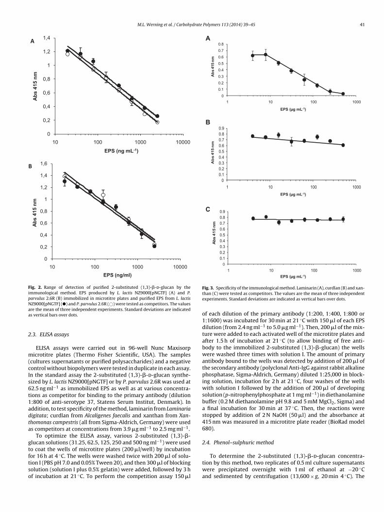
M.L. Werning et al. / Carbohydrate Polymers 113 (2014) 39–45 41
0
0,2
0,4
0,6
0,8
1
1,2
1,4
1,6
10 10 0 100 0 1000 0
Abs
415
nm
EPS (ng /ml)
0
0,2
0,4
0,6
0,8
1
1,2
1,4
10 10 0 100 0 1000 0
Abs
415
nm
EPS (ng mL-1)
A
B
Fig. 2. Range of detection of purified 2-substituted (1,3)-�-d-glucan by theimmunological method. EPS produced by L. lactis NZ9000[pNGTF] (A) and P.parvulus 2.6R (B) immobilized in microtitre plates and purified EPS from L. lactisNZ9000[pNGTF] (�) and P. parvulus 2.6R (©) were tested as competitors. The valuesaa
2
m(cIs6t1adta
gtftso
00.10.20.30.40.50.60.70.80.9
1000100101
Ab
s 41
5 n
m
B
A
0
0.1
0.2
0.3
0.4
0.5
0.6
0.7
0.8
1000100101
Abs
415
nm
C
00.10.20.30.40.50.60.70.80.9
1000100101
Abs
415
nm
EPS (µg mL-1)
EPS (µg mL-1)
EPS (µg mL-1)
To determine the 2-substituted (1,3)-�-d-glucan concentra-
re the mean of three independent experiments. Standard deviations are indicateds vertical bars over dots.
.3. ELISA assays
ELISA assays were carried out in 96-well Nunc Maxisorpicrotitre plates (Thermo Fisher Scientific, USA). The samples
cultures supernatants or purified polysaccharides) and a negativeontrol without biopolymers were tested in duplicate in each assay.n the standard assay the 2-substituted (1,3)-�-d-glucan synthe-ized by L. lactis NZ9000[pNGTF] or by P. parvulus 2.6R was used at2.5 ng ml−1 as immobilized EPS as well as at various concentra-ions as competitor for binding to the primary antibody (dilution:800 of anti-serotype 37, Statens Serum Institut, Denmark). Inddition, to test specificity of the method, laminarin from Laminariaigitata; curdlan from Alcaligenes faecalis and xanthan from Xan-homonas campestris (all from Sigma-Aldrich, Germany) were useds competitors at concentrations from 3.9 �g ml−1 to 2.5 mg ml−1.
To optimize the ELISA assay, various 2-substituted (1,3)-�-lucan solutions (31.25, 62.5, 125, 250 and 500 ng ml−1) were usedo coat the wells of microtitre plates (200 �l/well) by incubationor 16 h at 4 ◦C. The wells were washed twice with 200 �l of solu-
ion I (PBS pH 7.0 and 0.05% Tween 20), and then 300 �l of blockingolution (solution I plus 0.5% gelatin) were added, followed by 3 hf incubation at 21 ◦C. To perform the competition assay 150 �lFig. 3. Specificity of the immunological method. Laminarin (A), curdlan (B) and xan-than (C) were tested as competitors. The values are the mean of three independentexperiments. Standard deviations are indicated as vertical bars over dots.
of each dilution of the primary antibody (1:200, 1:400, 1:800 or1:1600) was incubated for 30 min at 21 ◦C with 150 �l of each EPSdilution (from 2.4 ng ml−1 to 5.0 �g ml−1). Then, 200 �l of the mix-ture were added to each activated well of the microtitre plates andafter 1.5 h of incubation at 21 ◦C (to allow binding of free anti-body to the immobilized 2-substituted (1,3)-�-glucan) the wellswere washed three times with solution I. The amount of primaryantibody bound to the wells was detected by addition of 200 �l ofthe secondary antibody (polyclonal Anti-IgG against rabbit alkalinephosphatase, Sigma-Aldrich, Germany) diluted 1:25,000 in block-ing solution, incubation for 2 h at 21 ◦C, four washes of the wellswith solution I followed by the addition of 200 �l of developingsolution (p-nitrophenylphosphate at 1 mg ml−1) in diethanolaminebuffer (0.2 M diethanolamine pH 9.8 and 5 mM MgCl2, Sigma) anda final incubation for 30 min at 37 ◦C. Then, the reactions werestopped by addition of 2 N NaOH (50 �l) and the absorbance at415 nm was measured in a microtitre plate reader (BioRad model680).
2.4. Phenol–sulphuric method
tion by this method, two replicates of 0.5 ml culture supernatantswere precipitated overnight with 1 ml of ethanol at −20 ◦Cand sedimented by centrifugation (13,600 × g, 20 min 4 ◦C). The
42 M.L. Werning et al. / Carbohydrate Polymers 113 (2014) 39–45
Table 1Comparative analysis of exopolysaccharide production by ELISA and phenol–sulphuric methods.
Bacteria Method
ELISA Phenol–sulphuric Phenol–sulfuric/ELISA
EPS concentration
Totala (�g ml−1) Precipitatedb (�g ml−1) Precipitatedb/totala (%) Precipitatedb (�g ml−1) Precipitatedb/precipitatedb (%)
P. parvulus 2.6R 322 ± 15 200 ± 35 62 117 ± 21 58L. lactis NZ9000/pNGTF 235 ± 35 183 ± 19 78 117 ± 8 64O. oeni I4 118 ± 7 94 ± 9 80 59 ± 5 63L. diolivorans G-77 211 ± 40 166 ± 24 79 64 ± 6 39
s.rmined after ethanol precipitation.
paFob(
2
idccr1fowwrpL
2
ut2rfatagrus2
3
3
oNlt(
0
100
200
300
400
500
600
700
800
900
EPS
(µg
mL-
1 )
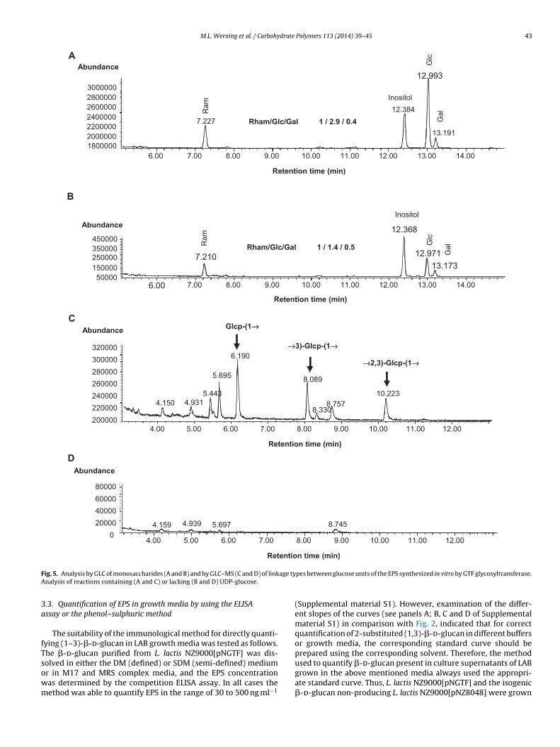
Fig. 4. Detection of 2-substituted (1,3)-�-d-glucan in culture supernatants. (A) Theindicated L. lactis strains were grown and expression of the EPS induced in eitherM17 medium. P. parvulus 2.6R and 2.6NR, Lb. diolivorans G-77 and O. oeni I4 weregrown in MRS medium. Triplicates of these cultures were then centrifuged and thesupernatants were directly used for quantification of the �-d-glucan present by the
primary antibody even at very high concentration (500 �g ml−1)
a Total: concentration of the EPS was determined directly in culture supernatantb Precipitated: concentration of the EPS present in culture supernatants was dete
recipitates were twice resuspended in 0.5 ml of distilled waternd ethanol reprecipitated and resedimented as described above.inally, after resuspension in distilled water, the concentrationf the biopolymer present in the precipitates was determinedy the phenol–sulphuric method as reported by Dubois et al.1956).
.5. Analysis of GTF glycosyltransferase activity
Membrane vesicles displaying GTF glycosyltransferase activ-ty, were prepared from L. lactis NZ9000[pNGTF] as previouslyescribed (Werning et al., 2008) and the enzymatic reactions werearried out as follows. Duplicate 100 �l reactions were preparedontaining 12.5, 25 or 50 �g of membrane proteins as well as aange of UDP-glucose (Sigma) concentrations (from 0.04 to 25 mM),
mM CaCl2, 9 mM MgCl2 and 50 mM NaCl. These were incubatedor 30 min at 30 ◦C then the reactions were stopped by additionf 900 �l of 100% ethanol. After 2 h incubation at 4 ◦C, samplesere centrifuged at 13,226 × g at 4 ◦C for 20 min, the precipitatesere air dried, and resuspended in 500 �l of PBS buffer pH 8.0. The
eaction product was quantified by the ELISA method. The kineticarameter, apparent Km and apparent Vmax were determined fromineweaver Burk plots.
.6. Chemical analysis of the enzymatic reaction product
The neutral sugar composition and linkage type was determinedsing polysaccharide synthesized in duplicate 1 ml reactions con-aining membrane vesicles (50 mg of total proteins) with or without5 mM UDP-glucose. The ethanol precipitates, after air drying, wereesuspended in 1 ml of distilled H2O plus 5 �l of 10% SDS, incubatedor 15 min at 37 ◦C and dialyzed (molecular mass cut-off 3.5 kDa)gainst distilled H2O for 24 h. Then, 100 �l aliquots containinghe membrane vesicles and the reaction product were hydrolyzed,cetylated and analyzed for the presence of neutral sugars byas–liquid chromatography (GLC) or after methylation, hydrolysis,eduction and acetylation for the determination of the structuralnit of the polysaccharide by gas–liquid chromatography–masspectrometry (GLC–MS) as previously described (Werning et al.,008).
. Results
.1. Sensitivity of the ELISA assay
To determine the sensitivity of the method two samplesf 2-substituted (1,3)-�-glucans synthesized by either L. lactis
Z9000[pNGTF] or P. parvulus 2.6R were purified and their molecu-ar masses determined by HP-SEC. The average molecular masses ofhe two main fractions were 6.6 × 10⁶ and 1.8 × 10⁴ Da for L. lactisFig. 1A) and of 9.6 × 10⁶ and 3.4 × 10⁴ Da for P. parvulus (Fig. 1B).
ELISA assay. The values are the mean of three independent experiments. Standarddeviations are indicated as vertical bars over bars.
The higher molecular mass fraction was the most abundant forboth strains constituting, respectively, 96% and 92% of the totalpolymer population. Both EPS preparations were used for testing asimmobilized �-glucans and as competitors for binding to the pri-mary antibody. Both EPS preparations were immobilized efficiently(Fig. 2A versus B) and both purified �-d-glucans could be quantifiedwithin a range of 20 to 2500 ng ml−1, independently of the originof the immobilized polymer (Fig. 2).
3.2. Specificity of the assay
Using the purified �-d-glucan from L. lactis (described above)as the immobilized substrate, we tested the specificity of themethod using three polysaccharides as competitors: (i) laminarin,a 6-substituted (1,3)-�-d-glucan (Fig. 3A); (ii) curdlan, a linear(1–3)-�-d-glucan (Fig. 3B) and (iii) xanthan, a branched het-eropolysaccharide (Fig. 3C). Xanthan was unable to bind to the
and curdlan also showed a very low affinity for the antibody at250 �g ml−1. Laminarin was able to compete with the immobilized2-substituted (1,3)-�-d-glucan in a linear mode, when tested in therange 10 to 250 �g ml−1.
M.L. Werning et al. / Carbohydrate Polymers 113 (2014) 39–45 43
A
GalRam
1 / 2.9 / 0.4Rham/Glc/Gal
Inositol
14.0013.0012.0011.0010.009.008.007.006.001800000200000022000002400000260000 02800000300000 0
Glc
7.22712.384
12.99 3
13.191
Abundance
Retention time (min)
B
Ino sitolAbundance
Glc
GalRam
1 / 1.4 / 0.5Rham/Glc/Gal
50000150000250000350000450000
6.00 14.0013.0012.0011.0010.009.008.007.00
7.210
12.36 8
12.97 113.173
Retention time (min)
AbundanceC
Glcp-(1→→
4.150 4.9315.443
5.695
6.190
8.089
8.3308.757
10.223
→→3)-Glcp-( 1→→
→→2,3)-Glcp-( 1→→
200000220000240000260000280000300000320000
12.0011.0010.009.008.007.006.005.004.00
Retention time (min)
DAbundance
12.0011.0010.009.008.007.006.005.004.00
4.159 4.939 5.697 8.7450
20000400006000080000
Retention time (min)
F age tyA
3a
fTsowm
ig. 5. Analysis by GLC of monosaccharides (A and B) and by GLC–MS (C and D) of linknalysis of reactions containing (A and C) or lacking (B and D) UDP-glucose.
.3. Quantification of EPS in growth media by using the ELISAssay or the phenol–sulphuric method
The suitability of the immunological method for directly quanti-ying (1–3)-�-d-glucan in LAB growth media was tested as follows.he �-d-glucan purified from L. lactis NZ9000[pNGTF] was dis-
olved in either the DM (defined) or SDM (semi-defined) mediumr in M17 and MRS complex media, and the EPS concentrationas determined by the competition ELISA assay. In all cases theethod was able to quantify EPS in the range of 30 to 500 ng ml−1pes between glucose units of the EPS synthesized in vitro by GTF glycosyltransferase.
(Supplemental material S1). However, examination of the differ-ent slopes of the curves (see panels A; B, C and D of Supplementalmaterial S1) in comparison with Fig. 2, indicated that for correctquantification of 2-substituted (1,3)-�-d-glucan in different buffersor growth media, the corresponding standard curve should beprepared using the corresponding solvent. Therefore, the method
used to quantify �-d-glucan present in culture supernatants of LABgrown in the above mentioned media always used the appropri-ate standard curve. Thus, L. lactis NZ9000[pNGTF] and the isogenic�-d-glucan non-producing L. lactis NZ9000[pNZ8048] were grown
4 drate
ip(oMwEvgw2ce(
pNlutot(tpctLtai3dwtb
3e
waTmigtt1f
rgIdarcphdctO
4 M.L. Werning et al. / Carbohy
n M17 medium as described previously (Werning et al., 2008). P.arvulus 2.6R and the isogenic �-d-glucan non-producing strain2.6NR) as well as O. oeni I4 and Lb. diolivorans G-77, and twother 2-substituted (1,3)-�-d-glucan producers, were grown inRS medium to stationary phase as described above. The culturesere then centrifuged and, after removal of the supernatants, the
PS still bound to the cells were released by resuspension into oneolume of PBS and the bacterial cells were removed by centrifu-ation. Then, the supernatants, after appropriate dilution in PBS,ere directly used in the ELISA assay. The results revealed that the
-substituted (1,3)-�-d-glucan secreted by the four producing-LABould be detected in the media used, whereas only background lev-ls were observed in the supernatants of the non-producing strainsFig. 4).
To compare the inhibition enzyme immunoassay and thehenol–sulphuric method, culture supernatants of L. lactisZ9000[pNGTF] grown in DM medium and P. parvulus 2.6R, Lb. dio-
ivorans G-77 and O. oeni I4 grown in SDM medium were analyzedsing both methods. Only these media were tested for detec-ion of the 2-substituted (1,3)-�-d-glucan, because the presence ofther polysaccharides in M17 and MRS media impairs the usage ofhe phenol–sulphuric method. The concentration of 2-substituted1,3)-�-d-glucan was determined directly in the supernatants byhe ELISA test or in their ethanol precipitates by the ELISA andhenol–sulphuric methods (Table 1). Determination of the EPS con-entration in the precipitates showed that for all the strains testedhe ELISA assay revealed a higher level of production (from 61% forb. diolivorans G-77 to 36% for L. lactis NZ9000[pNGTF]). In addi-ion, comparison of EPS detection by the ELISA method prior to, orfter, precipitation demonstrated that indeed precipitation resultsn a moderate loss of 2-substituted (1,3)-�-d-glucan in a range from8% to 20% for P. parvulus 2.6R and O. oeni I4 biopolymers. Theseifferences were probably due to low molecular weight EPS thatas not precipitated by ethanol. Therefore, these results indicate
hat the ELISA method allows a more reliable determination of theiopolymer after its synthesis by bacteria.
.4. Detection of 2-substituted (1,3)-ˇ-d-glucan produced in annzymatic reaction
Membrane vesicles containing the GTF glycosyltransferase,ere prepared from L. lactis NZ9000[pNGTF] and the enzymatic
ctivity was analyzed in vitro using UDP-glucose as substrate.he reaction product was detected and quantified by the ELISAethod. Kinetic analysis of the results (Supplemental material S2)
ndicated that the GTF glycosyltransferase has an affinity for UDP-lucose of Km = 123 ± 3 �M and a Vmax = 18 ± 0.8 ng of EPS/mg ofotal protein/min. This Km is almost identical to that reported forhe cellulose synthase BscA (125 �M) (Ross, Mayer, & Benziman,991) which, like GTF, belongs to the GT-2 family of glycosyltrans-erases.
To confirm the nature of the enzymatic reaction product,eactions performed with membrane vesicles and containing UDP-lucose (test) or lacking the substrate (control) were analyzed.n the presence or absence of the substrate, the GLC analysisetected rhamnose, glucose and galactose, in the ratios 1:2.9:0.4nd 1:1.4:0.5 for the test (Fig. 5A) and control (Fig. 5B) reactions,espectively. Thus, the L. lactis membrane vesicle preparationsontained three monosaccharides, whose origin could be due toolysaccharides bound to them and as expected only glucose wasighest in the test reaction. The GLC–MS analysis (Fig. 5C and D)
etected, in the test reaction only (Fig. 5C), the three peaks that areharacteristic of 2-substitute d-(1,3)-�-glucan, corresponding toerminal glucopyranose, 3-O-substituted glucopyranose, and 2-di--substituted glucopyranose. Thus, the product synthesized in vitroPolymers 113 (2014) 39–45
by the GTF glycosyltransferase was indeed 2-substituted (1,3)-�-d-glucan, confirming the ELISA detection of the product.
4. Discussion
ELISA tests have been developed previously for the detec-tion of both linear and 6-substituted (1,3)-�-d-glucans (Douwes,Doekes, Montijn, Heederik, & Brunekreef, 1996; Sander, Fleischer,Borowitzki, Brüning, & Raulf-Heimsoth, 2008) as well as for the spe-cific detection of 6-substituted (1,3)-�-d-glucans (Milton, UdeniAlwis, Fisette, & Muilemberg, 2001; Rao et al., 2007; Sander et al.,2008). However, as far as we know, this is the first instance ofan ELISA test developed for the detection of 2-substituted (1,3)-�-d-glucan. In the inhibition enzyme immunoassay developed inthis work the commercially available antibody (anti-S. pneumoniaeserotype 37) has been used. Our results show that this primary anti-body specifically detects 2-substituted (1,3)-�-d-glucan, has verylow binding affinity for 6-substituted (1,3)-�-d-glucan and doesnot bind linear (1,3)-�-d-glucan even at a concentration as highas 500 �g ml−1. The ELISA method allows detection of 30 ng ml−1
of the biopolymer in growth medium, a level of sensitivity simi-lar to that observed for other �-d-glucan, such us (1,6)-�-d-glucan(Vink et al., 2004) or (1,3)-�-d-glucan (Douwes et al., 1996), whenpolyclonal antibodies were used for their detection. This method isless sensitive than that obtained for the detection of other polysac-charides with monoclonal antibodies (in the range of pg ml−1) orthan the amoebocyte lysate assays for which commercial kits, suchas Glucatell® (Associates of Cape Cod, USA) or Fungitic-G (Seika-gaku, Japan) are available. However, the first mentioned type ofassay cannot be applied to the detection of 2-substituted (1,3)-�-d-glucan and the second type does not discriminate betweendifferent types of (1,3)-�-d-glucans. In addition, our results showthat the sensitivity of our ELISA test permits the detection of novel2-substituted (1,3)-�-d-glucan-producing bacteria by direct anal-ysis of culture supernatants even after growth in complex media.Moreover, the method can be used for the biochemical characteri-zation of the glycosyltransferases involved in their synthesis. Thesefindings will facilitate the identification of new probiotic strains andthe optimization of the production of this EPS.
Acknowledgements
We thank Dr. Stephen Elson for critical reading of themanuscript. This work was supported by the Spanish Min-istry of Economy and Competiveness grants AGL2009-12998 andAGL2012-40084.
Appendix A. Supplementary data
Supplementary material related to this article can be found,in the online version, at http://dx.doi.org/10.1016/j.carbpol.2014.06.072.
References
De Man, J. C., Rogosa, M., & Sharpe, M. E. (1960). A medium for the cultivation ofLactobacilli. Journal of Applied Bacteriology, 23(1), 130–135.
Dols-Lafargue, M., Lee, H. Y., Le Marrec, C., Heyraud, A., Chambat, G., & Lonvaud-Funel, A. (2008). Characterization of gtf, a glucosyltransferase gene in thegenomes of Pediococcus parvulus and Oenococcus oeni. Applied EnvironmentalMicrobiology, 74(13), 4079–4090.
Douwes, J., Doekes, G., Montijn, R., Heederik, D., & Brunekreef, B. (1996). Mea-surement of �-(1,3)-glucans in the occupational and home environment with
an inhibition enzyme immunoassay. Applied Environmental Microbiology, 62(9),3176–3182.Dubois, M., Gilles, K. A., Hamilton, J. K., Rebers, P. A., & Smith, F. (1956). Colori-metric methods for determination of sugars and related substances. AnalyticalChemistry, 28(3), 350–356.
drate
D
D
E
F
G
I
K
L
L
L
L
M
M
M.L. Werning et al. / Carbohy
uenas-Chasco, M. T., Rodríguez-Carvajal, M. A., Mateo, P. T., Franco-Rodríguez,G., Espartero, J., Irastorza-Iribas, A., et al. (1997). Structural analysis ofthe exopolysaccharide produced by Pediococcus damnosus 2.6. CarbohydrateResearch, 303(4), 453–458.
uenas-Chasco, M. T., Rodríguez-Carvajal, M. A., Tejero-Mateo, P., Espartero, J.L., Irastorza-Iribas, A., & Gil-Serrano, A. M. (1998). Structural analysis ofthe exopolysaccharides produced by Lactobacillus sp. Carbohydrate Research,307(1–2), 125–133. G-77
lizaquível, P., Sánchez, G., Salvador, A., Fiszman, S., Duenas, M. T., López, P., et al.(2011). Evaluation of yogurt and various beverages as carriers of lactic acidbacteria producing 2-branched (1,3)-�-d-glucan. Journal of Dairy Science, 94(7),3271–3278.
ernández de Palencia, P., Werning, M. L., Sierra-Filardi, E., Duenas, M. T., Irastorza,A., Corbí, A. L., et al. (2009). Probiotic properties of the 2-substituted (1,3)-�-d-glucan producing Pedioccus parvulus 2.6. Applied Environmental Microbiology,75(14), 4887–4891.
arai-Ibabe, G., Duenas, M. T., Irastorza, A., Sierra-Filardi, E., Werning, M. L.,López, P., et al. (2010). Naturally occurring 2-substituted (1,3)-�-d-glucan pro-ducing Lactobacillus suebicus and Pediococcus parvulus strains with potentialutility in the production of functional foods. Bioresource Technology, 101(23),9254–9263.
barburu, I., Soria Díaz, M. E., Rodríguez-Carvajal, M. A., Velasco, S. E., Tejero-Mateo,P., Gil-Serrano, A. M., et al. (2007). Growth and exopolysaccharide (EPS) produc-tion by Oenococcus oeni I4 and structural characterization of their EPSs. Journalof Applied Microbiology, 103(2), 477–486.
earney, N., Stack, H. M., Tobin, J. T., Chaurin, V., Fenelon, M. A., Fitzgerald, G. F., et al.(2011). Lactobacillus paracasei NFBC 338 producing recombinant �-glucan posi-tively influences the functional properties of yogurt. International Dairy Journal,21(8), 561–567.
ambo-Fodje, A. M., Leeman, M., Wahlund, K. G., Nyman, M., Öste, R., &Larsson, H. (2007). Molar mass and rheological characterisation of anexopolysaccharide from Pediococcus damnosus 2.6. Carbohydrate Polymers, 68(3),577–586.
laubères, R. M., Richard, B., Lonvaud, A., Dubourdieu, D., & Fournet, B. (1990). Struc-ture of an exocellular �-d-glucan from Pediococcus sp., a wine lactic bacteria.Carbohydrate Research, 203(1), 103–107.
lull, D., García, E., & López, R. (2001). Tts, a processive �-glucosyltransferase ofStreptococcus pneumoniae, directs the synthesis of the branched type 37 capsu-lar polysaccharide in Pneumococcus and other gram-positive species. Journal ofBiological Chemistry, 276(24), 21053–21061.
lull, D., Munoz, R., López, R., & García, E. (1999). A single gene (tts) located outsidethe cap locus directs the formation of Streptococcus pneumoniae type 37 capsu-lar polysaccharide: Type 37 Pneumococci are natural, genetically binary strains.Journal of Experimental Medicine, 190(2), 241–252.
årtensson, O., Biörklund, M., Lambo, M. A., Duenas-Chasco, M. T., Irastorza, A.,Holst, O., et al. (2005). Fermented ropy, oat-based products reduce cholesterollevels and stimulate the bifidobacteria flora in humans. Nutrition Research, 25(5),
429–442.ilton, D. K., Udeni Alwis, K., Fisette, L., & Muilemberg, M. (2001). Enzyme-linkedimmunosorbent assay specific for (1,6) branched, (1,3)-�-d-glucan detec-tion in environmental samples. Applied Environmental Microbiology, 67(12),5420–5424.
Polymers 113 (2014) 39–45 45
Notararigo, S., Nácher-Vázquez, M., Ibarburu, I., Werning, M. L., Fernández de Palen-cia, P., Duenas, M. T., et al. (2013). Comparative analysis of production andpurification of homo- and hetero-polysaccharides produced by lactic acid bacte-ria. Carbohydrate Polymers, 93(1), 57–64.
Rao, C. Y., Riggs, M. A., Chew, G. L., Muilenberg, M. L., Thorne, P. S., van Sickle, D.,et al. (2007). Characterization of airborne molds, endotoxins, in homes in NewOrleans after Hurricanes Katrina and Rita. Applied Environmental Microbiology,74(8), 1630–1634.
Ross, P., Mayer, R., & Benziman, M. (1991). Cellulose biosynthesis and function inbacteria. Microbiological Mololecular Biology Review, 55(1), 35–58.
Ruas-Madiedo, P., & de los Reyes-Gavilán, C. G. (2005). Invited Review: Methods forthe screening, isolation, and characterization of exopolysaccharides producedby lactic acid bacteria. Journal of Dairy Science, 88(3), 843–856.
Ruas-Madiedo, P., Abraham, A., Mozzi, F., & de los Reyes-Gavilán, C. G. (2008). Func-tionality of exopolysaccharides produced by lactic acid bacteria. In B. B. Mayo,P. López, & G. Pérez-Martín (Eds.), Molecular aspects of lactic acid bacteria fortraditional and new applications (pp. 137–166). Kerala, India: Research Signpost.
Russo, P., López, P., Capozzi, V., Fernández de Palencia, P., Duenas, M. T., Spano,G., et al. (2012). Beta-glucans improve growth, viability and colonization ofprobiotic microorganisms. International Journal of Molecular Science, 13(5),6026–6039.
Salminen, P., von Wright, A., Morelli, L., Marteau, P., Brassart, D., de Vos, W. M., et al.(1998). Demonstration of safety of probiotics—A review. International Journal ofFood Microbiology, 44(1–2), 93–106.
Sander, I., Fleischer, C., Borowitzki, G., Brüning, T., & Raulf-Heimsoth, M. (2008).Development of a two-site enzyme immunoassay based on monoclonalantibodies to measure airborne exposure to (1 → 3)-�-d-glucan. Journal ofImmunological Methods, 337(1), 55–62.
Sánchez, C., Neves, R. A., Cavalheiro, J., Santos, M., García-Quintáns, N., López, P., et al.(2008). The contribution of citrate metabolism to the growth of Lactococcus lactisCRL264 at low pH. Applied Environmental Microbiology, 74(4), 1136–1144.
Stack, H. M., Kearney, N., Stanton, C., Gerald, F., Fitzgerald, G. F., & Ross, R. P. (2010).Association of �-glucan endogenous production with increased stress toleranceof intestinal Lactobacilli. Applied Environmental Microbiology, 76(2), 500–507.
Terzaghi, B. E., & Sandine, W. E. (1975). Improved medium for lactic streptococci andtheir bacteriophages. Applied Microbiology, 29(6), 807–813.
Velasco, S. E., Areizaga, J., Irastorza, Duenas, A., Santamaría, M. T., & Munoz, A. M. E.(2009). Chemical and rheological properties of the �-glucan produced by Pedio-coccus parvulus 2.6. Journal of Agricultural Food Chemistry, 57(5), 1827–1934.
Ventura, M., Canchaya, C., Fitzgerald, G. F., Gupta, R. S., & van Sinderen, D. (2007).Genomics as a mean to understand bacterial phylogeny and ecological adapta-tion: the case of bifidobacteria. Antonie van Leeuwenhoek, 91(4), 351–372.
Vink, E., Rodríguez-Suárez, R. J., Gérard-Vincent, M., Ribas, J. C., de Nobe, H., van denEnde, H., et al. (2004). An in vitro assay for (1 → 6)-beta-d-glucan synthesis inSaccharomyces cerevisiae. Yeast, 21(13), 1121–1131.
Werning, M. L., Corrales, M. A., Prieto, A., Fernández de Palencia, P., Navas, J., &López, P. (2008). Heterologous expression of a 2-substituted-(1,3)-�-d-glucan
in Lactococcus lactis. Applied Environmental Microbiology, 74(16), 5259–5262.Werning, M. L., Ibarburu, I., Duenas, M. T., Irastorza, A., Navas, J., & López, P. (2006).Pediococcus parvulus gtf gene encoding the GTF glycosyltransferase and its appli-cation for specific PCR detection of �-d-glucan-producing bacteria in foods andbeverages. Journal of Food Protection, 69(1), 161–169.


![VOLCANIC ASH CLOUD DETECTION FROM SPACE: …...RST Ash Detection The RST technique computes two local variation indexes in order to detect volcanic ash plumes [Pergola, 2004], defined](https://static.fdocument.org/doc/165x107/5e88fcc6a79ac85418189436/volcanic-ash-cloud-detection-from-space-rst-ash-detection-the-rst-technique.jpg)