26147 2134.8 Number Description - Thermo Fisher … Description . 26147 Pierce Crosslink...
7
INSTRUCTIONS Pierce Biotechnology PO Box 117 (815) 968-0747 www.thermoscientific.com/pierce 3747 N. Meridian Road Rockford, lL 61105 USA (815) 968-7316 fax Number Description 26147 Pierce Crosslink Immunoprecipitation Kit, contains sufficient reagents to perform 50 reactions using 10μL of immobilized antibody support Kit Contents: Pierce Protein A/G Plus Agarose, 0.55mL of settled resin supplied as a 50% slurry (e.g., 100μL of 50% slurry is equivalent to 50μL of settled resin) 20X Coupling Buffer, 25mL, when diluted results in 0.01M sodium phosphate, 0.15M NaCl; pH 7.2 DSS (disuccinimidyl suberate), No-Weigh™ Format, 8 × 2mg microtubes IP Lysis/Wash Buffer, 2 × 50mL, 0.025M Tris, 0.15M NaCl, 0.001M EDTA, 1% NP-40, 5% glycerol; pH 7.4 100X Conditioning Buffer, 5mL, neutral pH buffer 20X Tris-Buffered Saline, 25mL, when diluted results in 0.025M Tris, 0.15M NaCl; pH 7.2 Elution Buffer, 50mL, pH 2.8, contains primary amine Lane Marker Sample Buffer, Non-reducing, (5X), 5mL, 0.3M Tris•HCl, 5% SDS, 50% glycerol, lane marker tracking dye; pH 6.8 Pierce Spin Columns – Screw Cap, 100 columns, includes accessories Microcentrifuge Collection Tubes, 2mL, 100 tubes Microcentrifuge Sample Tubes, 1.5mL, 50 tubes Pierce Control Agarose Resin (crosslinked 4% beaded agarose), 2mL of settled resin supplied as a 50% slurry (e.g., 100μL of 50% slurry is equivalent to 50μL of settled resin) Storage: Upon receipt store at 4°C. Store DSS desiccated at 4ºC. Kit is shipped at ambient temperature. Introduction The Thermo Scientific Pierce Crosslink Immunoprecipitation (IP) Kit enables highly effective and efficient antigen immunoprecipitations by covalently crosslinking antibodies onto Protein A/G resin. This kit combines the reliable crosslinking chemistry of DSS and the versatile high-binding capacity of Pierce Protein A/G Plus Agarose to produce an excellent method for performing IPs. The kit includes optimized buffers for high antigen yield using less than 10 µg of antibody and efficient spin columns and collection tubes for minimizing handling and mixing. The included 5X sample buffer is ideal for preparing samples for SDS-PAGE without significant dilution. This complete kit enables researchers to perform easy high-yield IPs that are free from antibody contamination. 2134.8 26147 Pierce ® Crosslink Immunoprecipitation Kit
-
Upload
nguyenminh -
Category
Documents
-
view
220 -
download
3
Transcript of 26147 2134.8 Number Description - Thermo Fisher … Description . 26147 Pierce Crosslink...
INSTRUCTIONS
Pierce Biotechnology PO Box 117 (815) 968-0747 www.thermoscientific.com/pierce 3747 N. Meridian Road Rockford, lL 61105 USA (815) 968-7316 fax
Number Description 26147 Pierce Crosslink Immunoprecipitation Kit, contains sufficient reagents to perform 50 reactions
using 10μL of immobilized antibody support Kit Contents: Pierce Protein A/G Plus Agarose, 0.55mL of settled resin supplied as a 50% slurry (e.g., 100μL of
50% slurry is equivalent to 50μL of settled resin) 20X Coupling Buffer, 25mL, when diluted results in 0.01M sodium phosphate, 0.15M NaCl; pH 7.2 DSS (disuccinimidyl suberate), No-Weigh™ Format, 8 × 2mg microtubes IP Lysis/Wash Buffer, 2 × 50mL, 0.025M Tris, 0.15M NaCl, 0.001M EDTA, 1% NP-40,
5% glycerol; pH 7.4 100X Conditioning Buffer, 5mL, neutral pH buffer 20X Tris-Buffered Saline, 25mL, when diluted results in 0.025M Tris, 0.15M NaCl; pH 7.2 Elution Buffer, 50mL, pH 2.8, contains primary amine Lane Marker Sample Buffer, Non-reducing, (5X), 5mL, 0.3M Tris•HCl, 5% SDS, 50% glycerol,
lane marker tracking dye; pH 6.8 Pierce Spin Columns – Screw Cap, 100 columns, includes accessories Microcentrifuge Collection Tubes, 2mL, 100 tubes Microcentrifuge Sample Tubes, 1.5mL, 50 tubes Pierce Control Agarose Resin (crosslinked 4% beaded agarose), 2mL of settled resin supplied as a
50% slurry (e.g., 100μL of 50% slurry is equivalent to 50μL of settled resin) Storage: Upon receipt store at 4°C. Store DSS desiccated at 4ºC. Kit is shipped at ambient
temperature. Introduction The Thermo Scientific Pierce Crosslink Immunoprecipitation (IP) Kit enables highly effective and efficient antigen immunoprecipitations by covalently crosslinking antibodies onto Protein A/G resin. This kit combines the reliable crosslinking chemistry of DSS and the versatile high-binding capacity of Pierce Protein A/G Plus Agarose to produce an excellent method for performing IPs. The kit includes optimized buffers for high antigen yield using less than 10 µg of antibody and efficient spin columns and collection tubes for minimizing handling and mixing. The included 5X sample buffer is ideal for preparing samples for SDS-PAGE without significant dilution. This complete kit enables researchers to perform easy high-yield IPs that are free from antibody contamination.
2134.8 26147
Pierce® Crosslink Immunoprecipitation Kit
Pierce Biotechnology PO Box 117 (815) 968-0747 www.thermoscientific.com/pierce 3747 N. Meridian Road Rockford, lL 61105 USA (815) 968-7316 fax
2
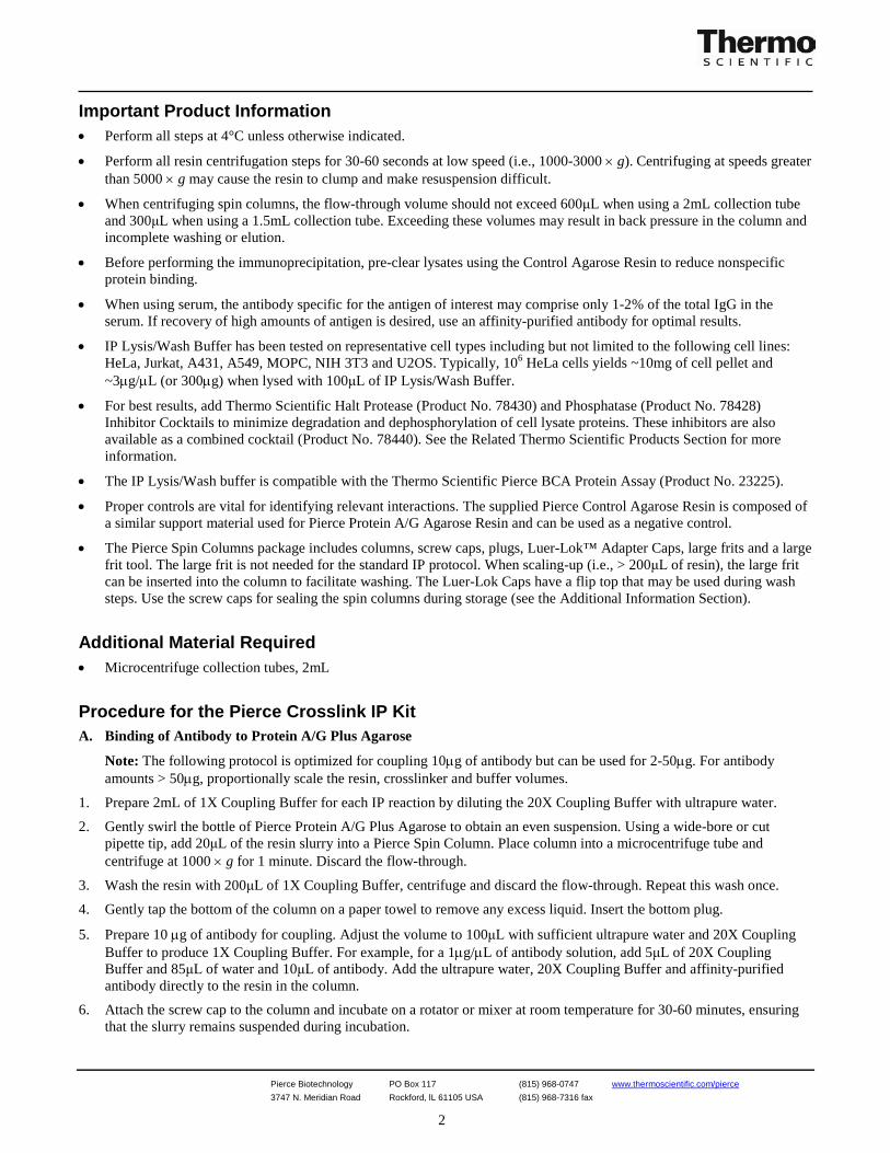
Important Product Information • Perform all steps at 4°C unless otherwise indicated.
• Perform all resin centrifugation steps for 30-60 seconds at low speed (i.e., 1000-3000 × g). Centrifuging at speeds greater than 5000 × g may cause the resin to clump and make resuspension difficult.
• When centrifuging spin columns, the flow-through volume should not exceed 600μL when using a 2mL collection tube and 300μL when using a 1.5mL collection tube. Exceeding these volumes may result in back pressure in the column and incomplete washing or elution.
• Before performing the immunoprecipitation, pre-clear lysates using the Control Agarose Resin to reduce nonspecific protein binding.
• When using serum, the antibody specific for the antigen of interest may comprise only 1-2% of the total IgG in the serum. If recovery of high amounts of antigen is desired, use an affinity-purified antibody for optimal results.
• IP Lysis/Wash Buffer has been tested on representative cell types including but not limited to the following cell lines: HeLa, Jurkat, A431, A549, MOPC, NIH 3T3 and U2OS. Typically, 106 HeLa cells yields ~10mg of cell pellet and ~3µg/µL (or 300µg) when lysed with 100μL of IP Lysis/Wash Buffer.
• For best results, add Thermo Scientific Halt Protease (Product No. 78430) and Phosphatase (Product No. 78428) Inhibitor Cocktails to minimize degradation and dephosphorylation of cell lysate proteins. These inhibitors are also available as a combined cocktail (Product No. 78440). See the Related Thermo Scientific Products Section for more information.
• The IP Lysis/Wash buffer is compatible with the Thermo Scientific Pierce BCA Protein Assay (Product No. 23225).
• Proper controls are vital for identifying relevant interactions. The supplied Pierce Control Agarose Resin is composed of a similar support material used for Pierce Protein A/G Agarose Resin and can be used as a negative control.
• The Pierce Spin Columns package includes columns, screw caps, plugs, Luer-Lok™ Adapter Caps, large frits and a large frit tool. The large frit is not needed for the standard IP protocol. When scaling-up (i.e., > 200μL of resin), the large frit can be inserted into the column to facilitate washing. The Luer-Lok Caps have a flip top that may be used during wash steps. Use the screw caps for sealing the spin columns during storage (see the Additional Information Section).
Additional Material Required • Microcentrifuge collection tubes, 2mL
Procedure for the Pierce Crosslink IP Kit A. Binding of Antibody to Protein A/G Plus Agarose
Note: The following protocol is optimized for coupling 10µg of antibody but can be used for 2-50µg. For antibody amounts > 50µg, proportionally scale the resin, crosslinker and buffer volumes.
1. Prepare 2mL of 1X Coupling Buffer for each IP reaction by diluting the 20X Coupling Buffer with ultrapure water.
2. Gently swirl the bottle of Pierce Protein A/G Plus Agarose to obtain an even suspension. Using a wide-bore or cut pipette tip, add 20μL of the resin slurry into a Pierce Spin Column. Place column into a microcentrifuge tube and centrifuge at 1000 × g for 1 minute. Discard the flow-through.
3. Wash the resin with 200μL of 1X Coupling Buffer, centrifuge and discard the flow-through. Repeat this wash once.
4. Gently tap the bottom of the column on a paper towel to remove any excess liquid. Insert the bottom plug.
5. Prepare 10 µg of antibody for coupling. Adjust the volume to 100μL with sufficient ultrapure water and 20X Coupling Buffer to produce 1X Coupling Buffer. For example, for a 1µg/µL of antibody solution, add 5μL of 20X Coupling Buffer and 85μL of water and 10μL of antibody. Add the ultrapure water, 20X Coupling Buffer and affinity-purified antibody directly to the resin in the column.
6. Attach the screw cap to the column and incubate on a rotator or mixer at room temperature for 30-60 minutes, ensuring that the slurry remains suspended during incubation.
Pierce Biotechnology PO Box 117 (815) 968-0747 www.thermoscientific.com/pierce 3747 N. Meridian Road Rockford, lL 61105 USA (815) 968-7316 fax
3
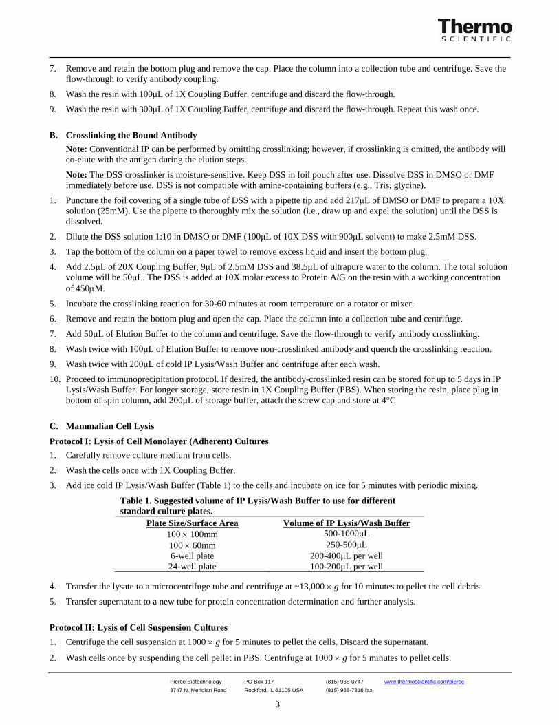
7. Remove and retain the bottom plug and remove the cap. Place the column into a collection tube and centrifuge. Save the flow-through to verify antibody coupling.
8. Wash the resin with 100μL of 1X Coupling Buffer, centrifuge and discard the flow-through.
9. Wash the resin with 300μL of 1X Coupling Buffer, centrifuge and discard the flow-through. Repeat this wash once.
B. Crosslinking the Bound Antibody
Note: Conventional IP can be performed by omitting crosslinking; however, if crosslinking is omitted, the antibody will co-elute with the antigen during the elution steps.
Note: The DSS crosslinker is moisture-sensitive. Keep DSS in foil pouch after use. Dissolve DSS in DMSO or DMF immediately before use. DSS is not compatible with amine-containing buffers (e.g., Tris, glycine).
1. Puncture the foil covering of a single tube of DSS with a pipette tip and add 217μL of DMSO or DMF to prepare a 10X solution (25mM). Use the pipette to thoroughly mix the solution (i.e., draw up and expel the solution) until the DSS is dissolved.
2. Dilute the DSS solution 1:10 in DMSO or DMF (100μL of 10X DSS with 900μL solvent) to make 2.5mM DSS.
3. Tap the bottom of the column on a paper towel to remove excess liquid and insert the bottom plug.
4. Add 2.5μL of 20X Coupling Buffer, 9μL of 2.5mM DSS and 38.5μL of ultrapure water to the column. The total solution volume will be 50μL. The DSS is added at 10X molar excess to Protein A/G on the resin with a working concentration of 450µM.
5. Incubate the crosslinking reaction for 30-60 minutes at room temperature on a rotator or mixer.
6. Remove and retain the bottom plug and open the cap. Place the column into a collection tube and centrifuge.
7. Add 50μL of Elution Buffer to the column and centrifuge. Save the flow-through to verify antibody crosslinking.
8. Wash twice with 100μL of Elution Buffer to remove non-crosslinked antibody and quench the crosslinking reaction.
9. Wash twice with 200μL of cold IP Lysis/Wash Buffer and centrifuge after each wash.
10. Proceed to immunoprecipitation protocol. If desired, the antibody-crosslinked resin can be stored for up to 5 days in IP Lysis/Wash Buffer. For longer storage, store resin in 1X Coupling Buffer (PBS). When storing the resin, place plug in bottom of spin column, add 200μL of storage buffer, attach the screw cap and store at 4°C
C. Mammalian Cell Lysis
Protocol I: Lysis of Cell Monolayer (Adherent) Cultures 1. Carefully remove culture medium from cells.
2. Wash the cells once with 1X Coupling Buffer.
3. Add ice cold IP Lysis/Wash Buffer (Table 1) to the cells and incubate on ice for 5 minutes with periodic mixing.
Table 1. Suggested volume of IP Lysis/Wash Buffer to use for different standard culture plates.
Plate Size/Surface Area Volume of IP Lysis/Wash Buffer 100 × 100mm 500-1000μL 100 × 60mm 250-500μL 6-well plate 200-400μL per well
24-well plate 100-200μL per well 4. Transfer the lysate to a microcentrifuge tube and centrifuge at ~13,000 × g for 10 minutes to pellet the cell debris.
5. Transfer supernatant to a new tube for protein concentration determination and further analysis.
Protocol II: Lysis of Cell Suspension Cultures 1. Centrifuge the cell suspension at 1000 × g for 5 minutes to pellet the cells. Discard the supernatant.
2. Wash cells once by suspending the cell pellet in PBS. Centrifuge at 1000 × g for 5 minutes to pellet cells.
Pierce Biotechnology PO Box 117 (815) 968-0747 www.thermoscientific.com/pierce 3747 N. Meridian Road Rockford, lL 61105 USA (815) 968-7316 fax
4
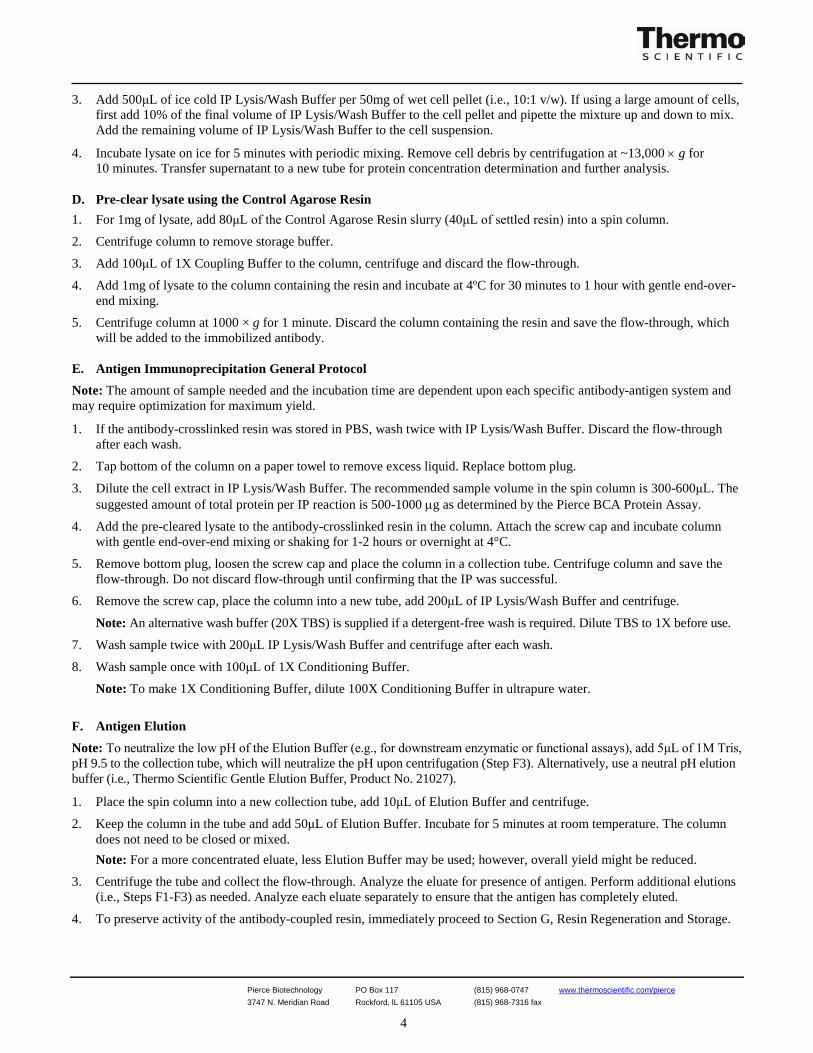
3. Add 500μL of ice cold IP Lysis/Wash Buffer per 50mg of wet cell pellet (i.e., 10:1 v/w). If using a large amount of cells, first add 10% of the final volume of IP Lysis/Wash Buffer to the cell pellet and pipette the mixture up and down to mix. Add the remaining volume of IP Lysis/Wash Buffer to the cell suspension.
4. Incubate lysate on ice for 5 minutes with periodic mixing. Remove cell debris by centrifugation at ~13,000 × g for 10 minutes. Transfer supernatant to a new tube for protein concentration determination and further analysis.
D. Pre-clear lysate using the Control Agarose Resin 1. For 1mg of lysate, add 80μL of the Control Agarose Resin slurry (40μL of settled resin) into a spin column.
2. Centrifuge column to remove storage buffer.
3. Add 100μL of 1X Coupling Buffer to the column, centrifuge and discard the flow-through.
4. Add 1mg of lysate to the column containing the resin and incubate at 4ºC for 30 minutes to 1 hour with gentle end-over-end mixing.
5. Centrifuge column at 1000 × g for 1 minute. Discard the column containing the resin and save the flow-through, which will be added to the immobilized antibody.
E. Antigen Immunoprecipitation General Protocol Note: The amount of sample needed and the incubation time are dependent upon each specific antibody-antigen system and may require optimization for maximum yield.
1. If the antibody-crosslinked resin was stored in PBS, wash twice with IP Lysis/Wash Buffer. Discard the flow-through after each wash.
2. Tap bottom of the column on a paper towel to remove excess liquid. Replace bottom plug.
3. Dilute the cell extract in IP Lysis/Wash Buffer. The recommended sample volume in the spin column is 300-600μL. The suggested amount of total protein per IP reaction is 500-1000 µg as determined by the Pierce BCA Protein Assay.
4. Add the pre-cleared lysate to the antibody-crosslinked resin in the column. Attach the screw cap and incubate column with gentle end-over-end mixing or shaking for 1-2 hours or overnight at 4°C.
5. Remove bottom plug, loosen the screw cap and place the column in a collection tube. Centrifuge column and save the flow-through. Do not discard flow-through until confirming that the IP was successful.
6. Remove the screw cap, place the column into a new tube, add 200μL of IP Lysis/Wash Buffer and centrifuge.
Note: An alternative wash buffer (20X TBS) is supplied if a detergent-free wash is required. Dilute TBS to 1X before use.
7. Wash sample twice with 200μL IP Lysis/Wash Buffer and centrifuge after each wash.
8. Wash sample once with 100μL of 1X Conditioning Buffer.
Note: To make 1X Conditioning Buffer, dilute 100X Conditioning Buffer in ultrapure water.
F. Antigen Elution Note: To neutralize the low pH of the Elution Buffer (e.g., for downstream enzymatic or functional assays), add 5μL of 1M Tris, pH 9.5 to the collection tube, which will neutralize the pH upon centrifugation (Step F3). Alternatively, use a neutral pH elution buffer (i.e., Thermo Scientific Gentle Elution Buffer, Product No. 21027).
1. Place the spin column into a new collection tube, add 10μL of Elution Buffer and centrifuge.
2. Keep the column in the tube and add 50μL of Elution Buffer. Incubate for 5 minutes at room temperature. The column does not need to be closed or mixed. Note: For a more concentrated eluate, less Elution Buffer may be used; however, overall yield might be reduced.
3. Centrifuge the tube and collect the flow-through. Analyze the eluate for presence of antigen. Perform additional elutions (i.e., Steps F1-F3) as needed. Analyze each eluate separately to ensure that the antigen has completely eluted.
4. To preserve activity of the antibody-coupled resin, immediately proceed to Section G, Resin Regeneration and Storage.
Pierce Biotechnology PO Box 117 (815) 968-0747 www.thermoscientific.com/pierce 3747 N. Meridian Road Rockford, lL 61105 USA (815) 968-7316 fax
5
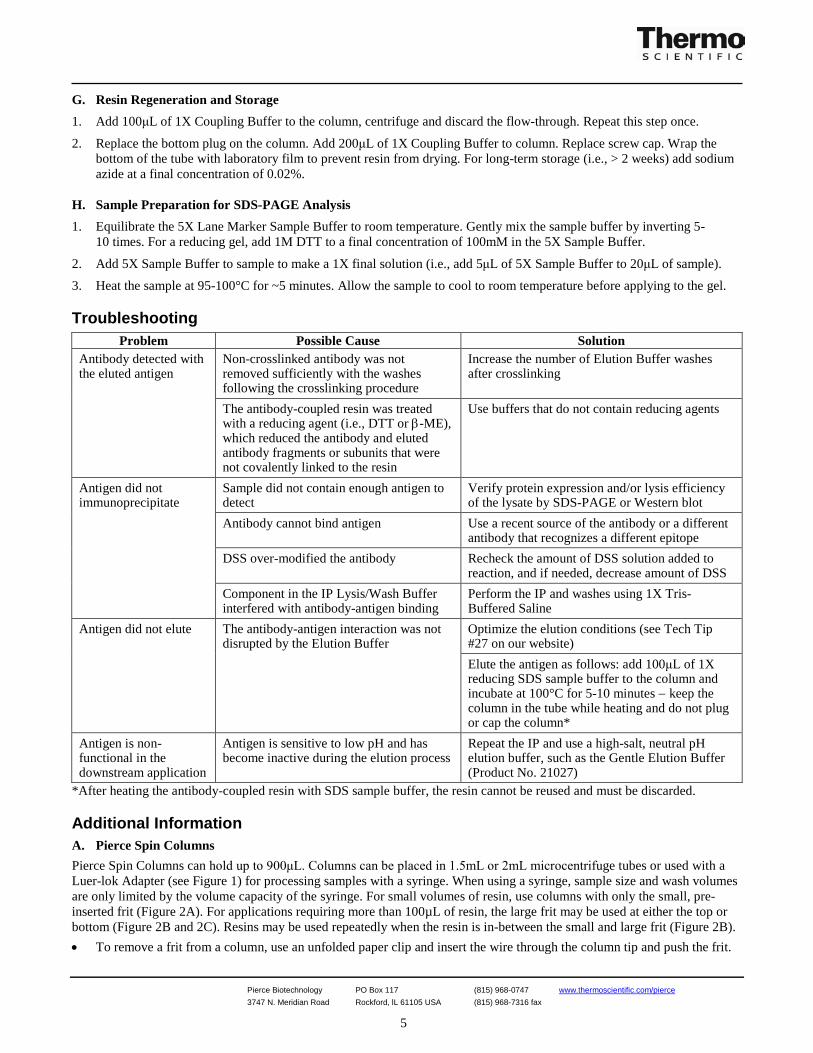
G. Resin Regeneration and Storage 1. Add 100μL of 1X Coupling Buffer to the column, centrifuge and discard the flow-through. Repeat this step once.
2. Replace the bottom plug on the column. Add 200μL of 1X Coupling Buffer to column. Replace screw cap. Wrap the bottom of the tube with laboratory film to prevent resin from drying. For long-term storage (i.e., > 2 weeks) add sodium azide at a final concentration of 0.02%.
H. Sample Preparation for SDS-PAGE Analysis 1. Equilibrate the 5X Lane Marker Sample Buffer to room temperature. Gently mix the sample buffer by inverting 5-
10 times. For a reducing gel, add 1M DTT to a final concentration of 100mM in the 5X Sample Buffer.
2. Add 5X Sample Buffer to sample to make a 1X final solution (i.e., add 5μL of 5X Sample Buffer to 20μL of sample).
3. Heat the sample at 95-100°C for ~5 minutes. Allow the sample to cool to room temperature before applying to the gel. Troubleshooting
Problem Possible Cause Solution Antibody detected with the eluted antigen
Non-crosslinked antibody was not removed sufficiently with the washes following the crosslinking procedure
Increase the number of Elution Buffer washes after crosslinking
The antibody-coupled resin was treated with a reducing agent (i.e., DTT or β-ME), which reduced the antibody and eluted antibody fragments or subunits that were not covalently linked to the resin
Use buffers that do not contain reducing agents
Antigen did not immunoprecipitate
Sample did not contain enough antigen to detect
Verify protein expression and/or lysis efficiency of the lysate by SDS-PAGE or Western blot
Antibody cannot bind antigen Use a recent source of the antibody or a different antibody that recognizes a different epitope
DSS over-modified the antibody Recheck the amount of DSS solution added to reaction, and if needed, decrease amount of DSS
Component in the IP Lysis/Wash Buffer interfered with antibody-antigen binding
Perform the IP and washes using 1X Tris-Buffered Saline
Antigen did not elute The antibody-antigen interaction was not disrupted by the Elution Buffer
Optimize the elution conditions (see Tech Tip #27 on our website) Elute the antigen as follows: add 100μL of 1X reducing SDS sample buffer to the column and incubate at 100°C for 5-10 minutes − keep the column in the tube while heating and do not plug or cap the column*
Antigen is non-functional in the downstream application
Antigen is sensitive to low pH and has become inactive during the elution process
Repeat the IP and use a high-salt, neutral pH elution buffer, such as the Gentle Elution Buffer (Product No. 21027)
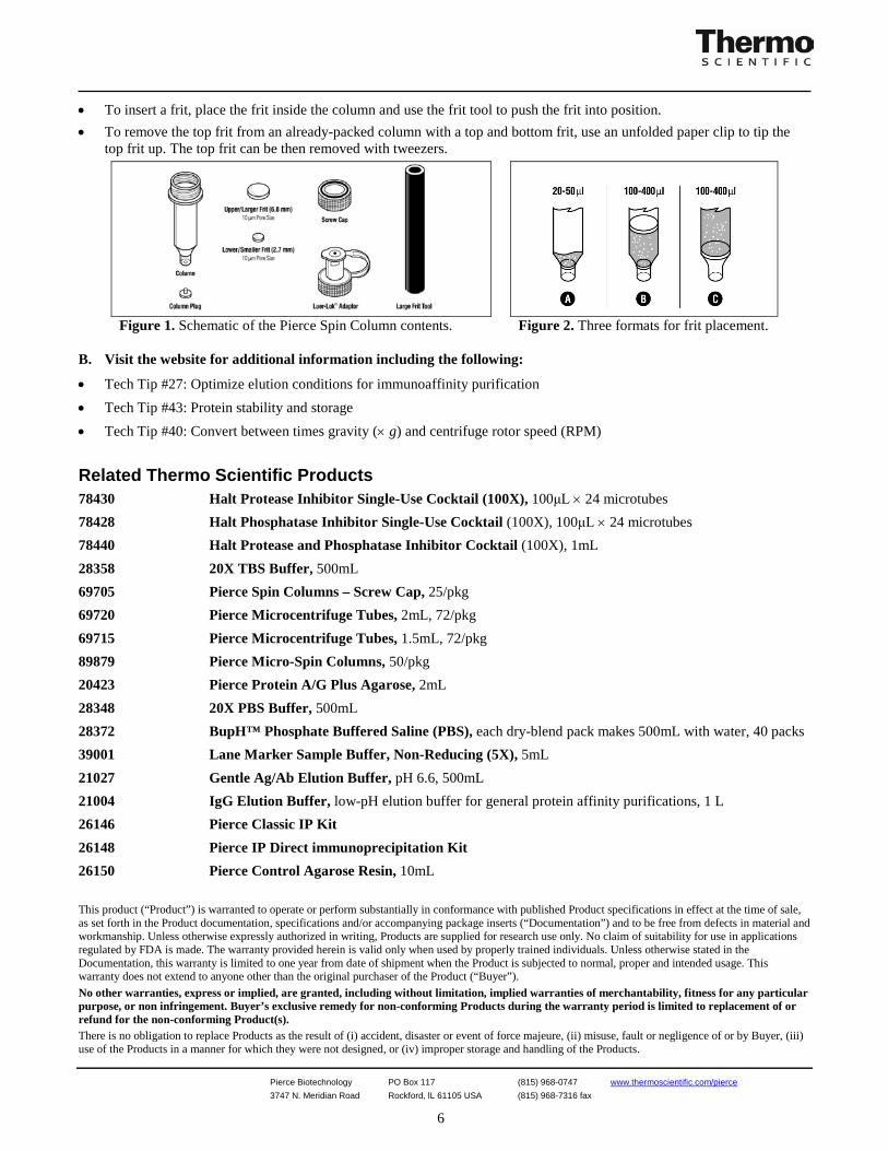
*After heating the antibody-coupled resin with SDS sample buffer, the resin cannot be reused and must be discarded. Additional Information A. Pierce Spin Columns Pierce Spin Columns can hold up to 900μL. Columns can be placed in 1.5mL or 2mL microcentrifuge tubes or used with a Luer-lok Adapter (see Figure 1) for processing samples with a syringe. When using a syringe, sample size and wash volumes are only limited by the volume capacity of the syringe. For small volumes of resin, use columns with only the small, pre-inserted frit (Figure 2A). For applications requiring more than 100µL of resin, the large frit may be used at either the top or bottom (Figure 2B and 2C). Resins may be used repeatedly when the resin is in-between the small and large frit (Figure 2B). • To remove a frit from a column, use an unfolded paper clip and insert the wire through the column tip and push the frit.
Pierce Biotechnology PO Box 117 (815) 968-0747 www.thermoscientific.com/pierce 3747 N. Meridian Road Rockford, lL 61105 USA (815) 968-7316 fax
6
• To insert a frit, place the frit inside the column and use the frit tool to push the frit into position. • To remove the top frit from an already-packed column with a top and bottom frit, use an unfolded paper clip to tip the
top frit up. The top frit can be then removed with tweezers.
Figure 1. Schematic of the Pierce Spin Column contents. Figure 2. Three formats for frit placement.
B. Visit the website for additional information including the following:
• Tech Tip #27: Optimize elution conditions for immunoaffinity purification • Tech Tip #43: Protein stability and storage • Tech Tip #40: Convert between times gravity (× g) and centrifuge rotor speed (RPM)
Related Thermo Scientific Products 78430 Halt Protease Inhibitor Single-Use Cocktail (100X), 100μL × 24 microtubes 78428 Halt Phosphatase Inhibitor Single-Use Cocktail (100X), 100μL × 24 microtubes 78440 Halt Protease and Phosphatase Inhibitor Cocktail (100X), 1mL 28358 20X TBS Buffer, 500mL 69705 Pierce Spin Columns – Screw Cap, 25/pkg 69720 Pierce Microcentrifuge Tubes, 2mL, 72/pkg 69715 Pierce Microcentrifuge Tubes, 1.5mL, 72/pkg 89879 Pierce Micro-Spin Columns, 50/pkg 20423 Pierce Protein A/G Plus Agarose, 2mL 28348 20X PBS Buffer, 500mL 28372 BupH™ Phosphate Buffered Saline (PBS), each dry-blend pack makes 500mL with water, 40 packs 39001 Lane Marker Sample Buffer, Non-Reducing (5X), 5mL 21027 Gentle Ag/Ab Elution Buffer, pH 6.6, 500mL 21004 IgG Elution Buffer, low-pH elution buffer for general protein affinity purifications, 1 L 26146 Pierce Classic IP Kit 26148 Pierce IP Direct immunoprecipitation Kit 26150 Pierce Control Agarose Resin, 10mL This product (“Product”) is warranted to operate or perform substantially in conformance with published Product specifications in effect at the time of sale, as set forth in the Product documentation, specifications and/or accompanying package inserts (“Documentation”) and to be free from defects in material and workmanship. Unless otherwise expressly authorized in writing, Products are supplied for research use only. No claim of suitability for use in applications regulated by FDA is made. The warranty provided herein is valid only when used by properly trained individuals. Unless otherwise stated in the Documentation, this warranty is limited to one year from date of shipment when the Product is subjected to normal, proper and intended usage. This warranty does not extend to anyone other than the original purchaser of the Product (“Buyer”). No other warranties, express or implied, are granted, including without limitation, implied warranties of merchantability, fitness for any particular purpose, or non infringement. Buyer’s exclusive remedy for non-conforming Products during the warranty period is limited to replacement of or refund for the non-conforming Product(s). There is no obligation to replace Products as the result of (i) accident, disaster or event of force majeure, (ii) misuse, fault or negligence of or by Buyer, (iii) use of the Products in a manner for which they were not designed, or (iv) improper storage and handling of the Products.
Pierce Biotechnology PO Box 117 (815) 968-0747 www.thermoscientific.com/pierce 3747 N. Meridian Road Rockford, lL 61105 USA (815) 968-7316 fax
7
Current product instructions are available at www.thermoscientific.com/pierce. For a faxed copy, call 800-874-3723 or contact your local distributor. © 2012 Thermo Fisher Scientific Inc. All rights reserved. Unless otherwise indicated, all trademarks are property of Thermo Fisher Scientific Inc. and its subsidiaries. Printed in the USA.